Frog Dissection - Apple
Frog Dissection - Apple
Frog Dissection - Apple

Create successful ePaper yourself
Turn your PDF publications into a flip-book with our unique Google optimized e-Paper software.
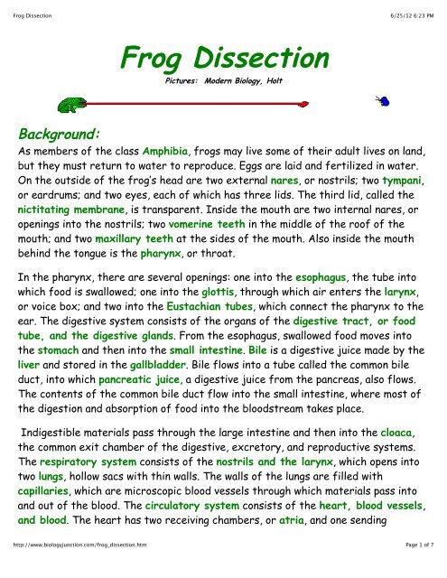
<strong>Frog</strong> <strong>Dissection</strong><br />
http://www.biologyjunction.com/frog_dissection.htm<br />
<strong>Frog</strong> <strong>Dissection</strong><br />
Pictures: Modern Biology, Holt<br />
6/25/12 6:23 PM<br />
Background:<br />
As members of the class Amphibia, frogs may live some of their adult lives on land,<br />
but they must return to water to reproduce. Eggs are laid and fertilized in water.<br />
On the outside of the frog’s head are two external nares, or nostrils; two tympani,<br />
or eardrums; and two eyes, each of which has three lids. The third lid, called the<br />
nictitating membrane, is transparent. Inside the mouth are two internal nares, or<br />
openings into the nostrils; two vomerine teeth in the middle of the roof of the<br />
mouth; and two maxillary teeth at the sides of the mouth. Also inside the mouth<br />
behind the tongue is the pharynx, or throat.<br />
In the pharynx, there are several openings: one into the esophagus, the tube into<br />
which food is swallowed; one into the glottis, through which air enters the larynx,<br />
or voice box; and two into the Eustachian tubes, which connect the pharynx to the<br />
ear. The digestive system consists of the organs of the digestive tract, or food<br />
tube, and the digestive glands. From the esophagus, swallowed food moves into<br />
the stomach and then into the small intestine. Bile is a digestive juice made by the<br />
liver and stored in the gallbladder. Bile flows into a tube called the common bile<br />
duct, into which pancreatic juice, a digestive juice from the pancreas, also flows.<br />
The contents of the common bile duct flow into the small intestine, where most of<br />
the digestion and absorption of food into the bloodstream takes place.<br />
Indigestible materials pass through the large intestine and then into the cloaca,<br />
the common exit chamber of the digestive, excretory, and reproductive systems.<br />
The respiratory system consists of the nostrils and the larynx, which opens into<br />
two lungs, hollow sacs with thin walls. The walls of the lungs are filled with<br />
capillaries, which are microscopic blood vessels through which materials pass into<br />
and out of the blood. The circulatory system consists of the heart, blood vessels,<br />
and blood. The heart has two receiving chambers, or atria, and one sending<br />
Page 1 of 7
<strong>Frog</strong> <strong>Dissection</strong><br />
6/25/12 6:23 PM<br />
chamber, or ventricle. Blood is carried to the heart in vessels called veins. Veins<br />
from different parts of the body enter the right and left atria. Blood from both<br />
atria goes into the ventricle and then is pumped into the arteries, which are blood<br />
vessels that carry blood away from the heart.<br />
The urinary system consists of the frog’s kidneys, ureters, bladder, and cloaca.<br />
The kidneys are organs that excrete urine. Connected to each kidney is a ureter, a<br />
tube through which urine passes into the urinary bladder, a sac that stores urine<br />
until it passes out of the body through the cloaca. The organs of the male<br />
reproductive system are the testes, sperm ducts, and cloaca. Those of the<br />
female system are the ovaries, oviducts, uteri, and cloaca. The testes produce<br />
sperm, or male sex cells, which move through sperm ducts, tubes that carry sperm<br />
into the cloaca, from which the sperm move outside the body. The ovaries produce<br />
eggs, or female sex cells, which move through oviducts into the uteri, then through<br />
the cloaca outside the body.<br />
The central nervous system of the frog consists of the brain, which is enclosed in<br />
the skull, and the spinal cord, which is enclosed in the backbone. Nerves branch out<br />
from the spinal cord. The frog’s skeletal and muscular systems consist of its<br />
framework of bones and joints, to which nearly all the voluntary muscles of the<br />
body are attached. Voluntary muscles, which are those over which the frog has<br />
control, occur in pairs of flexors and extensors. When a flexor of a leg or other<br />
body part contracts, that part is bent. When the extensor of that body part<br />
contracts, the part straightens.<br />
Objectives:<br />
• Describe the appearance of various organs found in the frog.<br />
• Name the organs that make up various systems of the frog.<br />
Purpose:<br />
In this lab, you will dissect a frog in order to observe the external and internal<br />
structures of frog anatomy.<br />
http://www.biologyjunction.com/frog_dissection.htm<br />
Page 2 of 7
<strong>Frog</strong> <strong>Dissection</strong><br />
Materials:<br />
• safety goggles, gloves, and a lab apron<br />
• forceps<br />
• preserved frog<br />
• dissecting pins (6–10)<br />
• dissecting tray and paper towels<br />
• plastic storage bag and twist tie<br />
• scissors<br />
• marking pen<br />
• dissecting needle<br />
Procedure:<br />
1. Put on safety goggles, gloves, and a lab apron.<br />
http://www.biologyjunction.com/frog_dissection.htm<br />
6/25/12 6:23 PM<br />
2. Place a frog on a dissection tray. To determine the frog’s sex, look at the<br />
hand digits, or fingers, on its forelegs. A male frog usually has thick pads on<br />
its "thumbs," which is one external difference between the sexes, as shown in<br />
the diagram below. Male frogs are also usually smaller than female frogs.<br />
Observe several frogs to see the difference between males and females.<br />
3. Use the diagram below to locate and identify the external features of the<br />
Page 3 of 7
<strong>Frog</strong> <strong>Dissection</strong><br />
head. Find the mouth, external nares, tympani, eyes, and nictitating<br />
membranes.<br />
http://www.biologyjunction.com/frog_dissection.htm<br />
6/25/12 6:23 PM<br />
4. Turn the frog on its back and pin down the legs. Cut the hinges of the mouth<br />
and open it wide. Use the diagram below to locate and identify the structures<br />
inside the mouth. Use a probe to help find each part: the vomerine teeth, the<br />
maxillary teeth, the internal nares, the tongue, the openings to the<br />
Eustachian tubes, the esophagus, the pharynx, and the slit-like glottis.<br />
5. Look for the opening to the frog’s cloaca, located between the hind legs. Use<br />
forceps to lift the skin and use scissors to cut along the center of the body<br />
from the cloaca to the lip. Turn back the skin, cut toward the side at each leg,<br />
and pin the skin flat. The diagram above shows how to make these cuts<br />
6. Lift and cut through the muscles and breast bone to open up the body cavity.<br />
If your frog is a female, the abdominal cavity may be filled with dark-colored<br />
eggs. If so, remove the eggs on one side so you can see the organs underlying<br />
them.<br />
7. Use the diagram below to locate and identify the organs of the digestive<br />
Page 4 of 7
<strong>Frog</strong> <strong>Dissection</strong><br />
6/25/12 6:23 PM<br />
system: esophagus, stomach, small intestine, large intestine, cloaca, liver,<br />
gallbladder, and pancreas.<br />
http://www.biologyjunction.com/frog_dissection.htm<br />
Page 5 of 7
<strong>Frog</strong> <strong>Dissection</strong><br />
6/25/12 6:23 PM<br />
8. Again refer to the diagram below to identify the parts of the circulatory and<br />
respiratory systems that are in the chest cavity. Find the left atrium, right<br />
atrium, and ventricle of the heart. Find an artery attached to the heart and<br />
another artery near the backbone. Find a vein near one of the shoulders. Find<br />
the two lungs.<br />
9. Use a probe and scissors to lift and remove the intestines and liver. Use the<br />
diagram on the next page to identify the parts of the urinary and reproductive<br />
http://www.biologyjunction.com/frog_dissection.htm<br />
Page 6 of 7
<strong>Frog</strong> <strong>Dissection</strong><br />
6/25/12 6:23 PM<br />
systems. Remove the peritoneal membrane, which is connective tissue that lies<br />
on top of the red kidneys. Observe the yellow fat bodies that are attached to<br />
the kidneys. Find the ureters; the urinary bladder; the testes and sperm<br />
ducts in the male; and the ovaries, oviducts, and uteri in the female.<br />
10. Remove the kidneys and look for threadlike spinal nerves that extend from<br />
the spinal cord. Dissect a thigh, and trace one nerve into a leg muscle. Note<br />
the size and texture of the leg muscles.<br />
11. Dispose of your materials according to the directions from your teacher.<br />
12. Clean up your work area and wash your hands before leaving the lab.<br />
http://www.biologyjunction.com/frog_dissection.htm<br />
Click here for worksheet<br />
BACK<br />
Page 7 of 7