

5. carbohydrates-i – general structure of - Cry my name
5. carbohydrates-i – general structure of - Cry my name
5. carbohydrates-i – general structure of - Cry my name

Create successful ePaper yourself
Turn your PDF publications into a flip-book with our unique Google optimized e-Paper software.
P A R T<br />
2<br />
Biomolecules<br />
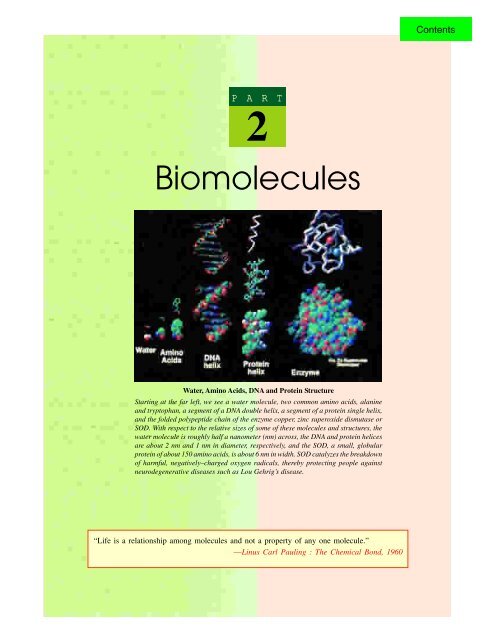
Water, Amino Acids, DNA and Protein Structure<br />
Starting at the far left, we see a water molecule, two common amino acids, alanine<br />
and tryptophan, a segment <strong>of</strong> a DNA double helix, a segment <strong>of</strong> a protein single helix,<br />
and the folded polypeptide chain <strong>of</strong> the enzyme copper, zinc superoxide dismutase or<br />
SOD. With respect to the relative sizes <strong>of</strong> some <strong>of</strong> these molecules and <strong>structure</strong>s, the<br />
water molecule is roughly half a nanometer (nm) across, the DNA and protein helices<br />
are about 2 nm and 1 nm in diameter, respectively, and the SOD, a small, globular<br />
protein <strong>of</strong> about 150 amino acids, is about 6 nm in width. SOD catalyzes the breakdown<br />
<strong>of</strong> harmful, negatively<strong>–</strong>charged oxygen radicals, thereby protecting people against<br />
neurodegenerative diseases such as Lou Gehrig’s disease.<br />
“Life is a relationship among molecules and not a property <strong>of</strong> any one molecule.”<br />
—Linus Carl Pauling : The Chemical Bond, 1960<br />
Contents
Contents
CONTENTS<br />
• Importance<br />
Nomenclature and Definition<br />
•<br />
• Classification<br />
• Asymmetry<br />
• Isomerism<br />
• Kiliani Cyanohydrian<br />
Synthesis<br />
• Optical Isomerism<br />
• Formulation <strong>of</strong><br />
Monosaccharides<br />
<strong>Cry</strong>stals <strong>of</strong> glucose, a key molecule in<br />
carbohydrate metabolism, viewed under<br />
polarised light.<br />
C H 5A P T E R<br />
Carbohydrates-I<br />
General Structure <strong>of</strong><br />
Monosaccharides<br />
IMPORTANCE<br />
he <strong>carbohydrates</strong>, <strong>of</strong>ten termed as sugars, are the<br />
‘staff <strong>of</strong> life’ for most organisms. On the basis <strong>of</strong><br />
mass, they are the most abundant class <strong>of</strong><br />
biomolecules in nature. Carbohydrates are also known as<br />
saccharides (sakcharon G = sugar or sweetness) since many<br />
<strong>of</strong> those <strong>of</strong> relatively small molecular weight have a sweet<br />
taste, although this is not true <strong>of</strong> those with large molecules.<br />
They are widely distributed molecules (moles L T<br />
= mass) in<br />
both plant and animal tissues. They are indispensable for<br />
living organisms, serving as skeletal <strong>structure</strong>s in plants<br />
and also in insects and crustaceans. They also occur as<br />
food reserves in the storage organs <strong>of</strong> plants and in the<br />
liver and muscles <strong>of</strong> animals. In addition, they are an<br />
important source <strong>of</strong> energy required for the various<br />
metabolic activities <strong>of</strong> the living organisms; the energy<br />
being derived as a result <strong>of</strong> their oxidation. They also<br />
serve to lubricate skeletal joints, to provide adhesion<br />
between cells and to confer biological specificity on the<br />
surface <strong>of</strong> animal calls.<br />
Plants are considerably richer in <strong>carbohydrates</strong> in<br />
comparison to the animals. In fact, animal and plant<br />
tissues differ widely in the relative abundance <strong>of</strong> the various<br />
major classes <strong>of</strong> constituent chemicals (Table 5<strong>–</strong>1).<br />
73<br />
Contents
74 FUNDAMENTALS OF BIOCHEMISTRY<br />
Table 5<strong>–</strong>1. Percentage composition <strong>of</strong> various major classes <strong>of</strong> constituent chemicals in<br />
plants and animals<br />
Organism Water Carbohydrates Proteins Lipids Ash<br />
Animal 60 1 20 15 4<br />
Plant 60 30 5 1 4<br />
NOMENCLATURE AND DEFINITION<br />
The term ‘carbohydrate’ was originally coined for this class <strong>of</strong> compounds as most <strong>of</strong> them<br />
were ‘hydrates <strong>of</strong> carbon’ or could be represented by the <strong>general</strong> formula, Cx (H2O) y . Later, it was<br />
found that some <strong>of</strong> them, such as deoxyribose (C5H10O4 ) and rhamnose (C6H12O5 ) do not have the<br />
required ratio <strong>of</strong> hydrogen to oxygen. In addition, certain other <strong>carbohydrates</strong> are now known to<br />
possess nitrogen (e.g., glucosamine, C6H13O5N), phosphorus or sulfur also and obviously do not<br />
coincide with the above <strong>general</strong> formula. Furthermore, formaldehyde (H.CHO or CH2O), acetic<br />
acid (CH3 .COOH or C2H4O2 ) and lactic acid (CH3 .CHOH.COOH or C3H6O3 ) which have C, H<br />
and O and the ratio <strong>of</strong> H : O is also the same as in water, but are not a <strong>carbohydrates</strong>. Hence,<br />
the continued usage <strong>of</strong> the term ‘carbohydrate’ is for convenience rather than exactness.<br />
To accommodate a wide variety <strong>of</strong> compounds, the <strong>carbohydrates</strong> are nowadays broadly<br />
defined as polyhydroxy aldehydes or ketones and their derivatives or as substances that yield<br />
one <strong>of</strong> these compounds on hydrolysis.<br />
CLASSIFICATION<br />
Carbohydrates are usually classified in 3 groups :<br />
A. Monosaccharides or Monosaccharoses (mono G = one; sakcharon G = sugar). The<br />
monosaccharides, <strong>of</strong>ten called simple sugars, are compounds which possess a free aldehyde (—CHO)<br />
or ketone (= CO) group and 2 or more hydroxyl (—OH) groups. They are, in fact, the simplest sugars<br />
and cannot be hydrolyzed into smaller units. Their <strong>general</strong> formula is Cn (H2O) n or CnH2nOn .<br />
The monosaccharides may be subdivided into trioses, tetroses, pentoses, hexoses, heptoses<br />
etc., depending upon the number <strong>of</strong> carbon atoms they possess; and as aldoses or ketoses, depending<br />
upon whether they contain aldehyde or ketone group. Some important examples are :<br />
Name Formula Aldoses Ketoses<br />
(Aldo sugars) (Keto sugars)<br />
Trioses C 3 H 6 O 3 Glycerose Dihydroxyacetone<br />
Tetroses C 4 H 8 O 4 Erythrose Erythrulose<br />
Pentoses C 5 H 10 O 5 Ribose Ribulose<br />
Hexoses C 6 H 12 O 6 Glucose Fructose<br />
Heptoses C 7 H 14 O 7 Glucoheptose Sodoheptulose<br />
Both these characters (i.e., the number <strong>of</strong> carbon atoms and the nature <strong>of</strong> functional group<br />
present) may also be combined into one. Thus, for example, glycerose (= glyceraldehyde) is an<br />
aldotriose; ribulose, a ketopentose and glucose, an aldohexose. It is noteworthy that, except fructose,<br />
ketoses are not as common as aldoses. The most abundant monosaccharide in nature is the 6carbon<br />
sugar, D-glucose.<br />
Sometimes, a distinction in naming between aldoses and ketoses is also maintained. The suffix<br />
Contents
-oses is kept reserved for the aldoses and the suffix -<br />
uloses is used for ketoses. Thus, glucose is a hexose<br />
and fructose, a hexulose. However, a few ketoses are<br />
<strong>name</strong>d otherwise, such as fructose (fructus L = fruit) as<br />
fruits are a good source <strong>of</strong> this sugar.<br />
B. Oligosaccharides or Oligosaccharoses<br />
(oligo G = few). These are compound sugars that yield<br />
2 to 10 molecules <strong>of</strong> the same or different<br />
GENERAL STRUCTURE OF MONOSACCHARIDES 75<br />
monosaccharides on hydrolysis. Accordingly, an oligosaccharide yielding 2 molecules <strong>of</strong><br />
monosaccharide on hydrolysis is designated as a dissaccharide, and the one yielding 3 molecules<br />
<strong>of</strong> monosaccharide as a trisaccharide and so on. The <strong>general</strong> formula <strong>of</strong> disaccharides is<br />
C n (H 2 O) n <strong>–</strong> 1 and that <strong>of</strong> trisaccharides is C n (H 2 O) n <strong>–</strong> 2 and so on. A few examples are :<br />
Disaccharides <strong>–</strong> Sucrose, Lactose, Maltose, Cellobiose, Trehalose,<br />
Gentiobiose, Melibiose<br />
Trisaccharides <strong>–</strong> Rhamninose, Gentianose, Raffinose (= Melitose),<br />
Rabinose, Melezitose<br />
Tetrasaccharides <strong>–</strong> Stachyose, Scorodose<br />
Pentasaccharide <strong>–</strong> Verbascose<br />
The molecular composition <strong>of</strong> the 3 legume oligosaccharides (viz., raffinose, stachyose and<br />
verbascose) is shown below :<br />
α-Galactose (1<strong>–</strong>6) α-Glucose (1<strong>–</strong>2) β-Fructose Raffinose<br />
α-Galactose (1<strong>–</strong>6) α-Galactose (1<strong>–</strong>6) α-Glucose (1<strong>–</strong>2) β-Fructose Stachyose<br />
α-Galactose (1<strong>–</strong>6) α-Galactose (1<strong>–</strong>6) α-Galactose (1<strong>–</strong>6) α-Glucose (1<strong>–</strong>2) β-Fructose Verbascose<br />
C. Polysaccharides or Polysaccharoses (poly G = many). These are also compound sugars<br />
and yield more than 10 molecules <strong>of</strong> monosaccharides on hydrolysis. These may be further<br />
classified depending on whether the monosaccharide molecules produced as a result <strong>of</strong> the hydrolysis<br />
<strong>of</strong> polysaccharides are <strong>of</strong> the same type (homopolysaccharides) or <strong>of</strong> different types<br />
(heteropolysaccharides). Their <strong>general</strong> formula is (C6H10O5 ) x . Some common examples are :<br />
Homopolysaccharides <strong>–</strong>Starch, Glycogen, Inulin, Cellulose, Pectin, Chitin<br />
Heteropolysaccharides <strong>–</strong> “Specific soluble sugar” <strong>of</strong> pneumococcus type III,<br />
Hyaluronic acid, Chondrotin<br />
ASYMMETRY<br />
Jacobus H. van’t H<strong>of</strong>f, the first Nobel Laureate in Chemistry (1901) and Joseph A. Le Bel,<br />
in 1894, introduced the concept <strong>of</strong> tetrahedral carbon atom.<br />
Fig. 5<strong>–</strong>1. Spatial arrangement <strong>of</strong> the valences <strong>of</strong> carbon<br />
Anselme Payen, codiscoverer <strong>of</strong> diastase,<br />
also isolated a compound common to cell<br />
walls <strong>of</strong> higher plants, which he <strong>name</strong>d<br />
`cellulose'. His naming <strong>of</strong> this<br />
polysaccharide, in fact, introduced the<br />
-ose suffix into the nomenclature <strong>of</strong><br />
<strong>carbohydrates</strong>.<br />
Contents
76 FUNDAMENTALS OF BIOCHEMISTRY<br />
JACOBUS HENRICUS VAN’T HOFF<br />
(LT, 1852-1911)<br />
A Dutch chemist. Best<br />
known for his hypothesis<br />
about the <strong>structure</strong> <strong>of</strong> carbon<br />
atom. Made notable<br />
contributions in the field <strong>of</strong><br />
Physical Chemistry,<br />
particularly on the theory <strong>of</strong><br />
dilute solutions. Won the<br />
first-ever Noble Prize in<br />
Chemistry in 1901 for work<br />
on rates <strong>of</strong> reactions,<br />
chemical equilibrium and osmotic pressure.<br />
It is now recognized that the carbon atom has the shape <strong>of</strong> a tetrahedron in which the carbon<br />
nucleus resides in the centre <strong>of</strong> this tetrahedron and the 4 covalent bonds extend out to the corners<br />
<strong>of</strong> it. The angle between any two covalent bonds is 109°28′ (Fig. 5<strong>–</strong>1).<br />
The 5th Incarnation <strong>of</strong> Carbon<br />
Carbon is one <strong>of</strong> the most important elements in the universe since life itself, as we know it, is<br />
carbon-based. The versatility <strong>of</strong> water to form a huge variety <strong>of</strong> compounds and their stability even<br />
under extreme conditions are the secret <strong>of</strong> carbon acting as the vehicle <strong>of</strong> life. Versatility <strong>of</strong> carbon<br />
compounds apart, the element itself has become a centre <strong>of</strong> great attention in recent years. For long,<br />
we knew carbon could manifest itself in vividly contrasting styles. It was known to assume a s<strong>of</strong>t and<br />
black form as graphite; it was known equally well in its hard and sparkling - white incarnation <strong>of</strong><br />
diamond which is the hardest material on earth. Then it was discovered (in 1985) to have had the<br />
shape <strong>of</strong> a soccer ball (called fullerenes), and <strong>of</strong> a cylindrical tube (called nanotubes). And now an<br />
announcement has been made that carbon can also assume a foa<strong>my</strong> shape, called nan<strong>of</strong>oam.<br />
This new spongy form <strong>of</strong> carbon is also a modification <strong>of</strong> graphitic form. It is extremely lightweighted<br />
form and was created when some researchers, at the Australian National University in<br />
Canberra, bombarded a piece <strong>of</strong> graphite with a high-power laser beam. The graphite piece vapourised<br />
as temperature rose to more than 10,000°C. On cooling, the vapourised<br />
carbon atoms formed a web <strong>of</strong> tubes. But these tubes instead <strong>of</strong> being<br />
aligned in any particular direction (as happens in the case <strong>of</strong> nanotubes),<br />
were found to be highly dispersed, criss-crossing with one another to<br />
form a highly-porous mass. This web <strong>of</strong> nanotubes is seen as a non<strong>of</strong>oam.<br />
Surprisingly, the nan<strong>of</strong>oam, unlike all the other forms <strong>of</strong> carbon, is magnetic.<br />
Although the magnetism is ephemeral at room temperature, it is<br />
an entirely new and unexpected property in the case <strong>of</strong> carbon. Because<br />
<strong>of</strong> light weight, foa<strong>my</strong> nature and magnetic property, nan<strong>of</strong>oam is being<br />
seen as useful for cancer and brain scans through magnetic resonance<br />
imaging (MRI). Besides, it might also be useful in the treatment <strong>of</strong><br />
tumours. Being foam, it would be a bad conductor <strong>of</strong> heat and if injected<br />
into tumours, and heated, it would retain the heat locally and kill<br />
tumours almost without affecting other body parts. But it needs confir-<br />
mation through a series <strong>of</strong> studies.<br />
In the formula given above for glucose, it may be observed that a<br />
different group is attached to each <strong>of</strong> the 4 valences <strong>of</strong> carbon atoms 2<br />
to <strong>5.</strong> For example, the 4 groups attached at carbon atom 2 are :<br />
JOSEPH A. LE BEL<br />
(LT, 1847-1930)<br />
A French chemist. Best<br />
known for his<br />
stereochemical theory<br />
<strong>of</strong> carbon and its<br />
compounds. Codiscoverer<br />
<strong>of</strong> the cause<br />
<strong>of</strong> optical activity with<br />
van’t H<strong>of</strong>f. Synthesised<br />
the first optically-active<br />
compound with<br />
asymmetric nitrogen atom.<br />
[The carbon atoms <strong>of</strong> a<br />
sugar are numbered from<br />
the end closest to the<br />
aldehyde or ketone group.]<br />
Contents
GENERAL STRUCTURE OF MONOSACCHARIDES 77<br />
Thus, a carbon atom to which 4 different atoms or groups <strong>of</strong> atoms are attached is said to be<br />
asymmetric or chiral (cheir G = hand).<br />
ISOMERISM<br />
The term isomer (isos G = equal ; meros G = part) was originally applied by Jönes Jacob<br />
Berzelius, in 1827, to different compounds with the same molecular formula, and the phenomenon<br />
was called isomerism. The presence <strong>of</strong> asymmetric carbon atoms in the <strong>carbohydrates</strong> makes<br />
possible the formation <strong>of</strong> isomers in them. It shall, hence, be beneficial at this stage to discuss<br />
in brief about isomers.<br />
The isomers are <strong>of</strong> 2 types : structural isomers and stereoisomers. Structural isomers have<br />
the same molecular formula but possess different <strong>structure</strong>s. The difference in <strong>structure</strong> may be<br />
exhibited either in the length <strong>of</strong> the carbon chain (chain isomers) or in the position <strong>of</strong> a substituent<br />
group (positional isomers) or in possessing different functional groups (functional-group isomers).<br />
Stereoisomers, on the other hand, have the same molecular formula and the same <strong>structure</strong><br />
but differ only in spatial configuration. Stereoisomers are <strong>of</strong> 2 types : geometrical and optical.<br />
Geometrical or cis-trans (cis L = same side; trans L = across) isomers arise from peculiar geometry<br />
<strong>of</strong> compounds having a double bond within the carbon chain. It may be illustrated by cis-trans pair,<br />
maleic and fumaric acids.<br />
Optical isomers (= enantiomers) differ from each other in the disposition <strong>of</strong> the various atoms<br />
or groups <strong>of</strong> atoms in space around the asymmetric carbon atom. These are, in fact, the mirror<br />
image <strong>of</strong> each other. These may also be likened to left- and right-handed gloves. For example,<br />
the glyceraldehyde has only one asymmetric carbon atom (numbered as 2) and it can, therefore,<br />
exist in 2 isomeric forms :<br />
One form in which H atom at carbon 2 is projected to the left side and OH group to the right is<br />
designated as D-form and the other form where H atom is projected to the right side and OH group<br />
to the left is called as L-form (note the use <strong>of</strong> small capital letters D and L) Similarly, a compound<br />
having 2 asymmetric carbon atoms (e.g., a tetrose) might exist in 4 optical isomeric froms.<br />
The number <strong>of</strong> possible isomers <strong>of</strong> any given compound, thus, depends upon the number <strong>of</strong><br />
asymmetric carbon atoms present in its molecule. According to the rule <strong>of</strong> n (also called as<br />
Contents
78 FUNDAMENTALS OF BIOCHEMISTRY<br />
Le Bel-van’t H<strong>of</strong>f rule), the total number <strong>of</strong> optical isomers <strong>of</strong> a compound will be equal to 2 n ,<br />
where n represents the number <strong>of</strong> asymmetric carbon atoms present in the molecule.<br />
In fact, in sugars with 2 or more asymmetric carbon atoms, the designation <strong>of</strong> D- and L- forms<br />
depends upon the orientation <strong>of</strong> the H and OH groups around the lowermost asymmetric carbon<br />
atom (i.e., the asymmetric carbon furthest from the aldehyde or keto group). The distribution <strong>of</strong><br />
the H and OH groups on the other carbon atoms in the molecule is <strong>of</strong> no importance in this<br />
connection. It is interesting to note that majority <strong>of</strong> the monosaccharides found in the human body<br />
are <strong>of</strong> D type. However, some sugars do occur naturally in their L form such as L-arabinose, a<br />
pentaaldose.<br />
KILIANI CYANOHYDRIN SYNTHESIS<br />
A method for the synthesis <strong>of</strong> monosaccharides was first proposed by Heinrich Kiliani in<br />
1886. It is, in fact, a method by which the chain length <strong>of</strong> a carbohydrate may be increased. The<br />
application <strong>of</strong> Kiliani synthesis to D-glyceraldeyde resulting in the production <strong>of</strong> 2 tetroses, Derythrose<br />
and D-threose is shown in Fig. 5<strong>–</strong>2.<br />
The process is based upon the addition <strong>of</strong> HCN to the carbonyl group <strong>of</strong> aldehydes (or<br />
ketones) <strong>of</strong> the sugars forming cyanohydrin (Reaction-1). This reaction creates a new asymmetric<br />
carbon atom, marked by an asterisk in the figure.<br />
Thus, two compounds differing in conformation<br />
about the newly-formed asymmetric carbon atom<br />
are formed. These two cyanohydrins are, then,<br />
hydrolyzed to produce carboxylic acids (Reaction-<br />
2) which are later converted to γ-lactones or inner<br />
esters (Reaction-3). Finally, the lactones are<br />
reduced to the corresponding aldoses, containing<br />
one carbon atom more than their parent sugar<br />
(Reaction-4).<br />
The process can be repeated and the 4<br />
isomeric D-pentoses may be produced; and from<br />
these the 8 isomeric D-hexoses would also result.<br />
The structural relationships <strong>of</strong> the<br />
monosaccharides (the aldoses, for example) may<br />
be visualized by the formulae given in Fig. 5<strong>–</strong>3.<br />
It is easy to remember the configurations <strong>of</strong> the<br />
aldoses by recalling the Rosan<strong>of</strong>f scheme, drawn<br />
out on the chart and mneumonic—ET; RAXL;<br />
AAGMGIGT.<br />
The term ‘conformation’ was first introduced<br />
by Walter Norman Haworth (1929), an English<br />
chemist. In its broadest sense, conformation has<br />
been used to describe different spatial<br />
arrangements <strong>of</strong> a molecule which are not<br />
superimposable. This means that, in effect, the<br />
terms conformation and configuration are<br />
equivalent. In the classical sense, the definition<br />
<strong>of</strong> conformation does not include the internal<br />
forces acting on the molecule. The term<br />
conformation, however, is the spatial<br />
arrangement <strong>of</strong> the molecule when all the<br />
internal forces acting on the molecule are taken<br />
into account. In more restricted sense, the term<br />
conformation is used to denote different spatial<br />
arrangements arising due to twisting or rotation<br />
<strong>of</strong> bonds <strong>of</strong> a ‘given’ configuration (used in the<br />
classical sense). The terms rotational isomers<br />
and constellations have also been used in the<br />
same sense as conformations.<br />
Any two sugars which differ from each other only in the configuration around a single<br />
asymmetric carbon atom other than the carbonyl carbon atom are called epimers. Glucose and<br />
galactose, for example, form an epimeric pair as they differ with respect to carbon 4 only. Similarly,<br />
glucose is also epimeric with mannose (differing in C 2 configuration) and allose with altrose<br />
(differing also in C 2 configuration).<br />
The two D- and L-forms <strong>of</strong> a compound constitute a pair <strong>of</strong> enantiomers (enantios G =<br />
opposite) or enantiomorphs. Thus, D-erythrose and L-erythrose are enantiomers; also D-glucose<br />
and L-glucose. The enantiomers (also called optical isomers or double image isomers) are<br />
nonsuperimposable mirror images <strong>of</strong> each other but are chemically identical in their reactions.<br />
They agree in their melting points, solubility etc., but differ in their ability to rotate the plane <strong>of</strong><br />
polarized light in a polarimeter; a solution <strong>of</strong> one <strong>of</strong> the two enantiomers rotates the plane to the<br />
right, and a solution <strong>of</strong> the other to the left.<br />
Contents
GENERAL STRUCTURE OF MONOSACCHARIDES 79<br />
Fig. 5<strong>–</strong>2. Application <strong>of</strong> the cyanohydrin synthesis to D-glyceraldehyde<br />
The conversion <strong>of</strong> D-glyceraldehyde into an aldotetrose yields D-erythrose and D-threose.<br />
These are called diastereoisomers i.e., isomers but not mirror images <strong>of</strong> each other. In fact,<br />
diastereoisomers are different forms <strong>of</strong> a compound with two asymmetric centres. It may, hence,<br />
Contents
80 FUNDAMENTALS OF BIOCHEMISTRY<br />
1<br />
2<br />
CHO<br />
HCOH<br />
HCOH<br />
HCOH<br />
CHO<br />
HCOH<br />
HCOH<br />
CHO<br />
HCOH<br />
CH OH<br />
CHO<br />
HOCH<br />
HCOH<br />
CHO CHO<br />
CHO<br />
HOCH HCOH<br />
HOCH<br />
HCOH HOCH<br />
HOCH<br />
HCOH HCOH<br />
HCOH<br />
CHO CHO CHO CHO CHO CHO CHO CHO<br />
HCOH HOCH HCOH HOCH HCOH HOCH HCOH HOCH<br />
3<br />
CH OH<br />
2<br />
CH OH<br />
2<br />
HCOH HCOH HOCH HOCH HCOH HCOH HOCH HOCH<br />
4<br />
HCOH HCOH HCOH HCOH HOCH HOCH HOCH HOCH<br />
5<br />
HCOH HCOH HCOH HCOH HCOH HCOH HCOH HCOH<br />
6<br />
CH OH<br />
2<br />
1<br />
2<br />
3<br />
4<br />
5<br />
D-ribose<br />
CH OH<br />
2<br />
1<br />
2<br />
3<br />
4<br />
D-erythrose<br />
CH OH<br />
2<br />
CH OH<br />
2<br />
CH OH<br />
2<br />
1<br />
2<br />
3<br />
2<br />
D-glycerose<br />
CH OH<br />
2<br />
CH OH<br />
2<br />
CH OH<br />
2<br />
CH OH<br />
2<br />
D-threose<br />
CH OH<br />
D-arabinose D-xylose<br />
D-lyxose<br />
CH OH<br />
2<br />
2<br />
CH OH<br />
D-allose D-altrose D-glucose D-mannose D-gulose D-idose D-galactose D-talose<br />
Fig. 5<strong>–</strong>3. D. Aldoses containing three, four, five and six carbon atoms<br />
Aldoses contain an aldehyde group (shown in blue) and have the absolute configuration <strong>of</strong> D-glyceraldehyde at the<br />
asymmetric centre (shown in red) farthest from the aldehyde group. The numbers indicate the standard designations<br />
for each carbon atom. The formulae <strong>of</strong> L-aldoses are in each case the mirror images <strong>of</strong> these <strong>structure</strong>s.<br />
be inferred that if two optical isomers are not enantiomers, they are related as diastereoisomers.<br />
They differ from each other in their melting points, solubilities and the chemical properties, in<br />
<strong>general</strong>.<br />
2<br />
Contents
GENERAL STRUCTURE OF MONOSACCHARIDES 81<br />
OPTICAL ISOMERISM<br />
The presence <strong>of</strong> asymmetric carbon atoms in the compound also confers optical activity to it.<br />
When a beam <strong>of</strong> polarized light is passed through an optically active solution, it may be rotated<br />
either to the right or to the left, depending upon the type <strong>of</strong> optical isomer present. A compound<br />
rotating the plane <strong>of</strong> polarized light to the right is called as dextrorotatory (dexter L = right, on the<br />
right side; rotatus L = pp <strong>of</strong> rotare L , to turn) or clockwise and is designated as d or (+) type. If the<br />
compound causes rotation <strong>of</strong> polarized light to the left, it is said to be levorotatory (laevus L = left,<br />
on the left side) or anticlockwise and is denoted as l or (<strong>–</strong>) type. The direction in which the light<br />
is rotated (or in other words, the optical rotation) is a specific property <strong>of</strong> the molecule. It should,<br />
however, be emphasized that the optical rotation is not at all related with the two D and L forms<br />
<strong>of</strong> a compound. Thus, D-glucose is dextrorotatory and D-fructose is levorotatory. If it is desired<br />
to indicate the direction <strong>of</strong> rotation, the two <strong>name</strong>s may be written as D(+)-glucose and<br />
D(<strong>–</strong>)-fructose. Similarly, D-erythrose is levorotatory and D-threose, dextrorotatory.<br />
When equal amounts <strong>of</strong> extrorotatory and levorotatory isomers are present, the resulting<br />
mixture becomes optically inactive because the optical activities <strong>of</strong> each isomer cancel each other.<br />
Such a mixture is called a racemic or dl-mixture or (±)-conglomerate and this process <strong>of</strong> converting<br />
an optically active compound into the racemic modification is known <strong>of</strong> racemisation (racemus L<br />
= grape). Compounds produced synthetically are invariably racemic in nautre because equal<br />
chances exist for the formation <strong>of</strong> two types <strong>of</strong> optical isomers or the d and l antipodes.<br />
It may be concluded that all monosaccharides are optically active (except aldobiose and<br />
ketotriose) since they contain one or more asymmetric carbon atoms in their molecules.<br />
Mutarotation<br />
When a monosaccharide is dissolved in water, the optical rotatory power <strong>of</strong> the solution<br />
gradually changes until it reaches a constant value (Dubrunfaut, 1846). A freshly-prepared aqueous<br />
solution <strong>of</strong> α-D-glucose, for instance, has a specific rotation, [ ] 20<br />
α <strong>of</strong> +112.2°. And when this<br />
D<br />
solution is allowed to stand, the rotation falls to +52.7° and remains constant at this value. The<br />
final stage can be attained more quickly either by heating the solution or by adding some catalyst<br />
which may be an acid or an alkali. This gradual change in specific rotation is known as mutarotation<br />
or changing rotation. In fact, this term reflects the discovery <strong>of</strong> the phenomenon by way <strong>of</strong> changes<br />
in the optical rotation <strong>of</strong> certain <strong>carbohydrates</strong>. The terms multirotation and birotation have<br />
sometimes also been used for mutarotation. The value <strong>of</strong> mutarotation for α-D-glucose is (+112.2°)<br />
<strong>–</strong> (+52.7°) or +59.5°. A fresh solution <strong>of</strong> β-D-glucose, on the other hand, has a rotation value <strong>of</strong><br />
+18.7°; on standing, it also changes to the same value, +52.7°. All reducing sugars (except a few<br />
ketoses) undergo mutarotation. The specific rotations for the anomers <strong>of</strong> glucose and for the<br />
aqueous equilibrium mixture are :<br />
[ ] 20<br />
α = + 112.2° ⎯⎯→<br />
D<br />
[ ] 20<br />
α = + 52.7° ←⎯⎯<br />
D<br />
[ ] 20<br />
D<br />
α = + 18.7°<br />
α-D-glucose Mutarotational β-D-glucose<br />
equilibrium mixture<br />
These substances (open and ring forms) interconvert due to a dynamic equilibrium. Although<br />
the ring form predominates, each molecule spends some time in the open form, when asymmetry<br />
is lost. When the open-chain form passes back to the ring form, it is possible for it to give rise<br />
to either anomer. Thus, a pure sample <strong>of</strong> an anomer only remains so when in the solid state. In<br />
solution, particularly at alkaline pH, a pure sample <strong>of</strong> one anomer will change to an equilibrium<br />
mixture <strong>of</strong> the two forms. There need not be equal quantities <strong>of</strong> the two forms at equilibrium<br />
because there are other centres for asymmetry and so anomers (like allo and threo pairs <strong>of</strong> amino<br />
acids) do not have identical physical properties, including ∆G° <strong>of</strong> formation. Equilibrium between<br />
Contents
82 FUNDAMENTALS OF BIOCHEMISTRY<br />
the anomers is called mutarotation and it can be followed by observing optical rotation<br />
(Fig. 5<strong>–</strong>4).<br />
Optical Rotation<br />
Mid point<br />
+ 112.2°<br />
6<strong>5.</strong>4°<br />
+ 52.7°<br />
+ 18.7°<br />
-glucose<br />
-glucose<br />
Time<br />
Fig. 5<strong>–</strong>4. The experimental detection <strong>of</strong> mutarotation<br />
Mechanism <strong>of</strong> mutarotation. The phenomenon <strong>of</strong> mutarotation appears to be due to the changes<br />
<strong>of</strong> α- to β-forms and vice versa via the straight chain aldo or keto form (refer to the subsequent<br />
section). When an equilibrium is attained, the characteristic rotation is reached. According to<br />
Lowry (1925), mutarotation can take place only in the presence <strong>of</strong> an amphiprotic solvent (i.e., a<br />
solvent which can act both as an acid and a base). Water is one such solvent. Thus, Lowry and<br />
Faulkner (1925) showed that mutarotation is checked in cresol solution (an acidic solvent) and in<br />
pyridine solution (a basic solvent). It is assumed that when mutarotation takes place, the ring opens<br />
and then recloses either in the inverted position or in the original position. The existence <strong>of</strong> the<br />
open chain form in mutarotation is proved by certain evidences. For example, the absorption<br />
spectra <strong>of</strong> fructose and sorbose in aqueous solution indicate the presence <strong>of</strong> open chain forms ; the<br />
aldoses, however, give negative results (Bednarczyk et al, 1938).<br />
FORMULATION OF MONOSACCHARIDES<br />
Formula <strong>of</strong> Glucose—Linear Form<br />
An organic chemist intends to incorporate as many characteristics as possible in writing the<br />
formulae (Fig. 5<strong>–</strong>5). When the molecular formula <strong>of</strong> glucose, C 6 H 12 O 6 is written in a form as<br />
propounded by Fittig and Baeyer, it tells us the presence <strong>of</strong> an aldehyde and 5 hydroxyl groups.<br />
Replacement <strong>of</strong> this formula by Fischer projection formula informs the reader about some more<br />
details. For example, the presence <strong>of</strong> 4 asymmetric carbon atoms in the molecule indicates towards<br />
the existence <strong>of</strong> 2 4 or 16 stereoisomers.<br />
Ring Form<br />
However, even Fischer formula fails to describe certain other properties <strong>of</strong> glucose. Hence,<br />
another method <strong>of</strong> representing the <strong>structure</strong> was searched for. The aldehyde or ketone group <strong>of</strong><br />
a sugar can react with hydroxyl groups <strong>of</strong> alcohols forming hemiacetals or hemiketals, respectively.<br />
For the larger sugars (n = >4), this happens within the same molecule to form a 5- or 6-membered<br />
ring.<br />
Contents
GENERAL STRUCTURE OF MONOSACCHARIDES 83<br />
Fig. 5<strong>–</strong><strong>5.</strong> Formulation <strong>of</strong> glucose<br />
In fact, the angles <strong>of</strong> the tetrahedral carbon atom tend to bend the glucose molecule forming<br />
a ring. The aldehyde group and the alcohol group <strong>of</strong> carbon 5 or carbon 4 readily approach each<br />
other forming intramolecular hemiacetals. Similarly, the keto group <strong>of</strong> a ketose can also approach<br />
the alcohol group <strong>of</strong> either carbon 5 or carbon 4 forming intramolecular hemiketals. This results<br />
in the formation <strong>of</strong> either a 6-membered ring (pyranose form) or a 5-membered ring (furanose<br />
form). A 7-membered ring, however, becomes too strained to allow participation <strong>of</strong> the OH group<br />
<strong>of</strong> carbon 6 <strong>of</strong> aldohexoses with the CHO group in ring formation. The pyranose form has a lower<br />
∆G° <strong>of</strong> formation than the furanose form.<br />
Contents
84 FUNDAMENTALS OF BIOCHEMISTRY<br />
The terminology <strong>of</strong> such <strong>structure</strong>s is, in fact, based on the 2 simple organic compounds,<br />
exhibiting a similar ring <strong>structure</strong>. These are pyran and furan. The pyranose forms <strong>of</strong> the sugars<br />
are more stable than the furanose forms in solution.<br />
The formation <strong>of</strong> a ring <strong>structure</strong> in the glucose molecule creates a new centre <strong>of</strong> asymmetry,<br />
i.e., the carbon 1 (carbon 2 in ketoses). One may, therefore, find that the ring form <strong>of</strong> glucose can<br />
exist as either <strong>of</strong> the 2 isomeric forms, termed as α and β isomers. These two isomers are, in fact,<br />
diastereoisomers rather than enantiomers because the α-form <strong>of</strong> D-glucose is not the mirror image<br />
<strong>of</strong> the β-form. They are also known as anomers (ano G = upper) as they differ in configuration only<br />
around the hemiacetal or anomeric carbon atom (i.e., carbon 1). The designation <strong>of</strong> α- and β-forms<br />
is based on the suggestion <strong>of</strong> Freudenberg (1933) who stated that those anomers having the same<br />
configuration at both the anomeric and penultimate carbon atoms should be called as α-form while<br />
in β-form the configuration would be different at both these carbon atoms. In other words, the<br />
configuration <strong>of</strong> H atoms at both these carbons (anomeric and penultimate) is erythro (written on<br />
the same side <strong>of</strong> the <strong>structure</strong>) in α-form and threo (written on opposite sides) in β-form. The<br />
α- and β-forms <strong>of</strong> D-glucose in both pyranose and furanose rings along with the open chain<br />
formula are shown in Fig. 5<strong>–</strong>6.<br />
As shown in the above figure, the hemiacetal formation is <strong>of</strong> reversible nature. Thus, Dglucose<br />
in aqueous solution exists as an equilibrium mixture <strong>of</strong> 5 compounds. The pyranose form,<br />
however, predominates in solution. An equilibrium mixture <strong>of</strong> glucose contains about one-third<br />
(37%) α anomer, two-thirds (62%) β anomer and very little (
H<br />
HO<br />
H<br />
H<br />
H<br />
HO<br />
H<br />
H<br />
HO<br />
C<br />
C<br />
C<br />
C<br />
C<br />
CH OH<br />
C<br />
C<br />
C<br />
C<br />
C<br />
2<br />
CH OH<br />
2<br />
OH<br />
H<br />
OH<br />
H<br />
OH<br />
- -gluc<strong>of</strong>uranose<br />
(0.5%)<br />
H<br />
D<br />
1<br />
2<br />
3<br />
4<br />
5<br />
6<br />
1<br />
2<br />
3<br />
4<br />
5<br />
6<br />
OH<br />
H<br />
OH<br />
-D-gluc<strong>of</strong>uranose<br />
(0.5%)<br />
GENERAL STRUCTURE OF MONOSACCHARIDES 85<br />
O<br />
O<br />
H<br />
HO<br />
H<br />
H<br />
1<br />
2<br />
3<br />
4<br />
5<br />
6<br />
C<br />
C<br />
C<br />
C<br />
C<br />
CH OH<br />
2<br />
O<br />
H<br />
OH<br />
H<br />
OH<br />
OH<br />
aldehydo- -glucose<br />
(0.003%)<br />
D<br />
H<br />
HO<br />
H<br />
H<br />
H<br />
HO<br />
H<br />
H<br />
H<br />
HO<br />
1<br />
2<br />
3<br />
4<br />
5<br />
6<br />
C<br />
C<br />
C<br />
C<br />
C<br />
CH OH<br />
C<br />
C<br />
C<br />
C<br />
C<br />
2<br />
CH OH<br />
2<br />
OH<br />
H<br />
OH<br />
H<br />
OH<br />
-D-glucopyranose<br />
(37%)<br />
1<br />
2<br />
3<br />
4<br />
5<br />
6<br />
OH<br />
H<br />
OH<br />
-D-glucopyranose<br />
(62%)<br />
Fig. 5<strong>–</strong>6. Various forms <strong>of</strong> d-glucose present in an aqueous solution<br />
[Note that in α-form, the OH group at anomeric carbon 1 is also on the right side like that at the penultimate<br />
carbon atom 5 ; whereas in β-form, the OH group at carbon 1 is on the left and that on carbon 5 is on the<br />
right side. Figures in parentheses indicate the molecular percentage present in an equilibrium mixture at<br />
pH 7.]<br />
O<br />
O<br />
Contents
86 FUNDAMENTALS OF BIOCHEMISTRY<br />
Table 5<strong>–</strong>2. Relative amounts <strong>of</strong> tautomeric forms for some monosaccharide sugars at<br />
equilibrium in water at 40ºC<br />
Monosaccharide Relative Amount (in%)<br />
α pyranose β pyranose α furanose β furanose Total furanose<br />
Ribose 20 56 06 18 24<br />
Lyxose 71 29 <strong>–</strong> a<br />
<strong>–</strong> a<br />
GENERAL STRUCTURE OF MONOSACCHARIDES 87<br />
1. Any group to the right <strong>of</strong> the carbon chain in Fischer formula is written down the plane<br />
<strong>of</strong> the ring in the Haworth formula ; those to the left are written up. A notable exception to this<br />
assumption is carbon atom 5 where H atom, present on the left side, is shown below the plane <strong>of</strong><br />
the ring instead <strong>of</strong> showing it above the plane <strong>of</strong> the ring. This is as a result <strong>of</strong> torsion needed<br />
to bring about ring closure. The carbon atom 5 rotates so that the O atom <strong>of</strong> the OH group on the<br />
anomeric carbon comes in the plane <strong>of</strong> the first five carbon atoms. Consequently, the H atom on<br />
carbon 5 is shifted to the other side <strong>of</strong> the carbon chain by rotating through more than 90°.<br />
2. Another important assumption is that the carbon atom (or atoms) not involved in ring<br />
formation (e.g., carbon 6 in aldopyranses) will be shown above the plane <strong>of</strong> the ring, if the oxide<br />
ring in Fischer formula is to the right. Conversely, if the oxide ring is to the left, the extra carbon<br />
atom (or atoms) would be shown below the plane <strong>of</strong> the ring in Haworth projection. Two examples<br />
(α-D-glucopyranose and β-L-glucopyranose) will illustrate the mode <strong>of</strong> conversion (Fig. 5<strong>–</strong>8).<br />
Fig. 5<strong>–</strong>8. Mode <strong>of</strong> conversion <strong>of</strong> two hexose sugars from Fischer projection formula to Haworth<br />
perspective formula<br />
For the sake <strong>of</strong> simplicity, while writing Haworth formulae, the H and OH groups are not<br />
written and the bond indicating the single hydrogen atom is not shown. Thickened line in lower<br />
half <strong>of</strong> the hexagon is also omitted. The extra carbon atoms are, however, written as such. Thus,<br />
the above two compounds may be simply represented as in Fig. 5<strong>–</strong>9.<br />
Contents
88 FUNDAMENTALS OF BIOCHEMISTRY<br />
Fig. 5<strong>–</strong>9. Simplified Haworth formulae for two hexose sugars<br />
The 6-membered pyranose ring, like cyclohexane, cannot be planar because <strong>of</strong> the tetrahedral<br />
geometry <strong>of</strong> its saturated carbon atoms. Instead, pyranose rings adopt chair and boat conformations<br />
(Fig. 5<strong>–</strong>10). The substituents on the ring carbon atoms have two orientations : axial and equatorial.<br />
Axial bonds are nearly perpendicular to the average plane <strong>of</strong> the ring, whereas equatorial bonds<br />
are almost parallel to this plane. Axial substituents emerge above and below the average plane <strong>of</strong><br />
the ring and sterically hinder each other if they emerge on the same side <strong>of</strong> the ring. By contrast,<br />
equatorial substituents emerge at the periphery and are less crowded. The chair form <strong>of</strong> β-Dglucopyranose<br />
predominates because all axial positions are occupied by hydrogen atoms. The<br />
bulkier —OH and —CH 2 OH groups emerge at the less hindered periphery. On the contrary, the<br />
boat form <strong>of</strong> glucose is highly disfavoured because it is much hindered sterically.<br />
More <strong>general</strong>ly, variations in the 3-‘D’ <strong>structure</strong> <strong>of</strong> biomolecules are described in terms <strong>of</strong><br />
configuration and conformation. However, great care should be exercised in the use <strong>of</strong> these terms<br />
as they are not synonyms. Conformation refers to the spatial arrangement <strong>of</strong> substituent groups that<br />
are free to assume different positions in space, without breaking any bonds, because <strong>of</strong> the freedom<br />
<strong>of</strong> bond rotation. In ethane (C 2 H 6 ), for example, there is nearly complete freedom <strong>of</strong> rotation around<br />
the single C—C bond. Many different interconvertible conformations <strong>of</strong> the ethane molecule are,<br />
therefore, possible, depending upon the degree <strong>of</strong> rotation. Two conformations are <strong>of</strong> special<br />
interest: the staggered conformation, which is more stable than others and thus predominates, and<br />
the eclipsed form, which is least stable. It is not possible to isolate either <strong>of</strong> these conformational<br />
forms because they are freely interconvertible without the breakage <strong>of</strong> any bonds and are in<br />
equilibrium with each other.<br />
On the contrary, configuration denotes the spatial arrangement <strong>of</strong> an organic molecule that is<br />
conferred by the presence <strong>of</strong> (a) double bonds, around which there is no freedom <strong>of</strong> rotation, or<br />
(b) chiral centres, around which substituent groups are arranged in a specific sequence. The diagnostic<br />
feature <strong>of</strong> configurational isomers is that they can be interconverted only by breaking one or more<br />
covalent bonds. For example, maleic acid is a conformational isomer <strong>of</strong> fumaric acid.<br />
e<br />
a<br />
a<br />
e<br />
a<br />
e<br />
a<br />
Axis<br />
O<br />
Boat form <strong>of</strong><br />
a pyranose<br />
a<br />
a<br />
e<br />
e<br />
e<br />
a<br />
e<br />
a<br />
e<br />
a<br />
Axis Axis<br />
O<br />
a<br />
e<br />
Chair form <strong>of</strong><br />
a pyranose<br />
a<br />
H<br />
CH OH<br />
HO<br />
O<br />
H<br />
H<br />
HO<br />
e<br />
H<br />
H<br />
OH<br />
OH<br />
- Glucopyranose<br />
(chair form)<br />
Fig. 5<strong>–</strong>10. Conformational formulae <strong>of</strong> the boat and chair forms <strong>of</strong> the pyranose ring<br />
Substituents on the ring carbons may be either axial (a), projecting almost parallel with the vertical axis<br />
through the ring or equatorial (e) projecting almost perpendicular to this axis. For sugars with large equatorial<br />
substituents, the chair form is energetically more favourable because it is less hindered. The boat conformation<br />
is uncommon except in derivatives with very bulky substituents.<br />
2<br />
D<br />
Contents
GENERAL STRUCTURE OF MONOSACCHARIDES 89<br />
The 5-membered furanose rings, like pyranose rings, are not planar. They can be puckered<br />
so that four atoms are nearly coplanar and the fifth is about 0.5Å away from this plane. This<br />
conformation is called envelope form because the <strong>structure</strong> resembles an opened envelope with the<br />
back flap raised (Fig. 5<strong>–</strong>11). In the ribose moiety <strong>of</strong> most biomolecules, either C-2 or C-3 is out<br />
<strong>of</strong> plane on the same side as C-<strong>5.</strong> These conformations are called C 2 -endo and C 3 -endo. [Note<br />
that the sugars in DNA double helix are in the C 2 -endo form whereas the sugars in RNA are in<br />
the C 3 -endo form.] Furanose rings can interconvert rapidly between different conformational states.<br />
Furanose rings are more flexible than pyranose rings and possibly this may be the reason for their<br />
selection as components <strong>of</strong> DNA and RNA.<br />
HO<br />
3<br />
H<br />
H<br />
2<br />
OH<br />
5<br />
CH OH<br />
4<br />
H<br />
2<br />
1<br />
O<br />
OH<br />
H<br />
Fig. 5<strong>–</strong>11. An envelope form <strong>of</strong> β-d-ribose<br />
The C3-endo conformation is shown. C-3 is out <strong>of</strong> plane on the same side as C-<strong>5.</strong><br />
See list following Chapter 8.<br />
REFERENCES<br />
PROBLEMS<br />
1. Account for the origin <strong>of</strong> the term carbohydrate.<br />
2. Indicate whether each <strong>of</strong> the following pairs <strong>of</strong> sugars consists <strong>of</strong> anomers, epimers, or<br />
an aldose-ketose pair :<br />
(a) D-glyceraldehyde and dihydroxyacetone<br />
(b) D-glucose and D-mannose<br />
(c) D-glucose and D-fructose<br />
(d) α-D-glucose and β-D-glucose<br />
(e) D-ribose and D-ribulose<br />
(f) D-galactose and D-glucose<br />
3. Glucose and other aldoses are oxidized by an aqueous solution <strong>of</strong> a silver<strong>–</strong>ammonia<br />
complex. What are the reaction products ?<br />
4. The specific rotations <strong>of</strong> the α and β anomers <strong>of</strong> D-glucose are + 112 degrees and + 18.7<br />
degrees, respectively. Specific rotation, [α] D , is defined as the observed rotation <strong>of</strong> light <strong>of</strong><br />
wavelength 589 nm (the D line <strong>of</strong> a sodium lamp) passing through 10 cm <strong>of</strong> a 1 g ml <strong>–</strong>1<br />
solution <strong>of</strong> a sample. When a crystalline sample <strong>of</strong> α-D-glucopyranose is dissolved in<br />
water, the specific rotation decreases from 112 degrees to an equilibrium value <strong>of</strong> 52.7<br />
degrees. On the basis <strong>of</strong> this result, what are the proportions <strong>of</strong> the α and β anomers at<br />
equilibrium ? Assume that the concentration <strong>of</strong> the open-chain form is negligible.<br />
<strong>5.</strong> Glucose reacts slowly with hemoglobin and other proteins to form covalent compounds.<br />
Why is glucose reactive ? What is the nature <strong>of</strong> the adduct formed ?<br />
6. Compounds containing hydroxyl groups on adjacent carbon atoms undergo carbon<strong>–</strong>carbon<br />
Contents
90 FUNDAMENTALS OF BIOCHEMISTRY<br />
<strong>–</strong><br />
bond cleavage when treated with periodate ion (IO4 ). How can this reaction be used to<br />
distinguish between pyranosides and furanosides ?<br />
7. Does the oxygen atom attached to C-1 in methyl α-D-glucopyranoside come from glucose<br />
or methanol ?<br />
8. Fructose in its β-D-pyranose form accounts for the powerful sweetness <strong>of</strong> honey. The β-<br />
D-furanose form, although sweet, is not as sweet as the pyranose form. The furanose form<br />
is the more stable form. Draw the two forms and explain why it may not always be wise<br />
to cook with honey.<br />
9. (a) Compare the number <strong>of</strong> reducing ends to nonreducing ends in a molecule <strong>of</strong> glcogen.<br />
(b) Glycogen is an important fuel storage form that is rapidly mobilized. At which end—<br />
the reducing or nonreducing—would you expect most metabolism to take place ?<br />
10. Draw Haworth projections for the following :<br />
(a) in α-furanose form. Name the sugar.<br />
(b) The L isomer <strong>of</strong> (a)<br />
(c) α-D-GlcNAc<br />
(d) α-D-fruct<strong>of</strong>uranose<br />
11. α-D-galactopyranose rotates the plane <strong>of</strong> polarized light, but the product <strong>of</strong> its reduction<br />
with sodium borohydride (galactitol) does not. Explain the difference.<br />
12. Provide an explanation for the fact that α-D-mannose is more stable than β-D-mannose,<br />
whereas the opposite is true for glucose.<br />
13. What is the natural polysaccharide whose repeating <strong>structure</strong> can be symbolized by<br />
GlcUAβ(1→3)GlcNAc, with these units connected by β(1→4) links ?<br />
14. A freshly prepared solution <strong>of</strong> the α form <strong>of</strong> D-galactose (1 g/mL in a 10 cm cell) shows<br />
an optical rotation <strong>of</strong> +150.7°. When the solution is allowed to stand for a prolonged<br />
period <strong>of</strong> time, the observed rotation gradually decreases and reaches an equilibrium<br />
value <strong>of</strong> + 80.2°. In contrast, a freshly prepared solution (1 g/mL) <strong>of</strong> the β form shows<br />
an optical rotation <strong>of</strong> only + 52.8°. Moreover, when the solution is allowed to stand for<br />
several hours, the rotation increases to an equilibrium value <strong>of</strong> +80.2°, identical to the<br />
equilibrium value reached by α-D-galactose.<br />
(a) Draw the Haworth perspective formulas <strong>of</strong> the α and β forms <strong>of</strong> galactose. What<br />
features distinguishes the two forms ?<br />
(b) Why does the optical rotation <strong>of</strong> a freshly prepared solution <strong>of</strong> the α form gradually<br />
decrease with time ? Why do solutions <strong>of</strong> the α and β forms (at equal concentrations)<br />
reach the same optical rotation at equilibrium ?<br />
(c) Calculate the percentage composition <strong>of</strong> the two forms <strong>of</strong> galactose at equilibrium.<br />
1<strong>5.</strong> An unknown substance containing only C, H, and O was isolated from goose liver. A<br />
0.423 g sample produced 0.620 g <strong>of</strong> CO 2 and 0.254 g <strong>of</strong> H 2 O after complete combustion<br />
in excess oxygen. Is the empirical formula <strong>of</strong> this substance consistent with its being a<br />
carbohydrate ? Explain.<br />
Contents