et al
et al
et al

Create successful ePaper yourself
Turn your PDF publications into a flip-book with our unique Google optimized e-Paper software.
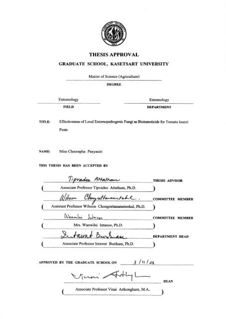
THESIS<br />
EFFECTIVENESS OF LOCAL ENTOMOPATHOGENIC FUNGI AS<br />
BIOINSECTICIDE FOR TOMATO INSECT PESTS<br />
CHEERAPHA PANYASIRI<br />
A Thesis Submitted in Parti<strong>al</strong> Fulfillment of<br />
the Requirements for the Degree of<br />
Master of Science (Agriculture)<br />
Graduate School, Kas<strong>et</strong>sart University<br />
2005<br />
ISBN 974-9846-84-2
ACKNOWLEDGMENTS<br />
I wish to express my deep gratitude to my advisor, Associate Professor Dr. Tipvadee<br />
Attathom who extends v<strong>al</strong>uable encouragement, suggestions and criticism throughout the course<br />
of my study. Her constant assistance and kindness in editing this thesis manuscript is highly<br />
appreciated.<br />
I would like to deeply thank my major committee, Assistant Professor Dr. Wiboon<br />
Chongratanam<strong>et</strong>eekul and my minor committee, Dr. Wanwilai Intanoo for their advices and<br />
v<strong>al</strong>uable comments for the compl<strong>et</strong>ion of this study.<br />
Many thanks are expressed to Miss Kannika Srinaunmak and members of insect<br />
pathology laboratory for their helpfulness and friendship during my graduate study, to Mrs.<br />
Apinun Sonong and Mrs. Yupin Srihirun for their technic<strong>al</strong> assistant on electron microscopic<br />
study and their friendliness.<br />
Most of <strong>al</strong>l, I am gratefully and sincerely thanks to my parents and my sister who<br />
mercifully provided me everything, especi<strong>al</strong>ly encouragement and education<strong>al</strong> supports<br />
throughout my study.<br />
This study was funded by the Nation<strong>al</strong> Research Council of Thailand (NRCT) and the<br />
Deutsche Forschungsgemeinschift (DFG, German Research Council) within frame work of the<br />
joint research project on “Integrated Management of Tomato Pests under Protected Cultivation<br />
Using Biologic<strong>al</strong> Products”. Speci<strong>al</strong> thanks are extended to Professor Dr. Michael Poehling,<br />
Institute of Plant Diseases and Plant Protection, Faculty of Horticulture, Hannover University,<br />
Germany for his v<strong>al</strong>uable suggestions and effort in providing MS. scholarship through the DFG<br />
project. This study was financi<strong>al</strong>ly supported, in part by the Graduate School, Kas<strong>et</strong>sart<br />
University.<br />
Cheerapha Panyasiri<br />
October 2005
TABLE OF CONTENTS<br />
Page<br />
TABLE OF CONTENTS…………………………………………………………………… i<br />
LIST OF TABLES…………………………………………………………………………. ii<br />
LIST OF FIGURES………………………………………………………………………… iv<br />
INTRODUCTION…………………………………………………………………………. 1<br />
LITERATURE REVIEW………………………………………………………………….. 4<br />
MATERIALS AND METHODS………………………………………………………….. 22<br />
RESULTS AND DISCUSSION…………………………………………………………... 32<br />
Insect survey and sample collection……………………………………………… 32<br />
Fung<strong>al</strong> isolation and identification ………………………………………………. 33<br />
Efficacy of entomopathogenic fungi against tomato insect pests……………..….. 49<br />
Pathologic<strong>al</strong> observation………………………………………………………….. 55<br />
Mass production of entomopathogenic fungi………….…………………………. 60<br />
Spray application in screen cage for insect control………………………………. 79<br />
CONCLUSION……………………………………………………………………………. 84<br />
LITERATURE CITED…………………………………………………………………….. 87<br />
APPENDIX………………………………………………………………………………… 104<br />
i
LIST OF TABLES<br />
Table Page<br />
1 Newly recovered of entomopathogenic fungi isolated from insect collected from<br />
field and greenhouse grown tomato. …………………………………………….. 34<br />
2 Isolates of entomopathogenic fungi obtained from government<strong>al</strong> institutes ……. 53<br />
3 Efficacy of entomopathogenic fungi against tomato insect pests.……… ………. 54<br />
4 Number of conidia of the fungus, Paecilomyces fumosoroseus (Acc. no. FWA3)<br />
produced on different substrates and harvested at different period of time………… 62<br />
5 Number of conidia of the fungus, Paecilomyces fumosoroseus (Acc. no. BCC<br />
7058) produced on different substrates and harvested at different period of time…. 64<br />
6 Number of conidia of the fungus, Beauveria bassiana (Acc. no. BCC 1658)<br />
produced on different substrates and harvested at different period of time……… 66<br />
7 Number of conidia of the fungus, M<strong>et</strong>arhizium anisopliae (Acc. no. KKU 2)<br />
produced on different substrates and harvested at different period of time……… 68<br />
8 Number of conidia of the fungus, Hypocrella hypocreoidea (Acc. no. BCC<br />
11370) produced on different substrates and harvested at different period of time… 70<br />
9 Number of conidia produced by the five effective isolates of entomopathogenic<br />
fungi cultured on different substrates used as growth medium……………………. 71<br />
10 An<strong>al</strong>ysis of LC 50 v<strong>al</strong>ue of entomopathogenic fungi against tomato insect pests at 4<br />
days after fung<strong>al</strong> spraying to tomato infested plants in insect screen cage………... 83<br />
ii
LIST OF TABLES (cont’d)<br />
Appendix Table Page<br />
1 Cost of each substrate used for conidia production………………………….......... 105<br />
2 Spray application of Paecilomyces fumosoroseus (Acc. no. FWA3) against thrips,<br />
Ceratothripoides claratris in insect screen cages………………………………… 106<br />
3 Spray application of M<strong>et</strong>arhizium anisopliae (Acc. no. KKU 2) against me<strong>al</strong>ybug,<br />
Pseudococcus cryptus in insect screen cages……………………………………… 106<br />
4 Spray application of Paecilomyces fumosoroseus (Acc. no. FWA3) against<br />
whitefly, Bemisia tabaci in insect screen cages…………………………………… 107<br />
iii
LIST OF FIGURES<br />
Figure Page<br />
1 Colony characteristics of the entomopathogenic fungi newly recovered from<br />
natur<strong>al</strong>ly infected insect pests of tomato. ……………………………………. 40<br />
2 Morphologic<strong>al</strong> diagram of the isolates of entomopathogenic fungi newly<br />
recovered from insect pests of tomato..………………………………………… 44<br />
3 Scanning electron micrographs demonstrated infection process of<br />
entomopathogenic fungi………………………………………………………… 57<br />
4 Scanning electron micrographs of insects infected with entomopathogenic fungi 58<br />
5 Scanning electron micrographs of conidiophores and conidia of highly effective<br />
fung<strong>al</strong> isolates produced on the surface of insect cadavers. …………………….. 59<br />
6 Mycelium growth and sporulation of Paecilomyces fumosoroseus (Acc. no.<br />
FWA3) on different solid substrates used as culture medium…………………… 72<br />
7 Mycelium growth and sporulation of Paecilomyces fumosoroseus (Acc. no.<br />
BCC 7058) on different solid substrates used as culture medium………………. 73<br />
8 Mycelium growth and sporulation of Beauveria bassiana (Acc. no. BCC 1658)<br />
on different solid substrates used as culture medium……………………………. 74<br />
9 Mycelium growth and sporulation of Hypocrella hypocreoidea (Acc. no. BCC<br />
11370) on different solid substrates used as culture medium……………………. 75<br />
10 Mycelium growth and sporulation of M<strong>et</strong>arhizium anisopiae (Acc. no. KKU 2)<br />
on different solid substrates used as culture medium……………………………. 76<br />
iv
EFFECTIVENESS OF LOCAL ENTOMOPATHOGENIC FUNGI AS<br />
BIOINSECTICIDE FOR TOMATO INSECT PESTS<br />
INTRODUCTION<br />
Tomato (Lycopersicon esculentum) is one of the economic important crops of Thailand. It is<br />
consumed as fresh table tomato and as an essenti<strong>al</strong> raw materi<strong>al</strong> for a vari<strong>et</strong>y of food processing<br />
industries. Tomato production faces many problems from sever<strong>al</strong> causes such as season<strong>al</strong> weather,<br />
temperature, humidity, diseases and insect pests. There are sever<strong>al</strong> insect species feed on tomato for<br />
examples: thrips, whitefly, tomato fruitworm, leaf miner, leafhopper, aphid, mites and me<strong>al</strong>ybug.<br />
Thrips, me<strong>al</strong>ybug and whitefly are particularly important pests and are the major causes for the<br />
reduction of tomato production.<br />
Thrips attack leads to the wilting of young plants. Leaves are silvered and flecked. It is <strong>al</strong>so a<br />
vector of spotted wilt virus (Morgan, 1979). In recent year, it has become one of the most<br />
economic<strong>al</strong>ly damaging pest to greenhouse crops (Osborne <strong>et</strong> <strong>al</strong>., 1990). In Thailand the crops most<br />
seriously attacked by thrips are chili, watermelon, cucumber, onion, sh<strong>al</strong>lot, garlic, eggplant,<br />
mushroom, potato, asparagus, yard-long bean, garden pea, okra and tomato (Bansiddhi and<br />
Poonchaisri, 1991). Me<strong>al</strong>ybug is an important pest in greenhouse. Outbreaks often occur in<br />
greenhouses even when densities are low in field. Sever<strong>al</strong> natur<strong>al</strong> enemies of me<strong>al</strong>ybug have been<br />
reported (Itioka and Inoue, 1996) and are thought to be important for suppressing the pest in the field.<br />
Whitefly is an important pest of horticultur<strong>al</strong> crops in greenhouse. The problems are increasing on<br />
outdoor crops such as strawberry, raspberry, pepper, cucumber, tomato, l<strong>et</strong>tuce, citrus and lima bean.<br />
Economic damage to crops are caused by ingestion of plant sap and contamination of crop products<br />
with honeydew that forms a substrate for the development of sooty mold (Johnson <strong>et</strong> <strong>al</strong>., 1992; Liu <strong>et</strong><br />
<strong>al</strong>., 1993)<br />
In many countries, control of tomato pests is based mainly on chemic<strong>al</strong> insecticides.<br />
Chemic<strong>al</strong> residues in fresh tomato raise awareness of adverse effects of chemic<strong>al</strong> insecticides to<br />
1
2<br />
human he<strong>al</strong>th and environment. In addition, extensive and indiscriminate uses of chemic<strong>al</strong><br />
insecticides had created resistant problem in many insect species. Growing tomatoes under protected<br />
cultivation is increasing due to the significant mark<strong>et</strong> size for pesticide-free tomatoes. However,<br />
some tiny, plant sucking insect such as thrips, Ceratothripoides claratris; me<strong>al</strong>ybug, Pseudococcus<br />
cryptus and whitefly, Bemisia tabaci can increase rapidly in greenhouse and cause costly damage to<br />
tomato. Concerns about the negative impacts of chemic<strong>al</strong> insecticides have led to <strong>al</strong>ternative<br />
strategies for pest control, the biologic<strong>al</strong> control using entomopathogens. There are many natur<strong>al</strong>ly<br />
occuring entomopathogens that are effective against insects, such as virus, bacteria, nematode and<br />
fungi. Entomopathogenic fungi are well-recognized as effective biocontrol agents for tiny plant<br />
sucking insects in particular, for example the fungi, Beauveria bassiana and Paecilomyces<br />
fumosoroseus were reported to be effective against whiteflies (Bemisia tabaci), Beauveria bassiana,<br />
Verticillium lecanii, Paecilomyces fumosoroseus and Hirsutella sp. were effective against thrips<br />
(Thrips tabaci) and Verticillium lecanii, Aschersonia sp. and Hypocrella sp. were effective against<br />
me<strong>al</strong>ybug (Pseudococcus cryptus) (Butt, 1997). This study aims to search for loc<strong>al</strong> strains of<br />
entomopathogenic fungi and ev<strong>al</strong>uate their effectiveness against some tomato insect pests. Attempts<br />
are made to develop those potenti<strong>al</strong> fungi as bioinsecticide for tomato pests control under protected<br />
cultivation.
OBJECTIVES<br />
1. To search for loc<strong>al</strong> strains of fungi that are pathogenic to tomato insect pests.<br />
2. To ev<strong>al</strong>uate and compare efficacy of the fungus isolates against some important tomato insect<br />
pests in greenhouse.<br />
3. To develop the effective fungus strains as bio-insecticide for the control of insect pests of tomato<br />
cultivated in greenhouse.<br />
3
Insect pests of tomato<br />
LITERATURE REVIEW<br />
The vast majority of tomato pests are aphids (Aulacorthum solani), leaf miner (Liromyza<br />
bryoniae), leafhopper (Zygina p<strong>al</strong>lidfrous), red spider mite (T<strong>et</strong>ramychus urticae), springtails, thrips<br />
(Thrips tabaci), tomato moth (Lacanobia oleracea), whitefly (Tri<strong>al</strong>euriodes vaporiorum), wire worm<br />
(Agriotes obscurus), and woodlice (Morgan, 1979) but the important pests are thrip (Thrips tabaci),<br />
whitefly (Bemisia tabaci (Genn.)), me<strong>al</strong>ybug (Ferrisia virgata (Ck<strong>al</strong>l)) and aphids (Aulacorthum<br />
solani) (Jones, 1999).<br />
1. Thrips<br />
Thrips is a world wide distributed insect species. It is classified in the order Thysanoptera.<br />
Larvae are p<strong>al</strong>e-yellow, adult thrips are sm<strong>al</strong>l, dark brown insect with elongated bodies, and fringed<br />
wings (Poonchaisri, 1990). The leaves of attacked plants become silvered and flecked. Heavy attacks<br />
lead to the wilting of young plants. They are vectors of spotted wilt virus (Morgan, 1979). In<br />
Thailand, plant crops that are seriously attacked by thrips are chili, watermelon, cucumber, onion,<br />
sh<strong>al</strong>lot, garlic, eggplant, tomato, potato, asparagus, yard-long bean, garden pea, okra and mushroom<br />
(Bansiddhi and Poonchaisri, 1991). Similar records of plants species attacked by thrips were <strong>al</strong>so<br />
reported in Taiwan (Chang, 1991).<br />
Life cycle: Thrips undergoes compl<strong>et</strong>e m<strong>et</strong>amorphosis in which the life cycle composed of<br />
egg, larva, pupa and adult stage. Hill (1983) described life cycle of thrips as follow: white eggs are<br />
laid in notch in the epidermis of the leaves and stems of young plants. They take 4-10 days to hatch.<br />
The larva feed on the leave by rasping the epidermis of the leaves and sucking the exudate. White or<br />
yellow nymphs moult twice in about five days. Pupation occurs in the soil, and takes 4-7 days. The<br />
adult is a sm<strong>al</strong>l, yellow-brown thrips, with darker transverse bands of 1 mm long across the thorax<br />
4
5<br />
and abdomen. It takes about 3 weeks for one generation and there are gener<strong>al</strong>ly sever<strong>al</strong> generations<br />
per year.<br />
Distribution: Thrips is <strong>al</strong>most compl<strong>et</strong>ely cosmopolitan insect species. Hill (1983) reported<br />
the distribution of thrips in West Africa, Canada and south Scandinavia to South Africa and New<br />
Ze<strong>al</strong>and. In Asia, there were records in Philippines which reve<strong>al</strong>ed that thrips feed only on tomato,<br />
pepper, eggplant, watermelon, mushmelon, cucumber, garlic and potato in low elevated areas.<br />
Species attacking sever<strong>al</strong> corps are predominantly Thrips p<strong>al</strong>mi Karny and Meg<strong>al</strong>urothrips usitatus<br />
Bagn<strong>al</strong>l (Bernardo, 1991). In Indonesia, T. tabaci Lind, T. p<strong>al</strong>mi Karny and T. parvispinus Karny are<br />
the major thrips species on veg<strong>et</strong>ables which include chili, potato, tomato, melons and amaranth<br />
(Sastrosiswojo, 1991). In M<strong>al</strong>aysia, T. p<strong>al</strong>mi Karny was found on cucumber (Cucumis s<strong>al</strong>ivus), chili<br />
(Capsicum annuum), brinj<strong>al</strong> (S<strong>al</strong>anum melongena) and tomato (Lycopersicon esculentum). It is the<br />
only vector reported to transmit tomato spotted wilt virus (TSWV) (Fauziah and Saharan, 1991). In<br />
Thailand, the important thrips species are Scirtothrips dors<strong>al</strong>is, T. parvispinus, T. tabaci, Haplothrips<br />
floricola and T. flavus (Poonchaisri, 1990).<br />
2. Me<strong>al</strong>ybug<br />
Me<strong>al</strong>ybugs are in the order Homoptera, family Pseudococcidae. Lindquist (1997)<br />
described that me<strong>al</strong>ybugs are the least sc<strong>al</strong>e-like of the group, mainly because they are soft-bodied,<br />
without the outer shell associated with insects as in the other sc<strong>al</strong>e insect families. Instead, me<strong>al</strong>ybugs<br />
are usu<strong>al</strong>ly covered with a white waxy powder, and have filamentous projections around the<br />
perim<strong>et</strong>er. Plant crops that are seriously attacked by me<strong>al</strong>ybugs are coffee, cocoa, citrus, cotton, jute,<br />
groundnut, beans, cassava, sugarcane, swe<strong>et</strong> potato, cashew, guava and tomato. Moderate damage<br />
<strong>al</strong>so occurs in many other plants. They feed on young shoots, berries and leaves. Heavy attacks of<br />
me<strong>al</strong>ybug usu<strong>al</strong>ly follow periods of prolonged drought.<br />
Life cycle: Hill (1983) described that fem<strong>al</strong>e me<strong>al</strong>ybug lays 300-400 eggs, the egg hatches in<br />
a few hours and the young nymph can move away quite rapidly. They are full grown in about 6
6<br />
weeks. The fem<strong>al</strong>e is a distinctive me<strong>al</strong>ybug with a pair of conspicuous longitudin<strong>al</strong> sub-median dark<br />
strips, long glassy wax threads, a pronounced tail, and a powdery waxy secr<strong>et</strong>ion. The entire life cycle<br />
takes about 40 days.<br />
Distribution: Borror <strong>et</strong> <strong>al</strong>. (1989) described that me<strong>al</strong>ybug is a large group, with more than<br />
300 species in America. There are three important pest species in this group. The citrus me<strong>al</strong>ybug,<br />
Planococcus citri (Risso) and the citrophilus me<strong>al</strong>ybug, Pseudococcus fragilis Brain, are serious pests<br />
of citrus and <strong>al</strong>so attack greenhouse plants. The longtailed me<strong>al</strong>ybug, Pseudococcus longispinus<br />
(Targioni- Tozz<strong>et</strong>ti), is often found in greenhouses, where it attacks a vari<strong>et</strong>y of plants. They are<br />
destructive insect pests in some area such as Austr<strong>al</strong>ia and South America (Hill, 1983). Species of<br />
me<strong>al</strong>ybug that are distributed throughout southern Europe, the Middle East, parts of Africa, South<br />
America and North America were first identified in C<strong>al</strong>ifornia in the Coachella V<strong>al</strong>ley in the early<br />
1990s (Gill, 1994).<br />
3. Whitefly<br />
Whitefly belongs to the order Homoptera, family Aleyrodidae and is close relatives of<br />
aphids, sc<strong>al</strong>e insects, me<strong>al</strong>ybugs, hoppers and cicadas. Sh<strong>et</strong>lar (2003) described whitefly as a white<br />
insect with p<strong>al</strong>e yellow body of approximately 2 mm long. It feeds by extracting plant fluids with<br />
sucking mouth parts. Feeding damage appears as yellow, stunted growth and, in severe cases,<br />
honeydew and sooty mold can develop on plant parts. Sooty mold is a black fungus that grows on<br />
honeydew. Honeydew and sooty mold can reduce photosynthesis and crop v<strong>al</strong>ue. Plant death can<br />
occur if large populations of whitefly are left untreated. Plants that are seriously attacked by whitefly<br />
are cotton, tomato, tobacco, swe<strong>et</strong> potato and cassava. Many wild and other cultivated plants are <strong>al</strong>so<br />
attacked by whitefly.<br />
Life cycle: Life cycle of whitefly had been described by Borror (1989) and Sh<strong>et</strong>lar (2003).<br />
In brief, the white egg is about 0.2 mm long and pear-shaped. It is laid on upper part of the leaf and<br />
egg hatches after about 7 days. The newly hatch larva can move a very short distance before s<strong>et</strong>ting
7<br />
down and starting to feed. The last instar larva is about 0.7 mm long and red eye of the adult can be<br />
seen. The nymph<strong>al</strong> stage lasts 2-4 weeks according to temperature and hatch to adult in a minute.<br />
The adults of both sexes are winged, and the wings are covered with the white dust or waxy powder.<br />
The adult is usu<strong>al</strong>ly active whitish insect that feed on leaves. The fem<strong>al</strong>e may lay 100 or more eggs.<br />
Distribution: The whiteflies are most abundant in the tropics and subtropics. It is a<br />
cosmopolitan insect species widespread occurring from Europe to Asia. Their population dynamics<br />
strongly suggested that biologic<strong>al</strong> control might provide the greatest capacity to reduce damage<br />
caused by Bemisia argentifolii (B. tabaci, strain B) in both greenhouse and field crops (van Lenteren<br />
and Wo<strong>et</strong>s, 1988; Baumgartner and Yano, 1990; Gerling, 1990).<br />
Recently its importance as a pest to field crops has increased. Damage estimates in 1991 by<br />
B. argentifoli to crops in the United State exceeded $500 million. Moreover its tranmission ability of<br />
a virus to the Florida tomato crops was <strong>al</strong>so reported (Perring <strong>et</strong> <strong>al</strong>., 1993). Entomogenous fungi,<br />
Paecilomyces fumosoroseus, Verticillium lecanii and Beauveria bassiana were used successfully for<br />
control of whitefly nymphs (Landa <strong>et</strong> <strong>al</strong>., 1994).<br />
4. Aphid<br />
Aphids are in the order Homoptera, family Aphididae. They infest a wide range of host<br />
plants. P<strong>al</strong>mer and Smith (1967) listed some important cultivated hosts which include potato, tomato,<br />
eggplant, sunflower, pepper, pea, bean, apple, turnip, corn, swe<strong>et</strong> potato, asparagus, clover and rose.<br />
Weeds such as ragweed, lambsquarters, jimsonweed, pigweed, shepherdspurse and wild l<strong>et</strong>tuce are<br />
<strong>al</strong>so common food plants. Sporadic in occurrence, aphid infestations are rarely severe enough to kill<br />
plants. Aphids pierce veins, stems, growing tips, and blossoms with their needle-like mouthparts. As<br />
a result, blossoms are shed and yield is reduced. New growth becomes stunted and curled. Heavily<br />
infested plants turn brown and die from the top down. Aphids tend to spread rapidly from field and<br />
transmitting a number of vir<strong>al</strong> diseases. These include various mosaics, leaf roll, spindle tuber, and<br />
unmottled curly dwarf diseases (Griffin, 2004).
8<br />
Aphids are soft-bodied, pear-shaped insect usu<strong>al</strong>ly wingless. The body color may be solid<br />
pink, green, yellow and pink mottled, or light green with a dark stripe. It is about 2.5 to 3.5 mm long<br />
and has a pair of long, slender tailpipe-like appendages known as cornicles at the dors<strong>al</strong> side of the<br />
fifth or sixth abdomin<strong>al</strong> segment. These cornicles secr<strong>et</strong>e a defensive fluid. Aphid <strong>al</strong>so excr<strong>et</strong>e<br />
honeydew, which is emitted from the anus. Adult fem<strong>al</strong>es give birth to live young, c<strong>al</strong>led nymphs.<br />
Although slightly sm<strong>al</strong>ler than adults, nymphs are similar in color and shape (Griffin, 2004).<br />
Life cycle: In Thailand, fem<strong>al</strong>e aphids feed and reproduce year round. No eggs or m<strong>al</strong>es are<br />
produced. Without mating, wingless fem<strong>al</strong>es give birth to about 50 live nymphs. During warm<br />
weather, each of these nymphs matures in 2 or 3 weeks. The life cycle continues in this manner until<br />
overcrowding occurs or food becomes scarce. At this time nymphs develop into winged adults and<br />
migrate to new host plants. Once s<strong>et</strong>tled down, these aphids begin reproducing and the life cycle<br />
continues as before. During winter, however, feeding and reproduction occur at a much slower rate.<br />
Many generations are produced each year (Lowe, 1973 and Borror, 1989).<br />
Distribution: Aphids are found <strong>al</strong>most widespread from Europe to Asia and occur throughout<br />
North American (Lowe, 1973). The aphids are tended by the ants, which transfer them from one food<br />
plant to another. The ants feed on the honeydew produced by the aphids. In many species the winged<br />
form migrates to a different host plant, and the reproductive process continues.<br />
Entomopathogenic Fungi<br />
Many insect pests are susceptible to infection by natur<strong>al</strong>ly occurring insect pathogenic fungi.<br />
Sever<strong>al</strong> fungi have been studied as potenti<strong>al</strong> mycoinsecticides. These fungi are very specific to<br />
insects, often to particular species and do not infect anim<strong>al</strong>s or plants. Fungi provide the only<br />
satisfactory microbi<strong>al</strong> means of biocontrol of plant sucking insects such as aphids and whiteflies that<br />
are not susceptible to bacteria and viruses. The well-studied insect pathogenic fungi include<br />
Beauveria bassiana for locusts and be<strong>et</strong>les, M<strong>et</strong>arhizium anisopliae and M. flavoviride for locusts and<br />
Verticillium lecanii for control of aphid. Other possible fung<strong>al</strong> candidates of biocontrol agents
9<br />
include B. brongniartii, Hirsutella thompsonii, Paecilomyces fumosoroseus, P. farinosus, Nomuraea<br />
rileyi and Aschersonia <strong>al</strong>eyrodis.<br />
1. History<br />
Boucias and Pendland (1998) summarized history of the entomogenous fungi as they<br />
originated more than 2000 years ago. The first record, Cordyceps(Ascomycota) was found to infect<br />
Lepidopteran larvae in ancient China. The colored fruiting body of this fungus usu<strong>al</strong>ly protrudes<br />
from the mouth or anus of the insect cadaver and is therefore very conspicuous. Throughout history,<br />
it has been used for religious, medicin<strong>al</strong> purposes and as food. Cordyceps was the subject of the first<br />
published account of an entomopathogenic fungus by Reaumus in 1726. Another fungus, Beauveria<br />
bassiana (Deuteromyc<strong>et</strong>es) was observed in about 900 AD in silkworms in Japan. It was used for<br />
medic<strong>al</strong> purposes as an antiseptic for wounds and sore throats. Moreover, it was through a study of<br />
this fungus that the germ theory of disease was postulated by A. Bassi in 1834. In the 1870’s, E.<br />
M<strong>et</strong>chnikoff pioneered the field of cellular immunity through his earlier research on other fung<strong>al</strong><br />
entomopathogen, the green muscardine fungus M<strong>et</strong>arhizium anisopliae (Deuteromyc<strong>et</strong>es).<br />
The use of B. bassiana and M. anisopliae as biologic<strong>al</strong> control agent has been studies with<br />
many insect pests. Both fungi <strong>al</strong>so have potenti<strong>al</strong> use against soil insects such as curculionids,<br />
scarabs and lepidopterous (Ferron, 1981)<br />
In Brazil, M. anisopliae has been used effectively against spittle bugs on sugarcane and the<br />
spittle bug population were reduced following spraying conidia products, usu<strong>al</strong>ly at 10 12 conidia/ha.<br />
Incidence of mort<strong>al</strong>ity from M. anisopliae followed treatment was expected to be at least 40% which<br />
increase sugarcane content sufficiently to justify fung<strong>al</strong> use (Roberts <strong>et</strong> <strong>al</strong>., 1991). Zimmermann<br />
(1993) reported that the host list of M. anisopliae was more than 200 insect species from seven<br />
orders.
10<br />
B. bassiana is available commerci<strong>al</strong>ly as a microbi<strong>al</strong> insecticide since B. bassiana can now<br />
be mass produced by a fermentation process and formulated to enable the fungus to withstand<br />
ultraviol<strong>et</strong> light, temperature and humidity extreme commonly encountered in the field. There are<br />
sever<strong>al</strong> products that contain B. bassiana and registered with EPA. B. bassiana takes 3-7 days to kill<br />
an insect and will take some time to suppress the pest population when using these product. Through<br />
spray coverage is essenti<strong>al</strong> because fung<strong>al</strong> spore must contact the insect for infection to occur<br />
(Hofmann and Frodsham, 1993).<br />
2. Natur<strong>al</strong> incidence<br />
The fungi isolated from or known to infect insects belong either to the class Hyphomyces<br />
or Zygomyces in the subdivisions Deuteromycotina and Zygomycotina, respectively.<br />
2.1 Zygomyc<strong>et</strong>es<br />
Observations of natur<strong>al</strong> epizootics in thrips populations had largely been restricted to<br />
Entomophthor<strong>al</strong>es. Neozygites parvispora, formerly known as Entomophthora parvispora, had been<br />
found frequenly in Thrips tabaci and T. fuscipennis populations on onion crops in centr<strong>al</strong> and<br />
southern Europe. Attempts to utilize this agent for management of thrips under field conditions were<br />
not successful, <strong>al</strong>though b<strong>et</strong>ter control of T. tabaci was obtained in greenhouses where environment<strong>al</strong><br />
conditions favoured infection and spread of the disease (Carl, 1975; MacLeod <strong>et</strong> <strong>al</strong>., 1976). In the<br />
N<strong>et</strong>herlands, epizootics caused by Entomophthora thripidum had been observed in T. tabaci<br />
infestations on a vari<strong>et</strong>y of greenhouse crops, but in field trails the fungus failed to suppress thrips<br />
populations below an economic<strong>al</strong>ly acceptable level (Ramakers, 1976; Samson <strong>et</strong> <strong>al</strong>., 1979).<br />
Saito <strong>et</strong> <strong>al</strong>. (1989) reported a low incidence of Neozygites parvispora in field-collected<br />
specimens of T. p<strong>al</strong>mi. This pathogen was <strong>al</strong>so recovered from Frankliniella occident<strong>al</strong>is infesting<br />
glasshouse-growth in It<strong>al</strong>y (Magano di san Lio <strong>et</strong> <strong>al</strong>., 1992). In glasshouse studies, Vacante <strong>et</strong> <strong>al</strong>.<br />
(1994) noted that N. parvispora caused up to 60 per cent mort<strong>al</strong>ity in motile development<strong>al</strong> stages of
11<br />
F. occident<strong>al</strong>is and reduced both the insect population density and the proportion of leaves and<br />
flowers infested. Development of an epizootic appeared to be less dependent upon high ambient<br />
relative humidity than for other isolates of this species.<br />
2.2 Hyphomyc<strong>et</strong>es (Imperfect fungi)<br />
Vertillium lecanii, M<strong>et</strong>arhizium anisopliae, Paecilomyces farinosus, P. lilacinus and<br />
Hirsutella sp. had been isolated from Taeniothrips inconsequens, a recurrent pest of sugar maple in<br />
the north-eastern USA (Skinner <strong>et</strong> <strong>al</strong>., 1991; Brownbrigde, 1995). Beauveria bassiana had been<br />
recovered from Thrips c<strong>al</strong>caratus, Frankliniella occident<strong>al</strong>is and Haplothrips tritici (Lyubenov,<br />
1961; Humber, 1992). H<strong>al</strong>l (1992) and H<strong>al</strong>l <strong>et</strong>. <strong>al</strong>. (1994) had reported Hirsutella sp. nov. causing an<br />
epizootic in T. p<strong>al</strong>mi in Trinidad and the occasion<strong>al</strong> incidence of P. fumosoroseus in collected<br />
specimens. Hirsutella sp. was <strong>al</strong>so isolated from Liothrips mikaniae (Greenwood and Mill, 1989).<br />
Although not gener<strong>al</strong>ly regarded as a virulent insect pathogen, an Aspergillus sp. had <strong>al</strong>so been<br />
reported to infect thrips (Dyadechko, 1977).<br />
A number of species of hyphomyc<strong>et</strong>ous imperfect fungi (Deuteromyc<strong>et</strong>es) had been reported<br />
to attack aphids. The fungi, Ceph<strong>al</strong>osporium aphidicola P<strong>et</strong>ch, C. muscarium P<strong>et</strong>ch, Cladosporium<br />
aphidis Thuem., Hirsutella aphidis P<strong>et</strong>ch and Paecilomyces (Spicaria) farinosus (Dicks ex. Fr) Brown<br />
and Smith were recorded from aphid hosts by P<strong>et</strong>ch (1932, 1948), Since they are seldom reported by<br />
researchers, it can be assumed that they occur rarely in nature and play a minim<strong>al</strong> role in the<br />
suppression of aphid populations which they are reported to attack.<br />
Numerous field trails were conducted with the fungus, Acrost<strong>al</strong>agmus aphidum Oud in<br />
Puerto Rico against the aphid, Myzus persicae (Sulzer) and Aphis gossypii Glover, on egg plant by<br />
Nolla (1929), who reported good control following spraying of infested plants with spore<br />
suspensions. Results of the studies conducted in the 1970’s by Wolcott using A. aphidum against<br />
heavy infestations of Sipha flava (Forbes) on young sucarcane in Puerto Rico were much more<br />
impressive. He found that under equ<strong>al</strong>ly optimum weather conditions of extended periods of rainf<strong>al</strong>l
12<br />
or high humidity at tropic<strong>al</strong> temperatures, application of infective stages of the fungus could bring<br />
mass infection and tot<strong>al</strong> destruction of the pest population on the treated foliage.<br />
The only other report on trails with A. aphidum is that of Shands <strong>et</strong> <strong>al</strong>. (1958), who claimed<br />
successful infection of sm<strong>al</strong>l numbers of Macrosiphum euphorbiae (Thomas) following application of<br />
the fungus. Moreover, the collection of a few infected aphids in check plots adjacent to the treated<br />
areas led them to believe that some spread of the fungus had taken place. This claim of the<br />
establishment and possible spread of the fungus were in doubt. Since during the following 10 years<br />
of study, thousands of dead, diseased aphids of sever<strong>al</strong> species infesting potatoes in northeastern<br />
Maine were collected and examined, none was found to be infected by A. aphidum (Shand <strong>et</strong> <strong>al</strong>.,<br />
1962; Shands <strong>et</strong> <strong>al</strong>., 1972). It is possible that the fungus requires tropic<strong>al</strong> weather conditions for<br />
development.<br />
Not <strong>al</strong>l stages in an insect’s life cycle are equ<strong>al</strong>ly susceptible to infection by<br />
entomopathogenic Hyphomyc<strong>et</strong>es. In some situations, immature insects are more susceptible to<br />
infection than mature insects. For example, young larvae of the European corn-borer (Ostrinia<br />
nubil<strong>al</strong>is) were more susceptible to B. bassiana than older larvae (Feng <strong>et</strong> <strong>al</strong>., 1985). In contrast,<br />
adults western flower thrips (Frankliniella occident<strong>al</strong>is) were more susceptible to V. lecanii than<br />
larvae (Vestergaard <strong>et</strong> <strong>al</strong>., 1995). Most host factors, such as insect development<strong>al</strong> rates, cannot be<br />
considered independent of environment (e.g. temperature). High temperatures accelerate insect<br />
development and will reduce the time b<strong>et</strong>ween molts, which can subsequently reduce the prev<strong>al</strong>ence<br />
of infection due to loss of inoculum on exuviae. Insect density is of particular importance in the<br />
epizootiology of disease. As the density of insects increases, there is a higher probability of an insect<br />
coming into contact with a pathogen (i.e. with infected individu<strong>al</strong>s or with the pathogen directly).<br />
The behavior of insects can influence epizootic development, and can affect the dispers<strong>al</strong> of an<br />
entomopathogen.
3. Infection path way of entomopathogenic fungi<br />
Poinar and Thomas (1984) described infection process of entomopathogenic fungi which<br />
is typic<strong>al</strong> for most of the fungi found to attack insect species. Most entomogenous fungi initiate their<br />
infection by a germinating spore or conidia which pen<strong>et</strong>rate the cuticle of an insect. The invasive<br />
hypha enters the host’s tissues (often the fat body is first attacked) and ramifies through the hemocoel.<br />
In some fungi such as M<strong>et</strong>arrhizium anisopliae, Beauveria bassiana, Conidiobolus coronata, and C.<br />
apiculatus, “hyph<strong>al</strong> bodies” or segments of hyphae break off and circulate in the host’s hemocoel<br />
during the early stages of infection. Upon contact with insect, the spore responds to biochemic<strong>al</strong> cues<br />
present in the waxy epicuticle and germinates within 8-16 hrs. Soon the fungus stops growing<br />
horizont<strong>al</strong>ly on the surface of the cuticle and initiate pen<strong>et</strong>ration, using a combination of mechanic<strong>al</strong><br />
pressure and a mixture of cuticle degrading enzymes, which attack and dissolve the cuticle. Once the<br />
fungus breaks through the cuticle and underlying epiderm<strong>al</strong>s, it tends to invade in haemocoel of the<br />
insect and proliferate in the haemolymph. The insect’s defense system in the haemocoel employs<br />
phagocytosis and the secr<strong>et</strong>ion. Usu<strong>al</strong>ly, within 24 hrs. of germination, the fungus rapidly proliferates<br />
through the insect. The infected insects stop feeding and become l<strong>et</strong>hargic. They may die relatively<br />
rapidly within 2-7 days (Jaronski, 1997). After filling the dying or dead insect with mycelium,<br />
hyphae emerge out through the insect’s integument, grow rapidly and produce spores on the extern<strong>al</strong><br />
surface of the host. These spores are dispersed by wind or rain and even by the parasitized insect<br />
during feeding or mating. In some instances the fungi are loc<strong>al</strong>ized in speci<strong>al</strong> organs of the host; e.g.,<br />
the fungi Massospora cicadina and Strongwellsea castrans occur only in the abdomen of adult hosts.<br />
Some fungi are host or stage specific, while others (e.g., Beauveria bassiana and M<strong>et</strong>arrhizium<br />
anisopliae) exhibit a wide host range. Fung<strong>al</strong> infection and especi<strong>al</strong>ly epizootic are mainly dependent<br />
on a large host population and ide<strong>al</strong> climatic conditions. The life cycle of the fungus is compl<strong>et</strong>ed<br />
when it sporulates on the cadaver of the host. Under the right conditions, particularly higher relative<br />
humidity, the fungus will break out through the body w<strong>al</strong>l of the insect producing aeri<strong>al</strong> spores. High<br />
humidity is critic<strong>al</strong> to spore germination, fung<strong>al</strong> survivorship and transmission from host to host. The<br />
dead insect’s body may be form an empty shell, often but not <strong>al</strong>ways with green, red or brown fung<strong>al</strong><br />
growth, either enveloping the body or emerging from joints and body segments. These extern<strong>al</strong><br />
13
14<br />
hyphae produce conidia that ripen and are released into the environment compl<strong>et</strong>ing the cycle.<br />
Adequate moisture and temperature are usu<strong>al</strong>ly required for successful sporulation and spore<br />
germination.<br />
Fungus as biologic<strong>al</strong> control agent<br />
1. Mass propagation of the fungus<br />
In laboratory, entomopathogenic fungi were usu<strong>al</strong>ly maintained as pure culture on Saboraud<br />
Dextrose Agar (SDA) and Potato Dextrose Agar (PDA). But field application for insect control, a<br />
large amount of spores or conidia is required. Therefore, mass production of entomopathogenic fungi<br />
that is cost-effective and provides numbers of viable spores is important for the successful<br />
development of fungi as mycoinsecticides.<br />
Daoust <strong>et</strong> <strong>al</strong>. (1983) described the procedure in which insect fungi produced as spore<br />
suspension containing approximately 5 x 10 7 – 1 x 10 8 conidia/ml. They were used to inoculate PDA<br />
agar (0.5 ml/plate) or rice medium (5 ml/bag). The rice medium, which contained 150 g of long-grain<br />
rice and 150 ml of water was sterilized high-density poly<strong>et</strong>hylene autoclave bag for 20 minutes at<br />
121°c. The bags were loosely se<strong>al</strong>ed with m<strong>et</strong><strong>al</strong> ties during autoclaving, cooled down, and then,<br />
tightly se<strong>al</strong>ed with only a sm<strong>al</strong>l amount of air using the same ties. Each bag was injected with 5ml of<br />
the conidi<strong>al</strong> suspension using a hypodermic syringe and the puncture was se<strong>al</strong>ed with tape. Bags<br />
were shaked vigorously to distribute the inoculum, and incubated for 14 days at 25°c - 26°c in the<br />
dark. Conidia were harvested and seived through 125 m particle size by the m<strong>et</strong>hod of Daoust and<br />
Roberts (1983). Conidia used in <strong>al</strong>l dry formulations were grown on rice medium while <strong>al</strong>l other<br />
formulations contained PDA-grown conidia. The other fungus growth substrates are <strong>al</strong>mond, coconut,<br />
corn, cottonseed, linseed, olive, peanut, soybean, sunflower, wheat germ and sorghum.<br />
Agudelo <strong>et</strong> <strong>al</strong>. (1983) maintained P. farinosus in solid media. The solid media employed<br />
were autoclaved moist bran (McCoy <strong>et</strong> <strong>al</strong>., 1941). The bran was thoroughly mixed with tap water
15<br />
(1.5:1 w/w) and autoclaved in a capped Erlenmeyer flask for 30 min. Potato egg yolk (G<strong>et</strong>zin, 1961)<br />
containing 5% potato, 1% fresh egg yolk, 1% active dry yeast, 1% Bacto peptone (Difco), and 1.5%<br />
agar. These ingredients were mixed in a Waring Blender, dispensed into P<strong>et</strong>ri dishes, then sterilized<br />
for 20 min. Be<strong>al</strong>l <strong>et</strong> <strong>al</strong>. (1939) reported the use of soil bean mass as fung<strong>al</strong> medium. One hundred<br />
grams of dry soybeans were soaked in 250 ml of water for 24 hr. The surplus water was poured off<br />
and the swollen beans were ground in Waring blender. The bean mash was put into P<strong>et</strong>ri dishes, and<br />
autoclaved for 30 min. The mycelia produced in solid media were sparse and form a tight mat-like<br />
growth with the substrate, which was difficult to suspend in water.<br />
Bourassa (1998) had identified B. bassiana as one of a series of isolated pathogenic to P.<br />
truncates. Conidia were produced by use of standard two-stage system (Jenkins <strong>et</strong> <strong>al</strong>., 1998) with<br />
rice as the solid substrate. Extracted and dried conidia were sieved with a 106 µm sieve to remove<br />
large hyph<strong>al</strong> fragments. Dried conidia powder contained 4.59 x 10 10 conidia/g and had a germination<br />
rate above 98%.<br />
2. Formulation of entomopathogenic fungus<br />
The development of a suitable formulation is essenti<strong>al</strong> to the successful utilization of<br />
mycoinsecticides. Of primary importance is the r<strong>et</strong>ention of viability and virulence of the infective<br />
units during storage and application. Formulation is mandatory in order to enhance spore application<br />
and efficacy. The type of formulation ultimately selected depends upon the biology and physic<strong>al</strong><br />
properties of the pathogen and the location and habits of the targ<strong>et</strong> pest (Daoust <strong>et</strong> <strong>al</strong>., 1983).<br />
Roberts and Yendol (1971) described the formulation that spore can be applied in dust,<br />
sprays or granules. The best formulation depends primarily upon the fung<strong>al</strong> species. Dust offer a<br />
slight advantage, in that they can be stores in a formulated condition, and their swirling in air j<strong>et</strong>s<br />
tends to adhere them to lower as well as upper surfaces of foliage. Selection of appropriate diluents is<br />
important since some materi<strong>al</strong>s inhibit spore germination. T<strong>al</strong>c, flour and milk powder have served as<br />
suitable diluents for dusts. Aqueous sprays of B. bassiana spores are gener<strong>al</strong>ly unsuitable unless a
16<br />
w<strong>et</strong>ting agent is included, <strong>al</strong>though it is advisible to test its effect on spore viability first. Some<br />
compounds which have proved satisfactory to be used as w<strong>et</strong>ting agent include Triton X-100, DuPont<br />
Spreader-sticker. Triton X-155 and the household d<strong>et</strong>ergent “Trend” at 1% in water. They had no<br />
d<strong>et</strong>ectable adverse effect on the spores of B. bassiana. Granules (corn me<strong>al</strong> or attapulgite) are more<br />
effective than sprays or dusts when treating corn with B. bassiana for corn borer control. Compounds<br />
which shield spores from ultra-viol<strong>et</strong> rays of the sun, increase moisture in the microclimate of the<br />
spores, and reduce the <strong>al</strong>lergenicity hazard from spores should receive speci<strong>al</strong> consideration by<br />
formulators of entomogenous fungi. Encapsulation, which conceivably could satisfy some of these<br />
requirements, has not been reported for fungi.<br />
Inglis <strong>et</strong> <strong>al</strong>. (1993) found that Beauveria bassiana conidia dispersed b<strong>et</strong>ter in oil than in<br />
0.05% aqueous Tween80, while substanti<strong>al</strong> clumping occurred in a 5% emulsion of the oil in water,<br />
even after homogenization.<br />
Smith (1994) reported that in dry conditions (45-100% RH day-night), Paecilomymes<br />
fumosoroseus conidia sprayed in oil (Shellsol T: rape seed oil, 7:3) and in two emulsions (1 and 10%<br />
Cadacide emulsifiable oil in water) killed 80-100% whitefly (Bemisia tabaci), but killed none when<br />
sprayed in 0.1% aqueous Tween80 (<strong>al</strong>l sprays applied at 2 ml per cucumber leaf on potted plants)<br />
M. anisopliae var. acridum is being developed as a Biologic<strong>al</strong> Control Agent of acridids in<br />
variuos regions of the world, including Africa (e.g. Lomer <strong>et</strong> <strong>al</strong>., 1997a), Austr<strong>al</strong>ia (e.g. Milner, 1997)<br />
and Brazil (e.g. Mag<strong>al</strong>haes <strong>et</strong> <strong>al</strong>., 2000). In a large research program conducted in Africa, conidia<br />
were produced on rice (Jenkins <strong>et</strong> <strong>al</strong>., 1998) and they were dried and stored for various periods of<br />
time at low (>18 months) and/or ambient (~ 12 months) temperatures (Lomer <strong>et</strong> <strong>al</strong>., 1997a). Conidia<br />
were formulated in oils (primarily paraffinic oils used <strong>al</strong>one or blends with botanic<strong>al</strong> oils) and applied<br />
at ultra-low volumes, using hand-held Micro-Ultra applicators. The efficacy of M. anisopliae var.<br />
acridum was probably enhanced by secondary spore pick-up. There were observations that acridids<br />
coming in contact with veg<strong>et</strong>ation infested with conidia pick up inoculum and often became infected<br />
and died by mycosis (Bateman <strong>et</strong> <strong>al</strong>., 1998; Thomas <strong>et</strong> <strong>al</strong>., 1998). Furthermore, the fungus can persist
17<br />
in fragments of infected grasshopper cadavers and survive adverse as a source of secondary inoculum<br />
(i.e. horizont<strong>al</strong> transmission), enhancing the efficacy of M. anisopliae var. acridum against acridids<br />
(Thomas <strong>et</strong> <strong>al</strong>., 1996). However, the biotic and abiotic factors regulating horizont<strong>al</strong> transmission in<br />
field s<strong>et</strong>ting are still poorly understood.<br />
3. Efficacy test against insect species<br />
James (2003) described application technique for entomopathogenic fungi in laboratory as<br />
follow: leaves with insects were sprayed using a Potter Precision Laboratory Spray Tower (Burkhard<br />
Manufaturing, Rickmansworth, England) with 0.7 kg/cm 2 of pressure and the fine-mist nozzle (as<br />
described by James and Jaronski, 2000). Each leaf was laid out flat on an acrylic plate with the<br />
abaxi<strong>al</strong> surface facing up, and then sprayed with 1 ml of each treatment preparation. After being<br />
sprayed, <strong>al</strong>l the leaves were s<strong>et</strong> upright by placing the water picks in test tube racks such that the<br />
leaves did not touch or overlap each other. The leaves were <strong>al</strong>lowed to air dry in the laboratory, and<br />
each rack of leaves was covered with a plastic bag to raise the humidity to ≥ 95%. The leaves were<br />
not arranged in any particular order within each rack. All the racks were incubated at 25 ๐ c with a<br />
photoperiod of 14:10 (L:D) h, and a sm<strong>al</strong>l temperature and relative humidity recorder was placed in<br />
one of the bags. The leaves were removed from the bag after 24 h and incubated at 70% RH, 25 ๐ c<br />
with photoperiod of 14:10 (L:D) h. Insect mort<strong>al</strong>ity was recorded 7 days after each spray application.<br />
Burges (1998) described laboratory work on the infection of M<strong>et</strong>arhizium and Beauveria<br />
conidia formulated in oil which has provided much evidence of infection at low humidities and the<br />
importance of pen<strong>et</strong>ration at intersegment<strong>al</strong> membranes. Previous reports indicated that conidi<strong>al</strong><br />
germination on cinch bugs (Blissus leucopterus) tended to be loc<strong>al</strong>ized at the articulation b<strong>et</strong>ween<br />
coxa and trochanter (Ramoska, 1984). Topic<strong>al</strong> appication of conidia in oil was more effective than in<br />
water plus w<strong>et</strong>ting agent on the mouth parts of the cocoa weevil by 36 times (Prior <strong>et</strong> <strong>al</strong>., 1988) and<br />
under the pronot<strong>al</strong> shield of locusts by 146 times at ca 35% RH (Bateman <strong>et</strong> <strong>al</strong>., 1993). The LT 50<br />
(l<strong>et</strong>h<strong>al</strong> time to kill h<strong>al</strong>f the insects) was lower if be<strong>et</strong>les were inoculated under the elytra than on the<br />
elytra where conditions were shown to be more fungistatic (Butt <strong>et</strong> <strong>al</strong>., 1995). Efficient killing in the
18<br />
presence of oil was indicated by steep dose-mort<strong>al</strong>ity curves more akin to those for chemic<strong>al</strong>s than for<br />
microbi<strong>al</strong> agents (Bateman <strong>et</strong> <strong>al</strong>., 1993)<br />
In field experiment, de la Rosa, <strong>et</strong> <strong>al</strong>. (2000) reve<strong>al</strong>ed that conidia were applied b<strong>et</strong>ween<br />
06.00 and 08.00 h with a previously c<strong>al</strong>ibrated manu<strong>al</strong> pressure knapsack sprayer. A dose of 1x10 9<br />
conidia per plant was applied for both B. bassiana and M. anisopliae; the number of conidia in<br />
suspension was d<strong>et</strong>ermined with a Neubauer chamber.<br />
A domestic strain of the fung<strong>al</strong> pathogen V. lecanii in Korea was tested for the control of<br />
aphid and whitefly (Kim <strong>et</strong> <strong>al</strong>., 2001). Of the six isolates collected in Korea, V. lecanii caused the<br />
highest mort<strong>al</strong>ity of cotton aphid (Aphid gossypii). V. lecanii was selected as a biologic<strong>al</strong> control<br />
agent for whitefly, based on bioassay and field tests. The pesticides dim<strong>et</strong>homorph and procymidone<br />
did not affect spore germination and myceli<strong>al</strong> growth of V. lecanii, provided they were used at the<br />
recommended concentration.<br />
Fargues <strong>et</strong> <strong>al</strong>. (1991) demonstrated that the reproductive potenti<strong>al</strong> of Colorado potato be<strong>et</strong>le<br />
fem<strong>al</strong>es surviving infection by B. bassiana was reduced at 22 ๐ C but not at 25 ๐ C. Adults of the<br />
hymenopteran parasitoid of Russian wheat aphid (Aphelinus asychis) treated with P. fumosoroseus<br />
and incubated at high humidity were significantly less active (e.g. percentage of time w<strong>al</strong>king,<br />
w<strong>al</strong>king speed and distance covered) than untreated insects (Lacey <strong>et</strong> <strong>al</strong>., 1997). Development<strong>al</strong><br />
times were prolonged and the predation efficacy of the coccinellid predator, Serangium<br />
parces<strong>et</strong>osum, was similarly reduced in individu<strong>al</strong>s sprayed with B. bassiana (Poprawski <strong>et</strong> <strong>al</strong>.,<br />
1998). Authurs and Thomas (1999) observed that down locusts (Locusta pard<strong>al</strong>ina) infected by M.<br />
anisopliae var. acridum were more susceptible to predation. Similar effects had been shown for<br />
Sahelian grasshoppers (Thomas <strong>et</strong> <strong>al</strong>., 1998). These recent reports demonstrated that subl<strong>et</strong>h<strong>al</strong> effects<br />
occured in insect pests infected with entomopathogenic fungi.<br />
Foliar application of B. bassiana was used effectively in conjunction with the bacterium B.<br />
thuringiensis. Densities of be<strong>et</strong>les in the fungus-bacterium-treated plots declined yearly relative to
19<br />
other treatments (Drummond and Groden, 1996). Poprawski <strong>et</strong> <strong>al</strong>.(1997) observed that applications<br />
of B. bassiana conidia at 3-4-day interv<strong>al</strong>s early in the season effectively reduced densities of older<br />
larvae and provided substanti<strong>al</strong> foliar protection; larv<strong>al</strong> densities were 10, 21 and 41 larvae per plant<br />
for the B. bassiana, insecticide (i.e. esfenv<strong>al</strong>erate, piperonyl butoxide, oxamyl and carbofuran) and<br />
control treatment, respectively. Lacey <strong>et</strong> <strong>al</strong>. (1999a) <strong>al</strong>so observed significant effects of B. bassiana<br />
against Colorado potato be<strong>et</strong>le.<br />
P. fumosoroseus causes rapid infection and death of <strong>al</strong>l whitefly stages. Under optim<strong>al</strong><br />
condition, hyphae are present in the haemocoel within 24 h of inoculation, death occurs b<strong>et</strong>ween 24<br />
and 48 h, hyphae emerges and conidiogenesis occurs on the surface of the cadaver within 72 h<br />
(Osborne <strong>et</strong> <strong>al</strong>., 1990a). Optim<strong>al</strong> growth rates are b<strong>et</strong>ween 20 and 30 ๐ C, with optima related to the<br />
microclimate of the fung<strong>al</strong> isolate’s biotype (Vid<strong>al</strong> <strong>et</strong> <strong>al</strong>., 1997a). Highly virulent isolates of P.<br />
fumosoroseus with considerable control potenti<strong>al</strong> against whiteflies are widespread and numerous<br />
(Lacey <strong>et</strong> <strong>al</strong>., 1996; Vid<strong>al</strong> <strong>et</strong> <strong>al</strong>., 1997b; Wraight <strong>et</strong> <strong>al</strong>., 1998) Wraight <strong>et</strong> <strong>al</strong>. (2000) demonstrated that<br />
infection can take place at ambient relative humidities as low as 25%. Hyph<strong>al</strong> bodies are more<br />
virulent than conidia (Lacey <strong>et</strong> <strong>al</strong>., 1999b) and can be rapidly produced in liquid culture, remaining<br />
viable and virulent following drying (Jackson <strong>et</strong> <strong>al</strong>., 1997). P. fumosoroseus demonstrated limited<br />
l<strong>et</strong>h<strong>al</strong> and sub-l<strong>et</strong>h<strong>al</strong> effects on Serangium parcesstosum, an important coccinelid predator of<br />
whiteflies, suggesting that the integration of these two control agents in IPM may be possible<br />
(Poprawski <strong>et</strong> <strong>al</strong>., 1998).<br />
Inundative application of P. fumosoroseus conidia or hyph<strong>al</strong> bodies had been used<br />
successfully to control whiteflies in field crops. In sm<strong>al</strong>l-sc<strong>al</strong>e field trails using portable air-assist<br />
sprayer, multiple applications of P. fumosoroseus at 4-7-day interv<strong>al</strong>s provided > 90% mort<strong>al</strong>ity of<br />
late-instar whiteflies on cucumber, cant<strong>al</strong>oupe melons and zucchini squash (Wraight <strong>et</strong> <strong>al</strong>., 2000).<br />
Although effects on nymphs were highly significant, the effects on adult whiteflies were minim<strong>al</strong>.<br />
Commerci<strong>al</strong> products based on this fungus are now available for whitefly control (Shah and Go<strong>et</strong>te,<br />
1999; Wraight <strong>et</strong> <strong>al</strong>., 2000). Mycotech Corporation, Burtte, Montana has developed an emulsifiable<br />
oil formulation, which can be readily targ<strong>et</strong>ed and applied with low-volume air-assist sprayers or
20<br />
moderate- to high-volume hydraulic sprayer (Wraight and Carruthers, 1999). Because whiteflies<br />
primarily inhabit the undersides of leaves, a speci<strong>al</strong> effort has to be made to adequately targ<strong>et</strong> this<br />
area. This can be accomplished by spraying upward from below the canopy level, using nozzles<br />
mounted on swivels on vertic<strong>al</strong> tubes. For crops with low canopies (e.g. cucurbits), high-pressure<br />
hydraulic sprayers, fitted with drop nozzles carried at or slightly above canopy levels are effective.<br />
Application of a w<strong>et</strong>table powder formulation of B. bassiana (Mycotrol) at 560 g/ha at 2-4-weekly<br />
applications in cucumbers and five to seven applications in cant<strong>al</strong>oupe melons consistenly provided<br />
65-75% control of first-generation whitefly larvae (Wraight <strong>et</strong> <strong>al</strong>., 2000).<br />
Sever<strong>al</strong> taxa of fungus had demonstrated excellent suppression of insect pests in<br />
greenhouses, including Aschersonia spp., B. bassiana, M. anisopliae, P. fumosoroseus and V. lecanii.<br />
Considerable research has focused on the development of V. lecanii against a vari<strong>et</strong>y of insect pests of<br />
glasshouse crops, including chrysanthemum (H<strong>al</strong>l and Burges, 1979; H<strong>al</strong>l, 1981)<br />
P. fumosoroseus (PEF-97®) and B. bassiana (Botaniguard®) are two other fungi that have<br />
recently been registered against an array of greenhouse pests, including aphids, thrips, whiteflies and<br />
spider mites (Shah and Go<strong>et</strong>tel, 1999).<br />
4. Factors affecting the efficacy of entomopathogenic fungi<br />
4.1 Dose<br />
Vestergaard <strong>et</strong> <strong>al</strong>. (1995) and Brownbridge <strong>et</strong> <strong>al</strong>. (1994) suggested that mort<strong>al</strong>ity of<br />
thrips exposed to B. bassiana, M. anisopliae or V. lecanii was dose dependent. High mort<strong>al</strong>ity was<br />
obtained using fung<strong>al</strong> concentrations of 10 7 and 10 8 conidia/ml. In re<strong>al</strong>ity, few thrips would be<br />
expected to receive this dose in greenhouse because of their cryptic habit (i.e. hiding in flower), but<br />
some control was obtained with M. anisopliae even at 10 5 conidia/ml (Vestergaard <strong>et</strong> <strong>al</strong>., 1995).<br />
Cadavers containing fungus could then act as a source of inoculum for infecting he<strong>al</strong>thy thrips.
4.2 Temperature<br />
Thrips were susceptible to M. anisopliae at <strong>al</strong>l temperatures tested, the optimum being<br />
23°C (Vestergaard <strong>et</strong> <strong>al</strong>., 1995). A f<strong>al</strong>l in temperatures of 3 to 5° C increases the time to death by 1<br />
day, which could be critic<strong>al</strong> in a heavily infested glasshouse, particularly if high humidities could not<br />
be sustained long enough for infection to occur (Vestergaard <strong>et</strong> <strong>al</strong>., 1995). Dramatic fluctuations in<br />
temperature will stunt fung<strong>al</strong> growth.<br />
4.3 Humidity<br />
Helyer <strong>et</strong> <strong>al</strong>. (1992) observed that four consecutive nights of high humidity per week<br />
or a cycle of two nights of high humidity and two nights of ambient humidity resulted in excellent<br />
control of aphids, thrips and whiteflies by V. lecanii with no adverse impacts on the crop. The<br />
efficacy of V. lecanii for controlling pests other than aphids, thrips and whiteflies (e.g. mites,<br />
nematode and rusts) has <strong>al</strong>so been demonstrated in a number of glasshouse crops (Verhaar <strong>et</strong> <strong>al</strong>.,<br />
1996). The ability of V. lecanii to infect other fungi (i.e. mycoparasites) appeard to be unique among<br />
the entomopathogenic Hyphomyc<strong>et</strong>es (Askara <strong>et</strong> <strong>al</strong>., 1998).<br />
4.4 The environment<br />
A vari<strong>et</strong>y of environment<strong>al</strong> factors have been shown to have dramatic effects on the<br />
efficacy of entomopathogens against insect pests. S<strong>al</strong>ient param<strong>et</strong>ers influencing the success of<br />
entomopathogenic Hyphomyc<strong>et</strong>es against insects are solar radiation, temperature, water availability,<br />
precipitation and wind. Although, attentions often focus on a particular variable, environment<strong>al</strong><br />
param<strong>et</strong>ers interact with each other in their impact on entomopathogens and, where possible, these<br />
factors should be addressed interactively (Butt <strong>et</strong> <strong>al</strong>., 2001).<br />
21
1. Insect survey and collection<br />
MATERIALS AND METHODS<br />
Insect surveys were undertaken in important tomato plantations in Thailand. They were in<br />
Pathum Thani, Nakhon Pathom, Ratchaburi, P<strong>et</strong>chaburi, Suphan Buri, Nakhon Ratchasima and Ubon<br />
Ratchatani provinces. In addition, the surveys were made in tomato greenhouses in Chiang Mai and<br />
Phrae provinces. Alive and dead insects were collected from tomato plants using sweep n<strong>et</strong> and<br />
aspirator. Collected insects which included insect pests and benefici<strong>al</strong> insects were identified and<br />
observed for disease infection. Cadavers of the dead insect were kept in the vi<strong>al</strong> with tight cover for<br />
identification of the causative agents.<br />
2. Fung<strong>al</strong> isolation and identification<br />
The objective of this study is to search for biologic<strong>al</strong> agents that are effective against insect<br />
species that attack tomato cultivated in protected condition such as in greenhouse. It is well known<br />
that entomopathogenic fungi are far more effective in controlling insect species under greenhouse<br />
condition than other pathogens of insect. This study was therefore, focused on searching and<br />
screening for effective entomopathogenic fungi against tomato insect pests in greenhouse or under<br />
protected cultivation. All collected <strong>al</strong>ive and dead insects were examined for strains of<br />
entomopathogenic fungi. Fungus recovery was made by placing the collected insects in moisture<br />
chamber, within 1-2 days the fung<strong>al</strong> mycelia appeared on the insect cadavers and grew rapidly to<br />
cover the whole bodies. The mycelia were picked up from the insect cadavers and cultured on potato<br />
dextrose agar (PDA). Subculturing was performed sever<strong>al</strong> times until pure cultures were obtained.<br />
Strains of fungi were maintained on PDA and incubated at 25 ๐ c for further study. The recovered<br />
fungus strains were identified to species level based on their morphology and growth appearance on<br />
medium according to Domsch <strong>et</strong> <strong>al</strong>. (1993) and de Hoog <strong>et</strong> <strong>al</strong>. (2000) with the kind assistance of<br />
Associate Professor Poonpilai Suwannarit at the Department of Microbiology, Faculty of Science,<br />
Kas<strong>et</strong>sart University, Bangkhen Campus, Bangkok. Morphologic<strong>al</strong> characteristics of the fungi were<br />
22
23<br />
confirmed by observation under scanning electron microscope provided at Centr<strong>al</strong> Laboratory and<br />
Greenhouse Complex, Kas<strong>et</strong>sart University Research and Development Institute, Kas<strong>et</strong>sart<br />
University, Kamphaengsaen Campus, Nakhon Pathom.<br />
3. Insect mass rearing<br />
Tomato (Lycopersicon esculentum) was used as host plant for mass rearing <strong>al</strong>l experiment<strong>al</strong><br />
insects. Tomato plants were cultivated in clay pot, three plants per pot and maintained in<br />
screenhouse. To initiate insect stock culture, field collected populations of whitefly (Bemisia tabaci),<br />
tomato thrips (Ceratothripoides claratris) and me<strong>al</strong>ybug (Pseudococcus cryptus) were separately<br />
released on one month old potted tomato plants. Individu<strong>al</strong> pot of tomato plant which had been<br />
<strong>al</strong>ready infested with each insect species was placed in insect screen cages to protect against entry of<br />
its natur<strong>al</strong> enemies. All of these experiment<strong>al</strong> units were then maintained in the air-conditioned<br />
glasshouse with the controlled temperature of approximately 25 ºC. Pots of fresh tomato plant were<br />
replaced when the old plants were no longer served as food. Thrips and whitefly would automatic<strong>al</strong>ly<br />
move to the newly supplied food plants and the life cycle took about three weeks to one month. For<br />
me<strong>al</strong>ybug, they were moved to new tomato plants with the aid of fine paint brush and the entire life<br />
cycle took about 40 days. Gener<strong>al</strong> caring of tomato plant in glasshouse was practiced regularly.<br />
Colony of each insect species was maintained in the glasshouse as described to obtain adequate<br />
number of insects for further experiments.<br />
4. Efficacy test of fungi against tomato insect pests<br />
Preliminary ev<strong>al</strong>uation was performed to d<strong>et</strong>ermine efficacy of <strong>al</strong>l fungus isolates against<br />
each targ<strong>et</strong> insect species. Bioassays were used for this preliminary screening.
4.1 Preparation of fungus inoculum<br />
The fungus isolates used in this study were strains that recovered from insect species<br />
collected from tomato field and greenhouse, as above-described and strains that were kindly provided<br />
by<br />
1.) Nation<strong>al</strong> Center for Gen<strong>et</strong>ic Engineering and Biotechnology (NCGEB), Ministry of<br />
Science, Technology and Energy<br />
2.) Associate Professor Dr. Sivilai Saksirirat, Department of Entomology, Faculty of<br />
Agriculture, Khon Kaen University<br />
3.) Department of Agriculture (DOA), Ministry of Agriculture and Cooperative.<br />
The fung<strong>al</strong> suspensions used as inoculum were prepared as follow : each fungus isolates was<br />
cultured in PDA medium till the diam<strong>et</strong>er of the colony reach 5 cm. Five millim<strong>et</strong>ers of 0.1% tween<br />
20 was dropped onto the fungus colony in culture plate. The colony was scraped out from the medium<br />
by fine paint brush. The suspension was then collected and vortexed to make a homogeneous fung<strong>al</strong><br />
suspension. Spore count was made using an Improved Neubauer Bright Line haemacytom<strong>et</strong>er and<br />
fung<strong>al</strong> concentration of approximately 10 5 to 10 7 conidia/ml was prepared. This suspension was used<br />
as inoculum for the efficacy tests of each fungus isolate.<br />
4.2 Bioassay procedure<br />
a. Bioassay against thrips, Ceratothripoides claratris<br />
Circular plastic containers, measuring 5 cm in diam<strong>et</strong>er and 2.5 cm in height with tight<br />
cover were prepared and sterilized before use with 70% <strong>al</strong>cohol. Piece of moisten cotton was<br />
placed into each container to provide humidity and tomato leaf was provided to serve as food for<br />
tested insect. Thrips at immature stage were dipped into each fungus inoculum prepared as<br />
24
25<br />
described in 4.1 for 30 seconds and placed on fresh tomato leaves in the containers. Ten<br />
individu<strong>al</strong> thrips were used in each treatment. The experiment<strong>al</strong> containers were tightly closed<br />
and placed at room temperature. Three replications were performed for each fungus isolate. The<br />
0.1%Tween 20 suspension was used in control units. Mort<strong>al</strong>ity was recorded in 4 days post<br />
inoculation. Ev<strong>al</strong>uation of the causative agent was accessed by placing the dead insect on PDA<br />
plate and the recovered fungus grew on the medium was observed, identified and compared with<br />
that in the origin<strong>al</strong> inoculum. Percent mort<strong>al</strong>ity was c<strong>al</strong>culated and corrected using Abbott’s<br />
formula (Abbott, 1925).<br />
b. Bioassay against me<strong>al</strong>ybug, Pseudococcus cryptus<br />
Twelve wells cell culture plates with cover were prepared and sterilized before use with<br />
70% <strong>al</strong>cohol. To absorb humidity in the well, the plaster cement was poured into the wells about<br />
0.3 mm and covered with filter paper. Two or three drops of water were applied onto the filter<br />
paper to provide moisture for tomato leaves and pieces of tomato st<strong>al</strong>k which were served as food<br />
for tested insects. Me<strong>al</strong>ybugs at immature stage were dipped into each fungus inoculum for 30<br />
second and placed on fresh tomato leaves or piece of tomato st<strong>al</strong>k in each well of the culture<br />
plate. Ten individu<strong>al</strong> me<strong>al</strong>ybugs were used in each treatment. Three replications were made for<br />
each fungus isolate. In control unit, fungus inoculum was replaced by 0.1% Tween 20. All<br />
experiment<strong>al</strong> units were se<strong>al</strong>ed by paraffin tape and were maintained at room temperature.<br />
Mort<strong>al</strong>ity was recorded in the fourth day post inoculation. The bioassay against each fungus<br />
isolate was repeated three times. Percent mort<strong>al</strong>ity was c<strong>al</strong>culated and corrected using Abbott’s<br />
formula (Abbott, 1925).<br />
c. Bioassay against whitefly, Bemisia tabaci<br />
Circular plastic containers, measuring 5 cm in diam<strong>et</strong>er and 2.5 cm in height with tight<br />
cover were prepared and sterilized before use with 70% <strong>al</strong>cohol. Piece of moisten cotton was<br />
placed into each container to provide humidity and tomato leaf was provided to serve as food for
26<br />
tested insect. A tomato leaf with ten whiteflies at immature stage (lower surface of the leaf) was<br />
dipped into each fungus inoculum prepared as described in 4.1 for 30 second, air-dried for the<br />
seconds and placed on fresh tomato leaves in the containers. The experiment<strong>al</strong> containers were<br />
tightly closed and placed at room temperature. Three replications were performed for each<br />
fung<strong>al</strong> isolate. The 0.1%Tween 20 suspension was used in control units. Mort<strong>al</strong>ity was recorded<br />
in 4 days post inoculation. Ev<strong>al</strong>uation of the causative agent was accessed by placing the dead<br />
insect on PDA plate and the recovered fungus grew on the medium was observed, identified and<br />
compared with that in the origin<strong>al</strong> inoculum. Percent mort<strong>al</strong>ity was c<strong>al</strong>culated and corrected<br />
using Abbott’s formula (Abbott, 1925).<br />
5. Specimen preparation for scanning electron microscope (SEM) observation<br />
He<strong>al</strong>thy insects, fung<strong>al</strong> infected insects and <strong>al</strong>l fung<strong>al</strong> isolates were observed using scanning<br />
electron microscope (SEM). The fungi were grown on PDA medium until sporulation. The media<br />
with arising conidiophores and conidia were then cut into cube of approximately 5 × 5 mm. The<br />
samples were prepared according to the m<strong>et</strong>hod described by Hoppert <strong>et</strong> <strong>al</strong>. (1998) which included<br />
the following processes:<br />
5.1 Fixation<br />
Osmium staining for SEM<br />
1. Place the sample in a P<strong>et</strong>ri dish at the periphery and incline the dish so that the sample will<br />
be located at the highest point.<br />
2. Place a few drops of OsO 4 in the dish, cover with a lid, se<strong>al</strong> tightly and incubate 2 hrs or<br />
overnight at 4 °C.<br />
3. Using a pip<strong>et</strong>te, remove the OsO 4 and carefully rinse the lower area with water.<br />
4. Level the dish out on a bench and surround the sample with water, <strong>al</strong>ways making sure that<br />
no water ever contacts the upper surface of the sample. Incubate for 15 min and repeat twice.
5. Excess of liquid is removed by transferring the sample on to filter paper.<br />
6. Dehydrate with <strong>et</strong>hanol series. Incubate for 15 min in each series (30%, 50%, 70%, 80%,<br />
90% and 100%).<br />
7. Place a few drops of amylac<strong>et</strong>ate in the dish. Incubate for 7 min.<br />
5.2 Dehydration and drying<br />
Dehydration was accomplished in critic<strong>al</strong> point dryer, Model HITACHI HCP-2. The<br />
consecutive steps were as the followings:<br />
1. Precool the apparatus to 5-10°c below the ambient temperature by flushing with liquid CO 2.<br />
2. Load samples immersed in <strong>et</strong>hanol (or ac<strong>et</strong>one) and se<strong>al</strong> the chamber.<br />
3. Open the CO 2 inl<strong>et</strong> v<strong>al</strong>ve and <strong>al</strong>low the chamber to fill with CO 2.<br />
4. Slightly open the exhaust v<strong>al</strong>ve to expel any <strong>et</strong>hanol.<br />
5. Close the exhaust v<strong>al</strong>ve and fill the chamber with CO 2 again.<br />
6. Close the inl<strong>et</strong> v<strong>al</strong>ve and <strong>al</strong>low the specimen to remain in liquid CO 2 for ~30 min, to <strong>al</strong>low<br />
time for the CO 2 to replace any dehydration fluid.<br />
7. Flush again with CO 2, until there is no <strong>et</strong>hanol left.<br />
8. Close both v<strong>al</strong>ves<br />
9. Turn on the heater and <strong>al</strong>low the temperature to rise above 31°c and the pressure to build up<br />
to 1100-1200 p.s.i.<br />
10. Once the critic<strong>al</strong> temperature and pressure are exceeded, slowly vent off the CO 2 while<br />
keeping the temperature elevated.<br />
11. The specimens are now ready to be mounted on stubs and sputter coated.<br />
27
5.3 Sputtering<br />
Conductivity of biologic<strong>al</strong> specimens has to be increased prior to visu<strong>al</strong>ization in the<br />
electron microscope. The fixation procedure may provide sufficient conductivity, but norm<strong>al</strong>ly,<br />
sputtering (Gold, Wolfram or Platinum, the latter two have a sm<strong>al</strong>ler particle size) or vacuum<br />
evaporation of Gold-P<strong>al</strong>ladium is necessary.<br />
Sputtering of the m<strong>et</strong><strong>al</strong> is done in a vacuum chamber equipped with a gold-plated cathode<br />
and a glow-discharge unit. Glow discharging results in the release of gold atoms which subsequently<br />
cover the sample (anode). All of the specimens used in this study were gold coated in FINE COAT<br />
ion sputter Model JEOL JFC-1100.<br />
The specimens were then observed and studied using scanning electron microscope<br />
(SEM) Model JEOL JSM-35CF operated at 10-15 kv.<br />
6. Mass production of fungi for greenhouse application<br />
6.1 Media preparation<br />
White-rice, brown rice (unmilled rice), broken-milled rice, corn and sorghum grains were<br />
ev<strong>al</strong>uated for their potenti<strong>al</strong> as the best substrate for fungus propagation. White-rice, brown rice and<br />
broken-milled rice were cooked in automatic rice cooker (rice : water = 1:1). Corn and sorghum<br />
grains were soaked in water for 48 hrs, then boiled until the grains were soft and swollen. Two<br />
hundred grams of each substrate were put into heat resistant plastic bags. One tiny hole was made on<br />
the bag for fungus inoculation afterward. The bag was perforated about 8-10 pores for ventilation,<br />
then tightly closed with a rubber band and the hole was immediately covered with tape. The substrate<br />
bags were then autoclaved for 30 min at 121°c and cooled down to norm<strong>al</strong> at room temperature<br />
before inoculation.<br />
28
6.2 Fung<strong>al</strong> inoculum<br />
Five fungus isolates were selected for mass production experiments on various substrates.<br />
They were two isolates of Paecilomyces fumosoroseus, Acc. no. BCC 7058 and FWA3 and one<br />
isolate of Beauveria bassiana, Acc. no. BCC 1658. These three isolates were proved to be effective<br />
against tomato thrips, Ceratothripoides claratris (more than 80% mort<strong>al</strong>ity). The other two isolates<br />
were M<strong>et</strong>arhizium anisopliae, Acc no. KKU 2 and Hypocrella hypocreoidea, Acc. no. BCC 11370<br />
which were proved to be effective against me<strong>al</strong>ybug, Pseudococcus cryptus (more than 70%<br />
mort<strong>al</strong>ity). The fung<strong>al</strong> suspension used as inoculum was prepared as follow: each fungus isolate was<br />
cultured on PDA medium till the diam<strong>et</strong>er of the colony reach 5 cm. One percent Tween 20 solution<br />
was dropped onto the fung<strong>al</strong> colony in culture plates. Suspension was prepared by gently scraping<br />
conidia from surface of the medium. The suspension was collected and vortexed in 0.1% Tween 20<br />
for 1 min and filtered through sterilized funnel equipped with screen to eliminate hyphae and pieces<br />
of agar. Fung<strong>al</strong> concentrations were measured using an Improved Neubauer Bright Line<br />
haemocytom<strong>et</strong>er and concentration of approximately 10 7 conidia/ml was prepared in double distilled<br />
water. This suspension was used as inoculum for further mass production tri<strong>al</strong>s.<br />
6.3 Inoculation procedure<br />
In laminar flow carbin<strong>et</strong>, one milliliter of each fung<strong>al</strong> suspension was quickly pip<strong>et</strong>ted<br />
and inoculated into the bags containing fungus medium through the hole previously made. The bags<br />
were then shook sever<strong>al</strong> times to make thorough distribution of fungus conidia in the substrate mass.<br />
Three bags of each substrate were made for each fungus isolate. All medium bags were incubated at<br />
25 ° C and >80% RH. After 48 hrs, the medium bags were shook again to increase space b<strong>et</strong>ween<br />
grains which accelerated spore germination and production. Conidia were harvested at 10, 12, 15 and<br />
20 days post inoculation by washing with 1% Tween 20.<br />
29
6.4 Ev<strong>al</strong>uation<br />
Potenti<strong>al</strong> substrates for fungus mass production were ev<strong>al</strong>uated on the basis of their<br />
ability to produce viable spores or conidia. The conidia were harvested at 10, 12, 15 and 20 days<br />
after inoculation by randomly collecting the contaminated medium from the substrate bags, three pick<br />
up points per bag and one gram for each point. The collected medium was then washed with 200 ml<br />
of 1% household d<strong>et</strong>ergent to release the conidia. Conidia or spore count was made under light<br />
microscope using an Improved Neubauer Bright Line haemacytom<strong>et</strong>er. Results were statistic<strong>al</strong>ly<br />
an<strong>al</strong>yzed using one way an<strong>al</strong>ysis of variance (ANOVA) and compared the efficiency of each substrate<br />
for conidia production using Duncan New Multiple Range Test.<br />
7. Utilization of fungi for tomato insect pests control<br />
Based on the result from the bioassays, the most effective fung<strong>al</strong> isolates against each insect<br />
species were selected and mass produced on the most suitable growth substrate. Utilization of those<br />
selected fung<strong>al</strong> isolates against important tomato insect pests were performed in insect screen cages.<br />
7.1 Preparation of the tomato plants infested with insect species<br />
a. Thrips, Ceratothripoides claratris<br />
To obtain thrips-infested tomato plant for the tri<strong>al</strong>s, one month old he<strong>al</strong>thy tomato<br />
plants, 1 plant per pot, were moved into thrips-rearing greenhouse. They were maintained in this<br />
greenhouse to <strong>al</strong>low infestation of thrips on the plants. The immature stages of thrips were observed<br />
on those he<strong>al</strong>thy plants within two weeks. Number of thrips in each plant were counted and recorded.<br />
b. Me<strong>al</strong>ybug, Pseudococcus cryptus<br />
Me<strong>al</strong>ybug’s infestations on one month old he<strong>al</strong>thy tomato plants were accomplished<br />
30
31<br />
by transferring five adult me<strong>al</strong>ybugs onto each plant. The infested plants were maintained in insect<br />
cage for 2 weeks for me<strong>al</strong>ybug colonization. Number of me<strong>al</strong>ybug in each plant were counted and<br />
recorded.<br />
c. Whitefly, Bemisia tabaci<br />
To obtain whitefly-infested tomato plants, ten adult whiteflies were transferred to each<br />
of one month old he<strong>al</strong>thy tomato plants using aspirator. The infested plants were maintained in insect<br />
cage. The immature stages of whitefly colonized on those he<strong>al</strong>thy plants within two or three weeks.<br />
Number of whitefly on each plant were counted and recorded.<br />
7.2 Spray application and ev<strong>al</strong>uation<br />
The fung<strong>al</strong> conidia harvested from the most suitable growth substrate (Result of Section<br />
6) were suspended in d.H 2O and the fung<strong>al</strong> concentrations were c<strong>al</strong>culated. Seri<strong>al</strong> dilutions were<br />
made and fung<strong>al</strong> concentrations of 10 1 , 10 3 , 10 5 , 10 7 and 10 9 conidia/ml were used. These prepared<br />
fungus inoculums were sprayed on the insect-infested tomato plants, 15 ml per plant. The application<br />
were thoroughly made on both, the upper surface and lower surface of neath. The plants were then<br />
brought into the insect screen cages. A cup of water was placed in each cage to provide humidity for<br />
conidia germination and fung<strong>al</strong> growth acceleration. Clean water was sprayed to plants in control<br />
unit. Mort<strong>al</strong>ity was recorded in 4 days post inoculation. Ev<strong>al</strong>uation was accessed by direct counting<br />
of the <strong>al</strong>ive and dead insects left on the plants. Results of the spray application and fung<strong>al</strong><br />
pathogenicity test in insect screen cage were subjected to statistic<strong>al</strong> an<strong>al</strong>ysis with the aid of computer<br />
program and the data were an<strong>al</strong>yzed using probit an<strong>al</strong>ysis (Finney, 1962). The median l<strong>et</strong>h<strong>al</strong><br />
concentration (LC 50) was fin<strong>al</strong>ly estimated.
1. Insect survey and sample collection<br />
RESULTS AND DISCUSSION<br />
Insect species collected from the surveys which included insect pests and benefici<strong>al</strong> insects<br />
were identified. The insect species found on tomato plants were whitefly, thrips, aphids, me<strong>al</strong>ybug,<br />
planthopper, tomato fruitworm, be<strong>et</strong> armyworm, diamond-back moth, leaf miners, grasshopper and<br />
flea be<strong>et</strong>le. The benefici<strong>al</strong> insects collected in tomato fields were dragonfly, lady-bird be<strong>et</strong>le, antlion,<br />
ant, long legged fly and some parasitic hymenopteran insects.<br />
The pest complex of tomato consists mainly of lepidopteran fruit and stemborers and a<br />
vari<strong>et</strong>y of plant sucking herbivores. Polyphagous species like the fruit borer, Helicoverpa (Heliothis)<br />
armigera (Lepidoptera: Noctuidae) and Phthorimea operculella (Lepidoptera: Gelechiidae), as well<br />
as Spodoptera spp. (Lepidoptera: Noctuidae) are the main lepidopteran pests of tomatoes in Southeast<br />
Asia (Deang, 1969; Gomaa <strong>et</strong> <strong>al</strong>., 1978; Krishnaiah <strong>et</strong> <strong>al</strong>., 1981; Kakar <strong>et</strong> <strong>al</strong>., 1990). Berlinger <strong>et</strong> <strong>al</strong>.<br />
(1993) reported that field-grown tomatoes, as well as those under protected cultivation, are attacked<br />
by aphids (e.g. Myzus persicae, Aphid gossypii), whiteflies (e.g. Tri<strong>al</strong>eurodes vaporariorum, Bemisia<br />
tabaci) and thrips (e.g. Thrips tabaci, T. flavus, T. p<strong>al</strong>mi, Frankliniella occident<strong>al</strong>is). These pests not<br />
only cause direct damage, but are <strong>al</strong>so important vectors of different virus diseases of tomatoes. In<br />
addition to these pests, spider mites and nematodes, in particular Meloidogyne spp., can be major<br />
constraints of tomato production in tropics and subtropics.<br />
Jones (1999) reported that insect species commonly affected tomato plants were aphids,<br />
Colorado potato be<strong>et</strong>les, corn earworms (tomato fruitworm), flea be<strong>et</strong>les, fruit flies, hornworms, leaf<br />
miners, pinworms, spider mites, stink bugs and whiteflies. Another insect that is becoming<br />
increasingly difficult to control is the thrips, a very sm<strong>al</strong>l insect that can damage the tomato plant as<br />
well as carry virus diseases.<br />
32
33<br />
Chaimongkol (n.d.) reported that in Thailand the key pests of tomato are whitefly (Bemisia<br />
tabaci), horn worm (Protoparce quingue, Maculate and P. sexta) and tomato fruitworm (Helicoverpa<br />
(Heliothis) zea).<br />
Most of the insect species previously reported to attack tomato plant were <strong>al</strong>so found in this<br />
study to cause damage to tomato, particularly insects that were able to attack both field tomato and<br />
tomato greenhouse. They were those tiny, plant sucking insects for example: whitefly, thrips,<br />
me<strong>al</strong>ybug and aphid. They were targ<strong>et</strong> insects for this study since they caused serious direct damage<br />
(consuming plant juice) and indirect damage (vector of virus diseases) to tomato in protected<br />
cultivation. It is worth to note that there were sever<strong>al</strong> insects found in tomato field and greenhouse<br />
that were identified as benefici<strong>al</strong> insects. Their roles either as parasite or predator in nature may be<br />
important in controlling some destructive insect species of tomato. It is interesting to further<br />
investigate their potenti<strong>al</strong> as biologic<strong>al</strong> control agents for controlling insect pests of tomato especi<strong>al</strong>ly<br />
in an integrated approach with entomopathogens.<br />
2. Fung<strong>al</strong> isolation and identification<br />
Diseased insect collected from tomato field and greenhouse were investigated for the<br />
causative agents. Twelve isolates of fungi were recovered from insect cadavers which include 6<br />
isolates from whiteflies and 6 isolates from thrips (Table 1). The accession number (Acc. no.) FWA<br />
and FWN were designated to the isolates recovered from adult and nymph of whitefly respectively.<br />
The accession number (Acc. no.) FTA was designated to the isolates recovered from adult thrips.<br />
These natur<strong>al</strong>ly occurring entomopathogenic fungi were identified on the basis of their morphology<br />
and growth appearance on medium according to Domsch <strong>et</strong> <strong>al</strong>. (1993) and de Hoog <strong>et</strong> <strong>al</strong>. (2000).<br />
Their colony characteristics and growth appearances as shown on both sides of the culture plates<br />
(upper surface and lower surface) and their morphology observed microscopic<strong>al</strong>ly were described as<br />
follow:
Table 1 Newly recovered of entomopathogenic fungi from insect collected from field and<br />
greenhouse grown tomato.<br />
Accession no. 1/<br />
FWA1<br />
FWA2<br />
FWA3<br />
FWA4<br />
FWA5<br />
FWN1<br />
FTA1<br />
FTA2<br />
FTA3<br />
FTA4<br />
FTA5<br />
FTA6<br />
Scientific name Host<br />
Fusarium solani<br />
Acremonium potronii<br />
Paecilomyces fumosoroseus<br />
Paecilomyces fumosoroseus<br />
Paecilomyces fumosoroseus<br />
Paecilomyces lilacinus<br />
Fusarium incarnatum<br />
Penicillium citrinum<br />
Penicillium verruculosum<br />
Fusarium solani<br />
Acremonium hy<strong>al</strong>inulum<br />
Mucor hiem<strong>al</strong>is<br />
1/ FWA = fungus isolates recovered from adult whitefly.<br />
FWN = fungus isolates recovered from nymph whitefly.<br />
FTA = fungus isolates recovered from adult thrips.<br />
Whitefly<br />
Whitefly<br />
Whitefly<br />
Whitefly<br />
Whitefly<br />
Whitefly<br />
Thrips<br />
Thrips<br />
Thrips<br />
Thrips<br />
Thrips<br />
Thrips<br />
34
1. Fusarium solani (Acc. no. FWA1)<br />
Colony characteristics: Colonies growing rapidly, with white to cream-colored aeri<strong>al</strong><br />
mycelium, usu<strong>al</strong>ly green to bluish-brown when sporodochia are present; the lower surface usu<strong>al</strong>ly<br />
colorless (Figure 1a, b).<br />
Microscopy: Conidiophores arising later<strong>al</strong>ly from aeri<strong>al</strong> hyphae, monophi<strong>al</strong>ides mostly<br />
with a rather distinct collar<strong>et</strong>te. Macroconidia produced on elongated, branched conidiophores which<br />
soon formed sporodochia, usu<strong>al</strong>ly moderately curved, with short, blunt apic<strong>al</strong> and indistinctly<br />
pedicellate bas<strong>al</strong> cells, mostly 3-septate, occasion<strong>al</strong>ly 5-septate. Microconidia usu<strong>al</strong>ly abundant,<br />
produced on shorter, som<strong>et</strong>imes verticillate conidiophores. Chlamydospores frequent, singly or in<br />
pairs, termin<strong>al</strong> or interc<strong>al</strong>ary, smooth or rough-w<strong>al</strong>led (Figure 2.1a, b).<br />
2. Acremonium potronii (Acc. no. FWA2)<br />
Colony characteristics: Colonies restricted, pink slimy, becoming powdery to<br />
granulose; the lower surface colorless (Figure 1c, d).<br />
Microscopy: Phi<strong>al</strong>ides simple, erect, arising from creeping hyphae. Conidia aggregating<br />
in slimy head, obovoid<strong>al</strong> or tear shaped, smooth-w<strong>al</strong>led (Figure 2.2).<br />
3. Paecilomyces fumosoroseus (Acc. no. FWA3, FWA4 and FWA5)<br />
Colony characteristics: Colonies grow considerably slower than the very fast growing<br />
fungi, for example Fusarium solani, mycelium floccose and pink in color (Figure 1e), at first colonies<br />
restricted and later fluffy with undefined boundery; the lower surface colorless (Figure 1e, f, g, h, i, j).<br />
Microscopy: Conidiophores erect, smooth-w<strong>al</strong>led, hy<strong>al</strong>ine, bearing sever<strong>al</strong> compact<br />
35
36<br />
whorls of phi<strong>al</strong>ides which have a strongly inflated base tapering into a long and narrow neck.<br />
Conidia cylindric<strong>al</strong> to fusiform, smooth-w<strong>al</strong>led, hy<strong>al</strong>ine to p<strong>al</strong>e pink (Figure 2.3).<br />
4. Paecilomyces lilacinus (Acc. no. FWN1)<br />
Colony characteristics: Colonies growing rapidly, floccose, vinaceous to viol<strong>et</strong>; the<br />
lower surface yellow-brown (Figure 1k, l).<br />
Microscopy: Conidiophores erect, mostly arising from submerged hyphae, occasion<strong>al</strong>ly<br />
forming tufts up to 2 mm high, bearing branches with densely clustered phi<strong>al</strong>ides; conidiophore stipes<br />
3-4 µm wide, yellow to purple, rough and smooth-w<strong>al</strong>led. Phi<strong>al</strong>ide consisting of a swollen bas<strong>al</strong> part,<br />
tapering into a thin neck. Conidia ellipsoid<strong>al</strong> to fusiform, smooth-w<strong>al</strong>led to slightly roughened,<br />
hy<strong>al</strong>ine, purple in mass, in divergent chains (Figure 2.4a, b).<br />
5. Fusarium incarnatum (Acc. no. FTA1)<br />
Colony characteristics: Colonies growing rapidly, aeri<strong>al</strong> mycelium floccose, at first<br />
whitish, later becoming avellaneous to buff-brown; the lower surface p<strong>al</strong>e, becoming peach-colored<br />
(Figure 1m, n).<br />
Microscopy: Conidiophores scattered in the aeri<strong>al</strong> mycelium, loosely branched;<br />
polyblastic conidiogenous cells abundant. Sporodochail macroconidia slightly curved, with foot cell,<br />
3-7-septate. Conidia on aeri<strong>al</strong> conidiophores (blastoconidia) usu<strong>al</strong>ly born singly on scattered<br />
denticles, fusiform to f<strong>al</strong>cate, mostly 3-5-septate. Microconidia sparse or absent. Chlamydospores<br />
sparse, spheric<strong>al</strong>, becoming brown, interc<strong>al</strong>ary, single or in chain (Figure 2.5a, b,c).
6. Penicillium citrinum (Acc. no. FTA2)<br />
Colony characteristics: Colonies with slow to moderate growth, velutinous to floccose;<br />
mycelium white to grayish-orange. Conidi<strong>al</strong> masses grayish-turquoise, frequently a p<strong>al</strong>e yellow to<br />
reddish-brown soluble pigment is produced; the lower surface colorless, p<strong>al</strong>e (Figure 1o, p).<br />
Microscopy: Conidiophore stipes smooth-w<strong>al</strong>led, penicilli biverticillate. M<strong>et</strong>ulae 12-15<br />
µm long, divergent, in whorls of 3-5. Phi<strong>al</strong>ides flask-shaped. Conidia spheric<strong>al</strong> to subsperic<strong>al</strong>,<br />
smooth-w<strong>al</strong>led or finely roughened (Figure 2.6a, b).<br />
7. Penicillium verruculosum (Acc. no. FTA3)<br />
Colony characteristics: Colonies velutinous or somewhat floccose to funiculose,<br />
mycelium white to bright yellow, conidi<strong>al</strong> mass green; the lower surface p<strong>al</strong>e to yellow (Figure 1q, r).<br />
Microscopy: Conidiophore stipes, smooth-w<strong>al</strong>led; penicilli usu<strong>al</strong>ly biverticillate.<br />
M<strong>et</strong>ulae in whorls of 7-10, 8-15 µm long. Phi<strong>al</strong>ides in whorls of 7-10, ampulliform to acerose.<br />
Conidia spheric<strong>al</strong> to subspheric<strong>al</strong>, with roughened w<strong>al</strong>ls (Figure 2.7a, b).<br />
8. Fusarium solani (Acc. no. FTA4)<br />
Colony characteristics: Colonies growing rapidly, with white to cream-colored aeri<strong>al</strong><br />
mycelium, usu<strong>al</strong>ly green to bluish-brown when sporodochia are present; the lower surface usu<strong>al</strong>ly<br />
colorless, becoming peach-colored (Figure 1s, t).<br />
Microscopy: Conidiophores arising later<strong>al</strong>ly from aeri<strong>al</strong> hyphae. Monophi<strong>al</strong>ides mostly<br />
with a rather distinct collar<strong>et</strong>te. Macroconidia produced on elongated, branched conidiophores which<br />
soon form sporodochia, usu<strong>al</strong>ly moderately curved, with short, blunt apic<strong>al</strong> and indistinctly<br />
pedicellate bas<strong>al</strong> cells, mostly 3-septate, occasion<strong>al</strong>ly 5-septate. Microconidia usu<strong>al</strong>ly abundant,<br />
37
38<br />
produced on shorter, som<strong>et</strong>imes verticillate conidiophores. Chlamydospores frequent, singly or in<br />
pairs, termin<strong>al</strong> or interc<strong>al</strong>ary, smooth or rough-w<strong>al</strong>led (Figure 2.8a, b).<br />
9. Acremonium hy<strong>al</strong>inulum (Acc. no. FTA5)<br />
Colony characteristics: Colonies growing rapidly, white to ochraceous, powdery to<br />
slightly floccose; the lower surface ochraceous to grayish-brown (Figure 1u, v).<br />
Microscopy: Conidiophores simple or repeatedly branched. Phi<strong>al</strong>ides in whorls on very<br />
short side branches, developing short, apic<strong>al</strong>, polyphi<strong>al</strong>idic branches with age, tip with loc<strong>al</strong>ized w<strong>al</strong>l<br />
thickenings. Conidia in chains, spindle-shaped with rounded upper ends, hy<strong>al</strong>ide, relatively thickw<strong>al</strong>led<br />
(Figure 2.9).<br />
10. Mucor hiem<strong>al</strong>is (Acc. no. FTA6)<br />
Colony characteristics: Colonies expanding, the mycelium tiered, grayish-ochraceous;<br />
the lower surface grayish-ochraceous (Figure 1w, x).<br />
Microscopy: Sporangiophores hy<strong>al</strong>ine, up to 15 mm high and 15 µm wide, erect,<br />
unbranched at first, later sparingly branched. Sporangia yellowish at first, becoming dark brown,<br />
with diffluent membranes, columellae ellipsoid<strong>al</strong>, spheric<strong>al</strong> when young. Sporangiospores<br />
ellipsoid<strong>al</strong>, som<strong>et</strong>imes flattened at one side, smooth-w<strong>al</strong>led. Oidia present in substrates hyphae<br />
(Figure 2.10a, b).<br />
The entomopathogenic fungi used in this study were <strong>al</strong>so obtained from other laboratories in<br />
Thailand. Nin<strong>et</strong>een isolates were from BIOTEC Culture Collection, Nation<strong>al</strong> Center for Gen<strong>et</strong>ic<br />
Engineering and Biotechnology (NCGEB), Thailand. They were designated with the Acession<br />
number BCC (Acc. no. BCC). Five isolates designated the accession number KKU were from<br />
Department of Entomology, Khonkaen University and two isolates of Hirsutella thompsonii
39<br />
designated the accession number DOA were from Department of Agriculture (DOA), Ministry of<br />
Agriculture and Cooperatives (Table 2). The tot<strong>al</strong> of 33 fung<strong>al</strong> isolates were subjected to efficacy<br />
tests against major insect pests of tomato cultivated in greenhouse.
Fusarium solani (Acc. no. FWA1)<br />
Acremonium protronii (Acc. no. FWA2)<br />
Paecilomyces fumosoroseus (Acc. no. FWA3)<br />
a<br />
c<br />
e<br />
Fusarium solani (Acc. no. FWA1)<br />
Acremonium protronii (Acc. no. FWA2)<br />
Paecilomyces fumosoroseus (Acc. no. FWA3)<br />
Figure 1 Colony characteristics of the entomopathogenic fungi newly recovered from natur<strong>al</strong>ly<br />
infected insect pests of tomato.<br />
Left = top view of PDA plates; right = bottom view of PDA plates<br />
b<br />
d<br />
f<br />
40
Paecilomyces fumosoroseus (Acc. no. FWA4)<br />
Paecilomyces fumosoroseus (Acc. no. FWA5)<br />
Paecilomyces lilacinus (Acc. no. FWN1)<br />
Figure 1 (continued)<br />
g<br />
i<br />
k<br />
Paecilomyces fumosoroseus (Acc. no. FWA4)<br />
Paecilomyces fumosoroseus (Acc. no. FWA5)<br />
Paecilomyces lilacinus (Acc. no. FWN1)<br />
h<br />
l<br />
j<br />
41
Fusarium incarnatum (Acc. no. FTA1)<br />
Penicillium citrinum (Acc. no. FTA2)<br />
Penicillium verruculosum (Acc. no. FTA3)<br />
Figure 1 (continued)<br />
m<br />
o<br />
q<br />
Fusarium incarnatum (Acc. no. FTA1)<br />
Penicillium citrinum (Acc. no. FTA2)<br />
Penicillium verruculosum (Acc. no. FTA3)<br />
n<br />
p<br />
r<br />
42
Fusarium solani (Acc. no. FTA4) s Fusarium solani (Acc. no. FTA4)<br />
Acremonium hy<strong>al</strong>inulum (Acc. no. FTA5)<br />
Mucor hiem<strong>al</strong>is (Acc. no. FTA6)<br />
Figure 1 (continued)<br />
u<br />
Acremonium hy<strong>al</strong>inulum (Acc. no. FTA5)<br />
w Mucor hiem<strong>al</strong>is (Acc. no. FTA6)<br />
x<br />
t<br />
v<br />
43
2.1a<br />
macroconidia<br />
conidiophores<br />
microconidia<br />
septate hypha<br />
Figure 2 Morphologic<strong>al</strong> diagram of the isolates of entomopathogenic fungi newly recovered from<br />
insect pests of tomato.<br />
2.1 Fusarium solani (Acc. no. FWA1) showing:<br />
2.1 a macroconidia on conidiophores<br />
2.1 b microconidia on conidiophores<br />
conidia<br />
2.1b<br />
44<br />
septate hypha (creeping hypha)<br />
phi<strong>al</strong>ide<br />
Figure 2 (continued) 2.2<br />
conidia.<br />
2.2 Acremonium protronii (Acc. no. FWA2) showing creeping hyphae, phailaides and
Figure 2 (continued)<br />
phi<strong>al</strong>ides<br />
m<strong>et</strong>ula<br />
conidia<br />
conidiophore<br />
2.3 Paecilomyces fumosoroseus (Acc. no. FWA3, FWA4, FWA5) showing: conidiophores,<br />
m<strong>et</strong>ula, conidia.<br />
conidia<br />
phi<strong>al</strong>ides<br />
m<strong>et</strong>ulae<br />
conidiophores<br />
2.4a 2.4b<br />
Figure 2 (continued)<br />
2.4 Paecilomyces lilacinus (Acc. no. FWN1) showing:<br />
2.4a. rough-w<strong>al</strong>led conidiophore, phailides and conidia<br />
2.4b. smooth-w<strong>al</strong>led conidiophore, phailides and conidia<br />
2.3<br />
45
Figure 2 (continued)<br />
Figure 2 (continued)<br />
septate hypha<br />
2.5a<br />
microconidia<br />
macroconidia<br />
phi<strong>al</strong>ides<br />
conidiophores<br />
aeri<strong>al</strong> conidiophore<br />
( )<br />
2.5b<br />
2.5 Fusarium incarnatum (Acc. no. FTA1) showing:<br />
2.5a. conidiophores, aeri<strong>al</strong> conidiophore, macroconidia<br />
2.5b. scattered denticles of blastoconidia<br />
2.5c. microconidia<br />
conidia<br />
phi<strong>al</strong>ides<br />
m<strong>et</strong>ulae<br />
phi<strong>al</strong>ophores<br />
septate hypha<br />
2.6a 2.6b<br />
2.6 Penicillium citrinum (Acc. no. FTA2) showing:<br />
2.6a. conidiophores, conidia, m<strong>et</strong>ulae, penicilli biverticillate<br />
2.6b. conidiophores, conidia, m<strong>et</strong>ulae, penicilli divergent<br />
2.5c<br />
46
2.7a<br />
Figure 2 (continued)<br />
conidia<br />
phi<strong>al</strong>ides<br />
m<strong>et</strong>ulae<br />
conidiophores<br />
septate hypha<br />
2.7 Penicillium verruculosum (Acc. no. FTA3) showing:<br />
2.7a. conidiophores, conidia, m<strong>et</strong>ulae<br />
2.7b. conidiophores, conidia, bim<strong>et</strong>ulae<br />
Figure 2 (continued)<br />
macroconidia<br />
conidiophores<br />
Branched conidiophore<br />
microconidia<br />
septate hypha<br />
2.7b<br />
2.8a 2.8b<br />
2.8 Fusarium solani (Acc. no. FTA4) showing:<br />
2.8a. conidiophore, macroconidia and microconidia<br />
2.8b. brached conidiophore and macroconidia<br />
47
conidia<br />
Figure 2 (continued)<br />
Septate hypha<br />
phi<strong>al</strong>ides<br />
conidiophores<br />
2.9 Acremonium hy<strong>al</strong>inulum (Acc. no. FTA5) showing conidiophores, phailide and conidia.<br />
sporangia<br />
sporangiospores<br />
Figure 2 (continued)<br />
2.10a<br />
columellae<br />
sporangiophores<br />
2.10 Mucor hiem<strong>al</strong>is (Acc. no. FTA6) showing:<br />
2.10a. sporagiophores and sporangia<br />
2.10b. sporagiophores, sporangiospores and columellae<br />
2.10b<br />
2.9<br />
48
3. Efficacy of entomopathogenic fungi against tomato insect pests.<br />
Thirty three isolates of the entomopathogenic fungi which included those from BIOTEC<br />
Culture Collection, NCGEB, from Khonkaen University, from DOA of Thailand and the newly<br />
recovered isolates from insect collected from tomato plants (table 2) were bioassayed against each<br />
species of tomato insect pests. In this study, efficacies of the fungi were classified into 4 categories;<br />
insects.<br />
1. Highly effective strains are those that caused more than 90% mort<strong>al</strong>ity of the tested<br />
2. Effective strains are those that caused less than 90% but more than 70% mort<strong>al</strong>ity of the<br />
tested insects.<br />
3. Low effective strains are those that caused less than 60% mort<strong>al</strong>ity of the tested insects.<br />
4. Ineffective strains are those that caused no mort<strong>al</strong>ity of the tested insects.<br />
3.1 Tomato thrips, Ceratothripoides claratris<br />
According to the purposed criteria for efficacy ev<strong>al</strong>uation, the bioassays against tomato thrips<br />
provided the following results (table3):<br />
A. Highly effective isolates are Paecilomyces fumosoroseus Acc. no. FWA3, FWA5 and<br />
BCC 7058<br />
B. Effective isolates (less than 90% but more than 70% mort<strong>al</strong>ity) are Beauveria bassiana<br />
(Acc. no. BCC 1658) and Paecilomyces fumosoroseus (Acc. no. BCC 1659 and FWA4) which caused<br />
80% mort<strong>al</strong>ity and B. bassiana (Acc. no. KKU 1) which caused 70% mort<strong>al</strong>ity of C. claratris.<br />
49
50<br />
C. Low effective isolates (less than than 60% mort<strong>al</strong>ity) are Hypocrella hypocreoidea (Acc.<br />
no. BCC 11370) and Hirsutella thompsonii (Acc. no. DOA 2064) which caused 40% mort<strong>al</strong>ity;<br />
Paecilomyces lilacinus (Acc. no. FWN1) which caused 30% mort<strong>al</strong>ity; M<strong>et</strong>arhizium anisopliae (Acc.<br />
no. KKU 3 and KKU 5) and Fusarium solani (Acc. no. FTA4) which caused 20% mort<strong>al</strong>ity;<br />
Verticillium lecanii (Acc. no. BCC 1535), M. anisopliae (Acc. no. KKU 2 and KKU 4), F. solani<br />
(Acc. no. FWA1) and Penicillium verruculosum (Acc. no. FTA3) which caused 10% mort<strong>al</strong>ity.<br />
D. Ineffective fungus isolates against tomato thrips are:<br />
Beauveria bassiana (Acc. no. BCC 1442), M. anisopliae (Acc. no. BCC 1555), V. lecanii<br />
(Acc. no.BCC 1560), Aschersonia hypocreoidea (Acc. no. BCC 1650), Nomuraea rileyi (Acc.<br />
no.BCC 2054 and BCC 2062), A. badia (Acc. no. BCC 2170), A. samoensis (Acc. no. BCC 2905),<br />
M<strong>et</strong>arhizium sp. (Acc. no. BCC 9735), Hirsutella thompsonii (Acc. no. DOA 1813), Acremonium<br />
protronii (Acc. no. FWA2), F. incarnatum (Acc. no. FTA1), P. citrinum (Acc. no. FTA2), A.<br />
hy<strong>al</strong>inulum (Acc. no. FTA5) and Mucor hiem<strong>al</strong>is (Acc. no. FTA6).<br />
3.2 Me<strong>al</strong>ybug, Pseudococcus cryptus<br />
According to the purposed criteria for efficacy ev<strong>al</strong>uation, the bioassays against me<strong>al</strong>ybug<br />
provided the following results (table3):<br />
A. Effective isolates (less than 90% but more than 70% mort<strong>al</strong>ity) are Hypocrella<br />
hypocreoidea (Acc. no. BCC 11370) and M. anisopliae (Acc. no. KKU 2).<br />
B. Low effective isolates (less than 60% mort<strong>al</strong>ity) are Fusarium solani (Acc. no. FTA4)<br />
which caused 60% mort<strong>al</strong>ity; P. fumosoroseus (Acc. no. BCC 7058), M. anisopliae (Acc. no. KKU<br />
3) and P. lilacinus (Acc. no. FWN1) which caused 40% mort<strong>al</strong>ity; Paecilomyces fumosoroseus<br />
(Acc. no. FWA3) which caused 30% mort<strong>al</strong>ity; Beauveria bassiana (Acc. no. BCC 1658),<br />
Aschersonia badia (Acc. no. BCC 2170), Hirsutella thompsonii (Acc. no. DOA 2064), B. bassiana
51<br />
(Acc. no. B1), P. fumosoroseus (Acc. no. FWA5), Penicillium verruculosum (Acc. no. FTA3) and<br />
Mucor hiem<strong>al</strong>is (Acc. no. FTA6) which caused 20% mort<strong>al</strong>ity; Verticillium lecanii (Acc. no. BCC<br />
1535), A. hypocreoidea (Acc. no. BCC 1650), P. fumosoroseus (Acc. no. BCC 1659), A. samoensis<br />
(Acc. no. BCC 2905), M<strong>et</strong>arhizium sp. (Acc. no. BCC 9735), H. thompsonii (Acc. no. 1813), M.<br />
anisopliae (Acc. no. KKU 4 and KKU 5), Acremonium protronii (Acc. no. FWA2), P. fumosoroseus<br />
(Acc. no. FWA4), Fusarium incarnatum (Acc. no. FTA1), Penicillium citrinum (Acc. no. FTA2) and<br />
A. hy<strong>al</strong>inulum (Acc. no. FTA5) which caused 10% mort<strong>al</strong>ity.<br />
C. Ineffective isolates against me<strong>al</strong>ybugs are:<br />
B. bassiana (Acc. no. BCC 1442), M. anisopliae (Acc. no. BCC 1555), V. lecanii (Acc.<br />
no.BCC 1560), Nomuraea rileyi (Acc. no.BCC 2054 and BCC 2062), F. solani (Acc. no. FWA1).<br />
3.3 Whitefly, Bemisia tabaci<br />
According to the purposed criteria for efficacy ev<strong>al</strong>uation, the bioassays against whitefly<br />
provided the following results (table 3):<br />
A. Effective isolates isolates against whiteflies (less than 90% but more than 70% mort<strong>al</strong>ity)<br />
are Fusarium solani (Acc. no. FWA1) and P. fumosoroseus (Acc. no. FWA3) which caused 80%<br />
mort<strong>al</strong>ity; P. fumosoroseus (Acc. no. FWA4 and FWA5) which caused 70% mort<strong>al</strong>ity.<br />
B. Low effective isolates (less than 60% mort<strong>al</strong>ity) are P. fumosoroseus (Acc. no. BCC<br />
7058) which caused 60% mort<strong>al</strong>ity; P. fumosoroseus (Acc. no. BCC 1659) which caused 40%<br />
mort<strong>al</strong>ity; V. lecanii (Acc. no. BCC 1560), B. bassiana (Acc. no. BCC 1658), M<strong>et</strong>arhizium sp. (Acc.<br />
no. BCC 9735), Penicillium verruculosum (Acc. no. FTA3) and Fusarium solani (Acc. no. FTA4)<br />
which caused 20% mort<strong>al</strong>ity; Verticillium lecanii (Acc. no. BCC 1535), Aschersonia hypocreoidea<br />
(Acc. no. BCC 1650), A. badia (Acc. no. BCC 2170), Beauveria bassiana (Acc. no. KKU 1),<br />
Acremonium protronii (Acc. no. FWA2), Paecilomyces lilacinus (Acc. no. FWN1), Fusarium
52<br />
incarnatum (Acc. no. FTA1) and Acremonium hy<strong>al</strong>inulum (Acc. no. FTA5) which caused 10%<br />
mort<strong>al</strong>ity.<br />
C. Ineffective isolates against whitefly are:<br />
B. bassiana (Acc. no. BCC 1442), M. anisopliae(Acc. no. BCC 1555), Nomuraea rileyi<br />
(Acc. no.BCC 2054 and BCC 2062), A. samoensis (Acc. no. BCC 2905), Hypocrella hypocreoidea<br />
(Acc. no. BCC 11370), Hirsutella thompsonii (Acc. no. DOA 1813), Hirsutella thompsonii (Acc. no.<br />
DOA 2064), M. anisopliae (Acc. no. KKU 2, KKU 3, KKU 4 and KKU 5), Penicillium citrinum<br />
(Acc. no. FTA2) and Mucor hiem<strong>al</strong>is (Acc. no. FTA6).
Table2 Isolates of entomopathogenic fungi obtained from government<strong>al</strong> institutes<br />
No. Accession No. Scientific name Insect host *<br />
1 BCC 1442 Beauveria bassiana Hemiptera-Podopidae<br />
2 BCC 1535 Verticillium lecanii Homoptera - sc<strong>al</strong>e insect<br />
3 BCC 1555 M<strong>et</strong>arhizium anisopliae Homoptera<br />
4 BCC 1560 Verticillium lecanii Homoptera - sc<strong>al</strong>e insect<br />
5 BCC 1650 Aschersonia hypocreoidea Homoptera - sc<strong>al</strong>e insect<br />
6 BCC 1658 Beauveria bassiana -<br />
7 BCC 1659 Paecilomyces fumosoroseus Buprestidae/leaf litter<br />
8 BCC 2054 Nomuraea rileyi -<br />
9 BCC 2062 Nomuraea rileyi -<br />
10 BCC 2170 Aschersonia badia Homoptera - sc<strong>al</strong>e insect<br />
11 BCC 2905 Aschersonia samoensis Homoptera - nymph<br />
12 BCC 7058 Paecilomyces fumosoroseus -<br />
13 BCC 9735 M<strong>et</strong>arhizium sp. -<br />
14 BCC 11370 Hypocrella hypocreoidea Homoptera - nymph<br />
15 DOA 1813 Hirsutella thompsonii Mite<br />
16 DOA 2064 Hirsutella thompsonii Mite<br />
17 KKU 1 Beauveria bassiana -<br />
18 KKU 2 M<strong>et</strong>arhizium anisopliae -<br />
19 KKU 3 M<strong>et</strong>arhizium anisopliae -<br />
20 KKU 4 M<strong>et</strong>arhizium anisopliae -<br />
21 KKU 5 M<strong>et</strong>arhizium anisopliae -<br />
BCC = BIOTEC Culture Collection, Nation<strong>al</strong> Center for Gen<strong>et</strong>ic Engineering and Nation<strong>al</strong><br />
Science Technology and Development Agency<br />
DOA = Department of Agriculture, Ministry of Agriculture and Cooperative<br />
KKU = KhonKaen University<br />
* - = no information<br />
53
Table 3 Efficacy of entomopathogenic fungi against tomato insect pests.<br />
Average % corrected mort<strong>al</strong>ity 2/<br />
Accession<br />
1/<br />
no.<br />
Control<br />
BCC 1442<br />
BCC 1535<br />
BCC 1555<br />
BCC1560<br />
BCC1650<br />
BCC 1658<br />
BCC 1659<br />
BCC 2054<br />
BCC 2062<br />
BCC 2170<br />
BCC 2905<br />
BCC 7058<br />
BCC 9735<br />
BCC 11370<br />
DOA 1813<br />
DOA 2064<br />
KKU 1<br />
KKU 2<br />
KKU 3<br />
KKU 4<br />
KKU 5<br />
FWA1<br />
FWA2<br />
FWA3<br />
FWA4<br />
FWA5<br />
FWN1<br />
FTA1<br />
FTA2<br />
FTA3<br />
FTA4<br />
FTA5<br />
FTA6<br />
Scientific name<br />
-<br />
Beauveria bassiana<br />
Verticillium lecanii<br />
M<strong>et</strong>arhizium anisopliae<br />
Verticillium lecanii<br />
Aschersonia hypocreoidea<br />
Beauveria bassiana<br />
Paecilomyces fumosoroseus<br />
Nomuraea rileyi<br />
Nomuraea rileyi<br />
Aschersonia badia<br />
Aschersonia samoensis<br />
Paecilomyces fumosoroseus<br />
M<strong>et</strong>arhizium sp.<br />
Hypocrella hypocreoidea<br />
Hirsutella thompsonii<br />
Hirsutella thompsonii<br />
Beauveria bassiana<br />
M<strong>et</strong>arhizium anisopliae<br />
M<strong>et</strong>arhizium anisopliae<br />
M<strong>et</strong>arhizium anisopliae<br />
M<strong>et</strong>arhizium anisopliae<br />
Fusarium solani<br />
Acremonium protronii<br />
Paecilomyces fumosoroseus<br />
Paecilomyces fumosoroseus<br />
Paecilomyces fumosoroseus<br />
Paecilomyces lilacinus<br />
Fusarium incarnatum<br />
Penicillium citrinum<br />
Penicillium verruculosum<br />
Fusarium solani<br />
Acremonium hy<strong>al</strong>inulum<br />
Mucor hiem<strong>al</strong>is<br />
Thrips Me<strong>al</strong>ybug Whitefly<br />
0.00 0.00 0.00<br />
0.00 0.00 0.00<br />
13.33 13.33 13.33<br />
3.33 0.00 3.33<br />
0.00 0.00 23.33<br />
0.00 6.67 10.00<br />
76.67 20.00 23.33<br />
80.00 10.00 36.67<br />
0.00 0.00 0.00<br />
0.00 0.00 0.00<br />
0.00 23.33 6.67<br />
0.00 10.00 0.00<br />
93.33 43.33 63.33<br />
0.00 13.33 23.33<br />
40.00 70.00 0.00<br />
3.33 6.67 0.00<br />
26.67 23.33 0.00<br />
73.33 20.00 10.00<br />
13.33 73.33 3.33<br />
23.33 36.67 0.00<br />
6.67 13.33 0.00<br />
20.00 10.00 0.00<br />
6.67 0.00 76.67<br />
0.00 10.00 10.00<br />
90.00 33.33 76.67<br />
80.00 13.33 73.33<br />
93.33 26.67 73.33<br />
26.67 43.33 13.33<br />
0.00 13.33 6.67<br />
0.00 6.67 0.00<br />
13.33 23.33 23.33<br />
16.67 56.67 23.33<br />
3.33 10.00 13.33<br />
3.33 23.33 0.00<br />
1/ Accession no. BCC : from Biotech Culture Collection<br />
Accession no. DOA : from Department of Agricultur<br />
Accession no. KKU : from Department of Entomology, Khonkaen University<br />
Accession no. FW and FT : newly recovered isolates<br />
2/ Corrected mort<strong>al</strong>ity using Abbott’s formula<br />
The most effective isolates against thrips.<br />
The most effective isolates against me<strong>al</strong>ybug.<br />
The most effective isolates against whitefly.<br />
54
4. Pathologic<strong>al</strong> observation<br />
The fungus isolates that have been proved to be effective against tomato insect pests in<br />
protected cultivation or greenhouse were observed for their pathogenicity using scanning electron<br />
microscope. Routes of infection for <strong>al</strong>l isolates were similar and followed the typic<strong>al</strong> process<br />
occurred in the majority of entomopathogenic fungi. The infection process could be described as<br />
follow:<br />
The fung<strong>al</strong> conidia are able to adhere to the insect cuticle, especi<strong>al</strong>ly on rough, less-waxy<br />
cuticle or cuticle with numbers of s<strong>et</strong>ae and hairs. At the favourable environment, the conidia<br />
germinated by forming a germ tube which afterward pen<strong>et</strong>rated through the cuticle and gained entry<br />
into the insect body cavity or hemocoel (Figure 3a). In some occasion, the conidia may never<br />
germinate for a number of reasons, including environment<strong>al</strong> factors such as low humidity or the<br />
presence of inhibitory factors on the cuticle. If germination does take place, germ tube may grow on<br />
the surface of the insect for short distances but never g<strong>et</strong> through the insect cuticle because the fungus<br />
lacks the enzymes required to soften the cuticle. On the cuticle of the tested insects it was found that,<br />
after attachment of the conidia, germinated germ tube pass through either spiracle (Figure 3b) or the<br />
cuticle and the epiderm<strong>al</strong> regions of the insect integument into the hemocoel (Figure 3c). Extensive<br />
veg<strong>et</strong>ative growth of the fungus, usu<strong>al</strong>ly by replication of yeast-like hyph<strong>al</strong> bodies or blastospores,<br />
then occured in the hemocoel. Nutrients in the hemolymph and surrounding fat body were depl<strong>et</strong>ed<br />
by such extensive fung<strong>al</strong> growth, and the host insects died of starvation, fung<strong>al</strong> invasion or<br />
toxification depended on species of the fungi. At or just prior to the time of death, hyph<strong>al</strong> bodies<br />
converted to mycelia, which grew and ramified throughout host tissues and exited the cadaver<br />
through the cuticle. The mycelium branched and grew on the surface of the cuticle which fin<strong>al</strong>ly<br />
covered the entire insect body (Figure 3d). Infective propagules or conidia were produced by the<br />
fungus on the exterior of the cadaver (Figure 3e). In nature these spores or conidia dispersed by<br />
environment<strong>al</strong> agents (wind, water) or by anim<strong>al</strong>s. The he<strong>al</strong>thy and mummified cadaver of thrips,<br />
me<strong>al</strong>ybug and whitefly were shown in Figure 4. Conidiophores and conidia of Paecilomyces<br />
fumosoroseus (Acc. no. FWA3), Beauveria bassiana (Acc. no. BCC 1658), Hypocrella hypocreoidea<br />
55
56<br />
(Acc. no. BCC 11370) and M<strong>et</strong>arhizium anisopliae (Acc. no. KKU 2) on insect cadavers were shown<br />
in Figure 5.<br />
Spore germination and pen<strong>et</strong>ration can be clearly illustrated with the aid of scanning electron<br />
microscope. St Legar <strong>et</strong> <strong>al</strong>. (1988) using scanning electron microscope to study germination of<br />
M<strong>et</strong>arhizium anisopliae conidia on the surface of the cuticle of the tobacco budworm. Once on the<br />
cuticle, the conidia responded to biochemic<strong>al</strong> cues present in the waxy epicuticle and germinates<br />
within 8-16 hrs. Soon the fungus stopped growing horizont<strong>al</strong>ly on the surface of the cuticle and<br />
initiated pen<strong>et</strong>ration, using a combination of mechanic<strong>al</strong> pressure and a mixture of cuticle degrading<br />
enzymes (lipases, proteases and chitinases), which attack and dissolve the cuticle. It tended to invade<br />
in haemocoel of the insect and proliferated in the hemolymph. Usu<strong>al</strong>ly, within 24 hrs of germination,<br />
the fungus rapidly proliferated through the insect. Growth could be in the form of mycelium or yeastlike<br />
blastospores. The life cycle of the fungus was compl<strong>et</strong>ed when it sporulated on the cadaver of<br />
host. McCoy <strong>et</strong> <strong>al</strong>., (1988) illustrated germination process of conidia of the white-muscardine fungus,<br />
Beauveria bassiana on the epicuticular surface of the corn earworm, Heliothis zea using scanning<br />
electron microscope. The germinating conidia, attached to the host, formed a germ tube and<br />
appressorium. The germination structures <strong>al</strong>so strengthened the adhesion of the fungus. Successful<br />
germination and pen<strong>et</strong>ration depend, not necessarily, on the tot<strong>al</strong> percentage of germination but <strong>al</strong>so<br />
on the duration of the germination time, mode of germination, aggressiveness of the fungus, the type<br />
of fung<strong>al</strong> spore and host susceptibility.
1 µm<br />
10 µm<br />
1 µm<br />
a<br />
1 µm<br />
c 100 µm<br />
d<br />
Figure 3 Scanning electron micrographs demonstrated infection process of entomopathogenic fungi<br />
(a) Conidia of Beauveria bassiana germinated as germ tube on cuticle of thrips.<br />
(b) Pen<strong>et</strong>ration of germ tube of Paecilomyces fumosoroseus into hemocoel of thrips<br />
through the spiracle (arrow).<br />
(c) Pen<strong>et</strong>ration of germ tube of B. bassiana into hemocoel of thrips through the cuticle (arrow).<br />
(d) Mummified cadaver of thrips infected with B. bassiana at the second day post inoculation.<br />
(e) Mycelium of B. bassiana ramified on the cuticle and produced conidia after pen<strong>et</strong>ration out<br />
from insect cadaver.<br />
e<br />
57<br />
b
100 µm<br />
100 µm<br />
100 µm c<br />
100 µm<br />
Figure 4 Scanning electron micrographs of insects infected with entomopathogenic fungi<br />
a<br />
100 µm e 100 µm<br />
f<br />
(a) He<strong>al</strong>thy thrips, Ceratothripoides claratris<br />
(b) Cadaver of thrips covered with Paecilomyces fumosoroseus<br />
(c) He<strong>al</strong>thy me<strong>al</strong>ybug, Pseudococcus cryptus<br />
(d) Cadaver of me<strong>al</strong>ybug covered with M<strong>et</strong>arhizium anisopliae<br />
(e) He<strong>al</strong>thy whitefly, Bemisia tabaci<br />
(f) Cadaver of whitefly covered with Paecilomyces fumosoroseus<br />
d<br />
58<br />
b
10 µm a 10 µm<br />
b<br />
100 µm<br />
c d<br />
100 µm<br />
Figure 5 Scanning electron micrographs of conidiophores and conidia of highly effective fung<strong>al</strong><br />
isolates produced on the surface of insect cadavers.<br />
(a) Paecilomyces fumosoroseus (Acc. no. FWA3)<br />
(b) Beauveria bassiana ( Acc. no. BCC 1658)<br />
(c) Hypocrella hypocreoidea (Acc. no. BCC 11370)<br />
(d) M<strong>et</strong>arhizium anisopliae (Acc. no. KKU 2)<br />
59
5. Mass production of entomopathogenic fungi<br />
Three effective isolates of entomopathogenic fungi against thrips namely, Paecilomyces<br />
fumosoroseus (Acc. no. FWA3 and BCC 7058) and Beauveria bassiana (Acc. no. BCC 1658) and<br />
two isolates against me<strong>al</strong>ybugs namely, Hypocrella hypocreoidea (Acc. no. BCC 11370) and<br />
M<strong>et</strong>arhizium anisopiae (Acc. no. KKU 2) were mass produced using different substrates to support<br />
fungus growth and sporulation. The substrate selected for this study were, white-rice, broken-milled<br />
rice, brown rice, corn grains and sorghum grains. All fung<strong>al</strong> isolates were cultured on these<br />
substrates and incubated at room temperature (25 – 30 ºC). Fung<strong>al</strong> growth on each substrate was<br />
observed and the conidia were harvested in 10, 12, 15 and 20 days of incubation period. Conidia<br />
counts were made using Improved Neubauer Bright Line haemacytom<strong>et</strong>er. The results could be<br />
summarized as the followings:<br />
5.1 Mass production of entomopathogenic fungi, Paecilomyces fumosoroseus (Acc. no.<br />
FWA3) on different substrates used as growth medium.<br />
On different substrates, P. fumosoroseus (Acc. no. FWA3) grew in similar manner. Two<br />
days after inoculation, the fung<strong>al</strong> mycelium formed on the substrates and covered parts of the<br />
substrates. They were discr<strong>et</strong>e unit which are easy to disperse the conidia. But in five days post<br />
inoculation, the mycelium growth over-crowded on the substrate. While the mycelia were produced<br />
well on <strong>al</strong>l substrates, number of the conidia obtained from each substrate varied considerably. The<br />
average number of conidia produced on white-rice, broken-milled rice, brown rice, corn grains and<br />
sorghum grains were 0.97 x 10 7 , 0.44 x 10 7 , 0.32 x 10 7 , 0.32 x 10 7 and 0.48 x 10 7 , conidia/ml<br />
respectively (Table 4). Figure 6 illustrated mycelium growth and sporulation of P. fumosoroseus<br />
(Acc. no. FWA3) on different substrate used in this study. It was observed that when grew on whiterice,<br />
the fungus produced abundant mycelia and conidia. The sporulation process was very fast, only<br />
after the day 10 th , <strong>al</strong>l of the substrates turned into pink or viol<strong>et</strong> color and floccose. The mycelia<br />
produced on corn grain and brown rice were sparse and formed tightly with the substrate (Figure 6),<br />
60
61<br />
which was difficult to wash off 1% household d<strong>et</strong>ergent. Results indicated that white-rice was the<br />
best substrate for P. fumosoroseus (Acc. no. FWA3) mass propagation.<br />
Statistic<strong>al</strong> an<strong>al</strong>ysis indicated that there were significant differences on the number of conidia<br />
produced by P. fumosoroseus (Acc. no. FWA3) when cultured on different substrates. However,<br />
when using brown rice and corn grains as medium, there were no significant differences. Results <strong>al</strong>so<br />
indicated that there were no significant differences on amounts of the conidia harvested at 10, 12, 15<br />
and 20 days after inoculation (Table 9).
62<br />
Table 4 Number of conidia of the fungus, Paecilomyces fumosoroseus (Acc. no. FWA3) produced<br />
on different substrates and harvested at different period of time.<br />
Harvesting time<br />
Number of conidia (x10 7 conidia/ml)*<br />
(day) White-rice Broken -milled rice Brown rice Corn grains Sorghum grains<br />
10 1.12 0.50 0.28 0.41 0.57<br />
12 1.09 0.35 0.26 0.24 0.49<br />
15 1.06 0.42 0.33 0.35 0.35<br />
20 0.61 0.48 0.42 0.30 0.51<br />
Tot<strong>al</strong> 3.88 1.74 1.28 1.30 1.92<br />
Average 0.97 0.44 0.32 0.32 0.48<br />
* average from three s<strong>et</strong>s of one gram of cultured medium per 200 ml of clean water supplemented<br />
with 1% household d<strong>et</strong>ergent
63<br />
5.2 Mass production of entomopathogenic fungi, Paecilomyces fumosoroseus (Acc. no. BCC<br />
7058) on different substrates used as growth medium.<br />
P. fumosoroseus (Acc. no. BCC 7058) grew on different substrates used in this study,<br />
demonstrated similar veg<strong>et</strong>ative growth and sporulation. The sporulation process was very similar to<br />
P. fumosoroseus (Acc. no. FWA3). After 10 days of incubation, the whole substrates turned into<br />
white color and cheese-liked. The mycelia were crowded on every grain especi<strong>al</strong>ly white rice,<br />
broken-milled rice and sorghum grains. Number of conidia produced on white-rice, broken-milled<br />
rice, brown rice, corn grains and sorghum grains were 0.44 x 10 7 , 0.25 x 10 7 , 0.24 x 10 7 , 0.04 x 10 7<br />
and 0.17 x 10 7 conidia/ml respectively (Table 5). Similar to P. fumosoroseus (Acc. no. FWA3) when<br />
grew on white rice, the fungus produced abundant mycelia and conidia. The mycelia formed on<br />
brown rice and corn grain adhered less tightly with the substrate than on the other cere<strong>al</strong>s but the<br />
conidia were produced on brown rice more than those produced on corn grains (Figure 7).<br />
Statistic<strong>al</strong> an<strong>al</strong>ysis indicated that there were significant differences on the number of conidia<br />
produced by P. fumosoroseus (Acc. no. BCC 7058) when cultured on different substrates which were<br />
similar to P. fumosoroseus (Acc. no. FWA3). However, when using broken-milled rice and sorghum<br />
grains as medium, there were no significant differences. Results <strong>al</strong>so indicated that there were no<br />
significant differences on amounts of the conidia harvested at 10, 12, 15 and 20 days after inoculation<br />
(Table 9).
Table 5 Number of conidia of the fungus, Paecilomyces fumosoroseus (Acc. no. BCC 7058)<br />
produced on different substrates and harvested at different period of time.<br />
Number of conidia (x10 7 Harvesting time<br />
conidia/ml)*<br />
(day) White-rice Broken -milled rice Brown rice Corn grains Sorghum grains<br />
10 0.49 0.24 0.25 0.04 0.14<br />
12 0.53 0.18 0.26 0.05 0.13<br />
15 0.39 0.33 0.25 0.03 0.20<br />
20 0.35 0.27 0.22 0.03 0.19<br />
Tot<strong>al</strong> 1.76 1.02 0.98 0.15 0.66<br />
Average 0.44 0.25 0.24 0.04 0.17<br />
* average from three s<strong>et</strong>s of one gram of cultured medium per 200 ml of clean water supplemented<br />
with 1% household d<strong>et</strong>ergent<br />
64
65<br />
5.3 Mass production of entomopathogenic fungi, Beauveria bassiana (Acc. no. BCC 1658)<br />
on different substrates used as growth medium.<br />
All substrates used in this study could support growth and sporulation of Beauveria bassiana<br />
(Acc. no. BCC 1658). The fungus grew in similar manner on <strong>al</strong>l substrates. The veg<strong>et</strong>ative growth<br />
and sporulation process was similar to that of P. fumosoroseus (Acc. no. FWA3). Two days after<br />
inoculation, the fungus mycelia occurred on the substrate. They grew rapidly covered parts of the<br />
substrates. After 10 days, the whole substrates turned into white color and cheese-liked. The mycelia<br />
were not floccose. They looked like some dust covered the periphery. Then the whole substrates<br />
turned into p<strong>al</strong>e color. Number of conidia produced on white-rice, broken-milled rice, brown rice,<br />
corn grains and sorghum grains were 0.90 x 10 7 , 0.98 x 10 7 , 0.88 x 10 7 , 0.37 x 10 7 and 0.29 x<br />
10 7 conidia/ml respectively (Table 6). This fungus differed from P. fumosoroseus (Acc. no. FWA3), it<br />
produced abundant mycelia and conidia when grew on broken-milled rice. The mycelia produced in<br />
sorghum grains were over-crowded but beared a few conidia when compared to other substrates.<br />
Results indicated that broken-milled rice was the best substrate for growing B. bassiana (Acc. no.<br />
BCC 1658) and sorghum grains was the least efficient substrate for fungus sporulation (Figure 8).<br />
Statistic<strong>al</strong> an<strong>al</strong>ysis indicated that there were no significant differences on the number of<br />
conidia produced by B. bassiana (Acc. no. BCC 1658) when cultured on white rice, broken-milled<br />
rice and brown rice but significantly different from those produced on corn grains and sorghum<br />
grains. The two latter cere<strong>al</strong>s could produced no significant different amounts of conidia. The<br />
amount of conidia produced within 10, 12, 15 and 20 days after inoculation were significantly<br />
differences. The best time for harvesting the conidia was 10 days after inoculation (Table 9).
Table 6 Number of conidia of the fungus, Beauveria bassiana (Acc. no. BCC 1658) produced<br />
on different substrates and harvested at different period of time.<br />
Number of conidia (x10 7 Harvesting time<br />
conidia/ml)*<br />
(day) White-rice Broken -milled rice Brown rice Corn grains Sorghum grains<br />
10 1.47 1.57 1.60 0.50 0.41<br />
12 1.14 0.82 0.94 0.23 0.31<br />
15 0.29 1.00 0.33 0.36 0.29<br />
20 0.69 0.55 0.66 0.38 0.15<br />
Tot<strong>al</strong> 3.58 3.94 3.53 1.47 1.16<br />
Average 0.90 0.99 0.88 0.37 0.29<br />
* average from three s<strong>et</strong>s of one gram of cultured medium per 200 ml of clean water supplemented<br />
with 1% household d<strong>et</strong>ergent<br />
66
67<br />
5.4 Mass production of entomopathogenic fungi, M<strong>et</strong>arhizium anisopiae (Acc. no. KKU 2)<br />
on different substrates used as growth medium.<br />
On most of the tested substrates, the mycelia of the fungus, M. anisopiae (Acc. no. KKU 2)<br />
occurred, grew and covered parts of the substrates within the second day after inoculation. However,<br />
on brown rice, the mycelia grew very slow. Sporulation process was fast on white-rice in which after<br />
10 days, the whole substrates turned into white color and then into green color when they produced<br />
many conidia in the next day. The mycelia grew on broken-milled rice, corn grains and sorghum<br />
grains were floccose. Number of conidia produced on white-rice, broken-milled rice, brown rice,<br />
corn grains and sorghum grains were 0.29 x 10 7 , 0.15 x 10 7 , 0.05 x 10 7 , 0.03 x 10 7 and 0.03 x<br />
10 7 conidia/ml respectively (Table 7). The mycelium produced on brown rice was sparse and formed<br />
tightly with the substrate. On white-rice, broken-milled rice and brown rice, the conidia were<br />
difficult to wash off from the substrate with 1% household d<strong>et</strong>ergent since the mycelium bearing<br />
conidia was deep-rooted into the substrate. Results indicated that white-rice was the best substrate for<br />
propagating M. anisopiae (Acc. no. KKU 2), corn grains and sorghum grains were the least efficient<br />
substrate for fungus sporulation (Figure 9).<br />
Statistic<strong>al</strong> an<strong>al</strong>ysis indicated that there were no significant differences on the number of<br />
conidia produced by M. anisopiae (Acc. no. KKU 2) when cultured on brown rice, corn grains and<br />
sorghum grains but were significant different from those cultured on white-rice and broken-milled<br />
rice. M. anisopiae (Acc. no. KKU 2) produced conidia on <strong>al</strong>l of the tested substrates less than the<br />
other isolates of fungi used in this study. There were significant differences on amounts of conidia<br />
harvested at 10, 12, 15 and 20 days after inoculation in which the best time for harvesting is 15 days<br />
after inoculation (Table 9).
68<br />
Table 7 Number of conidia of the fungus, M<strong>et</strong>arhizium anisopliae (Acc. no. KKU 2) produced on<br />
different substrates and harvested at different period of time.<br />
Number of conidia (x10 7 Harvesting time<br />
conidia/ml)*<br />
(day) White-rice Broken -milled rice Brown rice Corn grains Sorghum grains<br />
10 0.38 0.09 0.05 0.05 0.05<br />
12 0.25 0.13 0.08 0.01 0.04<br />
15 0.22 0.31 0.04 0.06 0.03<br />
20 0.33 0.06 0.05 0.02 0.03<br />
Tot<strong>al</strong> 1.18 0.58 0.22 0.14 0.14<br />
Average 0.29 0.15 0.05 0.03 0.03<br />
* average from three s<strong>et</strong>s of one gram of cultured medium per 200 ml of clean water supplemented<br />
with 1% household d<strong>et</strong>ergent
69<br />
5.5 Mass production of entomopathogenic fungi, Hypocrella hypocreoidea (Acc. no. BCC<br />
11370) on different substrates used as growth medium.<br />
Hypocrella hypocreoidea (Acc. no. BCC 11370) grew slowly on <strong>al</strong>l experimented substrates.<br />
It was considered a slow growing fungus. The mycelia were produced on <strong>al</strong>l substrate but not<br />
floccose. After 10 days, the whole substrates turned into white color. Number of conidia produced<br />
on white-rice, broken-milled rice, brown rice, corn grains and sorghum grains were 0.18 x 10 7 , 0.24 x<br />
10 7 , 0.17 x 10 7 , 0.04 x 10 7 and 0.11 x 10 7 conidia/ml respectively (Table 8). Similar to B. bassiana<br />
(Acc. BCC 1658) this fungus produced abundant mycelia and conidia when grew on broken-milled<br />
rice. The mycelia produced in brown rice, corn grains and sorghum grains were sparse and formed<br />
tightly with the substrate (Figure 10), hence difficult to wash off from the substrates with 1%<br />
household d<strong>et</strong>ergent. Results indicated that broken-milled rice was the best substrate for producing<br />
H. hypocreoidea (Acc. no. BCC 11370) and corn grains was the least efficient substrate for fungus<br />
sporulation (Figure 9).<br />
Statistic<strong>al</strong> an<strong>al</strong>ysis indicated that there were significant differences on the number of conidia<br />
produced by H. hypocreoidea (Acc. no. BCC 11370) when cultured on different substrates especi<strong>al</strong>ly<br />
when using corn grains and sorghum grains. However, when using white-rice and brown rice, there<br />
were no significant differences. Corn grains provided the least numbers of conidia of H.<br />
hypocreoidea (Acc. no. BCC 11370). The amount of conidia produced within 10, 12, 15 and 20 days<br />
after inoculation were significantly differences. The highest number of conidia was obtained when<br />
cultured the fungus for 20 days after inoculation (Table 9) which confirmed that fungus, H.<br />
hypocreoidea (Acc. no. BCC 11370) was a very slow growing fungus.
Table 8 Number of conidia of the fungus, Hypocrella hypocreoidea (Acc. no. BCC 11370)<br />
produced on different substrates and harvested at different period of time.<br />
Harvesting time<br />
Number of conidia (x10 7 conidia/ml)*<br />
(day) White-rice Broken -milled rice Brown rice Corn grains Sorghum grains<br />
10 0.12 0.21 0.16 0.04 0.08<br />
12 0.10 0.18 0.07 0.02 0.07<br />
15 0.08 0.35 0.07 0.06 0.12<br />
20 0.42 0.23 0.38 0.05 0.16<br />
Tot<strong>al</strong> 0.72 0.98 0.68 0.16 0.42<br />
Average 0.18 0.24 0.17 0.04 0.11<br />
* average from three s<strong>et</strong>s of one gram of cultured medium per 200 ml of clean water supplemented<br />
with 1% household d<strong>et</strong>ergent<br />
70
Table 9 Number of conidia produced by the five effective isolates of entomopathogenic fungi cultured on different substrates used as growth<br />
medium.<br />
Factor Paecilomyces fumosoroseus<br />
(Acc. no. FWA3)<br />
media (A)<br />
White-rice<br />
Broken -milled rice<br />
Brown rice<br />
Corn grains<br />
Sorghum grains<br />
F-test<br />
97.05 a 1/<br />
43.58 cb<br />
32.05 c<br />
32.38 c<br />
48.03 b<br />
Paecilomyces fumosoroseus<br />
(Acc. no.BCC 7058)<br />
44.10 a 1/<br />
25.47 b<br />
24.43 c<br />
3.83 d<br />
16.52 b<br />
No. of conidia (x10 5 conidia/ml)<br />
Beauveria bassiana<br />
(Acc. no. BCC 1658)<br />
89.57 a 1/<br />
98.52 a<br />
88.22 a<br />
36.65 b<br />
28.97 b<br />
M<strong>et</strong>arhizium anisopliae<br />
(Acc. no. KKU 2)<br />
29.45 a 1/<br />
14.52 b<br />
5.42 c<br />
3.47 c<br />
3.47 c<br />
Hypocrella hypocreoidea<br />
(Acc. no.BCC 11370)<br />
18.12 b 1/<br />
21.73 a<br />
16.98 b<br />
3.99 d<br />
10.60 c<br />
** ** ** ** **<br />
C.V. (%) 31.38 33.81 43.03 26.96 28.81<br />
time of harvesting (B)<br />
10 days<br />
57.65 a<br />
12 days<br />
15 days<br />
20 days<br />
1/<br />
23.00 a<br />
48.65 a<br />
49.89 a<br />
46.28 a<br />
1/<br />
11.09 a<br />
23.92 a<br />
24.16 a<br />
21.41 a<br />
1/<br />
1/<br />
12.17 ba 12.24 b<br />
68.64 b<br />
10.09 bc<br />
45.43 c<br />
13.17 a<br />
48.56 cb<br />
9.61 c<br />
1/<br />
8.69 c<br />
13.55 b<br />
24.79 a<br />
F-test<br />
ns ns ** * **<br />
C.V. (%) 31.38 33.81 43.03 26.96 28.81<br />
1/ Means follow by the same l<strong>et</strong>ter in a column are not significantly different by Tukey’s LSD test (P < 0.05).<br />
* 0.01 > P < 0.05<br />
** P < 0.01<br />
ns P > 0.01<br />
Paecilomyces fumosoroseus (Acc. no. FWA3 and BCC 7058) and Beauveria bassiana (Acc. no. BCC1658) were effective against thrips,<br />
Ceratothripoides claratris and whitefly, Bemisia tabaci.<br />
M<strong>et</strong>arhizium anisopliae (Acc. no. KKU 2) and Hypocrella hypocreoidea (Acc. no.BCC 11370) were effective against me<strong>al</strong>ybug, Pseudococcus<br />
cryptus.<br />
71
white rice a broken-milled rice<br />
brown rice<br />
sorghum grains<br />
c<br />
corn grains<br />
Figure 6 Mycelium growth and sporulation of Paecilomyces fumosoroseus (Acc. no. FWA3) on<br />
different solid substrates used as culture medium.<br />
e<br />
b<br />
d<br />
72
white rice<br />
broken-milled rice<br />
brown rice c corn grains<br />
d<br />
sorghum grains<br />
a<br />
Figure 7 Mycelium growth and sporulation of Paecilomyces fumosoroseus (Acc. no. BCC 7058) on<br />
different solid substrates used as culture medium.<br />
e<br />
b<br />
73
white rice a broken-milled rice<br />
brown rice c corn grains<br />
sorghum grains<br />
Figure 8 Mycelium growth and sporulation of Beauveria bassiana (Acc. no. BCC 1658) on<br />
different solid substrates used as culture medium.<br />
e<br />
b<br />
d<br />
74
white rice<br />
broken-milled rice<br />
brown rice c corn grains<br />
d<br />
sorghum grains<br />
a<br />
Figure 9 Mycelium growth and sporulation of M<strong>et</strong>arhizium anisopiae (Acc. no. KKU 2) on<br />
different solid substrates used as culture medium.<br />
e<br />
b<br />
75
white rice<br />
broken-milled rice<br />
brown rice c corn grains<br />
d<br />
sorghum grains<br />
a<br />
Figure 10 Mycelium growth and sporulation of Hypocrella hypocreoidea (Acc. no. BCC 11370) on<br />
different solid substrates used as culture medium.<br />
e<br />
b<br />
76
77<br />
Results from this study indicated that among <strong>al</strong>l cere<strong>al</strong>s tested, white-rice was the best<br />
substrate for fungus production. However, broken-milled rice and brown rice were <strong>al</strong>so good<br />
substrates for some fungus isolates. Mass production of any entomopathogen as bioinsecticide,<br />
cost/benefit ratio is a significant factor in d<strong>et</strong>ermining the acceptance of both the producers and<br />
consumers. Since broken-milled rice is cheaper than white-rice, it is suggested that broken-milled<br />
rice can be used to mass produce fungus as bioinsecticide <strong>al</strong>ternative to white-rice (Appendix table 1).<br />
The optimum time for spore or conidia production is 10 days of incubation period for most of the<br />
tested fungus isolates.<br />
Conidia productions of sever<strong>al</strong> fungi were depended on surface area of substrate provided as<br />
growing medium. Therefore, uniform distribution of fungus inoculum into the substrate masses was<br />
necessary. To accomplish this objective, frequently shaking the medium bags was recommended.<br />
Shaking the bags every 2-3 days during incubation period would ensure a homogeneous development<br />
of the fungus on <strong>al</strong>l grains. Moreover, the mycelium overgrown clumps of grains were crushed which<br />
facilitating mycelium growth and exposing more surface area of grains. It was observed that the<br />
enormous surface and the granular structure of the medium could be controlled more easily and the<br />
clumps b<strong>et</strong>ter crushed in the bags than in other containers. Heat-resistant bag was, considered, a<br />
suitable container for fungus production. Increasing surface area of the substrate and controlling<br />
substrate clumping were two param<strong>et</strong>ers that had significant effects on fungus productivity.<br />
Results from this study indicated that large quantities of conidia were produced faster on rice<br />
than other kinds of grain. This may be due to the sm<strong>al</strong>ler size of rice which provide higher ratio of<br />
surface per volume. There are enormous spaces for mycelium growth and at the same time sm<strong>al</strong>l size<br />
of rice grains provide less nutrition for fungus growth. Nutrients were depl<strong>et</strong>ed quickly, the<br />
mycelium was unable to develop and sporulation occurred.<br />
Among grains used as substrate in this study, the white rice had more surface area and space<br />
for fungus growth than broken-milled rice. Corn grains and sorghum grains had caryopsis cover the<br />
seed which might protect it from fungus pen<strong>et</strong>ration through seed coat for food supply, as a
78<br />
consequence, the fungus produced less mycelium and conidia. Brown rice had the pericarp cover<br />
which might have effect on mycelium growth and conidia productivity.<br />
Various kinds of cere<strong>al</strong> have been used extensively to mass produce the entomopathogenic<br />
fungi as bioinsecticide because they are loc<strong>al</strong>ly available and cheap. Fung<strong>al</strong> mass productions had<br />
been successfully developed in many laboratories around the world and were sc<strong>al</strong>ed up to commerci<strong>al</strong><br />
level using sever<strong>al</strong> m<strong>et</strong>hods (Feng <strong>et</strong> <strong>al</strong>, 1994).<br />
This study agreed with McCoy <strong>et</strong> <strong>al</strong>. (1988) who reported that conidia of M. anisopliae were<br />
successfully produced in se<strong>al</strong>ed plastic bag of rice for control of rhinoceros be<strong>et</strong>le, Oryctes<br />
rhinoceros, a serious pest of coconut p<strong>al</strong>ms. The fungus M. anisopliae could be grown on<br />
commerci<strong>al</strong>ly available media (e.g., Sabouraud dextrose agar) as solid substrate or in liquid or<br />
submerged culture. For inexpensive mass production, M. anisopliae conidia have been grown on<br />
steriled rice and membranes saturated with suspension of skim milk powder, dextrose or sucrose and<br />
potassium nitrate (Bailey and Rath, 1994). Using rice as fung<strong>al</strong> medium was <strong>al</strong>so reported by Meikle<br />
<strong>et</strong> <strong>al</strong>. (2001) who used the entomopathogenic fungus, B. bassiana to treat maize ears placed in<br />
tradition<strong>al</strong> grain stores against Prostephanus truncates. The experiments were conducted from<br />
September 1997 to March 1998 in the Benin Republic, West Africa. The conidia produced on whiterice<br />
were extracted and dried conidia were sieved through a 106-µm sieve to remove large hyph<strong>al</strong><br />
fragments. Dried conidia powder contained 4.59 x 10 10 conidia/g and had a germination rate above<br />
98%. Conidia were formulated on the day of application.<br />
In addition to the nutrition of each substrate used as medium for fung<strong>al</strong> propagation, it might<br />
be necessary to supplement in the medium with some additives to accelerate fung<strong>al</strong> growth and<br />
sporulation. Feng <strong>et</strong> <strong>al</strong>. (2004) had used B. bassiana and P. fumosoroseus for the control of<br />
Tri<strong>al</strong>eurodes vaporariorum on greenhouse grown l<strong>et</strong>tuce. Streamed rice with hard rubber-like<br />
firmness was used as a solid substrate for conidiation. To initiate the solid cultures, the streamed rice<br />
was mixed with 15% (v/w) of a 2-day liquid culture (shaken in Sabouraud dextrose broth at 23 – 25<br />
°C) supplemented with 0.6% KNO 3 (w/w) as a conidiation-stimulating agent, and then gently poured
79<br />
into steriled plastic trays, approximately 1.5 cm in depth. The inoculated tray plates were placed on<br />
shelves in an incubation room and were maintained at 25 ± 1 °C for 10 days. The conidia were<br />
harvested and formulated into dried conidia powder and reported in conidia per gram unit. The<br />
resulting product contained 1.48 × 10 11 conidia/g for B. bassiana and 1.35 × 10 11 conidia/g for P.<br />
fumosoroseus. In comparison, number of conidia they obtained when using rice as substrate was<br />
higher than that reported in this study. This was definitely due to the liquid culture (Sabouraud<br />
dextrose broth) supplemented with 0.6% KNO 3 (w/w) added into the rice medium. In this study, only<br />
cooked rice was used without any supplemented substances. Although some additives might<br />
accelerate fung<strong>al</strong> growth and sporulation, cost effectiveness of the production process should be<br />
awared. Moreover, other factors, for example, incubation period and incubation conditions could be<br />
adjusted in order to obtain the optim<strong>al</strong> conidi<strong>al</strong> production with high cost-effectiveness.<br />
6. Spray application in screen cage for insect pest control<br />
From bioassay studies the most effective fungus isolates against thrips, Ceratothripoides<br />
claratris were Paecilomyces fumosoroseus (Acc. no. FWA3, FWA4 and FWA5), against me<strong>al</strong>ybug,<br />
Pseudococcus cryptus were M<strong>et</strong>arhizium anisopliae (Acc. no. KKU 2) and Hypocrella hypocreoidea<br />
(Acc. no. BCC 11370) and against whitefly, Bemisia tabaci were P. fumosoroseus (Acc. no. FWA3)<br />
and Fusarium solani (Acc. no. FWA1). Two of the most virulent fungi against the targ<strong>et</strong> insect pests<br />
of tomato were selected as representative to explore their potenti<strong>al</strong> for spray application as<br />
bioinsecticide. The experiments were made in insect screen cage. P. fumosoroseus (Acc. no. FWA3)<br />
was used for spray application against thrips and whiteflies and M. anisopliae (Acc. no. KKU 2) was<br />
used against me<strong>al</strong>ybug. The results were as follow:
6.1 The fungus, Paecilomyces fumosoroseus (Acc. no. FWA3) against thrips,<br />
Ceratothripoides claratris.<br />
P. fumosoroseus (Acc. no. FWA3) was mass produced on white-rice and conidia were<br />
harvested after 10 days incubation time. The concentration of the initi<strong>al</strong> fung<strong>al</strong> suspension harvested<br />
from the substrate was 5 × 10 9 conidi<strong>al</strong>/ml and seri<strong>al</strong> dilutions for five different concentrations were<br />
made for spray tri<strong>al</strong>s. These fung<strong>al</strong> suspensions were sprayed to thrips-infested tomato plants in<br />
insect screen cages. Percent mort<strong>al</strong>ity of tomato thrips, C. claratris was observed in 4 days after<br />
spray application. Fung<strong>al</strong> suspensions at the concentration of 5 × 10 1 , 5 × 10 3 , 5 × 10 5 , 5 × 10 7 and 5 ×<br />
10 9 conidi<strong>al</strong>/ml caused 77.8, 54.5, 61.9, 62.1 and 80.7% mort<strong>al</strong>ity of tomato thrips respectively (table<br />
10). The l<strong>et</strong>h<strong>al</strong> concentration (LC 50) of P. fumosoroseus was c<strong>al</strong>culated as 9.51 × 10 2 conidia/ml<br />
(Table 13). Result from this study suggested that P. fumosoroseus (Acc. no. FWA3) was very<br />
effective against thrips. The fungus concentration as low as 10 3 conidia/ml could cause 50%<br />
mort<strong>al</strong>ity of thrips larvae. When higher percent mort<strong>al</strong>ity is needed, as in actu<strong>al</strong> field condition,<br />
higher concentration of the fungus is required for spray application.<br />
6.2 The fungus, M<strong>et</strong>arhizium anisopliae (Acc. no. KKU 2) against me<strong>al</strong>ybug,<br />
Pseudococcus cryptus<br />
After mass propagation on white-rice for 10 days, the conidia of M. anisopliae (Acc. no.<br />
KKU 2) were harvested. The concentration of the initi<strong>al</strong> fung<strong>al</strong> suspension was 4.5 × 10 9 conidi<strong>al</strong>/ml<br />
and the seri<strong>al</strong> dilutions for five different concentrations were made for spray tri<strong>al</strong>s. These fung<strong>al</strong><br />
suspensions were sprayed to me<strong>al</strong>ybug-infested tomato plants in insect screen cages. Percent<br />
mort<strong>al</strong>ity of me<strong>al</strong>ybug, P. cryptus was observed in 4 days after spray application. Fung<strong>al</strong> suspensions<br />
at the concentration of 4.5 × 10 1 , 4.5 × 10 3 , 4.5 × 10 5 , 4.5 × 10 7 and 4.5 × 10 9 conidi<strong>al</strong>/ml caused 48.8,<br />
24.8, 57.3, 23.5 and 49.2% mort<strong>al</strong>ity of me<strong>al</strong>ybug respectively (table 11). Since percent mort<strong>al</strong>ities<br />
of me<strong>al</strong>ybug highly varied and did not correspond with the concentrations of the fungus inoculum.<br />
Statistic<strong>al</strong> an<strong>al</strong>ysis indicated that the l<strong>et</strong>h<strong>al</strong> concentration (LC 50) of M. anisopliae against me<strong>al</strong>ybug<br />
could not be c<strong>al</strong>culated (Table 13).<br />
80
6.3 The fungus, Paecilomyces fumosoroseus (Acc. no. FWA3) against whitefly,<br />
Bemisia tabaci.<br />
The concentration of the initi<strong>al</strong> suspension of P. fumosoroseus (Acc. no. FWA3)<br />
harvested from the substrate was 6.85 × 10 9 conidi<strong>al</strong>/ml and the seri<strong>al</strong> dilutions were made for spray<br />
tri<strong>al</strong>s. These fung<strong>al</strong> suspensions were sprayed to whitefly-infested tomato plants in insect screen<br />
cages. Percent mort<strong>al</strong>ity of whitefly, Bemisia tabaci was observed in 4 days after spray application.<br />
Fung<strong>al</strong> suspensions at the concentration of 6.85 × 10 1 , 6.85 × 10 3 , 6.85 × 10 5 , 6.85 × 10 7 and 6.85 ×<br />
10 9 conidi<strong>al</strong>/ml caused 29.6, 37.0, 48.1, 74.1 and 92.6% mort<strong>al</strong>ity of whiteflies respectively (table<br />
12). The l<strong>et</strong>h<strong>al</strong> concentration (LC 50) of P. fumosoroseus was c<strong>al</strong>culated as 9.41 × 10 4 conidia/ml<br />
(table 13). Result from this study could suggest that fungus at the concentration of at least 10 4<br />
conidia/ml or higher is needed for the control of whitefly in tomato cultivated in greenhouse.<br />
Variation in the susceptibility of different insect stages may contribute to the differences in<br />
LC 50 v<strong>al</strong>ues. For example, Vestergaard <strong>et</strong> <strong>al</strong>. (1995) found that immatures of western flower thrips,<br />
Frankliniella occident<strong>al</strong>is (Pergande), were less susceptible to V. lecanii and M. anisopliae compared<br />
with adults. Brownbrigde <strong>et</strong> <strong>al</strong>. (2001) note that 2-d old Bemisia argentifolii nymphs were more<br />
sensitive to B. bassiana compared with neonates. Liu <strong>et</strong> <strong>al</strong>. (2002) using the second instar nymphs of<br />
Lygus lineolaris (Hemiptera: Miridae) to demonstrate pathogenicity of sever<strong>al</strong> isolates of B. bassiana.<br />
The fung<strong>al</strong> suspensions at the concentration ranging from 2 × 10 4 to 2 × 10 7 conidia/ml caused<br />
mort<strong>al</strong>ity ranging from 35 to 98%. Among the isolates, three of them produced mort<strong>al</strong>ity not<br />
significantly different from the water control. The LC 50 v<strong>al</strong>ues of the five most pathogenic isolates<br />
range from 0.8 to 5.0 × 10 5 conidia/ml. Hernandez and Garza (1994) reported the LC 50 of 4.3 × 10 8<br />
conidia/ml for P. fumosoroseus isolate PF2 sprayed against B. tabaci nymphs feed on Hibiscus rosasinensis.<br />
However, the amount of suspension sprayed was not indicated. In the assays, doses of 100<br />
conidia/mm 2 are produced by spraying suspensions containing approximately 1 × 10 7 conidia/ml. In<br />
previous reports, larv<strong>al</strong> stage was often used for pathogenicity assays and spray tri<strong>al</strong>s. This confirmed<br />
that larv<strong>al</strong> stage is more susceptible to fungus infection than adult, which is hold true for plants<br />
sucking insects, the targ<strong>et</strong> insects for this study.<br />
81
82<br />
Actu<strong>al</strong> field conditions are needed to confirm wh<strong>et</strong>her laboratory results reflect isolate<br />
performance in the field. Moreover, other fung<strong>al</strong> characteristics such as spore production,<br />
germination and hyph<strong>al</strong> growth rates and effects of environment<strong>al</strong> conditions ranging that influence<br />
persistence must <strong>al</strong>so be ev<strong>al</strong>uated so that the most appropriate strains can be further developed.<br />
Addition<strong>al</strong>ly, it should take into account that most bioassay are carried out at different climate<br />
s<strong>et</strong>tings, with different host-plant species. Meekes <strong>et</strong> <strong>al</strong>. (2000) found that bioassays carried out on<br />
poins<strong>et</strong>tia seemed to have a more hostile environment for entomopathogenic fungi than on cucumber<br />
and gerbera. It was, therefore, difficult to compare the result obtained from this study to the others<br />
since there were many pathogenicity-affecting factors involved which were differ in each situation.<br />
Simply apply the materi<strong>al</strong>s using convention<strong>al</strong> spray m<strong>et</strong>hods may not deliver sufficient<br />
inoculum to the insect to provide adequate control. None of the reported study gave evidence to<br />
convince that every insect test touched or received the fungi solution. In addition, the migratory<br />
behavior of the insect pest may <strong>al</strong>so be a hindrance to management because multiple applications may<br />
be required to targ<strong>et</strong> the newly invading individu<strong>al</strong>s, which occurs throughout much of the growing<br />
period. Therefore, future research and development focused on spray application techniques to<br />
overcome the concern factors is essenti<strong>al</strong> in order to achieve successful control of insect pests using<br />
entomopathogenic fungi.
83<br />
Table 10 An<strong>al</strong>ysis of LC 50 v<strong>al</strong>ue of entomopathogenic fungi against tomato insect pests at 4 days<br />
after fung<strong>al</strong> spraying to tomato infested plants in insect screen cage.<br />
Tomato<br />
Thrips,<br />
Me<strong>al</strong>ybug,<br />
Whitefly,<br />
insect Ceratothripoides claratris Pseudococcus cryptus Bemisia tabaci<br />
Fungi Paecilomyces fumosoroseus M<strong>et</strong>arhizium anisopliae Paecilomyces fumosoroseus<br />
(Acc. no. FWA3) (Acc. no. KKU 2) (Acc. no. FWA3)<br />
LC50 9.51 × 10 2<br />
N/A* 9.41 × 10 4<br />
Slope 0.105 ± 0.026 N/A 0.235 ± 0.044<br />
A 5.347 ± 5.483 N/A 5.143 ± 0.117<br />
M 16.281 N/A 15.582<br />
Regressive line : Y = A + Slope × (X+M)<br />
* Not available
CONCLUSION<br />
Thirty three isolates of entomopathogenic fungi were screened for their efficacy as biologic<strong>al</strong><br />
control agent for important tomato insect pests under protected condition or in greenhouse. These<br />
insect pest species were tomato thrips, Ceratothripoides claratris, me<strong>al</strong>ybug, Pseudococcus cryptus<br />
and whitefly, Bemisia tabaci. Twelve isolates of these fungi were newly recovered from natur<strong>al</strong>ly<br />
diseased insect collected from tomato field and greenhouse. Twenty one isolates were obtained from<br />
different government<strong>al</strong> institutes in Thailand. Bioassays of each fung<strong>al</strong> isolate against each targ<strong>et</strong><br />
insect species indicated that the highly effective fungi against thrips which caused 93.33% mort<strong>al</strong>ity<br />
were Paecilomyces fumosoroseus (Acc. no. FWA5 and BCC 7058), against me<strong>al</strong>ybug which caused<br />
73.33% mort<strong>al</strong>ity was M<strong>et</strong>arhizium anisopliae (Acc. no. KKU 2) and against whitefly which caused<br />
76.67% mort<strong>al</strong>ity were Fusarium solani (Acc. no. FWA1) and P. fumosoroseus (Acc. no. FWA3).<br />
The three effective isolates of entomopathogenic fungi against thrips and two isolates against<br />
me<strong>al</strong>ybugs were selected for mass propagation study using different cere<strong>al</strong>s as solid substrate to<br />
support fungus growth and sporulation. Five different cere<strong>al</strong>s, white rice, broken-milled rice, brown<br />
rice, corn grains and sorghum grains were prepared for mass production of Paecilomyces<br />
fumosoroseus (Acc. no. FWA3, BCC 7058), Beauveria bassiana (Acc. no. BCC 1658), H.<br />
hypocreoidea (Acc. no. BCC 11370) and M. anisopliae (Acc. no. KKU 2). Number of conidia<br />
produced on each substrate were recorded at 10, 12, 15 and 20 days of incubation period at 25 - 30°C.<br />
It was found that conidia production tended to increase as the time progressed and reached the<br />
maximum number on day 10. P. fumosoroseus (Acc. no. FWA3 and BCC 7058) cultured on white<br />
rice gave the maximum number of 0.97 × 10 7 and 0.44 × 10 7 conidia/ml, respectively. B. bassiana<br />
(Acc. no. BCC 1658) and M. anisopliae (Acc. no. KKU 2) gave the maximum number of 0.98 × 10 7<br />
and 0.24 × 10 7 conidia/ml when cultured on broken-milled rice. While H. hypocreoidea (Acc. no.<br />
BCC 11370) gave the maximum number of 0.29 × 10 7 conidia/ml when cultured on white-rice.<br />
Based on conidia productivity, it was suggested that white-rice was the most suitable medium for<br />
fungus mass production. The described fung<strong>al</strong> mass production procedure was considered the<br />
efficient one. The substrates used as fung<strong>al</strong> medium were cheap and loc<strong>al</strong>ly available. Particular<br />
84
85<br />
important, the m<strong>et</strong>hod was precise, simple and provided fast production of the conidia. This m<strong>et</strong>hod<br />
can be further developed to plant sc<strong>al</strong>e production to make available entomopathogenic fungi-based<br />
bioinsecticide commerci<strong>al</strong>ly.<br />
The highly effective fungus isolates against thrips, me<strong>al</strong>ybug and whitefly were selected for<br />
spray tri<strong>al</strong>s in insect screen cages. The fungus, Paecilomyces fumosoroseus (Acc. no. FWA3) was<br />
used against thrips and whitefly and M<strong>et</strong>arhizium anisopliae (Acc. no. KKU 2) against me<strong>al</strong>ybugs.<br />
For thrips control, P. fumosoroseus (Acc. no. FWA3) at the concentration of 5 × 10 1 , 5 × 10 3 , 5 × 10 5 ,<br />
5 × 10 7 and 5 × 10 9 conidi<strong>al</strong>/ml, were applied to thrips-infested tomato plants in insect screen cages.<br />
The fungus caused 77.8, 54.5, 61.9, 62.1 and 80.7% corrected mort<strong>al</strong>ity respectively and the l<strong>et</strong>h<strong>al</strong><br />
concentration (LC 50) was estimated as 9.51 × 10 2 conidia/ml. The same fungus was used for whitefly<br />
control and it was found that P. fumosoroseus (Acc. no. FWA3) at the concentration of 6.85 × 10 1 ,<br />
6.85 × 10 3 , 6.85 × 10 5 , 6.85 × 10 7 and 6.85 × 10 9 conidi<strong>al</strong>/ml, caused 29.6, 37.0, 48.1, 74.1 and 92.6%<br />
mort<strong>al</strong>ity respectively. The l<strong>et</strong>h<strong>al</strong> concentration (LC 50) of P. fumosoroseus for whitefly control was<br />
estimated as 9.41 × 10 4 conidia/ml. For me<strong>al</strong>ybug control, M. anisopliae (Acc. no. KKU 2) at the<br />
concentration of 4.5 × 10 1 , 4.5 × 10 3 , 4.5 × 10 5 , 4.5 × 10 7 and 4.5 × 10 9 conidi<strong>al</strong>/ml, caused 48.8, 24.8,<br />
57.3, 23.5 and 49.2% mort<strong>al</strong>ity respectively Since percent mort<strong>al</strong>ities of me<strong>al</strong>ybug highly varied and<br />
did not correspond with the concentrations of the fungus inoculum, therefore, the l<strong>et</strong>h<strong>al</strong> concentration<br />
(LC 50) of M. anisopliae for me<strong>al</strong>ybug control was not available.<br />
The study of entomopathogenic fungi for controlling insect pest is one branch of the<br />
biologic<strong>al</strong> control science. The role of entomopathogenic fungi in insect control has encouraged the<br />
interest of scientists to explore for their potenti<strong>al</strong> as bioinsecticide. This interest increased<br />
significantly in recent days in which chemic<strong>al</strong> control of insect pests had led to the problems of toxic<br />
residues and pollution threaten to human and anim<strong>al</strong>’s lifes and environment. However, the use of<br />
entomopathogenic fungi for controlling the insect has some limitations. These limitations are biotic<br />
factors such as strains of fungus, fung<strong>al</strong> virulence, host specific and biotic factors such as the<br />
environment<strong>al</strong> conditions, including light, humidity, temperature and application m<strong>et</strong>hod. Macro and<br />
microclimate differences b<strong>et</strong>ween field and protected condition (greenhouse or screenhouse) should
86<br />
have received particular attention in order to form an appropriate and most efficient insect control<br />
m<strong>et</strong>hod for each condition. For tomato insect pest control using entomopathogenic fungi, the<br />
investigation on the potenti<strong>al</strong> strain, economic<strong>al</strong> production m<strong>et</strong>hod, formulation and application<br />
strategies are very important for effective use of this biologic<strong>al</strong> agent.
LITERATURE CITED<br />
Abbott, W.S. 1925. A m<strong>et</strong>hod of computing the effectiveness of insecticide. J. Econ. Entomol.<br />
18:265-267.<br />
Agudelo, F. and L.A. F<strong>al</strong>con. 1983. Mass Production, Infectivity, and Field Application Studies with<br />
the Entomogenous Fungus, Paecilomyces farinosus. J. Invertebr. Pathol. 42: 124-132.<br />
Askary, H., Y. Carriere, R.R. Belanger and J. Brodeur. 1998. Pathogenicity of the fungus<br />
Verticillium lecanii to aphids and powdery mildew. Biocontrol Sci. Tech. 8: 23-32.<br />
Authers, S.P. and M.B. Thomas. 1999. Factors affecting horizont<strong>al</strong> transmission of entomopathogenic<br />
fungi in locusts and grasshoppers. In: M.B. Thomas and T. Kedwards (eds.). Ch<strong>al</strong>lenges in<br />
Applied Population Biology. Aspects of Applied Biology 53: pp. 89-97.<br />
Bailey, L.A. and A.C. Rath. 1994. Production of M<strong>et</strong>arhizium anisopliae spore using nutrient<br />
impregnated membranes and its economic an<strong>al</strong>ysis. Biocontrol Sci. Tech. 4: 29-37.<br />
Bansiddhi, K. and S. Poonchaisri. 1991. Thrips of Veg<strong>et</strong>ables and other commerci<strong>al</strong>ly important<br />
crops in Thailand. In: Thrips in Southeast Asia: Proceedings of a region<strong>al</strong> consultation<br />
workshop, Bangkok, Thailand, 13 March, 1991. Asian Veg<strong>et</strong>able Research and Development<br />
Center. pp. 34-39.<br />
Bateman, R. P., O.K. Douro-Kpindou, C. Kooyman, C. Lomer and Z. Ouambama. 1998. Some<br />
observations on the dose transfer of mycoinsecticide sprays to desert locusts. Crop Protection<br />
17: 151-158.<br />
______ , M. Carey, D. Moore and C. Prior. 1993. The enhanced infectivity of M<strong>et</strong>arhizium<br />
flavoviride in oil formulations to desert locusts at low humidities. Ann. Appl. Biol. 122: 145-<br />
87
152.<br />
Baugartner, J. and E. Yano. 1990. Whitefly population dynamics and modelling. In: “Whiteflies,<br />
Their Bionomics, Pest Status and Management”. D. Gerling (ed.). Intercept Ltd. Andover.<br />
pp. 123-146.<br />
Be<strong>al</strong>l, G., G.M. Stirr<strong>et</strong> and I.L. Conners. 1939. A field experiment on the control of the European<br />
corn borer, Pyrausta nubil<strong>al</strong>is Hubn. by Beauveria bassiana. Vuill. Sci. Agric. 19: 531-534.<br />
Berlinger, M.J., S.L. Mordechi, D. Fridja, R. Klein, Y.B. Don and Ahran, T. 1993. The<br />
development of an IPM programmed for greenhouse crops in Isre<strong>al</strong>. Bull. IOBC/OILB 16:<br />
18-21.<br />
Bernardo, E.N. 1991. Thrips on Veg<strong>et</strong>able Crops in the Philippines. In: Thrips in Southeast Asia:<br />
Proceedings of a region<strong>al</strong> consultation workshop, Bangkok, Thailand, 13 March 1991. Asian<br />
Veg<strong>et</strong>able Research and Development Center. pp. 5-11.<br />
Borror, D. J., C. A. Triplehorn and N. F. Johnson. 1989. An Introduction to the Study of Insect.<br />
Saunders Collage Publishing. Florida. 875p.<br />
Boucias, G. and J.C. Pendland. 1998. Principle of Insect Pathology. University of Florida.<br />
Institute of Food and Agricultur<strong>al</strong> Sciences. Boston, USA. 537p.<br />
Bourassa, C. 1998. Utilisation de Champignons Entomopathogenes Contre le Grand Capucin<br />
du Mais, Prostephanus truncates (Horn) (Col.: Bostrichidae), en Afrique de I’Ouest.<br />
M.S. thesis, University of Quebec at Montre<strong>al</strong>.<br />
Brownbridge, M., S. Costa and S.T. Jaronski. 2001. Effects of in vitro passage of Beauveria<br />
bassiana on virulence to Bemisia argentifolii. J. Invertebr. Pathol. 77: 280-283.<br />
88
______ . 1995. Prospects for mycopathogens in thrips management. In: Thrips Biology and<br />
Management. B.L. Parker , M. Skinner and T. Lewis (eds.). Plenum Press, New York,<br />
pp. 281-295.<br />
______ , D.L. McLean, B.L. Parker and M. Skinner. 1994. Use of fung<strong>al</strong> pathogens for<br />
insect control in greenhouses. In: Proceedings, Tenth Conference on Insect and Disease<br />
Management on Ornament<strong>al</strong>s. K. Robb, (ed.). D<strong>al</strong>las, Taxas. B<strong>al</strong>l Publishing, Batavia,<br />
Illinois. pp. 7-20.<br />
______ . 1998. “Formulation of mycoinsecticides”. In: Formulation of Microbi<strong>al</strong> Biopesticides:<br />
Benefici<strong>al</strong> Microorganisms, Nematodes and Seed Treatments. H.D. Burges, (ed.), Kluwer,<br />
Dordrecht, N<strong>et</strong>herlands. pp. 131-183.<br />
Butt, T.M. and M. Brownbridge. 1997. Fung<strong>al</strong> Pathogens of Thrips. In: Thrips as Crop Pests.<br />
T. Lewis, (ed.). CAB Internation<strong>al</strong>. London, USA., pp. 399-433.<br />
______ , C. Jackson and N. Magan. 2001. In: Fungi as Biocontrol Agents: Progress, Problems<br />
and Potenti<strong>al</strong>. T.M. Butt, C. Jackson and N. Magan, (eds.). CABI Publishing. pp. 1-8.<br />
______ , L. Ibrahim, S. J. Clark, and A. Beck<strong>et</strong>t. 1995. The germination behaviour of<br />
M<strong>et</strong>arhizium anisopliae on the surface of aphid and flea be<strong>et</strong>le cuticles. Mycol. Res. 99:<br />
945-950.<br />
Carl, K.P. 1975. An Entomophthora sp. (Entomophthor<strong>al</strong>es: Entomophthoraceae) pathogenic to<br />
Thrips spp. (Thysanoptera: Thripidae) and its potenti<strong>al</strong> as a biologic<strong>al</strong> control agent in<br />
glasshouses. Entomophthora 20: 381-388.<br />
Carruthers, R. I., S. P. Wraight and W. A. Jones. 1993. An overview of biologic<strong>al</strong> control of the<br />
swe<strong>et</strong> potato whitefly, Bemisia tabaci. In: Proceedsings, Beltwide Cotton Production and<br />
89
Research Conferences, 10-14 January 1993. Nation<strong>al</strong> Cotton Council, Memphis, TN.<br />
pp. 680-685.<br />
Chaimongkol, N. n.d. Tomato. (in Thai). Available Source: http://www.agric-prod. mju.ac.th./<br />
veg<strong>et</strong>able/File_link/tomato.pdf., September 6, 2005.<br />
Chang, N.T. 1991. Important thrips species in Taiwan. In: Thrips in Southeast Asia: Proceedings<br />
of a region<strong>al</strong> consultation workshop, Bangkok, Thailand, 13 March 1991. Asian Veg<strong>et</strong>able<br />
Research and Development Center. pp. 40-56.<br />
Daoust, R.A., M.G. Ward and D.W. Roberts. 1983. Effect of formulation on the viability of<br />
M<strong>et</strong>arhizium anisopliae conidia. J. Invertebr. Pathol. 41: 151-160.<br />
Deang, R.T. 1969. An annotated list of insect pests of veg<strong>et</strong>ables in the Philippines. Philippines<br />
Entomologist 1 (4): 313-333.<br />
De Hoog, G.S., J. Guarro, J. Gene and M.J. Figueras. 2000. Atlas of Clinic<strong>al</strong> Fungi, (2nd ed.).<br />
Centra<strong>al</strong>burean voor Schimmelcutures. Utrecht, The N<strong>et</strong>herlands. 1126 p.<br />
De la Rosa, W., R. Alatorre, S. Barrera and C. Toriello. 2000. Effect of Beauveria bassiana and<br />
M<strong>et</strong>arhizium anisiopliae (Deuteromyc<strong>et</strong>es) upon the Coffee Berry Borer (Coleoptera:<br />
Scolytidae) Under Field Condition. J. Econ. Entomol. 93(5): 1409-1414.<br />
Domsh, K.H, W. Gams and G. Anderson. 1993. Compendium of Soil Fungi. vol.1 (2nd ed.).<br />
Academic Press, London.<br />
Drummond, F.C and E. Groden. 1996. Insect pests and natur<strong>al</strong> enemies. In: The Ecology,<br />
Economics, and Management of the Potato Cropping Systems: A Report of the First Four<br />
Years of the Maine Potato Ecosystem Project. Maine Agricultur<strong>al</strong> and Forest Experiment<br />
90
Station Bull<strong>et</strong>in 843, Orono, Maine, USA. pp. 80-118.<br />
Dyadechko, N.P. 1977. Thrips or fringe-winged insects (Thysanoptera) of the European part of<br />
the USSR. Amerind Publishing Co., New Dehli, India.<br />
Fargues, J., J.C. Delmas, J. Auge and R.A. Lebrun. 1991. Fecundity and egg fertility in the adult<br />
Colorado be<strong>et</strong>le (Leptinotarsa decemlineata) surviving larv<strong>al</strong> infection by the fungus<br />
Beauveria bassiana. Entomol. Exp. Appl. 61: 45-51.<br />
Fauziah, I. And H.A. Saharan. 1991. Research on thrips in M<strong>al</strong>aysia. In: Thrips in Southeast Asia:<br />
Proceedings of a region<strong>al</strong> consultation workshop, Bangkok, Thailand, 13 March 1991.<br />
Asian Veg<strong>et</strong>able Research and Development Center. pp. 29-33.<br />
Feng, M.G., B. Chen and S.H. Yin. 2004. Tri<strong>al</strong>s of Beauveria bassiana, Paecilomyces<br />
fumosoroseus and Imidacloprid for manangment of Tri<strong>al</strong>eurodses vaporariorum (Homoptera:<br />
Aleyrodidae) on greenhouse grown l<strong>et</strong>tuce. Biocontr. Sci. Tech. 14: 531-544.<br />
______ , T. J. Poprawski and G. G. Khachatourians. 1994. Production, Formulation and Application<br />
of the Entomopathogenic Fungus Beauvaria bassiana for Insect Control: Current Status.<br />
Biocontr. Sci. Tech. 4: 3-34.<br />
______ , R.I Carruthers, D.W. Roberts and D.S. Robson. 1985. Age-specific dose-mort<strong>al</strong>ity effects<br />
of Beauveria bassiana (Deuteromycotina: Hyphomyc<strong>et</strong>es) on the European corn borer,<br />
Ostrinia nubil<strong>al</strong>is (Lepidoptera: Pyr<strong>al</strong>idae). J. Invertebr. Pathol. 46:259-264.<br />
Ferron, P. 1981. Pest control by the fungi Beauveria and M<strong>et</strong>arhizium, In: Microbi<strong>al</strong> Control of<br />
Pests and Plant diseases 1970-1980. H.D. Burges, (ed.). Academic Press, London.<br />
pp. 465-482.<br />
91
Finney, D.J. 1962. Probit an<strong>al</strong>ysis. (2nd ed.). Combridge University Press. 318p.<br />
Gerling, D. 1990. Natur<strong>al</strong> enemies of whiteflies, predator and parasitoids. In: “Whiteflies, Their<br />
Bionomics, Pest status and Management”. D. Gerling, (ed.), Intercept Ltd. Andover,<br />
pp. 147-185.<br />
Gill, S.S. 1994. Greenhouse Pests Insect and Mite Pests. Available source: http://floriculture.osu.<br />
edu/archive/oct94/index.html., March 27, 2004.<br />
Gomaa, A.A., S. El – Sherif and I.A. Hemeida. 1978. On the biology of the potato tuber worm<br />
Phthorimea operculella Zeller (Lepidoptera: Gelechiidae). Zeitschrift fur angewandte<br />
Entomologie 86: 290-294.<br />
Greenwood, S.R. and N.J. Mills. 1989. The biologic<strong>al</strong> control of Mikania micrantha for<br />
southwest Pacific countries. Commomwe<strong>al</strong>th Institute of Biologic<strong>al</strong> Control Report,<br />
Commonwe<strong>al</strong>th Institute of Biologic<strong>al</strong> Control, Silwood Park, Ascot, UK, 28p.<br />
Griffin, R.P. 2004. Tomato insect. In: Home and Garden Information Center. Department of<br />
Entomology, Clemson University. Available Source: http://www.hgic.clemson.edu/. June 17,<br />
2005.<br />
H<strong>al</strong>l, R.A., D. P<strong>et</strong>er and B. Ali. 1994. Fung<strong>al</strong> control of whitefly, Thrips p<strong>al</strong>mi and sugarcane<br />
froghopper in Trinidad and Tobago, In: Proceedings, Internation<strong>al</strong> Colloquium on<br />
Invertebrate Pathology and Microbi<strong>al</strong> Control, 28 August-2 September 1994, Montpellier,<br />
France, pp. 277-282.<br />
______ . 1981. The fungus Verticillium lecanii as a microbi<strong>al</strong> insecticide against aphids and sc<strong>al</strong>es.<br />
In: Microbi<strong>al</strong> Control of Pests and Plant Diseases 1970-1980. H.D. Burges, (ed.).<br />
Academic Press, New York, pp. 483-498.<br />
92
______ and H.D. Burges. 1979. Control of aphids in glasshouses with the fungus Verticillium<br />
lecanii. Ann. Appl. Biol. 93: 235-246.<br />
Helyer, N.L., G. Gill, A. Bywater and R. Chambers. 1992. Elevated humidities for control of<br />
chrysanthemum pests with Verticillium lecanii. Pesticide Sci. 36: 373-378.<br />
Hernandez, V.M. and E. Garza. 1994. Pathogenicidad de Paecilomyces fumosoroseus a mosquita<br />
blanca Bemisia tabaci (Genn.) (Homoptera, Aleyrodidae). In: “XVII Congreso Nacion<strong>al</strong> de<br />
Control BiologicoOaxaca”, Oax., Mexico, October, 1994. Sociedad Mexicana de Control<br />
Biologico”, pp. 42-43.<br />
Hill, D.S. 1983. Agricultured insect pests of the tropics and their control. Cambridge University<br />
Press. New York. USA, 746p.<br />
Hirose, Y. 1991. Pest status and biologic<strong>al</strong> control of Thrips p<strong>al</strong>mi in Southeast Asia. In: Thrips in<br />
Southeast Asia: Proceedings of a region<strong>al</strong> consultation workshop, Bangkok, Thailand, 13<br />
March 1991. Asian Veg<strong>et</strong>able Research and Development Center, pp. 57-60.<br />
Hoffmann, M.P. and A.C. Frodsham. 1990. Natur<strong>al</strong> Enemies of Veg<strong>et</strong>able Insect Pests.<br />
Cooperative Extension, Cornell University, Ithaca, New York, 63p.<br />
Hoppert, M. and A. Holzenburg. 1998. Electron Microscopy in Microbiology. BIOS Scientific<br />
Publisher Limited. Guildford, 112p.<br />
Humber, R.A. 1992. Collection of Entomopathogenic Fung<strong>al</strong> Cultures: ARSEF Cat<strong>al</strong>og of<br />
Strains, 1992. US Department of Agricuture, Agricultur<strong>al</strong> Research Service, ARS-110, 117p.<br />
Inglis, G. D., M.S. Go<strong>et</strong>tel and D. L. Johnson. 1993. Persistence of the entomopathogenic fungus,<br />
Beauveria bassiana, on phylloplanes of crested wheatgress and <strong>al</strong>f<strong>al</strong>fa. Biol. Control. 3: 258-<br />
93
270.<br />
Itioka, T. and T. Inoue. 1996. The role of predators and attendantants in the regulation and<br />
persistence of a population of the citrus me<strong>al</strong>ybug, Pseudococcus citriculus in a satsuma<br />
orange orchard. Appl. Entomol. Zool. 31:195–202.<br />
Jackson, M.A., M.R. McGuire, L.A. Lacey and S.P. Wraight. 1997. Liqiud culture production of<br />
desiccation tolerant blatospores of the bioinsecticid<strong>al</strong> fungus, Paecilomyces fuomosoroseus.<br />
Mycol. Res. 101: 35-41.<br />
James, R.R. 2003. Combinig Azadirachtin and Paecilomyces fumosoroseus (Deuteromyc<strong>et</strong>ina:<br />
Hyphomyc<strong>et</strong>es) to Control Bemisia argentifolii (Homoptera: Aleyrodidae). J. Econ.<br />
Entomol. 96(1): 25-30.<br />
______ and S. Jaronski. 2000. Effect of low viability on infectivity of Beauveria bassiana conidia<br />
toward the silverleaf whitefly. J. Invertbr. Pathol. 76: 227-228.<br />
Jaronski, S.T. 1997. “New paradigms in formulating mycoinsecticides”, In: Pesticide Formulations<br />
and Application Systems, vol. 17. G.R. Goss, M.J. Hopkinson and H.M. Collins, (eds.).<br />
American Soci<strong>et</strong>y for Testing of Materi<strong>al</strong>s, pp. 99-112.<br />
Jenkins, N.E., G. Heviefo, J. Langew<strong>al</strong>d, A.J. Cherry and C. Lomer. 1998. Development of mass<br />
production technology for aeri<strong>al</strong> conidia for use as mycopesticides. Biocontrol News and<br />
Information 19: 21N-31N.<br />
Jones, J.B. 1999. Tomato plant culture: in the field, greenhouse and home garden. CRC Press.<br />
Florida, 199p.<br />
Jonhson, D.L., M.S. Go<strong>et</strong>tel, C. Bradley, H. van der Paauw, and B. Maiga. 1992. Field trails with<br />
94
95<br />
the entomopathogenic fungus, Beauveria bassiana against grasshopper in M<strong>al</strong>i, West Africa,<br />
July 1990. In: Biologic<strong>al</strong> Control of Locusts and Grasshoppers. C.J. Lomer and C. Prior<br />
(eds.). CAB Internation<strong>al</strong>, W<strong>al</strong>lingford, UK, pp. 296-310.<br />
Kakar, K.L., J.P. Sharma and G.S. Dogra. 1990. Feasibility of using Trichogramma spp. against<br />
Heliothis armigera Hubner on tomato. J. Pl. Prot. 18(2): 273-289.<br />
Kim, J. J., M. H. Lee, C. Yoon, H. Kim, J. Yoo and K. Kim. 2001. Control of cotton aphid and<br />
greenhouse whitefly with a fungi pathogen. Available Source: http://www.agn<strong>et</strong>.org/library/.<br />
May12, 2004.<br />
Konononva, E.V. 1978. Selection of commerci<strong>al</strong> strains of the fungus, Beauveria bassiana (B<strong>al</strong>s.)<br />
Vuill., see Alyeshina, pp.172-191.<br />
Krishaiah, K., N.J. Mohan and V.G. Prasad. 1981. Efficacy of Bacillus thuringiensis for the<br />
control of lepidopterous pests of veg<strong>et</strong>able crops. Entomon. 6(2): 87-93.<br />
Lacey, L.A., D.R. Horton, R.L. Chauvin and J.M. Stocker. 1990 a. Comparatives efficacy of<br />
Beauveria bassiana, Bacillus thuringiensis, and <strong>al</strong>dicarb for control of Colorado potato be<strong>et</strong>le<br />
in an irrigated dessert agroecosystem and their effects on biodiversity. Entomologica<br />
Experiment<strong>al</strong>is <strong>et</strong> Applicata 93: 189-200.<br />
______ , A.A. Kirk, L. Millar, G. Mercadier and C. Vid<strong>al</strong>. 1999 b. Ovicid<strong>al</strong> and larvicid<strong>al</strong><br />
activity of conidia and blastospores of Paecilomyces fumosoroseus (Deutermycotina:<br />
Hyphomyc<strong>et</strong>es) against Bemisia argentifolii (Homoptera: Aleyrodidae). Biocontr. Sci. Tech.<br />
9: 9-18.<br />
______ , A.L.M. Mesquita, G. Mercadier, R. Debire, D.J. Kazmer and F. Leclant. 1997. Acute and<br />
subl<strong>et</strong>h<strong>al</strong> activity of the entomopathogenic fungus Paecilomyces fumosoroseus
96<br />
(Deuteromycotina: Hyphomyc<strong>et</strong>es) on adult Aphelinus asychis (Hymenoptera: Aphilinidae).<br />
Environ. Entomol. 26: 1452-1460.<br />
______ , J.J. Fransen, R. Carruthers. 1996. Glob<strong>al</strong> distribution of natur<strong>al</strong>ly occurring fungi of<br />
Bemisia, their biologies and use as biologic<strong>al</strong> control agents. In: Bemisia 1995: Taxonomy,<br />
Biology, Damage Control and Management. D. Gering and R.T. Mayer (eds.), Intercept,<br />
Ltd., Andover, pp. 401-433.<br />
Landa, Z., L. Osborne, F. Lopez and J. Ey<strong>al</strong>. 1994. A biosaay for d<strong>et</strong>ermining pathogenicity of<br />
entomogenous fungi on whiteflies. Biol. Control 4: 341-350.<br />
Lindquist, R. 1997. Me<strong>al</strong>ybugs. Available Source: http://floriculture.osu.edu/archive/oct97/index.<br />
html., March 27, 2004.<br />
Liu, H., M. Skinner, B.L. Parkers and M. Brownbridge. 2002. Pathogenicity of Beauveria bassiana,<br />
M<strong>et</strong>arhizium anisopliae (Deuteromycotina: Hyphomyc<strong>et</strong>es), and other entomopathogenic<br />
Fungi against Lygus lineolaris (Hemiptera: Miridae). J. Econ. Entomol. 95: 675-681.<br />
Liu, Z.I., R.J. Milner, C.F. Mcrae, G.G. Lutton. 1993. The use of dodine in selective media for the<br />
isolation of M<strong>et</strong>arhizium spp. from soil. J. Invertebr. Pathol. 62: 248-251.<br />
Lomer, C.J., C. Prior and C. Kooyman. 1997. Development of M<strong>et</strong>arhizium spp. For the control of<br />
grasshoppers and locusts. In: Microbi<strong>al</strong> Control of Grasshoppers and Locusts. M.S.<br />
Go<strong>et</strong>tel and D.L. Jonhson, (eds). Memories of the Entomologic<strong>al</strong> Soci<strong>et</strong>y of Canada 171, pp.<br />
265-286.<br />
Lowe, A.D. 1973. Perspectives in Aphid Biology. The Entomologic<strong>al</strong> Soci<strong>et</strong>y of New Ze<strong>al</strong>and<br />
(Inc.), Auckland, 123 p.
97<br />
Lyubenov, Y. 1961. A contribution to the bionomics of the wheat thrips (Haplothrips tritici Kurd) in<br />
Bulgaria and possibilities of reducing the injury done by it. Rev. Appl. Entomol. (A) 50: 348.<br />
MacLoed, D.M., D. Tyrrell and K.P. Carl. 1976. Entomophthora parvispora sp. nov., a pathogen of<br />
Thrips tabaci. Entomophthoga 21: 307-312.<br />
McCoy, C.W., R.A. Samson and D.G. Boucias. 1988. Entomogenous fungi. In: “CRC Handbook<br />
of Natur<strong>al</strong> Pesticides. Microbi<strong>al</strong> Insecticides, Part A. Entomogenous Protozoa and Fungi.”<br />
C.M. Ignoffe, (ed.), vol.5, pp. 151-236.<br />
Mag<strong>al</strong>haes, B.P., M. Lecoq, M.R. De Feria, F.G.V. Schmidt and W.D. Guerra. 2000. Field trails<br />
with the entomopathogenic fungus M<strong>et</strong>arhizium anisopliae var acridum against bands of the<br />
grasshopper Rhammatocercus schistocecoides in Brazil. Biocontr. Sci.Tech. 10: 427-441.<br />
Magano di San Lio, G., V. Vacante and G. Perrotta. 1992. Neozygites parvispora (MacLeod and<br />
Carl) Remaundiere and Keller a pathogen of western flower thrips, Micologia It<strong>al</strong>iana 2: 29-<br />
34.<br />
McCoy, C.W., R.A Samson and D.G. Boucias. 1988. Entomogenous fungi. In: Handbook of<br />
Natur<strong>al</strong> pesticides: vol 5. Microbi<strong>al</strong> pesticide, Part A, Entomogenous protozoa<br />
and fungi. C.M. Ignoffo and N.B. Mandava, (eds). CRC Press, New York. pp. 151-236.<br />
McCoy, E.E, and C.W. Carver. 1941. A m<strong>et</strong>hod for obtaining spores of the fungus Beauveria<br />
bassiana (B<strong>al</strong>s.) Vuill. in quantity. J.N.Y. Entomol. Soc. 49: 205-210.<br />
Meeks, E.T.M., S. van Voorst, N.N. Joosten, J.J. Fransen and J.C. van Lenteren. 2000.<br />
Persistence of the fung<strong>al</strong> whitefly pathogen Aschersonia <strong>al</strong>eyrodis, on three different plant<br />
species. Mycol. Res. 104: 1234-1240.
Meikle, W.G., A.J. Cherry, N. Holst, B. Hounna, and R.H. Markham. 2001. The effects of an<br />
Entomopathogenic Fungus, Beauveria bassiana (B<strong>al</strong>samo) Vuillemin (Hyphomyc<strong>et</strong>es), on<br />
Porstephanus truncates (Horn) (Coleoptera.: Curculionidae), and grain losses in stored maize<br />
in the Benin Republic. J. Invertebr. Pathol. 77: 198-205.<br />
Morgan, W.M. 1979. Pest and disease control in glasshouse crops. BCPC Publication. London.<br />
England, 81p.<br />
Nolla, J. A. B. 1929. Acrost<strong>al</strong>agmus aphidum Oud., and aphid control. J. Dept. Agr. Puerto Rico,<br />
13: 59-72.<br />
Osborne, L.S. and Z. Landa. 1992. Biologic<strong>al</strong> control of whiteflies with entomopathogenic fungi.<br />
Fla.Entomol. 75: 456-471.<br />
Osborne, L.S., K. Hoelmer and D. Gerling. 1990. Prospects for biologic<strong>al</strong> control of Bemisia tabaci.<br />
Internation<strong>al</strong> Organization for Biologic<strong>al</strong> Control/West P<strong>al</strong>earctic Region<strong>al</strong> Section<br />
(IOBC/WPRS) Bull<strong>et</strong>in 13: 153-160.<br />
P<strong>al</strong>mer, M.A. and C.F. Smith. 1967. Bibliography of the Aphididae of the world, Tech. Bull. N.<br />
Carol. Agric. Exp. Stn. 216: 717p.<br />
Perring, T.M., A.D. Cooper, R.J. Rodriguez, C.A. Farrar and T.S. Bellows. 1993. Identification of<br />
a whitefly species by genomic and behavior<strong>al</strong> studies. Science 259: 74-77.<br />
P<strong>et</strong>ch, T. 1932. Notes on Entomogenous fungi. Trans. Brit. Mycol. Soc. 16: 209-245.<br />
______ . 1948. A revised list of British entomogenous fungi. Trans. Brit. Mycol. Soc. 31: 286-304.<br />
Poinar, O.Jr. and G.M. Thomas. 1984. Laboratory Guide to Insect Pathologens and Parasites.<br />
98
99<br />
College of Natur<strong>al</strong> Resources University of C<strong>al</strong>ifornia. Berkeley, C<strong>al</strong>ifornia. Plenum Press.<br />
New York. USA, pp.105-170.<br />
Poonchaisri, S. 1990. Thrips and Preparing for Identification. Entomology and Zoology<br />
Division. Department of Agriculture, Ministry of Agriculture and Cooperative. pp. 25-28.<br />
(in Thai).<br />
Poprawski, T.J., J.C. Legaspi and P.E. Parker. 1998. Influence of entomopathogenic fungi on<br />
Serangium parces<strong>et</strong>osum (Coleoptera: Coccinellidae), an important predator of whiteflies<br />
(Homoptera: Aleyrodidae). Environ. Entomol. 27: 785-795.<br />
______ , R.I. Carruthers, J. Speese, D.C. Vacek and L. Wendel. 1997. Early-season<br />
applications of the fungus Beauveria bassiana and introduction of the hemipteran predator<br />
Perillus bioculatus for control of Colorado potato be<strong>et</strong>le. Biol. Control 10: 94-57.<br />
Prior, C., P. Jollands and G. Le Patourel. 1988. Infectivity of oil and water formulations of<br />
Beauveria bassiana (Deuteromycotina: Hyphomyc<strong>et</strong>es) to the cocoa weevil pest Pantorhytes<br />
plutus (Coleoptera: Curculionidae). J. Invertebr. Pathol. 52: 66-72.<br />
Ramakers, P.M.J. 1976. Preliminary observations on an Entomophthora sp. on Thrips tabaci Lind. in<br />
Dutch glasshouses. Bull<strong>et</strong>in of the Internation<strong>al</strong> Organization for Biologic<strong>al</strong> Control,<br />
West P<strong>al</strong>aearctic Region<strong>al</strong> Section 4, pp. 180-182.<br />
Romoska, W.A. 1984. The influence of relative humidity on Beauveria bassiana infectivity and<br />
replication in the chinch bug, Blissus leucopterus. J. Invertebr. Pathol. 43: 389-394.<br />
Roberts, D.W., J.R. Fuxa, R. Gaugler, M. Go<strong>et</strong>tel, R. Jaques and J. Maddox. 1991. Use of<br />
pathogens in insect control. In: Handbook of Pest Management in Agricuture, 2 nd eds.<br />
D. Pimentel, (ed.) CRC Press, Boca Raton, Florida. pp. 243-278.
______ and W.G. Yendol. 1971. Use of Fungi for Microbi<strong>al</strong> Control of Insect. In: Microbi<strong>al</strong><br />
Control of Insects and Mites. Academic Press. New York, 861p.<br />
Saito, T., S. Kubota and M. Shimura. 1989. A first record of the entomopathogenic fungus,<br />
Neozygites parvispora (MacLeod and Carl) Rem. and Kell., on Thrips p<strong>al</strong>mi in Japan. Appl.<br />
Entomol. and Zool. 24: 233-235.<br />
Samson, R.A., P.M.J. Ramakers and T. Osw<strong>al</strong>d. 1979. Entomophthora thripidum, a new fung<strong>al</strong><br />
pathogen of Thrips tabaci. Canadian J. Bot. 57: 1317-1323.<br />
SAS Institute. 1996. SAS Software: version 8. SAS Institue Inc., Cary, NC, USA.<br />
Sastrosiswojo, S. 1991. Thrips on Veg<strong>et</strong>able Crops in Indonesia. In: Thrips in Southeast Asia:<br />
Proceedings of a region<strong>al</strong> consultation workshop, Bangkok, Thailand, 13 March 1991. Asian<br />
Veg<strong>et</strong>able Research and Development Center, pp. 12-17.<br />
Shah, P.A. and M.S. Go<strong>et</strong>tel. 1999. Directory of Microbi<strong>al</strong> Control Products and services.<br />
Soci<strong>et</strong>y for Invertebrate Pathology, Gainesville, Florida.<br />
Shands, W. A., G. W. Simpson, I. M. H<strong>al</strong>l and C. C. Gordon. 1972. Further ev<strong>al</strong>uation of<br />
entomogenous fungi as a biologic<strong>al</strong> agent of aphid control in northeastern Maine. Maine<br />
Agric. Expt. Sta. Tech. Bull, 58, p.33.<br />
______ , ______ , and ______ . 1963. Importance of entomogenous fungi in controlling aphids on<br />
potatoes in northeastern Maine. Agric. Expt. Sta. Bull. T. 6 (Technic<strong>al</strong> Series), pp. 1-42.<br />
Shands, W. A., I. M. H<strong>al</strong>l and G. W. Simpson. 1962. Entomophthoraceous fungi attacking the<br />
potato aphids in northeastern Maine in 1960. J. Econ. Entomol. 55: 174-179.<br />
100
______ , C. G. Thompson, G. W. Simpson, and H. E. Wave. 1958. Preliminary studies of<br />
entomogenous fungi for the control of potato-infesting aphids in Maine. J. Econ. Entomol.<br />
51: 184-186.<br />
Sh<strong>et</strong>lar, D. J. 2003. Greenhouse Pests Insect & Mite Pests. Available Source:<br />
http://whiteflies.ifas.ufl.edu/, March27, 2004.<br />
Skinner, M., B.L. Parker and D.R. Bergdahl. 1991. Verticillium lecanii, isolated from larvae of pear<br />
thrips, Taeniothrips inconsequens, in Vermont. J.Invertebr. Pathol. 58: 157-163.<br />
Smith, P. 1994. Increased infectivity of oil and emulsifiable oil formulations of Paecilomyces<br />
fumosoroseus conidia to Bemisia tabaci, , In: Abstract of the VIth Internation<strong>al</strong><br />
Colloquium of invertebrate Pathology and Microbi<strong>al</strong> Control, vol.2, Montpellier, France,<br />
82 p.<br />
Steinhaus, E. A. and G. A. Marsh. 1962. Report of diagnoses of diseased insects 1951-1961.<br />
Hilgardia 33: 349-490.<br />
St Leger, R.J., P.K. Durrands, A.K. Charnley and R.M. Cooper. 1988. “Role of extracellular<br />
chymoelastase in the virulence of M<strong>et</strong>arhizium anisopliae for Manduca sexta”, J. Invertebr.<br />
Pathol. 52: 285-293.<br />
Tipyaduangdee, Arsorn. 1996. Efficacy Test of Thai Neem Seed Extracts from Kupilot Plant for<br />
the Control of Tomato Whitefly Bemisia tabaci (Gennadius). M.S.Thesis, Kas<strong>et</strong>sart Univ.<br />
Bangkok, 86p.<br />
Thomas, M.B., S. Blanford, C. Gbongbouli and C.J. Lomer. 1998. Experiment<strong>al</strong> studies to ev<strong>al</strong>uate<br />
spray applications of a mycoinsecticide against the rice grasshoppers, Hieroglpyhus<br />
daganensis, in northern Benin. Entomol. Exp. Appl. 87: 93-102.<br />
101
102<br />
______ , C. Gbongboui and C.L. Lomer. 1996. B<strong>et</strong>ween-season surviv<strong>al</strong> of the grasshopper pathogen<br />
M<strong>et</strong>arhizium flavoviride in the Sahel. Biocontr. Sci. Tech. 6: 569-573.<br />
Vacante, V., S.O. Cacciola and A.M. Pennisi. 1994. Epizootiologic<strong>al</strong> study of Neozygites parvispora<br />
(Zygomycotina: Entomophthoraceae) in a population of Frankliniella occident<strong>al</strong>is<br />
(Thysanoptera: Thripidae) on pepper in Sicily. Entomophaga 39: 123-130.<br />
van Lenteren, J.C. and J. Wo<strong>et</strong>s. 1988. Biologic<strong>al</strong> and integrated pest control in greenhouse.<br />
Annu. Rev. Entomol. 33: 239-269.<br />
Verhaar, M.A., T. Hijwegen and J.C. Zadocks. 1996. Glasshouse experiments on biocontrol of<br />
cucumber powdery mildew (Sphaerotheca fuliginea) by the mycoparasites Verticillium lecanii<br />
and Sporothrix rugulosa. Biol. Control 6: 353-360.<br />
Vestergaard, S., T.M. Butt, A.T. Gillespie, G. Schreiter and J. Eilenberg. 1995. Pathogenicity of<br />
the hyphomyc<strong>et</strong>e fungi Verticillium lecanii and M<strong>et</strong>arhizium anisopliae to the western flower<br />
thrips, Frankliniella occident<strong>al</strong>is. Biocontr. Sci.Tech. 5: 185-192.<br />
Vid<strong>al</strong>, C., J. Fargues and L.A. Lacey. 1997 a. Intraspecific variability of Paecilomyces fumosoroseus:<br />
effect of temperature on veg<strong>et</strong>ative growth. J. Invertebr. Pathol. 70: 18-26.<br />
______ , L.A. Lacey and J. Fargues. 1990 b. Pathogenicity of Paecilomyces fumosoroseus<br />
(Deuteromycotina: Hyphomyc<strong>et</strong>es) against Bemisia argentifolii (Homoptera: Aleyrodidae)<br />
with a description of a bioassay m<strong>et</strong>hod. J. Econ. Entomol. 90: 765-772.<br />
Wolcott, G. N. 1970. Experiences with entomogenous fungi in Puerto Rico. Agric. Expt. Sta. 130:<br />
1-19.<br />
Wraight, S.P. and R.I. Carruthers. 1999. Production, delivery and use of mycoinsecticides for control
103<br />
of insect pests of field crops. In: M<strong>et</strong>hods in Biotechnology, vol. 5, Biopesticides: Use and<br />
Delivery. F.R. H<strong>al</strong>l and J.J Menn. (eds.). Humana Press, Totowa, New Jersey, pp. 233-269.<br />
______ , ______ , C.A. Bradley, S.T. Jaronski, L.A. Lacey, P. Wood, and S. G<strong>al</strong>ainai-Wraight. 1998.<br />
Pathogenicity of the Entomopathogenic Fungi Paecilomyces spp. and Beauveria bassiana<br />
against the Silverleaf Whitefly, Bemisia argentifolii. J. Invertebr. Pathol. 71. 217-226.<br />
______ , ______ , ______ , ______ , C.J. Garza and S. G<strong>al</strong>aini-Wraight. 2000. Ev<strong>al</strong>uation of the<br />
entomopathogenic fungi Beauveria bassiana and Paecilomyces fumosoroseus for microbi<strong>al</strong><br />
control of the silverleaf whitefly, Bemisia argentifolii. Biol. Control 17: 203-217.<br />
Zimmermann, G. 1993. The entomopathogenic fungus M<strong>et</strong>arhizium anisopliae and its potenti<strong>al</strong> as a<br />
biocontrol. Pestic. Sci. 37: 375-379.
APPENDIX<br />
104
Potato Dextrose Agar<br />
Composition per liter:<br />
Potato Infusion:<br />
Composition per 10 ml:<br />
APPENDIX A<br />
Fung<strong>al</strong> media<br />
Agar 20.0 g<br />
Glucose 20.0 g<br />
Potato infusion 200.0 g<br />
Potato, unpeeled and sliced 200.0 g<br />
Preparation of Potato Infusion: Add potato slices to 1.0 l of distilled/deionized water. Gentlely heat<br />
and bring to boil. Continue boiling for 30 min. Filter through cheesecloth.<br />
105<br />
Preparation of Medium: Add components to distilled/deionized water and bring volume to 1.0 l. Mix<br />
thoroughly. Gentlely heat and bring to boil. Distribute into tubes or flasks. Autoclave for 15 min at<br />
15 psi, pressure at 121 °C. Pour into steriled P<strong>et</strong>ri dishes or leave in tubes.<br />
Appendix table 1 Cost of each substrate used for conidia production.<br />
Substrates Cost (Baht/kg)<br />
White rice<br />
12<br />
Broken-milled rice<br />
9<br />
Brown rice<br />
12<br />
Corn grains<br />
9<br />
Sorghum grains<br />
15
APPENDIX A<br />
Appendix table 2 Spray application of Paecilomyces fumosoroseus (Acc. no. FWA3) against thrips,<br />
Ceratothripoides claratris in insect screen cages.<br />
concentration Replication 1 Replication 2 Replication 3<br />
(spores/ml) Tot<strong>al</strong> treated Killed Tot<strong>al</strong> treated Killed Tot<strong>al</strong> treated Killed<br />
Control<br />
5 x 10 1<br />
5 x 10 3<br />
5 x 10 5<br />
5 x 10 7<br />
5 x 10 9<br />
56 0 56 0 56 0<br />
22 14 20 17 66 53<br />
36 30 78 30 29 18<br />
71 67 60 33 50 12<br />
54 40 50 27 20 10<br />
10 10 49 35 50 43<br />
Appendix table 3 Spray application of M<strong>et</strong>arhizium anisopliae (Acc. no. KKU 2) against me<strong>al</strong>ybug,<br />
Pseudococcus cryptus in insect screen cages.<br />
concentration Replication 1 Replication 2 Replication 3<br />
(spores/ml) Tot<strong>al</strong> treated Killed Tot<strong>al</strong> treated Killed Tot<strong>al</strong> treated Killed<br />
Control<br />
4.5 x 10 1<br />
4.5 x 10 3<br />
4.5 x 10 5<br />
4.5 x 10 7<br />
4.5 x 10 9<br />
53 0 53 0 53 0<br />
31 22 20 20 31 24<br />
66 57 27 6 26 22<br />
27 4 47 24 49 22<br />
31 19 47 34 9<br />
9<br />
41 10 48 34 43 20<br />
106
Appendix table 4 Spray application of Paecilomyces fumosoroseus (Acc. no. FWA3) against<br />
whitefly, Bemisia tabaci in insect screen cages.<br />
concentration Replication 1 Replication 2 Replication 3<br />
(spores/ml) Tot<strong>al</strong> treated Killed Tot<strong>al</strong> treated Killed Tot<strong>al</strong> treated Killed<br />
Control<br />
6.85 x 10 1<br />
6.85 x 10 3<br />
6.85 x 10 5<br />
6.85 x 10 7<br />
6.85 x 10 9<br />
9<br />
0<br />
9<br />
0<br />
9<br />
0<br />
9<br />
2<br />
9<br />
4<br />
9<br />
2<br />
9<br />
3<br />
9<br />
5<br />
9<br />
2<br />
9<br />
4<br />
9<br />
3<br />
9<br />
6<br />
9<br />
7<br />
9<br />
7<br />
9<br />
6<br />
9<br />
8<br />
9<br />
8<br />
9<br />
9<br />
107
CURRICULUM VITAE<br />
NAME : Miss Cheerapha Panyasiri<br />
BIRTH DATE : May 21, 1979<br />
BIRTH PLACE : Lampang, Thailand<br />
EDUCATION : B.S. (Agriculture), Kas<strong>et</strong>sart University (2002)<br />
SCHOLARSHIP/AWORD : Scholarship from the Deutsche Forschungsgemeinschift (DFG,<br />
German Research Council), under Protected Cultivation Using<br />
Biologic<strong>al</strong> Products”. Speci<strong>al</strong> thanks are extended to Professor<br />
Dr. Michael Poehling, Institute of Plant Diseases and Plant<br />
Protection, Faculty of Horticulture, Hannover University,<br />
Germany (2003-2005)<br />
: Scholarship for Master degree research from the Graduate School,<br />
Kas<strong>et</strong>sart University 2004<br />
108