Development of a Training Tool for Simulation of Tooth Movement
Development of a Training Tool for Simulation of Tooth Movement
Development of a Training Tool for Simulation of Tooth Movement

Create successful ePaper yourself
Turn your PDF publications into a flip-book with our unique Google optimized e-Paper software.
<strong>Development</strong> <strong>of</strong> a Computer Based System <strong>for</strong> <strong>Simulation</strong> <strong>of</strong> Orthodontic<br />
Treatments<br />
Maria Andréia Formico Rodrigues<br />
Universidade de Fortaleza - UNIFOR<br />
Centro de Ciências Tecnológicas<br />
mafr@uni<strong>for</strong>.br<br />
Milton Escóssia Barbosa Neto<br />
Universidade de Fortaleza - UNIFOR<br />
Centro de Ciências Tecnológicas<br />
milton@uni<strong>for</strong>.br<br />
Isabel Maria Magalhães Pinto Ribeiro<br />
Universidade de Fortaleza - UNIFOR<br />
Centro de Ciências da Saúde<br />
izabelribeiro@secrel.com.br<br />
Abstract<br />
Volumetric visualization and functional simulation <strong>of</strong> human body have been frequently utilized<br />
<strong>for</strong> medical consultation and treatment. In this paper, we aim to simulate the changes in shape that<br />
the dental arcades and teeth per<strong>for</strong>m during orthodontic treatment. In particular, we have a special<br />
interest in the behaviour <strong>of</strong> the main anatomical factors leading to complex teeth movement. A<br />
central issue in building such a computer-based simulation is to find a way to represent this<br />
behaviour so that the relationship between different mouth measurements, orthodontic appliances,<br />
and small translations and rotations the tooth executes over time can be simulated and analysed.<br />
This includes developing an understanding <strong>of</strong> how the 3D dental arcade behaves in response to the<br />
appliance used, when it is subjected to a variety <strong>of</strong> loading conditions.<br />
Key-words: Computer System, <strong>Simulation</strong>, Teeth <strong>Movement</strong>, Orthodontics.<br />
1. Introduction<br />
An interactive computer-based training tool <strong>for</strong> using in Orthodontics is aimed at students<br />
and experienced pr<strong>of</strong>essionals who need to per<strong>for</strong>m orthodontic medical treatment. The<br />
orthodontic treatment is commonly used to obtain the proper position <strong>of</strong> the teeth in dental<br />
arch, thus giving the correct occlusion with the best functional and aesthetic features [1]. The<br />
ideal teeth position in dental arch is geometrically defined by the Angle´s line <strong>of</strong> occlusion<br />
and by the 6 keys to normal occlusion by Andrews [2]. The treatment consists in applying<br />
<strong>for</strong>ces to the tooth crown by means <strong>of</strong> elastic de<strong>for</strong>mation <strong>of</strong> metallic wires. The mechanical
actions generate a stress state into the periodontal ligament and hence in the alveolar bone<br />
determining the tooth movement, and consequently, the bone remodelling. Usually, treatment<br />
planning and the choice <strong>of</strong> a proper appliance model are based exclusively on clinician<br />
expertise. To circumvent unexpectedly situations that eventually occur during the treatment,<br />
most orthodontists work on a trial and error basis, estimating an “ideal” loading condition that<br />
can lead to a precise and aimed tooth movement. According to experts in Orthodontics, it is<br />
very common to initially predict a specific tooth movement (caused by applying a continuous<br />
<strong>for</strong>ce during a certain period <strong>of</strong> time) that in practice does not occur. The tooth does not move<br />
at all or does not move enough as a response to the applied loading. The main disadvantage <strong>of</strong><br />
the present method is that, besides taking time, the orthodontist does not have an interactive<br />
and dynamic simulation tool that allows to visualize the temporal evolution <strong>of</strong> the treatment<br />
and a series <strong>of</strong> simulation trials to determine the most suitable strategies and appliance models<br />
to overcome possible dental arcade clinical problems. Consequently, many unpleasant sideeffects<br />
<strong>of</strong> applying inappropriate orthodontic treatments have been reported, including<br />
damage and extraction <strong>of</strong> tooth without need. This can cause serious problems to the<br />
functional and aesthetic features <strong>of</strong> the teeth in dental arch <strong>of</strong> the patient.<br />
We aim to simulate the changes in position that the dental arch and teeth undergo during<br />
the mechanical actions per<strong>for</strong>med. In particular, we have a special interest in finding a way to<br />
represent this behaviour so that the relationship between different mouth measurements, teeth<br />
movements, and appliances can be analysed. This includes investigating to which present<br />
computer aided orthodontic systems can contribute to the characterization <strong>of</strong> the behaviour <strong>of</strong><br />
the teeth movement and what are their main limitations, as well as developing an<br />
understanding <strong>of</strong> how the 3D dental arch and teeth behave when they are subjected to a<br />
variety <strong>of</strong> loading conditions. We expect our prototype to be a useful environment <strong>for</strong> training<br />
orthodontists, residents and students giving experience in both simulation and actual dental<br />
images. It is also one <strong>of</strong> the generic purposes <strong>of</strong> this research to assist in the Orthodontics<br />
field by constructing an image morphing tool that allows the clinician to simulate and<br />
visualize the temporal evolution <strong>of</strong> the treatment on dental arcade models <strong>of</strong> patient over time.<br />
As the warping process proceeds, the original image (be<strong>for</strong>e the planned treatment) will be<br />
gradually distorted and will fade out, while the final image (after the planned treatment) will<br />
start out toward the original and will fade in.<br />
2. Previous Computer Aided Orthodontic Systems<br />
Although no attempt was made to be comprehensive, we have carried out a considerable<br />
number <strong>of</strong> studies <strong>of</strong> the current commercial systems available in the market that permit a<br />
certain planning <strong>of</strong> orthodontic treatments (42 systems were investigated). The great majority<br />
<strong>of</strong> these systems do not act as valid clinical tools <strong>for</strong> treatment simulation, nor integrate<br />
several functionalities in one single tool. We have classified the commercial systems into 3<br />
main groups: clinical management systems, 2D tools <strong>for</strong> image analysis, and 2D and 3D<br />
systems <strong>for</strong> simulation <strong>of</strong> orthodontic treatments.<br />
The most common systems available are the clinical management s<strong>of</strong>twares<br />
[12,13,14,15,16]. In particular, there are just a few examples <strong>of</strong> systems developed in Brazil<br />
[15,16]. Basically these systems aim at financial management and following up <strong>of</strong> orthodontic<br />
treatments. In some cases, they include a cephalometric analysis module as one <strong>of</strong> its<br />
s<strong>of</strong>tware components. None <strong>of</strong> these commercial systems allows to get a 3D model <strong>of</strong> the<br />
arcade <strong>of</strong> the patient and effects a real simulation <strong>of</strong> tooth movement over time as an<br />
interactive computer-based tool. By contrast, we aim to have these basic functionalities<br />
available in our system. Moreover, we plan to integrate these main functionalities into one<br />
single computer-based tool. Motivated by the aim <strong>of</strong> reducing the human error (except <strong>for</strong><br />
errors <strong>of</strong> landmark identification) on doing cephalometric analysis, as well as per<strong>for</strong>ming the
analysis in less time than in normal registration (in situations where it is only necessary to<br />
identify the radiological points with the click <strong>of</strong> a mouse on a computer monitor, <strong>for</strong> example)<br />
many researchers have been modelling a great number <strong>of</strong> interesting and useful cephalometric<br />
analysis s<strong>of</strong>tware [17,18,19,20,21,22]. These systems have traditionally been accomplished<br />
by using a superimposition <strong>of</strong> the X-Ray and photograph into one manipulable image which<br />
shows both hard and s<strong>of</strong>t tissue. Cephalometric landmarks are then identified and anatomical<br />
measurements are compared to established norms and displayed <strong>for</strong> easy reference. Using<br />
clinically accurate hard tissue movement, the patient's pr<strong>of</strong>ile can be morphed to show the<br />
results <strong>of</strong> the proposed treatment. The relevance and advantage <strong>of</strong> these systems are<br />
demonstrated by the significantly increase <strong>of</strong> patient acceptance <strong>of</strong> proposed treatment plans<br />
after per<strong>for</strong>ming the cephalometric analysis using the s<strong>of</strong>tware. On the evidence <strong>of</strong> their own<br />
literature, the use <strong>of</strong> this group <strong>of</strong> systems has proved to be an invaluable approach to support<br />
dental treatment planning, and thus, has to be included in our computer prototype due its<br />
significant role in orthodontics. Some investigators concentrated on the development <strong>of</strong> 2D<br />
and 3D computer-based orthodontic treatment tools [23,24,25,26,27]. In these systems, data<br />
can be obtained by cephalometric measurements <strong>of</strong> the patient. In particular, the 2D models<br />
use elementary parameters in which full validation is necessary, and consequently, may be an<br />
excessive oversimplification <strong>of</strong> the orthodontic problem being considered because they do not<br />
include volumetric dental arch and appliance shapes. Just a few <strong>of</strong> these works are most<br />
closely related to ours in that they represent the teeth movement by a functional model<br />
composed by geometric restrictions <strong>of</strong> displacements in three-dimensions. However, they<br />
have some disadvantages because they are expensive commercial tools and usually do not<br />
<strong>of</strong>fer free code access to permit extensions. On the evidence <strong>of</strong> their own literature, there is a<br />
considerable number <strong>of</strong> commercial systems proposed <strong>for</strong> orthodontic practices. In no case,<br />
they advise on better type <strong>of</strong> treatment to apply to the patient as a tool that integrates different<br />
functionalities into one single public domain s<strong>of</strong>tware with free code access. Further, the great<br />
majority <strong>of</strong> these commercial systems are developed abroad. Few examples developed in<br />
Brazil are essentially focused on clinical managements [15,16] and 2D cephalometric analysis<br />
[16,17]. The goal <strong>of</strong> this work is to build a computer-based system that provides an integrated<br />
and public domain s<strong>of</strong>tware <strong>for</strong> simulation <strong>of</strong> orthodontic treatments. The prototype can be a<br />
valuable training tool <strong>for</strong> examining and interpreting large amount <strong>of</strong> clinical data.<br />
3. The Computer Based Orthodontic System<br />
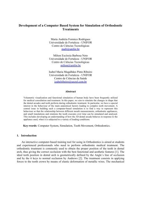
The proposed system consists <strong>of</strong> 3 basic modules: cephalometric analysis, volumetric<br />
mesh generator <strong>of</strong> dental arch and appliance models, and the orthodontic treatment simulator<br />
(as shown in Fig. 1). In particular, we propose an architecture model <strong>of</strong> the behaviour <strong>of</strong> the<br />
teeth as it is mechanically displaced by the appliances and loading conditions applied to them.<br />
A simple low resolution model <strong>of</strong> a face and skull was used to allow a better understanding <strong>of</strong><br />
the movements and provides more realism to the simulator and particularly do not influence<br />
the simulation results. The cephalometric analysis module is responsible <strong>for</strong> the X-Ray<br />
segmentations and the calculation <strong>of</strong> the ideal teeth position in dental arch based on the mouth<br />
measurements <strong>of</strong> a patient. Together with the actual dental cast, these measurements are used<br />
to build the 3D dental arch. The orthodontic treatment simulator provides an interface <strong>for</strong><br />
planning and simulation using different appliance models, as well as <strong>for</strong> helping to identify<br />
the best possible orthodontic techniques <strong>for</strong> the treatment <strong>of</strong> a patient based on its<br />
anatomy.The teeth movements are basically represented by translations and rotations.<br />
Dynamics are also used to give a more realistic behaviour based on the loading applied. We<br />
will investigate two subjects per<strong>for</strong>ming an analysis using our prototype. To represent the 3D<br />
dental model <strong>of</strong> an individual subject, the anatomy was generated by using the 3D dental<br />
cast, X-Ray images, and craneometric points (see Fig. 1). As part <strong>of</strong> the therapy planned, the
X-Ray Images<br />
Cephalometric<br />
Measurements<br />
and Image Analysis<br />
Cephalometric Analysis Module<br />
Morphing <strong>Tool</strong><br />
3D Mesh Generator Module<br />
Morphing <strong>Tool</strong><br />
3D Modelling<br />
(skull, jaw, teeth,<br />
and appliance)<br />
Orthodontic Treatment <strong>Simulation</strong> Module<br />
Dental Arch Features<br />
Extraction<br />
3D Dental Cast<br />
and Appliance<br />
Model<br />
Teeth <strong>Movement</strong> Simulator<br />
Simulated Graphical Results<br />
<strong>of</strong> Orthodontic Treatment<br />
Figure 1: The main architecture components <strong>of</strong> the orthodontic system, including the lateral<br />
radiography <strong>of</strong> the first subject investigated. A set <strong>of</strong> anatomical structures and points<br />
(craneometric points) are identified and used as a template <strong>for</strong> the application <strong>of</strong><br />
cephalometric analysis methods.
initial measurements <strong>of</strong> dental displacements will be realized during 12 clinical check-ups <strong>of</strong><br />
the patient over 1 year. The moved and the reference teeth on the cast model were marked <strong>for</strong><br />
identification, and particularly, we have used the molar and premolar teeth as reference teeth.<br />
The magnitude <strong>of</strong> translation and rotation will be calculated relative to the basis <strong>of</strong> these<br />
reference points be<strong>for</strong>e starting treatment. In particular, it was designed <strong>for</strong> this subject the<br />
fixed appliance model with brackets that already embeds established torsion values in its<br />
designed structure. The advantage is that the resultant torsion can be applied on an individual<br />
tooth or in a group <strong>of</strong> teeth at once. An extraction <strong>of</strong> the first premolars on both sides <strong>of</strong> the<br />
mandible and maxilla was planned <strong>for</strong> both subjects. A dental extraction results in bone<br />
remodelling, a process that takes time (21 days approximately). After the extraction process,<br />
the teeth will be aligned and levelled using rounded dental archs. The simulator module will<br />
per<strong>for</strong>m all the dynamics, including the distalization <strong>of</strong> the canines and incisors into the<br />
extraction space in direction <strong>of</strong> the molars, according to the model <strong>of</strong> dental arch appliance<br />
chosen (teeth displacement generally takes 8 months). As a fine stage, the teeth will be<br />
aligned again and a few <strong>of</strong> quite small torsions may be still applied to them. The morphing<br />
tool will illustrate the whole process.<br />
4. Discussion<br />
Our aim is to provide a computer model to help orthodontists to predict and deal with<br />
orthodontic problems, as well as suggesting possible treatments. We have approached this by<br />
defining a simple prototype. It is aimed to combine and improve the main ideas <strong>of</strong> some<br />
existing commercial models which are difficult to test and validate because they are not open<br />
source and free available tools. We believe that computer orthodontic models may be used as<br />
training tools <strong>for</strong> those seeking a better understanding <strong>of</strong> the geometric and dynamic factors<br />
involved in the control strategies <strong>of</strong> orthodontic treatments as well as to investigate the<br />
accuracy <strong>of</strong> the results and whether a specific planned treatment can be detrimental to the<br />
patient in any circumstance. Finally, as far as the s<strong>of</strong>tware components <strong>of</strong> the interactive<br />
computer model proposed are concerned, there is a great potential <strong>for</strong> modelling current<br />
treatment plans and possible new solutions to dental problems that can lead to expanded<br />
knowledge in orthodontics.<br />
References<br />
[1] Alcañiz, M., Monserrat, C., Grau, V., Chinesta, F., Ramón, A., Albalat, S., “An Advanced<br />
System <strong>for</strong> the <strong>Simulation</strong> and Planning <strong>of</strong> Orthodontic Treatment”, Medical Image Analysis,<br />
2(1), pp.61-79, 1998.<br />
[2] Marcotte, "Biomecânica em Ortodontia", Editora LS, 1993.<br />
[3] Foster, T.D., “Manual de Ortodontia”, Editora Santos, 3ª edição, 1993.<br />
[4] Enciso, R., Memon, A., Fidaleo, D.A., Neumann, U., Mah, J., “The Virtual Crani<strong>of</strong>acial<br />
Patient: 3D Jaw Modeling and Animation”, In Proc.<strong>of</strong> the 11th Annual Medicine Meets<br />
Virtual Reality Conference, pp.65-71, USA, 2003.<br />
[5] Bisler, A., Bockholt, U., Voss, G., “The Virtual Articulator - Applying VR Technologies<br />
to Dentistry”, In Proc.<strong>of</strong> the 6th IEEE International Conference on In<strong>for</strong>mation Visualisation,<br />
pp.600-602, USA, 2002.<br />
[6] Aoki, Y., “A 3D Head Model Construction <strong>of</strong> Individuals Utilizing Standard Models and<br />
Photogrammetry”, In Proc.<strong>of</strong> the 8th Korea-Japan Joint Workshop on Computer Vision,<br />
pp.98-104, 2002.<br />
[7] Fiorelli, G., "The 3-D Occlusogram S<strong>of</strong>tware", Am. Journal <strong>of</strong> Orthodontic and<br />
Dent<strong>of</strong>acial Orthopeditcs, pp.363-368,1999.
[8] Yamany, S.M., Farag, A.A., Mohamed, N.A., "Orthodontics Measurements Using<br />
Computer Vision", In Proc. <strong>of</strong> IEEE-EMBS, 20(2), pp.536-569, 1998.<br />
[9] Redmond, W.R., "Digital Models:a New Diagnostic <strong>Tool</strong>", Journal <strong>of</strong> Clinical<br />
Orthodontics, 35(6), 2001.<br />
[10] Garino, F., Garino, G.B., "Comparison <strong>of</strong> Dental Arch Measurements Between Stone and<br />
Digital Casts", World Journal <strong>of</strong> Orthodontics, 3(3), pp.250-254, 2002.<br />
[11] Ayoub, A.F., Wray, D., Moos, K.F., Jin, J., Niblett, T.B., Urquhart, C., Mow<strong>for</strong>th, P.,<br />
Siebert, P., "A Three Dimensional Imaging System <strong>for</strong> Archiving Dental Study Model: a<br />
Preliminary Report", International Journal <strong>of</strong> Adult Orthodontics Orthognathic Surgery,<br />
12(1), pp.79-84, 1997.<br />
[12] http://www.orthotrac.com/Ortho_pages/OPMSHome.asp, OPMS.<br />
[13] http://www.advanced-ortho.net/home.asp, Program Director.<br />
[14] http://www.acuscape.com/products/index.html, The Acuscape Product Family.<br />
[15] http://www.lssistemas.com/odontoway/odontoway.htm, Odontoway.<br />
[16] http://easys<strong>of</strong>t.com.br/, Easy (EasyDental 5.0, EasyPreview 1.7).<br />
[17] http://www.radiomemory.com.br/programas/radiocef/radiocef.html, Radiocef Studio.<br />
[18] http://www.orthotrac.com/Ortho_pages/ImagingHome.asp, Ortho Cephalometrics.<br />
[19] http://w4u.eexi.gr/~dhal/index.html, dHAL Orthodontic S<strong>of</strong>tware.<br />
[20] http://www.dolphinimaging.com, Dolphin Imaging, ImagingPlus.<br />
[21] http://www.quickceph.com/, Quick Ceph 2000.<br />
[22] http://www.yasunaga.co.jp/Eindex.html - Yasunaga computer system Co., Inc.<br />
[23] http://www.carriere.es/carriere/carreng.htm, S<strong>of</strong>tlander – Metrogamma S.A.<br />
[24] http://www.navimetric.com/marcos_dental.php?PHPSESSID=582d1607d8ddc6a1945<br />
a159372a0b33b, Orthometric.<br />
[25] http://www.dentalemodels.com/, GeoDigm Corporation (emodel).<br />
[26] http://www.orthocad.com/, Cadent - Computer-Aided Dentistry(OrthoCAD).<br />
[27] http://www.oc-j.com/june01/Invisalign.htm, Align Technology Inc., Invisalign.<br />
[28] Ferrario, V.F., S<strong>for</strong>za, C., Poggio, C., Serrao, G., “Facial Three-Dimensional<br />
Morphometry", Am. Journal <strong>of</strong> Orthodontic and Dent<strong>of</strong>acial Orthopeditcs, 109(1), pp.86-<br />
93,1996.<br />
[29] Ferrario, V.F., S<strong>for</strong>za, C., Puleo, A., Poggio, C., Schmitz, J. H., “Three-Dimensional<br />
Morphometry and Convencional Cephalometrics: a Correlation Study", International Journal<br />
<strong>of</strong> Adult Orthodontic & Orthognathic Surgery, 11,pp.329-338,1996.<br />
[30] Ferrario, V.F., S<strong>for</strong>za, C., Schmitz, J.H., Miani, A.Jr., Serrao, G., "A Three-Dimensional<br />
Computerized Mesh Diagram Analysis and its Application in S<strong>of</strong>t Tissue Facial<br />
Morphometry ", Am. Journal <strong>of</strong> Orthodontic and Dent<strong>of</strong>acial Orthopeditcs, 114, pp.404-<br />
413,1998.