Mutational analysis ofsimian virus 40 large T antigen DNA binding ...
Mutational analysis ofsimian virus 40 large T antigen DNA binding ...
Mutational analysis ofsimian virus 40 large T antigen DNA binding ...

Create successful ePaper yourself
Turn your PDF publications into a flip-book with our unique Google optimized e-Paper software.
K.AJones, R.M.Myers and R.Tjian<br />
-46 'O/52*X.<br />
late<br />
-<br />
. ~ ~<br />
a<br />
SoWw<br />
__<br />
- -~~ml -<br />
slo Wm<br />
_1--N.<br />
- -~~~~~~~<br />
;a 11 -ttM5<br />
f-i<br />
TATA<br />
III<br />
Ill<br />
11 _<br />
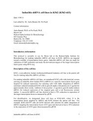
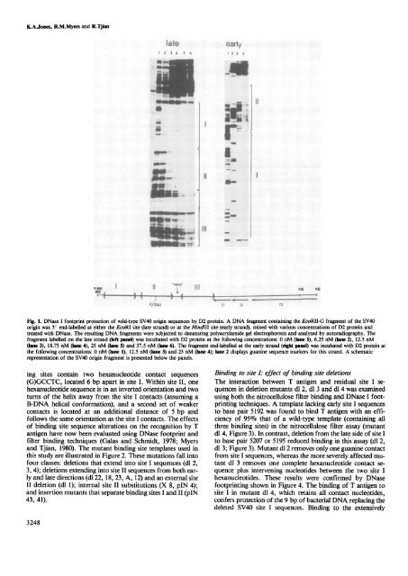
Fig. 1. DNase I footprint protection of wild-type SV<strong>40</strong> origin sequences by D2 protein. A <strong>DNA</strong> fragment containing the EcoRII-G fragment of the SV<strong>40</strong><br />
origin was 5' end-labelled at either the EcoRI site (late strand) or at the HindlII site (early strand), mixed with various concentrations of D2 protein and<br />
treated with DNase. The resulting <strong>DNA</strong> fragments were subjected to denaturing polyacrylamide gel electrophoresis and analyzed by autoradiography. The<br />
fragment labelled on the late strand (left panel) was incubated with D2 protein at the following concentrations: 0 nM (lane 1), 6.25 nM (lane 2), 12.5 nM<br />
(lane 3), 18.75 nM (lane 4), 25 nM (lane 5) and 37.5 nM (lane 6). The fragment end-labelled at the early strand (right panel) was incubated with D2 protein at<br />
the following concentrations: 0 nM (lane 1), 12.5 nM (lane 3) and 25 nM (lane 4); lane 2 displays guanine sequence markers for this strand. A schematic<br />
representation of the SV<strong>40</strong> origin fragment is presented below the panels.<br />
ing sites contain two hexanucleotide contact sequences<br />
(G)GCCTC, located 6 bp apart in site I. Within site II, one<br />
hexanucleotide sequence is in an inverted orientation and two<br />
turns of the helix away from the site I contacts (assuming a<br />
B-<strong>DNA</strong> helical conformation), and a second set of weaker<br />
contacts is located at an additional distance of 5 bp and<br />
follows the same orientation as the site I contacts. The effects<br />
of <strong>binding</strong> site sequence alterations on the recognition by T<br />
<strong>antigen</strong> have now been evaluated using DNase footprint and<br />
filter <strong>binding</strong> techniques (Galas and Schmidt, 1978; Myers<br />
and Tjian, 1980). The mutant <strong>binding</strong> site templates used in<br />
this study are illustrated in Figure 2. These mutations fall into<br />
four classes: deletions that extend into site I sequences (dl 2,<br />
3, 4); deletions extending into site II sequences from both early<br />
and late directions (dl 22, 18, 23, A, 12) and an external site<br />
II deletion (dl 1); internal site II substitutions (X 8, pIN 4);<br />
and insertion mutants that separate <strong>binding</strong> sites I and II (pIN<br />
43, 41).<br />
3248<br />
mEN<br />
.3Y<br />
-1<br />
early<br />
2 3 A<br />
2A<br />
I-<br />
-m-<br />
I<br />
II<br />
RIM RU<br />
Binding to site I: effect of <strong>binding</strong> site deletions<br />
The interaction between T <strong>antigen</strong> and residual site I sequences<br />
in deletion mutants dl 2, dl 3 and dl 4 was examined<br />
using both the nitrocellulose filter <strong>binding</strong> and DNase I footprinting<br />
techniques. A template lacking early site I sequences<br />
to base pair 5192 was found to bind T <strong>antigen</strong> with an efficiency<br />
of 95% that of a wild-type template (containing all<br />
three <strong>binding</strong> sites) in the nitrocellulose filter assay (mutant<br />
dl 4, Figure 3). In contrast, deletion from the late side of site I<br />
to base pair 5207 or 5195 reduced <strong>binding</strong> in this assay (dl 2,<br />
dl 3; Figure 3). Mutant dl 2 removes only one guanine contact<br />
from site I sequences, whereas the more severely affected mutant<br />
dl 3 removes one complete hexanucleotide contact sequence<br />
plus intervening nucleotides between the two site I<br />
hexanucleotides. These results were confirmed by DNase<br />
footprinting shown in Figure 4. The <strong>binding</strong> of T <strong>antigen</strong> to<br />
site I in mutant dl 4, which retains all contact nucleotides,<br />
confers protection of the 9 bp of bacterial <strong>DNA</strong> replacing the<br />
deleted SV<strong>40</strong> site I sequences. Binding to the extensively<br />
i