Mutational analysis ofsimian virus 40 large T antigen DNA binding ...
Mutational analysis ofsimian virus 40 large T antigen DNA binding ...
Mutational analysis ofsimian virus 40 large T antigen DNA binding ...

Create successful ePaper yourself
Turn your PDF publications into a flip-book with our unique Google optimized e-Paper software.
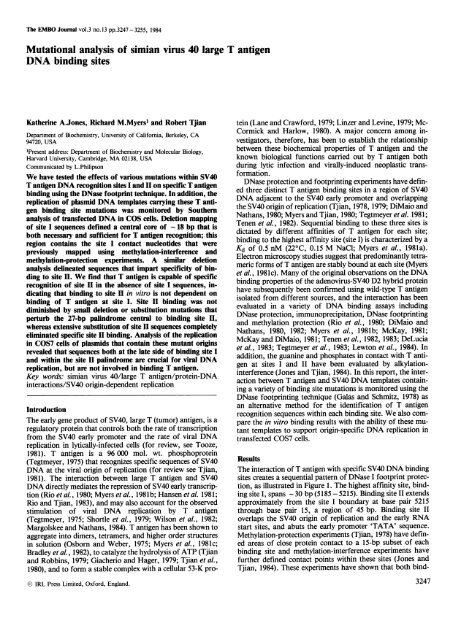
The EMBO Journal vol.3 no.13 pp.3247-3255, 1984<br />
<strong>Mutational</strong> <strong>analysis</strong> of simian <strong>virus</strong> <strong>40</strong> <strong>large</strong> T <strong>antigen</strong><br />
<strong>DNA</strong> <strong>binding</strong> sites<br />
Katherine A.Jones, Richard M.Myers1 and Robert Tjian<br />
Department of Biochemistry, University of California, Berkeley, CA<br />
94720, USA<br />
'Present address: Department of Biochemistry and Molecular Biology,<br />
Harvard University, Cambridge, MA 02138, USA<br />
Communicated by L.Philipson<br />
We have tested the effects of various mutations within SV<strong>40</strong><br />
T <strong>antigen</strong> <strong>DNA</strong> recognition sites I and II on specific T <strong>antigen</strong><br />
<strong>binding</strong> using the DNase footprint technique. In addition, the<br />
replication of plasmid <strong>DNA</strong> templates carrying these T <strong>antigen</strong><br />
<strong>binding</strong> site mutations was monitored by Southern<br />
<strong>analysis</strong> of transfected <strong>DNA</strong> in COS cells. Deletion mapping<br />
- of site I sequences defined a central core of 18 bp that is<br />
both necessary and sufficient for T <strong>antigen</strong> recognition; this<br />
region contains the site I contact nucleotides that were<br />
previously mapped using methylation-interference and<br />
methylation-protection experiments. A similar deletion<br />
<strong>analysis</strong> delineated sequences that impart specificity of <strong>binding</strong><br />
to site II. We find that T <strong>antigen</strong> is capable of specific<br />
recognition of site II in the absence of site I sequences, indicating<br />
that <strong>binding</strong> to site II in vitro is not dependent on<br />
<strong>binding</strong> of T <strong>antigen</strong> at site I. Site II <strong>binding</strong> was not<br />
diminished by small deletion or substitution mutations that<br />
perturb the 27-bp palindrome central to <strong>binding</strong> site II,<br />
whereas extensive substitution of site H sequences completely<br />
eliminated specific site II <strong>binding</strong>. Analysis of the replication<br />
in COS7 cells of plasmids that contain these mutant origins<br />
revealed that sequences both at the late side of <strong>binding</strong> site I<br />
and within the site II palindrome are crucial for viral <strong>DNA</strong><br />
replication, but are not involved in <strong>binding</strong> T <strong>antigen</strong>.<br />
Key words: simian <strong>virus</strong> <strong>40</strong>/<strong>large</strong> T <strong>antigen</strong>/protein-<strong>DNA</strong><br />
interactions/SV<strong>40</strong> origin-dependent replication<br />
Introduction<br />
The early gene product of SV<strong>40</strong>, <strong>large</strong> T (tumor) <strong>antigen</strong>, is a<br />
regulatory protein that controls both the rate of transcription<br />
from the SV<strong>40</strong> early promoter and the rate of viral <strong>DNA</strong><br />
replication in lytically-infected cells (for review, see Tooze,<br />
1981). T <strong>antigen</strong> is a 96 000 mol. wt. phosphoprotein<br />
(Tegtmeyer, 1975) that recognizes specific sequences of SV<strong>40</strong><br />
<strong>DNA</strong> at the viral origin of replication (for review see Tjian,<br />
1981). The interaction between <strong>large</strong> T <strong>antigen</strong> and SV<strong>40</strong><br />
<strong>DNA</strong> directly mediates the repression of SV<strong>40</strong> early transcription<br />
(Rio et al., 1980; Myers et al., 1981b; Hansen et al. 1981;<br />
Rio and Tjian, 1983), and may also account for the observed<br />
stimulation of viral <strong>DNA</strong> replication by T <strong>antigen</strong><br />
(Tegtmeyer, 1975; Shortle et al., 1979; Wilson et al., 1982;<br />
Margolskee and Nathans, 1984). T <strong>antigen</strong> has been shown to<br />
aggregate into dimers, tetramers, and higher order structures<br />
in solution (Osborn and Weber, 1975; Myers et al., 1981c;<br />
Bradley et al., 1982), to catalyze the hydrolysis of ATP (Tjian<br />
and Robbins, 1979; Giacherio and Hager, 1979; Tjian et al.,<br />
1980), and to form a stable complex with a cellular 53-K pro-<br />
IRL Press Limited, Oxford, England.<br />
tein (Lane and Crawford, 1979; Linzer and Levine, 1979; Mc-<br />
Cormick and Harlow, 1980). A major concern among investigators,<br />
therefore, has been to establish the relationship<br />
between these biochemical properties of T <strong>antigen</strong> and the<br />
known biological functions carried out by T <strong>antigen</strong> both<br />
during lytic infection and virally-induced neoplastic transformation.<br />
DNase protection and footprinting experiments have defined<br />
three distinct T <strong>antigen</strong> <strong>binding</strong> sites in a region of SV<strong>40</strong><br />
<strong>DNA</strong> adjacent to the SV<strong>40</strong> early promoter and overlapping<br />
the SV<strong>40</strong> origin of replication (Tjian, 1978, 1979; DiMaio and<br />
Nathans, 1980; Myers and Tjian, 1980; Tegtmeyer et al. 1981;<br />
Tenen et al., 1982). Sequential <strong>binding</strong> to these three sites is<br />
dictated by different affinities of T <strong>antigen</strong> for each site;<br />
<strong>binding</strong> to the highest affinity site (site I) is characterized by a<br />
Kd of 0.5 nM (22° C, 0.15 M NaCl; Myers et al., 1981a).<br />
Electron microscopy studies suggest that predominantly tetrameric<br />
forms of T <strong>antigen</strong> are stably bound at each site (Myers<br />
et al., 198 1c). Many of the original observations on the <strong>DNA</strong><br />
<strong>binding</strong> properties of the adeno<strong>virus</strong>-SV<strong>40</strong> D2 hybrid protein<br />
have subsequently been confirmed using wild-type T <strong>antigen</strong><br />
isolated from different sources, and the interaction has been<br />
evaluated in a variety of <strong>DNA</strong> <strong>binding</strong> assays including<br />
DNase protection, immunoprecipitation, DNase footprinting<br />
and methylation protection (Rio et al., 1980; DiMaio and<br />
Nathans, 1980, 1982; Myers et al., 1981b; McKay, 1981;<br />
McKay and DiMaio, 1981; Tenen et al., 1982, 1983; DeLucia<br />
et al., 1983; Tegtmeyer et al., 1983; Lewton et al., 1984). In<br />
addition, the guanine and phosphates in contact with T <strong>antigen</strong><br />
at sites I and II have been evaluated by alkylationinterference<br />
(Jones and Tjian, 1984). In this report, the interaction<br />
between T <strong>antigen</strong> and SV<strong>40</strong> <strong>DNA</strong> templates containing<br />
a variety of <strong>binding</strong> site mutations is monitored using the<br />
DNase footprinting technique (Galas and Schmitz, 1978) as<br />
an alternative method for the identification of T <strong>antigen</strong><br />
recognition sequences within each <strong>binding</strong> site. We also compare<br />
the in vitro <strong>binding</strong> results with the ability of these mutant<br />
templates to support origin-specific <strong>DNA</strong> replication in<br />
transfected COS7 cells.<br />
Results<br />
The interaction of T <strong>antigen</strong> with specific SV<strong>40</strong> <strong>DNA</strong> <strong>binding</strong><br />
sites creates a sequential pattern of DNase I footprint protection,<br />
as illustrated in Figure 1. The highest affinity site, <strong>binding</strong><br />
site I, spans -30 bp (5185-5215). Binding site II extends<br />
approximately from the site I boundary at base pair 5215<br />
through base pair 15, a region of 45 bp. Binding site II<br />
overlaps the SV<strong>40</strong> origin of replication and the early RNA<br />
start sites, and abuts the early promoter 'TATA' sequence.<br />
Methylation-protection experiments (Tjian, 1978) have defined<br />
areas of close protein contact to a 15-bp subset of each<br />
<strong>binding</strong> site and methylation-interference experiments have<br />
further defined contact points within these sites (Jones and<br />
Tjian, 1984). These experiments have shown that both bind-<br />
3247
K.AJones, R.M.Myers and R.Tjian<br />
-46 'O/52*X.<br />
late<br />
-<br />
. ~ ~<br />
a<br />
SoWw<br />
__<br />
- -~~ml -<br />
slo Wm<br />
_1--N.<br />
- -~~~~~~~<br />
;a 11 -ttM5<br />
f-i<br />
TATA<br />
III<br />
Ill<br />
11 _<br />
Fig. 1. DNase I footprint protection of wild-type SV<strong>40</strong> origin sequences by D2 protein. A <strong>DNA</strong> fragment containing the EcoRII-G fragment of the SV<strong>40</strong><br />
origin was 5' end-labelled at either the EcoRI site (late strand) or at the HindlII site (early strand), mixed with various concentrations of D2 protein and<br />
treated with DNase. The resulting <strong>DNA</strong> fragments were subjected to denaturing polyacrylamide gel electrophoresis and analyzed by autoradiography. The<br />
fragment labelled on the late strand (left panel) was incubated with D2 protein at the following concentrations: 0 nM (lane 1), 6.25 nM (lane 2), 12.5 nM<br />
(lane 3), 18.75 nM (lane 4), 25 nM (lane 5) and 37.5 nM (lane 6). The fragment end-labelled at the early strand (right panel) was incubated with D2 protein at<br />
the following concentrations: 0 nM (lane 1), 12.5 nM (lane 3) and 25 nM (lane 4); lane 2 displays guanine sequence markers for this strand. A schematic<br />
representation of the SV<strong>40</strong> origin fragment is presented below the panels.<br />
ing sites contain two hexanucleotide contact sequences<br />
(G)GCCTC, located 6 bp apart in site I. Within site II, one<br />
hexanucleotide sequence is in an inverted orientation and two<br />
turns of the helix away from the site I contacts (assuming a<br />
B-<strong>DNA</strong> helical conformation), and a second set of weaker<br />
contacts is located at an additional distance of 5 bp and<br />
follows the same orientation as the site I contacts. The effects<br />
of <strong>binding</strong> site sequence alterations on the recognition by T<br />
<strong>antigen</strong> have now been evaluated using DNase footprint and<br />
filter <strong>binding</strong> techniques (Galas and Schmidt, 1978; Myers<br />
and Tjian, 1980). The mutant <strong>binding</strong> site templates used in<br />
this study are illustrated in Figure 2. These mutations fall into<br />
four classes: deletions that extend into site I sequences (dl 2,<br />
3, 4); deletions extending into site II sequences from both early<br />
and late directions (dl 22, 18, 23, A, 12) and an external site<br />
II deletion (dl 1); internal site II substitutions (X 8, pIN 4);<br />
and insertion mutants that separate <strong>binding</strong> sites I and II (pIN<br />
43, 41).<br />
3248<br />
mEN<br />
.3Y<br />
-1<br />
early<br />
2 3 A<br />
2A<br />
I-<br />
-m-<br />
I<br />
II<br />
RIM RU<br />
Binding to site I: effect of <strong>binding</strong> site deletions<br />
The interaction between T <strong>antigen</strong> and residual site I sequences<br />
in deletion mutants dl 2, dl 3 and dl 4 was examined<br />
using both the nitrocellulose filter <strong>binding</strong> and DNase I footprinting<br />
techniques. A template lacking early site I sequences<br />
to base pair 5192 was found to bind T <strong>antigen</strong> with an efficiency<br />
of 95% that of a wild-type template (containing all<br />
three <strong>binding</strong> sites) in the nitrocellulose filter assay (mutant<br />
dl 4, Figure 3). In contrast, deletion from the late side of site I<br />
to base pair 5207 or 5195 reduced <strong>binding</strong> in this assay (dl 2,<br />
dl 3; Figure 3). Mutant dl 2 removes only one guanine contact<br />
from site I sequences, whereas the more severely affected mutant<br />
dl 3 removes one complete hexanucleotide contact sequence<br />
plus intervening nucleotides between the two site I<br />
hexanucleotides. These results were confirmed by DNase<br />
footprinting shown in Figure 4. The <strong>binding</strong> of T <strong>antigen</strong> to<br />
site I in mutant dl 4, which retains all contact nucleotides,<br />
confers protection of the 9 bp of bacterial <strong>DNA</strong> replacing the<br />
deleted SV<strong>40</strong> site I sequences. Binding to the extensively<br />
i
518.0 5190 5200 52105,0 52<strong>40</strong> 0 2<br />
AAGCTTTI T(;CAAAAGCCTACCCCTCCAAAAAA(;CCTCC-1 CACTACTTCTCGAAM1 ACC1 CA(;A(:(;CC(;A(;(:( (;( ( dac1(1(( Al AAA A<br />
0 a 0 0 0 0 0 0 0<br />
TTCGAAAAACGTTTTCGGATCCCGAGGTTTTTTCGGAGGAGTGATGAAGACCTTATCGAGTCTCCCGC1'CCGCC(;GAGGCUGAGACG'I'A'I 1 A<br />
A A<br />
Hi-d 2BgII<br />
dl 2 5207<br />
dl 3<br />
di14<br />
dl14<br />
s<br />
51951<br />
s5204<br />
dl22 5204.<br />
dl 18<br />
pIN43.<br />
pIN41-<br />
d123<br />
dli1<br />
piN 4A<br />
S -1I<br />
5215 -<br />
-~~0<br />
5238 l5243<br />
523%CCI'CGAC.,<br />
GCACCTCC<br />
'~kWWWAWVW'---<br />
u rn I 5241<br />
d 12 523:P<br />
T <strong>antigen</strong> <strong>binding</strong> and <strong>DNA</strong> replication mutants<br />
Fig. 2. Nucleotide sequences of the wild-type and mutant SV<strong>40</strong> origin templates used in these studies. The sequence of both strands of the wild-type origin is<br />
shown, with the HindIII (base pair 5182) and BgII (base pair 5243) restriction sites marked. The SV<strong>40</strong> BBB base pair numbering system is used (Buckman et<br />
al., 1980) and the approximate DNase footprint boundaries for each site are indicated by parentheses on the schematic template above the sequence. The solid<br />
bars within the parentheses represent the areas where guanine residues are protected from DMS methylation by the D2 protein. The number of the last SV<strong>40</strong><br />
base pair contained in each deletion mutant is given, and in all deletion mutants the SV<strong>40</strong> <strong>DNA</strong> sequences are abutted to bacterial pBR322 <strong>DNA</strong>. Mutants<br />
pIN 43 and pIN 41 contain insertions of 65 bp of bacterial polylinker <strong>DNA</strong> insertion between <strong>binding</strong> sites I and II. These two mutants differ only in the<br />
junction between bacterial and SV<strong>40</strong> site II sequences. Mutant pIN4 contains a substitution of sequences between base pairs 5220 and 9 (Rio and Tjian,<br />
1983).<br />
deleted mutant dl 3 is much less localized but appears to be<br />
centered about the single contact hexanucleotide sequence<br />
that remains in this mutant construction (Figure 4). Site I<br />
<strong>binding</strong> to mutant dl 2, which contains all contact nucleotides<br />
except one, appears normal at the T <strong>antigen</strong> concentrations<br />
used in this experiment. These experiments establish that sequences<br />
between base pairs 5192 and 5220 are required for the<br />
recognition of site I by T <strong>antigen</strong>; this region includes the sequences<br />
(5191 -5208) of close protein contact determined by<br />
methylation-protection and alkylation-interference experiments.<br />
Clearly the filter <strong>binding</strong> results could be misleading if the<br />
interaction of T <strong>antigen</strong> with additional <strong>binding</strong> sites on the<br />
template contributes to the retention of the fragment on<br />
nitrocellulose filters, since site II sequences are deleted in<br />
mutants dl 2 and dl 3 (not bound to the filter) and retained in<br />
mutant dl 4 (retained on the filter, Figure 3). In order to<br />
assess the contribution of multiple <strong>binding</strong> interactions to the<br />
filter assay, the interaction of T <strong>antigen</strong> with an isolated site I<br />
fragment derived from mutant pIN 41 was evaluated using<br />
the filter-<strong>binding</strong> technique (site II sequences were removed<br />
by nuclease digestion within the linker region between sites I<br />
and II and the site I fragment was purified by gel electrophoresis).<br />
T <strong>antigen</strong> complexes with the isolated site I<br />
_<br />
i<br />
sn221- @<br />
[ 1AI_ (mgnl )<br />
Fig. 3. Filter <strong>binding</strong> assay of site I mutant origin templates. Various concentrations<br />
of R284 T <strong>antigen</strong> were incubated with 5' end-labelled (Hinfdigested)<br />
mutant or wild-type <strong>DNA</strong> fragments for 10 min at 22° C and<br />
passed through nitrocellulose filters. <strong>DNA</strong> fragments tested were derived<br />
from wild-type (wt), dl 4, dl 2, dl 3, and control pBR plasmid <strong>DNA</strong> (c).<br />
3249
K.A.Jones, R.M.Myers and R.Tjian<br />
dl 2 d13<br />
2 3 A 5 6 11 3 4 5 t<br />
dl 3 &<br />
dl 4_ _.<br />
dl4 wt<br />
IL U<br />
B1%<br />
Fig. 4. DNase footprint protection of site I in wild-type and mutant templates by D2 protein. <strong>DNA</strong> fragments were 5' end-labeled at the Hinf site (base pair<br />
5135). The left panel shows the protection patterns for templates dl 2 and dl 3 in each case lane 1 contains guanine sequence markers and lanes 2-6 contain<br />
the DNase pattern in the presence of 0 nM, 12 nM, 15 nM, 20 nM and 30 nM of R284 T <strong>antigen</strong>, respectively. A schematic representation of each template<br />
is presented below the autoradiograms.<br />
fragments were retained by nitrocellulose filters with a 900/o<br />
efficiency relative to wild-type fragments containing all three<br />
<strong>binding</strong> sites (data not shown). These results indicate that<br />
filter <strong>binding</strong> is an accurate measure at least of site I <strong>binding</strong>,<br />
and that mutants dl 2 and dl 3 are defective for site I <strong>binding</strong><br />
in this assay whereas mutant dl 4 is not.<br />
Binding to site II in the absence of site I<br />
We previously reported that isolated site II sequences are not<br />
effectively bound to nitrocellulose in the presence of T <strong>antigen</strong><br />
even at concentrations exceeding that required for DNase<br />
footprint experiments (Myers et al., 1981b). With this assay<br />
we detected only low levels of specific <strong>binding</strong> of T <strong>antigen</strong> or<br />
Ad2D2 protein to templates lacking site I sequences<br />
(including mutants dl 22, dl 18, and dl 23; Figure 2), indicating<br />
that site II recognition is altered in the absence of site<br />
I. These mutant <strong>DNA</strong>s were re-examined using the DNase<br />
footprinting technique. Surprisingly, and in contrast with our<br />
results obtained using the filter-<strong>binding</strong> assay, specific <strong>binding</strong><br />
to site II sequences was observed using mutants lacking<br />
site I sequences (Figure 5, dl 22 and dl 18). Wild-type levels of<br />
protection from DNase digestion were also observed in<br />
mutants that contain polylinker <strong>DNA</strong> insertions that separate<br />
<strong>binding</strong> sites I and II (Figure 5, pIN 41 and pIN 43), and independent<br />
interactions were observed at both sites I and II.<br />
To eliminate the remote possibility that T <strong>antigen</strong> <strong>binding</strong> to<br />
site I was able to facilitate <strong>binding</strong> to site II even across a<br />
distance of 65 bp, the <strong>binding</strong> capacities of these mutants<br />
were re-evaluated after the removal of site I sequences.<br />
Plasmid pIN 41 or pIN 43 was cut at restriction sites in the<br />
polylinker sequence between sites I and II and in the vector<br />
3250<br />
wt<br />
dl 2<br />
is<br />
-1114D-<br />
AM.<br />
am<br />
::.: do<br />
.": tw ..<br />
<strong>40</strong> do-<br />
<strong>40</strong>.....<br />
1;.:- MD<br />
A 7<br />
I<br />
I<br />
-..- 4m<br />
we<br />
Im4m. --<br />
Q-41M.-<br />
qm 4m <strong>40</strong>0<br />
4m Ift. -..<br />
*a -- ,,<br />
""Mo-<br />
WAM<br />
"W ". %% 4ft ,,<br />
qft *ft ftb " ,a .,<br />
NW ". -..-<br />
l<br />
.p<br />
00<br />
<strong>DNA</strong>, and the site II fragment was isolated by gel electrophoresis.<br />
The isolated site II fragment was found to bind T<br />
<strong>antigen</strong> as effectively as a wild-type <strong>DNA</strong> fragment (Figure<br />
6A); an extensive titration of site II <strong>binding</strong> at lower T <strong>antigen</strong><br />
concentrations failed to reveal any significant difference in<br />
the <strong>binding</strong> affinities of these templates (data not shown). We<br />
also observed specific site II <strong>binding</strong> to a fragment containing<br />
a <strong>large</strong>r deletion of sequences on the early side of site II (deletion<br />
mutant dl 12; Figure 6B), although in this case we cannot<br />
rule out the possibility of a relatively small decrease (2- to<br />
4-fold) in affinity for site II in the mutant relative to the wildtype<br />
template. Therefore, we conclude that the filter-<strong>binding</strong><br />
assay is not accurate in the detection of specific site II protein-<br />
<strong>DNA</strong> complexes, and that the DNase protection assay widely<br />
used to monitor <strong>binding</strong> (Myers et al., 1981b; Tegtmeyer et<br />
al. 1983) appears to be unreliable when used to monitor site II<br />
<strong>binding</strong> because this procedure relies on nitrocellulose filter<br />
retention of protein-site II complexes.<br />
Binding to site II: effect of <strong>binding</strong> site mutations<br />
Because the affinity of T <strong>antigen</strong> for site II appears to be<br />
<strong>large</strong>ly independent of adjacent site I <strong>binding</strong>, we decided to<br />
map the nucleotides responsible for site II recognition directly,<br />
using DNase footprint <strong>analysis</strong> of <strong>DNA</strong> fragments containing<br />
site II sequence alterations. No specific site II protection<br />
was observed using a mutant which contains site I but in<br />
which 33 bp of site II are substituted by bacterial <strong>DNA</strong> (mutant<br />
pIN 4; Figure 7), indicating that nucleotides within the<br />
substituted region of base pair 5220 to 9 are specifically required<br />
for site II recognition. However, <strong>DNA</strong> fragments that<br />
contain either a 4-bp deletion (5239- 5242) or an 8-bp<br />
-J
dl 22<br />
goose<br />
2 3 4<br />
........I~<br />
_<br />
I_ 4<br />
a<br />
_<br />
'a 3..<br />
dl18<br />
* 3 4<br />
FE<br />
to.<br />
m .I<br />
_+r I<br />
Ii<br />
II<br />
11 _<br />
_<br />
plN43 plN41<br />
1 2 3 4 2 3 a<br />
I-i<br />
13<br />
ii<br />
sow_<br />
I<br />
I UI.I -.e .<br />
Al-,.<br />
NO<br />
Ls I-.:,<br />
WA.<br />
I* .-<br />
f o<br />
Ws m -w<br />
l wt<br />
dl22<br />
I ~~~~~~~~~~~~~~~~If<br />
ll~~~~~~~~~~~~<br />
d118<br />
i<br />
pIN43 i - -_ _ __ _<br />
pIN41<br />
I<br />
T <strong>antigen</strong> <strong>binding</strong> and <strong>DNA</strong> replication mutants<br />
Fig. 5. DNase I footprint <strong>analysis</strong> of mutant site II templates deleted of site I sequences. <strong>DNA</strong> fragments were 5' end-labeled on the late strand at the EcoRI<br />
site. Lanes 1-4 show the DNase pattern obtained in the presence of 0 nM, 12.5 nM, 25 nM and 37.5 nM of D2 protein, respectively. A schematic representation<br />
of the mutants is presented below the autoradiograms.<br />
substitution (5239- 5246) within site II were found to be<br />
unaffected for site II <strong>binding</strong> (dl 1 and pX 8; Figure 7). This<br />
result was surprising because these two mutants disrupt the<br />
symmetry of the 27-bp palindrome central to <strong>binding</strong> site II.<br />
Nevertheless, both of these mutants contain the 'major' hexanucleotide<br />
contact sequence identified in interference experiments<br />
(Jones and Tjian, 1984). Although the spacing or<br />
sequence of the 'weaker' hexanucleotide contacts is altered in<br />
the mutants, we note that this sequence is directly repeated<br />
and may provide an altemate set of contact nucleotides for T<br />
<strong>antigen</strong>. The site II <strong>binding</strong> affinities for these mutants also<br />
appear to be equal in DNase footprint titrations at lower T<br />
<strong>antigen</strong> concentrations (data not shown). Finally, a fragment<br />
that contains only the early half of site II was found to bind T<br />
<strong>antigen</strong> but confer DNase protection only to the residual site<br />
II sequences (mutant dl A, data not shown), indicating that T<br />
<strong>antigen</strong> requires sequences within both halves of <strong>binding</strong> site<br />
II in order to provide the complete footprint protection<br />
observed for site II in the wild-type template.<br />
Replication of mutant plasmids in COS7 cells<br />
The plasmids used in the <strong>binding</strong> studies above were tested<br />
for their ability to replicate in COS7 cells, a monkey cell line<br />
transformed with an origin-defective SV<strong>40</strong> <strong>virus</strong> (Gluzman et<br />
al., 1980; Gluzman, 1981). COS7 cells produce constitutive<br />
__-_ '<br />
II<br />
levels of T <strong>antigen</strong>s (Gluzman, 1981) sufficient to allow<br />
replication of plasmids containing the SV<strong>40</strong> origin of replication<br />
(Myers and Tjian, 1980). Wild-type and mutant origin<br />
plasmid <strong>DNA</strong> was introduced into COS7 cells by the DEAEdextran<br />
transfection procedure (McCutchan and Pagano,<br />
1968) and small mol. wt. <strong>DNA</strong> was isolated by the Hirt lysate<br />
procedure (Hirt, 1967) from the cells at 0, 12 and 48 h. Supercoiled<br />
and relaxed circular molecules were separated by<br />
agarose gel electrophoresis, transferred to nitrocellulose<br />
(Southern, 1975), and hybridized to radioactivity-labelled<br />
pBR322 <strong>DNA</strong>. In all cases, trace amounts of input supercoiled<br />
and relaxed circular <strong>DNA</strong> could be seen at 0 h. At 12 h<br />
after transfection, no supercoiled molecules were observed<br />
and trace amounts of relaxed circular <strong>DNA</strong> were present. In<br />
the case of plasmids containing the wild-type origin (pSV01),<br />
a <strong>large</strong> increase in the amount of supercoiled molecules was<br />
observed at 48 h, indicating that the plasmid replicated. Densitometry<br />
of autoradiograms was carried out comparing the<br />
levels of pSV01 and mutant supercoiled <strong>DNA</strong> accumulating<br />
at 48 h in transfected COS7 cells. The results are summarized<br />
in Table I. Mutants dl 4 and dl 22, which lack portions of T<br />
<strong>antigen</strong> <strong>binding</strong> site I, replicate at wild-type levels in COS7<br />
cells. Mutant dl 18, which lacks <strong>binding</strong> site I but contains the<br />
region between sites I and II, replicates 4- to 5-fold less effi-<br />
3251
K.AJones, R.M.Myers and R.Tjian<br />
ll<br />
A.<br />
pIN pIN<br />
d2183 43 41 rwt.<br />
l 2 3 1 2 3 1 2 3 1 2<br />
*? 's<br />
w<br />
l_-ai~<br />
_ _ a<br />
~ ~ ~ ~ a<br />
...^. am m*<br />
.<br />
o0ama<br />
"<br />
11 11<br />
B. w.t.<br />
G 1 2 34<br />
Ukm-.<br />
a d-,_g<br />
abg_i<br />
as<br />
so-<br />
_ r.<br />
aiam.0<br />
_ pk<br />
_j-_m_0 u<br />
Hg. 6. DNase footprint <strong>analysis</strong> of mutant site II templates. (A) The DNase pattern for mutant and wild-type SV<strong>40</strong> <strong>DNA</strong>, 5' end-labeled on the early strand.<br />
The wild-type <strong>DNA</strong> fragment was incubated in the absence of D2 protein (wt, lane 1), or with 25 nM D2 protein (lane 2). The site II fragments for mutants<br />
pIN 41, pIN 43, and mutant dl 18 were incubated in the absence of D2 protein (lane I in each panel) or at D2 concentrations of 25 nM (lane 2) and 50 nM<br />
(lane 3). (B) The DNase I footprint <strong>analysis</strong> of wild-type and mutant dl 12 <strong>DNA</strong> fragments in the presence of R284 T <strong>antigen</strong> at the following concentrations:<br />
0 nM (lane 1), 19 nM (lane 2), 28 nM (lane 3) and 57 nM (lane 4).<br />
ciently than wild-type <strong>DNA</strong>. Mutants pIN 43 and pIN 41,<br />
which contain a 65-bp insertion between sites I and II, do not<br />
replicate at all in COS7 cells and no replication of mutants dl<br />
23, dl 2, dl 1, X8, pIN 4, or dl A was observed. Thus, most of<br />
<strong>binding</strong> site I can be removed without affecting origin function.<br />
However, when site I is completely removed, the origin<br />
is moderately defective. Mutations that remove both site I<br />
and the sequences between sites I and II or that separate sites<br />
I and II by inserted bacterial sequences completely destroy the<br />
ability of the origin to replicate. Likewise, mutations affecting<br />
only <strong>binding</strong> site II destroy origin function.<br />
Discussion<br />
Here we describe the effects of various <strong>DNA</strong> template<br />
modifications on the interaction of SV<strong>40</strong> T <strong>antigen</strong> with <strong>binding</strong><br />
sites I and II at the SV<strong>40</strong> origin of replication. While we<br />
find that the nitrocellulose filter-<strong>binding</strong> and DNase footprint<br />
protection assays are equally suited for the <strong>analysis</strong> of site I<br />
<strong>binding</strong>, we observe no specific nitrocellulose filter retention<br />
of T <strong>antigen</strong>-site II complexes in the absence of <strong>binding</strong> site I.<br />
The observation that deletion mutant dl 23 (Figure 2) is not<br />
retained on nitrocellulose filters in the presence of T <strong>antigen</strong><br />
(Myers and Tjian, 1980) originally led to the conclusion that<br />
either sequences within site I or the <strong>binding</strong> of T <strong>antigen</strong> to<br />
site I promotes <strong>binding</strong> to site II, possibly in a manner consistent<br />
with positive cooperativity as observed in the <strong>binding</strong> of X<br />
repressor to adjacent <strong>binding</strong> sites at the ORM operator. Here<br />
we report that specific site I <strong>binding</strong>, apparently identical to<br />
3252<br />
JW;!- ....<br />
Q<br />
-b<br />
d112<br />
/ 1 2 3 4 G<br />
_4 'an<br />
4 _a<br />
the site II <strong>binding</strong> interactions observed with wild-type<br />
templates can be detected in the absence of site I sequences<br />
when the <strong>DNA</strong>se footprint assay is used (mutants dl 18, 41,<br />
43, dl 22; Figures 5, 6). We also observe the formation of T<br />
<strong>antigen</strong>-site II complexes with deletion mutant dl 22 using an<br />
immunoaffinity assay (Jones and Tjian, 1984) and the site II-<br />
T <strong>antigen</strong> complex can also be detected using the filter <strong>binding</strong><br />
assay if antibody directed against T <strong>antigen</strong> is present in<br />
the <strong>binding</strong> reaction (data not shown). These findings suggest<br />
that <strong>binding</strong> to sites I and II are independent events in vitro,<br />
and that T <strong>antigen</strong> contacts specific sequences within each<br />
<strong>binding</strong> site. We note that DNase footprint <strong>analysis</strong> of the<br />
more extensively deleted mutants dl 23 and dl 12 does not rule<br />
out the possibility of a relatively small effect on <strong>binding</strong> due<br />
to deletion of sequences between the two <strong>binding</strong> sites and,<br />
moreover, we have not eliminated the possibility of interactions<br />
between T <strong>antigen</strong> multimers bound at sites I and II that<br />
do not contribute to the <strong>binding</strong> affinity. Similar observations<br />
concerning the <strong>binding</strong> to site II in the absence of site I sequences<br />
have been reported by others (DiMaio and Nathans,<br />
1982; Tenen et al., 1983).<br />
The deletion <strong>analysis</strong> presented here indicates that site I<br />
<strong>binding</strong> is diminished or less specific in the absence of sequences<br />
between base pairs 5192 and 5220. The late boundary<br />
is established from the isolated site I fragment of mutant pIN<br />
41, which appears to have an unaltered affinity for site I when<br />
assayed by filter-<strong>binding</strong>. These sequences contain the close T<br />
<strong>antigen</strong> contacts as defined by methylation-protection (Tjian,<br />
_m<br />
_<br />
lof<br />
- -<br />
_g_b_WOqf<br />
--<br />
11
11<br />
wt<br />
dl 1<br />
X8<br />
piN 4<br />
plN4 X8 wt<br />
T <strong>antigen</strong> <strong>binding</strong> and <strong>DNA</strong> replication mutants<br />
Fig. 7. DNase footprint <strong>analysis</strong> of mutant site II templates. Wild-type and mutant dl-l <strong>DNA</strong> templates, 5' end-labeled at the early strand, were incubated<br />
with D2 protein at concentrations of 0 nM, 15 nM, 3 nM, 45 nM and 60 nM (left panel, lanes 1-5, respectively). The right panel shows the DNase footprint<br />
pattern of mutants pX8, pIN4, and the wild-type template in the presence of 0 nM, 20 nM, <strong>40</strong> nM and 60 nM of D2 protein (lanes 1-4, respectively).<br />
Table I. Mutant plasmid replication in COS7 cells<br />
wt dl 1<br />
1 2 3 4511 2 34 5<br />
I_ _ - ,__om--<br />
<strong>DNA</strong> % wild-type replication<br />
pSVo1 100<br />
dl2 n.d.<br />
dl 3 n.d.<br />
dl 4 100<br />
dl 22 100<br />
dl 18 20-25<br />
pIN 43
K.A.Jones, R.M.Myers and R.Tjian<br />
ped to the SV<strong>40</strong> minimal origin (base pairs 5208-30; Learned<br />
et al., 1981; Myers and Tjian, 1980; DiMaio and Nathans,<br />
1980). The <strong>binding</strong> of T <strong>antigen</strong> to site I in mutant dl 22 is<br />
characterized by a weak affinity and produces a severely truncated<br />
reigon of DNase protection (determined both by DNase<br />
footprinting and alkylation-interference techniques) indicating<br />
that complete site I <strong>binding</strong> is not required for<br />
replication but, rather, that sequences in the region of base<br />
pairs 5205 -5221 are critical for viral <strong>DNA</strong> replication. It is<br />
interesting in this regard that the origin of bidirectional<br />
replication in vivo has also been mapped to this region (base<br />
pairs 5210-5211; Hay and DePamphilis, 1982). In addition,<br />
the major initiation sites for Okazaki fragment synthesis map<br />
to nucleotides 5239 and 12 on the early mRNA strand within<br />
T <strong>antigen</strong> <strong>binding</strong> site II (Hay and DePamphilis, 1982). It is<br />
possible that the replicative failure of site II mutants dl 1 and<br />
pX 8 is due to the absence of appropriate primer initiation sequences<br />
in both mutants.<br />
Finally, we note that mutants in which <strong>binding</strong> sites I and<br />
II are separated by 65 bp are replication-deficient in COS7<br />
cells (pIN 41, pIN 43; Table I). Because these mutants bind T<br />
<strong>antigen</strong> at both sites with wild-type affinities, we conclude<br />
that the altered spacing between base pairs 5205 and 5212 in<br />
site I and essential sequences in site II somehow disrupts the<br />
normal replication process. Recently, these insertion mutants<br />
were also found to be defective in vitro in the repression of<br />
transcription by T <strong>antigen</strong> (Rio and Tjian, unpublished<br />
results). Therefore, interactions between T <strong>antigen</strong> multimers<br />
bound at each site may contribute to the mechanism of autoregulation,<br />
although the data presented here indicate that<br />
direct protein-protein interactions do not contribute significantly<br />
to the affinity of T <strong>antigen</strong> for each <strong>binding</strong> site.<br />
Materials and methods<br />
Restriction enzymes and nuclease Bal31 were purchased from Bethesda<br />
Research Laboratories. T4 polynucleotide kinase was from P-L Biochemicals<br />
and DNase I was from Worthington.<br />
Construction of plasmids containing wild-type and mtuant SV<strong>40</strong> origin sequences<br />
The 31 l-bp EcoRII G fragment of SV<strong>40</strong> was inserted at the EcoRI site of<br />
pBR322 (Myers and Tjian, 1980), creating plasmid pSVOI, the wild-type<br />
origin plasmid used in the replication studies. A plasmid containing these<br />
tandem copies of the EcoRII G fragment, pSVO7, was used in the <strong>binding</strong><br />
studies. Construction of deletion mutants dl 1, dl 4, dl 22, dl 23, and dl 12 has<br />
been previously described (Myers and Tjian, 1980), and the creation of<br />
substitution mutant pIN 4 was detailed by Rio and Tjian (1983). Mutant dl A<br />
was created by Bal31 digestion of plasmid pSV1096 cut at a HinclII site within<br />
site III; this mutant is lacking sequences from base pair 5241 to 52. Mutant<br />
pX 8 was a gift from Michael Fromm and Paul Berg (Fromm and Berg,<br />
1982). Insertion mutants pIN 41 and pIN 43 were constructed as follows: a<br />
fragment containing all of site I and 65 bp of bacterial <strong>DNA</strong> at the site II proximal<br />
side of site I was isolated from insertion mutant pIN 1 (Myers et al.,<br />
1981b) by cutting the polylinker sequence with BamHI, trimming the ends<br />
with nuclease Bal3J, followed by complete digestion with EcoRI. A vector for<br />
this insert was generated by cutting plasmid pSV01097 (a plasmid containing<br />
the origin fragment from CS1097; Shortle and Nathans, 1979) with HindIII<br />
and treating with nuclease Bal31 to remove site I sequences, followed by<br />
EcoRI digestion. The ligated plasmid forms the sequence site I -bacterial<br />
<strong>DNA</strong> -site II-site III, as diagrammed in Figure 2. Viral and plasmid <strong>DNA</strong>s<br />
were purified by a modified Hirt extraction (Hirt, 1967), followed by CsCl<br />
ethidium bromide gradient centrifugation. Ethidium was removed with<br />
Dowex AG 50W-X8 resin in 10 mM Tris, pH 7.9, 1 mM EDTA and 1 M<br />
NaCl and the samples were dialyzed extensively against 10 mM Tris 7.9, and<br />
I mM EDTA at 4° C.<br />
Purification of T <strong>antigen</strong><br />
We have used two sources of T <strong>antigen</strong> in the <strong>binding</strong> studies reported here,<br />
both derived from HeLa cells infected with defective adeno<strong>virus</strong>-SV<strong>40</strong> hybrid<br />
<strong>virus</strong>es. The Ad2+D2 <strong>virus</strong> encodes a 107-K protein that is the functional<br />
equivalent of SV<strong>40</strong><strong>large</strong> T <strong>antigen</strong> (DeLucia et al., 1983) consisting of - 10 K<br />
3254<br />
of an adeno<strong>virus</strong> polypeptide at the amino terminus (Hassell et al., 1978). The<br />
Ad-SVR284 hybrid <strong>virus</strong> produces full-length T <strong>antigen</strong> (Thummel et al.,<br />
1983). Both proteins were purified to homogeneity by the method of Tjian<br />
(1978), and were stored at 4-6 mg/ml concentrations in 10 mM Tris, pH 8,<br />
0.5 M, NaCl, 0.2 mM dithiothreitol, 10% glycerol at - 80° C. The source of<br />
T <strong>antigen</strong> used for each experiment is indicated in the individual figure<br />
legends.<br />
<strong>DNA</strong>-<strong>binding</strong> protocols<br />
The nitrocellulose filter-<strong>binding</strong> assay has been described (Myers and Tjian,<br />
1980). Origin-containing fragments were 5' end-labeled with [7y-32P]ATP and<br />
polynucleotide kinase and purified by agarose or polyacrylamide gel electrophoresis<br />
for use in footprinting experiments. The restriction site in the procedure<br />
is indicated in the individual figure legends. Approximately 10- 13 mol<br />
(150 ng) of each fragment was incubated with various concentrations of D2<br />
protein in 100 i1 25 mM 1,4-piperazinediethanesulfonic acid (Pipes), pH 6.8,<br />
0.1 M NaCl, 10% glycerol, 10 jug/ml BSA (5 min, 22° C) and cut 60 s with<br />
3 ng DNase I in 15 mM MgCl2, 100mM CaCI. The digestion was terminated by<br />
the addition of 200 /l 2 M ammonium acetate, 100 mM EDTA and<br />
150 jzg/ml sonicated calf thymus <strong>DNA</strong>, and the <strong>DNA</strong> was precipitated with<br />
2.5 volumes of ethanol. <strong>DNA</strong> pellets were resuspended in 200 Al 2.5 M ammonium<br />
acetate and reprecipitated from ethanol. After drying, the pellets<br />
were resuspended in 25 mM NaOH and 75% formamide, heated for 2 min at<br />
100° C and subjected to polyacrylamide gel electrophoresis (8-16%<br />
acrylamide, 19:1 acrylamide:bisacrylamide). The gels were analyzed by<br />
autoradiography for 8-24 h at -80° C using an X-ray intensifer screen.<br />
<strong>DNA</strong> replication assay<br />
The protocol followed in the <strong>analysis</strong> of plasmid replication in COS cells is<br />
outlined by Myers and Tjian (1980). COS7 cells, a monkey cell line constitutively<br />
producing SV<strong>40</strong> T <strong>antigen</strong>, were kindly provided by Yakov Gluzman.<br />
Acknowledgements<br />
We thank Dan DiMaio and Dan Nathans for providing cs1097 viral <strong>DNA</strong> and<br />
freely discussing unpublished data, Michael Fromm and Paul Berg for<br />
plasmid pX 8, and Don Rio for mutant pIN 4 <strong>DNA</strong>. We also thank Carl<br />
Thummel and Terri Grodzicker for Ad-SVR284 viral stocks, and Robin Clark<br />
for assistance in the purification of R284 T <strong>antigen</strong>. We are grateful to Karen<br />
Erdley for typing this manuscript. This work was funded by a National Institute<br />
of Environmental Health Sciences Center grant. K.A.J. was the recipient<br />
of a postdoctoral fellowship from the National Institutes of Health.<br />
References<br />
Bradley,M.K., Griffin,J.D. and Livingston,D.M. (1982) Cell, 28, 125-134.<br />
Buckman,A.R., Burnett,L. and Berg,P. (1980) in Tooze,J. (ed.), <strong>DNA</strong><br />
Tumor Viruses, Part II, Cold Spring Harbor Laboratory Press, NY, pp.<br />
799-829.<br />
DeLucia,A.L., Lewton,B.A., Tjian,R. and Tegtmeyer,P. (1983) J. Virol.,<br />
46, 143-150.<br />
DiMaio,D. and Nathans,D. (1980) J. Mol. Biol., 1<strong>40</strong>, 129-142.<br />
DiMaio,D. and Nathans,D. (1982) J. Mol. Biol., 156, 531-548.<br />
Fromm,M. and Berg,P. (1982) J. Mol. Appl. Genet., 1., 457-481.<br />
Galas,D.J. and Schmitz,A. (1978) Nucleic Acids Res., 5, 3157-3170.<br />
Giacherio,D. and Hager,L.P. (1979) J. Biol. Chem., 254, 8113-8116.<br />
Gluzman,Y. (1981) Cell, 23, 175-182.<br />
Gluzman,Y., Sambrook,J.F. and Frisque,R.J. (1980) Proc. Natl. Acad. Sci.<br />
USA, 77, 3898-3902.<br />
Hansen,U., Tenen,D.G., Livingston,D.M. and Sharp,P.A. (1981) Cell, 27,<br />
603-612.<br />
Hassell,J.A., Lukanidin,E., Fey,G. and Sambrook,J. (1978) J. Mol. Biol.,<br />
120, 209-247.<br />
Hay,R.T. and DePamphilis,M.L. (1982) Cell, 28, 767-779.<br />
Hirt,B. (1967) J. Mol. Biol., 26, 365-369.<br />
Jones,K.A. and Tjian,R. (1984) Cell, 36, 155-162.<br />
Lane,D.P. and Crawford,L.V. (1979) Nature, 278, 261-263.<br />
Learned,R.M., Myers,R.M. and Tjian,R. (1981) in Ray,D.S. and Fox,C.F.<br />
(eds.), ICN-UCLA Symposium on Molecular and Cellular Biology, Vol.<br />
21, Academic Press, NY, pp. 555-566.<br />
Lewton,B.A., DeLucia,A.L. and Tegtmeyer,P. (1984) J. Virol., 49, 9-13.<br />
Linzer,D.I.H. and Levine,A.J. (1979) Cell, 17, 43-52.<br />
Margolskee,R.F. and Nathans,D. (1984) J. Virol., 49, 386-393.<br />
McCormick,F. and Harlow,E. (1980) J. Virol., 34, 213-224.<br />
McCutchan,J.H. and Pagano,J.S. (1968) J. NatI. Cancer Inst., 41, 351-356.<br />
McKay,R.D.G. (1981) J. Mol. Biol., 145, 471-488.<br />
McKay,R. and DiMaio,D. (1981) Nature, 289, 810-813.<br />
Myers,R.M. and Tjian,R. (1980) Proc. Natl. Acad. Sci. USA, 77, 6491-<br />
6495.
Myers,R.M., Kligman,M. and Tjian,R. (1981a) J. Biol. Chem., 256, 10156-<br />
10160.<br />
Myers,R.M., Rio,D.C., Robbins,A.K. and Tjian,R. (1981b) Cell, 25, 373-<br />
384.<br />
Myers,R.M., Williams,R.C. and Tjian,R. (1981c) J. Mol. Biol., 148, 347-353.<br />
Osborn,M. and Weber, K. (1975) J. Virol., 15, 636-644.<br />
Rio,D.C. and Tjian,R. (1983) Cell, 32, 1227-12<strong>40</strong>.<br />
Rio,D., Robbins,A., Myers,R. and Tjian,R. (1980) Proc. Nat!. Acad. Sci.<br />
USA, 77, 5706-5710.<br />
Shortle,D. and Nathans,D. (1979) J. Mo!. Biol., 131, 801-817.<br />
Shortle,D.R., Margolskee,R.F. and Nathans,D. (1979) Proc. Nat!. Acad.<br />
Sci. USA, 77, 5706-5710.<br />
Southern,E.M. (1975) J. Mol. Biol., 26, 365-369.<br />
Tegtmeyer,P. (1975) J. Virol., 15, 613-618.<br />
Tegtmeyer,P., Anderson,B., Shaw,S.B. and Wilson,V.G. (1981) Virology,<br />
115, 75-87.<br />
Tegtmeyer,P., Lewton,B.A., DeLucia,A.L., Wilson,V.G. and Ryder,K.<br />
(1983) J. Virol., 46, 151-161.<br />
Tenen,D.G., Haines,L.L. and Livingston,D.M. (1982) J. Mol. Biol., 157,<br />
473-492.<br />
Tenen,D.G., Taylor,T.S., Haines,L.L., Bradley,M.K., Martin,R.G. and<br />
Livingston,D.M. (1983) J. Mol. Biol., 168, 791-808.<br />
Thummel,C., Tjian,R., Hu,S.-L. and Grodzicker,T. (1983) Cell, 33,455-464.<br />
Tjian,R. (1978) Cell, 13, 165-179.<br />
Tjian,R. (1979) Cold Spring Harbor Symp. Quant. Biol., 43, 655-662.<br />
Tjian,R. (1981) Cell, 26, 1-2.<br />
Tjian,R. and Robbins,A. (1979) Proc. Nat!. Acad. Sci. USA, 76, 610-614.<br />
Tjian,R., Robbins,A. and Clark,R. (1980) Cold Spring Harbor Symp. Quant.<br />
Biol., 44, 103-111.<br />
Tooze,J. (1981) Molecular Biology of Tumor Viruses, 2nd ed., <strong>DNA</strong> Tumor<br />
Viruses, published by Cold Spring Harbor Laboratory Press, NY.<br />
Wilson,V.G., Tevethia,M.J., Lewton,B.A. and Tegtmeyer,P. (1982) J.<br />
Virol., 44, 458-466.<br />
Received on 8 August 1984; revised on 28 September 1984<br />
T <strong>antigen</strong> <strong>binding</strong> and <strong>DNA</strong> replication mutants<br />
3255