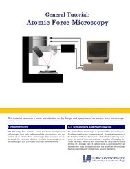
Full page fax print - Optical Sciences - Universiteit Twente
Full page fax print - Optical Sciences - Universiteit Twente
Full page fax print - Optical Sciences - Universiteit Twente

Create successful ePaper yourself
Turn your PDF publications into a flip-book with our unique Google optimized e-Paper software.
STIMULI RESPONSIVE POLYMER/QUANTUM<br />
DOT HYBRID PLATFORMS MODIFIED AT<br />
THE NANOSCALE
This research was financially supported by the MESA + Institute for Nanotechnology of the<br />
University of <strong>Twente</strong> (Strategic Research Orientation program Molecular Photonics) and the<br />
nanothechnology program NanoNed of the Dutch Ministry of Economic Affairs.<br />
Stimuli Responsive Polymer/Quantum Dot Hybrid Platforms Modified at the Nanoscale<br />
O. Tagit<br />
Ph. D Thesis<br />
© Oya Tagit, Enschede, 2010<br />
ISBN: 978-90-365-2982-2<br />
Publisher: Ipskamp Drukkers B. V., Josink Maatweg 43, 7545 PS, Enschede,<br />
The Netherlends, http://www.ipskampdrukkers.nl<br />
No part of this work may be reproduced by <strong>print</strong>, photocopy or any other means without the<br />
permission in writing of the author.
STIMULI RESPONSIVE POLYMER/QUANTUM<br />
DOT HYBRID PLATFORMS MODIFIED AT<br />
THE NANOSCALE<br />
PROEFSCHRIFT<br />
ter verkrijging van<br />
de graad van doctor aan de <strong>Universiteit</strong> <strong>Twente</strong>,<br />
op gezag van de rector magnificus,<br />
prof. dr. H. Brinksma,<br />
volgens besluit van het College voor Promoties<br />
in het openbaar te verdedigen<br />
op vrijdag 19 maart 2010 om 15.00 uur<br />
door<br />
Oya Tagit<br />
geboren op 21 mei 1981<br />
te Bursa, Turkije
Dit proefschrift is goedgekeurd door:<br />
Promotoren: Prof. dr. G.J. Vancso<br />
Prof. dr. J.L. Herek<br />
Assistent-promotor: dr. N. Tomczak
This thesis is dedicated to my family
Table of Contents<br />
Chapter 1 General introduction 1<br />
Chapter 2 Strategies towards fabrication of quantum dot/polymer assemblies 8<br />
2.1. General introduction to quantum dots 9<br />
2.2. <strong>Optical</strong> properties of quantum dots 10<br />
2.2.1. Surface modification strategies for quantum dots 13<br />
2.2.2. Poly(N-isopropylacryl amide) as encaging agent for<br />
quantum dots<br />
14<br />
2.3. Routes for designing polymer/quantum dot hybrid assemblies 18<br />
2.3.1. Layer-by-layer electrostatic assembly approach 18<br />
2.3.2. Loading quantum dots within polymeric matrices during the<br />
polymer synthesis<br />
21<br />
2.3.3. Grafting approaches 25<br />
2.3.4. In situ synthesis of quantum dots within polymeric matrices 27<br />
2.4. Conclusions 31<br />
2.5. References 31<br />
Chapter 3 Characterization methods of quantum dots and quantum dot /<br />
polymer assemblies<br />
3.1. Introduction 38<br />
3.2. Microscopy techniques 38<br />
3.2.1. Atomic force microscopy 38<br />
3.2.2. Confocal microscopy 42<br />
3.2.3. Combination of different microscopy techniques 43<br />
3.3. Spectroscopy techniques 44<br />
3.3.1. Time correlated single photon counting (TCSPC) 45<br />
3.3.2. Fluorescence correlation spectroscopy (FCS) 45<br />
3.4. References 47<br />
Chapter 4 Probing the morphology and nano-scale mechanics of single poly(Nisopropylacrylamide)<br />
microparticles across the lower critical<br />
solution temperature by atomic force microscopy<br />
37<br />
50
4.1. Introduction 51<br />
4.2. Experimental section 52<br />
4.3. Results and discussion 54<br />
4.4. Conclusions 64<br />
4.5. References 65<br />
Chapter 5 Temperature modulated quenching of quantum dots covalently<br />
coupled to chain ends of poly(N-isopropyl acrylamide) brushes on<br />
gold<br />
68<br />
5.1. Introduction 69<br />
5.2. Experimental section 70<br />
5.3. Results and discussion 71<br />
5.4. Conclusions 78<br />
5.5. References 78<br />
Chapter 6 Thermoresponsive quantum dot/PNIPAM assemblies 81<br />
6.1. Introduction 82<br />
6.2. Experimental section 83<br />
6.3. Results and discussion 85<br />
6.4. Conclusions 93<br />
6.5. References 94<br />
Chapter 7 <strong>Optical</strong> characterization of thermo-responsive polymer-quantum<br />
dot nanoparticles<br />
97<br />
7.1. Introduction 98<br />
7.2. Experimental section 100<br />
7.3. Results and discussion 101<br />
7.4. Conclusions 108<br />
7.5. References 109<br />
Chapter 8 Applications of quantum dots in bio-medicine: opportunities and<br />
ii<br />
risks<br />
8.1. Introduction 112<br />
8.2. Applications of quantum dots in biology and medicine 113<br />
111<br />
8.2.1. Applications of quantum dots as in vitro fluorescent labels 114
Summary<br />
Samenvatting<br />
Pêşgotîn bi Kurtayî<br />
Acknowledgements<br />
About the author<br />
8.2.2. Applications of quantum dots for in vivo imaging 117<br />
8.2.3. Applications of quantum dots in photodynamic therapy of<br />
cancer<br />
120<br />
8.3. QD toxicity 122<br />
8.4. Conclusions 124<br />
8.5. References 125<br />
128<br />
131<br />
134<br />
135<br />
138<br />
iii
List of Abbreviations<br />
QD quantum dot<br />
TOPO trioctylphosphine oxide<br />
PNIPAM poly(N-isopropylacryl amide)<br />
LCST lower critical solution temperature<br />
VPTT volume phase transition temperature<br />
PL photoluminescence<br />
LbL layer-by-layer<br />
AFM atomic force microscopy<br />
TCSPC time correlated single photon counting<br />
FCS fluorescence correlation spectroscopy<br />
BIS N,N’-methylenebisacrylamide<br />
KPS potassium persulfate<br />
MAA mercaptoacetic acid<br />
DTCA dithiodiundecane-11,1-diylbis{4[([(diethylamino)carbonothioyl]<br />
thioethyl)phenyl]carbamate}<br />
DDS 1,2-dioctadecyldisulfane<br />
ODT octadecane-1-thiol<br />
TEMPO 2,2,6,6-tetramethylpiperidine-1-oxyl<br />
EDC 1-ethyl-3-[3-dimethylaminopropyl]carbodiimide<br />
NHS N-hydroxysuccinimide<br />
DIPEA N,N-diisopropylethylamine<br />
FWHM full width at half maximum
General introduction<br />
Chapter 1
Chapter 1<br />
Nanotechnology aims at designing and creating functional materials, structures,<br />
devices and systems through the direct control of matter on the nanometer scale and at<br />
exploitation of novel phenomena and properties on this length scale, which is defined as<br />
being smaller than 100 nm [1]. Obtaining a fundamental understanding of the optical,<br />
electrical, magnetic and mechanical properties of nanostructures as well as controlled<br />
manipulation of these materials into complex, functional architectures requires<br />
multidisciplinary effort and cross-fertilization among different disciplines.<br />
Nanoscale engineering of materials enables controlled alteration, dynamical<br />
manipulation, and molecular functionalization of materials’ properties, and potentially creates<br />
entirely new properties, which are inaccessible otherwise.<br />
Complementary to traditional ‘Top-Down’ material processing approaches,<br />
nanotechnology have enabled ‘Bottom-Up’ processes, inspired by nature, involving building<br />
up materials from the molecular levels to nano/macrometer sized structures [2]. For the<br />
‘Bottom-Up’ approach, colloidal systems with diameters smaller than 50 nm are generally of<br />
interest [2]. These nano-sized particles, owing to their dimensions, have become a ‘hot’ topic<br />
in colloid and materials science [3] due to their unique electronic, optical, photoresponsive<br />
and catalytic properties [4], as well as to their applications in nanotechnology from biological<br />
labels to lasers and LEDs [5-14].<br />
Such particles display different properties than in the bulk, and are usually dependent<br />
on the shape and size of the individual particles, as well as on the distance between those<br />
particles. Among the nano-particles semiconductor nanocrystals, Quantum Dots, (QDs) have<br />
become of considerable scientific and technological interest due to the opportunity they offer<br />
in the quantum confined regime [15-20].<br />
QDs have recently entered the realm of biology owing to their advantages as<br />
biological probes including their nanoscale size (similar to biomolecules (Figure 1.1)), high<br />
quantum yield and molar extinction coefficients, versatility in surface modification, broad<br />
excitation spectra (for multicolor imaging) and narrow band emission (Figure 1.2), and<br />
tunable optical properties [21-24].<br />
2
Figure 1.1. Length scales showing the comparable sizes of biomolecules and QDs.<br />
General introduction<br />
While QDs easily mix in various solvents, utilization of their functionality in practical<br />
integrated photonic systems or in physiological environments requires QDs being distributed<br />
in a robust and highly functional matrix. In this respect organic polymers are of great<br />
potential as hosts for QDs [25, 26]. Polymers are usually transparent in a wide spectral range<br />
and can be easily processed, providing a flexible platform for devices based on optical<br />
properties of QDs [27].<br />
Emission Intensity (a. u.)<br />
1.0<br />
0.8<br />
0.6<br />
0.4<br />
0.2<br />
0.0<br />
500 550 600 650<br />
0.0<br />
700<br />
Wavelength (nm)<br />
Figure 1.2. Normalized absorption and emission spectra of TOPO-coated CdSe/ZnS QDs in<br />
chloroform solution.<br />
1.0<br />
0.8<br />
0.6<br />
0.4<br />
0.2<br />
Absorption (a. u.)<br />
3
Chapter 1<br />
Encaging QDs within polymer matrices not only enables the control over optical and<br />
spectroscopic properties of QDs but also introduces a strong resistance to chemical and<br />
photodegradation [28] as well as an enhanced compatibility to biological environment. In this<br />
respect, these colloidal fluorescent hybrid materials hold great promise for use as fluorescent<br />
probes in targeting, labeling and imaging applications [29-33].<br />
Stimuli-responsive polymers, which undergo large physical changes upon small<br />
variations of external stimuli, such as temperature [34], pH [35], electric field [36], ionic<br />
strength, solvent composition [37], have attracted a great deal of attention as QD-encaging<br />
matrices. Using these ‘smart’ polymers for encaging QDs, one could prepare biosensor<br />
devices that would be switched on and off in response to the external stimuli [38]. Poly(Nisopropylacrylamide)<br />
(PNIPAM) is one of the best studied temperature-responsive polymers,<br />
which exhibits a lower critical solution temperature (LCST) at ~32 o C [39]. The hybrid<br />
devices possessing optical properties of QDs with temperature-responsive properties of<br />
PNIPAM will be capable of sensing and detecting the state of biological systems and living<br />
organisms optically, electrically and magnetically at the single molecule level.<br />
The work presented in this thesis covers synthesis and characterization of CdSe/ZnS<br />
core/shell QDs, and synthesis and characterization of temperature-responsive polymer<br />
matrices made of PNIPAM, as carriers of QDs. Fabrication of QD/temperature responsive<br />
polymer assemblies are presented with potential applications as sensing devices to be used in<br />
bio-nanotechnology.<br />
Chapter 2 provides a basic background in the optical properties of QDs and a<br />
literature review regarding the recent developments in combining stimuli responsive<br />
polymers with QDs via different approaches, including layer-by-layer deposition,<br />
macromolecular grafting and in situ QD synthesis within polymeric matrices. Particular<br />
attention is paid to the assemblies of QDs with the temperature-responsive polymer<br />
PNIPAM.<br />
Chapter 3 describes the basics of some of the microscopy and spectroscopy<br />
techniques used for the characterization of QDs down to the single molecule level. In<br />
addition to atomic force microscopy and confocal optical microscopy, combined microscopy<br />
techniques for studying QDs and QD-bearing systems are explained. Spectroscopy<br />
techniques that are mentioned in this chapter include time correlated single photon counting<br />
and fluorescence correlation spectroscopy.<br />
Chapter 4 presents the study of the morphology and nano-mechanical properties of<br />
individual, isolated, PNIPAM microgel particles at the silicon/air, and silicon/water<br />
4
General introduction<br />
interfaces, below and above the PNIPAM volume phase transition temperature (VPTT) using<br />
atomic force microscopy. The force-indentation measurements performed in air and in water<br />
below and above the VPTT of PNIPAM were used for the determination of the modulus of<br />
the PNIPAM spheres.<br />
In Chapter 5 a thermo-responsive polymer/quantum dot platform based on PNIPAM<br />
brushes ‘grafted from’ a gold substrate and QDs covalently attached to the PNIPAM layer is<br />
presented. The influence of PNIPAM chain collapse above its lower critical solution<br />
temperature (LCST) on the QD luminescence is discussed.<br />
Chapter 6 describes the synthesis of maleic anhydride-based copolymers with grafted<br />
PNIPAM chains. The resulting amphiphilic polymers are used as coatings for hydrophobic<br />
QDs. Colloidal and optical characterization of QDs coated with the novel coatings in aqueous<br />
solutions as a function of temperature is presented.<br />
Chapter 7 includes a detailed analysis of the colloidal and optical properties of<br />
QD/PNIPAM assemblies presented in Chapter 6. Fluorescence correlation spectroscopy and<br />
time correlated single photon counting measurements carried out at temperatures below and<br />
above the LCST of PNIPAM are reported.<br />
In Chapter 8 applications of QDs in in vitro labeling, in vivo detection and photo<br />
dynamic therapy are reviewed. Potential risks of toxicity of QDs in biological systems are<br />
presented with suggestions for making QDs non-toxic to biological systems in an<br />
environment-friendly fashion.<br />
References<br />
1- Vancso, G. J., Eur Polym J 2004, 40: p. 883<br />
2- Sugunan, A.; Dutta, J., J Phys Sci Ide 2004, 4: p. 50<br />
3- Schneider, G.; Decher, G. Nano Lett, 2004, 4: p. 1833<br />
4- Shon, Y. S. Dekker Encyclopedia of Nanoscience & Nanotechnology 2004, Marcel<br />
Dekker, New York<br />
5- Chan, W.C.W.; Maxwell, D. J.; Gao, X.; Bailey, R. E.; Han, M.; Nie, S. Curr. Opin.<br />
Biotechnol. 2002, 13: p. 40<br />
6- Seydel, C., Science 2003, 300: p. 80<br />
7- Jovin, T., Nat. Biotechnol. 2004, 21: p. 32<br />
5
Chapter 1<br />
8- Jiang, W.; Papa, E.; Fischer, H.; Mardyani, S.; Chan, W.C.W. Trends Biotechnol.<br />
2004, 22: p. 607<br />
9- Chan, W. C.; Nie, S. Science 1998, 281: p. 2016<br />
10- Wuister, S. F.; Donega, C. M.; Meijerink, A. J. Phys. Chem. B 2004, 108: p. 17393<br />
11- Bruchez, M., Jr.; Moronne, M.; Gin, P.; Weiss, S.; Alivisatos, A.P. Science 1998,<br />
281: p. 2013<br />
12- Dubertret, B.; Skourides, P.; Norris, D. J.; Noireaux, V.; Brivanlou, A. H.; Libchaber,<br />
A. Science 2002, 298: p. 1759<br />
13- Klimov, V.I.; Mikhailovsky, A.A.; Xu, S.; Malko, A.; Hollingsworth, J.A.;<br />
Leatherdale, C. A. Eisler, H. Bawendi, M. G. Science 2000, 290: p. 314<br />
14- Coe, S. Woo, W.-K. Bawendi, M. G. Bulovic, V. Nature 2002, 420: p. 800<br />
15- Alivisatos A. P. Science 1996 271: p. 933<br />
16- Huynh, W. U.; Dittmer, J. J.; Alivisatos, A. P. 2002 Science 295: p. 2425<br />
17- Klein, D.; Roth, R.; Lim, A.K.L.; Alivisatos, A.P.; McEuen, P.L., 1997 Nature 389: p.<br />
699<br />
18- Eychmuller, A., 2000 J. Phys. Chem. B 104: p. 6514<br />
19- Trindade, T.; O’Brien, P.; Pickett, N.L., 2001 Chem. Mater. 13: p. 3843<br />
20- Trindade, T., 2003 NanoscaleMaterials ed L.M. Liz-Marz´an and P.V. Kamat<br />
(Dordrecht: Kluwer–Academic)<br />
21- Nehilla, B. J.; Vu, T. Q.; Desai, A. T. J. Phys. Chem. B 2005, 109: p. 20724<br />
22- Medintz, I. L.; Uyeda, H. T.; Goldman, E. R.; Mattoussi, H. Nature Mat. 2005, 4: p.<br />
435<br />
23- Ho, Y. P.; Kung, M. C.; Yang, S.; Wang, T. H. Nano Lett. 2005, 9: p. 1963<br />
24- Nagasaki, Y.; Ishii, T.; Sunaga, Y.; Watanabe, Y.; Otsuka, H.; Kataoka, K. Langmuir<br />
2004, 20: p. 6396<br />
25- Tessler, N.; Mededev, V.; Kazes, M.; Kan, S. H.; Banin, U.; Science 2002, 295: p.<br />
1506<br />
26- Bakueva, L.; Musikhin, S.; Hines, M. A.; Chang, T. W. F.; Tzolov, M.; Scholes, G.<br />
D.; Sargent, E. H. Appl. Phys. Lett. 2003, 82: p. 2895<br />
27- Olsson, Y. K.; Chen, G.; Rapaport, R.; Fuchs, D. T.; Sundar, V. C.; Steckel, J. S.;<br />
Bawendi, M. G.; Aharoni, A.; Banin, U. Appl. Phys. Lett. 2004, 85: p. 4469<br />
28- Medintz, I. L.; Sapsford, K. E.; Konnert, J. H.; Chatterji, A.; Lin, T.; Johnson, J. H.;<br />
Mattoussi, H. Langmuir, 2005, 21: p. 5501<br />
6
General introduction<br />
29- Jaiswal J. K.; Mattoussi, H.; Mauro, J. M.; Simon, S. M. Nat. Biotechnol. 2003, 21: p.<br />
47<br />
30- Jaiswal, J. K.; Simon, S. M. Trends Cell. Biol. 2004, 14: p. 497<br />
31- Ballou, B.; Lagerholm, B. C.; Ernst, L. A.; Bruschez, M. P.; Waggoner, A. S.<br />
Bioconjugate Chem. 2004, 15: p. 79<br />
32- Dahan, M.; Le´vi, S.; Luccardini, C.; Rostaing, P.; Riveau, B.; Triller, A. Science<br />
2003, 302: p. 442<br />
33- Chan, W. C. W.; Nie, S. M. Science 1998, 281: p. 2016<br />
34- Xulu, P. M.; Filipsei, G.; Zrinyi, M. Macromolecules 2000, 33: p. 1716<br />
35- Tanaka, T.; Fillmore, D.; Sun, S.-T.; Nishio, I. Phys. Rev. Lett.1980, 45: p.1636<br />
36- Tanaka, T.; Nishio, I.; Shun, S.-T.; Ueno-Nishio, S. Science 1982, 218: p. 467<br />
37- Pagonis, K.; Bokias, G. Polymer 2004, 45: p. 2149<br />
38- Tomczak, N.; Janczewski, D.; Han, M.-Y.; Vancso, G.J., Prog Polym Sci, 2009. 34: p.<br />
393.<br />
39- Schild, H. G.; Tirrell, D. A., Langmuir 1991, 7: p. 665<br />
7
Chapter 2<br />
Strategies towards fabrication of quantum dot/polymer<br />
assemblies *<br />
This chapter provides a basic background in the optical properties of QDs and a literature<br />
review of recent developments in fabrication of hybrid materials made of stimuli responsive<br />
polymers and QDs via different approaches, including layer-by-layer assembly,<br />
macromolecular grafting and in situ QD synthesis within polymeric matrices. A particular<br />
attention is paid to the assemblies of QDs with temperature-responsive poly(N-isopropylacryl<br />
amide), PNIPAM.<br />
* Parts of this chapter have been published in: Tomczak, N., Jańczewski, D., Tagit, O., Han, M-Y., Vancso, G.J.<br />
Surface Engineering of Quantum Dots with Designer Ligands Surface Design; Applications in Bioscience and<br />
Nanotechnology, Book chapter 4.3; Wiley-VCH 341 – 361 (2009)
2.1. General introduction to quantum dots<br />
Strategies towards fabrication of quantum dot/polymer assemblies<br />
One of the fundamental aims of life sciences is to understand the (bio)-molecules’<br />
complex spatial and temporal organization and their inter/intra-molecular interactions, from<br />
the perspective of a single molecule up to the integrative level. In order to study these<br />
interactions, researchers commonly employ fluorescent labeling for both in vivo imaging and<br />
in vitro detection [1]. The efficiency of a particular imaging or detection method depends to a<br />
large extent on the physicochemical and photophysical properties of the label used [2]. A<br />
suitable fluorescent label should fulfill certain requirements depending on the applications.<br />
For instance, an ideal fluorescent label (i) should be excitable and detectable with<br />
conventional instrumentation, (ii) should have high fluorescence quantum yield and should be<br />
stable under relevant conditions, (iv) should be soluble in relevant buffers, (v) should be<br />
suitable for surface modification for site-specific labeling [3]. Among the fluorescent labels,<br />
organic dyes and genetically encoded proteins have been most commonly used. These types<br />
of labels have known limitations. Figure 2.1 shows the absorption and emission spectra of<br />
Rhodamine 110, a commonly used organic dye. The broad absorption and emission bands<br />
mirror each other with a poor separation distance between their maxima that results in cross<br />
talk between individual dye molecules [3]. Additionally such chromophores display weak<br />
photostability, which limits their application in long-term and multiplexed imaging without<br />
complex instrumentation [4]. The problems associated with conventional organic<br />
fluorophores motivated development of alternative luminescent labels to replace the common<br />
dyes used in fluorescence detection. In this context, inorganic fluorescent semiconductor<br />
nanocrystals (quantum dots, QDs) can potentially solve many problems associated with<br />
organic fluorescent labels.<br />
Figure 2.1. Absorption and emission spectra of Rhodamine 110. This image was taken from<br />
http://probes.invitrogen.com/media/spectra/6479ph7.gif<br />
9
Chapter 2<br />
QDs are semiconductor nanocrystals, composed of elements of II-VI, III-V, or IV-VI<br />
periodic groups, ranging from 2 to10 nanometers in diameter. Because of their small size,<br />
they display unique optical and electronic properties. Their stable, size-tunable, and bright<br />
luminescence, high absorption coefficients and narrow emission lines give QDs significant<br />
advantages over common organic dyes as fluorescent labels [5]. In the last decade, they have<br />
attracted tremendous attention as a new class of fluorophores with a wide range of<br />
applications in diagnostics and sensors [6]. Labeling of peptides [7], proteins [8], and DNA<br />
[9] with QDs has been achieved in addition to successful sensing applications developed for<br />
small molecules and more complex structures [10]. QDs offer possibilities such as<br />
multiplexed imaging and long term investigations due to their tunable emission wavelengths<br />
and high photostabilities.<br />
2.2. <strong>Optical</strong> properties of quantum dots<br />
Semiconductor materials are composed of a large number of covalently bound atoms.<br />
The combination of overlapping atomic orbitals leads to molecular orbitals that are closely<br />
spaced in energy, forming a virtually continuous band [11]. The electronic band structure of a<br />
material describes the ‘allowed’ and ‘forbidden’ energy levels of an electron in the<br />
semiconductor. The range of forbidden energy levels where no electron may be present is<br />
called a ‘bandgap’. The highest occupied energy band is called the ‘valence band’; the lowest<br />
empty band is called the ‘conduction band’ (Figure 2.2). For semiconductors and insulators,<br />
the bandgap refers to the energy difference between the valence band and the conduction<br />
band. The electrons in the valence band can be promoted to the conduction band upon<br />
absorption of light and leave behind unoccupied states, ‘holes’, in the valence band. A bound<br />
electron-hole pair is called an ‘exciton’. The average physical separation between the electron<br />
and hole in an exciton is called the Bohr Radius [12].<br />
As the size of a semiconductor approaches the size of the material's Bohr Radius, a<br />
three dimensional confinement of the electrons and holes in the nanocrystal arises. As a result<br />
of this confinement, the electron energy levels become discreet and the bandgap increases as<br />
the confinement increases [11] (Figure 2.2).<br />
10
Strategies towards fabrication of quantum dot/polymer assemblies<br />
Figure 2.2. Schematic illustration of energy levels of a QD compared to a bulk<br />
semiconductor material. Quantum confinement in QDs results in discreet energy levels. This<br />
image was taken from:<br />
http://www.ncbi.nlm.nih.gov/pmc/articles/mid/NIHMS116385/figure/F2/.<br />
When excited with energy larger than the bandgap, an electron in the valence band<br />
can be promoted to the conduction band. As the electron falls back down across the bandgap,<br />
electromagnetic radiation with an energy corresponding to the energy it looses in the<br />
transition is emitted. Because the bandgap is size-dependent, by controlling the size of the<br />
nanocrystals one can tune the emission wavelength of the QDs. Tailored band gaps enable<br />
QDs to luminesce at wavelengths ranging from 350 nm to 2500 nm [12]. Therefore, desired<br />
photoluminescent properties can be obtained by a good control over the size of QDs (Figure<br />
2.3.). For instance, using a single synthetic route, QDs with photoluminescence varying from<br />
green to red can be obtained [13].<br />
11
Chapter 2<br />
Figure 2.3. Size dependent emission of CdSe/ZnS QDs. The excitation is at 350 nm in all<br />
cases. This image was taken from<br />
http://www.nrl.navy.mil/Review02/images/materialFig9.gif.<br />
In order to enhance the luminescence properties of QDs, an additional inorganic shell<br />
is commonly grown on the QD surface. This shell is composed of a second semiconducting<br />
material with a higher bandgap energy (Figure 2.4). The larger bandgap prevents surface<br />
oxidation, confines the excitons to the core, and passivates surface defects [14].<br />
Probably the most commonly studied QDs are those form the II-VI group of elements<br />
[15] including CdS, CdSe, ZnS, or, CdS/ZnS, CdSe/ZnS core-shell structures.<br />
CdSe<br />
ZnS<br />
Figure 2.4. Schematic presentation of the structure and energy levels of a core/shell<br />
CdSe/ZnS QD. The shell material has higher bandgap energy than the core material. All the<br />
charge carriers are confined to the core. The image was adapted from<br />
http://nanocluster.mit.edu/research.php.<br />
12
Strategies towards fabrication of quantum dot/polymer assemblies<br />
2.2.1. Surface modification strategies for quantum dots<br />
QDs are usually synthesized via wet chemical methods, in which the QDs are<br />
obtained as colloidal suspensions dispersed in nonpolar solvents. Trioctylphosphine oxide<br />
(TOPO) is commonly used as the stabilizing ligand (Figure 2.5). Due to the hydrophobic<br />
nature of TOPO, QDs can not be dispersed in aqueous buffers. This is an important limitation<br />
for the use of QDs in biological applications. To disperse the QDs in biologically relevant<br />
environment, i.e., water or serum, one has to coat the QDs with appropriate capping ligands,<br />
which would prevent aggregation of the QDs [16]. Various coating chemistries for QDs have<br />
been developed including silanization [17, 18], coating with mercaptoalkanoate ligands [5],<br />
organic dendrons [19], amphiphilic polymers [20], phospholipids micelles [21], recombinant<br />
proteins [22], or oligomeric phosphines.<br />
I II<br />
CdSe<br />
ZnS<br />
Figure 2.5. I) Schematic presentation of TOPO coated CdSe/ZnS quantum dots. II) Structure<br />
of the TOPO ligand on a CdSe surface.<br />
Image II was taken from http://www.chemistry.manchester.ac.uk/groups/pob/exafscdse.gif<br />
The most successful methods to render the QDs water soluble include replacement of<br />
TOPO with bi-functional ligands, such as cysteines [23], mercapto acids [24], oligomeric<br />
phosphines [25], having hydrophilic groups (carboxyl, amine, alcohol) in their structure. The<br />
second approach involves coating the TOPO layer with amphiphilic molecules. The<br />
hydrophilic part of the amphiphile is exposed to the surrounding medium and ensures water<br />
dispersability, while the hydrophobic part interacts with TOPO. For example, Dubertret et al.<br />
[26] and Geissbuehler et al. [27] reported successful encapsulation of QDs within<br />
13
Chapter 2<br />
phospholipid micelles using this approach. In other studies the QDs were transferred to water<br />
by covering the TOPO layer with a shell of amphiphilic polymers [28-31].<br />
In general, polymers are good materials for encapsulation of QDs [32] (Figure 2.6).<br />
They are transparent in a wide spectral range, including the near-IR and visible region. They<br />
can be easily processed and owing to their low curing temperatures they ensure that the<br />
optical properties of the QDs are minimally affected during the processing [33].<br />
CdSe<br />
ZnS<br />
Figure 2.6. Schematic presentation of a polymer-coated CdSe/ZnS core/shell QD. The<br />
organic coating further passivates the QD surface and offers a suitable platform for further<br />
surface modifications. The image was adapted from http://nanocluster.mit.edu/research.php.<br />
Such organic/inorganic hybrid nanoparticles composed of polymers and inorganic QDs offer<br />
the possibility to further modify the optical and electronic properties of nanoparticles at<br />
nanoscale [34]. The organic polymer shell also determines the chemical properties of the QDs<br />
and their interaction with the environment, while the photophysical properties of the QDs are<br />
mainly governed by the size of the inorganic core [35].<br />
2.2.2. Poly(N-isopropylacryl amide) as encaging polymer for quantum dots<br />
Stimulus-responsive polymers, which exhibit large, rapid and reversible changes in<br />
conformation, surface characteristics or solubility in response to relatively small<br />
environmental stimuli, are often referred to as ‘smart’. These polymers have attracted a great<br />
deal of attention for their ‘smart’ applications in combination with QDs. In a general concept<br />
the ‘smart’ polymers would modulate the photophysical properties of the QDs in response to<br />
14
Strategies towards fabrication of quantum dot/polymer assemblies<br />
an environmental stimulus. The stimuli may include temperature, pH, light, electric field,<br />
ionic strength, and presence of chemicals. One of the best studied ‘smart’ polymers is<br />
poly(N-isopropylacrylamide) (PNIPAM). PNIPAM displays reversible conformational<br />
transition in response to changes in temperature [36] (Figure 2.7. II). This transition is<br />
associated with the lower critical solution temperature (LCST) of the polymer occurring in<br />
water at ~32 o C [37]. At temperatures below the LCST, the polymer is hydrophilic and is<br />
soluble in water. As the temperature rises above the LCST, hydrogen bonds with the water<br />
molecules are destroyed and the intra-chain hydrophobic interactions dominate. The polymer<br />
becomes hydrophobic and collapses [38-40].<br />
I<br />
II<br />
Figure 2.7. I) Chemical structure of PNIPAM. II) Schematic presentation of the PNIPAM<br />
chain collapse at LCST.<br />
PNIPAM can be obtained in various forms including micelle [39], hydrogel [40] and<br />
tablet [41]. Moreover, PNIPAM-derived materials have been developed such as PNIPAMpolystyrene<br />
particles [42], PNIPAM-poly(butylmethacrylate) micelles [43], or PNIPAMpoly(D,L-lactide)<br />
micelles [39], where the LCST of PNIPAM can be finely tuned by<br />
adjusting the co-monomer ratios [44]. In this manner, the LCST of PNIPAM-derived<br />
materials can be increased up to around 37 o C, which is equal to the human body temperature.<br />
It has been previously shown that above LCST, PNIPAM chains interact with biocomponents,<br />
such as cells and proteins, whereas hydrated flexible chains do not interact with<br />
them [43]. Therefore, PNIPAM offers the possibility to develop new concepts in bio-related<br />
applications of QDs. For example Li et al. [45] reported the synthesis of a highly<br />
photoluminescent CdTe/PNIPAM hydrogel, and its photoluminescence was found to be<br />
sensitive to external temperature stimulus in a reversible way. The same group also prepared<br />
CdTe/p(NIPAM-acrylate) (AAc) microgels and studied their self-assembly on a glass<br />
substrate (Figure 2.8) [46]. The effects of the pH-dependent swelling properties of<br />
15
Chapter 2<br />
p(NIPAM-AAc) microgels and of the dipole moment of the CdTe QDs on the self assembly<br />
were studied. It was concluded that a combination of the physical and chemical properties of<br />
inorganic CdTe QDs with those of the organic polymer affected the self assembly process.<br />
The dipole moment of CdTe was an important driving force for the self assembly on a large<br />
scale and the self assembly could also be tuned by the pH dependent swelling behavior of the<br />
co-polymeric hydrogel. At low pH, the aggregation morphology was fractal and dendritic on<br />
a large scale. At high pH, the microgels aggregated to form a porous film and phase<br />
separation between the polymer and QDs occurred.<br />
Figure 2.8. Schematic illustration of the self assembly process of CdTe/p(NIPAM-AAc)<br />
microgels. Addition of CTAB into the QD solution results in formation of QD complexes (I).<br />
These complexes are mixed with p(NIPAM-AAc) microgels. At low pH the microgels are in<br />
a shrunken state (II) and their self assembly upon drying is investigated. The self assembly<br />
process is also investigated at high pH values (III), where the p(NIPAM-AAc) microgels are<br />
in a swollen state. This image was taken from [46].<br />
16
Strategies towards fabrication of quantum dot/polymer assemblies<br />
Wang et al. [47] have employed NIPAM and 4-viniylpyridine (VP), a pH responsive<br />
monomer, to fabricate pNIPVP spheres as colloidal carriers and to confine CdTe nanocrystals<br />
of different sizes within these hydrogel networks by changing the external pH. They also<br />
studied the controlled release of QDs by pH stimulus (Figure 2.9.). By absorbing the QDs of<br />
different size, they were able to achieve multicolour-coded microspheres with a LCST around<br />
34 o C. This system was designed to be used as a delivery agent for the QDs and their<br />
bioconjugates within the human body.<br />
Figure 2.9. Schematic illustration of the loading of CdTe QDs in pNIPVP hydrogels and<br />
their controlled release by pH stimulus. The pH-dependent phase transition of pNIPVP<br />
hydrogels is used for the internalization of QDs at pH 3. Upon increasing the pH, the<br />
hydrogel shrinks and the QDs are confined within the hydrogel. The release of QDs is<br />
achieved by increasing the pH to above 11. This image was taken from [47].<br />
When designing QD/polymer hybrid materials, a number of factors should be<br />
considered such as the size and shape of the polymer matrix, the amount of QDs to be<br />
entrapped within the matrix, as well as the spatial distribution/localization, separation and<br />
orientation of the QDs within the matrix. The fluorescent hybrid nanoparticles should be<br />
monodisperse and have relatively small size and large saturation intensity and be highly<br />
luminescent [48]. The requirements for their applications in single-molecule biological<br />
17
Chapter 2<br />
studies are even more stringent: they must be biocompatible, non-cytotoxic, chemically stable<br />
and offer conjugation chemistries for attachment of recognition molecules to their surfaces.<br />
The size of the host matrix may be few hundreds to tens of nanometers depending on<br />
the fabrication procedure and desired applications. QDs with thicker polymer coating tend to<br />
have better photostabilities and higher QYs, whereas QDs with thinner coatings would be<br />
more suitable as intracellular probes [48]. The size of the matrix plays also an important role<br />
in limiting the allowed number of QDs per matrix volume so that e.g. fluorescent resonance<br />
energy transfer (FRET) is prevented. Incorporation of QDs into spherical nano sized matrices<br />
is of interest for both fundamental studies on light-matter interactions and for practical<br />
applications [49]. These dot-in-a-dot structures confine electrons and photons in all three<br />
dimensions. The real success in developing polymer/QD nanohybrid materials is achievable<br />
only when the above requirements are satisfied. This necessitates a control over the size and<br />
shape of the matrix, and over the amount, spatial distribution/localization, separation and<br />
orientation of the QDs within the matrix.<br />
2.3. Designing polymer/quantum dot hybrid assemblies<br />
Prior to realizing most applications, QDs must be functionally integrated into matrices<br />
[34]. Some of the main routes, via which polymer/QD nanohybrid particles have been<br />
prepared, include layer-by-layer deposition of QDs on oppositely charged beads by<br />
electrostatic interactions, loading QDs into polymer beads during the synthesis of the<br />
polymeric nanoparticles, grafting polymeric shells from or to the QD surfaces, and in situ<br />
synthesis of QDs within polymeric matrices.<br />
2.3.1. Layer-by-layer electrostatic assembly approach<br />
Among the assembly techniques used to build functional structures, the electrostatic<br />
layer-by-layer (LbL) assembly evolved as a powerful method for the construction of<br />
supramolecular hybrid architectures by sequential absorption of oppositely charged<br />
polyelectrolytes [50], which enabled formation of functional thin film architectures [51]. By<br />
this simple and versatile method, it is possible to exert molecular-level control over the<br />
architecture, composition, and thickness of the films [52, 53]. The precise control that is<br />
offered by LbL assembly leads to remarkable improvements in organic optoelectronic<br />
devices owing to the large interfacial areas for charge separation and creation of efficient<br />
charge transfer pathways [54].<br />
18
Strategies towards fabrication of quantum dot/polymer assemblies<br />
Recently, a variety of schemes were introduced for the fabrication of QD multilayer<br />
films prepared with oppositely charged species. Mamedov et al. used positively charged<br />
polyelectrolyte (poly(diallyldimethylammonium chloride), PDDA) for the preparation of<br />
graded QD films [55]. Zhang et al. reported the assembly of aqueous CdTe nanoparticles with<br />
N-vinyl carbazole/4-vinyl pyridine copolymer [56]. Kotov’s group prepared a monolayer film<br />
of QDs (CdSe/PDDA or CdSe/CdS/PDDA) with homogenous, nearly close-packed coverage<br />
with little aggregation. [57]. However, these studies paid little attention to the<br />
photoluminescence (PL) efficiency of QDs in the films. Lesser et al. reported that the LbLassembled<br />
QDs had only 5% PL efficiency, although the initial PL efficiency of QDs in an<br />
aqueous colloidal solution was equal to 20% [58].<br />
For the fabrication of devices with patterned layers of QDs, one of the issues to<br />
consider is selectivity and non-specific interactions. Zhou et al. showed that the selectivity<br />
over patterned surfaces exhibited in the first few layers can decrease dramatically and almost<br />
disappears after tens of bilayers are assembled [59]. Therefore, it is important to improve the<br />
selectivity to the patterned features while reducing the non-specific interactions. The same<br />
group reported reduced non-specific interactions through the modification of QD surface<br />
coatings and employing a polymer with a hydrophilic backbone. Their method was based on<br />
patterning a gold substrate with self assembled monolayers (SAMs) of alkyl thiols terminated<br />
with hexa(ethylene glycol), which acted as the resistive coating due to its resistance against<br />
non-specific adsorption. Linear poly(ethyleneimine) (LPEI), a positively charged polymer,<br />
was used as the ‘assembly partner’ for 2-mercaptoethanesulfonic acid (MESA) terminated<br />
QDs (Figure 2.10). Using the LbL approach, the authors were able to selectively build up 3D<br />
fluorescent surface patterns.<br />
19
Chapter 2<br />
Figure 2.10. Fluorescence images at different stages of the LbL assembly of MESA-QDs and<br />
LPEI on a patterned substrate. a) After 2 cycles, b) after 19 cycles of assembly. Image size:<br />
30 µm x 30 µm. High signal-to-noise ratio shows that very little non-specific adsorption<br />
occurs during the assembly process. This image was taken from [59].<br />
Jaffar et al. [60] coated mercaptoacetic acid (MAA) treated QDs with cationic<br />
poly(allylamide) (PA) and subsequently with poly(vinylsulfonic acid) (PVSA) (Figure<br />
2.11.).<br />
Figure 2.11. Modification scheme of CdSe/ZnS QD by LbL. The ligand exchange reaction is<br />
performed to replace TOPO with MAA to render QDs water soluble and negatively charged.<br />
Subsquently, positively (PAA) and negatively (PVSA) charged polyelectrolytes are deposited<br />
on the QD surface. This image was taken from [60].<br />
20
Strategies towards fabrication of quantum dot/polymer assemblies<br />
In order to produce structured arrays, PAA-coated QDs (green) and MAA-treated<br />
QDs (red) were deposited on a glass substrate patterned by hyaluronic acid (Figure 2.12).<br />
Figure 2.12. Self-assembly scheme of modified QDs on HA patterned glass substrate.<br />
Anionic MAA-QDs and cationic PAA-QDs bind to the HA patterns on the glass substrate.<br />
This image was taken from [60].<br />
As an alternative to using electrostatic forces [45, 53, 61-63], covalent LbL assembly<br />
of polymers and QDs was also performed [52]. Liang et al. prepared robust and smooth,<br />
functional QD/polymeric thin films [52]. The QD/polymer hybrid structures displayed<br />
promising properties for applications in light-emitting diodes, photovoltaics, lasers and<br />
biosensors [64].<br />
In summary, LbL assembly of QDs using polymers offers control of their spatial<br />
organization over a range of length scales. These nanostructures prove useful as building<br />
blocks for opto-electronic device fabrication.<br />
2.3.2. Loading quantum dots into polymer matrices during the polymer synthesis<br />
Among the methods used for the incorporation of QDs into polymeric matrices,<br />
loading of QDs through emulsion [65] and suspension [66] polymerizations allows one to<br />
disperse the QDs through the volume of a spherical polymeric particle. A general procedure<br />
involves polymerizing the monomer in the presence of dispersed QDs. However, there are<br />
some challenges related to this approach, such as control over the amount and location of the<br />
QDs within the polymeric particles and control of the colloidal stability and monodispersity<br />
of the polymeric particles [65].<br />
21
Chapter 2<br />
Sheng et al. [49] reported a strategy for the incorporation of CdSe/ZnCdS/ZnS QDs<br />
into polystyrene (PS) microspheres by using functionalized oligomeric phosphine (OP)<br />
ligands. The TOP-coated QDs were first dispersed in OP/DMF solution, which was followed<br />
by prepolymerization of MMA groups to form a polymeric shell around the QDs. Formation<br />
of polystyrene particles in the presence of surface-modified QDs was achieved via free<br />
radical polymerization at high temperatures. (Figure 2.13.).<br />
Figure 2.13. Schematic illustration of the formation of QD-PS microspheres. The starting<br />
reaction mixture contains styrene monomer, initiator, stabilizer, OP ligand, and QDs treated<br />
with the OP ligand (I). Polymerization begins as the initiator decomposes (II). As the polymer<br />
chains reach a critical length they begin to aggregate into small particles which are stabilized<br />
by the stabilizer molecules (III). The polymerization continues inside the particles until all the<br />
monomer units are used up (IV). This image was taken from [49].<br />
Fluorescence imaging of the particles (Figure 2.14) showed that all the QD/PS hybrid<br />
particles were well separated, proving that QDs had been incorporated within the polymeric<br />
matrix and are still well protected by OP ligands.<br />
22
Strategies towards fabrication of quantum dot/polymer assemblies<br />
Figure 2.14. Fluorescence image of QD/PS particles. Scale bar: 5µm. This image was taken<br />
from [49].<br />
As an alternative strategy, radical polymerization in miniemulsion of QD/polymer<br />
nanocomposites without further modification of the QD surface was reported [65, 67, 68].<br />
Some features of the miniemulsion polymerization technique provide potential advantages for<br />
the encapsulation of organically capped QDs, namely, the ability to nucleate all the droplets<br />
containing the inorganic nanoparticles, a good control of the droplet size and size<br />
distribution, and the direct dispersion of the QDs within the oil phase [67]. The as-prepared<br />
QDs have their surface passivated with TOPO molecules, leaving the hydrophobic octyl<br />
chains directed outward [69, 70]. As a result, TOPO capped QDs are easily dispersed in a<br />
nonpolar media such as several organic solvents or in viscous liquid monomers. Therefore, in<br />
order to encapsulate the QDs within polymers via the miniemulsion polymerization strategy,<br />
there is no need for further surface derivatization.<br />
In the study reported by Esteves et al. [67], the incorporation of QDs into the polymer<br />
particles was achieved via polymerization of a mixture of TOPO-coated CdS or CdSe QDs<br />
and the monomer (Figure 2.15.). Two polymeric matrices, polystyrene (PS) and poly(n-butyl<br />
acrylate) (PBA), were investigated. In both cases, homogenous nanocomposites were<br />
obtained.<br />
23
Chapter 2<br />
Figure 2.15. Schematic representation of the miniemulsion polymerization of QD/polymer<br />
nanocomposites. TOPO-coated QDs are dispersed in the monomer and sonication of the<br />
monomer/water mixture results in miniemulsion formation. The polymerization of the<br />
monomer droplets results in QD/polymer nanocomposites. This image was taken from [67].<br />
Due to its high-luminescent properties, the PL of the CdSe/PBA nanocomposite was<br />
analyzed in more detail. The emission spectra of the CdSe/PBA nanocomposite and the<br />
respective CdSe QDs are shown in Figure 2.16. The narrow PL emission band is blueshifted<br />
from the bulk PL due to a strong quantum confinement effect. Therefore the emission<br />
observed in the CdSe/PBA nanocomposite proceeds directly from the unique properties of the<br />
Figure 2.16. PL spectra of CdSe and CdSe/PBA nanohybrid particles at room temperature.<br />
This image was taken from [67].<br />
24
Strategies towards fabrication of quantum dot/polymer assemblies<br />
constituent QDs. In this experiment, contrary to some other reports where conventional free<br />
radical initiators (such as AIBN) degraded the dots and totally quenched their optical<br />
properties [71], QD degradation or PL quenching was not observed. This is probably due to<br />
dispersion of QDs within monomer droplets and use of a water soluble initiator, KPS, which<br />
might reduce the contact between QDs and the free radicals.<br />
In order to overcome the quenching problems associated with the use of free radicals<br />
during the polymerization Zhang et al. [72] used an alternative method in which aqueous<br />
nanocrystals were used instead of hydrophobic QDs. The maximum PL was reported to be<br />
retained when the thickness of the Cd-thiol complexes around QDs was increased under<br />
proper conditions. This was also proven by some other reports where a shell of Cd-mercapto<br />
carboxylic acid complexes was formed around CdTe QDs [73]. This structure improved both<br />
the PL intensity and stability of aqueous QDs [74].<br />
Considering all the results mentioned above, miniemulsion polymerization seems to<br />
be a promising approach for the encapsulation of hydrophobic QDs within polymeric<br />
matrices. However, fabricating monodisperse hybrid particles with homogenously distributed<br />
QDs still remains a challenge.<br />
2.3.3. Grafting approaches<br />
Growing polymer chains around inorganic cores is one of the most popular methods<br />
used to obtain core/shell structures. In this approach, polymer chains are tethered by one end<br />
to the surface of the core particle [75]. Generally there are two methods to chemically attach<br />
polymer chains to a surface:<br />
i) Grafting-to method;<br />
ii) Grafting-from method.<br />
In grafting-to method, the end-functionalized polymers react with an appropriately<br />
functionalized surface, i.e., this method involves irreversible grafting of a presynthesized<br />
polymer chain [75, 76]. This method has certain limitations including:<br />
Relatively low surface coverage;<br />
Restricted diffusion of the polymer chain-ends to the surface;<br />
Island formation due to steric crowding of the reactive sites by the already-grafted<br />
polymers;<br />
25
Chapter 2<br />
Lack of complete control over the growth of stable polymer brushes at nanoscale [77].<br />
The second approach involves initiation of polymerization from the surface of the<br />
particles, which are functionalized by initiators [75]. This is called the grafting-from method<br />
and it provides a greater control over the density of the grafts [78].<br />
Both methods mentioned above are used mainly in combination with free radical<br />
polymerizations [78]. Greater control over the polymerization process is possible by using<br />
living polymerization or employing controlled radical polymerization scheme, where the<br />
concentration of the radicals is kept at minimum by equilibrating the reactive radicals with<br />
their reversibly-terminated counterparts. In such systems, surface initiation can be combined<br />
with atom transfer radical polymerization (ATRP), nitroxide-mediated radical polymerization<br />
(NMRP) or photoiniferter-controlled polymerization, where the reaction time determines the<br />
thickness of the polymeric shell [79].<br />
Using the grafting-from approach, a number of research groups synthesized polymer brushes<br />
on silica [80, 81], gold [82], QDs [71, 83], and magnetic nanoparticles [84, 85].<br />
Regarding QDs, Farmer and Patten prepared CdS/SiO2 core/shell nanoparticles and<br />
modified their surfaces with an ATRP initiator (Figure 2.17) [83]. The initiator-modified<br />
nanoparticles were then used in the polymerization of methyl methacrylate (MMA).<br />
Figure 2.17. Schematic representation of preparation of Cd/SiO2/PMMA nanoparticles via<br />
reverse microemulsion. Addition of Si(OEt)4 results in formation of a SiO2 coating on the QD<br />
surface. After modification of the QD surface with the initiator, the polymerization proceeds<br />
to form QD/polymer hybrids. This image was taken from [83].<br />
The TEM images of the particles (Figure 2.18) demonstrate that, it is possible to obtain<br />
QD/polymer hybrid particles with a single QD located in the centre of the matrix. This is<br />
difficult to achieve using other methods to incorporate QDs in polymeric matrices.<br />
26
Strategies towards fabrication of quantum dot/polymer assemblies<br />
Figure 2.18. TEM image of QD/SiO2 particles. This image was taken from [83].<br />
In another study Sill and Emrick [71] reported polystyrene and poly(styrene-methyl<br />
methacrylate) copolymer brushes grown directly from the surface of CdSe nanoparticles by<br />
nitroxide-mediated controlled free radical polymerization. Since free radicals can quench the<br />
fluorescence of the CdSe nanoparticles, nitroxide-mediated polymerization allows for the<br />
preparation of polymer-nanoparticle composites while maintaining the fluorescence of the<br />
nanoparticles.<br />
In summary, grafting polymers on the QD surfaces is a promising approach for the<br />
preparation of hybrid nanoparticles. However, more attention should be paid to the control<br />
over the polymerization reactions as well as over the grafting density of the polymer chains.<br />
In addition, new protocols should be developed in order to minimize the influence of the<br />
reaction conditions on the PL intensity of the QDs.<br />
2.3.4. In situ synthesis of quantum dots within polymeric matrices<br />
Synthesis of semiconductor nanoparticles in geometrically restricted environments<br />
has been well-studied [86]. The studies included block copolymer micelles [87], reverse<br />
micelles and micro emulsions [88], organic-inorganic matrices [18] and hydrogels [89, 90] as<br />
nanoreactors for the synthesis of semiconductor nanoparticles.<br />
27
Chapter 2<br />
The preparation of CdS in block ionomer reverse micelles in an organic solvent was<br />
reported by Moffitt et al. [91]. CdS was precipitated within the ionic cores of a PS-b-PACd<br />
diblock ionomer. In a follow-up study, aqueous solutions of polystyrene-b-poly (acrylic acid)<br />
(PS-b-PAA) compound micelles containing QDs were obtained [92].<br />
Other preparations of CdS QDs involved also triblock copolymers such as<br />
hydroxylated poly-(styrene-b-butadiene-b-styrene) micelles in toluene [93] and poly(ethylene<br />
oxide)-b-polystyrene-b-poly (acrylic acid) triblock copolymers (PEO-b-PS-b-PAA) [91].<br />
In the study reported by Duxin et al. [92] the authors obtained different polymer<br />
morphologies from the same triblock copolymer, which consisted of a cadmium acrylate<br />
(CdAcr) core, surrounded by PS chains and a PEO corona (Figure 2.19). The use of block<br />
copolymers in QD synthesis allows also for precise localization of the QDs within the bulk<br />
matrix, or in the surfaces of inverted micelles [87, 94-96].<br />
In another study by Chu et al. [97] a synthetic route for the preparation of luminescent<br />
and rodlike CdS nanocrystals embedded in poly(BA-co-GMA-co-GMA-IDA) (PBGM)<br />
copolymer templates by soap-free emulsion copolymerization was presented. In this study,<br />
GMA-IDA groups within the copolymer acted as coordination sites for chelating Cd 2+ , at<br />
which nanosized CdS nanocrystals were grown by the dry method (H2S) and the wet method<br />
(Na2S) (Figure 2.20). The hybrid semiconductor–polymer composites prepared with the<br />
above procedure are stable and free from other capping molecules.<br />
Recently, hydrogels have also been investigated as nanoreactors for producing<br />
semiconductor or metallic nanoparticles [89, 90]. By choosing a suitable polymer, the<br />
swollen-shrunk states of the hydrogels can be effectively controlled by external stimuli like<br />
temperature, pH, electric field, solvent, etc.<br />
The incorporation of QDs into the hydrogels is usually based on the “breathing in”<br />
technique, where the dry hydrogel is swollen with an aqueous solution containing the<br />
preformed nanoparticles [89, 90, 97]. An alternative approach is based on in situ formation of<br />
the nanoparticles using appropriate ionic precursors. This method has been successfully<br />
employed to form CdS nanoparticles on the surface of poly(methyl methacrylate-comethacrylicacid)<br />
latexes [98] or in the interior of poly(N-isopropylacrylamide-co-acrylic<br />
acid) microgels [99]. Because of the acrylate anions, these gels are negatively charged, and<br />
the introduction of the precursors was performed via ion exchange of the latex or microgel<br />
counterions with Cd 2+ . Furthermore, the formation of the nanoparticles was localized around<br />
the acrylate anions, which offered an opportunity to control the location of the QDs within<br />
the matrix.<br />
28
Strategies towards fabrication of quantum dot/polymer assemblies<br />
Figure 2.19. Schematic illustrations of the formation of PEO-b-PS-b-PAA assemblies. (a)<br />
Single triblock copolymer molecules in THF. (b) Ionically crosslinked triblock micelles.<br />
(b1) Primary spherical inverse micelles (PSIMs) in THF; (b2) wormlike micelles at higher<br />
water content. (c) Triblock copolymer structures with CdS quantum dots. (c1) Spheres in<br />
THF; (c2) rods in water-rich solutions. (d) PS core micelles in water, surrounded by CdS<br />
nanoparticles. (e) Multicore cadmium acrylate supermicelle (SM) structures, following the<br />
change of the solvent to water of the PSIMs shown in part b. (f) Water soluble SM triblocks<br />
with CdS cores. This image was taken from [92].<br />
29
Chapter 2<br />
Figure 2.20. Schematic illustration of the preparation of luminescent and rodlike CdS<br />
nanocrystals embedded in poly(BA-co-GMA-co-GMA-IDA) (PBGM) copolymer templates.<br />
This image was taken from [97].<br />
30
Strategies towards fabrication of quantum dot/polymer assemblies<br />
Bekiari et al. used the in situ synthesis to prepare CdS nanoparticles in a nonionic<br />
hydrogel, based on poly(N,N-dimethylacryl-amide), PDMAM [86]. Contrary to previous<br />
studies, the distribution of the CdS nanoparticles in the PDMAM hydrogel is expected to be<br />
homogeneous throughout the whole hydrogel volume since PDMAM is a nonionic,<br />
hydrophilic polymer. A special attention has been paid to the fate of the QDs as a function of<br />
the degree of swelling of the hydrogel.<br />
In summary, preparation of QDs/hydrogel nanohybrid particles is an important issue<br />
for the development of luminescent materials. The materials can serve as luminescent probes<br />
of macromolecules of biological importance.<br />
2.4. Conclusions<br />
Many future materials and devices based on QDs require their incorporation and<br />
organization in polymeric matrices. The growing need for photonic materials and devices<br />
encouraged the development of many different strategies to produce polymer/QD hybrid<br />
structures. The appropriate choice of a strategy depends mainly on the final application of the<br />
hybrid materials. For instance, if the material is designed to be used in biological<br />
applications, in addition to its luminescence efficiency, the compatibility of the material with<br />
the biological systems should be also considered. The fabrication methods which have been<br />
reported to date (for a recent review see [100]) have advantages and drawbacks. This makes<br />
the development of new routes for producing QD/polymer nanohybrid materials a hot<br />
scientific topic, since simultaneous control over the size and shape of the matrix, and over the<br />
amount, spatial distribution/localization, separation and orientation of the QDs within the<br />
matrix still remains a challenge to be solved.<br />
2.5. References<br />
1- Miyawaki, A., Dev. Cell 2003, 4: p. 295<br />
2- Waggoner, A., Curr. Opin. Chem. Biol. 2006, 10: p. 62<br />
3- Resh-Genger, U.; Grabolle, M.; Cavaliere-Jaricot, S.; Nitschke, R.; Nann, T., Nature<br />
Methods 2008, 5: p. 763<br />
4- Louis, C.; Bazzi, R.; Marquette, C. A.; Bridot, J. L.; Roux, S.; Ledoux, G.; Mercier,<br />
B.; Blum, L.; Perriat, P.; Tillement, O., Chem. Mater. 2005, 17: p. 1673<br />
5- Chan, W. C. W.; Nie, S. M., Science 1998, 281: p. 201638<br />
6- Whaley, S.R.; English, D.S.; Hu, E.L.; Barbara, P.F.; Belcher, A.M., Nature 2000,<br />
405: p. 665<br />
31
Chapter 2<br />
7- Bruchez, M., Jr.; Moronne, M.; Gin, P.; Weiss, S.; Alivisatos, A.P., Science 1998,<br />
281: p. 2013<br />
8- Mitchell, G.P.; Mirkin, C.A.; Letsinger, R.L., J. Am. Chem. Soc. 1999, 121: p. 8122<br />
9- Mednitz, I.L.; Uyeda, H.T.; Goldman, E.R.; Mattoussi, H., Nat. Mater. 2005, 4: p. 435<br />
10- Hezinger, A.F.E.; Teβmar, J.; Göpferich, A., Eur. J. Pharma. Biopharma. 2008, 68: p.<br />
1<br />
11- Alivisatos A. P., Science 1996, 271: p. 933<br />
12- http://www.evidenttech.com/products/evitags/overview.php<br />
13- Qu, L.; Peng, Z. A.; Peng, X., Nano Lett. 2001, 1: p. 333<br />
14- Ooba, H., AIST Today 2006, 6: p. 26<br />
15- Wilcoxon, J. P.; Provenico, P. P., J. Phys. Chem. B 2005, 109: p. 13461<br />
16- Zhang, S,; Yu, J.; Li, X.; Tian, W., Nanotechnology 2004, 15: p. 1108<br />
17- Bruchez, M.; Moronne, M.; Gin, P.; Weiss, S.; Alivisatos, A. P., 281: p. 2013.<br />
18- Gerion, D.; Pinaud, F.; Williams, S. C.; Parak, W. J.; Zanchet, D.; Weiss, S.;<br />
Alivisatos, A.P., J. Phys. Chem. B 2001, 105: p. 8861.<br />
19- Guo, W.; Li, J. J.; Wang, Y. A.; Peng, X., J. Am. Chem. Soc. 2003, 125: p. 3901.<br />
20- Larson, D. R.; Zipfel, W. R.; Williams, R. M.; Clark, S. W.; Bruchez, M. P.; Wise, F.<br />
W.; Webb, W.W., Science 2003, 300: p.1434.<br />
21- Dubertret, B.; Skourides, P.; Norris, D. J.; Noireaux, V.; Brivanlou, A. H.; Libchaber,<br />
A., Science 2002, 298: p. 1759.<br />
22- Mattoussi, H.; Mauro, J. M.; Goldman, E. R.; Green, T. M.; Anderson, G. P.; Sundar,<br />
V. C.; Bawendi, M. G., Phys. Status Solidi B 2001, 224: p. 277.<br />
23- Sukhanova, A.; Venteo, L.; Devy, J.; Artemyev, M.; Oleinikov, V.; Pluot, M.;<br />
Nabiev, I., Lab. InVest. 2002, 82: p. 1259<br />
24- Akerman, M. E.; Chan, P.; Laakkonen, S. N.; Bhatia, E.; Ruoslahti, E., PNAS 2002,<br />
99: p. 12617<br />
25- Kim, S.; Bawendi, M. G., J. Am. Chem. Soc. 2003, 125: p. 14652<br />
26- Dubertret, B.; Skourides, P.; Norris, D. J.; Noireaux, V.; Brivanlou, A. H.; Libchaber,<br />
A., Science 2002, 298: p. 1759<br />
27- Geissbuehler, I.; Hovius, R.; Martinez, K. L.; Adrian, M.; Thampi, K.;<br />
Ravindranathan, T.; Vogel, H., Angew. Chem., Int. Ed. 2005, 44: p. 1388<br />
28- Wu, X.; Liu, H.; Liu, J.; Haley, K. N.; Treadway, J. A.; Larson, J. P.; Ge, N.; Peale,<br />
32<br />
F.; Bruchez, M. P., Nat. Biotechnol. 2003, 21: p. 41
Strategies towards fabrication of quantum dot/polymer assemblies<br />
29- Pellegrino, T.; Manna, L.; Kudera, K.; Liedl, T.; Koktysh, D.; Rogach, A. L.; Keller,<br />
S.; Radler, J.; Natile, G.; Parak, W.P., Nano Lett. 2004, 4: p. 704<br />
30- Gao, X.; Cui, Y.; Levenson, R. M.; Chung, L. W.; Nie, S., Nat. Biotechnol. 2004, 22:<br />
p. 959<br />
31- Kim, S. W.; Kim, S.; Tracy, J. B.; Jasanoff, A.; Bawendi, M. G., J. Am. Chem. Soc.<br />
2005, 127: p. 4556<br />
32- Bakueva, L.; Musikhin, S.; Hines, M. A.; Chang, T. W. F.; Tzolov, M.; Scholes, G.<br />
D.; Sargent, E. H., Appl. Phys. Lett. 2003, 82: p. 2895<br />
33- Olsson, Y. K.; Chen, G.; Rapaport, R.; Fuchs, D. T.; Sundar, V. C.; Steckel, J. S.;<br />
Bawendi, M. G.; Aharoni, A.; Banin, U., Appl. Phys. Lett. 2004, 85: p. 4469<br />
34- Caruso, F., Adv. Mater. 2001, 13: p. 11<br />
35- Zhang, Y.; Luo, S.; Liu, S., Macromolecules 2005, 38: p. 9813<br />
36- Galaev, I. Y.; Mattiasson, B., Trends Biotechnol. 1999, 17: p. 335<br />
37- Heskins, M.; Guillet, J. E., J. Macromolecules Sci., Chem 1968, A2: p. 1441<br />
38- Schild, H. G.; Tirrell, D. A., Langmuir 1991, 7: p. 665<br />
39- Kohori, F.; Sakai, K.; Aoyagi, T.; Yokoyama, M.; Sakurai, Y.; Okano, T., J. Control.<br />
Rel. 1998, 55: p. 87<br />
40- Sershen, S.R.; Westcott, S.L.; Halas, N.J.; West, J.L., J. Biomed. Mater. Res. 2000,<br />
51: p. 293<br />
41- Yuk, S.H.; Cho, S.H.; Lee, S.H., Macromolecules 1997, 30: p. 6856<br />
42- Cammas, S.; Suzuki, K.; Sone, C.; Sakurai, Y.; Kataoka, K.; Okano, T., J. Control.<br />
Release 1997, 48: p. 157<br />
43- Chung, J.E.; Yokoyama, M.; Suzuki, K.; Aoyagi, T.; Sakurai, Y.; Okano, T., Colloid.<br />
Surface. B 1997, 9: p. 37<br />
44- Sershen, S. R.; Westcott, S. L.; Halas, N. J.; West, J. L., Appl. Phys. Lett. 2002, 80: p.<br />
4609<br />
45- Li, J.; Hong, X.; Liu Y.; Li, D.; Wang, Y.W.; Li, J.H.; Bai, Y.B.; Li, T.J., Adv. Mater.<br />
2005, 17: p. 163<br />
46- Li, J.; Liu, B.; Li, J., Langmuir 2006, 22: p. 528<br />
47- Kuang, M.; Wang, D. Y.; Bao, H. B.; Gao, M. Y. Mohwald, H.; Jiang, M., Adv.<br />
Mater. 2005, 17: p. 267<br />
48- Tsay, J. M.; Doose, S.; Pinaud, F.; Weiss, S., J. Phys. Chem. B 2005, 109: p. 1669<br />
49- Sheng, W.; Kim, S.; Lee, J.; Kim, S. W.; Jensen, K.; Bawendi, M. G., Langmuir 2006,<br />
22: p. 3782<br />
33
Chapter 2<br />
50- Decher, G., Science 1997, 277: p. 1232<br />
51- Decher, G.; Hong, J. D., Makromol. Chem. Macromol. Symp. 1991, 46: p. 321<br />
52- Liang, Z.; Dzienis, K. L.; Xu, J.; Wang, Q., Adv. Funct. Mater. 2006, 16: p. 542<br />
53- Yang, P.; Li, C. L.; Murase, N., Langmuir 2005, 21: p. 8913<br />
54- Li, F.B.; Albery, W.J., Adv. Mater. 1992, 4: p.673<br />
55- Mamedov, A. A.; Belov, A.; Giersig, M.; Mamedova, N. N.; Kotov, N. A., J. Am.<br />
Chem. Soc. 2001, 123: p. 7738<br />
56- Zhang, H.; Zhou, Z.; Liu, K.; Wang, R.; Yang, B., J. Mater. Chem. 2003, 13: p. 1356<br />
57- Tang, Z.; Wang, Y.; Kotov, N. A., Langmuir 2002, 18: p. 7035<br />
58- Lesser, C.; Gao, M.; Kirstein, S., Mater. Sci. Eng., C 1999, 8-9: p. 159<br />
59- Zhou, D.; Bruckbauer, A.; Abell, C.; Klenerman, D.; Kang, D.J., Adv. Mater. 2005,<br />
17: p. 1243<br />
60- Jaffar, S.; Nam, K. T.; Khademhosseini, A.; Xing, J.; Langer, R. S.; Belcher, A. M.,<br />
Nano Lett. 2004, 4: p. 1421<br />
61- Gorelikov, I.; Kumacheva, E., Chem. Mater. 2004, 16: p. 4122<br />
62- Schneider, G.; Decher, G., Nano Lett. 2006, 6: p. 530<br />
63- Lowman, G. M.; Nelson, S. L.; Graves, S. M.; Strouse, G. F.; Buratto, S. K.,<br />
Langmuir 2004, 20: p. 2057<br />
64- Colvin, V. L; Schlamp, M.C.; Alivisatos, A.P., Nature 1994, 370: p. 534<br />
65- Yang, X.; Zhang, Y., Langmuir 2004, 20: p. 6071<br />
66- Li, Y.; Liu, E.C.Y.; Pickett, N.; Skabara, P. J.; Cummins, S.S.; Ryley, S.; Sutherland,<br />
A.J.; O’Brien, P., J. Mater. Chem. 2005, 15: p. 1238<br />
67- Esteves, A.C.C.; Barros-Timmons, A.M.V.; Monteiro, T.; Trindade, T., J. Nanosci.<br />
Nanotechnol. 2005, 5: p. 766<br />
68- Peres, M.; Costa, L.C.; Neves, A.; Soares, M.J.; Monteiro, T.; Esteves, A.C.; Barros-<br />
Timmons, A.; Trindade, T.; Kholkin, A.; Alves, E., Nanotechnology 2005, 16: p.<br />
1969<br />
69- Becerra, L.R.; Murray, C.B.; Griffin, R.G.; Bawendi, M.G., J. Chem. Phys. 1994,<br />
100: p. 3297<br />
70- Bowen, K.J.E.; Colvin, V.L.; Alivisatos, A.P., J. Phys. Chem. 1994, 98: p. 4109<br />
71- Sill, K. and Emrick, T., Chem. Mater. 2004, 16: p. 1240<br />
72- Zhang, H.; Wang, C.; Li, M.; Ji, X.; Zhang, J.; Yang, B., Chem. Mater. 2005, 17: p.<br />
4783<br />
73- Bao, H.; Gong, Y.; Li, Z.; Gao, M. Y., Chem. Mater. 2004, 16: p. 3853<br />
34
Strategies towards fabrication of quantum dot/polymer assemblies<br />
74- Gao, M.Y.; Kirstein, S.; Mohwald, H.; Rogach, A.L.; Kornowski, A.; Eychmuller, A.;<br />
Weller, H., J. Phys. Chem. B 1998, 102: p. 8360<br />
75- Li, D.; Jones, G.L.; Dunlap, J.R.; Hua, F.; Zhao, B., Langmuir 2006, 22: p. 3344<br />
76- Jordan, R.; Graft, K.; Riegler, H.; Unger, K. K., Chem. Commun. 1996, 9: p. 1025<br />
77- de Boer, B.; Simon, H. K.; Werts, M. P.L.; van der Vegte, E. W.; Hadziioannou, G.,<br />
Macromolecules 2000, 33: p. 349<br />
78- Reddy, S.K.; Sebra, R.P.; Anseth, K.S.; Bowman, C.N., J. Polym. Sci. A, Polym.<br />
Chem. 2005, 43: p. 2134<br />
79- Luo, N.; Hutchison, J. B.; Kristi S. Anseth, K. S.; Bowman, C. N., Macromolecules<br />
2002, 35: p. 2487<br />
80- Ohno, K.; Morinaga, T.; Koh, K.; Tsujii, Y.; Fukuda, T., Macromolecules 2005, 38: p.<br />
2137<br />
81- Bai, J.; Qiu, K. Y.; Wei, Y., Polym. Int. 2003, 52: p. 853<br />
82- Ohno, K.; Koh, K.; Tsujii, Y.; Fukuda, T., Angew. Chem., Int. Ed. 2003, 42: p. 2751<br />
83- Farmer, S.C.; Patten, T.E., Chem. Mater. 2001, 13: p. 3920<br />
84- Vestal, C.R.; Zhang, Z.J., J. Am. Chem. Soc. 2002, 124: p. 14312<br />
85 - Matsuno, R.; Yamamoto, K.; Otsuka, H.; Takahara, A., Macromolecules 2004, 37: p.<br />
2203<br />
86- Bekiari, V.; Pagonis, K.; Bokias, G.; Lianos, P., Langmuir, 2004, 20: p.7972<br />
87- Zhao, H.; Douglas, E.P.; Harrison, B.S.; Schanze, K.S., Langmuir 2001, 17: p. 8428.<br />
88- Simmons, B.A.; Sichu, L.; Vijay, J.T.; McPherson, G.L.; Bose, A.; Zhou, W.; He, J.,<br />
Nano Lett. 2002, 2: p. 263<br />
89- Jones, C.D.; Serpe, M.J.; Schroeder, L.; Lyon, A., J. Am. Chem. Soc. 2003, 125: p.<br />
5292<br />
90- Xulu, P. M.; Filipsei, G.; Zrinyl, M., Macromolecules 2000, 33: p. 1716.<br />
91- Moffitt, M.; McMahon, L.; Pessel, V.; Eisenberg, A., Chem. Mater. 1995, 7: p. 1185<br />
92- Duxin, N.; Liu, F.; Vali, H.; Eisenberg, A., J. Am. Chem. Soc. 2005, 127: p. 10063<br />
93- Moffitt, M.; Vali, H.; Eisenberg, A., Chem. Mater. 1998, 10: p. 1021<br />
94- Wang, D.; Yaan, C.; Xintong, Z.; Zhiqiang, L.; Xinming, Q.; Xin, A.; Fengqi, L.;<br />
Dejun, W.; Yubai, B.; Tiejin, L.; Tang, X., Chem. Mater. 1999, 11: p. 392<br />
95- Zhao, H.; Douglas, E.P., Chem. Mater. 2002, 14: p. 1418<br />
96- Liu, T.; Burger, C.; Chu, B., Prog. Polym. Sci. 2002, 28: p. 5<br />
97- Chu, Y.C.; Wang, C.C.; Chen, C.Y., Nanotechnology 2005, 16: p. 58<br />
35
Chapter 2<br />
98- Pardo-Yissar, V.; Gabai, R.; Shipway, A.N.; Bourenko, T.; Willner, I., Adv. Mater.<br />
2001, 13: p. 1320<br />
99- Zhang, J.; Coombs, N.; Kumacheva, E.; Lin, Y.; Sargent, E.H., Adv. Mater. 2002, 14:<br />
p. 1756<br />
100- Tomczak, N.; Janczewski, D.; Han, M.-Y.; Vancso, G.J., Prog Polym Sci, 2009. 34: p.<br />
36<br />
393.
Chapter 3<br />
Characterization methods of quantum dots and quantum<br />
dot/polymer assemblies<br />
This chapter describes some of the microscopy and spectroscopy techniques used in this<br />
thesis for the characterization of QDs. In addition to atomic force microscopy and confocal<br />
microscopy, combined microscopy techniques for studying QDs and QD-bearing systems are<br />
explained. The spectroscopic techniques which are mentioned in this chapter include time<br />
correlated single photon counting and fluorescence correlation spectroscopy.
Chapter 3<br />
3.1. Introduction<br />
It is of great importance to obtain detailed information on the structure of QD surfaces<br />
due to their strong influence on the colloidal and optical properties of the QDs. Surface<br />
characterization methods are required for the monitoring of colloidal and optical properties<br />
each time a chemical modification is made. Colloidal properties such as particle size, size<br />
distribution and particle aggregation are monitored by transmission electron microscopy<br />
(TEM), atomic force microscopy (AFM), light scattering, chromatography, and<br />
electrophoresis [1]. Given the typical values of the relevant energy levels in QDs, the<br />
photophysical characterization methods usually involve UV/visible absorption and<br />
fluorescence emission spectroscopies.<br />
Monitoring the excited states of QDs proves useful for surface characterization of<br />
QDs, since luminescence behavior is influenced by the polarity, dielectric constant, or the<br />
presence of quenchers in the environment of the excited species [2]. In this context, timeresolved<br />
luminescence spectroscopy and time-resolved fluorescence lifetime measurements<br />
give information about the charge carrier dynamics on the QD surface. Simple quenching<br />
experiments with a number of quenchers with different redox potentials can be used to detect<br />
the energy levels of surface traps.<br />
In this chapter, some of the important microscopic and spectroscopic characterization<br />
methods are described.<br />
3.2. Microscopy techniques<br />
With recent advances in nanotechnology, the conventional microscopy has been<br />
replaced by more powerful detection methods with single molecule sensitivity. Single<br />
molecule detection enables determination of dynamic as well as static heterogeneities that are<br />
masked in ensemble-averaging methods.<br />
3.2.1. Atomic force microscopy<br />
Atomic force microscope (AFM) provides high spatial resolution, three-dimensional<br />
topographical information on sample surfaces in air and in aqueous environment, and<br />
therefore it is a powerful tool for imaging, characterization and manipulation of matter at the<br />
nanoscale [3]. Along with the topographic image, other properties, such as elasticity or<br />
adhesion force can be probed and mapped with this technique [4].<br />
An AFM consists of a cantilever with a sharp tip (probe) at its end, which is used to<br />
scan the specimen surface [5]. When the tip is brought into proximity of a sample surface,<br />
38
Characterization methods of QDs and QD/polymer assemblies<br />
intermolecular forces (e.g. electrostatic forces, Van der Waals forces, etc.) between the tip<br />
and the sample result in a deflection of the cantilever (Figure 3.1.). Typically, the deflection<br />
is measured using a laser spot reflected from the top of the cantilever and directed onto a<br />
photodiode array.<br />
A feedback mechanism is employed to adjust the deflection to maintain a constant<br />
force between the tip and the sample during imaging. Typically, the sample is mounted on a<br />
piezoelectric tube, which can move the sample in the z direction to maintain a constant force,<br />
and the x and y directions for scanning the sample.<br />
Figure 3.1. Schematic presentation of an atomic force microscope (the components are not<br />
drawn to scale). The devices for data acquisition and signal processing have been omitted for<br />
clarity. The image was taken from [4].<br />
The interactions between the AFM tip and the sample can be monitored by measuring<br />
the cantilever deflection as a function of the scanner movement along the z-axis. Figure 3.2<br />
shows the cantilever behavior as it approaches to, and retracts from, the sample surface.<br />
Initially there is a weak repulsive force (1) until the tip gets in contact with the surface (2).<br />
Further movement towards the surface results in bending of the cantilever (3) due to stiffness<br />
of the surface. During retraction, the cantilever remains at the surface (4) until it overcomes<br />
the adhesive forces (5) and detaches from the surface.<br />
39
Chapter 3<br />
approach<br />
1 2 3<br />
5<br />
4<br />
retract<br />
Figure 3.2. i) Schematic presentation of the cantilever deflection versus scanner movement<br />
when approaching to and retracting from the surface. ii) Schematic force-distance plot. (1) is<br />
the non-contact region. After initial contact with the surface (2) the cantilever bends due to<br />
further movement of the piezo (3). The cantilever remains at the surface (4) until it<br />
overcomes the adhesive forces (5) and completely detaches from the surface. This image was<br />
taken from [4].<br />
An AFM can be operated in a number of modes, depending on the application. For<br />
imaging purposes, the primary modes of operation are the contact and the tapping modes. In<br />
the contact mode, the force between the tip and the surface is kept constant during rasterscanning<br />
by maintaining a constant cantilever deflection. In the tapping mode, the cantilever<br />
is vibrating such that it comes in brief contact with the sample during each cycle, and then<br />
enough restoring force is provided by the cantilever spring to separate the tip from the<br />
sample.<br />
In addition to the determination of surface topography, AFM has been used as a tool<br />
for the determination of the elastic properties of polymer surfaces [6-8] via indentation<br />
experiments. For instance, AFM in the force spectroscopy mode has been used to study the<br />
mechanical properties of thin polymer layers [9], polyelectrolyte multi layers [10], as well as<br />
40<br />
3
Characterization methods of QDs and QD/polymer assemblies<br />
the viscoelastic and plastic properties of amorphous polymer surfaces as a function of<br />
temperature across the glass transition temperature (Tg) [11]. Due to the small dimension of<br />
the AFM probe, related nanoindentation experiments provided information on the<br />
nanomechanical properties, e.g. on the Young’s modulus, of these surfaces with<br />
unprecedented lateral resolution. [12-14].<br />
The indentation experiments involve acquisition of deflection-distance curves and<br />
conversion of the cantilever deflection and scanner extension data into force and indentation,<br />
respectively. A representative curve is shown in Figure 3.3. Either the approach or the<br />
retraction curve can be used for determination of the mechanical properties of the samples. A<br />
hard substrate with ‘zero indentation’ is used as a reference [15].<br />
Figure 3.3. Illustration of an indentation experiment and the corresponding force –<br />
displacement curves obtained for a hard substrate (solid) and a softer sample (dash). The<br />
contact regions are used for the determination of mechanical properties. The difference in the<br />
displacement between the hard substrate and the soft material corresponds to the indentation<br />
of the tip into the sample. This image was taken from [15].<br />
The versatility of AFM technique enables studying materials’ properties under<br />
different environments (air, or liquid environment) and at different temperatures.<br />
41
Chapter 3<br />
3.2.2. Confocal microscopy<br />
Confocal laser scanning fluorescence microscopy is a widely used technique in life<br />
sciences for obtaining high-resolution optical images with depth selectivity [16, 17]. By using<br />
a process called optical sectioning, in-focus images from the selected depths are acquired.<br />
During this process out-of-focus light is rejected, which improves the signal to noise ratio<br />
[17]. The schematic presentation of a confocal setup is shown in Figure 3.4.<br />
Figure 3.4. Schematic presentation of a confocal microscope. Blue: excitation path; red:<br />
detection path. The excitation beam is focused to a diffraction-limited volume. The emitted<br />
fluorescence is collected by the objective and directed to the detector by using a beamsplitter.<br />
The image was adapted from [18].<br />
In a confocal laser scanning microscope, the laser beam passes through an aperture<br />
and then is focused by an objective lens to a diffraction-limited focal volume. The objective<br />
lens re-collects the emitted fluorescence as well as scattered and reflected laser light. A<br />
beamsplitter sends the light to the detection apparatus, which is equipped with filters that<br />
selectively allow passing only the fluorescence. After passing through a pinhole, the<br />
fluorescence light intensity is detected by photomultiplier tubes (PMTs), or avalanche<br />
photodiodes (APDs).<br />
There have been a number of investigations of individual QDs immobilized on<br />
surfaces with confocal microscopy. For instance, Nirmal et al. for the first time reported<br />
blinking in the emission of single QDs (Figure 3.5) using a scanning stage confocal<br />
microscope, equipped with a CCD camera [19]. More recent works supported the<br />
42
Characterization methods of QDs and QD/polymer assemblies<br />
interpretation that blinking is caused by surface defects that can be altered by surface<br />
chemistries and buffer conditions [20-22].<br />
Figure 3.5. Left: Confocal microscopy image of single CdSe QDs embedded in a thin<br />
polyvinylbutyral film acquired by raster scanning the sample across a diffraction limited laser<br />
spot (532 nm). Field of view: 8x8 μm. Right: Fluorescence intensity versus time trace of a<br />
single QD. This image was taken from [19].<br />
3.2.3. Combination of different microscopy techniques<br />
AFM and optical microscopy have both experienced significant advancements in<br />
recent years [23]. For instance, several improvements enabled overcoming the diffraction<br />
limit in optical microscopy [24]. Similarly, AFM has been applied for the determination of<br />
electrostatic properties of biomolecules [25], and for the measurements of mechanical<br />
properties of such molecules [26, 27]. Combining these two techniques has opened up many<br />
exciting possibilities in biophysics, materials science and bionanotechnology by providing<br />
nanoscale resolution and quantitative force measurements as well as spectral characterization<br />
[23, 28, 29]. Owen et al. have reported on simultaneous AFM and confocal imaging of<br />
CdSe/ZnS core/shell QDs of 3.5 nm core size with high spatial resolution [23]. Lately,<br />
Ebenstein et al. showed how to utilize QDs as contrast agents for both AFM topography and<br />
fluorescence imaging with a combined setup [30]. In addition, Wang et al. employed QDs as<br />
bifunctional cell-surface markers for both AFM and fluorescence microscopy for quantitative<br />
43
Chapter 3<br />
investigation of the distribution and expression of membrane proteins on cell surface using<br />
simultaneous fluorescence and topography mapping [31]. In a study reported by Doak et al. a<br />
combined AFM and confocal microscopy setup was used for simultaneous detection of<br />
structural and fluorescent properties of PNT2 cells, a cell line derived from human prostate<br />
epithelial cells, labeled with QDs [28] (Figure 3.6). The topographic features of the cells<br />
were visualized by AFM and their molecular make-up was investigated using confocal<br />
microscopy.<br />
Figure 3.6.a) AFM image of PNT2 cells with QD labelled F-actin, and b) corresponding<br />
confocal microscopy image of the same region. This image was taken from [28].<br />
The combined microscopy techniques mentioned here have the potential to be applied<br />
to a variety of samples, including living cells, and can be coupled with AFM-based force<br />
measurements for further biophysical analysis. Therefore, these techniques will not only<br />
provide significant advances in imaging, but will also enable the characterization of structural<br />
constituents, the mechanical properties and structure-function relationships of biological<br />
molecules at the single molecule level.<br />
3.3. Spectroscopy techniques<br />
The key to understanding of the optical and colloidal properties of QDs is to study<br />
their interactions with light. Fluorescence and UV/visible absorption spectroscopy are two<br />
commonly used techniques for basic optical characterization [32]. The techniques have<br />
become very popular because of their sensitivity to changes in the structural and<br />
44
Characterization methods of QDs and QD/polymer assemblies<br />
photophysical properties of QDs. Steady-state or time resolved emission or absorption can be<br />
measured.<br />
Ultrafast spectroscopy provides essential information on the mechanistic and kinetic<br />
details of chemical and physical events that occur on the timescale of tens of femtoseconds to<br />
several nanoseconds. Such events are initiated by an ultrafast laser pulse (pump) and the<br />
photophysical and photochemical events are probed by another ultrafast laser pulse (probe).<br />
With sub femtoseconds pulse durations, it is possible to access the dynamics of the internal<br />
conversion, intramolecular vibrational relaxation and many other processes, which steer the<br />
energy into different reaction pathways [33] [34].<br />
3.3.1. Time correlated single photon counting (TCSPC)<br />
TCSPC is a technique used for recording low-level light signals with picosecond time<br />
resolution. It is based on the excitation with a periodic light source and measuring the arrival<br />
times of individual fluorescence photons [33]. The method makes use of the fact that for low<br />
light level and high excitation repetition rates, the probability of detecting one photon<br />
between excitation light pulses is much lower than one. The time between the excitation light<br />
pulse and the detected fluorescence photon is measured. This time corresponds to the excited<br />
state lifetime of the investigated species. Histogram of the photon arrival times corresponds<br />
to the time-averaged luminescence decay curves from which an average excited state lifetime<br />
can be obtained.<br />
TCSPC has been extensively used for the investigation of carrier dynamics in excited<br />
QDs [35-37].<br />
3.3.2. Fluorescence correlation spectroscopy (FCS)<br />
Fluorescence correlation spectroscopy is a sensitive single molecule detection method<br />
with a wide range of applications in different fields [38-41]. It is based on the monitoring of<br />
the fluctuating fluorescence signal of the emitter molecule as it passes through the confocal<br />
volume [42]. Autocorrelation function is then calculated according to Eq. (3.1):<br />
〈 δI<br />
( t)<br />
δI(<br />
t + τ ) 〉<br />
G(<br />
τ ) = 1+<br />
(3.1)<br />
2<br />
〈 I ( t)<br />
〉<br />
In this equation, t is the time variable corresponding to the time frame over which the<br />
intensity signal I is defined, while τ is the delay variable corresponding to the time frame,<br />
45
Chapter 3<br />
over which the relative fluctuations of I are measured, and the autocorrelation function G is<br />
defined. The fluorescence fluctuation around the average intensity, δI(t), is monitored and<br />
correlated to the function obtained at time t + τ to generate the autocorrelation function [42].<br />
The autocorrelation function of the fluctuations resulting from the diffusion of the molecules<br />
through the focal volume is used to obtain the diffusion coefficient by setting Eq. (3.1) equal<br />
to the autocorrelation function for a 3D Gaussian excitation volume shown in Eq. (3.2):<br />
1 ⎛ τ ⎞<br />
G(<br />
τ ) = 1+<br />
⎜<br />
⎜1+<br />
⎟<br />
N ⎝ τ D ⎠<br />
−1<br />
⎛<br />
⎜<br />
⎜1+<br />
S<br />
⎝<br />
2<br />
τ ⎞<br />
⎟<br />
τ D ⎠<br />
1<br />
−<br />
2<br />
(3.2)<br />
In the equation above, N represents the number of fluorophores in the confocal<br />
volume at any time, τD is diffusion time of the molecule through the confocal volume and S is<br />
the structure parameter, which is the ratio of axial (ωz) and radial (ωr) distances at which the<br />
intensity of the Gaussian excitation falls to e -2 of its maximum. The inverse relation between<br />
the autocorrelation signal and the number of fluorophores enables measurements in the<br />
nanomolar range.<br />
FCS performed on diffusing molecules has single molecule sensitivity and is<br />
compatible with high throughput analysis [43-45]. Despite using nanomolar concentrations, it<br />
can provide evidence on distributions of characteristic time constants and sample<br />
heterogeneities [46].<br />
FCS has been used in a wide range of biochemical assays [41]. It has been applied to<br />
monitor conformational fluctuations of biopolymers, detect binding events between<br />
macromolecules and their ligands, investigate photophysical properties of organic dyes and<br />
fluorescent proteins, or look at diffusion properties of labeled macromolecules in vitro and in<br />
vivo.<br />
Doose et al. reported use of FCS for characterization of the photophysical and<br />
colloidal properties of different water-stable QDs [46]. They have reported on the brightness<br />
per particle, saturation intensity, blinking, and hydrodynamic radii for different QDs and<br />
demonstrated that variations in the FCS amplitude reflected the sample inhomogeneities.<br />
More detailed discussions on the advances, challenges and opportunities of using FCS for<br />
characterization of QDs can be found in [47].<br />
The microscopy and spectroscopy techniques mentioned here are useful tools for<br />
characterization of QDs and QD/polymer hybrid structures in terms of their colloidal and<br />
46
Characterization methods of QDs and QD/polymer assemblies<br />
optical properties down to single molecule level. Combining different microscopy and<br />
spectroscopy techniques will enable simultaneous characterization of different properties<br />
such as morphology, optical and colloidal stability, etc.<br />
3.4. References<br />
1. Doose, S.; Tsay, J.M.; Pinaud, F.; Weiss, S., Anal Chem, 2005. 77: p. 2235.<br />
2. Thomas, J.K., J Phys Chem, 1987. 91: p. 267.<br />
3. Binnig, G.; Quate, C.F.; Gerber, C., Phys Rev Lett, 1986. 56: p. 930.<br />
4. Schönherr, H., From functional group ensembles to single molecules: scanning force<br />
microscopy of supramolecular and polymeric systems. Ph D Thesis 1999, University<br />
of <strong>Twente</strong>: Enschede.<br />
5. Peterson, C.A.; Sarid, D., Kirk-Othmer Encyclopedia of Chemical Technology:<br />
Atomic Force Microscopy. Vol. 3. 2007, New Jersey: Wiley.<br />
6. Matzelle, T.R.; Geuskens, G.; Kruse, N., Macromolecules, 2003. 36: p. 2926.<br />
7. Briscoe, B.; Fiori, L.; Pelillo, E., J Phys D, 1998. 31: p. 2395.<br />
8. Butt, H.-J.; Cappella, B.; Kappl, M., Surf Sci Rep, 2005. 59: p. 1.<br />
9. Dimitriadis, E.K.; Horkay, F.; Maresca, J.; Kachar, B.; Chadwick, R. S., Biophys J,<br />
2002. 82: p. 2798.<br />
10. Mermut, O.; Lefebre, J.; Gray, D. G.; Barrett, C. J., Macromolecules, 2003. 36: p.<br />
8819.<br />
11. Cappella, B.; Kaliappan, S.K.; Sturm, H., Macromolecules, 2005. 38: p. 1874.<br />
12. Chizhik, S.A.; Huang, Z.; Gorbunov, V. V.; Myshkin, N. K.; Tsukruk, V. V.,<br />
Langmuir, 1998. 14: p. 2606.<br />
13. Reynaud, C.; Sommer, F.; Quet, C.; El Bounia, M.; Duc, T. M., Surf Interface Anal,<br />
2000. 30: p. 185.<br />
14. Raghavan, D.; Gu, X.; Nguyen, T.; VanLandingham, M. R.; Karim, A.,<br />
Macromolecules, 2000. 33: p. 2573.<br />
15. Puttini, S.; Lekka, M.; Dorchies, O.M.; Saugy, D.; Incitti, T.; Ruegg, U.T.; Bozzoni,<br />
I.; Kulik, A.J.; Mermod, N., Mol Theraph, 2008. 17: p. 19.<br />
16. Fellers, T.J.; Davidson, M.W., Introduction to Confocal Microscopy. 2007, Olympus<br />
Fluoview Resource Center.<br />
17. Pawley, J.B., Handbook of Biological Confocal Microscopy. 2006, Berlin: Springer.<br />
18. Wright, S.J.; Wright, D.J., Methods Cell Biol, 2002. 70: p. 1.<br />
47
Chapter 3<br />
19. Nirmal, M.; Dabbousi, B. O.; Bawendi, M. G.; Macklin, J. J.; Trautman, J.K.; Harris,<br />
T. D.; Brus, L. E., Nature, 1996. 383: p. 802.<br />
20. Hohng, S.; Ha, T. J., J Am Chem Soc, 2004. 126: p. 1324.<br />
21. Kuno, M.; Fromm, D. P.; Hamann, H. F.; Gallagher, A.; Nesbitt, D. J., J Chem Phys,<br />
2000. 112: p. 3117.<br />
22. Shimizu, K.T.; Neuhauser, R. G.; Leatherdale, C. A.; Empedocles, S. A.; Woo, W. K.;<br />
Bawendi, M. G., Phys Rev B, 2001. 6320: p. U5395.<br />
23. Owen, R.J.; Heyes, C.D.; Knebel, D.; Rocker, C.; Nienhaus, G.U., Biopolymers,<br />
2006. 82: p. 410.<br />
24. Hell, S.W., Nat Biotechnol, 2003. 21: p. 1347.<br />
25. Muler, D.J.; Anderson, K., Trends Biotechnol, 2002. 20: p. S45.<br />
26. Oesterhelt, F.; Oesterhelt, D.; Pfeiffer, M.; Engel, A.; Gaub, H. E.; Muller, D. J.,<br />
Science, 2000. 288: p. 143.<br />
27. Fernandez, J.M.; Li, H., Science, 2004. 303: p. 1674.<br />
28. Doak, S.H.; Rogers, D.; Jones, B.; Francis, L.; Conlan, R.S.; Wright, C., Histochem<br />
Cell Biol, 2008. 130: p. 909.<br />
29. Ebenstein, Y.; Mokari, T.; Banin, U., Appl Phys Lett, 2002. 80: p. 4033.<br />
30. Ebenstein, Y.; Gassman, N.; Kim, S.; Weiss, S., J Mol Recog, 2009. 22: p. 397.<br />
31. Wang, Y.; Chen, Y.; Cai, J.; Zhong, L., Ultramicroscopy, 2009. 109: p. 268.<br />
32. Royer, C.A., Biophys J, 1995. 68: p. 1191.<br />
33. Lakowicz, J.R., Principles of fluorescence spectroscopy. 2006, New York, NY:<br />
Springer.<br />
34. Klimov, V.I., Annu Rev Phys Chem, 2007. 58: p. 635.<br />
35. Yamada, T.; Goushi, K.; Xu, X.; Otomo, A., Thin Solid Films, 2008. 517: p. 1507.<br />
36. Hong, M.; Guo-Hong, M.; Wen-Jun, W.; Xue-Xi, G.; Hong-Liang, M., Chinese Phys<br />
B, 2008. 17: p. 1280.<br />
37. Nikolaev, I.S.; Lodahl, P.; van Driel, A.F.; Koenderink, A.F.; Vos, W.L., Phys Rev B,<br />
2007. 75: p. 115302.<br />
38. Kinjo, M., Anal Chim Acta, 1998. 365: p. 43.<br />
39. Schwille, P., Cell Biochem Biopyhs, 2001. 34: p. 383.<br />
40. Sengupta, P.; Garai, K.; Balaji, J.; Periasamy, N.; Maiti, S., Biophys J, 2003. 84: p.<br />
1977.<br />
41. Hess, S.T.; Huang, S.; Heikal, A.A.; Webb, W.W., Biochemistry, 2002. 41: p. 697.<br />
42. Magde, D.; Elson, E.; Webb, W.W., Phys Rev Lett, 1972. 29: p. 705.<br />
48
Characterization methods of QDs and QD/polymer assemblies<br />
43. Zhang, P.; Li, L.; Dong, C.; Qian, H.; Ren, J., Anal Chim Acta, 2005. 546: p. 46.<br />
44. Akcakir, O.; Therrien, J.; Belomoin, G.; Barry, N.; Muller, J.D.; Gratton, E.; Nayfeh,<br />
M., Appl Phys Lett, 2000. 76: p. 1857.<br />
45. Pellegrino, T.; Manna, L.; Kudera, S.; Liedl, T.; Koktysh, D.; Rogach, A.L.; Keller,<br />
S.; Rädler, J.; Natile, G.; Parak, W.J., Nano Lett, 2004. 4: p. 703.<br />
46. Doose, S.; Tsay, J.M.; Pinaud, F.; Weiss, S., Anal Chem, 2005. 77: p. 2235.<br />
47. Heuff, R.F.; Swift, J.L.; Cramb, D.T., Phys Chem Chem Phys, 2007. 9: p. 1870.<br />
49
Chapter 4<br />
Probing the morphology and nano-scale mechanics of<br />
single poly(N-isopropylacrylamide) microparticles across<br />
the lower critical solution temperature by atomic force<br />
microscopy *<br />
This chapter discusses the morphology and nano-mechanical properties of individual,<br />
isolated, PNIPAM microgel particles at the silicon/air, and silicon/water interfaces, below<br />
and above the PNIPAM volume phase transition temperature (VPTT) using atomic force<br />
microscopy. The elastic modulus calculated from force-indentation curves obtained for each<br />
individual microgel sphere reveals that the stiffness of the hydrogel’s surface decreases by<br />
two orders of magnitude upon swelling in water. Additionally, the modulus of the PNIPAM<br />
spheres in water increases by one order of magnitude when crossing the VPTT indicating a<br />
more compact chain packing at the particle surface. A thorough characterization of the<br />
microparticles was necessary in view of subsequent sections in this thesis, focusing on<br />
QD/PNIPAM hybrid structures.<br />
* Parts of this chapter have been published in: Tagit,O., Tomczak, N., Vancso, G.J. Probing the Morphology<br />
and Nanoscale Mechanics of Single Poly(N-isopropylacrylamide) Microgels Across the Lower-Critical-Solution<br />
Temperature by Atomic Force Microscopy Small 4 119 - 126 (2008)
Probing the morphology and nanoscale mechanics of single PNIPAM particles across LCST..<br />
4.1. Introduction<br />
Responsive polymer hydrogels display swelling and shrinking behavior strongly<br />
influenced by an external stimulus [1] such as temperature [2], ionic strength [3], pH [4],<br />
light [5], or electric field [6]. These materials, due to their reversible changes in optical,<br />
mechanical or electronic properties in response to changes in their environment [7] are often<br />
referred to as "smart" and have found numerous applications in catalysis [8], chemical<br />
separation [9], in biology and medicine [10] for tissue engineering [11], controlled drug<br />
delivery [12], biological sensors [13], and biomolecule immobilization [14].<br />
Poly(N-isopropylacrylamide) (PNIPAM) [15] is one of the best studied<br />
thermoresponsive polymers exhibiting a lower critical solution temperature (LCST), above<br />
which the solubility of the polymer in water decreases dramatically and the polymer phaseseparates.<br />
This transition (around 32 o C for PNIPAM [16]) manifests itself as e.g. a sharp<br />
decrease in the microgel volume. For mildly crosslinked microgel systems a reversible<br />
volume–volume phase transition (VPT) is therefore observed, which usually occurs at the<br />
same critical temperature (i.e., LCST) as that of the uncrosslinked systems.<br />
Recently, there has been growing interest in the fabrication of responsive<br />
organic/inorganic hybrid nanomaterials [17]. In this context, immobilization of metal [18–22]<br />
or semiconductor nanoparticles [23–25] in stimulus-responsive polymer hydrogels and<br />
microgels provides novel platforms with unique combination of properties for applications in<br />
chemical sensing or biodiagnostics. However, prior to realizing applications of these<br />
platforms it is necessary to study and understand the factors that affect their VPTT, as well as<br />
the shape and physical properties of the microparticles. The associated uptake and release of<br />
water from the hydrogel’s interior influence the macroscopic dimensions of the polymers as<br />
well as the polymer chain packing at the molecular level. The microscopic structure of the<br />
polymer materials, in turn, greatly influences the materials’ mechanical properties.<br />
Additionally, loading of nanoparticles inside the polymer (e.g., quantum dots for sensing)<br />
would influence the mechanical properties of the hybrid materials. The extent to which<br />
swollen/shrunken states influence the mechanical properties of the hydrogels is therefore<br />
crucial for their final applications [26]. The interactions between the polymer- coated<br />
nanoparticles and biological tissue or cells are complex and topography and mechanical<br />
properties of the nanoparticles, their chemistry, and behavior in various biological buffers all<br />
play a role. The knowledge of the nanomechanical properties of such materials is therefore of<br />
fundamental interest for the understanding of how mechanical properties affect the<br />
interaction of these materials with their environments, for example, with cells. This<br />
51
Chapter 4<br />
necessitates the study of changes in the nanoscale mechanical properties of the polymers at<br />
interfaces in response to environmental stimuli.<br />
Atomic force microscopy (AFM) has emerged as a unique, versatile tool for the<br />
determination of the elastic properties of polymer surfaces on the nanoscale [26–28]. For<br />
instance, AFM in force-spectroscopy mode has been used to study the mechanical properties<br />
of thin polymer layers [29], polyelectrolyte multilayers [30] , and the viscoelastic and plastic<br />
properties of amorphous polymer surfaces as a function of temperature [31] . Due to the small<br />
dimensions of the AFM probe, related nanoindentation experiments have provided<br />
information on the nanomechanical properties, for example, the Young’s modulus, of these<br />
surfaces with unprecedented lateral resolution [27–29, 32–34]. Recently, AFM has also been<br />
used as an efficient tool for the determination of mechanical properties of polymeric systems<br />
as a function of environmental conditions such as temperature, [35] pH, [36] or surface<br />
charge [37]. Although the structural, as well as thermodynamic and kinetic aspects of the<br />
VPT of PNIPAM have been studied in great detail, [15, 38, 39] the nanoscale mechanical<br />
properties of PNIPAM microgels across the LCST have not yet been investigated in depth<br />
[26, 40-42]. Two studies on the nanomechanical properties of microgels with simultaneous<br />
observations of the VPTT behavior by AFM were only recently reported in the literature [42,<br />
43].<br />
Hence, in this chapter, we report on the simultaneous structural and surface<br />
nanomechanical properties of individual PNIPAM microgels. PNIPAM microgels are<br />
synthesized by surfactant free emulsion radical polymerization. Changes in the particle<br />
characteristic dimensions and changes of the surface elastic modulus above and below the<br />
VPTT are monitored by in situ Atomic Force Microscopy as a function of imaging<br />
conditions. It is found that depending on the imaging conditions, (in air and in water at<br />
different temperatures) the PNIPAM surface modulus values span two orders of magnitude<br />
form 1.8 MPa to 125.9 MPa. The changes in the surface modulus values obtained correspond<br />
to changes in the surface crosslink densities in the range from 10 -4 and 10 -2 mol cm -3 .<br />
4.2. Experimental section<br />
Synthesis of PNIPAM particles: The monomer N-isopropylacrylamide (NIPAM),<br />
crosslinker N,N’-methylenebisacrylamide (BIS), and polymerization initiator potassium<br />
persulfate (KPS) were all purchased from Aldrich (Germany). Before use the NIPAM<br />
monomer was recrystallized from hexane (HPLC grade, Biosolve). Milli-Q water (18.2 mΩ,<br />
52
Probing the morphology and nanoscale mechanics of single PNIPAM particles across LCST..<br />
Millipore) was used for the synthesis and purification procedures. PNIPAM particles were<br />
synthesized by surfactant free radical polymerization according to the procedure reported by<br />
Pelton and Chibante [44]. 14 g/L of NIPAM and 1.4 g/L of BIS were mixed together in 700<br />
mL of Milli-Q water by continuous stirring under N2 atmosphere in a three-necked flask<br />
equipped with a condenser, and placed in a heating bath. The temperature was raised to 70 ºC<br />
and then 20 mL of 0.83 g/L of KPS solution was added to initiate the polymerization<br />
reaction. The reaction medium turned turbid after 5 minutes, and the reaction was allowed to<br />
proceed for 4 hours at 70 ºC. After the reaction was stopped, the solution was cooled down to<br />
room temperature. To remove the unreacted monomer and low molecular weight oligomeric<br />
impurities, the reaction solution was dialyzed using a cellulose membrane (12-14 kDa, 4<br />
Spectra/Por, USA) for one week against deionized water.<br />
Characterization of PNIPAM particles: The VPTT of the synthesized PNIPAM<br />
microgels was determined by temperature-dependent light scattering and UV-Vis absorption<br />
measurements. The mean diameter value of the PNIPAM microgels in water solutions at<br />
room temperature was obtained using a Malvern Zetasizer 4000 (633 nm, 90 o scattering<br />
angle). The VPTT of the particles was assessed by measuring the particle diameters at<br />
different temperatures from 25 o C to 40 o C (±0.5 o C). A Varian Cary 300 UV-VIS<br />
spectrophotometer (USA) equipped with a temperature controller was used to determine the<br />
effect of temperature on the absorbance of PNIPAM particles. The absorbance of the polymer<br />
solutions was monitored at 500 nm and recorded for temperatures from 25 o C to 40 o C (±0.2<br />
o<br />
C). The diameter of the air-dried microgels was obtained by Scanning Electron Microscopy<br />
(SEM) (Jeol JSM-5600 LV, Japan) operated at 5 kV. The microgel particles were deposited<br />
onto cleaned silicon substrates prior to measurements. The substrates were cleaned using a<br />
solution of 30% (v/v) hydrogen peroxide and 70% (v/v) concentrated sulfuric acid and<br />
subsequently stored in ethanol.<br />
Atomic force microscopy: All AFM experiments were performed with a Multimode<br />
NanoScope III (Veeco-Digital Instruments, CA, USA). The instrument was equipped with a<br />
J-scanner (100x100 μm 2 x-y scan size) and with a high-temperature accessory, diTAC,<br />
provided by Veeco (USA). Commercially available triangular silicon nitride cantilevers were<br />
used (Veeco, USA).<br />
Concentrated or diluted PNIPAM solutions were deposited directly onto 1x1 cm 2<br />
pieces of a silicon wafer and dried in air. The particles were first visualized in air, which was<br />
followed by observations in the liquid cell in aqueous solutions. The sample topography in<br />
53
Chapter 4<br />
air, and water, was obtained by measurements in the contact mode. Temperature dependent<br />
experiments in the liquid cell were performed at 25 °C and 40 °C. To perform simultaneous<br />
topographical and force-spectroscopy measurements the AFM was operated in the Force-<br />
Volume (FV) mode. The sample height images (512 samples per line) and force-distance<br />
curves (64 samples per line, with 64 points per force-distance curve) were acquired at a<br />
frequency of 9.3006 Hz. The spring constant of the cantilevers used was calculated<br />
according to the procedure reported in [45], where a reference cantilever with a known spring<br />
constant (kref) is used. The sensitivity of the cantilever deflection detection system was<br />
measured on a hard substrate (δtot) and at the free end of a reference cantilever (δtest). The<br />
following formula (Eq. 4.1) was used to calculate the spring constant of the cantilever (ktest):<br />
k<br />
test<br />
δ tot − δ test<br />
= kref<br />
(4.1)<br />
δ cosθ<br />
test<br />
where θ is the angle formed between the cantilever under test and the reference cantilever.<br />
The calculated cantilever spring constant was equal to 0.46 ± 0.05 N/m.<br />
For the evaluation of the surface nanomechanical properties of PNIPAM only the<br />
force distance curves on top of each of the spheres were taken for subsequent analysis. To<br />
estimate the Young’s modulus, the raw deflection-piezo travel data were transformed into<br />
force-indentation curves. The sensitivity of the deflection measurements was determined<br />
form the slope of the contact part of the deflection-piezo travel curve obtained on a hard<br />
silicon substrate. The force (F) was obtained from the deflection data by applying Hooke’s<br />
law F=ktestd, where d is the cantilever deflection. For each point on the curve, the indentation<br />
values were calculated by subtracting the obtained deflection from the piezo displacement.<br />
4.3. Results and discussion<br />
The crosslinked PNIPAM microgels were synthesized using surfactant-free radical<br />
polymerization. The synthesis was carried out at temperatures above the LCST of PNIPAM<br />
in the presence of a crosslinker, the result of which was that as the polymerization proceeded,<br />
PNIPAM phase separated and formed a crosslinked network. The stability of the resulting<br />
PNIPAM colloidal system is a result of a combination of steric and electrostatic repulsion<br />
effects [44].<br />
54
Probing the morphology and nanoscale mechanics of single PNIPAM particles across LCST..<br />
The diameter of representative crosslinked microgel particles as a function of<br />
temperature is presented in Figure 4.1. At room temperature (RT), the particles have an<br />
average diameter of 550 nm with a polydispersity index of 0.12 ± 0.08 as determined by<br />
dynamic light scattering. When the temperature was increased, the diameter decreased<br />
slightly; a drop of the particle diameter was observed and then finally the diameter reached a<br />
value of approximately 280 nm.<br />
Diameter [nm]<br />
550<br />
500<br />
450<br />
400<br />
350<br />
300<br />
250<br />
24 26 28 30 32 34 36 38 40 42<br />
Temperature [ o C]<br />
0,16<br />
0,15<br />
0,14<br />
0,13<br />
0,12<br />
Absorbance<br />
0,11<br />
@<br />
0,10<br />
0,09<br />
500<br />
0,08<br />
nm<br />
Figure 4.1 . PNIPAM particles diameter (■) and absorbance (□) as a function of temperature.<br />
The PNIPAM particle diameter decreases by almost 50 % when crossing the polymer VPTT.<br />
This decrease in particle diameter corresponds to a ~7.6-fold decrease in particle<br />
volume. Such a behavior is a signature of a VPTT, and the temperature, at which the<br />
transition occurs, is associated with the LCST of the polymer. The mechanism of this<br />
transition is related to the decreasing solvency of PNIPAM in water with increasing<br />
temperature [15, 46, 47].<br />
Using Figure 4.1 and by plotting the first derivative of the particle diameter versus<br />
temperature, we estimate the VPTT of the sample to be around 32 °C. This value is in good<br />
agreement with the data reported in the literature for the same system [8]. The VPTT can be<br />
also monitored by UV-Vis spectroscopy. Temperature-dependent UV-Vis measurements<br />
were performed in order to monitor the temperature dependence of the absorbance of the<br />
polymer solution at 500 nm [48, 49]. The increase of the optical density (OD) of the<br />
PNIPAM solution at the monitoring wavelength as a function of temperature is shown in<br />
Figure 4.1. It is observed that the OD doubles at T>LCST, when compared to the value<br />
55
Chapter 4<br />
measured at RT. Such increase in OD implies that thermo-responsive PNIPAM particles<br />
collapse at T>LCST and aggregate.<br />
In Figure 4.2a and 2c scanning electron microscopy (SEM) and AFM height images<br />
of dry PNIPAM particles obtained at 22 o C deposited from low-concentration solutions onto<br />
the native oxide layer of a silicon wafer are shown, respectively.<br />
(a)<br />
1 μm<br />
(b)<br />
0 1.00 2.00<br />
(c)<br />
0 2.00 4.00<br />
6.00<br />
2.00<br />
1.00<br />
0<br />
µm<br />
6.00<br />
4.00<br />
2.00<br />
0<br />
µm<br />
71.7 nm<br />
35.9 nm<br />
0.0 nm<br />
400.0 nm<br />
200.0 nm<br />
Figure 4.2. (a) SEM image of PNIPAM particles on silicon oxide substrate. AFM height<br />
images of PNIPAM deposited from high (b) and low concentration solutions (c). The<br />
particles are spherical with a narrow size distribution as evidenced by particle packing<br />
exhibiting long-range order in part (b).<br />
When deposited from concentrated solutions (Figure 4.2b), the PNIPAM particles<br />
formed a closely packed structure. This structure is likely formed as a result of packing<br />
56<br />
0.0 nm
Probing the morphology and nanoscale mechanics of single PNIPAM particles across LCST..<br />
constraints and particle interaction. Additionally, the negative charges present at the surface<br />
of the PNIPAM particles can stabilize the packing [44].<br />
Height images and cross sections of PNIPAM particles deposited onto silicon<br />
substrates, in water, at temperatures below and above the VPTT are shown in Figure 4.3a,<br />
and Figure 4.3b, respectively.<br />
(a)<br />
0 2.50<br />
(b)<br />
0 2.50<br />
5.00<br />
2.50<br />
0<br />
5.00<br />
µm<br />
5.00<br />
2.50<br />
0<br />
5.00<br />
µm<br />
600.0 nm<br />
300.0 nm<br />
0.0 nm<br />
600.0 nm<br />
300.0 nm<br />
0.0 nm<br />
Figure 4.3. AFM height images and corresponding cross sections for PNIPAM particles in<br />
water at (a) 22 ο C and at (b) 40 ο C.<br />
The cross section analysis was performed on isolated particles and height and width<br />
values were extracted. The vertical and horizontal distances between the arrows were taken as<br />
apparent height and half-width of the particles. More than 100 isolated particles were<br />
analyzed in order to calculate statistically relevant average height and width values for each<br />
experimental condition. The results obtained are shown in Table 4.1. The corresponding<br />
histograms are displayed in Figure 4.4.<br />
57
Chapter 4<br />
Table 4.1. Average height and apparent width values for PNIPAM nanoparticles obtained by<br />
AFM imaging in air at 22 ο C, in water at 22 ο C, and in water at 40 ο C.<br />
Experimental Conditions Height [nm] Width [nm]<br />
Air, 22 ◦ C 142± 18 581 ± 59<br />
H2O, 22 ◦ C 169 ± 25 405 ± 76<br />
H2O, 40 ◦ C 203 ± 31 390 ± 55<br />
AFM imaging of the PNIPAM particles at 22 o C in air revealed that the dried particles<br />
exhibited a flattened shape with average particle height values being almost four times<br />
smaller than their (apparent) widths. Upon introduction of water the particles swelled and an<br />
increase in height and a decrease in width were observed (at 22 o C). Upon increasing the<br />
temperature above the LCST (40 o C), the width of the particles further decreased. However,<br />
the height of the particles increased as well. A scheme representing the morphology of<br />
particles under three imaging conditions (air at 22 o C, water 22 o C, and water at 40 o C) is<br />
shown in Figure 4.4c.<br />
In air, at 22 o C, the dried PNIPAM particles adsorbed on the silicon surface adopt a<br />
“pancake”-like shape (case I in Figure 4.4c) indicating that the polymer chain network is<br />
mildly crosslinked and relatively porous. The increase in height accompanied by a decrease<br />
in width for the particles in water indicates that the particles adopt a more rounded shape<br />
(case II in Figure 4.4c). At temperature lower than the VPTT the polymer is hydrophilic and<br />
water is able to penetrate into the interior of the PNIPAM spheres. As the temperature is<br />
increased above the VPTT, a further increase in height was observed with a decrease in width<br />
(case III in Figure 4.4c).<br />
58
Probing the morphology and nanoscale mechanics of single PNIPAM particles across LCST..<br />
Occurrence (%)<br />
Occurrence (%)<br />
(c)<br />
20<br />
(a)<br />
15<br />
10<br />
5<br />
0<br />
H 2 O, 40 o C H2 O, 22 o C<br />
Air, 22 o C<br />
(b)<br />
30<br />
25<br />
20<br />
15<br />
10<br />
5<br />
0<br />
H 2 O, 40 o C<br />
H 2 O, 22 o C<br />
Air, 22 o C<br />
250<br />
200<br />
150<br />
100<br />
200<br />
300<br />
400<br />
500<br />
600<br />
700<br />
800<br />
Height (nm)<br />
Width (nm)<br />
Figure 4.4. (a) Height and, (b) width histograms of PNIPAM particles under different<br />
experimental condition, as obtained from the AFM height images. (c) Proposed morphologies<br />
of the PNIPAM particles at different experimental conditions. In air, at 22 ο C, (I) the particle<br />
are dry and collapsed (T
Chapter 4<br />
When crossing the LCST of PNIPAM the polymer becomes hydrophobic and the<br />
water should be expelled from the particle interior, resulting in a decrease of the average<br />
particle size. The increase in height observed in our study seems to contradict this statement.<br />
However, it should be kept in mind that the PNIPAM particles are adsorbed onto a<br />
hydrophilic silicon oxide substrate. It is reasonable to argue that morphological changes of<br />
surface-adsorbed PNIPAM will be controlled not only by the interactions between the<br />
polymer chains with each other, and with the surrounding medium, but also with the<br />
substrate. The native oxide surface of the silicon substrates becomes hydrophilic after<br />
treatment with a Piranha solution. At T>VPTT the PNIPAM particles become hydrophobic<br />
and tend to decrease the contact area with the hydrophilic substrate. This in turn will result in<br />
an increase in height. Interactions between the PNIPAM chains and the substrate at TLCST. The wetting properties of the substrate therefore play a major role in defining<br />
the final particle morphology.<br />
The particle volume of the microgel was estimated from AFM height and width data<br />
assuming that the particles have spheroid morphology on the surface. For PNIPAM on silicon<br />
substrate the particle volume at TLCST. This increase is significantly lower than the eight fold change in<br />
volume obtained from light scattering experiments. However, due to tip convolution effects,<br />
the real width, thus the volume, can not be accurately determined.<br />
AFM force-volume (FV) imaging was used for the simultaneous determination of<br />
topography and indentation modulus of the PNIPAM particles. FV imaging is an AFM<br />
surface-mapping technique, in which a force-distance curve is obtained at each pixel of the<br />
image. The force-distance curves can be used to determine the value of the indentation<br />
modulus. It is therefore possible to correlate topographical features with the adhesive and<br />
mechanical properties. An example of such force-volume images obtained for PNIPAM<br />
deposited on silicon in air, at 22 o C, is shown in Figure 4.5. A clear correlation between the<br />
topography (Figure 4.5a) and the force-volume image (Figure 4.5b) is visible. This allows<br />
us to preferentially choose the force-distance curves obtained only on top of the PNIPAM<br />
spheres, i.e. to probe indentation modulus in the surface-normal direction. Such procedure<br />
60
Probing the morphology and nanoscale mechanics of single PNIPAM particles across LCST..<br />
also ensures that e.g. no probing at the nanoparticle side-walls was performed (increased<br />
contact areas can influence the measured values).<br />
(a)<br />
0 1.00<br />
2.00<br />
2.00<br />
1.00<br />
0<br />
µm<br />
500.0 nm<br />
250.0 nm<br />
0.0 nm<br />
Figure 4.5. Representative height (a) and force volume (b) images obtained simultaneously<br />
for isolated PNIPAM spheres in air at 22 o C. Correlation between the height and force-volume<br />
is clearly visible. The force distance curves are collected on top of each of the spheres. High<br />
adhesion corresponds to light color in the force volume image.<br />
In order to quantitatively compare the surface mechanical properties we proceed with<br />
the calculation of the surface elastic modulus. In order to calculate the elastic modulus of the<br />
samples, the cantilever deflection (d) and piezo displacement (z) data were converted into<br />
force and indentation, respectively. The analysis method employed in this study assumes that<br />
the material recovery during the initial unloading is largely elastic. Another assumption is<br />
that during the initial withdrawal of the AFM tip indenter, the contact area between the<br />
indenter and the sample remains constant [50, 51]. To further ensure that the deformations<br />
probed are largely elastic only the initial slope of the retraction part of the force-indentation<br />
curve was used for the calculations. The elastic modulus was calculated according to the<br />
method reported by Oliver and Pharr as shown in Eq. 4.2 [50, 51]:<br />
2 ( 1−ν<br />
)<br />
π dP 1<br />
E = (4.2)<br />
2 dh A<br />
where ν is the Poisson ratio, dP/dh is the slope of the retraction curve and A is the contact<br />
area between the tip and the sample. The contact area was calculated using the Eq. 4.3:<br />
(b)<br />
61
Chapter 4<br />
3 3 p h A =<br />
2 2 tan<br />
α<br />
(4.3)<br />
where α is the half angle of the indenter and hp is the effective penetration depth [51]<br />
determined from the force-indentation curve.<br />
A set of force-indentation curves for PNIPAM particles and the native oxide surface<br />
of the silicon substrate under each experimental condition are shown in Figure 4.6.<br />
Figure 4.6. Representative force-indentation curves obtained from force-distance curves<br />
collected on top of the spheres for isolated PNIPAM spheres in air at 22 o C, in water at 22 o C<br />
and in water at 40 o C. The penetration depth is the highest for the swollen particles measured<br />
in water at 22 o C. The indentation is the smallest for the air-dried samples measured at 22 o C.<br />
Two sets of curves are shown for each experimental condition. Those curves with a sharp<br />
increase in force correspond to curves acquired on hard substrate, while the remaining ones<br />
were obtained on the microgels.<br />
62
Probing the morphology and nanoscale mechanics of single PNIPAM particles across LCST..<br />
The ‘zero indentation’ for hard silicon surface was taken as the reference for<br />
determination of penetration depths for PNIPAM particles. The highest penetration was<br />
observed in the measurements performed at 22 o C in water. Crossing the VPTT, the<br />
penetration depth decreased by approximately 50 %, indicating presence of a stiffer surface.<br />
The initial slopes of the retraction curves were used to calculate the modulus values. It should<br />
be noted at this stage that because the maximum indentation depths are in the range of tens of<br />
nanometers, the assumption of pyramidal tip geometry (Eq. 4.3) can introduce errors into<br />
calculations and the calculated modulus values could be slightly overestimated. In addition,<br />
we applied the approach reported by Akhremitchev et al. [52] to estimate the correction<br />
factors related to the substrate effects on the modulus values.<br />
The value of the elastic modulus obtained for dry PNIPAM spheres in air was<br />
estimated to be 125.9 ± 0.9 MPa. When the spheres swell in water below the VPTT their<br />
modulus dramatically decreases by almost two orders of magnitude to 1.8 ± 0.2 MPa. Above<br />
the VPTT the elastic modulus of the spheres increases an order of magnitude to 12.8 ± 3.6<br />
MPa. In other words, upon shrinking the cross-linked particles lose water and become stiffer.<br />
This trend in the elastic modulus of PNIPAM particles at temperatures above the LCST is in<br />
good agreement with the values reported e.g. by Wiedemair et al. [42], where a 15 fold<br />
decrease in elasticity was observed above the LCST of PNIPAM.<br />
Rubber elasticity behavior of crosslinked systems is governed by the effective<br />
crosslink density of the elastically active junctions. It was previously reported that in the case<br />
of ‘ideal’ vulcanized rubber containing no network defects in the form of ‘loose ends’, or,<br />
intramolecular crosslinks, the resulting network elasticity can be described by the Eq. 4.4<br />
[53]:<br />
E ρRT<br />
G = =<br />
3<br />
M c<br />
(4.4)<br />
whereG is the shear modulus (Pa), E is the Young’s modulus of elasticity (Pa), c M ρ is<br />
the density of the elastically active crosslinks (mol cm -3 ) and R , and T are the universal gas<br />
constant and temperature, respectively. Here, we correlate the elasticity of PNIPAM particles<br />
measured under three different conditions (air at 22 o C, H2O at 22 o C, and H2O at 40 o C) to the<br />
crosslink densities per probed volume by using the Eq. 4.4. The crosslink density of the<br />
elastically active network junctions (physical as well as chemical crosslinks) determined for<br />
our air-dried particles at 22 o C is 1.71 (± 0.1) X 10 -2 (mol cm -3 ) and this value decreases<br />
dramatically to 2.44 (± 0.27) X 10 -4 (mol cm -3 ) upon water uptake by the particles. At<br />
63
Chapter 4<br />
temperatures higher than LCST of the PNIPAM, the crosslink density increases almost ten<br />
fold to 1.64 (± 0.46) X 10 -3 (mol cm -3 ).<br />
For PNIPAM microgels in water an almost 10-fold increase in crosslink density is<br />
observed for a twofold decrease in the particle volume. Considering the correlation reported<br />
in Reference [52] for natural rubber, we would anticipate only a twofold increase in crosslink<br />
density for a twofold decrease in the particle volume. It follows that the AFM indentation<br />
experiments probe not only the chemical crosslinks but also the formation of new physical<br />
crosslinks at temperatures higher than LCST [15].<br />
The synthesis of PNIPAM microgels takes place at temperatures higher than LCST,<br />
where the individual PNIPAM chains are in the collapsed state. As the temperature is<br />
lowered to 22 o C, the particles swell in water. During swelling the physical crosslinks are<br />
inevitably broken. Upon increasing the temperature above the LCST, the chains collapse and<br />
the effective increase in the number of crosslinks is attributed to physical crosslinks. These<br />
crosslinks can be associated with the hydrophobic interactions between isopropyl groups.<br />
It has been shown that PNIPAM microgel particles (obtained under very similar<br />
conditions) exhibit heterogeneous core/shell structure with a highly crosslinked core and a<br />
nearly noncrosslinked shell [54]. Hence, we may assume that the crosslinks sensed by AFM,<br />
which are near the corona, are mostly physical. Thus, the observed change in crosslink<br />
density can be primarily attributed to the changes in the number of physical crosslinks upon<br />
passing the LCST.<br />
Since the formation of physical crosslinks requires intermolecular interactions<br />
between polymer chains, it is important to study how the amount of the crosslinker<br />
introduced during the synthesis procedure would influence the formation of the physical<br />
crosslinks. In particular, functionalized nanoparticles would act as multiple crosslink points<br />
and influence the nanomechanical behavior of the PNIPAM hydrogels by preventing<br />
intermolecular interactions [25].<br />
4.4. Conclusions<br />
AFM was used to probe the morphology and the nanomechanical properties of<br />
individual PNIPAM microgels deposited on surfaces of silicon oxide. The<br />
particles were imaged in air and in water at temperatures below (22 o C) and above (40 o C) the<br />
polymer VPTT (32 o C). The measured height and apparent width values of the PNIPAM<br />
particles in water revealed that the particle volume changed by a factor of approximately two<br />
upon crossing the VPTT. The particle morphology also changed, surprisingly, from a flat<br />
64
Probing the morphology and nanoscale mechanics of single PNIPAM particles across LCST..<br />
pancake-like to a more spherical, round shape upon increasing the temperature. Decreased<br />
solubility of the particles at T>VPTT, as well as the changes in the interactions between the<br />
particles with the substrate were mainly responsible for the observed morphological changes.<br />
Modulus values and corresponding effective crosslink densities were quantitatively<br />
determined by AFM nanoindentaion experiments. The change of the surface modulus in<br />
water when crossing the VPTT was approximately ten fold. We attribute the changes in the<br />
crosslink densities to a combination of the chemical crosslinks and introduction of large<br />
number of physical crosslinks above the VPTT of PNIPAM microgels.<br />
4.5. References<br />
1- Winnik, F.M.; Whitten, D.G., Editorial Langmuir 2007, 23: p. 1.<br />
2- Li, Y.; Tanaka, T. J. Chem. Phys. 1990, 92: p. 1365.<br />
3- Duracher, D.; Sauzedde, F.; Elaissari, A.; Perrin, A.; Pichot, C., Colloid Polym. Sci.<br />
1998, 276: p. 219.<br />
4- Moselhy, J.; Wu, X.Y.; Nicholov, R.; Kodaria, K., J. Biomater. Sci. Polym. Ed. 2000,<br />
11: p. 123.<br />
5- Sershen, S.R.; Westcott, S.L.; Halas, N.J.; West, J.L., J. Biomed. Mater. Res. 2000,<br />
51: p. 293.<br />
6- Tanaka, T.; Nishio, I.; Sun, S.T.; Ueno-Nishio, S., Science 1982, 218: p. 467.<br />
7- Osada, Y.; Gong, J.P., Adv. Mater. 1998, 10: p. 827.<br />
8- Heskins, M.; Guillet, J.E., J. Macromol. Sci. Chem 1968, A2: p. 1441.<br />
9- Kasgoz, H.; Ozgumus, S.; Orbay, M., Polymer 2004, 44: p. 1785.<br />
10- Peppas, N.A.; Hilt, J.Z.; Khademhosseini, A.; Langer, R., Adv. Mater, 2006 18: p.<br />
1345.<br />
11- Lee, K.Y.; Mooney, D.J., Chem.Rev. 2001, 101: p. 1869.<br />
12- Lopez, C.V.; Raghavan, S. L.; Snowden, M. J., React. Funct. Polym. 2004, 58: p. 175.<br />
13- Gerlach, G.; Günther, M.; Suchaneck, G.; Sorber, J.; Arndt, K.-F.; Richter, A.,<br />
Macromol. Symp. 2004, 21: p. 403.<br />
14- Cornelius, V. J.; Snowden, M. J.; Silver, J.; Fern, G. R., React. Funct. Polym. 2004,<br />
58: p.165.<br />
15- Schild, H.G., Prog. Polym. Sci. 1992, 17: p. 163.<br />
16- Pelton, R.H., Macromol Symp. 2004, 207: p. 57.<br />
17- Lu, Y.; Mei, Y.; Drechsler, M.; Ballauff, M., Angew.Chem. 2006, 118: p. 827;<br />
Angew. Chem. Int. Ed. 2006, 45: p. 813.<br />
65
Chapter 4<br />
18- Suzuki, D.; Kawaguchi, H., Langmuir 2006, 22: p. 3818.<br />
19- Pardo-Yissar, V.; Gabai, R.; Shipway, A.N.; Bourenko, T.; Willner, I., Adv. Mater.<br />
2001, 13: p. 1320.<br />
20- Wang, C.; Flynn, N.T., Langer, R., Adv. Mater. 2004, 16: p. 1074.<br />
21- Kim, S.-W.; Kim, S.; Tracy, J.B.; Jasanoff, A.; Bawendi, M.G., J. Am. Chem. Soc.<br />
2005, 127: p. 4556.<br />
22- Zhao, X.; Ding, X.; Deng, Z.; Zheng, Z.; Peng, Y.; Long, X., Macromol. Rapid<br />
Comm. 2005, 26: p. 1784.<br />
23- Gatas-Asfura, K.M.; Zheng, Y.; Micic, M.; Snedaker, M.J.; Ji, X.; Sui, G.; Orbulescu,<br />
J.; Andreopoulos, F.M.; Pham, S.M.; Wang, C.; Leblanc, R.M., J. Phys. Chem. B<br />
2003, 107: p. 10464.<br />
24- Kuang, M.; Wang, D.; Bao, H.; Gao, M.; Möhwald, H.; Jiang, M., Adv. Mater. 2005,<br />
17: p. 267.<br />
25- Gong, Y.; Gao, M.; Wang, D.; Möhwald, H., Chem. Mater. 2005, 17: p. 2648.<br />
26- Matzelle, T.R.; Geuskens, G.; Kruse, N., Macromolecules 2003, 36: p. 2926.<br />
27- Briscoe, B.; Fiori, L.; Pelillo, E., J. Phys. D 1998, 31: p. 2395.<br />
28- Butt, H.-J. ; Cappella, B. ; Kappl, M., Surf. Sci. Rep. 2005, 59: p. 1.<br />
29- Dimitriadis, E.K.; Horkay, F.; Maresca, J.; Kachar, B.; Chadwick, R.S., Biophys. J.<br />
2002, 82: p. 2798.<br />
30- Mermut, O.; Lefebre, J.; Gray, D.G.; Barrett, C.J., Macromolecules 2003, 36: p. 8819.<br />
31- Cappella, B.; Kaliappan, S.K.; Sturm, H., Macromolecules 2005, 38: p. 1874.<br />
32- Chizhik, S.A.; Huang, Z.; Gorbunov, V.V.; Myshkin, N.K.; Tsukruk, V.V., Langmuir<br />
1998, 14: p. 2606.<br />
33- Reynaud, C.; Sommer, F.; Quet, C.; El Bounia, M.; Duc, T.M., Surf. Interface Anal.<br />
2000, 30: p. 185.<br />
34- Raghavan, D.; Gu, X.; Nguyen, T.; VanLandingham, M.R.; Karim, A.,<br />
Macromolecules 2000, 33: p. 2573.<br />
35- Kaliappan, S.K. and Cappella, B., Polymer 2005, 46: p. 11416.<br />
36- Yao, K.D.; Liu, W.G.; Lin, Z.; Qiu, X.H., Polym. Int. 1999, 48: p. 794.<br />
37- Tan, S.; Sherman Jr., R.L.; Ford, W.T., Langmuir 2004, 20: p. 7015.<br />
38- Rice, C.V., Biomacromolecules 2006, 7: p. 2923.<br />
39- Tanaka, T.; Sato, E.; Hirokawa, Y.; Hirotsu, S.; Peetermans, J., Phys. Rev. Lett. 1985,<br />
55: p. 2455.<br />
66
Probing the morphology and nanoscale mechanics of single PNIPAM particles across LCST..<br />
40- Suzuki, A.; Yamazaki, M.; Kobiki, Y.; Suzuki, H., Macromolecules 1997, 30: p.<br />
2350.<br />
41- Harmon, M.E., Langmuir 2003, 19: p. 10660.<br />
42- Wiedemair, J.; Serpe, M.J.; Kim, J.; Masson, J-F.; Lyon, L.A.; Mizaikoff, B.; Kranz,<br />
C., Langmuir 2007, 23: p. 130.<br />
43- Hofl, S.; Zitzler, L.; Hellweg, H.; Herminghaus, S.; Mugele, F., Polymer 2007, 48: p.<br />
245.<br />
44- Pelton, R.H.; Chibante, P., Colloids & Surfaces 1986, 20: p. 247.<br />
45- Tortonese, M.; Kirk, M., Proc. SPIE 1997, 3009: p. 53.<br />
46- Pelton, R.H., Adv. Coll. Int. Sci. 2000, 85: p.1.<br />
47- Nayak, S.; Lyon, L.A., Angew.Chem. 2005, 117: p. 7862; Angew. Chem. Int. Ed.<br />
2005, 44: p. 7686.<br />
48- Maeda, T.; Kanda, T.; Yonekura, Y.; Yamamoto, K.; Aoyagi, T., Biomacromolecules<br />
2006, 7: p.545.<br />
49- Mori, T.; Maeda, M., Langmuir 2004, 20: p. 313.<br />
50- Pharr, G.M.; Oliver, W.C.; Botzen, F.R., J. Mater. Res. 1992, 7: p. 613.<br />
51- Oliver, W.C.; Pharr, G.M., J. Mater. Res. 1992, 7: 1564.<br />
52- Guo, S.; Akhremitchev, B.B., Biomacromolecules 2006, 7: p. 1630.<br />
53- Treloar, L. R. G. The Physics of Rubber Elasticity, 3rd ed.; Oxford Clarendon Press:<br />
England, 1975 ; 160 pp.<br />
54- Fernandez-Barbero, A.; Fernandez-Nieves, A. ; Grillo, I. ; Lopez-Cabaros, E. , Phys<br />
Rev E 2002, 66 : p. 051803<br />
67
Chapter 5<br />
Temperature modulated quenching of quantum dots<br />
covalently coupled to chain ends of poly(N-isopropyl<br />
acrylamide) brushes on gold *<br />
A thermo-responsive polymer/quantum dot platform based on poly(N-isopropyl acrylamide)<br />
(PNIPAM) brushes ‘grafted from’ a gold substrate and quantum dots (QDs) covalently<br />
attached to the PNIPAM layer is presented. The PNIPAM brushes are grafted from the gold<br />
surface using an iniferter initiated controlled radical polymerization. The PNIPAM chainends<br />
are functionalized with amine groups for coupling to water-dispersible COOHfunctionalized<br />
QDs. Upon increasing the temperature above the lower critical solution<br />
temperature (LCST) of PNIPAM the QD luminescence is quenched. The luminescence was<br />
observed to recover upon decreasing the temperature below the LCTS. The data obtained are<br />
consistent with temperature-modulated thickness changes of the PNIPAM layer and<br />
quenching of the QDs by the gold surface via nonradiative energy transfer. Such systems may<br />
constitute useful combination of QDs and polymers for example in sensing applications.<br />
* Parts of this chapter have been published in: Tagit, O., Tomczak, N., Benetti, E.M., Cesa, Y., Blum, C.,<br />
Subramaniam, V., Herek, J.L., Vancso G.J. Temperature-modulated quenching of quantum dots covalently<br />
coupled to chain ends of poly(N-isopropyl acrylamide) brushes on gold Nanotechnology 20 1 - 6 (2009)
Temperature modulated quenching of QDs covalently coupled to chain ends of PNIPAM…...<br />
5.1. Introduction<br />
<strong>Optical</strong> transduction of stimulus-induced events in polymeric thin film materials is<br />
often explored for various sensing applications [1, 2]. In this context, polymer brushes [3,4]<br />
made of polymers undergoing distinct conformational changes [5] in response to changes in<br />
pH [1] [6, 7], electric field, solvent composition [8], light absorption, temperature [9, 10], or<br />
to biochemical stimulus [11] are conveniently used as responsive layers. Poly(N-<br />
isopropylacrylamide) (PNIPAM) is a temperature-responsive polymer that undergoes a<br />
reversible phase transition in water when crossing the polymer lower critical solution<br />
temperature (LCST, ~32°C) [12]. Above the LCST, the PNIPAM chains are hydrophobic and<br />
are in a collapsed state while below the LCST the PNIPAM chains are hydrophilic and<br />
extend in water. <strong>Optical</strong> transduction of this temperature modulated PNIPAM phase<br />
transition can be realized by combining nanoparticles with polymer brushes and monitoring<br />
their optical properties as a function of temperature. Here, we probe the phase transition of<br />
PNIPAM brushes using semiconductor nanocrystals (i.e. quantum dots, QDs).<br />
QDs have gained much attention as optical transducers due to their unique<br />
composition and size-tuneable optical properties, broad excitation and narrow emission<br />
spectra, and high chemical and photochemical stability [13, 14]. Strategies to immobilize<br />
QDs on different types of polymeric surfaces include adhesion [15], adsorption via<br />
electrostatic interactions [16], ligand exchange [17] and covalent attachment [7]. Covalent<br />
attachment of QDs to polymer layers offers some advantages. Namely, it reduces the possible<br />
problems associated with particle aggregation, weak or reversible bonding, and polymer film<br />
infiltration [17]. Combining stimulus responsive surfaces and semiconductor nanoparticles<br />
allows one to fabricate robust and cost-effective sensors for various applications.<br />
In this contribution, we prepare a hybrid multilayer Au/PNIPAM/QD assembly and<br />
probe its optical response to changes of temperature in water. The PNIPAM brushes are<br />
grafted from the gold surface using an iniferter initiated controlled radical polymerization.<br />
The PNIPAM chain ends are functionalized with amine groups for covalent coupling to<br />
carboxylic acid-functionalized QDs. Upon heating and crossing the LCST of PNIPAM, the<br />
brushes collapse and QD luminescence intensity decreases. Upon cooling the assembly, the<br />
QD luminescence increases again, demonstrating the reversibility of the system. We attribute<br />
the decrease and recovery of the luminescence intensity to the non-radiative energy transfer<br />
to the gold surface. The data obtained are consistent with temperature modulated thickness<br />
changes of the PNIPAM layer and quenching of the QDs by the proximity of the gold<br />
surface.<br />
69
Chapter 5<br />
5.2. Experimental section<br />
Core/shell CdSe/ZnS quantum dots were obtained as reported in [18]. The QDs were<br />
dispersed in water using mercaptoacetic acid (MAA, 97%, Aldrich) as reported in [19].<br />
Photopolymerization of NIPAM from the gold substrate was performed using a disulfide-<br />
based photo-initiator dithio diundecane-11,1-diylbis{4[([(diethylamino) carbonothioyl]<br />
thioethyl)phenyl]carbamate}(DTCA) synthesized as reported in [20]. 1,2-<br />
dioctadecyldisulfane (DDS) was obtained by oxidation of octadecane-1-thiol (ODT, Sigma)<br />
in chloroform using an equimolar aqueous solution of iodine and potassium iodide (Merck<br />
and Sigma, respectively). To obtain the initiator layer on the gold surface, the gold substrates<br />
were incubated overnight in 10 mM chloroform mixed solution of the initiator DTCA and<br />
DDS (1:1 molar ratio). The substrates were then placed in a quartz flask equipped with a 280<br />
nm cut-off optical filter and containing 5% aqueous solution of NIPAM (Aldrich,<br />
recrystallized from hexane before use). The flasks were purged with nitrogen and<br />
subsequently irradiated for 10 minutes by UV-B lamps (280-320 nm range, 15W G15T8E,<br />
Ushio Japan) at a distance of 20 cm. After the photopolymerization, the substrates were<br />
extensively rinsed with water and methanol and dried in nitrogen. After grafting the<br />
PNIPAM to the gold surface, the substrates were placed in a quartz flask containing 5mL of a<br />
0.01M aqueous solution of 4-Amino-2,2,6,6-tetramethylpiperidine-1-oxyl (4-amino-TEMPO)<br />
and were irradiated again for 5 minutes by UV-B lamps. The substrates were subsequently<br />
rinsed with methanol and dried in nitrogen stream. It was previously shown that the height of<br />
PNIPAM brushes obtained with this procedure is around 10 nm [20].<br />
To attach MAA-functionalized QDs covalently to the PNIPAM chain ends, 76 mg of<br />
(1-ethyl-3-[3-dimethylaminopropyl]carbodiimide) (EDC, Aldrich) and 12 mg of N-<br />
hydroxysuccinimide (NHS, Aldrich) were separately dissolved in 1mL PBS buffer (0.01 M,<br />
pH 7.4) and then mixed in a quartz flask containing gold substrates with PNIPAM brushes.<br />
The reaction was left to proceed with gentle shaking for 30 minutes in dark conditions. After<br />
the reaction, in order to remove the excess of physisorbed QDs, the substrates were rinsed<br />
with methanol and dried in nitrogen.<br />
The UV-Vis absorption spectra of TOPO-coated CdSe/ZnS QDs in chloroform and<br />
MAA-coated QDs in water were obtained using a Varian Cary 300 UV-Vis spectrometer.<br />
The concentration of the QD solutions and the diameter of the QDs was estimated based on<br />
the absorbance of the first absorption peak following the procedure reported in [21].<br />
Luminescence emission spectra were obtained using an Edinburgh FL 900<br />
spectrofluorometer.<br />
70
Temperature modulated quenching of QDs covalently coupled to chain ends of PNIPAM…...<br />
Atomic force microscope (AFM) was used to characterize the morphology of the gold<br />
substrates and the PNIPAM polymer brushes before and after QD immobilization. The<br />
imaging was performed using a NanoScope III (Veeco-Digital Instruments, CA, USA) AFM<br />
equipped with an EV scanner (15 x 15 μm scan size). Commercially available triangular<br />
silicon nitride cantilevers (k = 0.3 N/m, Veeco, CA, USA) were used for all measurements.<br />
Fluorescence images of the QD/PNIPAM brush layers were obtained using a custom-<br />
built microscope based on an inverted Olympus IX71 microscope incorporating spectroscopic<br />
capabilities and equipped with suitable filters and dichroic mirrors. Fluorescence images were<br />
obtained using a Mercury lamp (Olympus, Japan) as excitation light source, a 40X objective<br />
and a sensitive colour camera (AxioCam HRC, Zeiss, Germany). Fluorescence spectra were<br />
obtained by 469 nm laser excitation and confocal detection of the emitted light via a prism<br />
spectrometer by a cooled CCD camera (Newton EMCCD, Andor, Northern Ireland). The size<br />
of the excitation spot was ~ 3 μm. For measurement in aqueous environment, a drop of Milli<br />
Q (Millipore, 18.2 MΩ) water was placed on top of the sample. A PDMS seal was used to<br />
avoid water-spillage. A custom made heating stage was used to perform the above-described<br />
photoluminescence imaging at 25 o C (+/-1 o C) and 50 o C (+/- 2 o C). The imaging experiments<br />
were performed 30 minutes after the input of the initial temperature settings ensuring that the<br />
temperature drift was minimized.<br />
5.3. Results and discussion<br />
CdSe/ZnS core-shell quantum dots are commonly synthesized by pyrolysis of<br />
organometallic precursors in the presence of organic coordinating solvents [18]. Among the<br />
methods used to render the QDs dispersible in water, a ligand exchange reaction with<br />
bifunctional ligands is commonly performed [19, 22, 23]. This type of ligand has a functional<br />
group which is able to bind to the surface of the QDs and a hydrophilic part (usually COOH<br />
or OH) which makes the QDs dispersible in aqueous medium. We have replaced the TOPO<br />
ligands on the QD surface with MAA (see experimental section). MAA-coated QDs were<br />
dispersible in water with no obvious signs of nanoparticle aggregation. We present the<br />
absorption and emission (excited at 460 nm) spectra of QDs before and after MAA coating in<br />
Figure 5.1., which was chosen among the ligand exchange reactions we performed for QDs<br />
of different sizes.<br />
71
Chapter 5<br />
Figure 5.1. Absorption (dashed line) and normalized emission (solid line) spectra of QDs in<br />
chloroform (grey) and in water (black).<br />
The lowest energy absorption bands of the spectra of both compounds peak at a<br />
similar wavelength ~550 nm, although the spectrum of the MAA-coated QDs is slightly<br />
broader. The emission peak of the MAA-coated QDs (~ 570 nm) is slightly shifted with<br />
respect to that observed for the TOPO-coated QDs (~ 565 nm). It is known that exchanging<br />
the TOPO ligands on the surface of QDs with thiols (or other types of ligands) may cause<br />
changes in the absorption and emission profiles [24]. Nevertheless, the slight differences<br />
observed here could be related simply to the changes in the dielectric environment of the QDs<br />
(different solvents) or to the presence of charges on the QD surface. The size of the CdSe<br />
core calculated from the first absorption peak is ~ 3 nm [21].<br />
The PNIPAM polymer films were obtained by an “iniferter-type” controlled radical<br />
polymerization. Iniferters are initiators of radical polymerization that also act as propagation<br />
and termination agents [20]. In this study, we synthesized a disulfide-based initiator, DTCA,<br />
to graft the polymer chains to the gold substrate [20]. (The structure of the initiator is shown<br />
in Figure 5.2.a). In context of the present work there are three attractive features which such<br />
type of polymerization may provide. First, the initiator was shown to bind effectively to the<br />
gold surface, while retaining its polymerization initiating functions [20]. This feature allows<br />
us to grow polymeric chains directly form the gold substrate.<br />
72
Temperature modulated quenching of QDs covalently coupled to chain ends of PNIPAM…...<br />
a<br />
b<br />
Figure 5.2. a) The chemical structure of the iniferter DTCA, b) scheme of the gold surface<br />
functionalization with PNIPAM and QDs. The polymer chains are grown directly from the<br />
Au surface by UV initiated controlled radical polymerization. The radical chain ends are<br />
transformed to amines for coupling to COOH modified QDs (see experimental section).<br />
Second, controlled living radical polymerization allows us to set the molar mass of the<br />
polymer, and therefore the resulting film thickness. Finally, the reactive<br />
diethyldithiocarbamyl group remaining at the end of the growing PNIPAM chain can be<br />
exchanged for another chemical group to introduce functionality for further coupling with<br />
QDs [25]. A more detailed description and characterization of the PNIPAM brushes can be<br />
found in [20]. In order to couple the QDs to the PNIPAM chain-ends we exchanged the<br />
radical chain ends of PNIPAM with 4-amino-TEMPO groups. EDC-assisted coupling<br />
between carboxylic acids and primary amines permits linking the functionalized PNIPAM<br />
chain ends to the MAA-coated QDs, resulting in an amide bond formation [26]. Addition of<br />
NHS improves the efficiency of the reaction by increased stability of the NHS ester<br />
intermediate. The general experimental scheme is presented in Figure 5.2b.<br />
Fluorescence imaging of a sample with a removed initiator layer (by scratching it<br />
from the glass surface) showed that, after washing the substrate, emission originated only<br />
from the areas where the brush has been grown and the quantum dots were covalently<br />
attached to the chain ends (Figure 5.3).<br />
73
Chapter 5<br />
Figure 5.3. Scratched surface of iniferter coated gold substrate. The attachment of QDs<br />
occurs only in the non-scratched portion of the surface, where the iniferter-terminated chain<br />
ends are present.<br />
The surface of the QD/PNIPAM layer was investigated by AFM. Figure 5.4 shows<br />
AFM height images of the PNIPAM brushes obtained before and after the attachment of<br />
QDs. Upon attachment of the QDs the surface roughness and variations in height increase<br />
significantly, indicating the presence of the nanoparticles on top of the PNIPAM brushes. The<br />
root mean square (rms) roughness of the surface calculated before (0.8 nm) and after (2.3 nm)<br />
QD attachment is in good agreement with the results obtained by Gupta et al., where the rms<br />
values for PAA brushes without immobilized QDs was 0.7 nm, and 2.2 nm after QD<br />
attachment [7]. AFM results suggest that the amino end-functionalized PNIPAM brushes can<br />
serve as efficient layers for covalent immobilization of carboxy-terminated QDs.<br />
a<br />
Figure 5.4. AFM height images of PNIPAM brush layer on gold before (a) and after (b)<br />
attachment of the QDs. (Scan size: 1 x 1 μm).<br />
74<br />
b
Temperature modulated quenching of QDs covalently coupled to chain ends of PNIPAM…...<br />
The central analysis of the hybrid polymer/QD platform was performed via<br />
temperature variation. The PNIPAM brushes were shown previously to display LCST<br />
behaviour similar to that of linear polymers or polymer networks [25, 27, 28]. Upon<br />
increasing the temperature and crossing the LCST, the polymer chains collapse, reducing the<br />
thickness of the polymer layer on the substrate. Upon decreasing the temperature below the<br />
LCST, the brush layer swells in water to restore the original thickness. This behaviour<br />
suggests that in the present study, in which a layer of QDs sits on top of the brush layer, it<br />
should therefore be possible to modulate the distance of the nanoparticles to the gold<br />
substrate. The temperature-induced collapse and swelling will result in a modulation of the<br />
distance between the QDs and gold substrate, which will be reflected in the luminescence<br />
properties.<br />
The temperature response of the QD/PNIPAM luminescence upon heating and<br />
cooling the system in an aqueous environment is shown in Figure 5.5 (left). The temperature<br />
was varied from 25°C to 50°C. During heating, a decrease in the luminescence intensity was<br />
observed. Upon crossing the LCST and reaching 50°C, almost no luminescence (94 % less<br />
intensity than initially) could be observed from the sample (Figure 5.5(2)). Subsequently, the<br />
heating was switched off and the luminescence intensity could be observed to recover (68 %<br />
of the initial intensity) (Figure 5.5(3)).<br />
1<br />
2<br />
3<br />
Figure 5.5. Left: Luminescence images of Au/PNIPAM/QD assembly obtained at 25 o C (1),<br />
after heating to 50 o C (2), and after cooling back to 25 o C (3). Right: Spectra measured at<br />
below the LCST (1,3) and above the LCST (2), showing the quenching and recovery<br />
behaviour.<br />
75
Chapter 5<br />
Corresponding spectral measurements obtained for temperatures below and above the<br />
LCST are shown in Figure 5.5 (right). The emission peak upon excitation of the sample with<br />
a 469 nm laser line is centred around 547 nm and no apparent shift is observed comparing to<br />
the emission peak of MAA coated QDs in water. The incomplete recovery of the<br />
fluorescence upon cooling the sample below its LCST could be explained by the hysteresis in<br />
the swelling/deswelling processes, which are usually observed in PNIPAM-related systems<br />
due to intra- and inter-chain interactions formed in the collapsed state at T>LCST [27]. Such<br />
hysteresis in the first cycle was reported to increase for thin hydrogel films due to constrain<br />
imposed by the crosslink points [27]. In our system, the immobilized QDs might act as<br />
additional surface crosslink points influencing the hysteresis.<br />
The hybrid polymer/QD system shows a remarkable ON/OFF switching and good<br />
recovery of the emission spectra, indicating high photostability of both the QDs and assembly<br />
during prolonged heating and irradiation. In order to elucidate the mechanism of switching,<br />
we consider a number of possible scenarios. There is a possibility that the QDs could be<br />
incorporated into the polymer layer by swelling and hydrogen bond formation with PNIPAM.<br />
However, at T>LCST, PNIPAM becomes hydrophobic and would expel the noncovalently<br />
attached QDs. The good recovery of the fluorescence intensity after each heating/cooling<br />
cycle indicates that only the emission originating from the covalently bound QDs is observed.<br />
Additionally Snaith et al [17] reported that QDs with diameters larger than 3 nm form bilayer<br />
structures on polymer brushes rather than infiltrate the brush interior due to unfavourable<br />
entropic penalties. Hence, we can conclude that the observed intensity changes are related to<br />
changes in the Au/PNIPAM/QD assembly that originate from the temperature induced<br />
thickness variations of the PNIPAM layer.<br />
The photoluminescence of QDs embedded in various matrices (also polymers) was<br />
found to be temperature dependent and to decrease linearly by 1.3 %/°C with increasing<br />
temperature [29]. Given the temperature range investigated here, this effect would correspond<br />
to around 30 % of decrease in intensity, which is far below the quenching observed in our<br />
study.<br />
We have additionally investigated the effects of the temperature on the emission of<br />
the bare QDs in solution and found a decrease in luminescence only around 15%, much<br />
below the 87% decrease observed when the QDs are coupled to the PNIPAM brushes grown<br />
from the gold substrate (Figure 5.6).<br />
76
Temperature modulated quenching of QDs covalently coupled to chain ends of PNIPAM…...<br />
Figure 5.6. Luminescence emission of MAA coated QDs in solution as a function of<br />
temperature. The emission was first measured at 20 o C (RT), luminescence intensity drops<br />
and slightly red shifts upon heating using a Peltier thermoelectric cuvette holder (55 o C) by<br />
15 % and recovers on cooling the solution to 20 o C (RT-cooled).<br />
The most plausible explanation for the observed emission changes is quenching of the<br />
luminescence of the QDs by the Au substrate (Figure 5.7). In general, the interaction<br />
between QDs and metals may result in enhanced photoluminescence [30, 31] or in increased<br />
nonradiative decay rates due to energy transfer via electromagnetic interaction [31, 32].<br />
Figure 5.7. Proposed structure and temperature-dependent behaviour of the Au/PNIPAM/QD<br />
assembly. At TLCST the<br />
PNIPAM chains collapse reducing the brush film thickness and bringing the QDs closer to<br />
the gold surface where pronounced quenching takes place. The transition is reversible and<br />
upon cooling the PNIPAM brushes swell in water and the luminescence is recovered.<br />
77
Chapter 5<br />
Photoluminescence enhancement is not expected to be pronounced in our study as we<br />
excited the QDs at frequencies far from the gold surface plasmon resonance. Quenching of<br />
the luminescence by energy transfer to metals is a short-distance (in the 10 nm range) effect,<br />
which is modulated by the spectral overlap and distance between the energy donor and<br />
acceptor, and which is sensitive to surface roughness and to the local environment. In our<br />
system the distance between the QDs and gold is modulated via temperature-induced<br />
swelling and deswelling of the PNIPAM layer. This in turn influences the decay rates and<br />
results in luminescence quenching. The photoluminescence decay rate is a sum of the internal<br />
decay rates (radiative and nonradiative) of the QD and the energy transfer rate from the QD<br />
to the metal. To elucidate the exact mechanism for the quenching a more detailed study<br />
including life time measurements shall be performed in the future.<br />
5.4. Conclusions<br />
We have demonstrated a thermo-responsive metal/polymer/quantum dot platform<br />
based on PNIPAM brushes grafted from gold substrate using an iniferter-type<br />
polymerization, and QDs covalently attached to the PNIPAM chain ends. The EDC/NHS<br />
chemistry was shown to be efficient in coupling of COOH functionalized QDs to amineterminated<br />
PNIPAM brushes. Luminescence imaging and, luminescence spectra<br />
measurements showed that upon increasing the temperature above the LCST of PNIPAM the<br />
QD emission is quenched. When decreasing the temperature below the LCTS, recovery of the<br />
luminescence can be observed. The results obtained are consistent with temperature<br />
modulated thickness of the PNIPAM brushes and quenching of the nanoparticles<br />
luminescence by the gold substrate. The Au/PNIPAM/QD system reported here may be<br />
explored in sensing and surface thermometry. The iniferter–type polymerization would also<br />
allow one to control the film thickness and improve the sensitivity and time response of the<br />
system.<br />
5.5. References<br />
1. Tokareva, I., Minko, S., Fendler, J.H., Hutter, E. J Am Chem Soc, 2004. 126(49): p.<br />
15950.<br />
2. Mack, N.H., Wackerly, J.W., Malyarchuk, V., Rogers, J.A., Moore, J.S.; Nuzzo, R.G.,<br />
Nano Lett, 2007. 7(3): p. 733.<br />
3. Zhao, B.B., Brittain, W.J., Prog Pol Sci, 2000. 25(5): p. 677.<br />
78
Temperature modulated quenching of QDs covalently coupled to chain ends of PNIPAM…...<br />
4. Advincula, R.C., Brittain, W.J., Caster, K.C., Rühe, J. Polymer Brushes: Synthesis,<br />
Characterization, Applications. 1st ed. 2004, Weinheim: Wiley-VCH.<br />
5. Luzinov, I., Minko, S., Tsukruk, V.V., Prog Pol Sci, 2004. 29(7): p. 635.<br />
6. Ionov, L., Sapra, S., Synytska, A., Rogach, A.L., Stamm, M., Diez, S., Adv Mater,<br />
2006. 18(11): p. 1453.<br />
7. Gupta, S., Uhlmann, P., Agrawal, M., Lesnyak, V., Gaponik, N., Simon, F., Stamm,<br />
M., Eychmüller, A., J Mater Chem, 2008. 18(2): p. 214.<br />
8. Pardo-Yissar, V., Bourenko, T., Wasserman, J., Willner, I., Adv Mater, 2002. 14(9):<br />
p. 670.<br />
9. Mitsuishi, M., Koishikawa, Y., Tanaka, H., Sato, E., Mikayama, T., Matsui, J.,<br />
Miyashita, T., Langmuir, 2007. 23(14): p. 7472.<br />
10. Ionov, L., Synytska, A., Diez, S. ,Adv Funct Mater, 2008. 18(10): p. 1501.<br />
11. Alexander, C., Shakesheff, K.M. , Adv Mater, 2006. 18(24): p. 3321.<br />
12. Schild, H.G., Prog Pol Sci, 1992. 17(2): p. 163.<br />
13. Bruchez, M., Jr., Moronne, M., Gin, P., Weiss, S., Alivisatos, A.P., Science, 1998.<br />
281(5385): p. 2013.<br />
14. Chan, W.C., Maxwell, D.J., Gao, X., Bailey, R.E., Han, M., Nie, S. , Curr Opin<br />
Biotechnol, 2002. 13(1): p. 40.<br />
15. Shavel, A., Gaponik, N., Eychmuller, A. ,Chem Phys Chem, 2005. 6(3): p. 449.<br />
16. Ostrander, J.W., Mamedov, A.A., Kotov, N.A., J Am Chem Soc, 2001. 123(6): p.<br />
1101.<br />
17. Snaith, H.J., Whiting, G.L., Sun, B., Greenham, N.C., Huck, W.T.S., Friend, R.H.,<br />
Nano Lett, 2005. 5(9): p. 1653.<br />
18. Janczewski, D., Tomczak, N., Khin, Y.W., Han, M.Y., Vancso, G.J. , Eur Polym J,<br />
2009, 45: p. 3.<br />
19. Chan, W.C.W., and Nie, S., Science, 1998. 281(5385): p. 2016.<br />
20. Benetti, E.M., Zapotoczny, S., Vancso, G.J., Adv Mater, 2007. 19(2): p. 268.<br />
21. Yu, W.W., Qu, L., Guo, W., Peng, X., Chem Mater, 2003. 15(14): p. 2854.<br />
22. Aldana, J., Wang, Y.A., Peng, X., J Am Chem Soc, 2001. 123(36): p. 8844.<br />
23. Wuister, S.F., Swart, I., van Driel, F., Hickey, S.G., de Mello Donega, C., Nano Lett,<br />
2003. 3(4): p. 503.<br />
24. Mitchell, G.P., Mirkin, C.A., Letsinger, R.L., J Am Chem Soc, 1999. 121(35): p.<br />
8122.<br />
25. Matsuda, T. and Ohya, S., Langmuir, 2005. 21(21): p. 9660.<br />
79
Chapter 5<br />
26. Cameron, P.J., Zhong, X., Knoll, W., J Phys Chem C, 2007. 111(28): p. 10313.<br />
27. Cao, Z., Du, B., Chen, T., Li, H., Xu, J., Fan, Z., Langmuir, 2008. 24(10): p. 5543.<br />
28. Tagit, O., Tomczak, N., Vancso, G.J., Small, 2008. 4(1): p. 119.<br />
29. Walker, G.W., Sundar, V.C., Rudzinski, C.M., Wun, A.W., Bawendi, M.G., Nocera,<br />
D.G., Appl Phys Lett, 2003. 83(17): p. 3555.<br />
30. Kulakovich, O., Strekal, N., Yaroshevich, A., Maskevich, S., Gaponenko, S., Nabiev,<br />
I., Woggon, U., Artemyev,M., Nano Lett, 2002. 2(12): p. 1449.<br />
31. Matsuda, K., Ito, Y., Kanemitsu, Y., Appl Phys Lett, 2008. 92(21): p. 211911.<br />
32. Ueda, A., Tayagaki, T., Kanemitsu, Y., Appl Phys Lett, 2008. 92(13): p. 133118.<br />
80
Chapter 6<br />
Thermoresponsive quantum dot/PNIPAM assemblies *<br />
Synthesis, characterization, and application of novel thermoresponsive polymeric coatings<br />
for Quantum Dots (QDs) is presented. Comb-copolymers featuring hydrophobic alkyl groups,<br />
carboxylic groups and poly(N-isopropylacrylamide) (PNIPAM) side chains with molar<br />
masses ranging from 1000 g/mol to 25400 g/mol were obtained. The amphiphilic combcopolymers<br />
were shown to efficiently transfer the QDs to aqueous media. The PNIPAMcoated<br />
QD materials display a lower critical solution temperature (LCST). The absorbance,<br />
luminescence emission, size of the assemblies, and electrophoretic mobility were followed as<br />
a function of temperature and the reversibility of the temperature induced changes is<br />
demonstrated by cyclic heating and cooling.<br />
* Parts of this chapter have been submitted for publication to: Tagit, O., Jańczewski, D., Tomczak, N., Han, M-<br />
Y., Herek, J.L., Vancso, G.J. Thermoresponsive quantum dot/PNIPAM assemblies Macromolecules.
Chapter 6<br />
6.1. Introduction<br />
Stimulus responsive organic-inorganic hybrid materials find applications in drug<br />
delivery [1], bionanotechnology [2, 3] and sensing [4], among other areas. The organic part<br />
of the hybrids is made of so-called “smart” polymers, i.e., polymers that undergo<br />
conformational changes in response to external stimuli such as temperature [5], pH [6], or<br />
solvent [7]. Poly(N-isopropyl acrylamide), PNIPAM, is probably the best-known and the<br />
most-studied thermoresponsive polymer exhibiting a lower critical solution temperature<br />
(LCST). Below the critical transition, the polymer is hydrophilic and disperses well in<br />
aqueous solutions. Above the LCST (~ 32 °C for PNIPAM), the polymer becomes<br />
hydrophobic. Thus, at or above the LCST the polymer deswells and the chains collapse [8].<br />
The LCST behavior of PNIPAM has been widely explored for drug delivery [9] and sensing<br />
applications [10]. The inorganic part of the hybrids may serve not only as a scaffold for the<br />
organic part but may also display functionalities such as magnetic or luminescent properties.<br />
Regarding hybrid materials, thermoresponsive polymeric coatings have been applied to<br />
magnetic iron oxide [1, 3, 4, 11-14], gold [15-22] and silica nanoparticles [18, 23-25].<br />
Semiconductor nanoparticles such as quantum dots (QDs), gained much attention as<br />
luminescent materials due to their unique composition and size-tunable properties, broad<br />
excitation and narrow emission spectra, and high photochemical stability [26]. Combining<br />
QDs with thermoresponsive coatings, such as PNIPAM, offers many attractive possibilities,<br />
but has not yet been widely explored [27]. Hybrid thermoresponsive polymer/QD assemblies<br />
have previously been prepared by grafting end-functionalized polymer chains [28, 29],<br />
surface initiated polymerization [30], hydrogen bond formation [31], or solvent induced<br />
encapsulation [32]. Alternatively, one can employ amphiphilic polymers to coat the surface<br />
of hydrophobic QDs [33-35]. A major advantage of amphiphilic coatings is the ability to<br />
introduce diverse functionalities at the QD surface. Coatings with grafted polymer side chains<br />
can also be prepared, and a one-step coating procedure may be employed to prepare QDs<br />
with grafted polymer chains. Such coating would endow the QDs with water-dispersability<br />
and thermoresponsiveness. Application of amphiphilic coatings with thermoresponsive side<br />
polymer chains for QDs however was not studied to date. The limits imposed on the molar<br />
mass of the polymer side-chains for effective transfer of the QDs to water using amphiphilic<br />
polymers are also not known. The molar mass of the thermoresponsive polymer grafted onto<br />
the nanoparticles may in addition influence the thermally induced collapse of the hybrids<br />
82
Thermoresponsive quantum dot/PNIPAM assemblies<br />
[20]. Thus, some consideration should be given to the molar mass of the thermoresponsive<br />
polymeric coatings on the QD surface.<br />
In this chapter, we show that aqueous dispersions of hybrid PNIPAM/QD assemblies<br />
can be prepared using TOPO-coated QDs and engineered polymeric coatings. The coatings<br />
consist of an amphiphilic polymer bearing PNIPAM side chains. CdSe/ZnS QDs are grafted<br />
with PNIPAM chains with molar masses of 1.0 x 10 3 , 10.8 x 10 3 , and 25.4 x 10 3 g/mol and<br />
are transferred into water. The hybrid materials exhibit a well-pronounced LCST behavior.<br />
Above the LCST the PNIPAM chains on the surface of the QDs become hydrophobic and<br />
collapse. However, the presence of carboxylate groups on the amphiphilic coating prevents<br />
the nanoparticles from aggregating. As a result, higher electrophoretic mobility is observed<br />
above LCST. The transition is fully reversible, as observed during a number of heating and<br />
cooling cycles.<br />
6.2. Experimental section<br />
Core-shell CdSe/ZnS QDs were synthesized as reported previously [35]. Aminoterminated<br />
PNIPAM (NH2-PNIPAM) with three different molar masses (Mw) 1.0 x 10 3 , 10.8<br />
x 10 3 , and 25.4 x 10 3 g/mol were purchased from Polymer Source Inc. (Canada). noctylamine,<br />
N,N-diisopropylethylamine (DIPEA), poly(isobutylene-alt-maleic anhydride)<br />
(Mw= 6000 g/mol) and other chemicals were purchased from Sigma-Aldrich.<br />
Synthesis of polymer 1 (P1). 0.4 mL of n-octylamine and 1.0 mL of DIPEA, were added to<br />
300 mL of a THF solution containing 0.35 g of isobutylene-alt-maleic anhydride and stirred<br />
until a clear solution was obtained. 1.00 g of PNIPAM (1 x 10 3 g/mol, Mw/Mn: 1.8) was<br />
added to the solution and the reaction was left to proceed for 12 hours at 50 °C. Subsequently<br />
THF was evaporated and the resulting material was suspended in water with small excess of<br />
NaOH (pH= 11) with respect to the carboxylic groups on the polymer backbone. After<br />
evaporation of water and DIPEA, the remaining residue was dissolved in water and dialyzed<br />
against 0.01 M NaOH and pure water for a week. Evaporation of water by freeze-drying<br />
resulted with 0.4877 g of polymer P1 as white powder. The polymer composition was<br />
determined by 1 H NMR : δH(400 MHz; D2O): 7.35 (9 H, br), 3.93 (153 H, br), 3.77 – 2.45<br />
(112 H, m), 2.45 – 1.85 (255 H, m), 1.85 – 1.47 (350 H, br), 1.38 (253 H, br), 1.20 (921 H,<br />
br), 1.15 – 0.82 (284 H, m).<br />
83
Chapter 6<br />
Synthesis of polymer 2 (P10). 0.06 mL of n-octylamine and 0.15 mL of DIPEA, were added<br />
to 300 mL of a THF solution containing 0.14 g of isobutylene-alt-maleic anhydride and<br />
stirred until a clear solution was obtained. 1.00 g of PNIPAM (10.8 x 10 3 g/mol, Mw/Mn:<br />
1.55) was added to the solution and the reaction was left to proceed for 12 hours at 50 °C.<br />
Subsequently THF was evaporated and the resulting material was suspended in water with<br />
small excess of NaOH (pH= 11) with respect to the carboxylic groups on the polymer<br />
backbone. After evaporation of water and DIPEA, the remaining residue was dissolved in<br />
water and dialyzed against 0.01 M NaOH and pure water for a week. Evaporation of water by<br />
freeze-drying resulted with 1.0708 g of polymer P10 as white powder. The polymer<br />
composition was determined by 1 H NMR: δH(400 MHz; D2O): 7.34 (7 H, br), 3.93 (281 H,<br />
br), 3.79 – 2.45 (102 H, m), 2.45 – 1.88 (362 H, m), 1.85 – 1.42 (539 H, br), 1.33 (257 H, br),<br />
1.18 (1687 H, br), 1.15 – 0.82 (285 H, m).<br />
Synthesis of polymer 3 (P25). 0.12 mL of n-octylamine and 0.3 mL of DIPEA, were added<br />
to 300 mL of a THF solution containing 0.31 g of isobutylene-alt-maleic anhydride and<br />
stirred until a clear solution was obtained. 1.00 g of PNIPAM (25.4 x 10 3 g/mol, Mw/Mn:<br />
2.49) was added to the solution and the reaction was left to proceed for 12 hours at 50 °C.<br />
Subsequently THF was evaporated and the resulting material was suspended in water with<br />
small excess of NaOH (pH= 11) with respect to the carboxylic groups on the polymer<br />
backbone. After evaporation of water and DIPEA, the remaining residue was dissolved in<br />
water and dialyzed against 0.01 M NaOH and pure water for a week. Evaporation of water by<br />
freeze-drying resulted with 1.318 g of polymer P25 as white powder. The polymer<br />
composition was determined by 1 H NMR: δH(400 MHz; D2O): 7.33 (9 H, br), 3.92 (92 H, br),<br />
3.79 – 2.45 (68 H, m), 2.45 – 1.84 (167 H, m), 1.84 – 1.42 (213 H, br), 1.32 (179 H, br), 1.17<br />
(567 H, br), 1.15 – 0.75 (266 H, m).<br />
Coating of QDs with amphiphilic polymers. 3 mg of purified QDs were suspended in 3 mL<br />
of THF. 30, 60 and 30 mg, of polymers P1, P10 and P25, respectively, were dissolved in 2<br />
mL of water. The polymer and QD solutions were mixed while purging with argon.<br />
Following the evaporation of THF and half of the volume of water, the solutions were diluted<br />
with 2 mL of water. The resulting aqueous solutions of polymer coated QDs R1, R10 and<br />
R25 were filtered through a 0.22 μm MILEX PES membrane filter and centrifuged at 10000<br />
rpm for 30 minutes.<br />
84
Thermoresponsive quantum dot/PNIPAM assemblies<br />
Characterization methods. 1 H NMR spectra were obtained using a Bruker spectrometer<br />
(DRX 400 MHz). UV–Vis absorption was recorded using a Shimadzu spectrophotometer<br />
(UV-1601). A Cary Eclipse spectrofluorometer with a Peltier thermoelectric cuvette holder<br />
was used to obtain the luminescence spectra of the QD solutions. Particle size and surface<br />
charge measurements were performed with a Zetasizer Nano ZS (Malvern Instruments,<br />
Malvern, UK) using light with a wavelength of 633 nm (4 mW, 173 o measurement angle).<br />
Dynamic light scattering (DLS) is a powerful tool to characterize the effective Stokes<br />
hydrodynamic size of micro- or nanoparticles, including polymers in solution, and the<br />
electrophoretic mobility of charged particles [36]. In principle, if absorbing (and/or<br />
luminescent) particles are present in the scattering volume, a complex refractive index must<br />
be used in the Rayleigh formalism of light scattering. Chromophore absorption causes<br />
intensity loss, heating, and convection effects in the scattering volume. The (incoherent,<br />
scattering angle independent) emission intensity is added to the scattered intensity measured.<br />
As a result, the scattering function becomes distorted. However, in DLS essentially only local<br />
heating can perturb the experiment in routine characterization studies, which can be reduced<br />
by low concentration of the particles, low incident light intensity (at the cost of experimental<br />
sensitivity), and using a heat dissipating experimental geometry [37]. If proper precautions<br />
are taken, DLS can be used with success to characterize the hydrodynamic dimensions,<br />
electrophoretic mobility and stability of water soluble QDs capped with various surface<br />
ligands as shown by Mattoussi et al. [38]. In our work we used a low intensity laser and a<br />
Malvern particle sizer to characterize the QD-PNIPAM assemblies.<br />
6.3. Results and discussion<br />
Core-shell CdSe/ZnS QDs were synthesized in the presence of trioctylphosphine<br />
oxide (TOPO), triphenylphosphine (TOP) and n-hexadecylamine according to the reported<br />
procedure [35]. The size of the QDs was controlled by the reaction temperature and the time<br />
elapsed until the shell material was added to the reaction mixture after the initial core<br />
formation. Reaction at 235 °C for 30 minutes resulted in red-emitting QDs with a first<br />
absorption peak located at 620 nm and with a narrow emission spectrum (35 nm full width at<br />
half maximum) with a peak located at 638 nm (Figure 6.1). The core size of the QDs was<br />
estimated to be equal to ~5.6 nm by using the reported empirical equation [39].<br />
85
Chapter 6<br />
Emission Intensity (a. u.)<br />
1.0<br />
0.8<br />
0.6<br />
0.4<br />
0.2<br />
0.0<br />
500 550 600 650<br />
0.0<br />
700<br />
Wavelength (nm)<br />
Figure 6.1. Normalized absorption and emission spectra of TOPO-coated CdSe/ZnS QDs in<br />
chloroform solution.<br />
Our strategy to fabricate QD materials with temperature responsiveness is based on<br />
the design of suitable polymeric coatings for QDs, which include functional parts composed<br />
of PNIPAM. To this end, we have employed an amphiphilic polymer structure, which was<br />
already reported to efficiently coat hydrophobically-functionalized QDs [35]. A number of<br />
different functionalities including bulky side groups and longer PEG chains appended to the<br />
amphiphilic polymer were shown not to preclude the transfer of the QDs into water. The<br />
synthesis of functionalized amphiphilic polymers is based on anhydride ring opening with<br />
amine-functionalized molecules in presence of an amine catalyst. For the polymer to serve as<br />
an efficient amphiphilic coating one needs to include hydrophobic side groups for<br />
interactions with the hydrophobic ligands on the QD surface, and hydrophilic groups to attain<br />
colloidally stable suspension of the nanoparticles in water. Reaction of the anhydride polymer<br />
backbones with n-octylamine and amine-terminated PNIPAM of different molar masses<br />
resulted in a series of amphiphilic polymers with a general structure shown in Figure 6.2.<br />
86<br />
1.0<br />
0.8<br />
0.6<br />
0.4<br />
0.2<br />
Absorption (a. u.)
a<br />
b<br />
Thermoresponsive quantum dot/PNIPAM assemblies<br />
Figure 6.2. a) Schematic structure of the amphiphilic polymeric coatings after grafting of<br />
NH2-terminated PNIPAM and n-octylamine. (l + m + n = 38) to a poly(isobutylene-altmaleic<br />
anhydride) backbone. The number of monomer units in the PNIPAM chain is denoted<br />
with x. b) Schematic representation of a TOPO-QD coated with the amphiphilic polymeric<br />
coating bearing PNIPAM side chains. The zoomed-in section shows details of the polymeric<br />
coating.<br />
Upon completion of the reaction the amphiphilic polymers include octyl side groups<br />
for interaction with TOPO present on the QD surface, carboxyl groups for water<br />
dispersability and PNIPAM chains to induce temperature responsiveness. The compositions<br />
of polymers P1, P10, and P25 were evaluated by 1 H NMR and are presented in Table 6.1.<br />
87
Chapter 6<br />
Table 6.1. Composition of amphiphilic, PNIPAM-functionalized polymers used to transfer<br />
QDs into water.<br />
Polymer<br />
Mn a x 10 3<br />
[g/mol]<br />
PNIPAM<br />
Mw x 10 3<br />
[g/mol]<br />
% of functional groups<br />
Indices<br />
l, m, n, x carboxylic n-octyl<br />
PNIPAM<br />
amide<br />
P1 28.7 1 0, 19, 19, 8 50 25 25<br />
P10 41.9 10.8 16, 19, 3, 95 71 25 4<br />
P25 19.8 b 25.4 25, 13, 0.4, 220 82.7 16.7 0.6<br />
a<br />
Number molar mass calculated from the NMR spectrum.<br />
b Average molar mass for a mixture of two polymers. †<br />
The number of PNIPAM chains per poly(isobutylene-alt-maleic anhydride) backbone<br />
was equal to 19, 3, and 0.4 for polymers P1, P10, and P25, respectively. All polymers<br />
displayed an LCST with a transition onset occurring at temperatures to within 1 °C equal to<br />
that of the PNIPAM (Figure 6.3). Therefore, grafting of the PNIPAM chains to the anhydride<br />
backbone did not modify their LCST behavior.<br />
Absorption@500 nm<br />
1.0<br />
0.8<br />
0.6<br />
0.4<br />
0.2<br />
0.0<br />
20 25 30 35 40 45 50<br />
Temperature ( o C)<br />
Figure 6.3. Normalized absorption of aqueous solutions of amphiphilic polymers P1 (□), P10<br />
(○) and P25 (∆) measured at 500 nm as a function of temperature.<br />
† Considering the size of PNIPAM and n = 0.4 there are most likely two types of chains<br />
present, one without any PNIPAM side chain (n = 0 Mw = 9000 g/mol) and the other with a<br />
single PNIPAM side chain (n = 1, Mw = 34 000 g/mol).<br />
88
Thermoresponsive quantum dot/PNIPAM assemblies<br />
Hydrophobic TOPO-coated QDs were transferred from organic solution to water<br />
using polymers P1, P10 and P25; as a result assemblies R1, R10 and R25 were formed. All<br />
polymers efficiently coated the QD surface rendering the QD/polymer assemblies<br />
hydrophilic.<br />
The absorption and emission spectra of QDs R1, R10, and R25 coated with the<br />
polymers P1, P10 and P25, respectively, are shown in Figure 6.4. The position of the first<br />
absorption peak and that of the emission peak was not altered significantly by the presence of<br />
the polymeric coating. However, the emission intensity clearly decreased in comparison with<br />
TOPO-coated QDs of the same concentration (data not shown). Although such a decrease in<br />
intensity was reported previously for hydrophilic, polymer-coated QDs [34], we cannot rule<br />
out that other factors, including multiple particle aggregation play here a role. Once<br />
transferred to water the PNIPAM-coated QDs displayed colloidal stability for a period of<br />
several months with no visible precipitation. The stability of the polymer coating covering the<br />
QD surface is a result of hydrophobic-hydrophobic interactions between the octylamine<br />
groups of the polymer with the TOPO layer at the QD surface. The colloidal stability of<br />
QD/PNIPAM assemblies in water is provided by the hydrophilic nature of PNIPAM and the<br />
carboxyl groups on the polymer backbone.<br />
Normalized Intensity (a. u.)<br />
0.8<br />
0.4<br />
0.0<br />
0.8<br />
0.4<br />
0.0<br />
0.8<br />
0.4<br />
R25<br />
R10<br />
R1<br />
0.8<br />
0.4<br />
0.0<br />
0.8<br />
0.4<br />
0.0<br />
0.8<br />
0.4<br />
0.0<br />
0.0<br />
400 500 600 700<br />
Wavelength (nm)<br />
Figure 6.4. Normalized emission (dash) and absorption (solid) spectra of water solutions of<br />
CdSe/ZnS QDs R1, R10 and R25 coated with polymers P1, P10 and P25, respectively.<br />
Absorption (a. u.)<br />
89
Chapter 6<br />
In order to characterize the temperature response of QDs coated with the new<br />
amphiphilic coatings, a series of temperature-dependent absorption and emission<br />
measurements was performed (Figure 6.5). Upon reaching temperatures above the LCST, the<br />
absorbance of samples R1, R10 and R25 increased. A sharp transition at 32 °C was observed<br />
for sample R25. The transitions for samples R10 and R1 occurred at 34 °C and 33 °C,<br />
respectively.<br />
Absorption (a. u.)<br />
0.12<br />
0.06<br />
0.00<br />
0.12<br />
0.06<br />
0.00<br />
0.12<br />
R25<br />
R10<br />
0.06<br />
0.00<br />
R1<br />
20 30 40 50<br />
Temperature ( o C)<br />
Emission Intensity (a. u.)<br />
1.0 R25<br />
0.5<br />
0.0<br />
1.0<br />
0.5<br />
0.0<br />
1.0<br />
0.5<br />
R10<br />
R1<br />
0.0<br />
20 30 40 50<br />
Temperature ( o C)<br />
Figure 6.5. Absorption at 500 nm (left), and luminescence emission at 640 nm (right)<br />
recorded during a heating (Χ) and subsequent cooling (○) cycle. For comparison, the initial<br />
measurements were normalized to 1.0 on the emission plots.<br />
Interestingly, the QD luminescence emission intensity was also influenced by<br />
temperature. Pronounced transitions in the luminescence intensity for all polymer-coated QD<br />
samples, as shown in Figure 6.5, occurred to within the experimental error at the same<br />
temperatures as the transitions visible in the absorbance. This suggests that the LCST<br />
behavior of PNIPAM was responsible for the changes in the luminescence emission of the<br />
QDs. We performed a control experiment, where we heated the same QDs coated with a<br />
PEG-modified amphiphilic polymer [35] instead of PNIPAM. No transition was observed<br />
within the probed temperature range, and the changes in the emission intensity followed a<br />
similar trend as reported in the literature [5]. This indicates that changes within the PNIPAM<br />
shell were responsible for the observed behavior.<br />
90
Thermoresponsive quantum dot/PNIPAM assemblies<br />
The transitions in absorption and luminescence associated with the LCST of PNIPAM<br />
were clearly reversible (Figure 6.5). The absorption and the luminescence intensities<br />
recovered back to the initial values when the temperature was decreased below the LCST.<br />
Similar trends in the emission and absorption, including the extent of the hysteretic behavior<br />
was observed for each of the samples, giving additional evidence that PNIPAM is responsible<br />
for the observed changes. The reversibility of the LCST-induced transitions was also studied<br />
by heating above LCST to 50 °C and cooling down to 25 °C over multiple heating/cooling<br />
cycles (Figure 6.6). To compare the influence of temperature on the absorption and emission<br />
intensities for different samples, the initial values were normalized to 0.1 for absorption and<br />
to 1.0 for emission.<br />
Absorption<br />
(a. u.)<br />
Emission<br />
Intensity (a. u.)<br />
1.0<br />
0.8<br />
0.6<br />
0.4<br />
0.2<br />
0.0<br />
1.0<br />
0.8<br />
0.6<br />
0.4<br />
0.2<br />
0.0<br />
0 2 4 6 8 10<br />
0 2 4 6 8 10<br />
Heating/cooling Cycles<br />
Figure 6.6. <strong>Optical</strong> absorption at 500 nm and emission intensity at λ = 640 nm of R1 (□),<br />
R10 (○) and R25 (∆) recorded at 25 °C (odd numbers) and at 50 °C (even numbers) in water.<br />
Different response to changes in temperature depending on the polymer coating is a<br />
result of a convolution of different factors including the molar mass of the grafted PNIPAM<br />
chains to the anhydride backbone, its grafting density, and the number of the PNIPAM chains<br />
on the QD surface. Unfortunately we are unable to separate each of these factors. For our<br />
system, it is notoriously difficult to estimate the number of polymer chains in the polymeric<br />
coating on the QD surface. Additionally, we do not rule out that larger assemblies are formed<br />
in water, especially for coatings with longer PNIPAM chains.<br />
91
Chapter 6<br />
Changes in the morphology of the samples upon crossing the LCST of PNIPAM were<br />
followed by temperature-dependent light scattering experiments (Figure 6.7). Upon heating<br />
to 50 °C, the hydrodynamic radius decreased. A transition was clearly observed at<br />
temperatures slightly higher than the onset temperatures of the transitions observed in<br />
absorption and luminescence experiments, however still within the transitions widths.<br />
R h,T /R h,25 o C in %<br />
120<br />
100<br />
80<br />
60<br />
40<br />
20<br />
0<br />
25 30 35 40 45 50<br />
Temperature ( o C)<br />
Figure 6.7. Effective hydrodynamic radius (Rh,T) of PNIPAM-coated QDs R1 (□), R10 (○)<br />
and R25 (∆) as a function of temperature normalized to the hydrodynamic radius at 25 °C<br />
(Rh,25 °C).<br />
Temperature dependent electrophoretic mobility experiments shown in Figure 6.8<br />
revealed that regardless of the polymeric coating used, the samples displayed low<br />
electrophoretic mobility at T
Thermoresponsive quantum dot/PNIPAM assemblies<br />
polymers where the collapse of the polymer chains on the surface of the nanoparticles leads<br />
to aggregation [4, 28-30].<br />
Mobility (µmcm/Vs)<br />
0<br />
-1<br />
-2<br />
-3<br />
-4<br />
25 30 35 40 45 50<br />
Temperature ( o C)<br />
Figure 6.8. Electrophoretic mobility of samples R1 (□), R10 (○) and R25 (∆) measured as a<br />
function of temperature.<br />
The above-presented results suggest a complicated process during the LCST transition<br />
involving PNIPAM chain collapse and modification of inter-dot interactions resulting in a<br />
decrease of the assemblies’ size. More studies on the morphology of the samples, which is<br />
beyond the scope of this thesis, are needed to elucidate the exact mechanism for the changes<br />
in the luminescence intensity. Regardless, it is important to note that our approach offered an<br />
easy QD functionalization protocol based on amphiphilic polymeric coatings, and that the<br />
PNIPAM-coated QDs display temperature induced changes in the emission. Additionally the<br />
presence of the carboxylate groups on the surace of the QD/polymer assembly prevented<br />
aggregation of the QDs above LCST. This may prove useful in context of QD applications in<br />
drug delivery and sensing.<br />
6.4. Conclusions<br />
An efficient method for the fabrication of water soluble, temperature-responsive<br />
QD/PNIPAM assemblies was presented. A maleic anhydride-based polymer was used as<br />
backbone for grafting PNIPAM chains of three different molar masses. The QDs coated with<br />
the polymers were successfully transferred to water. The hybrid nanoparticles exhibited<br />
93
Chapter 6<br />
LCST behavior at temperature similar to those of free polymers. These nano-sized,<br />
temperature responsive hybrid particles hold a great promise towards realization of sensor<br />
devices with a broad range of applications.<br />
6.5. References<br />
1. Zhang, J.L., Srivastava, R.S.; Misra, R.D.K., Langmuir 2007. 23: p. 6342.<br />
2. Hoffman, A.S. and Stayton, P.S. Prog Polym Sci, 2007. 32: p. 922.<br />
3. Narain, R.; Gonzales, M.; Hoffman, A.S.; Stayton, P.S.; Krishnan, K.M. Langmuir,<br />
2007. 23(11): p. 6299-304.<br />
4. Lai, J.J.; Hoffman, J.M.; Ebara, M.; Hoffman, A.S.; Estournes, C.; Wattiaux, A.;<br />
Stayton, P.S., Langmuir, 2007. 23(13): p. 7385-91.<br />
5. Tagit, O.; Tomczak, N.; Benetti, E.M.; Cesa, Y.; Blum, C.; Subramaniam, V.; Herek,<br />
J.L.; Vancso, G.J., Nanotechnology, 2009. 20: p. 185501.<br />
6. Wang, S.; Song, H.; Ong, W.Y.; Han, M.Y.; Huang, D., Nanotechnology, 2009. 20: p.<br />
425102.<br />
7. Ionov, L.; Sapra, S.; Synytska, A.; Rogach, A.L.; Stamm, M.; Diez, S., Adv Mater,<br />
2006. 18(11): p. 1453.<br />
8. Schild, H.G., Prog Polym Sci, 1992. 17(2): p. 163.<br />
9. Schmaljohann, D., Adv Drug Deliv Rev, 2006. 58: p. 1655.<br />
10. Wu, K.; Shi, L.; Zhang, W.; An, Y.; Zhu, X.X., J App Polym Sci, 2006. 102: p. 3144.<br />
11. Deng, Y.H.; Yang, W.L.; Wang, C.C.; Fu, S.K., Adv Mater, 2003. 15: p. 1729.<br />
12. Zhang, S.; Zhang, L.; He, B.; Wu, Z., Nanotechnology, 2008. 19: p. 325608.<br />
13. He, J.; Chen, J.-Y.; Wang, P.; Wang, P.-N.; Guo, J.; Yang, W.-L.; Wang, C.-C.; Peng,<br />
Q., Nanotechnology, 2007. 18(41): p. 415101.<br />
14. Cheng, Z.; Liu, S.; Gao, H.; Tremel, W.; Ding, N.; Liu, R.; Beines, P.W.; Knoll, W.,<br />
Macromol Chem Phys, 2008. 209: p. 1145.<br />
15. Zhu, M.Q.; Wang, L.Q.; Exarhos, G.J.; Li, A.D., J Am Chem Soc, 2004. 126(9): p.<br />
2656.<br />
16. Raula, J.; Shan, J.; Nuopponen, M.; Niskanen, A.; Jiang, H.; Kauppinen, E.I.; Tenhu,<br />
H., Langmuir, 2003. 19: p. 3499.<br />
17. Shan, J.; Nuopponen, M.; Jiang, H.; Kauppinen, E,; Tenhu, E., Macromolecules,<br />
2003. 36: p. 4526.<br />
18. Li, D.; He, Q.; Cui, Y.; Wang, K.; Zhang, X.; Li, J,.Chem A Eur J, 2007. 13(8): p.<br />
2224.<br />
94
Thermoresponsive quantum dot/PNIPAM assemblies<br />
19. Shan, J.; Nuopponen, M.; Jiang, H.; Viitala, T.; Kauppinen, E.; Kontturi, K.; Tenhu,<br />
H., Macromolecules, 2005. 38: p. 2918.<br />
20. Shan, J.; Zhao, Y,; Granqvist, N.; Tenhu, H., Macromolecules, 2009. 42: p. 2696.<br />
21. Salmaso, S.; Caliceti, P.; Amendola, V.; Meneghetti, M.; Magnusson, J.P.;<br />
Pasparakis, G.; Alexander, C., J Mater Chem, 2009. 19: p. 1608.<br />
22. Kim, D.J.; Kang, S.M.; Kong, B.; Kim, W.-J.; Paik, H.-J.; Choi, H.; Choi, I.S.,<br />
Macromol Chem Phys, 2005. 206: p. 1941.<br />
23. Liu, J.; Pelton, R.; Hrymak, A.N., J Colloid Interface Sci, 2000. 227(2): p. 408.<br />
24. Li, D.; Jones, G.L.; Dunlap, J.R.; Hua, F.; Zhao, B., Langmuir, 2006. 22(7): p. 3344.<br />
25. Karg, M.; Pastoriza-Santos, I.; Liz-Marzan, L.M.; Hellweg, T., Chem Phys Chem,<br />
2006. 7(11): p. 2298-301.<br />
26. Bruchez, M., Jr.; Moronne, M.; Gin, P.; Weiss, S.; Alivisatos, A.P., Science, 1998.<br />
281(5385): p. 2013-6.<br />
27. a) Tomczak, N.; Janczewski, D.; Han, M.-Y.; Vancso, G.J., Prog Polym Sci, 2009. 34:<br />
p. 393. b) Janczewski, D.; Tomczak, N.; Han, M.-Y.; Vancso, G.J., Macromolecules,<br />
2009. 42: p. 1801.<br />
28. Hou, Y.; Ye, J.; Gui, Z.; Zhang, G., Langmuir, 2008. 24: p. 9682.<br />
29. Ye, J.; Hou, Y.; Zhang, G.; Wu, C., Langmuir, 2008. 24: p. 2727.<br />
30. Zhou, L.; Gao, C.; Xu, W., J Mater Chem, 2009. 19: p. 5655.<br />
31. Shiraishi, Y.; Adachi, K.; Tanaka, S.; Hirai, T., J Photochem Photobiol A: Chem,<br />
2009. 205: p. 51.<br />
32. Cheng, Z.; Liu, S.; Beines, P.W.; Ding, N.; Jakubowicz, P.; Knoll, W., Chem Mater,<br />
2008. 20: p. 7215.<br />
33. Yu, W.W.; Chang, E.; Falkner, J. C.; Zhang, J.; Al-Somali, A. M.; Sayes, C. M.;<br />
Johns, J.; Drezek, R.; Colvin, V. L., J Am Chem Soc, 2007. 129(10): p. 2871.<br />
34. Pellegrino, T.; Manna, L.; Kudera, S.; Liedl, T.; Koktysh, D.; Rogach, A.L.; Keller,<br />
S.; Rädler, J.; Natile, G.; Parak, W.J. H, Nano Lett, 2004. 4(4): p. 703.<br />
35. Jańczewski, D.; Tomczak, N.; Khin, Y.W.; Han, M.Y.; Vancso, G.J., Eur Poly J,<br />
2009. 45(1): p. 3.<br />
36. Berne, B.J.; Pecora, R. Dynamic Light Scattering with Applications to Chemistry,<br />
Biology and Physics, Wiley, New York, 1976.<br />
37. Van Blaaderen, A.; Vrij, A., Langmuir 1992. 8(12): p.2921<br />
38. Pons, T.; Uyeda, H.T.; Medintz, I.L.; Mattoussi, H., J. Phys.Chem.B. 2006, 110(41):<br />
p. 20308<br />
95
Chapter 6<br />
39. Yu, W.W.; Qu, L.; Guo, W.; Peng, X., Chem Mater, 2003. 15(14): p. 2854<br />
40. Hou, Y.; Ye, J.; Wei, X.; Zhang, G., J Phys Chem B, 2009. 113: p. 7457<br />
96
Chapter 7<br />
<strong>Optical</strong> characterization of thermo-responsive polymer-<br />
quantum dot nanoparticles<br />
Structural and optical characterization of water soluble, thermo-responsive quantum dot/<br />
poly(N-isopropyl acrylamide) (QD/PNIPAM) hybrid particles using fluorescence correlation<br />
spectroscopy (FCS) and time correlated single photon counting (TCSPC) measurements<br />
performed at temperatures below and above the lower critical solution temperature (LCST)<br />
of PNIPAM is reported. By increasing the temperature above the LCST, the collapse of the<br />
PNIPAM chains covering the QDs is revealed by FCS. Despite the significant structural<br />
change, the TCSPC measurements show that fluorescence lifetimes remain on the same order<br />
of magnitude at T>LCST. Such QD/PNIPAM hybrid particles with water solubility and<br />
robust thermo-responsive behavior at physiologically relevant temperatures will prove useful<br />
for in vivo applications.
Chapter 7<br />
7.1. Introduction<br />
The unique optical properties of quantum dots (QDs), such as broad absorption<br />
spectrum and narrow, size-tunable emission peaks [1, 2], make them attractive for many<br />
applications in life sciences and optoelectronics [3, 4]. Stimuli-responsive polymers have<br />
received great interest due to their physical or chemical changes in response to changes in<br />
environmental conditions such as temperature, pH, ionic strength, and electromagnetic<br />
radiation [5]. Poly(N-isopropyl acrylamide) (PNIPAM) is a well-known example of a thermo-<br />
responsive polymer, which undergoes a sharp coil–globule transition in water at its lower<br />
critical solution temperature (LCST, ~32 °C). The polymer changes from a hydrophilic state<br />
below the LCST into a hydrophobic state above the critical temperature [6]. Fortuitously, the<br />
phase transition of PNIPAM occurs in the physiological temperature range, making the<br />
polymer especially promising for biological applications. Recently, integration of PNIPAM<br />
with QDs has been pursued in an attempt to create smart, nanosized hybrid luminescent<br />
materials [7-12]. The synthesis of such materials is discussed in Chapter 6. The optical<br />
characterization of QDs and QD/polymer assemblies as a function of temperature has been<br />
limited to simple absorption and emission measurements [13], and the colloidal properties,<br />
such as particle size distribution and aggregation behavior have been monitored mostly by<br />
light scattering and TEM measurements [14]. Conventional light scattering measurements of<br />
QDs lack single molecule sensitivity, and bulk measurements can be influenced by<br />
aggregation of the nanoparticles [15]. TEM measurements give accurate and quantitative size<br />
values; however, it is not possible to study the materials in solution and at high temperatures<br />
with this technique. Therefore, it is necessary to use complementary techniques to determine<br />
the colloidal and optical properties of temperature-responsive QD/polymer hybrid assemblies<br />
[16] at different temperatures.<br />
Fluorescence correlation spectroscopy (FCS) is a sensitive optical detection method<br />
with a wide range of applications [17-21]. FCS is based on the measurement of the<br />
fluorescence fluctuations as a light emitter passes through the confocal volume. In contrast to<br />
photon correlation spectroscopy based on dynamic light scattering, which is also used for<br />
characterization of diffusing molecules, FCS provides information about the photophysical<br />
properties of the fluorescent particles, such as photon count rates. FCS is therefore a<br />
particularly attractive method for studying the optical and colloidal properties of engineered<br />
QDs for situations, where the surface coating affects both the colloidal and photophysical<br />
stabilities. In addition, detection of fluorescence rather than scattered light makes FCS a more<br />
sensitive and specific technique for luminescent particles.<br />
98
<strong>Optical</strong> characterization of thermo-responsive polymer-quantum dot nanoparticles<br />
The theory of FCS has been described elsewhere [19, 21, 22]. Briefly, the observed<br />
fluorescence intensity fluctuations are translated into the autocorrelation function shown in<br />
Eq. (7.1):<br />
g( τ 〉<br />
2<br />
τ ) = 〈 δI(<br />
t)<br />
δI<br />
( t + ) 〉 + 〈 I(<br />
t)<br />
(7.1)<br />
The fluorescence fluctuation around the average intensity, δI(t), is monitored and correlated<br />
to the intensity obtained at time t + τ to generate the autocorrelation function. Normalization<br />
of the autocorrelation function yields Eq. (7.2.):<br />
〈 δI<br />
( t)<br />
δI(<br />
t + τ ) 〉<br />
G(<br />
τ ) = 1+<br />
(7.2)<br />
2<br />
〈 I(<br />
t)<br />
〉<br />
The normalized autocorrelation function of the fluorescence fluctuations resulting<br />
from the diffusion of the particles through the confocal volume is used to obtain information<br />
about the number of particles per detection volume and the characteristic timescale of<br />
diffusion, by equating Eq. (7.2) to the autocorrelation function obtained for a 3D Gaussian<br />
volume shown in Eq. (7.3):<br />
G(<br />
1<br />
−1<br />
−<br />
1 ⎛ τ ⎞ ⎛ 2<br />
2 τ ⎞<br />
τ ) = 1+<br />
⎜1+<br />
⎟ ⎜1+<br />
S ⎟<br />
(7.3)<br />
N ⎜<br />
⎝<br />
τ ⎟<br />
D ⎠<br />
⎜<br />
⎝<br />
τ ⎟<br />
D ⎠<br />
where, N represents the number of fluorophores in the confocal volume at any time, τD is the<br />
diffusion time of the molecule through the confocal volume, and S is the structure parameter.<br />
S is equal to the ratio of axial (ωz) and radial (ωr) distances at which the intensity of the<br />
Gaussian excitation falls to e -2 of its maximum [14]. The inverse relation between the<br />
autocorrelation signal and the number of fluorophores enables measurements in the<br />
nanomolar range.<br />
FCS has been recently employed to determine the diffusion behavior of fluorescent<br />
nanoparticles [14, 23, 24], including Si nanoparticles and QDs [15, 25, 26], and the optical<br />
properties of fluorescent nanoparticles [27]. Since polymer coatings influence both the<br />
colloidal stability and the emission properties of the QDs, FCS turns out to be a very<br />
99
Chapter 7<br />
attractive characterization method for QDs coated with thermo-responsive polymers as a<br />
function of temperature.<br />
A complementary technique, TCSPC, allows measurements of fluorescence decay<br />
profiles [28]. TCSPC has been widely used for the characterization of colloidal<br />
semiconductor nanoparticles [29], organic fluorophores [30], and molecules labeled with<br />
fluorescent moieties [31] with picosecond time resolution down to the single molecule level<br />
[32]. The pertaining details of both methods can be found in Chapter 3.<br />
In this chapter, we report a detailed optical characterization study of QD/PNIPAM<br />
hybrid nanoparticles fabricated using an amphiphilic coating (synthesis of these particles is<br />
discussed in Chapter 6) at temperatures below and above the LCST of PNIPAM, using<br />
fluorescence correlation spectroscopy (FCS) and time-correlated single photon counting<br />
(TCSPC). These techniques can give novel insights into the colloidal and optical properties of<br />
the investigated systems.<br />
7.2. Experimental section<br />
The synthesis of CdSe/ZnS core/shell QDs and grafting of poly(isobutylene-altmaleic<br />
anhydride) (Mn= 6000 g/mol) with n-octylamine and amino-terminated PNIPAM<br />
(NH2-PNIPAM, with three different molar masses (Mn) 1000 g/mol, P1; 10800 g/mol, P10;<br />
and 25400 g/mol, P25) was described in Chapter 6. The aqueous solutions of polymer-coated<br />
QDs R1, R10 and R25 were filtered through a 0.22 μm MILEX PES membrane filter and<br />
centrifuged at 10000 rpm for 30 minutes prior to measurements.<br />
The polymers P1, P10 and P25 used for coating the QDs had different PNIPAM<br />
content, of 19%, 3%, and 0.4%, respectively, as determined by 1 H NMR. This means that in<br />
addition to different molar mass of the PNIPAM chains, the grafting density of the PNIPAM<br />
chains was also different. The details of the temperature-dependent steady-state absorption<br />
and emission measurements, and light scattering experiments (DLS) were described in<br />
Chapter 6.<br />
Fluorescence correlation spectroscopy (FCS) measurements were performed using a<br />
ConfoCor2/LSM510 microscope (Carl Zeiss, Germany). Excitation light was provided by an<br />
Ar ion laser (458, 488 and 514 nm) and two He-Ne lasers (543 and 633 nm). The ConfoCor2<br />
microscope contained a set of appropriate excitation, dichroic and emission filters. The<br />
excitation laser power was set at approximately 2 µW. Light was focused onto the sample and<br />
the luminescence was collected using a water immersion C-Apochromat 40x objective lens<br />
with a numerical aperture of 1.2 (Carl Zeiss, Germany), and detected using two fiber coupled<br />
100
<strong>Optical</strong> characterization of thermo-responsive polymer-quantum dot nanoparticles<br />
avalanche photodiodes. AIM software (Zeiss-EMBL) was used for the autocorrelation<br />
analysis. Data analysis was performed on a PC workstation equipped with either the AIM<br />
software or with an FCS Data Processor 1.4 (Scientific Software Technologies Software<br />
Centre, Blarus), which allows global fitting using an autocorrelation function describing<br />
three-dimensional diffusion with triplet state kinetics. The volume of the confocal spot was<br />
determined using a solution of 50 nM Rhodamine 110 with known diffusion coefficient (D =<br />
420 µm 2 /s).<br />
Time-resolved fluorescence data were obtained at an emission wavelength of 640 nm<br />
by using a HORIBA Jobin Yvon FluoroMax-4 equipped with a NanoLED pulsed laser<br />
diode excitation source from IBH (FWHM=1 ns, 462 nm) and a TCSPC detection system<br />
(FluoroHub, HORIBA Jobin Yvon) based on time to amplitude conversion (TAC). The<br />
fluorescence time-resolved data were fitted by using the DAS6 Decay Analysis Software<br />
package from HORIBA Jobin Yvon. An instrument response function was recorded on<br />
strongly scattering particles (Ludox) in water by using identical settings as when measuring<br />
the samples; only the emission wavelength was adjusted. Deconvolution analysis of the<br />
luminescence decay with 3 exponentials was carried out. The data were recorded at 20 o C and<br />
at 55 o C.<br />
7.3. Results and discussion<br />
The QDs exhibited a broad absorption spectrum with a first absorption peak at ~ 620<br />
nm and a narrow emission spectrum (35 nm full width at half maximum) located at 638 nm.<br />
Coating the QDs with the polymer did not induce any noticeable changes in the positions of<br />
the absorption and emission peaks; however, the fluorescence intensity was observed to<br />
decrease compared to that of the bare QDs in chloroform at the same concentration (see<br />
Chapter 6).<br />
Fluorescence correlation spectroscopy (FCS) at temperatures below and above the<br />
LCST of PNIPAM was performed in order to determine the colloidal properties of<br />
QD/PNIPAM hybrid particles using the fluctuations in their luminescence intensities. A<br />
representative raw data set, showing the intensity time-traces and corresponding correlation<br />
curves for QDs coated with thermo-responsive PNIPAM at temperatures below (20 o C) and<br />
above (55 o C) the LCST is shown in Figure 7.1. Comparison of the raw data for each sample<br />
(only R10 is shown here) obtained at TLCST show distinct changes in the<br />
shape and amplitude of the correlation curves.<br />
101
Chapter 7<br />
Intensity<br />
g(τ)<br />
100<br />
80<br />
60<br />
40<br />
20<br />
0<br />
40<br />
40<br />
50 60<br />
Time (s)<br />
70<br />
30<br />
20<br />
10<br />
0<br />
10 -5<br />
10 -3<br />
10 -1<br />
10 1<br />
τ (ms)<br />
Figure 7.1. Raw correlation curves and intensity time-traces of sample R10 measured at<br />
TLCST (gray).<br />
Crossing the LCST resulted in higher amplitudes and steeper slopes of the correlation<br />
curves. The observed increase in the amplitudes of the correlation curves at T>LCST can be<br />
due to decrease in the number of luminescent species in the solution with unchanged<br />
luminescence intensity or to decrease of the luminescence intensity with unchanged particle<br />
number, or both. Dong et al. previously reported that the amplitudes of the FCS curves for<br />
CdTe QDs solutions increase upon addition of a quencher [27]. The increase of the<br />
amplitudes was explained by a decrease in the number of bright QDs present in the solution,<br />
causing the apparent number of molecules in the confocal volume to decrease, giving rise to<br />
higher g amplitudes. In another study, the dependence of the shape of the FCS autocorrelation<br />
curves on the excitation laser power was presented [33]. At higher intensities the<br />
autocorrelation intensity (g0) drops due to excitation saturation [34]. In our measurements the<br />
excitation laser power was kept constant to compare the effects of the different coatings, and<br />
to disentangle the intensity-dependence of the FCS curve. The potential artifacts that would<br />
be introduced due to heating were eliminated by considering the temperature-induced<br />
changes in the physical parameters, such as viscosity of water in our calculations. Therefore,<br />
it is very likely that temperature-induced changes in the correlation curves are solely due to<br />
changes in the properties of QD/PNIPAM hybrids. As reported in Chapter 6, the steady-state<br />
measurements at T>LCST showed that the luminescence intensity of the hybrid particles<br />
decrease at T>LCST. Such decrease in the luminescence intensity at T>LCST is most likely<br />
102<br />
10 3<br />
10 5
<strong>Optical</strong> characterization of thermo-responsive polymer-quantum dot nanoparticles<br />
the reason of the higher amplitudes observed in the FCS autocorrelation curves upon crossing<br />
the LCST.<br />
The G(τ) function used to obtain the diffusion times were averaged over 5<br />
measurements, and normalized to G(0)= 1 for each sample. As a control, the autocorrelation<br />
curve of Rh110 was measured in parallel (Figure 7.2).<br />
(G 2 (t)/G 2 (0))<br />
1.0<br />
0.8<br />
0.6<br />
0.4<br />
0.2<br />
0.0<br />
10 -5<br />
10 -3<br />
10 -1<br />
10 1<br />
τ (ms)<br />
Figure 7.2. Normalized correlation functions of Rh110 measured at TLCST (55 o C, dotted).<br />
The correlation curves obtained at T>LCST had steeper slopes when compared to<br />
those obtained at T
Chapter 7<br />
The diffusion times of the samples were determined using the slopes of the<br />
autocorrelation curves. At T>LCST, the hybrid particles were observed to diffuse faster<br />
through the confocal volume, as indicated by the sharper decay in the correlation functions.<br />
In addition, the diffusion times of the nanoparticles increased as a function of PNIPAM<br />
chain-size grafted to the QD coatings.<br />
The diffusion coefficients of the samples at temperatures below and above the LCST<br />
of PNIPAM were calculated using the Eq. (7.4):<br />
2<br />
ω0<br />
τ D = (7.4)<br />
4D<br />
where τ D is the diffusion time of the particles through the confocal volume, ω0 is the radius<br />
of the confocal volume, and D is the diffusion coefficient. ω0 was calculated by inserting the<br />
τ D obtained for Rhodamine 110 into Eq. 7.4.<br />
The diffusion coefficients obtained for each sample at temperatures below (20 o C) and<br />
above (55 o C) the LCST of PNIPAM are compared in Table 7.1.<br />
Table 7.1. Diffusion coefficients of R1, R10 and R25 at TLCST as calculated<br />
from FCS correlation curves and light scattering measurements (DLS).<br />
Diffusion Coefficient D (x10 -12 m 2 /s)<br />
FCS DLS<br />
R1 R10 R25 R1 R10 R25<br />
TLCST 7.7 4.9 1.00 3.6 0.7 4.4<br />
We attribute the changes in the diffusion coefficients at different temperatures to changes in<br />
the morphology of the PNIPAM chains at the QD surface. The hydrodynamic radius of the<br />
QDs depends on the conformation of the PNIPAM chains. Extended chains will increase the<br />
apparent hydrodynamic radius and as a result, the diffusion coefficient will decrease. The<br />
most pronounced changes in the diffusion coefficients upon crossing the LCST were<br />
observed for samples R10 (more than ten fold) and R1 (more than five fold). It is likely that<br />
the low grafting density of PNIPAM chains (0.4 %) for sample R25 resulted in a morphology<br />
that was less sensitive to chain collapse at T>LCST, and therefore the diffusion coefficient<br />
was less affected.<br />
104
<strong>Optical</strong> characterization of thermo-responsive polymer-quantum dot nanoparticles<br />
The degrees of change in the diffusion coefficients are in good agreement as<br />
determined by both FCS and DLS techniques. For the sample R25, however, light scattering<br />
experiments showed that diffusion coefficient increased by a factor of ~15 at T>LCST, while<br />
FCS results suggested that the change was not remarkable. FCS is the fluorescence-based<br />
analogue of DLS and there are a number of factors to consider before comparing the results<br />
obtained from these techniques. In DLS, the diffusion of the particles through the scattering<br />
volume and changes in the refractive index are probed, whereas in FCS fluctuations in the<br />
absorption cross-section, quantum yield and local particle concentration due to diffusion are<br />
probed. In other words, molecular brightness as well as the molecular size contributes to the<br />
observed fluctuations in FCS. In addition, DLS and FCS differ in their spatial resolution. The<br />
volume, from which the scattered light is acquired and the diffusion behavior is averaged is<br />
on the order of cubic milliliters in DLS, whereas in FCS this range is on the order of cubic<br />
microns. Therefore, the number of probed particles is much lower in FCS resulting in higher<br />
sensitivity and selectivity. Using confocal volume in FCS measurements greatly reduces the<br />
background signal, however in DLS any contaminants would contribute to the observed<br />
scattering fluctuations. As shown in Chapter 6, the absorption of the samples increases upon<br />
crossing the LCST due to PNIPAM chain collapse, which might contribute to the refractive<br />
index changes in the scattering volume and thus to the observed scattering intensities.<br />
In summary, the FCS measurements showed that crossing the LCST resulted in<br />
changes in both optical and colloidal properties of the QD/PNIPAM hybrid structures as<br />
determined from the amplitudes and widths of the autocorrelation curves. The temperatureinduced<br />
changes in the colloidal properties were confirmed also with the DLS measurements.<br />
For further investigation of the influence of temperature on the optical properties of the<br />
samples, TCSPC measurements were performed at temperatures below and above the LCST.<br />
The luminescence decay curves were best fitted with a tri-exponential decay function.<br />
Figure 7.4 displays the measured decays and the corresponding fits. The goodness of the fits<br />
(χ 2 value) varied from 1.2 to 2.0, which is very close to the ideal value of 1.<br />
105
Intensity (a. u.)<br />
Chapter 7<br />
10 4<br />
10 3<br />
10 2<br />
a b<br />
c<br />
10 20 30 40 50 10 20 30 40 50 10 20 30 40 50<br />
Time (ns)<br />
Figure 7.4. Luminescence decay curves of samples R1 (a), R10 (b) and R25 (c) measured at<br />
TLCST (55 o C, □) and fitted functions (gray curves).<br />
Each sample exhibited a fast (sub-nanosecond) component (τ1), which varied widely<br />
in time constant and amplitude (Table 7.2). In contrast, the second and third time constants<br />
were nearly the same for all samples: 3.5 ± 0.5 ns and 16 ± 1 ns. When compared to the<br />
unmodified QDs, it was observed that the polymer coating process resulted in an overall<br />
decrease in the lifetimes of each sample, as well as in an increase of the relative weight of the<br />
fast component.<br />
Sadhu et al. showed similar tri-exponential decay profiles for CdS QDs [35]. The<br />
fastest decay component was equal to ~ 300 ps; the second and third components were 3 ns<br />
and 23 ns, respectively.<br />
Analogous to previous descriptions of QD photophysics [36], the fastest decay (< ns)<br />
component is tentatively assigned to a trapping processes caused by surface defects or<br />
impurities. The second decay component (3-4 ns) corresponds to the presence of charged<br />
excitons and increases with the increasing laser power. The third component, which is ~ 15-<br />
23 ns, is taken to be the intrinsic lifetime, resulting from radiative electron-hole<br />
recombination. These assignments are made for unmodified QDs. In order to assess the effect<br />
of the polymer coating, we further explore the trend with temperature, length and grafting<br />
density of PNIPAM chains.<br />
106
<strong>Optical</strong> characterization of thermo-responsive polymer-quantum dot nanoparticles<br />
Table 7.2. Decay components of the luminescence of R1, R10 and R25 and unmodified QDs<br />
(in chloroform solution) measured at TLCST (55 o C). Each<br />
measurement has 10% uncertainty. The fastest decay components that were below the<br />
instrument response are omitted in the table (
Chapter 7<br />
The influence of the increased temperature on the sample luminescence is twofold.<br />
First, the temperature-induced PNIPAM chain collapse can induce changes in the decay<br />
kinetics. The second aspect involves the potential influence of temperature on the intrinsic<br />
photoluminescence of QDs. In order to determine the influence of temperature on the<br />
intrinsic luminescence of QD, we performed lifetime measurements for unmodified QDs at<br />
both temperatures below and above the LCST (Table 7.2). Increasing the temperature above<br />
the LCST did not cause any substantial change in the lifetime of the QDs. In addition, we<br />
performed another control measurement using QDs modified with the same polymer coating,<br />
but the PNIPAM chains were replaced by a non-temperature-responsive polymer,<br />
poly(ethylene glycol) (PEG-QDs). The PEG-QDs showed no substantial change in their<br />
lifetimes as measured at 20 o C and 55 o C (data not shown). These findings suggest that the<br />
observed changes in the average lifetimes of samples R1, R10 and R25 as a function of<br />
temperature are mainly due to PNIPAM chain collapse.<br />
Lee et. al. reported the temperature dependence of fluorescence decay components of<br />
QDs, which were characterized by a bi-exponential function [37]. It was found that at<br />
temperatures above 200 K, the slow component remained constant; however a gradual<br />
decrease in the amplitude of fast decay component accompanied with a decrease of the<br />
photoluminescence intensity was observed. This decrease was attributed to the predominance<br />
of nonradiative recombination process resulting from thermal activation. Extending this trend<br />
in the decrease of the fast decay component to our samples, we predict a decrease in the fast<br />
decay component of 11 % at 55 o C. The observed decrease (excluding the sample R10) is ~<br />
25 %, i.e. considerably higher than would be expected from increased temperature alone,<br />
which also supports the idea that observed changes in the average lifetimes of the samples<br />
resulted from PNIPAM chain collapse.<br />
To obtain more detailed insights into the mechanisms of relaxation dynamics of<br />
QD/PNIPAM hybrid assemblies requires better time resolution to capture the fastest<br />
processes. To this end, transient absorption spectroscopy, which would enable direct<br />
characterization of the charge recombination kinetics [38] could be performed.<br />
7.4. Conclusions<br />
The colloidal and optical properties of QD/PNIPAM hybrid materials at temperatures<br />
below and above the LCST of PNIPAM have been characterized. The FCS and TCSPC data<br />
provide new insights on the temperature-induced chain collapse of PNIPAM at T>LCST. The<br />
colloidal and optical stability of QD/PNIPAM hybrids at T>LCST of PNIPAM suggest that<br />
108
<strong>Optical</strong> characterization of thermo-responsive polymer-quantum dot nanoparticles<br />
surface engineering of QDs with thermo-responsive PNIPAM chains enable realization of<br />
hybrid assemblies with switchable optical and colloidal properties. These materials are in<br />
particular attractive for in vivo applications, given that the LCST of PNIPAM is close to<br />
physiologically-relevant temperatures.<br />
7.5. References<br />
1. Henglein, A., Chem Rev, 1989. 89: p. 1861.<br />
2. Alivisatos, A.P., Science, 1996. 271: p. 933.<br />
3. Bruchez, M., Jr.; Moronne, M.; Gin, P.; Weiss, S.; Alivisatos, A.P, Science, 1998.<br />
281: p. 2013.<br />
4. Brus, L., Appl Phys A, 1991. 53: p. 465.<br />
5. Gil, E.S.; Hudson, S.M., 2004. 29: p. 1173.<br />
6. Schild, H.G., Prog Polym Sci, 1992. 17(2): p. 163.<br />
7. Tomczak, N.; Janczewski, D.; Han, M.-Y.; Vancso, G.J., Prog Polym Sci, 2009. 34: p.<br />
393.<br />
8. Janczewski, D.; Tomczak, N.; Han, M.-Y.; Vancso, G.J., Macromolecules, 2009. 42:<br />
p. 1801.<br />
9. Ye, J.; Hou, Y.; Zhang, G.; Wu, C., Langmuir, 2008. 24: p. 2727.<br />
10. Zhou, L.; Gao, C.; Xu, W., J Mater Chem, 2009. 19: p. 5655.<br />
11. Shiraishi, Y.; Adachi, K.; Tanaka, S.; Hirai, T., J Photochem Photobiol A: Chem,<br />
2009. 205: p. 51.<br />
12. Cheng, Z.; Liu, S.; Beines, P.W.; Ding, N.; Jakubowicz, P.; Knoll, W., Chem Mater,<br />
2008. 20: p. 7215.<br />
13. Doose, S.; Tsay, J.M.; Pinaud, F.; Weiss, S., Anal Chem, 2005. 77: p. 2235.<br />
14. Murcia, M.J.; Shaw, D.L.; Long, E.C.; Naumann, C.A., Optics Commun, 2008. 281:<br />
p. 1771.<br />
15. Zhang, P.; Li, L.; Dong, C.; Qian, H.; Ren, J., Anal Chim Acta, 2005. 546: p. 46.<br />
16. Tagit, O.; Janczewski, D.; Tomczak, N.; Han, M.-Y.; Herek, J.L.; Vancso, G.J.,<br />
Macromolecules, submitted.<br />
17. Bacia, K.; Scherfeld, D.; Kahya, N.; Schwille, P., Biophys J, 2004. 87: p. 1034.<br />
18. Kinjo, M., Anal Chim Acta, 1998. 365: p. 43.<br />
19. Schwille, P., Cell Biochem Biopyhs, 2001. 34: p. 383.<br />
109
Chapter 7<br />
20. Sengupta, P.; Garai, K.; Balaji, J.; Periasamy, N.; Maiti, S., Biophys J, 2003. 84: p.<br />
1977.<br />
21. Hess, S.T.; Huang, S.; Heikal, A.A.; Webb, W.W., Biochemistry, 2002. 41: p. 697.<br />
22. Magde, D.; Elson, E.; Webb, W.W., Phys Rev Lett, 1972. 29: p. 705.<br />
23. Heuff, R.F.; Cramb, D.T.; Marrocco, M., Chem Phys Lett, 2008. 454: p. 257.<br />
24. Heuff, R.F.; Swift, J.L.; Cramb, D.T., Phys Chem Chem Phys, 2007. 9: p. 1870.<br />
25. Akcakir, O.; Therinnen, J.; Belomoin, G.; Barry, N.; Muller, J.D.; Gratton, E.;<br />
Nayfeh, M., Appl Phys Lett, 2000. 76: p. 1857.<br />
26. Pellegrino, T.; Manna, L.; Kudera, S.; Liedl, T.; Koktysh, D.; Rogach, A.L.; Keller,<br />
S.; Rädler, J.; Natile, G.; Parak, W.J., Nano Lett, 2004. 4: p. 703.<br />
27. Dong, C.; Huang, X.; Ren, J., Ann NY Acad Sci, 2008. 1130: p. 253.<br />
28. Lakowicz, J.R., Principles of fluorescence spectroscopy. 2006, New York, NY:<br />
Springer.<br />
29. Pelton, M.; Smith, G.; Scherer, N.F.; Marcus, R.A., PNAS, 2007. 104: p. 14249.<br />
30. Xie, X.S.; Trautman, J.K., Annu Rev Phys Chem, 1998. 49: p. 441.<br />
31. Weiss, S., Science, 1999. 283: p. 1676.<br />
32. Yamada, T.; Goushi, K.; Xu, X.; Otomo, A., Thin Solid Films, 2008. 517: p. 1507.<br />
33. Ito, S.; Toitani, N.; Pan, L.; Tamai, N.; Miyasaka, H., J Phys: Condens Matter, 2007.<br />
19: p. 486208.<br />
34. Larson, D.R.; Zipfel, W.R.; Williams, R.M.; Clark, S.W.; Bruchez, M.P.; Wise, F.W.;<br />
Webb, W.W., Science, 2003. 300: p. 1434.<br />
35. Sadhu, S.; Patra, A., Chem Phys Chem, 2008. 9: p. 2052.<br />
36. Salman, A.A.; Tortschanoff, A.; van der Zwan, G.; van Mourik, F.; Chergui, M.,<br />
Chem Phys, 2009. 357: p. 96.<br />
37. Lee, W.Z.; Shu, G.W.; Wang, J.S.; Shen, J.L.; Lin, C.A.; Chang, W.H.; Ruaan, R.C.;<br />
Chou, W.C.; Lu, C.H.; Lee, Y.C., Nanotechnology, 2005. 16: p. 1517.<br />
38. Dibbell, R.S.; Watson, D.F., J Phys Chem C, 2009. 113: p. 3139.<br />
110
Chapter 8<br />
Applications of quantum dots in biosciences and medicine:<br />
Opportunities and risks<br />
In this chapter, applications of QDs in in-vitro labeling, in-vivo detection and photo dynamic<br />
therapy are reviewed. Potential risks related to the toxicity of QDs in biological systems are<br />
presented with the suggestions for making QDs non-toxic to humans and the environment.
Chapter 8<br />
8.1. Introduction<br />
It has been 40 years since the idea of manipulating matter at the molecular and atomic<br />
scales was proposed by Richard Feynman in his famous talk ‘There’s Plenty of Room at the<br />
Bottom’ [1]. Since then, nanotechnology has evolved into a rapidly-growing applied science<br />
with a considerably high socio-economic value. The opportunities offered by nanoscale<br />
technologies and materials have high impact on a wide range of industries including<br />
medicine, electronics, and engineering [2]. By the year 2012, the nanotechnology industry is<br />
estimated to have about $1 trillion of economic value world-wide [3]. This vast expansion<br />
and integration of nanotechnology into almost all branches of industry suggests that both<br />
human and environmental exposure to nanoscale-engineered materials will increase.<br />
However, the effects of nanomaterials on health and the environment remain largely<br />
unexplored. Lack of standard testing protocols makes it difficult to rule out potential risks of<br />
nanomaterials on human health and the environment. Therefore, it is essential that further<br />
studies explore the side effects of these nanomaterials [4, 5].<br />
In this chapter, biological and medical applications of QDs are highlighted, with a<br />
special emphasis on related toxicology studies.<br />
8.2. Applications of quantum dots in biology and medicine<br />
The unique optical and electronic properties, combined with the possibility of tuning<br />
these properties, make QDs especially valuable for biomedical imaging [6, 7]. Recent<br />
progress achieved in the design and fabrication of various platforms of QD-based materials<br />
has enabled realization of many applications of such materials.<br />
Surface engineering strategies have been widely used to render QDs suitable for<br />
biological applications. The basic requirements involve water solubility and the ability of<br />
bioconjugation. There are two general strategies for rendering QDs water soluble. The first<br />
method involves exchanging the surface–capping organic hydrophobic ligand with a water<br />
soluble ligand (Figure 8.1.a). The hydrophilic stabilizers should be able to strongly attach to<br />
the QD surface to maintain colloidal stability. Bifunctional ligands with a thiolate group at<br />
one end and a hydrophilic group at the other end, such as mercaptoacetic acid, are most<br />
commonly used in the ligand exchange reactions [8]. The second strategy for making QDs<br />
water soluble is to coat them with an amphiphilic polymer without removing the hydrophobic<br />
capping ligand (Figure 8.1.b). Several polymers have been used, such as PEG-derivatized<br />
phospholipids [9], octylamine-modified polyacrylic acid [10], and amphiphilic block<br />
copolymers [11].<br />
112
Applications of QDs in biosciences and medicine: Opportunities and risks<br />
Figure 8.1. Schematic presentation of different approaches used for conjugation of QDs to<br />
biomolecules: a) Attachment of a biomolecule to QD surface by using a bifunctional ligand<br />
such as mercaptoacetic acid. b) Binding of a biomolecule to QD surface modified with<br />
acrylic acid polymer via hydrophobic interactions. c) Solubilization and bioconjugation of<br />
QDs using mercapto-silane compound. d) Linking negatively charged QDs to positively<br />
charged biomolecules via electrostatic interactions. e) Incorporation of QDs within<br />
nanobeads with functional groups on the surface and conjugation to biomolecules. This<br />
image was taken from [12].<br />
A number of ligands that were used to make QDs water soluble have also been<br />
reported to enable conjugation of QDs to biomolecules. Figure 8.1 shows the conjugation<br />
strategies commonly used for attaching proteins to the QD surface. The different conjugation<br />
strategies suggest that the water solubilization and conjugation processes will result in<br />
modified colloidal properties of the QDs including surface charge and hydrodynamic size.<br />
Therefore, each strategy needs to be tailored depending on the nature of the target biological<br />
system [13]. For example, the resulting increase in the size of QDs may make the<br />
intracellular delivery difficult and could increase cytotoxicity [14]. In addition, it is also a<br />
challenge to control the number and orientation of biomolecules attached to QD surface [12].<br />
These and other important technical issues regarding the surface coating chemistry still<br />
remain to be solved, especially when in vivo conditions are considered. The surface coating<br />
113
Chapter 8<br />
chemistry should be designed in a way that it provides minimal non-specific binding while<br />
maintaining stability, avoiding oxidation and resisting salt concentration levels within the<br />
cells.<br />
8.2.1. Applications of quantum dots as in vitro fluorescent labels<br />
One of the most common visualization techniques used in cell biology is fluorescent<br />
labeling, largely because cells are mostly transparent to visible light and their compartments<br />
are too small to be observed directly [15, 16]. The idea is to chemically attach a fluorescent<br />
probe to the receptor molecules that specifically and selectively bind to desired cell<br />
component. First studies on using bioconjugated QDs for bio-imaging were reported by<br />
Bruchez et al. [17] and Chan and Nie [18]. These studies represented a breakthrough in<br />
applications of QDs in biological systems. Since then, there has been a growing interest in<br />
improving the cellular targeting of modified QDs. A variety of examples are highlighted<br />
below: DNA-mediated arrangement of QDs on a surface via complementary nucleotide<br />
binding is presented in Figure 8.2. QDs of two different sizes are conjugated with two<br />
different oligonucleotide sequences. The complementary sequences of these oligonucleotides<br />
are immobilized on different substrates. Upon incubation of the substrates with a mixed<br />
solution of QDs, only those modified with the corresponding complementary sequences<br />
attach to the surface [19].<br />
Figure 8.2. Green and red emitting QDs are modified with different oligonucleotide<br />
sequences. The substrate is modified with complementary sequences to green (a) and to red<br />
(b) QD-oligonucleotide conjugates. After incubation and several washing steps, only the QDs<br />
conjugated to oligonucleotides that have the complementary sequence to those immobilized<br />
on the surface bind. This image was taken from [19].<br />
114
Applications of QDs in biosciences and medicine: Opportunities and risks<br />
Several other research groups attached QDs directly to a biomolecule of interest.<br />
Zhang et al. reported a conjugation scheme of a ribosome-inactivating protein (trichosanthin,<br />
TCS) to mercapto-modified QDs [20] for studying the uptake, localization and distribution of<br />
TCS-QDs in cancerous cells.<br />
The strongest known biological non-covalent interaction is between avidin and biotin<br />
molecules (Ka= 10 15 ) [21]. Avidin is a tetrameric glycoprotein that is soluble in aqueous<br />
solutions and is stable over a wide range of pH and temperature. Biotin is a vitamin that plays<br />
a role in cell growth, the production of fatty acids and in fat and amino acid metabolism. The<br />
high affinity binding of avidin to biotin provides an excellent method for labeling and<br />
targeting. Streptavidin, isolated from Streptomyces avidinii, was used to modify QDs for<br />
labeling biotinylated F-actin fibres [10] (Figure 8.3).<br />
Figure 8.3. Fluorescence image of mouse 3T3 fibroblasts. Actin filaments are stained with<br />
biotinylated phalloidin and streptavidin-modified QDs. The nuclei are counterstained with<br />
Hoechst blue dye. This image was taken from [10].<br />
In another study, streptavidin-conjugated QDs were used for chromosomal detection<br />
using fluorescence in situ hybridization [22] with biotinylated single stranded DNA (ssDNA)<br />
as probes. Although the brightness and the resistance to photobleaching was a clear<br />
advantage of QDs for quantitative analysis, blinking and intermittency of QD fluorescence<br />
115
Chapter 8<br />
was also observed. The blinking behavior was attributed to surface chemistry and the<br />
environment of QDs [23].<br />
As mentioned in Chapter 2, one major advantage of QDs is their broad absorption<br />
and narrow-band emission profiles, which make multiplex detection possible. Mattheakis et<br />
al. recently reported a QD system for encoding, imaging, and decoding single cells for<br />
multiplexing and other assay applications [24] (Figure 8.4). The multicolor QDs were<br />
delivered to different mammalian cells and each cell type was encoded separately with a<br />
unique and spectrally resolvable QD code.<br />
Figure 8.4. Schematic illustration of a multiplex cellular screening assay using QDs.<br />
Different cell lines are encoded separately with specific QD cell codes. The cells are mixed<br />
and aliquots from the master mix are deposited into the wells of the assay plate. The<br />
compounds are also added to the assay plate and the plate is imaged using fluorescence<br />
microscope. This image was taken from [24].<br />
Five separate cultures of mammalian cells were encoded with 566, 582, 608, 630 and<br />
647 nm QDs. The fluorescence microscopy image of the mixed cell population and the<br />
corresponding resolved spectra are shown in Figure 8.5.<br />
116
Applications of QDs in biosciences and medicine: Opportunities and risks<br />
Figure 8.5. A. Fluorescence microscopy image of a mixed cell population encoded with<br />
different QDs. B. The normalized emission spectra of around 60 cells from the mixed cell<br />
population. Each emission wavelength for each QD-type is resolved. This image was taken<br />
from [24].<br />
With the use of five different colors of QDs and the three different intensity levels for<br />
each, hundreds of unique codes can be created.<br />
8.2.2. Applications of quantum dots for in vivo imaging<br />
Using QDs for in vivo imaging is still in the early stages of development. One of the<br />
first studies of QDs as in vivo labeling agents was reported by Dubertret et al [9]. QDs<br />
encapsulated in phospholipid block copolymer micelles were injected to Xenopus embryos<br />
and were visualized using bright field and fluorescence imaging (Figure 8.6). When injected<br />
into Xenopus embryos, the QD-micelles were reported to be stable, non-toxic, and resistant to<br />
photobleaching.<br />
Other studies of in vivo labeling using QDs involved targeting tumors [25-27], or<br />
imaging vasculature in different tissues [28]. Morgan et al. reported real time in vivo optical<br />
imaging of a tumor in mice [29]. Near infrared (NIR) emitting QDs were coated with bovine<br />
serum albumin (BSA) and were injected either subcutaneously or intravenously into mice<br />
(Figure 8.7). The QDs used in this study were proven to be useful as contrast agents as there<br />
was no significant photobleaching or degradation of the QDs after continuous excitation for 1<br />
hour.<br />
117
Chapter 8<br />
Figure 8.6. Labeling of Xenopus embryos using QDs at different stages of development. A)<br />
Injection of QDs embedded within phospholipids block copolymer micelles to an individual<br />
blastomere during early stages of development. B) Injection of one cell of eight-cell-stage<br />
embryo results in individual labeling of blastomeres. C) The same embryo as in B, after 1<br />
hour. The daughter cells are labeled with QDs. D) and E) show the later stages of<br />
development. The QDs are visible through the pigmented layer of epidermis. Scale bars: 0.5<br />
mm. This image was taken from [9].<br />
Figure 8.7. Thoracic and abdominal regions of a mouse imaged after injection of QDs before<br />
(left) and after (right) the incision. The image was taken from [29].<br />
In another study, QDs were modified with a number of marker proteins for specific<br />
targeting to lung, blood vessels or lymphatic vessels in tumors [25] in mice. The schematic<br />
presentation of QD targeting is shown in Figure 8.8.<br />
118
Applications of QDs in biosciences and medicine: Opportunities and risks<br />
Figure 8.8. The upper scheme shows the intravenous injection of QDs. The lower scheme<br />
shows the design of the surface coating of QDs used for targeting. The QDs were coated with<br />
either peptides only, or, peptides and PEG. PEG helps maintaining the colloidal stability in<br />
aqueous environment and minimizes nonspecific binding. This image was taken from [25].<br />
The injected QDs were observed to accumulate in the targeted tissues. The Figure 8.9<br />
demonstrates the resulting fluorescence images.<br />
Figure 8.9. Selective binding of QDs to targeted tissues. The blood vessels are visualized<br />
coinjecting tomato lectin (green). a) Colocalization of QDs with lectin in the blood vessels in<br />
the tumor tissue. b) QDs with a different surface coating accumulated in the blood vessel but<br />
not colocalized with the blood vessel marker. c) Red and green QDs with different targets<br />
injected into the same tumor. This image was taken from [25].<br />
119
Chapter 8<br />
Attaching PEG to the QD surface in addition to targeting proteins prevents the nonselective<br />
accumulation of QDs in the tissues. When compared to organic dyes, tissue penetration of<br />
QDs was reduced, which was likely a result of the relatively large size of the PEG-modified<br />
QDs. Nonetheless, these results encourage the construction of more complex nanostructures<br />
with simultaneous capabilities for recognition, sensing and drug delivery.<br />
At present, there is relatively little work published on QDs as fluorescent markers for<br />
whole body imaging. One of the most important difficulties is the potential toxicity of the<br />
QDs, which will be discussed in detail in the following sections. Other challenges for in vivo<br />
imaging using QDs include absorbance and scattering by the tissues and autofluorescence<br />
[12]. The absorption and scattering by the tissue is much lower in the near-infrared region<br />
(NIR, 700 – 1000 nm) [28], therefore QDs emitting at NIR would be better labels for in vivo<br />
applications.<br />
8.2.3. Applications of quantum dots in photodynamic therapy of cancer<br />
Photodynamic therapy (PDT) is regarded as a new modality in the treatment of<br />
cancer, though it was first introduced in the 1980s [30, 31]. The basic idea in PDT is to<br />
combine a photosensitizing agent and light. Both light and the photosensitizer are harmless<br />
by themselves; however, their combination in the presence of oxygen can result in selective<br />
tumor destruction. The concept of PDT is shown in Figure 8.10.<br />
Figure 8.10. a) The photosensitizing drug is administrated intravenously to the patient. b)<br />
The sensitizer is localized in the tissue bearing the tumor. c) The tumor area is irradiated with<br />
light of appropriate wavelength for a predetermined time. d) After several sessions, the tumor<br />
disappears. This image was taken from [21].<br />
120
Applications of QDs in biosciences and medicine: Opportunities and risks<br />
In PDT the singlet oxygen that is produced by the sensitizer destroys the neighboring<br />
tissue. With proper localization of the sensitizer within the tumor tissue, the singlet oxygen<br />
can be confined to the targeted area. The efficiency of singlet oxygen generation determines<br />
the effectiveness of the PDT. The most commonly used photo sensitizers include porphyrins,<br />
chlorins, and bacteriochlorins [21]. Recently, Samia et al. reported two strategies for the use<br />
of QDs as PDT agents [32]. The first approach involved using QDs directly as<br />
photosensitizers (Figure 8.11). In the second approach QDs were used to excite a<br />
conventional photosensitizer, phthalocyanine (Pc4).<br />
Figure 8.11. Schematic illustration of singlet oxygen generation through QD-excitation. In<br />
the first case (right) the QDs are used directly as photosensitizers. Singlet oxygen is<br />
generated via a triplet energy transfer (TET) process. In the second case (left), the QD is used<br />
as an energy transfer agent to excite a photosensitive Pc4 molecule via fluorescence<br />
resonance energy transfer (FRET), which then generates the singlet oxygen via the triplet<br />
energy transfer process. This image was taken from [32].<br />
The basic requirements for QDs to be used in PDT for photo-sensitizer enhancement<br />
are, (i) the QD should have an emission spectrum that matches the absorption spectrum of the<br />
sensitizer; (ii) the luminescence efficiency of QDs should be high; (iii) the QD should be<br />
easily linked to the sensitizer; (iv) and the QD should be non-toxic, water soluble and stable<br />
in biological environment [21].<br />
The proposed mechanisms of PDT induced by QDs are shown in Figure 8.12.<br />
121
Chapter 8<br />
Figure 8.12. The PDT mechanisms induced by the QDs. QDs or QD-photo-sensitizer<br />
complexes labeled with antibodies are targeted to tumor tissue. Excited QDs may directly<br />
induce the formation of reactive oxygen species (ROS), or, they might transfer their energy to<br />
the photo-sensitizers. Free cadmium ions or singlet oxygen species induce apoptosis<br />
(programmed cell death). This image was taken from [21].<br />
Figure 8.12 shows the possible mechanisms of PDT that the QD/sensitizer conjugates<br />
can induce. Using QDs in combination with photo sensitizers provides flexibility in using<br />
variable excitation wavelengths to activate the sensitizer molecule. QDs are also known to<br />
generate other active oxygen and nitrogen species. For instance presence of CdSe/ZnS QDs<br />
resulted in increased nitric oxide generation during the photolysis of water [33]. In addition, it<br />
was also proposed that QDs exposed to UV radiation release Cd 2+ and Se 2+ ions that might<br />
generate oxidative stress in living cells [34].<br />
So far, the above mentioned findings show that QDs have been successfully applied to<br />
biological systems for labeling, imaging and therapeutic purposes. Then the question arises:<br />
how safe is it to use QDs in biological systems?<br />
8.3. QD toxicity<br />
Exposure of cells to cytotoxic compounds may result in: i) decrease in cell viability,<br />
where the cells stop actively growing and dividing; ii) cell necrosis, where the cells loose the<br />
membrane integrity and die as a result of cell lysis; iii) cell apoptosis, where the cells activate<br />
122
Applications of QDs in biosciences and medicine: Opportunities and risks<br />
a genetic code for programmed cell death [35]. A compound which causes at least one of the<br />
three cases is referred to as being cytotoxic.<br />
Some in vitro studies have suggested that certain types of QDs may be cytotoxic [8,<br />
11, 34, 36, 37]. The extent of cytotoxicity has been found to depend on a number of factors<br />
such as size, capping material, dose of QDs, surface chemistry, and processing parameters<br />
[36, 38].<br />
A number of mechanisms have been suggested to be responsible for the QD<br />
cytotoxicity. These mechanisms include desorption of free cadmium (core degradation) to the<br />
surrounding tissue, free radical formation, and interaction of QDs with cellular organelles<br />
[34]. Derfus et al. studied QD cytotoxicity using a hepatocyte culture. Exposure of CdSe QDs<br />
to an oxidative environment caused decomposition of the QD core and detachment of<br />
cadmium ions. High levels of Cd 2+ were found both in the air-oxidized sample (126 ppm) and<br />
UV-exposed sample (82 ppm), whereas the non-oxidized CdSe solution contained only 6<br />
ppm of Cd 2+ . The cytotoxicity rates observed for oxidized and non-oxidized CdSe QDs were<br />
95-98 % and 0 %, respectively. The same group studied also the influence of the surface<br />
coating of the QDs on their cytotoxicity. They coated the CdSe cores with ZnS shells and<br />
modified the surface by attaching bovine serum albumin (BSA). The CdSe/ZnS QDs had<br />
66% cell viability, whereas this number increased to 98% for CdSe/ZnS QDs coated with<br />
BSA, which shows that successive addition of an inorganic layer and organic ligands<br />
minimizes the cytotoxicity of QDs.<br />
Lovric et al. reported that CdTe QDs with mercaptopropionic acid (MPA) and<br />
cysteamine coatings were toxic to rat cell cultures in the 10 µg/mL concentration range,<br />
however this range dropped down to 1 µg/mL when uncoated QDs were used [36]. The size<br />
of the QDs was also found to influence the cytotoxicity. At equal concentrations, cytotoxicity<br />
was found to be more pronounced for smaller QDs (2.2 ± 0.1 nm) than bigger QDs (5.2 ± 0.1<br />
nm). The size of QDs was also observed to affect the distribution of QDs within the cell, such<br />
that smaller QDs were localized in the nuclear compartment whereas the larger QDs were<br />
localized in the cytosol.<br />
Hoshino et al [39] reported a cytotoxicity study where the capping materials of QDs,<br />
mercaptoundecanoic acid (MUA) and cysteamine, were tested in the absence of core QDs.<br />
MUA was reported to cause severe cytotoxicity in the rat cells at 100µg/mL concentration<br />
after 12 hours of exposure. Cysteamine of the same concentration was tested for the same<br />
duration and was found to be weakly toxic. Hoshino et al. attributed the cytotoxicity to the<br />
capping material rather than to the QD itself. In a similar study reported by Shiohara et al, it<br />
123
Chapter 8<br />
was observed that MUA-coated CdSe/ZnS QDs were cytotoxic to HeLa cells and primary<br />
human hepatocytes at 100µg/mL concentration [38].<br />
There have been a number of in vitro and in vivo studies of QDs reporting lack of<br />
evidence for QD-induced cytotoxicity [9, 40-43]. However, some of the reports suggest that<br />
QDs may affect cell growth and viability at certain concentrations. For instance, Dubertret et<br />
al. reported that QDs with PEG surface coatings resulted in cell abnormalities when injected<br />
into Xenopus blastomeres at concentrations of 5 x 10 9 QDs/cell, whereas those that were<br />
injected with 2 x 10 9 concentrations displayed a normal phenotype [9]. The dose dependency<br />
was also shown in a study by Hoshino et al. where the cell viability decreased at QD<br />
concentrations above 0.1 mg/mL [39]. After 10 days of incubation, the fluorescence intensity<br />
was observed to concentrate in the endosomes, suggesting intracellular QD degradation.<br />
Interestingly, in vivo studies with the same QDs showed no evidence for toxicity when<br />
injected to mice intravenously.<br />
Ballou et al. intravenously injected QDs that were coated with an amphiphilic<br />
polymer and conjugated with PEG into mice [40]. The concentration of QDs was chosen to<br />
be 20 picomol of QD per gram of animal weight. There were no signs of necrosis at the sites<br />
of tissue deposition and the mice were viable until the necropsy. In addition, no signs of in<br />
vivo QD degradation were observed in TEM studies performed after necropsy. The in vivo<br />
QD stability was attributed to the presence of the amphiphilic coating.<br />
Overall, the reports mentioned here suggest that QD toxicity depends on the<br />
combination of many factors including QD size, charge, and concentration, coating material<br />
and oxidative, photolytic and colloidal stability. The nature of the functional surface coating<br />
and QD core stability are also significant factors influencing cytotoxicity.<br />
8.4. Conclusions<br />
With the recent advances in nano-fabrication technologies it is very likely that the use<br />
of QD products will increase. It is necessary to elucidate the potential adverse effects of these<br />
materials to the human health and the environment. Given the potential benefits offered by<br />
the QD technology, clarifying the mechanisms and sources of QD toxicity will help avoid<br />
problems that could be encountered due to misapplication of this technology. Only with this<br />
knowledge it will be possible to achieve a QD technology that is compatible with the social<br />
and ecologic systems.<br />
124
Applications of QDs in biosciences and medicine: Opportunities and risks<br />
8.5. References<br />
1. Feynman, R.P., Plenty of Room at the Bottom 1956, Presentation to American<br />
Physical Society.<br />
2. Hardman, R., Environ Health Pers, 2006. 114: p. 165.<br />
3. Service, R.F., Science, 2004. 304: p. 1732.<br />
4. Colvin, V.L., Nat Biotechnol, 2003. 21: p. 1166.<br />
5. Oberdorster, G.; Oberdorster, E.; Oberdorster, J., Environ Health Perspect, 2005. 113:<br />
p. 823.<br />
6. Alivisatos, P., Nat Biotechnol, 2004. 22: p. 47.<br />
7. Chan, W.C.W.; Maxwell, D.J.; Gao, X.; Bailey, R.E.; Han, M.; Nie, S., Curr Opin<br />
Biotechnol 2002. 13: p. 40.<br />
8. Medintz, I.L.; Uyeda, H.T.; Goldman, E.R.; Mattoussi, H., Nat Mater, 2005. 4: p. 435.<br />
9. Dubertret, B.; Skourides, P.; Norris, D.J.; Noireaux, V.; Brivanlou, A.H.; Libchaber,<br />
A., Science, 2002. 298: p. 1759.<br />
10. Wu, X.; Liu, H.; Liu, J.; Haley, K.N.; Treadway, J.A.; Larson, J.P.; Ge, N.; Peale, F.;<br />
Bruchez, M.P., Nat Biotechnol, 2002. 21: p. 41.<br />
11. Kirchner, C.; Liedl, T.; Kudera, S.; Pellegrino, T.; Javier, A.M.; Gaub, H.E.; Stölzle,<br />
S.; Fertig, N.; Parak, W.J., Nano Lett, 2004. 5: p. 331.<br />
12. Jamieson, T.; Bakhshi, R.; Petrova, D.; Pocock, D.; Imani, M.; Seifalian, A.M.,<br />
Biomaterials, 2007. 28: p. 4717.<br />
13. Luccardini, C.; Tribet, C.; Vial, F.; Marchi-Artzner, V.; Dahan, M., Langmuir, 2006.<br />
22: p. 2304.<br />
14. Weng, J.F.; Ren, J.C., Curr Med Chem, 2006. 13: p. 897.<br />
15. Enenkel, C.; Lehmann, A.; Kloetzel, P.M., Mol Biol Rep, 1999. 26: p. 131.<br />
16. Gao, X.; Chan, W.C.; Nie, S., J Biomed Opt, 2002. 7: p. 532.<br />
17. Bruchez, M.; Moronne, M.; Gin, P.; W eiss, S.; Alivisatos, A.P., Science, 1998. 281:<br />
p. 2013.<br />
18. Chan, W.C.W.; Nie, S.M., Science, 1998. 281: p. 2016.<br />
19. Parak, W.J.; Gerion, D.; Pellegrino, T.; Zanchet, D.; Micheel, C.; Williams, S.C.;<br />
Boudreau, R.; Le Gros, M.A.; Larabell, C.A.; Alivisatos, A.P. , Nanotechnology,<br />
2003. 14: p. R15.<br />
20. Zhang, C.Y.; Ma, H.; Nie, S.M.; Ding, Y.; Jin, L.; Chen, D.Y., Analyst, 2000. 125: p.<br />
1029.<br />
21. Chen, W., J Nanosci Nanotechnol, 2008. 8: p. 1019.<br />
125
Chapter 8<br />
22. Xiao, Y.; Barker, P.E., Nucleic Acids Res, 2004. 32: p. e28.<br />
23. Shimizu, K.T.; Neuhauser, R.G.; Leatherdale, C.A.; Empedocles, S.A.; Woo, W.K.;<br />
Bawendi, M.G., Pyhs Rev B, 2001. 63: p. 205316.<br />
24. Mattheakis, L.C.; Dias, J.M.; Choi, Y.J.; Gong, J.; Bruchez, M.P.; Liu, J.Q.; Wang,<br />
E., Anal Biochem, 2004. 327: p. 200.<br />
25. Akerman, M.E.; Chan, W.C.W.; Laakkonen, P.; Bhatia, S.N.; Ruoslahti, E., PNAS,<br />
2002. 99: p. 12617.<br />
26. Gao, X.H.; Cui, Y.Y.; Levenson, R.M.; Chung, L.W.K.; Nie, S.M., Nat Biotechnol,<br />
2004. 22: p. 969.<br />
27. Chen, L.M.; Zurita, A.J.; Ardelt, P.U.; Giordano, R.J.; Arap, W.; Pasqualini, R.,<br />
Chem Biol, 2004. 11: p. 1081.<br />
28. Lim, Y.T.; Kim, S.; Nakayama, A.; Stott, N.E.; Bawendi, M.G.; Frangioni, J.V., Mol<br />
Imaging, 2003. 2: p. 50.<br />
29. Morgan, N.Y.; English, S.; Chen, W.; Chernomordik, V.; Russo, A.; Smith, P.D.;<br />
Gandjbakhche, A. , Acad Radiol, 2005. 12: p. 313.<br />
30. Dougherty, T.J.; Gomer, C.J.; Henderson, B.W.; Jori, G.; Kessel, D.; Korbelik, M.;<br />
Moan, J.; Peng, Q., J Nation Cancer Inst, 1998. 90: p. 889.<br />
31. De Rosa, F.S.; Bentley, M.V., Pharm Res, 2000. 17: p. 1447.<br />
32. Samia, A.C.S.; Chen, X.B.; Burda, C., J Am Chem Soc, 2003. 125: p. 15736.<br />
33. Neuman, D.; Ostrowski, A.D.; Mikhailovsky, A.A.; Absalonson, R.O.; Strouse, G.F.;<br />
Ford, P.C., J Am Chem Soc, 2008. 130: p. 168.<br />
34. Derfus, A.M.; Chan, W.C.W.; Bhatia, S.N., Nano Lett, 2004. 4: p. 11.<br />
35. Tsay, J.M.; Michalet, X., Chem Biol, 2005. 12: p. 1159.<br />
36. Lovric, J.; Bazzi, H.S.; Cuie, Y.; Fortin, G.R.A.; Winnik, F.M.; Maysinger, D., J Mol<br />
Med, 2005. 83: p. 377.<br />
37. Hsieh, S.C.; Wang, F.F.; Hung, S.C.; Chen, Y.; Wang, Y.J., J Biomed Mater Res Part<br />
B-Appl Biomater, 2006. 79B: p. 95.<br />
38. Shiohara, A.; Hoshino, A.; Hanaki, K.; Suzuki, K.; Yamamoto, K., Microbiol<br />
Immunol, 2004. 48: p. 669.<br />
39. Hoshino, A.; Fujioka, K.; Oku, T.; Suga, M.; Sasaki, Y.F.; Ohta, T.; Yasuhara, M.;<br />
Suzuki, K.; Yamamoto, K., Nano Lett, 2004. 4: p. 2163.<br />
40. Ballou, B.; Lagerholm, B.C.; Ernst, L.A.; Bruchez, M.P.; Waggoner, A.S., Bioconjug<br />
Chem, 2004. 15: p. 79.<br />
126
Applications of QDs in biosciences and medicine: Opportunities and risks<br />
41. Hoshino, A.; Hanaki, K.; Suzuki, K.; Yamamoto, K., Biochem Biophys Res<br />
Commun, 2004. 314: p. 46.<br />
42. Jaiswal, J.K.; Mattoussi, H.; Mauro, J.M.; Simon, S.M., Nat Biotechnol, 2003. 21: p.<br />
47.<br />
43. Voura, E.B.; Jaiswal, J.K.; Mattoussi, H.; Simon, S.M., Nat Med, 2004. 10: p. 993.<br />
127
Summary<br />
Quantum dots, QDs, receive growing attention from many research disciplines owing<br />
to their advantages as fluorescent probes including their nanoscale size (similar to<br />
biomolecules), high quantum yield and molar extinction coefficients, versatility in surface<br />
modification, broad excitation spectra (for multicolor imaging) and narrow band emission<br />
and tunable optical properties. Fabricating QD/polymer hybrid nanostructures enables<br />
realization of many potential applications as optoelectronic devices, biological sensors, and<br />
photonic structures because encaging QDs within polymer matrices not only enables the<br />
control over optical and spectroscopic properties of QDs but also introduces a strong<br />
resistance to chemical and photodegradation. The research described in this thesis aims at<br />
synthesis and characterization of CdSe/ZnS core/shell QDs, synthesis and characterization of<br />
temperature-responsive polymer matrices made of poly(N-isopropylacryl amide), PNIPAM,<br />
as carriers of QDs, and fabrication of QD/PNIPAM assemblies with potential applications as<br />
sensing devices to be used in bio-nanotechnology.<br />
Chapter 1 was a general introduction to this thesis. Chapter 2 provided a basic<br />
background in the optical properties of QDs and reviewed the recent developments on<br />
combining stimuli responsive polymers with QDs via different approaches, including layerby-layer<br />
deposition, macromolecular grafting and in situ QD synthesis within polymeric<br />
matrices.<br />
Chapter 3 described the basics of some of the microscopy and spectroscopy<br />
techniques used for the characterization of QDs down to the single molecule level. In<br />
addition to atomic force microscopy and confocal optical microscopy, combined microscopy<br />
techniques for studying QDs and QD-bearing systems were explained. Spectroscopy<br />
techniques that were mentioned in this chapter included fluorescence correlation<br />
spectroscopy and time correlated single photon counting. The microscopy and spectroscopy<br />
techniques mentioned are useful tools for characterization of QDs and QD/polymer hybrid<br />
structures in terms of their colloidal and optical properties at single molecule level.<br />
Combining different microscopy and spectroscopy techniques will enable simultaneous<br />
characterization of different properties such as morphology, optical and colloidal stability,<br />
etc…<br />
In Chapter 4 the morphology and the nanomechanical properties of individual<br />
PNIPAM microgels as probed by atomic force microscopy, AFM, at different experimental
Summary<br />
conditions were discussed. The particles were imaged in air and in water at temperatures<br />
below and above the polymer volume-volume phase transition temperature, VPTT. The<br />
measured height and width values of the PNIPAM particles adsorbed at the silicon surface<br />
revealed that the particles undergo large changes in volume and characteristic dimensions as<br />
a function of experimental conditions. Decreased solvency of the particles at T>VPTT as well<br />
as the changes in the interactions between the particles with the substrate were mainly<br />
responsible for the observed morphological changes. As probed by AFM-based<br />
nanoindentaion experiments, the related changes in the crosslink densities at the nanoparticles<br />
surface resulted form a combination of the chemical crosslinks and introduction of large<br />
amount of physical crosslinks above the lower critical solution temperature, LCTS, of<br />
PNIPAM. The results presented will serve as a reference for the studies of surface<br />
mechanical properties of hybrid polymer/nanoparticle materials.<br />
In Chapter 5, a thermo-responsive metal/polymer/quantum dot platform based on<br />
PNIPAM brushes grafted from gold substrate using an iniferter-type polymerization, and<br />
QDs covalently attached to the PNIPAM chain ends was demonstrated. The EDC/NHS<br />
chemistry is shown to be efficient in coupling of COOH functionalized QDs to amineterminated<br />
PNIPAM brushes. Luminescence imaging and luminescence spectra<br />
measurements showed that upon increasing the temperature above the LCST of PNIPAM the<br />
QD emission was quenched. When decreasing the temperature below the LCTS, recovery of<br />
the luminescence could be observed. The results obtained were consistent with temperature<br />
modulated thickness of the PNIPAM brushes and quenching of the nanoparticles<br />
luminescence by the gold substrate. The Au/PNIPAM/QD system reported here may be<br />
explored in sensing and surface thermometry. The iniferter–type polymerization would also<br />
allow one to control the film thickness and improve the sensitivity and time response of the<br />
system.<br />
Chapter 6 described an efficient method for the fabrication of water soluble,<br />
temperature-responsive QD/PNIPAM assemblies. A maleic anhydride-based polymer was<br />
used as backbone for grafting PNIPAM chains of three different molar masses. The QDs<br />
coated with the polymers were successfully transferred to water. The hybrid nanoparticles<br />
exhibited LCST behavior at temperature similar to those of free polymers. These nano-sized,<br />
temperature responsive hybrid particles hold a great promise towards realization of sensor<br />
devices with a broad range of applications.<br />
In Chapter 7 the colloidal and optical properties of QD/PNIPAM hybrid materials at<br />
temperatures below and above the LCST of PNIPAM were characterized. Taken together, the<br />
129
Summary<br />
FCS and TCSPC data provided strong evidence on the temperature-induced chain collapse of<br />
PNIPAM at T>LCST. The proven colloidal and optical stability of QD/PNIPAM hybrids at<br />
T>LCST of PNIPAM suggested that surface engineering of QDs with thermo-responsive<br />
PNIPAM chains enable realization of hybrid assemblies with switchable optical and colloidal<br />
properties as a function of temperature. These materials are also suitable for in vivo<br />
applications, where the temperature is readily higher than the LCST of PNIPAM.<br />
In Chapter 8 applications of QDs in bioscience and medicine were reviewed with a<br />
special emphasis on the QD cytotoxicity. With the recent advances in nano-fabrication<br />
technologies it is very likely that the use of QD products will increase. Therefore, it is<br />
necessary to elucidate the potential adverse effects of these materials not only for the<br />
concerns about the human health and environmental protection, but also for the industry to<br />
make use of these promising materials in larger scales.<br />
Given the potential benefits offered by QD technology, clarifying the mechanisms and<br />
sources of QD toxicity will help avoid problems that could be encountered due to<br />
misapplication of this technology. Only with this knowledge it will be possible to achieve a<br />
QD technology that is compatible with the social and ecologic systems, and that has the<br />
public support.<br />
130
Samenvatting<br />
Quantum dots (QDs) krijgen steeds meer aandacht vanuit veel onderzoeksdisciplines<br />
wegens hun voordelen als fluorescente bakens, waaronder de nanoschaal grootte<br />
(overeenkomstig met de grootte van biomoleculen), de hoge quantum efficiëntie, hoge<br />
molaire uitdovingscoëfficienten, veelzijdigheid op het gebied van oppervlakte modificatie,<br />
brede excitatie spectra (voor meerkleuren imaging) en een nauwe emissieband en instelbare<br />
optische eigenschappen. Het fabriceren van QD/polymeer hybride nanosctructuren maakt de<br />
realisatie mogelijk van veel potentiële applicaties zoals opto-electronische apparaten,<br />
biologische sensoren, en fotonische structuren, omdat het insluiten van QDs in polymeer<br />
matrices niet enkel de controle over de optische en spectroscopische eigenschappen van de<br />
QDs mogelijk maakt, maar ook een sterke weerstand tegen chemische en fotonische<br />
degradatie mogelijk maakt. Het onderzoek dat is beschreven in dit proefschrift heeft als eerste<br />
doel de synthese en karakterisatie van CdSe/ZnS core/shell QDs, en van temperatuur-<br />
responsieve polymeer matrices gemaakt van poly(N-isopropylacryl amide), PNIPAM, als<br />
dragers van QDs. Het tweede doel is de fabricatie van QD/PNIPAM structuren die potentieel<br />
gebruikt kunnen worden als meetinstrumen in de bio-nanotechnologie.<br />
Hoofdstuk 1 is een algemere introductie van dit proefschrift. Hoofdstuk 2 geeft<br />
achtergrand informatie over de optische eigenschappen van QDs. Daarnaast worden de<br />
recente ontwikkelingen met betrekking tot het combineren van stimulusgevoelige polymeren<br />
met QDs via verschillende methodes, waaronder laag-voor-laag depositie, macromoleculaire<br />
grafting en in situ QD synthese in polymeer matrices bechouwd.<br />
Hoofdstuk 3 beschrijft de basis van enkele microscopie en spectroscopie technieken<br />
die worden gebruikt voor de karakterisatie van QDs tot op het niveau van een enkel<br />
molecuul. Naast atomic force microscopie en confocale optische microscopie, worden<br />
gecombineerde microscopie technieken, die gebruikt zijn voor het besturderen van QDs en<br />
systemen die QDs bevatten uitgelegd. Onder de ultrasnelle spectroscopie technieken welke in<br />
dit hoofdstuk genoemd worden vallen onder andere time correlated single photon counting en<br />
fluorescence correlation spectroscopie. De microscopie en spectroscopie technieken die<br />
worden besproken zijn een nuttig gereedschap voor de karakterisatie van QDs en<br />
QD/polymeer hybride structuren op het gebied van hun colloïdale en optische eigenschappen<br />
op het niveau van een enkel molecuul. Het combineren van verschillende microscopie en
Samenvatting<br />
spectroscopie technieken zal het simultaan karakteriseren van verschillende eigenschappen<br />
zoals morfologie, optische en colloïdale stabiliteit, etc. mogelijk maken.<br />
In Hoofdstuk 4 worden de morfologie en de nanomechanische eigenschappen<br />
beschreven van individuele PNIPAM microgels, zoals onderzocht met de AFM onder<br />
verschillende experimentele condities toestanden. De deeltjes worden afgebeeld in lucht en in<br />
water bij temperaturen zowel onder als boven de polymeer VPTT. De gemeten hoogte en<br />
breedte waardes van de PNIPAM deeltjes welke geadsorbeerd zijn op het silicium oppervlak,<br />
onthullen dat de deeltjes een grote verandering in volume en karakteristieke dimensies<br />
ondergaan, welke afhankelijk is van de experimentele toestanden. Zowel een afgenomen<br />
oplosbaarheid van de deeltjes bij T>VPTT als de veranderingen in de interacties tussen de<br />
deeltjes met het substraat zijn hoofdzakelijk verantwoordelijk voor de waargenomen<br />
morfologische veranderingen. De gerelateerde veranderingen in de crosslink dichtheden aan<br />
het oppervlak van de nanodeeltjes, zoals gemeten met op AFM gebaseerde nanoindentatie<br />
experimenten, komen voort uit een combinatie van de chemische crosslinks en vele<br />
geïntroduceerde fysische crosslink boven de LCTS van PNIPAM. De resultaten die worden<br />
gepresenteerd dienen als een referentie voor het bestuderen van mechanische eigenschappen<br />
van hybride polymeer/nanodeeltjes materialen.<br />
In Hoofdstuk 5 wordt een thermoresponsief metaal/polymeer/QD platform gebaseerd<br />
op PNIPAM borsteltjes grafted op een goud substraat met gebruik van een iniferter-type<br />
polymerisatie, met QDs covalent gebonden aan het einde van de PNIPAM ketens,<br />
gedemonstreerd. Er wordt aangetoond dat de EDC/NHS chemie efficient is met betrekking<br />
tot het verbinden van COOH gefunctionaliseerde QDs aan amine-getermineerde PNIPAM<br />
borsteltjes. Luminescentie imaging en metingen van luminescentie spectra laten zien dat de<br />
QD emissie gequenched wordt als de temperatuur boven de LCST van PNIPAM komt. Als<br />
de temperatuur wordt verlaagd tot onder de LCTS, is herstel van de luminescentie<br />
waarneembaar. De verkregen resultaten zijn consistent met de temperatuur gemoduleerde<br />
dikte van de PNIPAM borsteltjes en uitdoving van de luminescentie van de nanodeeltjes door<br />
het gouden subtraat. Het Au/PNIPAM/QD systeem waar hier melding van wordt gemaakt<br />
biedt mogelijkheden voor oppervlakte thermometrie. De iniferter-type polymerisatie stelt<br />
men in staat om de dikte van de film, en hiermee de gevoeligheid en tijdsrespons van het<br />
systeem, te beïnvloeden.<br />
Hoofdstuk 6 beschrijft een efficiënte methode voor de fabricatie van, in water<br />
oplosbare en temperatuur gevoelige, QD/PNIPAM structuren. Een maleic anhydridegebaseerd<br />
polymeer wordt gebruikt als backbone voor het graften van PNIPAM ketens met<br />
132
Samenvatting<br />
drie verschillende molaire massas. De met polymeren gecoated QDs zijn succesvol<br />
overgebracht in water. De hybride nanodeeltjes tonen LCST gedrag bij temperaturen<br />
vergelijkbaar met die van vrije polymeren. Deze nanogrootte, temperatuur responsieve<br />
hybride deeltjes zijn veelbelovend voor de realisatie van sensors met een grote<br />
verscheidenheid aan applicaties.<br />
In Hoofdstuk 7 worden de colloïdale en optische eigenschappen van QD/PNIPAM<br />
hybride materialen gekarakteriseerd bij temperaturen boven en onder de LCST van PNIPAM.<br />
Samen leveren de FCS en TCSPC een sterk bewijs voor de temperatuur geïnduceerde<br />
ineenstorting van PNIPAM bij T>LCST. Verschillende graden van QD confinement met<br />
polymeer backbones, welke een verschillend aantal PNIPAM ketens van verschillende lengte<br />
bevatten, resulteren in hybride materialen met verschillende mate van responsiviteit met<br />
betrekking tot temperatuur. Deze materialen zijn tevens geschikt voor in vivo toepassingen,<br />
waar de temperatuur doorgaans hoger is dan de LCST van PNIPAM.<br />
In Hoofdstuk 8 worden toepassingen van QDs in de biowetenschap en geneeskunde<br />
belicht met een nadruk op de cytotoxiciteit van QDs. Met de recente ontwikkelingen in nanofabricage<br />
technologieën is het zeer waarschijnlijk dat het gebruik van QD producten toe zal<br />
nemen. Daarom is het noodzakelijk om een licht te schijnen op de mogelijk nadelige effecten<br />
van deze materialen. Niet enkel wegens de zorgen met betrekking tot gezondheid en<br />
milieubescherming, maar tevens voor de industrie, opdat zij op grotere schaal gebruik kan<br />
maken van deze veelbelovende materialen.<br />
Gegeven de potentiële voordelen welke QD technologie brengt, zal het ophelderen<br />
van de mechanismen en de oorzaken van QD toxiciteit helpen om problemen te voorkomen<br />
welke tegengekomen kunnen worden bij de verkeerde toepassing van deze technologie. Enkel<br />
met deze kennis zal het mogelijk zijn om een QD technologie te bereiken welke verenigbaar<br />
is met de sociale en ecologische systemen, en de steun van het publiek heeft.<br />
133
Pêşgotîn bi Kurtayî<br />
Niqtayên Kuantumê, (NK) probên fuloresan bi dîmenên nanometerîn. Ji ber xisûseten<br />
optîkî qu dikarin werin sazkirin, niqtayên kuantumê li lekolînên zanîyarî alakayêk mezin<br />
dikşînin. Ji ber ku niqtayên kuantumê di zanîstê bîyolojî were baş karkirin rûyên wan ger<br />
werîn bi polîmeran modîfe kirin. Bi alîkarîyên polîmeran mirov dikarê xisûseten optîkî ye<br />
niqtayên kuantumê kontol bike, li alîyên din mirov dikarê niqtayên kuantumê ji xirabbûnên<br />
kîmyayî biparêze. Armanca vê têzê niqtayên kuantumê bi polîmerek (N-izopropilakrilamid),<br />
PNIPAM, ku hesasê germahî yê, yekbike û bi vê yêkê çêkirina cîhazên hîbrît bi sensor ku<br />
xwedîyê dîmenên nanometerîn.<br />
Beşa 1, li vê têzê bi babêtê giştî dirîne. Beşa 2, xisûseten optîkî yê niqtayên kuantumê<br />
li ser bingêha teorîk û tetbiqên ku di lîteratûrda hatîn nîşandan ihtîva dike. Beşa 3, behsa hin<br />
metodên mikroskopî û spektroskopî ku ji bo karekterîzasyonê niqtayên kuantumê dike. Beşa<br />
4, behsa lekolîna bi alîkarîyê mikroskopek atomî li ser perçêyen PNIPAM ku hesasê<br />
germahîyêne û dîmêne van 500 nanometerê dike. Beşa 5, behsa çekirin û tetbîqkırına<br />
sensorêkî germahiyê ku ji metal, polîmerên hesasê germahîyê û ji niqtayên kuantumê hatîye<br />
avakirin dike. Beşa 6, behsa sentez û tatbîqa block co-polîmer dike ku ji niqtayên kuantumêre<br />
îmkana helîna van li awê dide û van hesasê germahîye dike. Beşa 7, behsa karekterîzasyona<br />
optîk bi detayî yen sentezên ku li beşa 6 da hatin kirin dike. Beşa 8, behsa potansîyale<br />
bandûra tênişt yen niqtayên kuantumê li ser sîstemên bîyolojî dike. Dîsa li vir behsa çend<br />
metodên ku van bi bîyo ahengdar dike tekirin.
Acknowledgements<br />
There are certain events in life that change the entire direction of the rest of the ‘road’.<br />
If I was asked to name, coming to Enschede to pursue my Ph D degree would definitely be<br />
one of those. By giving me the opportunity to work on this project, Prof. G. Julius Vancso<br />
definitely contributed the most to the realization of this ‘new-beginning’ in my life. Dear<br />
Julius, thank you very much for accepting me as a member of MTP. I have really enjoyed all<br />
the scientific and intellectual discussions we had, which had occasionally been quite harsh. I<br />
have learned a lot from your expertise. Prof. Jennifer L. Herek, as my promoter, also helped<br />
me a lot with her expertise in this project. Dear Jennifer, I really enjoyed all the scientific and<br />
social activities we had with the OS group. Unfortunately I had the opportunity to work with<br />
you only towards the end of my project. Thank you for your great contributions to this project<br />
within such a short period of time.<br />
I would like to thank Dr. Nikodem Tomczak, the master Yoda, my daily supervisor.<br />
Dear Nikodem, I really appreciate your efforts to supervise me from the other side of the<br />
world. I really enjoyed our e-discussions. Working with you in Singapore for a couple of<br />
months was a great opportunity. Dr. Dominik Janczewski also helped me a lot during my stay<br />
in Singapore. Thank you very much Dominik. I would like to thank Prof. Ming-Yong Han<br />
(IMRE, Singapore) for the collaborations we had.<br />
This work would not be possible without the financial support of MESA + Institute for<br />
Nanotechnology. Working in such an interdisciplinary project, I had the opportunity to<br />
collaborate with other groups at University of <strong>Twente</strong>. I would like to thank Prof. Vinod<br />
Subramaniam (BPE) for kindly agreeing to collaborate. During this collaboration I had the<br />
opportunity to work with Dr. Christian Blum and Dr. Yanina Cesa. Dear Christian and<br />
Yanina, I would like to thank you for our fruitful discussions as well as your technical<br />
assistance with the measurements.<br />
The work presented in this thesis could not be realized without the help of many<br />
colleagues and friends. I’d like to thank Dr. Aliakbar Jafarpour for his contributions to the<br />
seventh Chapter of this thesis. I’d also like to thank Eric Theodore Cartman, the coolest<br />
person I’ve seen (after Vali Zakaee), for all his support. Many thanks to the rest of <strong>Optical</strong>
Acknowledgements<br />
<strong>Sciences</strong> group, especially to the female members Leila, Di and Jincy, who are the living<br />
proofs of the fact that physics is not a ‘man’ business. I really appreciate help of Alexander in<br />
proofreading of this thesis and translating the summary into Dutch. Many thanks to Dr.<br />
Herman Offerhaus, Martin, Dan, Bob, Kasper, Erik, Erwin and Robert for creating a nice<br />
working environment at the 10 th floor of Hogekamp. Jeroen, you were our savior in the lab.<br />
Frans, I really appreciate your dedication to doing sports (even at minus 10 degrees). Thank<br />
you guys for all the technical support in the lab. Karen, thank you very much for your help<br />
with the administrative and financial issues. I promise that I’ll keep the train tickets for<br />
reimbursement.<br />
During my Ph D, I really enjoyed the nice working atmosphere in Langezijds, which<br />
was made possible by the members of MTP. I enjoyed discovering Netherlands and drinking<br />
sessions with Eugenia, Denis and Eddy when I first arrived here. Guys, all of you are<br />
scattered around the world now and apparently I’ll be the last one to leave Enschede. I would<br />
like to thank Janet (my dear friend with the mother attitude) for all the moments we spent<br />
together. Otel odalarinda sigara icip cildinin gozeneklerini kapamama ragmen bana<br />
katlanabildigin icin tesekkurler ☺. Ayrica Kris gibi sahane bi hatuna ablalik yaptigin icin de<br />
sagol ;). Mine (welcome to Enschede!), Edit, Joost (the joker), Qi (papa), Yujie (mama), Jing<br />
(mamma mia), Gabriella (oh lala), Wilma, Anika, XiaoFeng, Mathijs, and all the students of<br />
MTP, thank you for being great colleagues and friends! Maisha and Gervin, thank you very<br />
much for your contributions to my work in your Bachelor’s projects. I’d also like to thank the<br />
staff members of MTP; Peter, Mark and Clemens. Clemens, I think Denis was right when he<br />
was saying that you would repair a spaceship with a screwdriver and a tape. Genevieve, thank<br />
you for having such great spirit all the time. I always appreciated your high mood.<br />
Looking back to when I started my Ph D, I realize that I’m really missing many<br />
colleagues/friends who used to work at MTP. Szczepan, Holger, Thomas, Davide, Monique,<br />
In Yee, Marina & Miguel (M&M), Melba, Cynthia, Nina, Ewa and many others. It was really<br />
great to work with you.<br />
During the 2 months I spent in Singapore I made very nice friends. I’d like to thank<br />
Ani, Sagar, Pur, Aiden, Nicky, Nguyen, Adit, Tom, Soubhik and all the ‘SIC’ residents, who<br />
made those two months unforgettable for me.<br />
136
Acknowledgements<br />
Speaking of residents, I should also mention the members of ‘the residents’ Edit,<br />
Berk, Cem and Pinar for the nice time we spend together with the company of port with jazz,<br />
or palinka with movie. Guys, we should design that table sometime!<br />
Jacob, the grandpa, thank you very much for all the lunch meetings we had, where<br />
you had to listen to me complaining for hours. I always enjoyed your company and<br />
appreciated your advices. I’d also like to thank Bilge, Jealemy, Stefano (why am see yeay)<br />
Francesca, Albert, Kim, Pelagia, Erhan, Arzu gizim, Zeynep, Aytac, Banu, Oktay, Duygu,<br />
Engin, Hasan, Selim, Feridun, Gregory, Chaitanya, Mukund, Can, Elif, Siggi, XingYi and<br />
many other friends that I can not recall now for making Enschede a fun place. Ankara 100.<br />
Yil atmosferini Utrecht’e tasiyan Rasim ve Ozlem’e de ayrica tesekkurler.<br />
I’d like to thank again Edit Kutnyanszky and Alexander van Rhijn for accepting to be<br />
my paranimfen.<br />
Without the constant and unconditional support of my wonderful parents Rabiye and<br />
Dursun, my sister Ozge and my brother Cem, I would not be the person who I am now.<br />
Annecigim, babacigim, baciconam ve abbasiconam, size ne kadar tesekkur etsem yine de<br />
eksik kalir bi’yerlerde bi’seyler. Bana verdikleriniz icin, inandiginiz ve guvendiginiz icin cok<br />
tesekkur ederim. Sizi cok seviyorum. Iyi ki varsiniz. Spas!<br />
And finally, Vypiem za Rodinu!<br />
I wish all the best for all of you.<br />
Oya,<br />
137
About the Author<br />
Oya Tagit was born in 21st May, 1981 in Bursa, Turkey. She obtained her bachelor<br />
degree of Biology from the Middle East Technical University (METU) in the year of 2003.<br />
She continued her master education in the same university and joined the group of<br />
Biomaterials and Tissue Engineering headed by Prof. Dr. Vasif Hasirci at METU. She<br />
received her master degree of Biotechnology in June 2005, with the research project entitled<br />
“Development and analysis of controlled release polymeric rods containing vancomycin”. In<br />
October 2005, she started her PhD under the supervision of Prof. Dr. G. Julius Vancso and<br />
Prof. Dr. Jennifer L. Herek in the Material Science and Technology of Polymers (MTP) and<br />
<strong>Optical</strong> <strong>Sciences</strong> (OS) groups at the University of <strong>Twente</strong>, on the development and<br />
characterization of stimuli-responsive polymer/quantum dot hybrid platforms with potential<br />
applications in bio-nanotechnology. The results of this research work are described in this<br />
thesis.