In Silico Warfare on the West Nile Virus - Accelrys
In Silico Warfare on the West Nile Virus - Accelrys
In Silico Warfare on the West Nile Virus - Accelrys
Create successful ePaper yourself
Turn your PDF publications into a flip-book with our unique Google optimized e-Paper software.
Pharmaceutical case study<br />
From Gene to Functi<strong>on</strong>: <str<strong>on</strong>g>In</str<strong>on</strong>g> <str<strong>on</strong>g>Silico</str<strong>on</strong>g><br />
<str<strong>on</strong>g>Warfare</str<strong>on</strong>g> <strong>on</strong> <strong>the</strong> <strong>West</strong> <strong>Nile</strong> <strong>Virus</strong><br />
Anne Marie Quinn, Luke Fisher and Dana Haley-Vicente<br />
<strong>Accelrys</strong> <str<strong>on</strong>g>In</str<strong>on</strong>g>c., San Diego, CA<br />
Can we produce reliable structural models of proteins encoded by viral<br />
genomes even when <strong>the</strong> sequence identity am<strong>on</strong>g homologs is low? Such<br />
structural informati<strong>on</strong> about viral proteins is critical to fur<strong>the</strong>r efforts to<br />
design drugs to fight <strong>the</strong> <strong>on</strong>slaught of disease. Here we show how Discovery<br />
Studio® (DS) Gene, DS Modeling, DS GeneAtlas®, and DS MODELER can be<br />
used to produce reliable structural and functi<strong>on</strong>al annotati<strong>on</strong> of <strong>the</strong> proteins<br />
encoded by <strong>the</strong> <strong>West</strong> <strong>Nile</strong> <strong>Virus</strong> (WNV) genome.<br />
<str<strong>on</strong>g>In</str<strong>on</strong>g>troducti<strong>on</strong><br />
The WNV is a single-stranded RNA flavivirus. It has a single open reading<br />
frame encoding a polypeptide that is cleaved by co- and post-translati<strong>on</strong>al<br />
processing to produce protein products. No crystal or NMR structures have<br />
been produced for any of <strong>the</strong>se proteins although a low-resoluti<strong>on</strong> cryo-electr<strong>on</strong><br />
microscopy structure was determined for <strong>the</strong> virus 1 .<br />
Structural informati<strong>on</strong> can be used to classify protein functi<strong>on</strong> and to identify<br />
<strong>the</strong> potential binding sites of new drug targets to combat <strong>the</strong> virus. The use<br />
of 3D homology models in <strong>the</strong> absence of an atomic resoluti<strong>on</strong> structure has<br />
been shown to be an effective alternative to study <strong>the</strong> structure and search for<br />
new lead candidates using structure-based drug design (SBDD). A recent publicati<strong>on</strong><br />
employs <strong>the</strong> DS GeneAtlas pipeline to annotate <strong>the</strong> SARS virus in<br />
preparati<strong>on</strong> for SBDD 2 . Here we apply DS GeneAtlas to <strong>the</strong> WNV proteome to<br />
functi<strong>on</strong>ally annotate <strong>the</strong> proteins and determine potential binding sites.<br />
Methods<br />
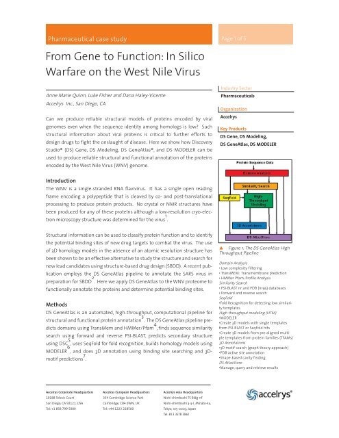
DS GeneAtlas is an automated, high-throughput, computati<strong>on</strong>al pipeline for<br />
structural and functi<strong>on</strong>al protein annotati<strong>on</strong> 3 . The DS GeneAtlas pipeline predicts<br />
domains using TransMem and HMMer/Pfam 4 , finds sequence similarity<br />
search using forward and reverse PSI-BLAST, predicts sec<strong>on</strong>dary structure<br />
using DSC 5 , uses SeqFold for fold recogniti<strong>on</strong>, builds homology models using<br />
MODELER 6 , and does 3D annotati<strong>on</strong> using binding site searching and 3Dmotif<br />
predicti<strong>on</strong>s 7 .<br />
<strong>Accelrys</strong> Corporate Headquarters<br />
10188 Telesis Court<br />
San Diego, CA 92121, USA<br />
Tel: +1 858 799 5000<br />
<strong>Accelrys</strong> European Headquarters<br />
334 Cambridge Science Park<br />
Cambridge, CB4 0WN, UK<br />
Tel: +44 1223 228500<br />
<strong>Accelrys</strong> Asia Headquarters<br />
Nishi-shimbashi TS Bldg 11F<br />
Nishi-shimbashi 3-3-1, Minato-ku,<br />
Tokyo, 105-0003, Japan<br />
Tel: 81 3 3578 3861<br />
Page 1 of 5<br />
<str<strong>on</strong>g>In</str<strong>on</strong>g>dustry Sector<br />
Pharmaceuticals<br />
Organizati<strong>on</strong><br />
<strong>Accelrys</strong><br />
Key Products<br />
DS Gene, DS Modeling,<br />
DS GeneAtlas, DS MODELER<br />
Figure 1: The DS GeneAtlas High<br />
Throughput Pipeline<br />
Domain Analysis<br />
• Low complexity Filtering<br />
TransMEM: Transmembrane predicti<strong>on</strong><br />
HMMer Pfam: Profile Analysis<br />
Similarity Search<br />
PSI-BLAST nr and PDB (nr95) databases<br />
Forward and reverse search<br />
SeqFold<br />
Fold Recogniti<strong>on</strong> for detecting low similarity<br />
templates<br />
High throughput modeling (HTM)<br />
MODELER<br />
Create 3D models with single templates<br />
from PSI-BLAST or SeqFold hits<br />
Create 3D models from pre-aligned multiple<br />
templates from protein families (TFAMs)<br />
3D Annotati<strong>on</strong>s<br />
3D motif search (graph <strong>the</strong>ory approach)<br />
PDB active site annotati<strong>on</strong><br />
Shape-based cavity finding<br />
DS AtlasStore<br />
Manage, query and retrieve results
Pharmaceutical case study c<strong>on</strong>tinued<br />
Alignments for homology modeling are selected from ei<strong>the</strong>r PSI-BLAST or<br />
SeqFold hits reaching a predetermined cutoff value. Models are scored based<br />
<strong>on</strong> <strong>the</strong> Verify/Profiles-3D and Potential Mean Force (PMF) scores. The<br />
Verify/Profiles-3D score measures <strong>the</strong> compatibility of a protein model with its<br />
sequence using a 3D profile 8 , while <strong>the</strong> PMF score uses statistical potentials of<br />
mean force to assess <strong>the</strong> accuracy of a model 9 . The final model score that is<br />
used to select <strong>the</strong> best models is used to measure <strong>the</strong> c<strong>on</strong>fidence of <strong>the</strong> structural<br />
annotati<strong>on</strong> generated from <strong>the</strong> models. This score is <strong>the</strong> normalized<br />
combinati<strong>on</strong> of <strong>the</strong> model Verify/Profiles-3D and PMF scores. Models with values<br />
higher than 0 are c<strong>on</strong>sidered to be sufficiently reliable.<br />
DS Gene software for molecular sequence analysis was used to identify <strong>the</strong><br />
signalase and dibasic viral protease cleavage sites in <strong>the</strong> WNV genome<br />
sequence. Twelve protein products derived from <strong>the</strong> polyprotein sequence of<br />
<strong>the</strong> genome were <strong>the</strong>n analyzed with DS GeneAtlas. These protein sequences<br />
include <strong>the</strong> capsid (C), membrane (M), and envelope (E) structural proteins, as<br />
well as <strong>the</strong> n<strong>on</strong>-structural proteins NS1, NS2a, NS2B, NS3, NS4A, NS4B, and NS5<br />
(Figure 2). The precapsid includes <strong>the</strong> C-terminal signal sequence for <strong>the</strong> Mprotein.<br />
PreM is <strong>the</strong> mature M protein with <strong>the</strong> signal sequence cleaved, and<br />
M is <strong>the</strong> fully processed protein.<br />
Figure 2: The <strong>West</strong> <strong>Nile</strong> Polyprotein.<br />
DS Gene was used to annotate <strong>the</strong> WNV polyprotein sequence with <strong>the</strong> co-translati<strong>on</strong>al<br />
and post-translati<strong>on</strong>al cleavage sites reported by Chambers et. al. 10 .<br />
Results and Discussi<strong>on</strong><br />
Reliable structural and functi<strong>on</strong>al annotati<strong>on</strong>s were generated for <strong>the</strong> capsid,<br />
envelope, NS1, NS3, and NS5 proteins (see Table 1). Note that despite low<br />
sequence similarity and high (worse) PSI-BLAST values, DS GeneAtlas can still<br />
predict good models (high model scores) and thus associate homologous<br />
functi<strong>on</strong> with <strong>the</strong> protein databank (PDB) template protein(s). The membrane,<br />
NS2a, NS2b, NS4a, and NS4b proteins had low similarity to PDB template<br />
proteins and bad model scores and thus are less reliable, but useful<br />
informati<strong>on</strong> was still revealed by DS GeneAtlas through o<strong>the</strong>r methods such<br />
as HMMer Pfam. Only <strong>the</strong> capsid, envelope, NS1, NS3, and NS5 proteins will be<br />
discussed below; however, more details are available up<strong>on</strong> request.<br />
Page 2 of 5
Pharmaceutical case study c<strong>on</strong>tinued<br />
Protein Descripti<strong>on</strong> of<br />
Template<br />
Functi<strong>on</strong><br />
Capsid<br />
Protein<br />
Envelope<br />
Protein<br />
Post-translati<strong>on</strong>ally<br />
modified by<br />
c-terminal cleavage<br />
Envelope protein<br />
291-791<br />
NS1 N<strong>on</strong>-structural protein<br />
1<br />
NS2a N<strong>on</strong>-structural (multipletransmembrane<br />
domain)<br />
protein 2a<br />
NS3 N<strong>on</strong>structural protease<br />
complex<br />
NS5<br />
6-269<br />
NS5<br />
370-777<br />
Uncleaved, n<strong>on</strong>structural<br />
c<strong>on</strong>served<br />
cytoplasmic protein<br />
Uncleaved, n<strong>on</strong>structural<br />
c<strong>on</strong>served<br />
cytoplasmic protein<br />
DS<br />
GeneAtlas<br />
Methods*<br />
HTM 1bvs<br />
DNA BINDING<br />
PROTEIN (RUVA)<br />
HTM and<br />
PB90<br />
HTM and<br />
PB90<br />
PDB Template Scores**<br />
1oam<br />
DENGUE 2 VIRUS<br />
ENV PROTEIN<br />
1gzy<br />
INSULIN-LIKE<br />
GROWTH FACTOR<br />
HTM 1jbi<br />
COCHLIN CHAIN<br />
A;<br />
LCCL MODULE.<br />
HTM and<br />
PB90<br />
HTM and<br />
PB90<br />
HTMM<br />
And PB90<br />
1cu1<br />
HYDROLASE<br />
1l9k<br />
DENGUE METHYL-<br />
TRANSFERASE<br />
1c2p-1gx5-[1nb4]<br />
(Template family)<br />
Viral RNA polymerase<br />
PSI-BLAST E =0.04<br />
% Identity = 25%<br />
Model Score = 0.11<br />
PSI-BLAST E = 0<br />
% Identity = 46%<br />
Model Score = 0.99<br />
PSI-BLAST E = 0.001<br />
% Identity = 40%<br />
Model Score =0 .63<br />
PSI-BLAST E = 0.45<br />
% Identity = 37%<br />
Model Score = 0.17<br />
Psi_BLAST E = 2.6e-<br />
98% Identity =<br />
19%Model Score =<br />
0.88<br />
PSI-BLAST E = 0<br />
% Identity = 63%<br />
Model Score = 1.0<br />
PSI-BLAST E = 0<br />
% Identity = 20%<br />
Model Score = 0.80<br />
Table 1: Highlights of <strong>the</strong> DS GeneAtlas results for some of <strong>the</strong> <strong>West</strong> <strong>Nile</strong> proteins.<br />
Only <strong>on</strong>e hit for each protein sequence with <strong>the</strong> best PSI-BLAST E-Value is shown.<br />
*HTM = high throughput modeling, HTMM = High throughput multiple template modeling,<br />
PB90 = PSI-BLAST against NR database (GenBank) at 90% sequence identity or less<br />
**Model score corresp<strong>on</strong>ds to linear combinati<strong>on</strong> score.<br />
The capsid protein was assigned a single transmembrane domain by<br />
TransMEM from residue positi<strong>on</strong> 44 to 66. Approximately <strong>the</strong> same regi<strong>on</strong>,<br />
positi<strong>on</strong> 44 to 58, was predicted to be helical by sec<strong>on</strong>dary structure analysis.<br />
This regi<strong>on</strong> may corresp<strong>on</strong>d to <strong>the</strong> residues that anchor to <strong>the</strong> WNV membrane.<br />
HTM indicates nucleotide binding functi<strong>on</strong>ality when evaluating <strong>the</strong><br />
PDB template functi<strong>on</strong>ality. One binding site (178 Å3) was also predicted and<br />
may corresp<strong>on</strong>d to <strong>the</strong> RNA binding site. Note that <strong>the</strong> WNV is an RNA virus,<br />
thus differences in <strong>the</strong> binding site residues may distinguish RNA binding<br />
from <strong>the</strong> template DNA binding.<br />
The best PDB template based <strong>on</strong> model score (0.99) for <strong>the</strong> E (Envelope) protein<br />
is <strong>the</strong> crystalline structure of <strong>the</strong> Dengue E protein (Figure 3). This hit was<br />
determined by a combinati<strong>on</strong> of methods in DS GeneAtlas (SeqFold, HTM, and<br />
PB90). At <strong>the</strong> sequence level, <strong>the</strong> Dengue and <strong>West</strong> <strong>Nile</strong> proteins are 46%<br />
identical and 64% similar.<br />
Page 3 of 5
Pharmaceuticals case study c<strong>on</strong>tinued<br />
This model spans <strong>the</strong> length of <strong>the</strong> <strong>West</strong> <strong>Nile</strong> protein, from positi<strong>on</strong> 3 to 400,<br />
excluding <strong>the</strong> last 101 amino acids of <strong>the</strong> C-terminus. Transmembrane analysis<br />
indicates that <strong>the</strong> C-terminal regi<strong>on</strong> anchors this protein in <strong>the</strong> viral membrane.<br />
The template structure is in complex with N-Octyl-Beta-D-Glucoside,<br />
lending <strong>the</strong> potential for homology-based docking experiments <strong>on</strong> <strong>the</strong> <strong>West</strong><br />
<strong>Nile</strong> E protein, as many of <strong>the</strong> residues in <strong>on</strong>e of <strong>the</strong> four binding pockets are<br />
c<strong>on</strong>served. Fur<strong>the</strong>r annotati<strong>on</strong> by HMMer indicates an immunoglobulin-like<br />
(Ig-like) domain from positi<strong>on</strong> 291 to 409 in <strong>the</strong> protein sequence. This Ig-like<br />
fold is also seen in <strong>the</strong> homology modeling. The active site, predicted by CSC 7 ,<br />
shows a c<strong>on</strong>served 3D motif (Thr-Asp-His) in a cavity. To better represent <strong>the</strong><br />
biological assembly of <strong>the</strong> E protein, <strong>the</strong> entire dimer must be remodeled (i.e.<br />
using MODELER).<br />
Seven homology models for NS1 with good model scores ranging from 0.2 to<br />
0.6 corresp<strong>on</strong>d to 40 residues in <strong>the</strong> C terminus from positi<strong>on</strong> 279 to 318 in <strong>the</strong><br />
primary sequence. All of <strong>the</strong> templates for this domain are in <strong>the</strong> same SCOP<br />
family (Structural Classificati<strong>on</strong> of Proteins) and indicate an insulin-like<br />
growth factor fold with a c<strong>on</strong>served disulfide b<strong>on</strong>d. The best of model with<br />
<strong>the</strong> highest model score is based <strong>on</strong> <strong>the</strong> PDB entry 1gzy (B chain), <strong>the</strong> crystalline<br />
structure for insulin-like growth factor I (Figure 4).<br />
The DS GeneAtlas pipeline initially identified a good model for <strong>the</strong> NS3 protein<br />
from <strong>the</strong> alpha chain of <strong>the</strong> PDB entry 1cu1 (A-chain), a hydrolase protein. This<br />
was remodeled in DS Modeling (DS MODELER) using both chains (A and B) of<br />
<strong>the</strong> 1cu1 template (Figure 5). The dimer template protein is from <strong>the</strong> Hepatitis<br />
C virus. The results indicate a bifuncti<strong>on</strong>al protein with both serine protease<br />
and RNA helicase properties. The active site of <strong>the</strong> protease features a c<strong>on</strong>served<br />
histidine residue. This template as well as several o<strong>the</strong>rs determined<br />
by DS GeneAtlas produced excellent model scores ranging from 0.8 to 0.9<br />
despite low sequence similarity between <strong>the</strong> NS3 target and templates. Fold<br />
recogniti<strong>on</strong> using SeqFold reports compatible sec<strong>on</strong>dary structure with good<br />
alignment and larger coverage across <strong>the</strong> length of <strong>the</strong> sequence.<br />
HMMer Pfam analysis in DS GeneAtlas identified two distinct domains for<br />
NS5-an N terminal FtsJ-like methyltransferase domain and a C-terminal RNA<br />
polymerase domain-thus indicating a bifuncti<strong>on</strong>al protein (Figure 6). These<br />
two domains cover <strong>the</strong> majority of this 905 residue protein, but a complete<br />
model was not available that covered both domains. The 119KDa structure<br />
from Dengue virus methyltransferase serves as a template to build a model of<br />
<strong>the</strong> first domain. At <strong>the</strong> sequence level, <strong>the</strong>se proteins are 63% identical and<br />
yield a model with a predictably high reliability rating. The polymerase<br />
domain was created from a multiple template model using <strong>the</strong> alignment of<br />
<strong>the</strong> template structures 1gx5, 1c2p, and 1nb4, all from Hepatitis C virus. All of<br />
<strong>the</strong> templates are part of <strong>the</strong> RNA-dependent RNA polymerase SCOP family.<br />
Page 4 of 5<br />
Figure 3: Model of <strong>the</strong> <strong>West</strong> <strong>Nile</strong><br />
envelope protein shown in blue,<br />
superimposed <strong>on</strong> Dengue virus envelope<br />
protein template shown in red.<br />
501 residues overlap for good coverage,<br />
with high sequence identity and<br />
predicted binding sites shown in<br />
orange.<br />
C<strong>on</strong>served disulfide b<strong>on</strong>d<br />
Figure 4: A model of <strong>the</strong> regi<strong>on</strong><br />
near <strong>the</strong> C terminus of <strong>the</strong> <strong>West</strong> <strong>Nile</strong><br />
NS1 protein shown in blue, based <strong>on</strong><br />
<strong>the</strong> superimposed template from PDB<br />
entry 1gzy, shown in red. Sequence<br />
identity with <strong>the</strong> template is 40%<br />
over 40 residues shown here.<br />
C<strong>on</strong>served active site histidine.<br />
Figure 5: A model of <strong>the</strong> <strong>West</strong><br />
<strong>Nile</strong> NS3 protein (blue) superimposed<br />
with <strong>the</strong> 1cu1 dimerized template<br />
protein from Hepatitis C virus (red).<br />
Similarity between <strong>the</strong> template and<br />
NS3 sequences is <strong>on</strong>ly 19%. The<br />
superimpositi<strong>on</strong> = 1.38 Å RMSD over<br />
1173 residues.
Pharmaceuticals case study c<strong>on</strong>tinued<br />
The alignment is based <strong>on</strong> structural properties and sequence c<strong>on</strong>servati<strong>on</strong> of<br />
all <strong>the</strong> templates. The percent sequence identity between WNV NS5 polymerase<br />
domain and any of <strong>the</strong> template sequences is less than 19%. The<br />
active site annotati<strong>on</strong> in <strong>the</strong> template sequences is perfectly c<strong>on</strong>served in <strong>the</strong><br />
WNV sequence, residues D536, D668, and D669 and <strong>the</strong> model score is high<br />
(see Table 1), illustrating <strong>the</strong> advantage of <strong>the</strong> multiple template approach to<br />
generate reliable homology models, especially when sequence similarity is<br />
low.<br />
A<br />
Figure 6A:<br />
A model of <strong>the</strong> N-terminal domain of <strong>the</strong><br />
<strong>West</strong> <strong>Nile</strong> NS5 protein is shown in blue,<br />
with <strong>the</strong> Dengue methyltransferase template<br />
shown in red (superimpositi<strong>on</strong> = 0<br />
.22 Å over 260 residues). The dengue protein<br />
was crystallized in <strong>the</strong> presence of Sadenosyl-l-homocysteine<br />
and sulfate i<strong>on</strong>s<br />
(green). The homocysteine active site is<br />
indicated in orange. The green atoms<br />
corresp<strong>on</strong>d to sulfate i<strong>on</strong>s.<br />
Figure 6B:<br />
A model of <strong>the</strong> C-terminal domain of<br />
<strong>the</strong> <strong>West</strong> <strong>Nile</strong> n<strong>on</strong>structural NS5 protein<br />
shown in blue, superimposed with <strong>the</strong><br />
alpha chain of <strong>the</strong> 1nb4 template, <strong>the</strong><br />
crystal structure of <strong>the</strong> Hepatitis C virus<br />
RNA polymerase in red (superimpositi<strong>on</strong><br />
= 1.06 Å RMSD over 495 residues).<br />
C<strong>on</strong>clusi<strong>on</strong>s<br />
This study shows how <strong>the</strong> DS GeneAtlas high-throughput structural annotati<strong>on</strong><br />
pipeline can help to characterize proteins of unknown functi<strong>on</strong>. 3D<br />
Models generated by <strong>the</strong> pipeline can <strong>the</strong>n be used for structure-based drug<br />
design. The predicted structure of <strong>the</strong> protein, combined with active site<br />
annotati<strong>on</strong>, can reveal <strong>the</strong> residues involved in substrate recogniti<strong>on</strong> and can<br />
provide a structural basis for exploring mutati<strong>on</strong>al effects <strong>on</strong> activity. DS<br />
GeneAtlas was used to predicte reliable 3D models for <strong>the</strong> WNV proteins NS3<br />
and NS5, despite very low sequence similarity with <strong>the</strong> available templates.<br />
Fur<strong>the</strong>r analysis is underway now using <strong>the</strong> Ludi program for rati<strong>on</strong>al drug<br />
design to generate a specific serine-protease inhibitor against <strong>the</strong> NS3 protein.<br />
A poster presentati<strong>on</strong> of <strong>the</strong> WNV analysis presented at <strong>the</strong> ISMB/ECCB 2004<br />
c<strong>on</strong>ference in Scotland (August 2004) is available up<strong>on</strong> request.<br />
From Gene to Functi<strong>on</strong>: <str<strong>on</strong>g>In</str<strong>on</strong>g> <str<strong>on</strong>g>Silico</str<strong>on</strong>g> <str<strong>on</strong>g>Warfare</str<strong>on</strong>g> <strong>on</strong> <strong>the</strong> <strong>West</strong> <strong>Nile</strong> <strong>Virus</strong><br />
B<br />
C<strong>on</strong>served catalytic triad<br />
Page 5 of 5<br />
References<br />
1. Mukhopadhyay, S. et al., Science<br />
2003, 302, 248.<br />
2. Yan, L., et.al., FEBS Letters 2003,<br />
554, 257-263.<br />
3. Kits<strong>on</strong>, D.H., et al., Briefings in<br />
Bioinformatics 2002, 3, 32-44.<br />
4. Bateman, A., et al., Nucleic Acids<br />
Research 2002, 30, 276-280.<br />
5. King, R.D. and Sternberg, M.J.E.,<br />
Protein Science 1996, 5, 2298.<br />
6.Sali, A. and Blundell T.L., J. Mol.<br />
Biol. 1993, 234, 779-815.<br />
7. Milik, M. et al., Protein<br />
Engineering 2003, 16, 543-552.<br />
8. Fischer D. and Eisenberg D.,<br />
Protein Science 1996, 5, 947-955.<br />
9. Melo, F. Sanchez, and R. Sali, A.<br />
Protein Science 2002, 11, 430-448.<br />
10. Chambers, T.J., et al., Annual<br />
Review of Microbiology 1990, 44,<br />
649-688.


