Pdf-download - AN DER GRUB Bio Research Gmbh
Pdf-download - AN DER GRUB Bio Research Gmbh
Pdf-download - AN DER GRUB Bio Research Gmbh

Create successful ePaper yourself
Turn your PDF publications into a flip-book with our unique Google optimized e-Paper software.
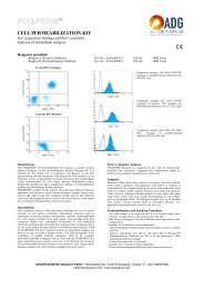
MONOCLONAL <strong>AN</strong>TIBODIES<br />
TO HUM<strong>AN</strong> LEUKOCYTE <strong>AN</strong>TIGENS<br />
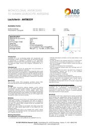
CD45 <strong>AN</strong>TIBODY<br />
Available Forms:<br />
Purified Antibody Cat. No.: GM-4061 2ml 0,2mg<br />
Fluorescein Conjugate Cat. No.: GM-4062 2ml 100 Tests<br />
Specification<br />
Specificity (Synonyms): CD45, leukocyte factor antigen<br />
Clone: VIT200<br />
Immunogl. Class: IgG2a<br />
Species: Mouse<br />
Purification: Chromatography<br />
Fluorochrome: un-coupled or PE conjugated<br />
Storage Buffer: PBS pH 7.2, 1% BSA, 0.05% NaN 3<br />
Introduction<br />
The CD45 molecule is typically expressed at high levels on all<br />
hematopoietic cells. CD45 is a major component of the glycocalix<br />
of these cells and can be expressed in different isoforms. Antibody<br />
VIT200 recognizes a pan CD45 epitope, which is expressed on all<br />
hematopoietic cells.<br />
Intended use<br />
The VIT200 antibody permits the identification and enumeration of<br />
human leukocytes using flow cytometry.<br />
Results must be put within the context of other diagnostic tests as well<br />
as the clinical history of the patient by a certified professional before<br />
final interpretation. Analyses performed with this antibody should be<br />
paralleled by positive and negative controls. If unexpected results<br />
are obtained which cannot be attributed to differences in laboratory<br />
procedures, please contact us.<br />
Specificity<br />
The CD45 mAb (clone VIT200) recognizes a pan CD45 epitope.<br />
Storage<br />
<strong>AN</strong> <strong>DER</strong> <strong>GRUB</strong> monoclonal antibody reagents contain optimal<br />
concentrations of affinity-purified antibody. For stability reasons this<br />
monoclonal antibody solution contains sodium azide. These<br />
reagents should be stored at 2-8°C (DO NOT FREEZE!) and protected<br />
from prolonged exposure to light. Stability of the reagent: Please refer<br />
to the expiry date printed onto the vial. The use of the reagent after<br />
the expiration date is not recommended.<br />
Samples<br />
<strong>Bio</strong>logical fluids (blood, bone marrow, and others) must be collected<br />
under sterile conditions. Anticoagulation with EDTA or heparin is<br />
recommended. The samples should be stored at room temperature<br />
until used. For optimal results, samples should be processed and<br />
analyzed within 24 hours.<br />
Samples with high numbers of non-viable cells might cause false<br />
results, such cases require determination of cell viability with e.g.<br />
propidium iodide.<br />
All biological samples have to be handled with caution. Always<br />
consider them as potentially infective. Use appropriate precautions<br />
such as gloves, lab-coat, etc.<br />
Staining Procedure<br />
Direct Immunofluorescence (Staining Procedure)<br />
ADG fluorochrome labeled antibodies are designed for use with<br />
either whole blood or isolated mononuclear cell (MNC) preparations.<br />
PB: Leukogate<br />
CD45 - FITC<br />
Proposed staining procedure for whole blood in short:<br />
- For each sample add 50 µl of EDTA anti-coagulated blood to<br />
a 3-5 ml tube<br />
- Add 20 µl of the appropriate <strong>AN</strong> <strong>DER</strong> <strong>GRUB</strong> monoclonal<br />
antibody conjugate<br />
- Incubate the tube for 15 minutes at 4°C or at room<br />
temperature in the dark<br />
- Add 100 µl ADG-LYSE (Cat.No. GAS-003) to each tube and<br />
incubate for 10 minutes at room temperature<br />
- Add 3-4 ml of destilled water and vortex, incubate for 5-10<br />
minutes at room temperature<br />
- Centrifuge tube for 5 minutes at 300 g<br />
- Aspirate supernatant and resuspend pellet in 0.3 ml of sheath<br />
fluid<br />
- Analyze immediately or store samples at 2-8° C in the dark<br />
and analyze within 24 hours<br />
For “No-Wash” protocol please refer to www.andergrub.com<br />
Proposed staining procedure for MNC in short:<br />
- Carefully add 20 µl antibody conjugate and 50-100 µl MNC to<br />
the bottom of a tube<br />
- Vortex at low speed for 1-2 seconds<br />
- Incubate for 15-30 minutes at 2-8°C or at room temperature<br />
- Centrifuge tubes for 5 minutes at 300 g<br />
- Remove supernatant, resuspend cells in 2-5 ml of phosphate<br />
buffered saline (PBS) and centrifuge cells again for 5 minutes<br />
at 300 g<br />
- Remove supernatant and resuspend cells in sheath fluid for<br />
immediate analysis or resuspend cells in 0.5 ml 1 %<br />
formaldehyde and store them at 2-8°C in the dark. Analyze<br />
fixed cells within 24 hours<br />
Indirect Immunofluorescence (Staining Procedure)<br />
- Mix 20 µl <strong>AN</strong> <strong>DER</strong> <strong>GRUB</strong> purified antibody with 50 µl whole<br />
blood or MNC suspension<br />
- Incubate for 15 minutes at 2-8°C<br />
- Wash cells with phosphate buffered saline (PBS)<br />
- Add to cell pellet 20 µl of affinity purified, fluorochrome<br />
labeled F(ab’) 2 anti mouse Ig antibodies<br />
- Incubate for 15 minutes at 2-8°C<br />
- Wash cells with phosphate buffered saline (PBS) or proceed as<br />
described for direct staining<br />
Sensitivity<br />
The sensitivity of VIT200 mAb is determined by staining well-<br />
defined blood samples from representative donors with serialfold<br />
mAb dilutions to obtain a titration curve that allows relating<br />
<strong>AN</strong> <strong>DER</strong> <strong>GRUB</strong> <strong>Bio</strong> <strong>Research</strong> GmbH • Gerichtsberg 28 • A-2572 Kaumberg • Austria • F: +43/1/489421450<br />
office@andergrub.com • www.andergrub.com<br />
side scatter
the mAb concentration to the percentage of stained cells and<br />
geometric MFI (mean fluorescence intensity). For this purpose, a<br />
mAb-concentration range is selected to include both the saturation<br />
point (i.e. the mAb dilution expected to bind all epitopes on the<br />
target cell) and the detection threshold (i.e. the mAb dilution<br />
expected to represent the least amount of mAb needed to detect<br />
an identical percentage of cells). In practice, 50 µl of leukocytes<br />
containing 10 7 cells/ml are stained with 20 µl mAb of various dilutions<br />
to obtain a titration curve and to identify the saturation point and<br />
detection threshold. The final concentration of the product is then<br />
adjusted to be at least 3-fold above the detection threshold. In<br />
addition and to control lot-to-lot variation, the given lot is compared<br />
and adjusted to fluorescence standards with defined intensity.<br />
Limitations of the technique<br />
Flow cytometry should be performed by professional users only.<br />
Improper alignment of the flow cytometer, inaccurate<br />
compensation of fluorescence leaking into other channels as well as<br />
incorrect positioning of regions may lead to false results.<br />
Lysis of red cells might be impossible for various reasons. In such<br />
instances it is recommended to isolate mononuclear cells (MNC) via<br />
density gradient centrifugation prior to staining.<br />
Results will be correct and reproducible as long as the procedures<br />
used respect the technical recommendations and obey good<br />
laboratory practice.<br />
The antibody is provided in a concentration that will allow to<br />
unequivocally detect specific cells. It is therefore strongly<br />
recommended to stick to the staining protocol in terms of<br />
concentration and volume regarding cells and antibody.<br />
The therapeutic use of antibodies might influence the recognition of<br />
target-antigens by this antibody. The reaction pattern of VIT200 mAb<br />
alone is not sufficient to diagnose “leukemia”. Combination with<br />
other antibodies in multi-color stainings is strongly recommended.<br />
Precautions<br />
For professional users only.<br />
This reagent contains sodium azide. To avoid the development of<br />
hazardous conditions, reagents containing azide should be diluted in<br />
running water prior to be discarded. Similar to the work with<br />
other biological products, proper handling procedures are<br />
recommended.<br />
Warranty<br />
The products sold hereunder are warranted only to conform to the<br />
quantity and contents stated on the label at the time of delivery<br />
to the customer. There are no warranties, expressed or implied,<br />
that extend beyond the description on the label of the product.<br />
ADG´s sole liability is limited to either replacement of the products<br />
or refund of the purchase price. ADG is not liable for property<br />
damage, personal injury, or economic loss caused by the<br />
product.<br />
Selected References<br />
Battifora, H. & Trowbridge, I. S. (1983) Cancer 51, 816-21.<br />
Brocklebank, A. M. & Sparrow, R. L. (2001) Cytometry 46, 254-<br />
61.<br />
Cobbold, S., Hale, G. & Waldmann, H. (1987) In Leukocyte<br />
Typing III (Oxford University Press, Oxford-New York-Tokyo) p788-<br />
803.<br />
Dalchau, R., Kirkley, J. & Fabre, J. W. (1980) Eur J Immunol 10,<br />
737-44.<br />
Jing, S., Ralph, S., Head, M. T. A., Chain, A. & Trowbridge, I.<br />
(1987) In Leukocyte Typing III (Oxford University Press, Oxford-<br />
New York-Tokyo) p899-905.<br />
Nicholson, J. K., Hubbard, M. & Jones, B. M. (1996) Cytometry<br />
26, 16-21.<br />
Omary, M. B., Trowbridge, I. S. & Battifora, H. A. (1980) J Exp<br />
Med 152, 842-52.<br />
Schraven, B., Roux, M., Hutmacher, B. & Meuer, S. C. (1989) Eur<br />
J Immunol 19, 397-403.<br />
Sugita, K., Majdic, O., Stockinger, H., Holter, W., Burger, R. &<br />
Knapp, W. (1987) Transplantation 43, 570-4.<br />
Sun, T., Sangaline, R., Ryder, J., Gibbens, K., Rollo, C., Stewart,<br />
S. & Rajagopalan, C. (1997) Am J Clin Pathol 108, 152-7.<br />
Thomas, M. L. (1989) Annu Rev Immunol 7, 339-69.<br />
For In Vitro Diagnostic Use For professional use only.<br />
Explanation of symbols<br />
REF Catalog number<br />
IVD In vitro diagnostic medical device<br />
� Consult instructions for use<br />
2˚-8˚ Temperature limitation<br />
� Keep away from sunlight<br />
LOT Batch code<br />
� Use by<br />
� Manufacturer<br />
Contains sufficient for (N) Tests<br />
<strong>AN</strong> <strong>DER</strong> <strong>GRUB</strong> <strong>Bio</strong> <strong>Research</strong> GmbH • Gerichtsberg 28 • A-2572 Kaumberg • Austria • F: +43/1/489421450<br />
office@andergrub.com • www.andergrub.com