Hematuria - Diagnostic Centers of America
Hematuria - Diagnostic Centers of America
Hematuria - Diagnostic Centers of America

You also want an ePaper? Increase the reach of your titles
YUMPU automatically turns print PDFs into web optimized ePapers that Google loves.
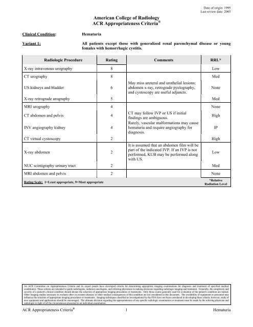
Clinical Condition: <strong>Hematuria</strong><br />
<strong>America</strong>n College <strong>of</strong> Radiology<br />
ACR Appropriateness Criteria ®<br />
Date <strong>of</strong> origin: 1995<br />
Last review date: 2005<br />
Variant 1: All patients except those with generalized renal parenchymal disease or young<br />
females with hemorrhagic cystitis.<br />
Radiologic Procedure Rating Comments RRL*<br />
X-ray intravenous urography 8 Low<br />
CT urography 8 Med<br />
US kidneys and bladder 6<br />
May miss ureteral and urothelial lesions;<br />
abdomen x-ray, retrograde pyelography,<br />
and cystoscopy are useful adjuncts.<br />
X-ray retrograde urography 5 Med<br />
MRI urography 4 None<br />
CT abdomen and pelvis 4<br />
INV angiography kidney 4<br />
CT may follow IVP or US if initial<br />
findings are ambiguous.<br />
Rarely, vascular malformations may cause<br />
hematuria and require angiography for<br />
diagnosis.<br />
CT virtual cystoscopy 2 High<br />
X-ray abdomen 2<br />
It is assumed that an abdomen film will be<br />
part <strong>of</strong> the indicated IVP. If an IVP is not<br />
performed, KUB may be performed along<br />
with US.<br />
NUC scintigraphy urinary tract 2 Med<br />
MRI abdomen and pelvis 2 None<br />
Rating Scale: 1=Least appropriate, 9=Most appropriate<br />
An ACR Committee on Appropriateness Criteria and its expert panels have developed criteria for determining appropriate imaging examinations for diagnosis and treatment <strong>of</strong> specified medical<br />
condition(s). These criteria are intended to guide radiologists, radiation oncologists, and referring physicians in making decisions regarding radiologic imaging and treatment. Generally, the complexity and<br />
severity <strong>of</strong> a patient's clinical condition should dictate the selection <strong>of</strong> appropriate imaging procedures or treatments. Only those exams generally used for evaluation <strong>of</strong> the patient's condition are ranked.<br />
Other imaging studies necessary to evaluate other co-existent diseases or other medical consequences <strong>of</strong> this condition are not considered in this document. The availability <strong>of</strong> equipment or personnel may<br />
influence the selection <strong>of</strong> appropriate imaging procedures or treatments. Imaging techniques classified as investigational by the FDA have not been considered in developing these criteria; however, study <strong>of</strong><br />
new equipment and applications should be encouraged. The ultimate decision regarding the appropriateness <strong>of</strong> any specific radiologic examination or treatment must be made by the referring physician and<br />
radiologist in light <strong>of</strong> all the circumstances presented in an individual examination.<br />
ACR Appropriateness Criteria ® 1 <strong>Hematuria</strong><br />
None<br />
High<br />
IP<br />
Low<br />
*Relative<br />
Radiation Level
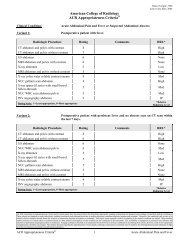
Clinical Condition: <strong>Hematuria</strong><br />
Variant 2: Due to generalized renal parenchymal disease.<br />
Radiologic Procedure Rating Comments RRL*<br />
US kidneys and bladder 8<br />
X-ray chest 6<br />
For renal volume and morphology and as<br />
localizer for biopsy.<br />
For cardiopulmonary and pleural<br />
manifestations <strong>of</strong> renal diseases.<br />
X-ray retrograde urography 3 Med<br />
INV angiography kidney 2 IP<br />
CT virtual cystoscopy 2 High<br />
CT urography 2 Med<br />
NUC scintigraphy urinary tract 2 Med<br />
MRI urography 2 None<br />
MRI abdomen and pelvis 2 None<br />
CT abdomen and pelvis 2 Routine. High<br />
X-ray abdomen 1 Low<br />
X-ray intravenous urography 1 Low<br />
Rating Scale: 1=Least appropriate, 9=Most appropriate<br />
An ACR Committee on Appropriateness Criteria and its expert panels have developed criteria for determining appropriate imaging examinations for diagnosis and treatment <strong>of</strong> specified medical<br />
condition(s). These criteria are intended to guide radiologists, radiation oncologists, and referring physicians in making decisions regarding radiologic imaging and treatment. Generally, the complexity and<br />
severity <strong>of</strong> a patient's clinical condition should dictate the selection <strong>of</strong> appropriate imaging procedures or treatments. Only those exams generally used for evaluation <strong>of</strong> the patient's condition are ranked.<br />
Other imaging studies necessary to evaluate other co-existent diseases or other medical consequences <strong>of</strong> this condition are not considered in this document. The availability <strong>of</strong> equipment or personnel may<br />
influence the selection <strong>of</strong> appropriate imaging procedures or treatments. Imaging techniques classified as investigational by the FDA have not been considered in developing these criteria; however, study <strong>of</strong><br />
new equipment and applications should be encouraged. The ultimate decision regarding the appropriateness <strong>of</strong> any specific radiologic examination or treatment must be made by the referring physician and<br />
radiologist in light <strong>of</strong> all the circumstances presented in an individual examination.<br />
ACR Appropriateness Criteria ® 2 <strong>Hematuria</strong><br />
None<br />
Min<br />
*Relative<br />
Radiation Level<br />
Variant 3: Hemorrhagic cystitis in females less than 40 years old (hematuria completely clears<br />
with therapy).<br />
Radiologic Procedure Rating Comments RRL*<br />
MRI abdomen and pelvis 2 None<br />
INV angiography kidney 2 IP<br />
CT virtual cystoscopy 2 High<br />
MRI urography 2 None<br />
NUC scintigraphy urinary tract 2 Med<br />
CT urography 2 Med<br />
X-ray retrograde urography 2 Med<br />
CT abdomen and pelvis 2<br />
This and other imaging are rarely needed<br />
for diagnosis. Routine.<br />
X-ray abdomen 1 Low<br />
US kidneys and bladder 1 None<br />
X-ray intravenous urography 1 Low<br />
Rating Scale: 1=Least appropriate, 9=Most appropriate<br />
High<br />
*Relative<br />
Radiation Level
Expert Panel on Urologic Imaging:<br />
Peter L. Choyke, MD 1 ; Edward I. Bluth, MD 2 ;<br />
William H. Bush, Jr, MD 3 ; David D. Casalino, MD 4 ;<br />
Isaac R. Francis, MD 5 ; S. Zafar H. Jafri, MD 6 ;<br />
Akira Kawashima, MD, PhD 7 ; Alan Kronthal, MD 8 ;<br />
Robert A. Older, MD 9 ; Nicholas Papanicolaou, MD 10 ;<br />
Parvati Ramchandani, MD 11 ; Arthur T. Rosenfield, MD 12 ;<br />
Carl M. Sandler, MD 13 ; Arthur J. Segal, MD 14 ;<br />
Clare Tempany, MD 15 ; Martin I. Resnick, MD. 16<br />
Summary <strong>of</strong> Literature Review<br />
<strong>Hematuria</strong> is one <strong>of</strong> the most common presentations <strong>of</strong><br />
patients with urinary tract diseases and <strong>of</strong> patients<br />
referred for urinary imaging. This review summarizes<br />
practice for the radiologic approach to such patients. It is<br />
limited to adults and does not refer to patients, whose<br />
hematuria coexists with other clinical situations reviewed<br />
in other ACR Appropriateness Criteria ® topics, including<br />
acute trauma, infection, renal failure, symptoms <strong>of</strong> acute<br />
stone disease, known renal masses, and prostatism. It is<br />
also limited to initial tests; follow-up <strong>of</strong> normal or<br />
abnormal first tests is beyond its scope.<br />
The initial decision to be made is whether all patients<br />
with any degree <strong>of</strong> hematuria need imaging evaluation.<br />
Patients whose urinary tracts have no detectable<br />
abnormalities normally release small amounts <strong>of</strong> blood<br />
into the urine, so that several red cells per high-power<br />
field may be seen upon microscopic examination <strong>of</strong> the<br />
spun sediment. This fact, together with the low<br />
prevalence <strong>of</strong> clinically detectable disease in some groups<br />
<strong>of</strong> patients with asymptomatic microscopic hematuria, has<br />
led some investigators to suggest that minimal<br />
microhematuria in an asymptomatic young adult needs no<br />
evaluation [1].<br />
Unfortunately, no threshold number <strong>of</strong> red blood cells per<br />
high-power field has been found that separates patients<br />
with clinically important disease from those with no<br />
detectable urinary tract abnormalities. The distinction<br />
between gross and microscopic hematuria is not a useful<br />
guideline to distinguish between patients who need<br />
evaluation and those who do not, and the ranges <strong>of</strong> red<br />
1 Review Author and Panel Chair, National Institutes <strong>of</strong> Health, Bethesda, Md;<br />
2 Ochsner Foundation Hospital, New Orleans, La; 3 University <strong>of</strong> Washington<br />
Medical Center, Seattle, Wash; 4 Northwestern University, Chicago, Ill; 5 University<br />
<strong>of</strong> Michigan, Ann Arbor, Mich; 6 William Beaumont Hospital, Royal Oak, Mich;<br />
7 Mayo Clinic, Rochester, Minn; 8 W B & A Imaging, Rockville Md; 9 University <strong>of</strong><br />
Virginia Medical Center, Charlottesville, Va; 10 Hospital <strong>of</strong> University <strong>of</strong><br />
Pennsylvania, Philadelphia, Pa; 11 Hospital <strong>of</strong> University <strong>of</strong> Pennsylvania,<br />
Philadelphia, Pa; 12 Yale-New Haven Hospital, New Haven, Conn; 13 UT MD<br />
Anderson Cancer Center, Houston, Texas; 14 Rochester General Hospital,<br />
Rochester, NY; 15 Brigham & Women’s Hospital, Boston, Mass; 16 University<br />
Hospital <strong>of</strong> Cleveland, Cleveland, Ohio, <strong>America</strong>n Urological Association.<br />
Reprint requests to: Department <strong>of</strong> Quality & Safety, <strong>America</strong>n College <strong>of</strong><br />
Radiology, 1891 Preston White Drive, Reston, VA 20191-4397.<br />
HEMATURIA<br />
cells per high-power field in patients with “normal”<br />
hematuria and those in whom microhematuria indicates<br />
important or even life-threatening disease have sufficient<br />
overlap that many authorities claim that any amount <strong>of</strong><br />
hematuria, no matter how slight, should be considered an<br />
indication <strong>of</strong> urinary tract malignancy until proven<br />
otherwise [2,3], and that all cases <strong>of</strong> hematuria therefore<br />
need complete work-up.<br />
There may, however, be specific circumstances in which<br />
complete radiologic work-up is not necessary [4]. Young<br />
women with a clinical picture <strong>of</strong> simple cystitis and<br />
whose hematuria completely and permanently resolves<br />
after successful therapy, can probably be spared any<br />
imaging [5-7]. Patients who have clear-cut evidence <strong>of</strong><br />
glomerulopathy also constitute a special group; although<br />
they should probably have chest radiography [8] to search<br />
for any <strong>of</strong> the numerous manifestations <strong>of</strong><br />
glomerulonephritis (including cardiac enlargement,<br />
pleural and pericardial effusions, pulmonary congestion<br />
and edema, and pulmonary bleeding) and ultrasound (US)<br />
(to display the site and number <strong>of</strong> kidneys prior to biopsy<br />
and to screen for renal morphologic abnormalities that<br />
may coexist by chance in a patient with<br />
glomerulonephritis), they probably do not need extensive<br />
work-up to exclude a surgical lesion that may be bleeding<br />
[7,9-11]. However, the decision to pursue this course<br />
requires firm demonstration that the glomerular<br />
abnormality is responsible for the bleeding; such evidence<br />
includes heavy proteinuria (sufficient to indicate that<br />
plasma proteins, rather than proteins in red cells, account<br />
for the protein in the urine), red cell casts, or (in<br />
institutions that have reliable traditions <strong>of</strong> identifying<br />
such abnormalities) evidence <strong>of</strong> severe red cell<br />
dysmorphism. Patients on anticoagulants have a<br />
sufficiently high prevalence <strong>of</strong> important disease that<br />
work-up cannot be forgone [12].<br />
All other adult patients⎯especially those specifically<br />
referred for evaluation <strong>of</strong> hematuria⎯require imaging<br />
evaluation [6,7,13]. This evaluation will almost always be<br />
accompanied by cystoscopy, since many bleeding urinary<br />
tract lesions arise in the lower tract and no imaging<br />
procedure is highly sensitive in diagnosing most <strong>of</strong> them.<br />
It goes without saying that a complete history, physical<br />
examination, urine analysis, and appropriate serologic<br />
tests should precede or accompany the imaging<br />
examinations. At the time <strong>of</strong> cystoscopy, bilateral<br />
retrograde pyelography is <strong>of</strong>ten employed to evaluate the<br />
upper tracts for pathology [4].<br />
There is not universal agreement about the first imaging<br />
examination to choose. Traditionally, excretory<br />
urography (IVP) was standard [4,6], but the establishment<br />
<strong>of</strong> this practice preceded the development <strong>of</strong> high-quality<br />
An ACR Committee on Appropriateness Criteria and its expert panels have developed criteria for determining appropriate imaging examinations for diagnosis and treatment <strong>of</strong> specified medical<br />
condition(s). These criteria are intended to guide radiologists, radiation oncologists, and referring physicians in making decisions regarding radiologic imaging and treatment. Generally, the complexity and<br />
severity <strong>of</strong> a patient's clinical condition should dictate the selection <strong>of</strong> appropriate imaging procedures or treatments. Only those exams generally used for evaluation <strong>of</strong> the patient's condition are ranked.<br />
Other imaging studies necessary to evaluate other co-existent diseases or other medical consequences <strong>of</strong> this condition are not considered in this document. The availability <strong>of</strong> equipment or personnel may<br />
influence the selection <strong>of</strong> appropriate imaging procedures or treatments. Imaging techniques classified as investigational by the FDA have not been considered in developing these criteria; however, study <strong>of</strong><br />
new equipment and applications should be encouraged. The ultimate decision regarding the appropriateness <strong>of</strong> any specific radiologic examination or treatment must be made by the referring physician and<br />
radiologist in light <strong>of</strong> all the circumstances presented in an individual examination.<br />
ACR Appropriateness Criteria ® 3 <strong>Hematuria</strong>
US [14], computed tomography (CT), and magnetic<br />
resonance imaging (MRI). Subsequently, real-time US<br />
was investigated and found to be useful in the search for<br />
bleeding urinary tract lesions. Very recently, a<br />
combination <strong>of</strong> urinary tract CT with various ways <strong>of</strong><br />
obtaining IVP-like images <strong>of</strong> the collecting systems,<br />
ureters, and bladder has been proposed, as have similar<br />
formats <strong>of</strong> MRI examinations (CT urography and MR<br />
urography). (Urinary tract scintigraphy [15] possesses<br />
insufficient spatial resolution to screen for any but large<br />
intrarenal or obstructing lesions.)<br />
There is some literature dealing with the choice between<br />
US and excretory urography as the initial imaging study<br />
for patients with hematuria [14,16,17]. With respect to the<br />
wide range <strong>of</strong> abnormalities [3,17,19] that may be<br />
encountered in such patients (including urinary tract<br />
neoplasms <strong>of</strong> all sorts, stone disease, inflammatory<br />
processes, congenital abnormalities, vascular lesions, and<br />
obstruction from a wide variety <strong>of</strong> lesions), both exams<br />
are felt to have moderately high sensitivity. Precise<br />
comparisons <strong>of</strong> the two are lacking for several reasons:<br />
false-negative rates have not been evaluated in large<br />
numbers <strong>of</strong> patients due to the cost and invasiveness <strong>of</strong><br />
the follow-up procedures that would be necessary;<br />
sensitivities need to be individually evaluated for each <strong>of</strong><br />
the many kinds <strong>of</strong> lesions, so that a careful comparative<br />
study would require thousands <strong>of</strong> patients for appropriate<br />
statistical power; and there has been little careful<br />
definition <strong>of</strong> the patient groups in whom the two<br />
modalities have been compared. Nevertheless, it appears<br />
that there are only slight differences between the two<br />
modalities with regard to the rate <strong>of</strong> diagnosing clinically<br />
important lesions [20].<br />
US and urography tend to miss different sorts <strong>of</strong> lesions.<br />
US is not likely to detect nonobstructing ureteral stones or<br />
small urothelial abnormalities, and urography with<br />
nephrotomography may miss small exophytic anterior and<br />
posterior renal masses and small bladder lesions [21,22].<br />
The choice <strong>of</strong> exam may be affected by clinical<br />
circumstances (a positive urinary cytologic analysis may<br />
make urography crucial, whereas serious risk factors for<br />
contrast reactions may make US more appropriate). When<br />
US is negative and the source <strong>of</strong> hematuria remains<br />
obscure, urography should be added; if urography is<br />
negative, CT (or US) may be ordered [6,22,23]. When US<br />
is used as the primary screening modality, the yield from<br />
imaging may be increased by adding a plain film <strong>of</strong> the<br />
abdomen.<br />
CT <strong>of</strong> the entire urinary tract can be augmented by images<br />
<strong>of</strong> the contrast-opacified collecting systems, ureters and<br />
bladder (24); the combined exam is known as CT<br />
urography. The IVP-like portions <strong>of</strong> the exam may be<br />
obtained by exposing film (or direct digital) images when<br />
contrast administered for the CT has opacified the hollow<br />
urinary organs. Images may alternatively be produced by<br />
reformatting delayed CT images to show this anatomy.<br />
Presumably, the pyelogram portion <strong>of</strong> this exam could be<br />
comparable to a standard IVP exam, and the CT should<br />
be more sensitive and specific (both statistically and<br />
pathologically) than US or nephrotomography with<br />
regard to focal renal parenchymal abnormalities. For<br />
these reasons, a distinction should be made between<br />
routine CT <strong>of</strong> the abdomen and pelvis that may not be<br />
optimized for the urinary tract and a dedicated CT<br />
urogram that is tailored to evaluate the urinary tract for<br />
sources <strong>of</strong> hematuria. The latter study typically employs<br />
oral water instead <strong>of</strong> oral positive contrast media. A<br />
noncontrast CT <strong>of</strong> the kidneys is obtained to evaluate<br />
renal calculi. This is followed by the injection <strong>of</strong><br />
iodinated contrast media with the acquisition <strong>of</strong> a highresolution<br />
(1-2 mm thick sections) nephrographic phase<br />
and high-resolution delayed (5-10 minutes) phase. The<br />
latter can be reconstructed to evaluate the urinary tract<br />
and bladder. Some investigators employ a hybrid <strong>of</strong> CT<br />
urography and IVP-like delayed images to form one<br />
complete study, which is also known as CT urography.<br />
CT urography, taken as a group, has shown equal or<br />
superior sensitivity to IVP for causes <strong>of</strong> hematuria<br />
[25,26].<br />
MR urography currently serves as an alternative imaging<br />
technique for children and pregnant women and for<br />
patients with a contraindication to iodinated contrast<br />
media [27]. It has the potential to be useful in the search<br />
for important abnormalities that cause hematuria. Initial<br />
work demonstrating the feasibility <strong>of</strong> its performance has<br />
been published. But the examination has not been adopted<br />
in clinical practice, is expensive, and has not been<br />
evaluated for efficacy, so it cannot be recommended as an<br />
initial examination.<br />
Several authors have suggested that virtual cystoscopy,<br />
the acquisition <strong>of</strong> high-resolution CT images<br />
reconstructed to allow virtual “fly-throughs” <strong>of</strong> bladder,<br />
be used to evaluate the bladder for causes <strong>of</strong> hematuria<br />
[28]. Virtual cystoscopy is inaccurate for small lesions<br />
and lesions located near the ureteric orifices. The urethra<br />
cannot be evaluated. Thus, while promising, virtual<br />
cystoscopy cannot replace actual cystoscopy.<br />
In summary, most adults with hematuria <strong>of</strong> any degree<br />
require urinary tract imaging. Glomerulopathies may be<br />
appropriately investigated with renal US and chest<br />
radiography; most other patients require urography, CT<br />
urography, or US and a few carefully chosen patients may<br />
need no imaging at all.<br />
References<br />
1. Froom P, Ribak J, Benbassat, J, et al. Significance <strong>of</strong> hematuria in<br />
young adult men. Br Med 1984; 288(6410):20-22.<br />
An ACR Committee on Appropriateness Criteria and its expert panels have developed criteria for determining appropriate imaging examinations for diagnosis and treatment <strong>of</strong> specified medical<br />
condition(s). These criteria are intended to guide radiologists, radiation oncologists, and referring physicians in making decisions regarding radiologic imaging and treatment. Generally, the complexity and<br />
severity <strong>of</strong> a patient's clinical condition should dictate the selection <strong>of</strong> appropriate imaging procedures or treatments. Only those exams generally used for evaluation <strong>of</strong> the patient's condition are ranked.<br />
Other imaging studies necessary to evaluate other co-existent diseases or other medical consequences <strong>of</strong> this condition are not considered in this document. The availability <strong>of</strong> equipment or personnel may<br />
influence the selection <strong>of</strong> appropriate imaging procedures or treatments. Imaging techniques classified as investigational by the FDA have not been considered in developing these criteria; however, study <strong>of</strong><br />
new equipment and applications should be encouraged. The ultimate decision regarding the appropriateness <strong>of</strong> any specific radiologic examination or treatment must be made by the referring physician and<br />
radiologist in light <strong>of</strong> all the circumstances presented in an individual examination.<br />
ACR Appropriateness Criteria ® 4 <strong>Hematuria</strong>
2. Lowe FG, Brendler CB. Evaluation <strong>of</strong> the urologic patient. In:<br />
Walsh PC, et al, eds. Campbell’s Urology. Philadelphia, Pa: WB<br />
Saunders; 1992:307-317.<br />
3. Messing EM, Young TB, Hunt VB, et al. The significance <strong>of</strong><br />
asymptomatic microhematuria in men 50 or more years old. J Urol<br />
1987; 137(5):919-922.<br />
4. Grossfeld, GD, Wolf, JS Jr., Litwin MS, et al. Asymptomatic<br />
microscopic hematuria in adults: summary <strong>of</strong> the AUA best<br />
practice policy recommendations. Am Fam Physician 2001;<br />
63(6):1145-1154.<br />
5. Abuelo JG. The diagnosis <strong>of</strong> hematuria. Arch Int Med 1983;<br />
143(5):967-970.<br />
6. Benson GS, Brewer ED. <strong>Hematuria</strong>: algorithms for diagnosis.<br />
JAMA 1981; 246(9):993-995.<br />
7. Copley JB. Isolated asymptomatic hematuria in the adult. Am J<br />
Med Sci 1981; 291(2):101-111.<br />
8. Pulmonary hypertension and edema. In: Fraser RG, et al, eds.<br />
Diseases <strong>of</strong> the Chest. Philadelphia, Pa: WB Saunders; 1990:1823-<br />
1968.<br />
9. Abuelo JG. Evaluation <strong>of</strong> hematuria. Urology 1983; 21(3): 215-<br />
225.<br />
10. Fairley K. Urinalysis. In: Schrier RW, Gottschalk CW, eds.<br />
Diseases <strong>of</strong> the kidney. 4th edition. Boston, Mass: Little Brown;<br />
1988:359-383.<br />
11. Sutton JM. Evaluation <strong>of</strong> hematuria in adults. JAMA 1990;<br />
263(18):2475-2480.<br />
12. Avidor Y, Nadu A, Matzkin H. Clinical significance <strong>of</strong> gross<br />
hematuria and its evaluation in patients receiving anticoagulant and<br />
aspirin treatment. Urology 2000; 55(1):22-24.<br />
13. Golin AL, Howard RS. Asymptomatic microscopic hematuria. J<br />
Urol 1980; 124(3):389-391.<br />
14. Datta SN, Allen GM, Evans R, et al. Urinary tract ultrasonography<br />
in the evaluation <strong>of</strong> hematuria—a report <strong>of</strong> over 1,000 cases. Ann R<br />
Coll Surg 2002; 84(3): 203-205.<br />
15. Chisholm RA. The investigation <strong>of</strong> painless hematuria – a<br />
comparison <strong>of</strong> intravenous urography and DMSA scintingraphy.<br />
Clin Radiol 1988; 39(5):494-495.<br />
16. Corwin HL, Silverstein MD. The diagnosis <strong>of</strong> neoplasia in patients<br />
with microscopic hematuria. J Urol 1988; 139(5):1002-1006.<br />
17. Murakami S, Igarashi T, Shigeru H, et al. Strategies for<br />
asymptomatic microscopic hematuria: a prosepective study <strong>of</strong><br />
1,034 patients. J Urol 1990; 144(1):99-101.<br />
18. Mariani AJ, Mariani MC, Macchioni C, et al. The significance <strong>of</strong><br />
adult hematuria: 1,000 hematuria evaluations including a riskbenefit<br />
and cost-effectiveness analysis. J Urol 1989; 141(2): 350-<br />
355.<br />
19. Mohr DN, Offord KP, Owen RA, et al. Asymptomatic<br />
microhematuria and urologic disease. A population-based study.<br />
JAMA 1986; 256(2):224-229.<br />
20. Aslaksen A, Gadeholt G, Gothlin JH, et al. US vs. IVU in the<br />
evaluation <strong>of</strong> patients with microscopic hematuria. Br J Urol 1990;<br />
66(2):144-147.<br />
21. Amendola MA, Bree RL, Pollack HM, et al. Small renal cell<br />
carcinomas; resolving a diagnostic dilemma. Radiology 1988;<br />
166(3):637-641.<br />
22. Glen DA, Gilbert FJ, Bayliss AP, et al. Renal carcinoma missed by<br />
urography. Br J Urol 1989; 63(5):457-459.<br />
23. Barkin M, Lopatin W, Herschorn S, Comisarow R. Unexplained<br />
hematuria. Can J Surg 1983; 26(6):501-503.<br />
24. McNicholas MM, Raptopoulos VD, Schwartz RK, et al. Excretory<br />
phase CT urography for opacification <strong>of</strong> the urinary collecting<br />
system. AJR 1998; 170(5): 1261-1267.<br />
25. J<strong>of</strong>fe SA, Servaes S, Okon S, Horowitz M. Multi-detector row CT<br />
urography in the evaluation <strong>of</strong> hematuria. Radiographics 2003;<br />
23(6):1441-1455; discussion 1455-1456. Review.<br />
26. McTavish JD, Jinzaki M, Zou KH, et al. Multi-detector row CT<br />
urography: comparison <strong>of</strong> strategies for depicting the normal<br />
urinary collecting system. Radiology 2002; 225(3):783-790.<br />
27. Kawashima A, Glockner JF, King BF Jr. CT Urography and MR<br />
Urography. Radiol Clin North Am 2003; 41(12):945-961.<br />
28. Nambirajan T, Sohaib SA, Muller-Pollard C, et al. Virtual<br />
cystoscopy from computed tomography: a pilot study. BJU Int<br />
2004; 94(6):828-831.<br />
An ACR Committee on Appropriateness Criteria and its expert panels have developed criteria for determining appropriate imaging examinations for diagnosis and treatment <strong>of</strong> specified medical<br />
condition(s). These criteria are intended to guide radiologists, radiation oncologists, and referring physicians in making decisions regarding radiologic imaging and treatment. Generally, the complexity and<br />
severity <strong>of</strong> a patient's clinical condition should dictate the selection <strong>of</strong> appropriate imaging procedures or treatments. Only those exams generally used for evaluation <strong>of</strong> the patient's condition are ranked.<br />
Other imaging studies necessary to evaluate other co-existent diseases or other medical consequences <strong>of</strong> this condition are not considered in this document. The availability <strong>of</strong> equipment or personnel may<br />
influence the selection <strong>of</strong> appropriate imaging procedures or treatments. Imaging techniques classified as investigational by the FDA have not been considered in developing these criteria; however, study <strong>of</strong><br />
new equipment and applications should be encouraged. The ultimate decision regarding the appropriateness <strong>of</strong> any specific radiologic examination or treatment must be made by the referring physician and<br />
radiologist in light <strong>of</strong> all the circumstances presented in an individual examination.<br />
ACR Appropriateness Criteria ® 5 <strong>Hematuria</strong>