Superior Semicircular Canal Dehiscence - Stanford Hospital & Clinics
Superior Semicircular Canal Dehiscence - Stanford Hospital & Clinics
Superior Semicircular Canal Dehiscence - Stanford Hospital & Clinics

Create successful ePaper yourself
Turn your PDF publications into a flip-book with our unique Google optimized e-Paper software.
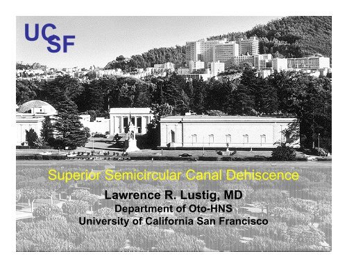
UC SF<br />
<strong>Superior</strong> <strong>Semicircular</strong> <strong>Canal</strong> <strong>Dehiscence</strong><br />
Lawrence R. Lustig, MD<br />
Department of Oto-HNS<br />
University of California San Francisco
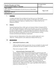
54 y.o. man with sound-evoked vertigo and<br />
oscillopsia<br />
• History:<br />
- Sensation of motion (vertigo) when he heard loud noises in his right<br />
ear<br />
- “Fluttering vision” (oscillopsia) when he made whistling, trilling, or<br />
humming noises<br />
- No past history of otologic disease<br />
• Exam:<br />
- Negative for: spontaneous, gaze-evoked, or positional nystagmus;<br />
head-shaking induced nystagmus; head thrust sign<br />
- Normal otoscopic exam<br />
- Weber (512 Hz) lateralized to right<br />
- Normal air and bone conduction thresholds<br />
- Eye movements evoked by sound in right ear
54 year old with sound-induced vertigo
Brödel M: Unpublished drawing of the anatomy of the human ear
Overview<br />
• Eye movements<br />
• Mechanism<br />
• Audiometric Diagnosis<br />
• Etiology<br />
• CT imaging<br />
• Surgical correction<br />
• Apparent conductive hearing loss
Overview<br />
• Eye movements<br />
• Mechanism<br />
• Audiometric Diagnosis<br />
• Etiology<br />
• CT imaging<br />
• Surgical correction<br />
• Apparent conductive hearing loss
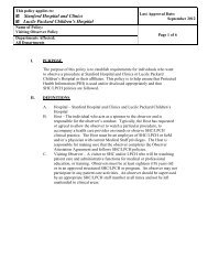
Eye movements evoked by stimulation of<br />
individual semicircular canals
Hennebert’s Sign in a left ear with SSCD
Overview<br />
• Eye movements<br />
• Mechanism<br />
• Audiometric Diagnosis<br />
• Etiology<br />
• CT imaging<br />
• Surgical correction<br />
• Apparent conductive hearing loss
Minor et al. Arch Otolaryngol Head Neck Surg 1998; 124:249-258
Third mobile window
Physiology of a 3 rd Mobile Window<br />
Merchant et al, 2009
Overview<br />
• Eye movements<br />
• Mechanism<br />
• Audiometric Diagnosis<br />
• Etiology<br />
• CT imaging<br />
• Surgical correction<br />
• Apparent conductive hearing loss
Cervical VEMP Responses<br />
• Example of normal<br />
VEMP from click<br />
stimulation of right<br />
ear.<br />
• The early 2 waves<br />
(p13-n23<br />
components) seem<br />
to be of vestibular<br />
origin.<br />
click<br />
R SCM<br />
50 μV<br />
L SCM<br />
Colebatch & Halmagyi Neurology 42:1635-1636
Lowered cVEMP Threshold in SCD<br />
41-year-old man with left SCD syndrome<br />
Streubel et al. Acta Otolaryngol Suppl 2001; 545:41-495
VEMP Responses in SCD Syndrome<br />
• 25 patients (diagnosis = 3-D eye recordings, CT)<br />
• VEMP threshold in dehiscent ears = 75 +/- 7 dB*<br />
• VEMP threshold for normal controls = 98 +/- 4<br />
dB*<br />
(*p < 0.001)<br />
• VEMP from contra ear did not differ from normal<br />
controls<br />
Streubel et al, Acta Otolaryngol 2001
ECoG = Alternate Diagnostic Modality<br />
• Increased SP/AP<br />
ratio (>0.4)<br />
• Seen in 14/15<br />
cases of SSCD<br />
• Reverses with<br />
surgical correction<br />
Arts et al, O & N, 2008
Symptoms in 53 patients with SSCD<br />
Sound-induced vertigo/oscillopsia 44 (83%)<br />
Pressure-induced vertigo/oscillopsia 39 (74%)<br />
Conductive hyperacusis 27 (51%)<br />
9 patients had undergone previous middle ear<br />
exploration for possible perilymphatic fistula<br />
Minor et al, Otol Neurotol 2003
Additional auditory Sx:<br />
• autophony (40%)<br />
• hyperacusis to bodily sounds (65%)<br />
• hearing loss (40%)<br />
• aural pressure (45%<br />
• tinnitus (35%)<br />
Yuen et al, O & N, 2009
Signs in 53 patients with SSCD<br />
• Sound-induced eye movements 38 (72%)<br />
• Valsalva-induced eye movements 37 (70%)<br />
• EAC pressure-induced eye movements 23 (43%)<br />
• Sound-induced head tilt 8 (15%)<br />
Minor et al, Otol Neurotol 2003
Overview<br />
• Eye movements<br />
• Mechanism<br />
• Audiometric Diagnosis<br />
• Etiology<br />
• CT imaging<br />
• Surgical correction<br />
• Apparent conductive hearing loss
Temporal Bone Study<br />
Carey JC et al, Arch Otolaryngol, 2001<br />
• 1000 bones screened for MF dehiscences<br />
from the JHU temporal bone archives<br />
• 8 “thinned” (
Range of bone over SC
Middle<br />
Fossa<br />
<strong>Dehiscence</strong><br />
sps
Bilateral SC dehiscences
Is there a congenital basis?<br />
24 wks gest. 2 mths<br />
4 mths<br />
10 mths<br />
Courtesy of John Cary, Johns Hopkins
• Unknown<br />
Genetics<br />
• 1 patient also with simultaneous COCH<br />
mutation (DFNA9)<br />
Hildebrand et al Am J Med Genet, 2008
A good CT<br />
1-mm slice thickness<br />
Coronal reconstruction<br />
Filtered to remove noise<br />
…and a better one<br />
0.5-mm slice thickness<br />
<strong>Canal</strong> plane reconstruction<br />
Minimal filtering
<strong>Superior</strong> <strong>Canal</strong> <strong>Dehiscence</strong> by CT<br />
1.0-mm<br />
collimation CT<br />
Sensitivity 27/27<br />
(100%)<br />
Specificity 112/139<br />
(81%)<br />
0.5-mm<br />
collimation CT<br />
27/27<br />
(100%)<br />
159/161<br />
(99%)<br />
Belden et al. Radiology 226: 337-43, 2003
<strong>Superior</strong> <strong>Canal</strong> <strong>Dehiscence</strong><br />
Positive<br />
Predictive Value<br />
Negative<br />
Predictive Value<br />
1.0-mm<br />
collimation CT<br />
27/54<br />
(50%)<br />
112/112<br />
(100%)<br />
Belden et al. Radiology 226: 337-43, 2003<br />
0.5-mm<br />
collimation CT<br />
27/29<br />
(93%)<br />
159/159<br />
(100%)
3D CT of SSCD, Crane et al, O & N, 2008
Overview<br />
• Eye movements<br />
• Mechanism<br />
• Audiometric Diagnosis<br />
• Etiology<br />
• CT imaging<br />
• Surgical correction<br />
• Apparent conductive hearing loss
Middle Fossa Approach
Surgical correction:<br />
Resurfacing or plugging with fascia in lumen
Surgical Correction<br />
18 patients (9 men, 9 women)<br />
Age: 27 – 59 yrs (median: 40)<br />
Symptoms and signs completely resolved: 13<br />
Partial relief of symptoms and signs: 2<br />
Transient sensorineural hearing loss: 2<br />
Minor et al, Otol Neurotol 2003
Surgical Correction – Post Op<br />
• 38% have vestibular hypofunction following<br />
correction Agarwal et al, O & N, 2009<br />
Early > late dysfunction<br />
Larger dehiscence = more hypofunction<br />
• 94% show improved autophony<br />
Crane et al, O & N, 2010<br />
• Significant SNHL 5-16%<br />
eg. Phillips, J Clin Neurosci, 2010
Meta-analysis of Surgical results<br />
Vlastarakos et al, Eur Arch Otorhinolaryngol,2009<br />
• 64 operations<br />
• Indications: 56 vestibular, 7 auditory<br />
• Success Rate:<br />
33 plugging (bone pate/wax) – 97%<br />
16 resurfacing (bone/fascia) – 50%<br />
15 capping (hydroxyapetite) – 93%
Transmastoid Correction<br />
Deschenes et al, Laryngoscope, 2009<br />
• Mastoidectomy<br />
• Identify posterior and superior canals<br />
• Blue-line SSC<br />
• Fenestrate on either end of dehiscence<br />
• Plug with bone wax and pate<br />
• Cartilage graft to MF<br />
• Alt: just resurface through MF<br />
(Larry Lundy, personal communication)
Overview<br />
• Eye movements<br />
• Mechanism<br />
• Audiometric Diagnosis<br />
• Etiology<br />
• CT imaging<br />
• Surgical correction<br />
• Apparent conductive hearing loss
48 y.o. man with pulsatile oscillopsia<br />
• CC: “My eyes jump with my heartbeat”<br />
• Oscillopsia also induced by pressure & Valsalva<br />
• 25 dB air-bone gap noted for right ear<br />
ipsi- and contralateral acoustic reflexes intact<br />
tympanometry: pulsation on right<br />
• Right stapedectomy<br />
post-op: no change in thresholds<br />
• Revision right stapedectomy<br />
air conduction thresholds slightly worse
48 y.o. man with pulsatile oscillopsia<br />
• Torsional eye movements in time with pulse<br />
• Valsalva against pinched nostrils: vertical-torsional<br />
nystagmus with slow phase components directed<br />
upward and superior pole of right eye moving<br />
counterclockwise<br />
• Intact VEMP response from right ear
Audiogram
CT Scan showing dehiscence
Comparison of pre- and post-op thresholds<br />
Post-op<br />
Pre-op
Inner Ear Conductive Hearing Loss<br />
“Inner ear conductive hearing loss is a term we have<br />
used to describe a situation in which there is a<br />
consistent conductive element in the hearing<br />
impairment but no evidence of tympanic membrane<br />
or ossicular problem.”<br />
“We do not have an explanation for this phenomenon<br />
but suspect that there is some inner ear abnormality<br />
not visible on the polytome X-ray.”<br />
House JW et al. Laryngoscope 1980
Summary<br />
• Etiology: developmental abnormality and<br />
then erosion or traumatic event resulting in<br />
disruption of bone<br />
• Audiological findings: air-bone gap in the<br />
absence of middle ear pathology, bone<br />
conduction thresholds better than 0 dB NHL<br />
• CT imaging: specificity improved with 0.5 mm<br />
collimation and multiplanar reconstruction,<br />
should never be used as the exclusive<br />
method for diagnosis
Summary<br />
• VEMPs: lowered thresholds<br />
• Treatment:<br />
surgical plugging or resurfacing of the<br />
dehiscence when symptoms and signs are<br />
debilitating<br />
Middle Fossa or Transmastoid