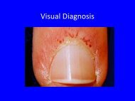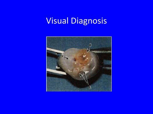
Visual Diagnosis
Visual Diagnosis
Visual Diagnosis

You also want an ePaper? Increase the reach of your titles
YUMPU automatically turns print PDFs into web optimized ePapers that Google loves.
<strong>Visual</strong> <strong>Diagnosis</strong>
Things That Can Happen to Your<br />
Belly Button<br />
Kathleen Forcier
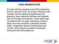
Case of Umbilical Drainage<br />
• 1 mo old ex FT F with persistent umbilical<br />
drainage<br />
• Cord fell off at 2 weeks of life<br />
• History of umbilical granuloma s/p silver<br />
nitrate@2 ½ weeks old
• Afebrile<br />
Physical Exam<br />
• Gen: alert, NAD, shirt has 1 inch area of<br />
moisture<br />
• Chest: CTAB<br />
• CV: no murmurs, 2 + femoral pulses b/l<br />
• Abd: umbilicus glistens/wet, no purulent<br />
discharge, no granuloma, no surrounding<br />
erythema, normal BS, no HSM
Differential <strong>Diagnosis</strong>
Studies?
Our Patient<br />
• Pyridium PO x 1 day – looking to see if<br />
discharge turned orange<br />
• Orange umbilical discharge = patent urachus,<br />
if not may be urachal cyst<br />
• Keflex ppx<br />
• Abd U/S, Renal U/S, VCUG
Results<br />
• No orange drainage from<br />
umbilicus (not a true<br />
patent urachus)<br />
• U/S showed “anechoic<br />
tubular structure extending<br />
from the dome of the<br />
bladder to the umbilicus<br />
with 1 cm x 0.8 cm cystic<br />
structure seen approx 2 cm<br />
from bladder dome”
What We’ll Cover<br />
• Embryology review of umbilical cord contents<br />
• Omphalomesenteric duct anomalies<br />
• Urachal anomalies<br />
• Other common umbilical findings
Embryology<br />
• 4 th week the umbilical cord contains the umbilical<br />
vessels, the urachus and the omphalomesenteric<br />
duct.<br />
• The omphalomesenteric duct connects the gut to<br />
the yolk sac (involutes 9 th week of gestation)<br />
• The allantois (diverticulum of the hindgut)<br />
becomes the urachus and connects the bladder<br />
to the umbilicus (involutes to become the median<br />
umbilical ligament in month 5 of gestation)
Problems with Omphalomesenteric Duct
Meckel’s Diverticulum<br />
• Painless rectal bleeding from ectopic gastric<br />
mucosa<br />
• Can be a lead point for intussusception
Patent Omphalomesenteric Duct<br />
• Drainage from the<br />
umbilicus (serous,<br />
bilious, feculent)<br />
• Dx with fistulogram –<br />
inject dye into the<br />
umbilicus and look for<br />
contrast in the small<br />
intestine
<strong>Visual</strong> <strong>Diagnosis</strong>
Fibrous Band<br />
• Can result in volvulus and intestinal<br />
obstruction
Problems of the Urachus
Symptoms<br />
• 43% with umbilical drainage<br />
• 40% of urachal cysts presented with infection<br />
• 15% had palpable suprapubic mass<br />
• Adults present with hematuria and pain, 51%<br />
had evidence of adenocarcinoma on<br />
histopathology<br />
• Excision is controversial
Urachal Cyst<br />
• Palpable periumbilical mass<br />
• Infected cysts present with abdominal pain,<br />
erythema, periumbilical swelling<br />
• If the cyst ruptures into intraperitoneal space<br />
it can cause acute abdomen<br />
• Usual organism is Staph aureus
Patent Urachus<br />
• Free communication between the bladder and<br />
the umbilicus.<br />
• Persistent wet draining cord<br />
• Can cause UTIs<br />
• May be a pop‐off valve in posterior urethral<br />
valves
VCUG
Umbilical Granuloma<br />
• Soft, moist, pink, friable lesion of granulation<br />
tissue<br />
• Can cause persistent drainage<br />
• Treatment is silver nitrate
Umbilical Polyps<br />
• Omphalomesenteric ductal or urachal<br />
remnants<br />
• Larger than granulomas<br />
• Don’t respond to silver nitrate, require<br />
surgical excision<br />
• Send the tissue to path to determine if it is<br />
urachal or omphalomesenteric origin
Omphalitis<br />
• Infection of the umbilical stump or<br />
surrounding tissue<br />
• Erythema, induration, swelling of the skin<br />
• Systemic signs may include lethargy, fever,<br />
irritability, poor<br />
feeding
Omphalitis<br />
• Umbilicus grants access to the portal vein (via<br />
umbilical vein) can cause portal vein<br />
thrombosis or liver abscess<br />
• Can cause peritonitis<br />
• Can become necrotizing fasciitis<br />
• Usually polymicrobial infection involving S.<br />
aureus, GAS, and GN bacteria<br />
• Treatment Amp + Gent, may need vanco
Umbilical Hernia<br />
6‐10x more common in African Americans than<br />
whites<br />
Rarely incarcerate<br />
Most resolve by 3 y/o<br />
If present at 4 y/o<br />
Unlikely to resolve on<br />
its own
Single Umbilical Artery<br />
• Occurs in
Delayed Separation<br />
• Normal time to separation is about 1 week<br />
• Presence of the cord past 3 weeks is<br />
considered delayed<br />
• Causes include immunodeficiency ( leukocyte<br />
adhesion deficiency), urachal abnormality<br />
• Evaluate neutrophil function in neonates with<br />
delayed separation and signs of infection
Take Home Points<br />
• Omphalomesenteric duct connects the small<br />
bowel to the umbilicus<br />
• Urachus connects the bladder to the<br />
umbilicius<br />
• Omphalitis is an emergency and requires<br />
prompt antibiotics to prevent significant<br />
morbidity
Sources<br />
• Ashley et al. Urachal Anomalies: A Longitudinal Study of Urachal<br />
Remnants in Children and Adults. The Journal of Urology. 2007:<br />
178: 1615‐1618.<br />
• Galati et al. Management of Urachal Remnants in Early Childhood.<br />
The Journal of Urology. 2008; 180, 1824‐1827.<br />
• Hinson et al. Picture of the Month. Archives of Pediatric and<br />
Adolescent Medicine. 1997., 151: 1161‐1162.<br />
• O’Donnell et al. Pediatric Umbilical Problems. Pediatric Clinics of<br />
North America. 1998: 45; 4 791‐799.<br />
• Palazzi and Brandt. Care of the umbilicus and management of<br />
umbilical disorders. Up to Date. September 25, 2009.<br />
• Pomeranz. Anomalies, abnormalities and care of the umbilicus.<br />
Pediatric Clinics of North America. 2004: 51; 819‐827.<br />
• Ueno et al. Urachal Anomalies: Ultrasonography and Management.<br />
Jounral of Pediatric Surgery. 2003: 38; 1203‐1207.