The digital Plateosaurus II - Acta Palaeontologica Polonica
The digital Plateosaurus II - Acta Palaeontologica Polonica
The digital Plateosaurus II - Acta Palaeontologica Polonica

Create successful ePaper yourself
Turn your PDF publications into a flip-book with our unique Google optimized e-Paper software.
<strong>The</strong> <strong>digital</strong> <strong>Plateosaurus</strong> <strong>II</strong>: An assessment of the range of<br />
motion of the limbs and vertebral column and of previous<br />
reconstructions using a <strong>digital</strong> skeletal mount<br />
HEINRICH MALLISON<br />
Mallison, H. 2010. <strong>The</strong> <strong>digital</strong> <strong>Plateosaurus</strong> <strong>II</strong>: An assessment of the range of motion of the limbs and vertebral column<br />
and of previous reconstructions using a <strong>digital</strong> skeletal mount. <strong>Acta</strong> <strong>Palaeontologica</strong> <strong>Polonica</strong> 55 (3): 433–458.<br />
Scientific literature and museum exhibits are full of explicit and implicit claims about the possible postures and motion<br />
ranges of dinosaurs. For the example of the prosauropod <strong>Plateosaurus</strong> engelhardti I assessed the motion range of limbs<br />
and vertebral column in a CAD program using a 3D virtual skeletal mount. <strong>The</strong> range of motion of the forelimb is very<br />
limited, allowing the grasping of objects placed directly ventrally and ventrolaterally of the anterior torso. <strong>The</strong> manus is<br />
adapted for grasping. <strong>The</strong> powerful fore limb can barely reach in front of the shoulder, making a quadrupedal walking cy−<br />
cle impractical. Only a digitigrade pose of the pes with a steeply held metatarsus is feasible, and the morphology of the<br />
stylopodium and zeugopodium indicates a slightly flexed limb posture. Hind limb protraction and retraction are limited<br />
by the pelvic architecture. <strong>The</strong> neck has significant mobility both dorsoventrally and laterally, but blocks torsion. <strong>The</strong><br />
dorsal vertebral column is flexible to a degree similar to the neck, mainly in the anterior half, but blocks torsion totally in<br />
the anterior and posterior thirds. <strong>The</strong> anterior dorsals are similar in shape to the posterior cervicals and significantly in−<br />
crease the motion range of the neck. <strong>The</strong> tail is highly flexible due to its large number of elements, showing more lateral<br />
than dorsoventral mobility. <strong>The</strong>se results are compared to reconstruction drawings and museum skeletal mounts, high−<br />
lighting a pattern of errors specific to certain widely used reconstruction methods.<br />
Introduction<br />
Key words: Prosauropoda, <strong>Plateosaurus</strong>, reconstruction, <strong>digital</strong> skeleton, 3D model, accuracy.<br />
Heinrich Mallison [heinrich.mallison@googlemail.com], Glambecker Weg 6, 13467 Berlin, Germany.<br />
Received 26 May 2009, accepted 25 February 2010, available online 8 March 2010.<br />
<strong>Plateosaurus</strong> engelhardti Meyer, 1837 from the Upper Tri−<br />
assic of Central Europe is one of the best−known dinosaurs,<br />
with a large number of strikingly well−preserved skeletons<br />
found in Frick (Switzerland), Halberstadt and Trossingen<br />
(both Germany), and additional disarticulated finds from<br />
many other localities. P. engelhardti was also the fifth dino−<br />
saur to be described, and the first outside England (see<br />
Galton 2001). In addition to the holotype material, many<br />
skeletons were found in articulation in the first third of the<br />
19 th century, and the excavation of some of these was docu−<br />
mented in detail, with figures published of the specimens as<br />
found in the ground (Jaekel 1913–1914; Huene 1926). One<br />
of the best finds of this kind, SMNS F33 from Trossingen,<br />
was prepared so that the association of all bones was re−<br />
tained, and remains in this state to this day (Fig. 1A). This<br />
specimen is the only one in which the anterior body does not<br />
rest on one side, but is upright (Sander 1992), offering insight<br />
into the articulation of the shoulder girdle difficult to gain<br />
from other specimens. Some other individuals, notably the<br />
most complete specimen ever found from Frick (MSF 23,<br />
Sander 1992), were also retained in articulation (Fig. 1B). On<br />
<strong>Acta</strong> Palaeontol. Pol. 55 (3): 433–458, 2010<br />
the basis of this huge amount of detailed data, one could as−<br />
sume that reconstructing the overall body shape, posture and<br />
locomotion of this animal is simple. To the contrary, the liter−<br />
ature not only of the early 19 th century, but even of recent<br />
years is full of contradictory reconstruction drawings and re−<br />
marks on the locomotion of <strong>Plateosaurus</strong>. <strong>The</strong> history of<br />
biomechanical interpretations of the genus, the graphical<br />
representation of which will be discussed here, is nearly as<br />
complex and confusing as that of its taxonomy. Moser (2003)<br />
gives the latest detailed review, concluding that Plateosau−<br />
rus is monospecific, with P. engelhardti as the sole species.<br />
However, Moser (2003) cautions that there is ample material<br />
labeled “<strong>Plateosaurus</strong>” in collections that does not belong to<br />
the genus. Yates (2003) assigns a second, more gracile and<br />
much rarer species, previously Sellosaurus gracilis Huene,<br />
1907–1908, to <strong>Plateosaurus</strong> as P. gracilis. Here, “Plateo−<br />
saurus” refers to the Halberstadt, Trossingen, Ellingen (Ger−<br />
many) and Frick material belonging to P. engelhardti.<br />
<strong>The</strong> easiest way for a researcher to communicate his view<br />
of the body shape, locomotion and general appearance of an<br />
extinct animal is usually the creation of a reconstruction<br />
drawing. <strong>The</strong> tradition goes back to Marsh (e.g., Triceratops<br />
and “Brontosaurus” in Marsh 1891), and in recent years, in−<br />
doi:10.4202/app.2009.0075
434 ACTA PALAEONTOLOGICA POLONICA 55 (3), 2010
MALLISON—DIGITAL RECONSTRUCTION OF PLATEOSAURUS 435<br />
clusion of such a drawing in publications has almost become<br />
de rigueur. A certain standard has developed: the animals are<br />
depicted in lateral view, with the bones drawn as outlines,<br />
and a suggested body outline surrounding them in black.<br />
Other views, dorsal, anterior or cross−sections, are some−<br />
times also provided (e.g., Paul 1996, 2000). Some artists and<br />
researchers offer “rigorous” drawings of specific specimens,<br />
in which the preserved parts are hatched (e.g., Wellnhofer<br />
1994), so that the viewer can judge what is actually know<br />
from the fossil and what is inferred from other finds of the<br />
same or similar species. This distinction is not moot, as<br />
shown by <strong>The</strong>rrien and Henderson (2007), because many an−<br />
imals do not scale linearly compared to their close relatives,<br />
so that linear scaling will deliver inaccurate size and mass es−<br />
timate.<br />
<strong>The</strong> first reconstruction drawing of <strong>Plateosaurus</strong> can be<br />
found in Jaekel (1913–1914). Most famous, though, is the<br />
drawing in Huene (1926: pl. 7). It depicts SMNS 13200, a<br />
nearly complete skeleton of “P. quenstedti”, a junior synonym<br />
to P. engelhardti (Galton 1985). <strong>The</strong> animal is shown in lateral<br />
view, in a bipedal pose with a steeply inclined body, appar−<br />
ently running quickly. Galton wrote a series of publications on<br />
<strong>Plateosaurus</strong> and related prosauropods, several of which<br />
(Galton 1971b, 1986a, 1990, 2000, 2001) also contain lateral<br />
view drawings. Those in Galton (2000) and (2001) are copies<br />
of that in Galton (1990), which is based on Huene (1926), as<br />
stated in Galton (2001) and some print editions of Galton<br />
(2000). <strong>The</strong> drawings in Galton (1971b) and (1976) are exact<br />
depictions of the museum mount of AMNH 6810. Weis−<br />
hampel and Westphal (1986) published a guide to the Plateo−<br />
saurus exhibit in the IFGT, which contains a drawing in lateral<br />
view. Wellnhofer (1994) describes material from Ellingen in<br />
Bavaria. His “rigorous” drawing depicts <strong>Plateosaurus</strong> in a<br />
quadrupedal pose, with a tail base bent sharply downward.<br />
<strong>Plateosaurus</strong> receives an extensive figure treatment by Paul<br />
(1987, 1997, 2000). A lateral view of the skeleton with body<br />
outline, apparently in a rapid walk, is supplemented by a dor−<br />
sal view and a drawing combining an anterior view at the level<br />
of the pectoral girdle and a posterior view at the level of the<br />
pelvic girdle, in the same pose. A second lateral view portrays<br />
the animal as a musculature reconstruction in a full gallop,<br />
with the pedes almost overstepping the manus. On request,<br />
Scott Hartmann (www.skeletaldrawing.com) provided a res−<br />
toration drawing in lateral view. Skeletal restorations dis−<br />
cussed below are reproduced in Fig. 2.<br />
<strong>The</strong> public’s perception of an extinct animal is shaped<br />
more by mounted skeletons in museums than by scientific<br />
publications. Due to the distribution of <strong>Plateosaurus</strong> through−<br />
out Central Europe, mounted skeletons or casts can be found<br />
in Frick (MSF), München (BSPG), Tübingen (IFGT), Stutt−<br />
gart (SMNS), Frankfurt (SNG), Halberstadt (MHH) and<br />
Berlin (MFN). <strong>The</strong> IFGT houses two mounts. GPIT1 (Fig.<br />
1D), the source data for the virtual skeletal mount (Fig. 1E)<br />
employed in this study, is a nearly complete individual<br />
mounted in a bipedal standing pose, snout close of the ground<br />
as if feeding or drinking (see Gunga et al. 2007; Mallison<br />
2007, 2010). GPIT2 is a composite of similar size to GPIT1. It<br />
consists of two individuals, one consisting of a skeleton nearly<br />
complete to the level of the last dorsal, the other lacking all<br />
parts anterior to the pelvis. Several copies of mounts can be<br />
found in other museums worldwide. Some of the mounts have<br />
been altered or taken down in recent years. For example, the<br />
SMNS housed a total of four mounted casts of SMNS 13200,<br />
in a variety of poses (Ziegler 1988: cover illustration, fig. 4;<br />
Moser 2003: fig. 4). All but one in a quadrupedal walking pose<br />
(Fig. 1H, G) have been taken down for an exhibition redesign<br />
in 2007. Similarly, the MFN mount has been dismantled and<br />
not been recreated because of a museum renovation in<br />
2006/2007. <strong>The</strong> lizard−like sprawling old mount of SMNS<br />
13200 (Fraas and Berckhemer 1926) in the old Stuttgart mu−<br />
seum was dismantled during World War <strong>II</strong> and never re−<br />
mounted. Outside Europe, the AMNH mount of a nearly com−<br />
plete Trossingen skeleton (AMNH 6810; Fig. 1F) from the<br />
joint 1921–1923 excavation has been used by most US re−<br />
searchers as the typical <strong>Plateosaurus</strong>. I here only discuss<br />
mounts that I was able to inspect personally, including mounts<br />
that have since been taken down, as reliance on photographs<br />
alone is prone to cause errors.<br />
Even more memorable than drawings or mounts, but<br />
more difficult to assess for their accuracy, are models of the<br />
living animal. In a tradition going back to Benjamin Water−<br />
house’s plaster dinosaurs created for Richard Owen in 1851<br />
and exhibited in the Crystal Palace (London, UK), many mu−<br />
seums today enliven their exhibits with life−sized models. In<br />
the SMNS, the old skeletal mounts were accompanied by<br />
two models, one in a tripodal (Fig. 1C) and one in a quadru−<br />
pedal pose, at the scale of the largest <strong>Plateosaurus</strong> ever<br />
found. Together with the new skeletal mounts, new 3D mod−<br />
els were set up in 2007, in quadrupedal (Fig. 1I, L), bipedal<br />
(Fig. 1J) and a resting pose. Additionally, toy versions of the<br />
old tripodal and the new quadrupedal (Fig. 1K) models (by<br />
Bullyland TM ) are available, which were created in collabora−<br />
tion with scientific advisors from the SMNS. While their<br />
small size leads to inaccuracies in some areas for technical<br />
reasons, they are overall proportioned almost like the life−<br />
sized models. <strong>The</strong>y can stand in their stead for the analysis<br />
here, because they can be digitized accurately and thus help<br />
avoid the inherent inaccuracies of photograph analysis due to<br />
perspective distortion.<br />
In addition to these classic reconstructing methods, digi−<br />
tal techniques have also been used in recent years. Gunga et<br />
al. (2007, 2008) used high resolution laser scanning to obtain<br />
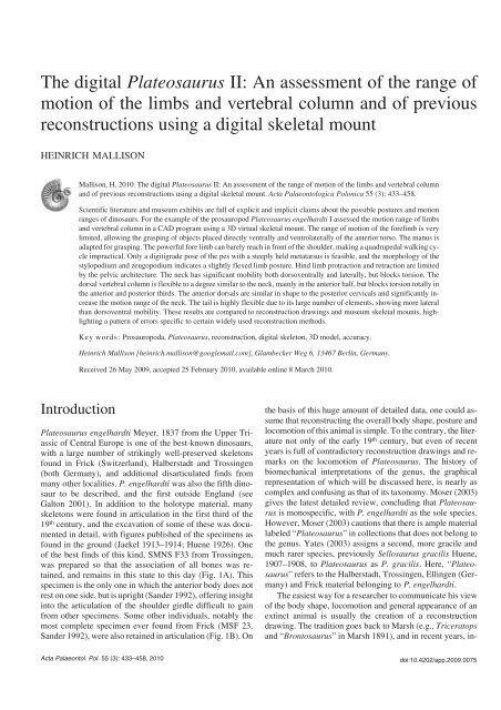
Fig. 1. Prosauropod <strong>Plateosaurus</strong> engelhardti Meyer, 1837 skeleton mounts and life reconstructions. A. SMNS F33, in position as found. B. MSF 23, in posi−<br />
tion as found. C. SMNS old tripodal model. D.GPIT1.E. GPIT1 virtual mount based on CT data. F. AMNH 6810. G, H. SMNS 13200 (cast). I, L.SMNSnew<br />
quadrupedal model in anterior (I) and lateral (L)views.J. SMNS new bipedal model. K. Toy model of SMNS new quadrupedal model by Bullyland. All but<br />
I femur/thigh length ~0.6 m, I ~5 m high. Photographs by the author except C ©GPIT. A, C–K from Trossingen, Germany, B from Frick, Switzerland.<br />
doi:10.4202/app.2009.0075
436 ACTA PALAEONTOLOGICA POLONICA 55 (3), 2010<br />
Fig. 2. Skeletal reconstructions of prosauropod <strong>Plateosaurus</strong> engelhardti Meyer, 1837, redrawn from: A. Paul (1987, 2000). B. Wellnhofer (1994). C. Jaekel<br />
(1913–1914). D. Huene (1926). E. Galton (1990). F. Weishampel and Westphal (1986). G. Scott Hartmann. (www.skeletaldrawing.com). Typical femur<br />
length of <strong>Plateosaurus</strong> is0.6to0.8m.<br />
3D point cloud models of various mounted skeletons, includ−<br />
ing the mount of GPIT1 in the IFGT. Based on this excellent<br />
base they created two versions of a <strong>digital</strong> 3D model, one in−<br />
tentionally fat, the other slim, to assess the possible range of<br />
soft tissue reconstruction.<br />
With all kinds of reconstructions it is important to provide<br />
a high level of accuracy. Pictures and models tell an “at one<br />
glance” message about an animal’s body shape and propor−<br />
tions that immediately tells the viewer much more than just<br />
the shape and sizes of the actual (bony) specimen. Small in−<br />
accuracies can lead to drastic misinterpretations. A simple<br />
example is Henderson (2006), who used a drawing by Paul<br />
(1987) to create a 3D model of <strong>Plateosaurus</strong>, based on which<br />
the total mass and the center of mass position were calcu−<br />
lated. For unknown reasons, the scale bar in the figure by<br />
Paul (1987: 29) appears in altered form in Henderson (2006:<br />
fig. 11). In fact, the size of the animal is considerably reduced<br />
by this small change, so that Henderson calculates a body<br />
weight of only 279 kg for <strong>Plateosaurus</strong> (Henderson 2006:<br />
table 4). <strong>The</strong> scaling difference is not immediately visible,<br />
so that there is a high risk that other researchers will use<br />
Henderson’s mass value as the body weight of an adult<br />
<strong>Plateosaurus</strong> in future works. Similarly, the many drawings<br />
and skeletal mounts of <strong>Plateosaurus</strong> in a quadrupedal pos−<br />
ture have led to an increasing number of artists’ reconstruc−<br />
tions in similar poses. Today the internet is a prime research<br />
tool for scientists and laypeople alike. Practically every<br />
internet site offers pictures with their text (or vice versa), and<br />
there is the increasing trend to reduce the text and emphasize<br />
the visual presentation. This means that pictures gain in im−<br />
portance relative to textual descriptions. Thus, inaccurate<br />
figures can have a profound influence on both the public’s<br />
and the scientific community’s perception of an extinct<br />
animal, and should be avoided at all cost.<br />
An animal’s range of motion, especially in limb joints,<br />
determines motion capabilities such as grasping ability or<br />
possible stride length. For an assessment of locomotion capa−<br />
bilities, it is of paramount importance to consider the motion<br />
ranges of the humerus and femur, as well as the ability to<br />
pronate the hand. Also, the lateral stiffness or flexibility of<br />
the vertebral column can provide insight into the animal’s<br />
ability to use undulating motions for locomotion, either on
MALLISON—DIGITAL RECONSTRUCTION OF PLATEOSAURUS 437<br />
land or in water, while dorsoventral bending influences stride<br />
length during galloping and leaping. To date, detailed discus−<br />
sions of the mobility of extinct dinosaurs are limited almost<br />
exclusively to theropods, with an emphasis on their fore−<br />
limbs (e.g., Galton 1971a; Carpenter and Smith 2001;<br />
Gishlick 2001; Carpenter 2002; Senter and Robins 2005;<br />
Senter 2006a, 2006b; Senter and Parrish 2006). Sauropodo−<br />
morphs are rarely treated in detail, with the exceptions of, for<br />
example, Galton 1971a (<strong>Plateosaurus</strong>), or the discussion of<br />
manus pronation capabilities in <strong>Plateosaurus</strong> and Masso−<br />
spondylus in Bonnan and Senter (2007).<br />
Here, I address the mentioned reconstruction drawings,<br />
mounts and 3D models with regards to accuracy, attempting<br />
to determine which factors have the strongest influence on<br />
accuracy. Also, I test claims about joint motion range, using<br />
the 3D virtual skeleton of GPIT1. This method is much easier<br />
to use than manual manipulation of casts or real bones, as<br />
e.g., conducted by Senter and Robins (2005). Aside from al−<br />
lowing extensive study without the risk of damaging the<br />
specimens, it also can be performed without access to the<br />
material, once it has been digitized. Also, there is no need to<br />
create elaborate support for the bones in larger assemblages<br />
(see Senter and Robins [2005: fig 1] for an example concern−<br />
ing a theropod forelimb). Claims about motion range can be<br />
explicit, e.g., Paul (2000: 83): “[in] most bipedal dinosaurs<br />
[…] the lower arm bones, radius and ulna, could rotate<br />
around each other”, they can be implicit in a drawing (e.g.,<br />
showing a strongly dorsiflexed neck), or even in a remark on<br />
locomotion or behavior of the animal, e.g., Paul (2000: 91):<br />
“<strong>The</strong> long−backed prosauropods may have run with a bound−<br />
ing gallop, but the heavily built, shorter−footed melanoro−<br />
saurs probably galloped less and trotted more”. This implies<br />
that <strong>Plateosaurus</strong> was capable of quadrupedal locomotion,<br />
did not have a sprawling gait, and could use gaits with<br />
unsupported phases.<br />
Institutional abbreviations.—AMNH, American Museum of<br />
Natural History, New York, USA; BSPG, Bayerische Staats−<br />
sammlung für Geologie und Paläontologie, München, Ger−<br />
many; GPIT, see IFGT; IFGT, Institut für Geowissenschaften,<br />
Eberhardt−Karls−Universität Tübingen, Tübingen, Germany<br />
(collection numbers: GPIT); IPFUB, see FUB; FUB, Freie<br />
Universität Berlin, Germany (collection numbers of osteolo−<br />
gical collection IPFUB OS); MB.R., see MFN; MFN, Mu−<br />
seum für Naturkunde – Leibniz−Institut für Evolutions− und<br />
Biodiversitätsforschung an der Humboldt−Universität zu<br />
Berlin, Berlin, Germany (collection numbers: MB.R.); MHH,<br />
Museum Heineanum Halberstadt, Halberstadt, Germany;<br />
MSF, Sauriermuseum Frick, Frick, Switzerland; SMA, Saurier−<br />
museum Aathal, Aathal−Seegräben, Switzerland; SMNS,<br />
Staatliches Museum für Naturkunde Stuttgart, Stuttgart, Ger−<br />
many; SNG, Senckenbergische Naturforschende Gesell−<br />
schaft, Frankfurt, Germany.<br />
Other abbreviations.—ONP, osteologically neutral pose.<br />
Two vertebrae are in ONP if their centra faces are parallel<br />
and the zygapophyses overlap fully.<br />
Material<br />
For creation of the virtual mount of <strong>Plateosaurus</strong>, the nearly<br />
complete individual GPIT1 was CT−scanned at the UHT by<br />
Burkhard Ludescher. Scanning and file extraction details are<br />
given in Mallison (2010). Several elements of GPIT1 are<br />
missing or severely damaged. Where possible these were re−<br />
placed with CT−scan based files from GPIT2, a composite<br />
skeleton from the same excavation and of similar size<br />
(see Huene 1926, 1928, 1932). <strong>The</strong>se include the whole left<br />
manus, and in order to avoid misinterpretations on the proper<br />
articulation and the motion range of the wrist, the entire left<br />
forelimb of GPIT2 was used. Also, the left pes was mirrored<br />
and used in stead of the right pes, in which most metatarsals<br />
are incompletely preserved. Several elements are missing in<br />
both GPIT1 and GPIT2, or so incompletely preserved that<br />
they are not useful for the work presented here. <strong>The</strong>se are: both<br />
clavicles, all cervical ribs not attached to the cervical verte−<br />
brae, and the sternals. <strong>The</strong> gastralia could not be scanned due<br />
to being embedded in large sediment slabs. Results concern−<br />
ing missing, deformed or potentially deformed elements of<br />
GPIT1 and GPIT2 were compared to other individuals of<br />
<strong>Plateosaurus</strong>, both from Trossingen and Halberstadt. <strong>The</strong>se<br />
include SMNS F33 (Fig. 1A), MB.R. 4429, an articulated<br />
neck with skull from Halberstadt, MSF 23 (Fig. 1B), and SMA<br />
unnumbered, an articulated neck with partial skull and the first<br />
six dorsals from Frick. <strong>The</strong>se and other specimens, as well as<br />
the diagram of GPIT1 as found in the field by Huene (1928: pl.<br />
10) were also used for assessing the correct articulation of the<br />
skeleton, especially the manus and pes, and the possible mo−<br />
tion range of the neck.<br />
<strong>The</strong> 3D files (Polymesh [*.stl] format) are available on re−<br />
quest from the author, providing previous permission for use<br />
by the IFGT.<br />
Methods<br />
Computer programs and digitizer<br />
All CAD tasks (mounting the skeleton, motion range analy−<br />
sis) were conducted using McNeel Associates Inc. “Rhinoc−<br />
eros® NURBS modeling for Windows®” versions 3.0 and<br />
4.0. Digitizing of toy models also took place in Rhinoceros©,<br />
using an Immersion TM Microscribe 3D digitizer. <strong>The</strong> result−<br />
ing point clouds were meshed in Geomagic Inc. Geomagic<br />
Qualify 8.0© (time limited evaluation version).<br />
Methods of CAD virtual skeleton creation and<br />
range of motion assessment<br />
<strong>The</strong> virtual skeleton of GPIT1 was created in a bipedal stand−<br />
ing pose (Mallison 2010). For the assessment of mobility,<br />
those elements of importance to the joint being investigated<br />
(e.g., two consecutive vertebrae or humerus, radius and ulna)<br />
doi:10.4202/app.2009.0075
438 ACTA PALAEONTOLOGICA POLONICA 55 (3), 2010<br />
were copied into a new Rhinoceros® file. One object was<br />
given a different color and made immobile (“locked”), the<br />
other one duplicated and moved to an extreme position (e.g.,<br />
maximum dorsiflexion). Other copies were moved into fur−<br />
ther extreme position (e.g., extreme lateral flexion) until all<br />
possible positions had been created. Combinations of ex−<br />
tremes (e.g., maximum lateral flexion at maximal dorsi−<br />
flexion) were also created. Throughout the process, the artic−<br />
ulation of the elements was checked in six axial views and a<br />
freely rotatable perspective view. Where necessary, objects<br />
were made translucent to allow better judgment of the feasi−<br />
bility of the created articulation. Angles were measured in<br />
the CAD program either directly using bone landmarks, or<br />
by adding a line to the bone before motions were tested,<br />
copying and moving this line with the bone, and then mea−<br />
suring the angle between the two lines. <strong>The</strong> resulting files for<br />
each motion between vertebrae were imported into one new<br />
file, to illustrate e.g., maximum dorsiflexion of the entire dor−<br />
sal vertebral column. Similarly, for the limbs the files from<br />
stylopodium, zeugopodium and autopodium were united.<br />
Here, all non−redundant positions were combined, to illus−<br />
trate the total motion range of the limb.<br />
Motion between vertebrae was assumed to be possible if<br />
any zygapophysal overlap was retained. This means that in<br />
contrast to Stevens and Parrish (1999, 2005a, b), 50% over−<br />
lap is not required. Stevens and Parrish (1999) use this value,<br />
but as they point out (Stevens and Parrish 2005a), giraffes re−<br />
tain only minimal overlap, and avian cervicals retain be−<br />
tween 25% and 50% of overlap. Here, minimal overlap is<br />
therefore used, and the resulting position then assessed for its<br />
feasibility. Overlap of 50% is used when higher values lead<br />
to unrealistic results as mentioned below.<br />
Influence of soft tissues on the range of motion<br />
Ligaments, muscles and tendons.—Aside from the bones,<br />
soft tissues have a large influence on the motion range of<br />
joints. For example, ligaments play an important role in block−<br />
ing or strictly limiting certain degrees of freedom in many<br />
joints (McGinnis 2004; Stevens and Parrish 2005a). Some−<br />
times these limitations are obvious from the bone shapes. For<br />
example, the shape of the distal condyles of the human femur<br />
indicates that the knee is, as a first approximation, limited to<br />
flexion/extension only, even though the bones alone would al−<br />
low other motions such as antero−posterior sliding. Similarly<br />
shaped femora in extinct animals can thus be assumed to have<br />
had a similar complement of ligaments, and the knee therefore<br />
a similarly limited amount of degrees of freedom. If insertion<br />
sites of ligaments can be found on the fossil, and the corre−<br />
sponding ligament in extant taxa reliably identified, the accu−<br />
racy of a motion range analysis can increase, but in the case of<br />
<strong>Plateosaurus</strong> few traces were found, and no corresponding<br />
ligaments could be identified with certainty.<br />
Muscles and their tendons can also limit motions<br />
(McGinnis 2004), because they cannot be stretched at will.<br />
<strong>The</strong> greater the distance between a muscle’s path and the joint<br />
it works on, the lesser the angle the moved body part can<br />
cover. <strong>The</strong> prime example is the caudofemoralis longus mus−<br />
cle, running from the underside of the anterior tail to the fourth<br />
trochanter of the femur. Its moment arm on the hip is accord−<br />
ingly large, and the more distal position of the fourth tro−<br />
chanter in dinosaurs compared to lizards and crocodiles means<br />
that the angle the femur can cover is smaller. <strong>The</strong> exact rota−<br />
tion angle, however, depends on many unknown variables,<br />
among them the internal structure of the muscle (parallel−<br />
fibred or pennate?), the relative length of its tendon, and the<br />
path both take through other soft tissues, so that the resolution<br />
that can be achieved by modeling the muscle is insufficient to<br />
be of help for a range of motion study (contra Paul 2005).<br />
In such cases evidence from skeletons found in life−like posi−<br />
tions can help determine minima for joint deflection angles.<br />
Many skeletons of <strong>Plateosaurus</strong> were found belly down, with<br />
strongly flexed hind limbs, e.g., MSF 23 (Fig. 1B) and espe−<br />
cially SMNS F33 (Fig. 1A). Although sediment compaction<br />
has compressed SMNS F33 to the point where the femur, tibia<br />
and metatarsus are in contact with each other along the bone<br />
shafts, it is reasonable to assume that <strong>Plateosaurus</strong> could in−<br />
deed adopt a resting pose in which the pelvic girdle supported<br />
part of the animal’s weight. SMNS F33 can be used to recon−<br />
struct this position, by attempting to place the <strong>digital</strong> skeleton<br />
into the same pose without disarticulating any joints. Where<br />
this is not possible the <strong>digital</strong> bones must be placed in the clos−<br />
est possible position that is dorsoventrally of greater extent,<br />
because SMNS F33 was mainly dorsoventrally compressed.<br />
<strong>The</strong> resulting pose of the <strong>digital</strong> skeleton (see below) is then<br />
the best estimate for the posture SMNS F33 had before sedi−<br />
ment compaction. <strong>The</strong> resulting protraction angle of the hind<br />
limb and the flexion angle of the knee and ankle are required<br />
minima. In the following, if angles determined using this or<br />
similar approaches are smaller than those suggested by bone−<br />
bone articulation, the smaller angle is given.<br />
Motion range limitations are especially important and dif−<br />
ficult to determine from the bones alone where muscles or<br />
tendons cross more than on joint. <strong>The</strong> maximum length of the<br />
muscle may be insufficient to allow full range of motion in<br />
all joints, effectively reducing the range of motion, while sin−<br />
gle−joint muscles rarely do so (McGinnis 2004). For exam−<br />
ple, Crocodilus porosus (IPFUB OS 13) shows limits of<br />
about 13 per joint in the tail for lateral flexion based on the<br />
bones alone at 50% zygapophsal overlap and 22 with mini−<br />
mal overlap, but is limited to roughly 6 per joint when the<br />
complete tail is flexed to one side, based on measurements of<br />
overall curvature of the tail on photographs (Fig. 3A; own<br />
unpublished data). In contrast, if the tail is placed in a sinu−<br />
soidal curve, so that e.g., only ten articulations flex to the left,<br />
and the following ten flex to the right, the soft tissues do not<br />
reduce the maximum angle as much, allowing 10 of motion<br />
per joint (own unpublished data). <strong>The</strong> osteoderms of croco−<br />
diles have an even larger influence blocking flexion and ex−<br />
tension of the trunk. Overall, Crocodylus porosus is capable<br />
of roughly a 45 extension in the dorsal series (own unpub−<br />
lished data), while the vertebrae alone would allow more
MALLISON—DIGITAL RECONSTRUCTION OF PLATEOSAURUS 439<br />
Fig. 3. Examples for the influence of soft tissues on joint motions. A. Outline drawing of caudals 5 and 6 of salt−water crocodile Crocodylus porosus, IPFUB<br />
OS 13 in dorsal view. Anterior is up. Caudal 6 is shown in positions with full, 50% and minimal zyapophysal overlap (0,10,21, respectively). Width of<br />
caudal 5 across transverse processes is 113 mm. B–D. Ulnae of stegosaur Kentrosaurus aethiopicus Hennig, 1915 from the Upper Jurassic Tendaguru For−<br />
mation of Tanzania, in anterior (B 1–D 1) and lateral (B 2–D 2) views. Right (B, field number St [unknown]) and left (C, field number St 113) ulnae, both part<br />
of GPIT 1424 (mounted skeleton). D. Left ulna (part of skeletal mount in MFN) MB.R.4800.33 (length 306 mm) shows cartilage preservation on the distal<br />
and especially proximal end, preserving a large olceranon process.<br />
than 90. <strong>The</strong> influence of soft tissues on the motion range of<br />
a joint depends on many factors, and cannot be taken into ac−<br />
count in detail here, especially because there is no direct fos−<br />
sil evidence of soft tissues known in <strong>Plateosaurus</strong>.<br />
Articular cartilage.—Especially difficult to estimate is the<br />
influence of articular cartilage on joint motion. In many<br />
joints of dinosaurs, including <strong>Plateosaurus</strong>, the amount of<br />
cartilage was small, comparable to extant mammals and<br />
birds. Especially in the digits and along the vertebral column<br />
there are well developed bony articulation surfaces, so that<br />
good matches exist between the bones. However, the ends of<br />
limb bones as well as the articular edges of e.g., the scapulae,<br />
coracoids and sternals retained a large amount of cartilage, so<br />
that the shape of the preserved bone may not correlate closely<br />
to the actual articulation surface. In general, there appears to<br />
be a good correlation of the general shape and main features<br />
(but not the fine details) in adult archosaurs, but juveniles of<br />
all and adults of some selected taxa show massive differ−<br />
ences (Bonnan et al. 2009; own unpublished data). Cartilagi−<br />
nous tissues are rarely preserved on fossils, so the thickness<br />
of cartilage caps in dinosaurs is unclear. Often, it is claimed<br />
that even large dinosaurs had only thin layers of articular car−<br />
tilage, as seen in extant large mammals, because layers pro−<br />
portional to extant birds would have been too thick to be ef−<br />
fectively supplied with nutrients from the synovial fluid.<br />
This argument is fallacious, because it assumes that a thick<br />
cartilage cap on a dinosaur long bone would have the same<br />
internal composition as the thin cap on a mammalian long<br />
bone. Mammals have a thin layer of hyaline cartilage only,<br />
but in birds the structure is more complex, with the hyaline<br />
cartilage underlain by thicker fibrous cartilage pervaded by<br />
numerous blood vessels (Graf et al. 1993: 114, fig. 2), so that<br />
nutrient transport is effected through blood vessels, not diffu−<br />
sion. This tissue can be scaled up to a thickness of several<br />
centimeters without problems.<br />
An impressive example for the size of cartilaginous struc−<br />
tures in dinosaurs is the olecranon process in the stegosaur<br />
Kentrosaurus aethiopicus Hennig, 1915. In the original de−<br />
scription a left ulna (MB.R.4800.33, field number St 461) is<br />
figured (Hennig 1915: fig. 5) that shows a large proximal<br />
process. However, other ulnae of the same species lack this<br />
process, and are thus far less distinct from other dinosaurian<br />
ulnae (Fig. 3B, C). <strong>The</strong> process on MB.R.4800.33 and other<br />
parts of its surface have a surface texture that can also be<br />
found on other bones of the same individual, and may indi−<br />
cate some form of hyperostosis or another condition that<br />
leads to ossification of cartilaginous tissues. Fig. 3B–D com−<br />
pares MB.R.4800.33 and two other ulnae of K. aethiopicus<br />
from the IFGT skeletal mount. It is immediately obvious that<br />
the normally not fossilized cartilaginous process has a signif−<br />
icant influence on the ability to hyperextend the elbow, be−<br />
cause it forms a stop to extension. Similarly large cartilagi−<br />
nous structures may have been present on a plethora of bones<br />
in any number of dinosaur taxa, so that range of motion anal−<br />
yses like the one presented here are at best cautious approxi−<br />
mations.<br />
Methods for comparing the virtual skeleton<br />
to previous reconstructions<br />
For comparison of a reconstruction drawing to the virtual<br />
skeleton, the drawing was imported into Rhinoceros© as a<br />
background image and scaled to fit the virtual skeleton. If<br />
possible, a fit was created for the overall length of the dorsal<br />
series and the femur length. If this was not possible due to<br />
differences in proportion, the dorsal series alone was used.<br />
Initial attempts to include the sacral series as well failed, be−<br />
cause of different deformations of the sacra of GPIT1 and<br />
SMNS 13200, the latter of which is the base for most recon−<br />
struction drawings. If there were several views of a recon−<br />
doi:10.4202/app.2009.0075
440 ACTA PALAEONTOLOGICA POLONICA 55 (3), 2010<br />
struction available, they were imported into one Rhinoc−<br />
eros© file and arranged as background images in the appro−<br />
priate axial viewports (dorsal view in the “top” viewport, an−<br />
terior view in the “left” viewport, etc.). <strong>The</strong> virtual skeleton<br />
was then posed to conform to the drawing as well as possible.<br />
In case of length differences between bones, the virtual bone<br />
was shifted to the proximal/anterior end of the corresponding<br />
element in the drawing, creating a visible gap in the next dis−<br />
tal articulation. Ribs were ignored in most cases, due to the<br />
relatively high degree of deformation in the ribs of GPIT1,<br />
and the usually quite schematic style of the ribs in the draw−<br />
ings. <strong>The</strong> sole exception is the set of drawings by Paul (1987,<br />
1997, 2000). Paul considers his drawings “technical recon−<br />
structions”, even when they are based on old photographs of<br />
mounts, and uses them for taxonomic investigations (Paul<br />
2008). Clearly, these drawings must thus conform to higher<br />
standards than others that are created purely for illustrating<br />
general proportions of the animal. In the case of multiple<br />
views, the bones were first arranged according to the lateral<br />
view, then in the other views. If this required misaligning<br />
them in lateral view, a duplicate was created instead. <strong>The</strong> re−<br />
sulting pose was assessed in all axial and the perspective<br />
views.<br />
Skeletal mounts in museums were inspected for correct<br />
articulation visually and on photographs. Because several<br />
mounts were visited several years ago, before the conception<br />
of the work presented here, and have since been dismantled,<br />
their assessment was based on photographs alone. Here, and<br />
for life−sized 3D models, the problem of edge distortion had<br />
to be solved. Where possible, a number of photographs were<br />
taken with a 50 mm lens from a fixed distance at a right angle<br />
to the long and transverse axes of the mount or model, a 25%<br />
frame cut away from them, and the remaining, least distorted<br />
central parts combined into a composite image. Alterna−<br />
tively, a 300 mm lens was used to take one picture at a large<br />
distance. While these photographs are more distorted than a<br />
composite picture, they are better than a wide angle shot<br />
from a short distance.<br />
One problem that could not be solved easily unless at<br />
least two orthogonal views were available is that of perspec−<br />
tive distortion in dynamically posed mounts. If, e.g., the neck<br />
or tail curves towards or away from the viewer, they will ap−<br />
pear shorter than they really are. This effect is hard to judge<br />
in lateral view alone. Where no picture from another perspec−<br />
tive was available, I forced the virtual skeleton to fit the lat−<br />
eral view. This may hide articulation errors in the mounts or<br />
scaling errors in the models.<br />
<strong>The</strong> <strong>digital</strong> 3D models were imported into Rhinoceros©<br />
directly, while the toy models were first mechanically digi−<br />
tized using the point cloud digitizing procedure detailed in<br />
Mallison at al. (2009), and meshed to create a 3D body in<br />
Geomagic Qualify 8.0©. <strong>The</strong> virtual skeleton was arranged to<br />
conform to them in the same way as for multiple view draw−<br />
ings. <strong>The</strong> 3D models by Gunga et al. (2007) were compared<br />
directly to the point cloud scan file of the IFGT GPIT1 skeletal<br />
mount, on the basis of which the models were created.<br />
Results<br />
Range of motion<br />
In the following, maximum motion ranges with (where nec−<br />
essary) considerations of forces acting on articulations will<br />
be discussed for separate functional body units, such as the<br />
hindlimb or the tail. Note that the determined motion ranges<br />
are the extremes possible for these parts alone, not for the en−<br />
tire skeleton. In some joints, motions are possible to degrees<br />
that are blocked by other body parts given certain articulation<br />
angles of other joints. <strong>The</strong>se combined motion ranges are dis−<br />
cussed afterwards.<br />
Vertebral column: cervical series.—<strong>Plateosaurus</strong> has 10<br />
cervical vertebrae (plus a rudimentary proatlas), 15 dorsals<br />
and three sacrals (Huene 1926). Huene (1926) also lists 41<br />
preserved and at least 15 missing distal caudals for SMNS<br />
13200. In GPIT1 45 caudals are present, and only a very<br />
small number may be missing at the tip of the tail. Atlas and<br />
axis of GPIT1 are attached to the skull and were not available<br />
for CT scanning. For reference, the cervical column was<br />
placed so that the neural canal of the last vertebra was hori−<br />
zontal. <strong>The</strong> neck was mounted in neutral pose (following<br />
Stevens and Parrish [1999], the pose with perfectly overlap−<br />
ping zygapophyses and parallel centra faces is often also<br />
termed osteologically neutral pose [ONP]), although there<br />
are strong indications that the posture has no special signifi−<br />
cance for an animal’s habitual posture (Christian and Dzem−<br />
ski 2007; Taylor et al. 2009). In lateral view the neck then<br />
projects upwards slightly, forming a 7 angle between the<br />
horizontal and the line connecting the posterior opening of<br />
the neural canal on the last and the anterior one on first pre−<br />
served cervical. However, there is no marked angling of the<br />
centrum−centrum articulations at the base of the neck, nor<br />
marked keystoning. Rather, the centra are trapezoidal in lat−<br />
eral aspect, with the anterior face dorsally displaced in rela−<br />
tion to the posterior face. This shift is present in all but the<br />
first two cervicals, “stepping up” the neck while the inter−<br />
vertebral discs are parallel to each other. <strong>The</strong> anterior three<br />
cervical centra are slightly keystoned, creating a ventrally<br />
concave arch that places the anterior surface of the centrum<br />
of the epistropheus in a near vertical position. <strong>The</strong> shape of<br />
the atlas and its articulation with the epistropheus result in a<br />
downwards inclined position of the skull, as described by<br />
Huene (1926: 28).<br />
Any definitive assessment of neck mobility requires<br />
knowledge of the shape and potential flexibility of the cervi−<br />
cal ribs. <strong>The</strong>se, however, are not fully preserved in GPIT1 to−<br />
day, and have not been prepared separately in other individu−<br />
als of <strong>Plateosaurus</strong>. Several articulated necks have been re−<br />
tained in the position they were found in. Especially well pre−<br />
served are SMNS F33, SMA unnumbered, and MB.R.4429.<br />
Of these, SMNS F33 and MB.R.4429 curve strongly, are<br />
well articulated, and their cervical ribs are distally slightly<br />
bent, indicating some flexibility in vivo. Although the cervi−
MALLISON—DIGITAL RECONSTRUCTION OF PLATEOSAURUS 441<br />
Fig. 4. Range of motion of prosauropod <strong>Plateosaurus</strong> engelhardti Meyer, 1837 using the <strong>digital</strong> skeleton mount of GPIT, from Trossingen, Germany.<br />
A. Lateral view of cervicals in neutral articulation, maximal dorsiflexion and maximal ventriflexion. B. Dorsal view of cervicals in neutral articulation and<br />
maximal lateral flexion. C–F. Dorsal vertebral column and ribcage in dorsal view in maximal lateral flexion (C), lateral view in maximal ventriflexion (D),<br />
lateral view in maximal dorsiflexion (E); air exchange volume determination (F). Pink ribs and dark green volume = exhaled volume, red ribs and translu−<br />
cent green volume = inhaled volume. See text for further explanation. G. Tail in lateral view, showing (top to bottom) dorsiflexion at 10 and at 5 per joint,<br />
neutral articulation, maximum ventriflexion. H. Tail in dorsal view, straight and at 10 lateral flexion. Length of cervical series 103 cm, length of dorsal se−<br />
ries 137 cm, length of caudal series 261 cm. Anterior to the left in A–C and F–H, to the right in D and E.<br />
cal ribs in <strong>Plateosaurus</strong> overlap the next vertebra, it is un−<br />
likely that they blocked intervertebral motion. For the <strong>digital</strong><br />
analysis presented here I therefore ignored them, instead<br />
comparing my results to the articulated necks.<br />
Ventral mobility of the neck is limited to a 100 curve,<br />
measured as the angle between the anterior face of cervical 2<br />
and the posterior face of cervical 10. Mobility is greatest be−<br />
tween the anterior vertebra, and decreases with the reducing<br />
length of the vertebrae posteriorly. In the posteriormost three<br />
cervicals, ventriflexion is minimal, both because of the dorsal<br />
displacement of the zygapophysal articulation, and the trape−<br />
zoidal shape of the centra. Dorsiflexion is possible to a slightly<br />
larger extent (110), with the posterior half of the neck con−<br />
tributing the largest part. <strong>The</strong> anterior part is in comparison<br />
quite stiff (Fig. 4A).<br />
Lateral motions of the neck cannot be accurately judged<br />
because of the imperfect preservation of the cervical ribs.<br />
However, a 180 curve appears plausible as a minimum, plac−<br />
ing the head 0.6 m laterally and 0.3 m above the neck base,<br />
with the snout pointing caudally (Fig. 4B). SMNS F33,<br />
MB.R.4429, and MSF 23 do not contradict this interpretation.<br />
All are less flexed than appears possible from the virtual<br />
mount, but all also appear not to be flexed to the limits of the<br />
intervertebral joints. An articulated neck in the SMA (unnum−<br />
doi:10.4202/app.2009.0075
442 ACTA PALAEONTOLOGICA POLONICA 55 (3), 2010<br />
bered) from Frick shows a similar curvature as SMNS F33, al−<br />
though created by both dorsiflexion and lateral flexion. Galton<br />
(2001: fig. 1c) figures the skeleton of AMNH 6810 as found<br />
based on an unpublished drawing by Friedrich von Huene. Its<br />
neck is curved ~180, with the largest flexion angles occurring<br />
in the middle part of the neck. However, it is not clear whether<br />
these vertebrae are still articulated. In all, it seems reasonable<br />
to assume that the values given above are possible, but a<br />
slightly more restricted range of motion cannot be ruled out.<br />
Torsion is almost totally blocked by the medially angled<br />
zygapophysal articulations along the entire neck.<br />
Vertebral column: dorsal series.—ONP articulation creates<br />
a slight dorsally concave arch in the anterior five dorsals, and a<br />
slight ventrally concave curve in dorsals 6 to 10. From dorsal<br />
11 on, the series is straight. As in SMNS 13200, the zygapo−<br />
physal faces of GPIT1 are medially inclined about 45 be−<br />
tween dorsals 1 and 2, measured by placing a rectangle on the<br />
articulation surface of each side in the CAD program, and hav−<br />
ing the program calculate the angle this plane forms with the<br />
vertical axis. From dorsal 3 on, the zygapophyses are oriented<br />
progressively less steep up to dorsal 6, where the angle is<br />
about 18. Further posteriorly, they are oriented more steeply<br />
again, and reach approximately 35 between dorsal 15 and the<br />
first sacral. This angle is 10 lower than in SMNS 13200. It is<br />
unclear whether this difference is caused by intraspecific vari−<br />
ation or deformation. <strong>The</strong> angles for GPIT1 were determined<br />
on the right side only, because the lateral processes and zyga−<br />
pophyses of the left side are all displaced dorsally by post−<br />
mortem deformation. <strong>The</strong> zygapophysal alignment indicates a<br />
high mobility in dorsoventral direction in the posterior and<br />
even more so in the anterior third of the dorsal series. <strong>The</strong> mid−<br />
dle part is somewhat less flexible in dorsoventral direction,<br />
and additionally allows strong lateral flexion. Torsion is<br />
blocked by steeply oriented zygapophyses, and thus only pos−<br />
sible, to a limited degree, in the middle third of the dorsal se−<br />
ries. Articulation of the <strong>digital</strong> files confirms this interpreta−<br />
tion. However, lateral flexion of the vertebrae alone is possible<br />
to an extent that appears unreasonable, allowing nearly a full<br />
circle for the entire dorsal series. Lateral flexion between con−<br />
secutive vertebrae of over 30, as possible between, e.g., dor−<br />
sals 7 and 8, leads to intersection of the ribs. Apparently, lat−<br />
eral motion in vivo is not blocked osteologically, but rather by<br />
the maximum compression of the intercostal tissues. Addition<br />
of the ribs to the 3D file leads to a maximum lateral curvature<br />
of the dorsal series of about 110 (Fig. 4C). Flexion is also<br />
blocked by the ribs, not the intervertebral articulations, allow−<br />
ing a 85 curve (Fig. 4D). Extension was limited by the inter−<br />
costal and ventral soft tissues, the effect of which is difficult to<br />
determine. However, a 90 curve for the entire dorsal series<br />
seems possible (Fig. 4E). In life, the dorsal mobility of the an−<br />
terior dorsals was certainly greater, as rib motion was ignored<br />
here. This would have increased the motion range of the neck<br />
by moving the neck base.<br />
Ribcage.—<strong>The</strong> ribs of GPIT1 all show the marks of post−<br />
mortem deformation and distortion. Comparison between<br />
contralateral elements shows that this damage is not signifi−<br />
cant enough to make a reconstruction of the ribcage width<br />
impossible. <strong>The</strong> capitulum can be matched accurately with<br />
the parapophysis in most ribs, but the dorsal displacement of<br />
the left transverse processes of many dorsal vertebrae results<br />
in a dorsal displacement of the diapophysis (term used here<br />
sensu Huene [1926] and Wilson [1999], referring to the artic−<br />
ulation surface, not the entire transverse process). In some<br />
dorsals, the tuberculum cannot be articulated with it at all, in<br />
others the resulting position elevates part of the rib shaft as<br />
high as the top of the neural arch. Similarly, on the right side<br />
some neural arches, and with them the diapophyses, have<br />
been displaced ventrally. Here, the respective ribs are tilted<br />
medially. In the virtual skeleton an attempt was made to cor−<br />
rect for these errors, placing the ribs as symmetrically as pos−<br />
sible. This results in a high−oval ribcage cross−section, in<br />
which the transverse axis at the widest point (dorsal 6) is<br />
slightly shorter than the vertical axis, if the latter is measured<br />
from the ventral end of the corresponding rib. <strong>The</strong> 4th to 9th<br />
ribs are almost equal in length, and the cross sections only<br />
slightly less wide than in the 6th. Fitting an ellipse into the rib<br />
pairs of this region results in a ratio between the transverse<br />
and the vertical axis of 0.85. In the first to third rib pairs a<br />
greater difference exists between the axes of the ellipse, with<br />
the transverse axis roughly 0.6 times the length of the vertical<br />
axis. <strong>The</strong> posterior third of the ribcage is only slightly wider<br />
than the anterior third, but the ribs are much shorter. <strong>The</strong>ir<br />
distal ends form a posteriorly ascending line that corresponds<br />
to the pubes.<br />
While the correct angle between the longitudinal axis of<br />
the animal and the rib shafts cannot be determined with ease,<br />
it is nevertheless possible to estimate the tidal volume of<br />
<strong>Plateosaurus</strong> on the basis of the reconstructed ribcage. As in<br />
tyrannosaurids (Hirasawa 2009), the ribs rotate around an<br />
axis defined by the articulations between capitulum and<br />
parapophysis, and tuberculum and diapophysis. <strong>The</strong> orienta−<br />
tion of these axes is one of the main factors defining thorax<br />
kinematics. <strong>The</strong> axes of rotation were determined by creating<br />
cylindrical bodies that were arranged with one end touching<br />
the diapophysis and the other end the parapophysis. Five<br />
point objects were created on the medial side of each rib,<br />
splitting the medial face of the rib into four segments, and<br />
grouped with the rib, so that any motion of the rib also affects<br />
the points. All ribs were rotated around their respective artic−<br />
ulation axes by +5, while a copy of each rib (with the at−<br />
tached points) was rotated by −5, creating a 10 difference<br />
between the two rib sets. This angle results in rib kinematics<br />
similar to those observed during rest or moderate activity in<br />
extant basal birds (Claessens 2009) or reconstructed for<br />
tyrannosaurids (Hirasawa 2009), with lateral displacement<br />
of the ventral end of the rib always surpassing ventral dis−<br />
placement. Based on the points on each rib, curves were cre−<br />
ated that in general terms follow the rib shape, forming<br />
U−shapes open ventrally. <strong>The</strong>se were closed by creating a<br />
straight curve section closing the gap. From each set of<br />
curves, a NURBS body with straight sections was lofted, and
MALLISON—DIGITAL RECONSTRUCTION OF PLATEOSAURUS 443<br />
its volume determined (Fig. 4F). Similar bodies were also<br />
created for the neutral rib positions, and for rib rotations of<br />
+10 and −10. <strong>The</strong> difference between the volumes for +5<br />
and −5 rotation is almost 1900 cm 3 , or 19 liters. Values for<br />
neutral to +/−10 are ~1500 cm 3 and ~2500 cm 3 , so 2000 cm 3<br />
appears to be a good average estimate of the tidal volume.<br />
While there is no reason to assume that all ribs rotated<br />
through the exact same angle during breathing, the approach<br />
chosen here is sufficient for a rough estimate of the air ex−<br />
change volume, because small changes of the angle in the an−<br />
terior and posterior ribs have only a minuscule influence on<br />
the volume. Altering the angle of the ribs in their supposed<br />
neutral position has only a minimal effect on the air exchange<br />
volume, and a strong posterior angling results in a bio−<br />
mechanically disadvantageous position of the anterior ribs,<br />
in which compressive stresses on the pectoral girdle would<br />
work to laterally compress the anterior body even further.<br />
Also, the flattened shafts in the anterior ribs only form a con−<br />
tinuous curve if the ribs are angled close to the position cho−<br />
sen here. Tilting them posteriorly would lead to the attaching<br />
musculature creating torsion forces in the rib shafts.<br />
Vertebral column: caudal series.—In GPIT1 the zyga−<br />
pophyses of the caudals are angled between 50 and 60,<br />
blocking torsion of the tail as well as limiting lateral flexion<br />
to ~10 to 12 per intervertebral joint. Due to the large num−<br />
ber and short length of the vertebrae, the tail can still cover a<br />
large arc (Fig. 4G, H), allowing a lateral divergence of the<br />
proximal ten caudals of about 45 form the long axis of the<br />
animal even if only a 5 lateroflexion per intervertebral joint<br />
is assumed. Flexion is blocked by the haemapophyses at an<br />
angle of roughly 7 per joint, rather than by the zygapophses.<br />
Except for the first two, the haemapophyses are at least as<br />
long as the corresponding vertebra is high, but are strongly<br />
posteriorly inclined. In all, the distance between the distal<br />
ends of the haemapophyses and the ventral rim of the centra<br />
makes up 45% of the height of the tail, emphasizing the rela−<br />
tively greater role played by the caudofemoral musculature<br />
in prosauropods compared to crocodiles, where the same dis−<br />
tance corresponds to less than 30% of tail height (Crocodylus<br />
porosus IPFUB OS 13).<strong>The</strong> first twenty haemapophyses are<br />
distally slightly thickened, and from caudal 15 on they tend<br />
to show a slight posterior curvature, but there is no distinct<br />
change of shape at any point. Extension to values greater<br />
than 10 per intervertebral joint creates large gaps between<br />
the centra, but there is no osteological block below 17, when<br />
the neural spines collide. However, at such an angle no room<br />
is left for soft tissues. Taking soft tissues into account, a max−<br />
imum angle of 5 to 7 per articulation appears reasonable<br />
(Fig. 4G). Lateral motion was probably limited by soft tis−<br />
sues. <strong>The</strong> bones alone easily allow for lateral flexion between<br />
7 and 10 per intervertebral joint with 50% zygapophysal<br />
overlap (Fig. 4H), which would allow the entire tail to curve<br />
over 360. A sensible assumption is that mobility in vivo was<br />
comparable to extant crocodilians, in which the zygapo−<br />
physes (except for those of the first 5 caudals, which are less<br />
steeply oriented) are angled medially at slightly smaller val−<br />
ues (40 to 55; Crocodylus porosus IPFUB OS 13), allow−<br />
ing the tail tip to barely touch the side of the body. This corre−<br />
sponds to joint lateroflexion values of about 6.<br />
Pectoral girdle.—Both clavicles of GPIT1 were preserved<br />
complete and nearly in articulation (Huene 1926: 41). <strong>The</strong><br />
fact that they as well as the anterior margins of the coracoids<br />
were in contact when excavated in both GPIT1 and GPIT2<br />
indicates that this position reflects in vivo articulation, as<br />
pointed out by Huene (1926). Other finds show a similar ar−<br />
rangement, with closely spaced coracoids, e.g., MSF 23 and<br />
SMNS F 33, as well as several individuals from Halberstadt<br />
(Germany, Jaekel 1913–1914). Medially touching clavicles<br />
were reported for the closely related prosauropd Masso−<br />
spondylus by Yates and Vasconcelos (2005). <strong>The</strong>refore, the<br />
pectoral girdle formed a narrow U−shape, allowing little or<br />
no rotation of the scapula against the ribcage.<br />
In GPIT1, the left coracoid is badly deformed. <strong>The</strong> ante−<br />
rior and dorsal margins of the coracoid have been folded me−<br />
dially, but the rest of the bone as well as its association with<br />
the scapula appears unremarkable. However, the right cora−<br />
coid has a much weaker curvature in dorsal view, as does the<br />
right compared to the left scapula blade. Other coracoids of<br />
<strong>Plateosaurus</strong> show great variability in both the curvature and<br />
the angle they forms with the scapula (see Moser 2003).<br />
When constructing the virtual mount I used an average of the<br />
bones of GPIT1.<br />
<strong>The</strong> exact position of the pectoral girdle on the ribcage is<br />
impossible to determine, because of several uncertainties.<br />
First of all, it is unclear how far cranially the midline of the<br />
pectoral girdle projected. Positions below the 8 th cervical to<br />
the 2 nd dorsal are theoretically possible. However, very cau−<br />
dal positions require rotating the ribs improbably far back,<br />
and very cranial positions create too much room for soft tis−<br />
sues between the scapulae and the ribs, or force an ex−<br />
tremely dorsal position of the scapulae. This is problematic,<br />
because the dorsal ends of the scapula blades then project<br />
nearly as far dorsally as the tips of the neural spines. Only<br />
positions with the anterior edges of the coracoids below<br />
the last two cervicals appear possible (Figs. 1D; SOM 1 at<br />
http://app.pan.pl/SOM/app55-Mallison_SOM.pdf). Also, the<br />
angle between the scalupar blade and the vertebral column<br />
cannot be determined exactly, but an angle steeper than 45<br />
for the long axis of the scapula seems likely (see Bonnan<br />
and Senter 2007; Remes 2008; Mallison 2010, in press).<br />
<strong>The</strong> resulting position with a far ventrally, anteriorly and<br />
steeply angled scapula is similar to that of sauropods (Remes<br />
2008) and even highly derived ornithischians, as evidenced<br />
by an articulated Triceratops find (Fujiwara 2009).<br />
Forelimb: shoulder.—<strong>The</strong> glenoid on both the left and the<br />
right scapulacoracoid in GPIT1 is a simple U−shaped trough,<br />
despite the massive deformation of the left element. This is<br />
also true of all other well preserved specimens that can un−<br />
equivocally be assigned to the same genus as GPIT1 (GPIT2,<br />
SMNS13200, MB.R.4404, MSF 23, see Galton [2000]; Moser<br />
doi:10.4202/app.2009.0075
444 ACTA PALAEONTOLOGICA POLONICA 55 (3), 2010<br />
[2003]). <strong>The</strong>refore, humerus motion was limited to a simple<br />
rotation around an axis transverse to the scapula blade when<br />
significant forces acted on the glenoid. <strong>The</strong> narrow U−shape of<br />
the pectoral girdle and the curvature of the scapulacoracoid<br />
dictate that this transverse axis is angled between 35 and 50<br />
medially compared to the long axis of the animal, depending<br />
on the exact arrangement of the pectoral girdle chosen. Simi−<br />
larly, the axis can be tilted medioventrally up to 15, orrun<br />
horizontal, depending on the dorsoventral position of the scap−<br />
ulae on the ribcage. In the articulation deemed most probable<br />
here (roughly the middle of possible scapular positions in an−<br />
terior and lateral views), the axis is angled 45 medially in dor−<br />
sal view, and horizontal in anterior view. <strong>The</strong>refore, humerus<br />
protraction and retraction under load is limited to an antero−<br />
medial−posterolateral plane, displacing the elbow laterally<br />
during retraction (Fig. 5A, E). <strong>The</strong> humerus can only cover an<br />
angle of approximately 80 in extension and flexion (Fig. 5B,<br />
D). This angle is not split evenly by the orthogonal to the scap−<br />
ula long axis through the glenoid, but only 35 extend in front<br />
of it in GPIT1 (angle varies for other skeletons due to different<br />
deformation of the scapulacoracoids). Thus, the humerus can−<br />
not be protracted to a vertical position if the scapula is placed<br />
with the long axis steeper than 35. However, angles lower<br />
than 45 for the scapula are unreasonable (Mallison 2010; see<br />
also Bonnan and Senter 2007; Remes 2008), so that humerus<br />
protraction to vertical was impossible.<br />
Maximal abduction (humeral elevation) and adduction<br />
angles are difficult to determine, due to the large amount of<br />
cartilage that must have been present in the shoulder. <strong>The</strong><br />
glenoid is 8.0 cm wide on both sides, while the humeral<br />
head measures 5.4 cm on the left and 5.5 cm on the right hu−<br />
merus of GPIT1. <strong>The</strong>refore, cartilage made up around 15%<br />
of the anteroposterior extent of the humeral head, assuming<br />
that cartilage thickness was equal on the humerus and<br />
glenoid. More probably, this figure is too low, and the carti−<br />
lage cover of the glenoid was much thinner than that of the<br />
humeral head. It is in any case moot to try to reconstruct the<br />
shape of the cartilage cap in detail. Following Senter and<br />
Robins (2005), Galton (1971a) and Gishlick (2001) I here<br />
assume that the extents of the preserved articular surfaces<br />
on each bone represent the limits of possible motion. Large<br />
abduction and adduction angles require the glenoid to be<br />
free of significant shearing forces, so that the forelimb can<br />
not have played a significant role in locomotion, which<br />
would require a sprawling position. Fig. 5A, C, E and SOM<br />
1 (http://app.pan.pl/SOM/app55-Mallison_SOM.pdf) depict<br />
the maximum abduction and adduction angles that may<br />
have been possible under compacting forces. Larger angles<br />
were probably not possible if significant forces acted on the<br />
joint. Humeral elevation to the level of the glenoid is impos−<br />
sible expect at large retraction angles (i.e., not possible by<br />
abduction, Fig. 5A–D), but the manus can cross the body<br />
midline ventrally (Fig. 5A, E). Rotation of the humerus<br />
head in the glenoid, as seen in animals with a sprawling pos−<br />
ture (e.g., Alligator and Varanus [Goslow and Jenkins 1983;<br />
Landsmeer 1983, 1984]), was probably very limited. In ap−<br />
parent contrast to the limited motion range, the wide proxi−<br />
mal end of the humerus indicates that the shoulder muscula−<br />
ture was voluminous and required large moment arms in<br />
<strong>Plateosaurus</strong>, similar to all other prosauropods (see Remes<br />
2008), indicating a powerful forelimb.<br />
Forelimb: elbow.—<strong>The</strong> axis through the distal condyli of the<br />
humerus is rotated against the transverse axis of the proximal<br />
end, which corresponds to the rotation axis in the glenoid, by<br />
about 30, with the ulnar condyle displaced ventrally. This<br />
means that flexion and extension of the elbow occurs in a plane<br />
that is only slightly rotated against the median plane of the ani−<br />
mal. Extension in the elbow is limited to slightly less than<br />
a straight position (which is here equivalent to 180), the<br />
zeugopodium forming a nearly straight line with the stylopo−<br />
dium (Fig. 5B). Flexion is possible to at least 70 (Fig.5B).For<br />
SOM 1 (http://app.pan.pl/SOM/app55-Mallison_SOM.pdf), I<br />
chose a spaced articulation between radius and ulna, in order to<br />
maximize pronation potential. Because of this, flexion to an−<br />
gles below 120 requires a supinating motion by 15 to sustain<br />
articulation between the radius head and the radial condyle.<br />
Manus pronation by longitudinal rotation of the radius<br />
around the ulna is not possible to a degree that allows a ventral<br />
direction of the palm in <strong>Plateosaurus</strong>. <strong>The</strong> proximal articular<br />
end of the radius is much wider than the shaft, and strongly el−<br />
liptical in proximal aspect (Fig. 5F, G) in contrast to, e.g.,<br />
Homo sapiens and Felis silvestris. <strong>The</strong>se species have a circu−<br />
lar proximal radius head with a shallow circular articular pit,<br />
allowing rotation of the radius at all flexion angles of the el−<br />
bow. In contrast, GPIT1 and all other individuals of Plateo−<br />
saurus examined for this publication consistently exhibit a<br />
shallow trough, the curvature of which is the same or very<br />
similar to that of the proximal articular facet of the ulna (Fig.<br />
5F). As with the glenoid, much cartilage must be missing from<br />
the elbow joint, so the possibility cannot be ruled out that carti−<br />
laginous structures allowed a modest amount of radius rota−<br />
tion. However, even assuming a rotation by 90 does not lead<br />
to manus pronation sufficient to allow the palm to face fully<br />
ventrally. Examination of extant taxa such as Gorilla gorilla,<br />
Homo sapiens, several species of Manis and Felis cattus<br />
(MFN collection) shows that the degree of rotation of the ra−<br />
dius that is possible against the ulna can be reliably estimated<br />
by taking a photograph of the radius in proximal view and fit−<br />
ting a circle to the medial outline. <strong>The</strong> angle corresponding to<br />
the arc conforming to the medial outline of the radius head is<br />
somewhat greater than the maximum angle the radius can be<br />
rotated. In <strong>Plateosaurus</strong>, the corresponding angle is roughly<br />
80 (Fig.5G),40 supinating and 40 pronating from the best<br />
fit between radius and ulna. This apparent neutral position is<br />
with the palms facing 60 medially from dorsal, thus a 40<br />
rotation leads to medially directed palms at best. For SOM 1<br />
(http://app.pan.pl/SOM/app55-Mallison_SOM.pdf),<br />
pronation capability was maximized by spacing radius and<br />
ulna as far apart as possible. Also, the radius rotates 55,much<br />
more than possible. Despite this, the palm faces at best 10<br />
ventrally of medially. <strong>The</strong>se results confirm, together with
MALLISON—DIGITAL RECONSTRUCTION OF PLATEOSAURUS 445<br />
Fig. 5. Range of motion of the fore limb of prosauropod <strong>Plateosaurus</strong> engelhardti Meyer, 1837 using the <strong>digital</strong> skeleton mount of GPIT1, from Trossingen,<br />
Germany. A–E. Left scapula and fore limb in anterior (A), anterolateral (B), anteromedial (C), lateral (D), and dorsal (E) views. Equal colors are identical<br />
positions. B is parallel, C is perpendicular to the main axis (flexion/extension) of the glenoid. Length of humerus 350 mm. Dashed line(s) refer to: body<br />
midline (A), orthogonal to scapula blade long axis (B), body midline and main axis of glenoid (C). Red numbers in B refer to elbow, black to humerus<br />
flexion/extension. Numbers in C refer to humerus abduction/adduction versus the vertical. F. Left radius and ulna in articulation in (top row) proximo−<br />
lateral, medial view, (bottom row) distal and lateral views. Length of ulna 237 mm. G. Radius and ulna in proximal view. Dotted line indicates main joint<br />
axis of elbow. Circle and lines show method for determination of theoretical maximal pronation angle. H–M. Left manus. H, I. Left to right: flexion, neutral<br />
position and extension in dorsal (H) and palmar (I) views. Digit IV duplicated in neutral position views to show lateromedial deviation range. J–M. Oblique<br />
views of flexion (J, K) and extension (L, M). Length of metacarpal <strong>II</strong>I 97 mm.<br />
doi:10.4202/app.2009.0075
446 ACTA PALAEONTOLOGICA POLONICA 55 (3), 2010<br />
manual manipulation of specimens and <strong>digital</strong> manipulation<br />
of 3D data from several individuals (e.g., GPIT1, GPIT2,<br />
MB.R.4402, MB.R.4404), the results of Bonnan and Senter<br />
(2007), who studied AMNH 2409 exclusively. Bonnan and<br />
Senter (2007) also show conclusively that alternative methods<br />
for manus pronation, e.g., proximo−distal motions of the ra−<br />
dius, are not possible in plateosaurid dinosaurs. Additionally,<br />
as pointed out by Mallison (2010), the posture of articulated<br />
skeletons in the field argues against <strong>Plateosaurus</strong> being capa−<br />
ble of turning the palm ventrally without massive humerus ab−<br />
duction.<br />
Forelimb: carpus, metacarpus and digits.—<strong>The</strong> block−<br />
like structure of the wrist appears to block rotating motions<br />
(see also Bonnan and Senter 2007), but to allow significant<br />
extension and flexion. <strong>The</strong> digits flex strongly, with the<br />
claws of digits <strong>II</strong> and <strong>II</strong>I potentially able to touch the palm. In<br />
contrast to the description by Huene (1926), the digits are not<br />
widely splayed and the metacarpus does not show a signifi−<br />
cant transverse curvature. Only the whole of metacarpal V<br />
and the distal end of metacarpal 4 are placed clearly palmar<br />
in relation to the others (see also Jaekel 1913–14: fig. 20).<br />
This is a marked contrast to the tubular manus of sauropods<br />
(Bonnan 2003). <strong>The</strong> distal articular facet of metacarpal IV,<br />
however, is slightly tilted against those of metacarpals <strong>II</strong> and<br />
<strong>II</strong>I. This directs the digits slightly medially during flexion.<br />
Digit <strong>II</strong>I and especially digit IV also have some lateral mobil−<br />
ity between the metacarpal and the basal phalanx, allowing<br />
lateral deviation and therefore some countering action of the<br />
flexed digit against digits I and <strong>II</strong> (Fig. 5G–J). <strong>The</strong> angle is<br />
quite limited at ~25, comparable to the lateral mobility pres−<br />
ent in the digit IV of humans. Fully flexed, the digits can<br />
grasp around objects, and digit V opposes digits <strong>II</strong> and <strong>II</strong>I<br />
(Fig. 5G–J, Mallison 2010: fig. 6). Digit I, as has also been<br />
pointed out for the closely related Anchisaurus by Galton<br />
(1976), is not angled strongly against digit <strong>II</strong> (Fig. 5G–K;<br />
Mallison 2010: fig. 6; contra Galton 1971a, b), and flexion<br />
reduces the angle to only 13 compared to the long axis of<br />
metacarpal <strong>II</strong>I (Galton 1971a, b).<br />
<strong>The</strong> right manus of SMNS F 33 is preserved in articulation,<br />
as is SMNS 13200 k, a right manus assigned to Pachysaurus<br />
by Huene (1932), a genus regarded as a junior synonym of<br />
<strong>Plateosaurus</strong> by Galton (1985). <strong>The</strong>se two hands are the sole<br />
articulated prosauropod manus from the German Keuper pre−<br />
served without significant transverse compression and whose<br />
elements have not been separated during preparation. In both,<br />
metacarpals <strong>II</strong> through IV are articulated with their shafts<br />
nearly parallel, forming an angle of less than 20,andtheen−<br />
tire metacarpus has no transverse arching. <strong>The</strong> visible parts of<br />
the bones do not suggest that this is a taphonomic artifact, con−<br />
firming the results obtained for GPIT2.<br />
<strong>The</strong> manus appears to allow significant digit extension<br />
(Fig. 5G, H, K, L). <strong>The</strong> distal condyles of the metacarpals are<br />
smoothly rounded trochleae, and there are small and shallow<br />
hyperextension pits visible on the distal ends of metacarpals<br />
<strong>II</strong> through IV. <strong>The</strong> phalanges show only very shallow or no<br />
hyperextension pits. Although the manus could certainly by<br />
used to support part of the animals weight, it does not show<br />
any special adaptations for a significant role in locomotion<br />
beyond the strong hyperextension of the digits. For example,<br />
the metacarpus is not elongated, and shows a significantly<br />
lesser development of inter−element contact surfaces, and<br />
thus stability, than the pes.<br />
Hindlimb: hip.—Both hindlimbs of GPIT1 are preserved al−<br />
most completely. <strong>The</strong> left pes lacks the distal tarsals and the<br />
ungual of digit IV, while the right hindlimb has large plaster<br />
replacements on the metatarsus as well as the phalanges.<br />
<strong>The</strong>refore, a copy of the left tarsals, metatarsus and toes was<br />
used instead of the original right side elements. Most bones<br />
do not show any indications of significant deformations, ex−<br />
cept for the left fibula. This bone is damaged slightly below<br />
midshaft, and the remaining parts have been glued together<br />
in a slightly bent position. Overall, the left fibula is 1.7 cm<br />
shorter than the contralateral element. In the virtual skeletal<br />
mount it was positioned with the distal end correctly articu−<br />
lated with the tibia and the tarsals, therefore the proximal end<br />
is shifted distally compared to the tibia.<br />
In the parasagittal plane, the femur can cover an angle of<br />
65 without abduction (Fig. 6B), if retraction is assumed to<br />
end at the level of the ischia, and protraction before collision<br />
with the pubes. Further retraction may well have been possi−<br />
ble, depending on the exact architecture of the m. caudo−<br />
femoralis longus. However, resting positions are only feasi−<br />
ble if the femora can be protracted at least 15 beyond the<br />
pubes (Fig. 6A, C, see below).<br />
<strong>The</strong>se limits on motion of the hindlimb in the parasagittal<br />
plane indicate that the long axis of the sacrum must have been<br />
held in a roughly horizontal position for speedy locomotion, as<br />
already concluded by Christian et al. (1996) and Christian and<br />
Preuschoft (1996). In a steep position as suggested for Anchi−<br />
saurus by Marsh (1893a, b) and for <strong>Plateosaurus</strong> by Jaekel<br />
(1913–1914) and Huene (1907–1908, 1926), hardly any room<br />
is left for femur motions in the parasagittal plane, because the<br />
ischia approach a near−vertical position.<br />
Adduction and abduction limits are difficult to determine.<br />
Bringing the foot under the body midline requires 15 of<br />
adduction, measured from the vertical (Fig. 6C), and abduc−<br />
tion angles of as much as 45 may have been possible. Clear−<br />
ing the pubes at large protraction angles requires at least 22<br />
of abduction (Fig. 6C). However, the missing cartilage in the<br />
hip joint does not allow an accurate assessment.<br />
Hindlimb: knee.—<strong>The</strong> knee of <strong>Plateosaurus</strong> appears to be a<br />
simple hinge joint at first approximation, as in all extant taxa<br />
with parasagittal limbs. <strong>The</strong> exact degree of inversion and<br />
eversion possible cannot be determined due to the lack of<br />
preserved articular cartilage. <strong>The</strong> preserved bone shape of<br />
the distal femur end does not form a smooth continuous cur−<br />
vature in lateral aspect. Rather, there is a marked flattened<br />
part of the articular end, at a 110 angle to the long axis of the<br />
femur shaft. Assuming that this shape corresponds closely to<br />
the shape of the actual articular surface leads to the conclu−
MALLISON—DIGITAL RECONSTRUCTION OF PLATEOSAURUS 447<br />
Fig. 6. Range of motion of the hind limb of prosauropod <strong>Plateosaurus</strong> engelhardti Meyer, 1837 using the <strong>digital</strong> skeleton mount of GPIT1, from<br />
Trossingen, Germany. A–H. Left pes in left to right: flexion, probable standing pose, extension, in lateral (A), medial (B), oblique (C–F), plantar (G), and<br />
dorsal (H) views. Length of metatarsal <strong>II</strong>I 231 mm. I–K. Pelvis and left hind limb, in lateral (I, J) and anterior (K) views. I, K, probable standing (blue) and<br />
minimally possible flexion (resting) pose; J, maximum femur protraction and retraction angles for locomotion, resulting stride length 1.34 m. L. Left hind<br />
limb showing knee range of motion. Crus positions left to right: maximal extension, maximum flexion under large loads, maximum flexion for resting. M.<br />
Crus in lateral view, showing maximum ankle flexion and extension under load. Length of fibula 463 mm.<br />
doi:10.4202/app.2009.0075
448 ACTA PALAEONTOLOGICA POLONICA 55 (3), 2010<br />
sion that there is a preferred articulation angle, in which the<br />
joint is positioned more stably than at neighboring flexion<br />
values, a sort of low−grade locking mechanism. However,<br />
this assumption is unreasonable for a joint that has to un−<br />
dergo rotation at a constant rate under loading, which is the<br />
case in the knee joint during locomotion. <strong>The</strong> distal condyles<br />
of the femur must therefore have been covered with a sub−<br />
stantial amount of cartilage. Unless one assumes a cap that at<br />
its thickest point is 3 cm thick or more on the femur, there is<br />
no smooth curve possible over the distal end, and continuous<br />
extension and flexion under compression (i.e., during the<br />
stance phase of walking) would have been hindered by the<br />
“preferred orientation” of the joint. A similar bone and carti−<br />
lage shape relation can be seen in juvenile and subadult ex−<br />
tant birds, e.g., Gallus domesticus and Struthio camelus (own<br />
unpublished data), where the lateral aspect of the bony distal<br />
femoral condyles is also flattened, while the covering carti−<br />
lage cap is well rounded distally. <strong>The</strong> tibiotarsus of Gallus<br />
also has a massive cartilage layer on the proximal end, simi−<br />
lar in thickness to that of the femur. Here, however, the dif−<br />
ference in shape between cartilage and bone is less pro−<br />
nounced, so that the bones give a good approximation of<br />
joint mobility.<br />
A conservative estimate of the motion range is used here,<br />
based on the preserved surfaces and my own measurements<br />
and manipulations of Gallus knees. Maximum extension in−<br />
cludes a 165 angle between the long axes of the shafts of the<br />
femur and the tibia, a maximum flexion angle of 100 under<br />
loading (e.g., during rapid locomotion), and a flexion to at<br />
least 40 without significant loading (e.g., in a resting pose)<br />
(Fig. 6D).<br />
Hindlimb: ankle.—Like the knee, the intertarsal articula−<br />
tion allows motion mainly around the transverse axis. <strong>The</strong><br />
main rotation occurs between the astragalus and calcaneum<br />
proximally, and the distal tarsals distally, as has been pointed<br />
out by Huene (1926). Only here exists a large convex/con−<br />
cave set of articulation surfaces that allows a continuous ro−<br />
tation over a large angle under load. <strong>The</strong> distal articular sur−<br />
face of the astragalus covers an angle of 170. However, it<br />
appears unlikely that positions were possible in which the<br />
metatarsus is angled posteriorly in relation to the tibia, there−<br />
fore the motion range can be reconstructed as covering an−<br />
gles between the long axis of the tibia shaft and the long axis<br />
of metatarsal 3 from 175 (maximum extension) to 105<br />
(maximum flexion) under large loads (Fig. 6E). If no signifi−<br />
cant compressing force is active on the joint, flexion between<br />
the distal tarsal row and the metatarsus may have been possi−<br />
ble, to facilitate the high flexion angles required for resting<br />
poses. However, feet preserved in articulation appear to<br />
achieve flexion to 130 (MSF 23), 22 (GPIT unnumbered,<br />
articulated hindlimb) or even 0 (SMNS F33) between the<br />
proximal and distal tarsals, without requiring tarsal/metatar−<br />
sal motion. Obviously, these angles are affected by post−<br />
mortem compression of the skeletons, but the fact remains<br />
that the limbs could be flexed to a resting pose with ankle<br />
flexion at values far below 90 between the astragalus/calca−<br />
neum and the distal tarsals alone.<br />
Hindlimb: digits.—<strong>The</strong> range of flexion and hyperextension<br />
in the pedal digits is comparable to that in the manual digits<br />
(Fig. 6F–M). <strong>The</strong> claws of digits <strong>II</strong>I and IV can touch the plan−<br />
tar surface of the metatarsus, while the second toe follows the<br />
same curvature path in lateral view, but cannot reach the<br />
metatarsus due to the smaller number of elements. <strong>The</strong> first<br />
digit has similar flexion mobility, too. As in the manus, it has a<br />
medially angled articulation between the metapodial element<br />
and the basal phalanx. Fully flexed, it curves laterally by 25<br />
compared to the long axis of metatarsal <strong>II</strong>I, which is a smaller<br />
angle than in the second digit (28). Digit IV curves medially<br />
by 14.<br />
A telling detail for the interpretation of the manus and pes<br />
function is the slight difference in shape of the distal ends of<br />
the metatarsals and metacarpals <strong>II</strong> through IV. In the manus,<br />
the distal articular condyles are significantly wider ventrally<br />
than dorsally, while in the pes the ventral and dorsal trans−<br />
verse extent is subequal in digits <strong>II</strong> and <strong>II</strong>I. This indicates that<br />
the main forces acting on these articulation occurred during<br />
flexion in the hand, while there is no clear preference in the<br />
pes. This pattern is not compatible with quadrupedal locomo−<br />
tion. Additionally, in contrast to the knee and the metacar−<br />
pal/basal phalanx articulations, the metatarsal/basal phalanx<br />
joints have a preferred articulation angle. A flattened area of<br />
the distal articular end is orientated at roughly 70 to the long<br />
axis of the metatarsus on metatarsals <strong>II</strong> through IV. In con−<br />
trast to the knee, there is no indication that significant<br />
amounts of articular cartilage are missing. Rather, the articu−<br />
lation surfaces are well ossified. Articulating the basal pha−<br />
langes with the metatarsus to achieve maximum bony con−<br />
tact results in a position conformal to a high digitigrade<br />
stance. <strong>The</strong> basal phalanges create the shift from subvertical<br />
to horizontal, and the distal ends of the metatarsals are lifted<br />
off the ground (Fig. 6A, D). Under high compressive loads,<br />
e.g., when taking up the weight of the animal during rapid lo−<br />
comotion, or on soft substrates, the distal ends of the metatar−<br />
sals probably contacted the ground. <strong>The</strong> proximal surfaces of<br />
the basal digits are shaped to conform to the flattened area of<br />
the metatarsals, creating a weak locking mechanism. If the<br />
joint is compressed during the support phase, rotating or slid−<br />
ing motion away from the preferred articulation angle results<br />
in intra−joint forces that favor the preferred angle. A similar,<br />
though more elaborate structure is present in the wrists of<br />
ungulates, where roughly cubic carpals lock the joint in a<br />
straight position under pressure, but allow strong flexion<br />
(two times to 88) for folding the manus under the ante−<br />
brachium when the animal lies down.<br />
In digit IV, the distal end of the metatarsal has a medial<br />
condyle that is equally sized dorsally and ventrally, while the<br />
lateral condyle is limited to the ventral face (differing from the<br />
other two long pedal digits). Dorsally, there is a pronounced<br />
gap, leaving the dorsolateral edge of the proximal articulation<br />
surface of the basal phalanx (indistinguishable in shape from
MALLISON—DIGITAL RECONSTRUCTION OF PLATEOSAURUS 449<br />
those of digits <strong>II</strong> and <strong>II</strong>I) free of an immediate bony contact.<br />
<strong>The</strong> functional significance of this feature is unclear.<br />
Mobility of the complete skeleton<br />
For the creation of a complete skeletal mount, most skeleton<br />
parts were easily combined, e.g., the tail and the sacrum. How−<br />
ever, taphonomic damage to the first sacral has tilted the ante−<br />
rior surface of the centrum as well as the neural arch back−<br />
wards by an unknown amount. <strong>The</strong>refore, the prezygapo−<br />
physes have been displaced caudally, and their correct orienta−<br />
tion cannot be ascertained, making alignment of the dorsal and<br />
sacral series difficult. Most other sacra of <strong>Plateosaurus</strong> are<br />
also damaged (Moser 2003; own observation), but in general<br />
the first sacral has roughly parallel caudal and cranial faces of<br />
the centrum, and the center of the prezygapophysal articula−<br />
tion surface rests directly dorsal to the cranial face of the<br />
centrum. <strong>The</strong> virtual skeleton was mounted accordingly, caus−<br />
ing a wedge shaped gap between the last presacral and the first<br />
sacral. <strong>The</strong> resulting continuous curvature of the vertebral col−<br />
umn across the dorsal/sacral transition is further indication<br />
that this arrangement is correct.<br />
Fig. 5A–E and SOM 1 (http://app.pan.pl/SOM/app55−<br />
Mallison_SOM.pdf ) show the full motion range of the com−<br />
plete forelimb. Fig. 6B, D, E and SOM 2 (http://app.pan.pl/<br />
SOM/app55−Mallison_SOM.pdf) show the motion range of<br />
the hindlimb available for locomotion, without strong femur<br />
abduction, while Fig. 6A, C depict maximum limb flexion.<br />
Aside from neutral pose, various flexion/extension combi−<br />
nations were created to assess the overall mobility of the ani−<br />
mal. <strong>The</strong> aim was not to demonstrate the possible extremes of<br />
motion, but rather functional poses, such as resting poses, or a<br />
pose that brings the head to the ground, e.g., for drinking or<br />
feeding. <strong>The</strong>se occasionally highlighted collisions between<br />
body parts, limiting overall range of motion. When the dorsal<br />
series is ventrally flexed, the distal ends of the posterior ribs<br />
quickly reach the level of the pubes, at flexion angles of below<br />
5 in each of the last four intervertebral articulations. While<br />
some overlap may have been possible, further ventriflexion<br />
compresses the digestive tract to an improbable degree. Simi−<br />
larly, large adduction angles combined with large retraction<br />
angles place the distal end of the humeri within the ribcage.<br />
Ground feeding.—One pose (Fig. 7A) was created to show<br />
how the “stepping up” of the neck caused by the trapezoidal<br />
cervical centra affects the animal’s ability to bring the snout<br />
to the ground, e.g., for drinking to feeding. Huene (1928)<br />
suggests that <strong>Plateosaurus</strong> had to perform a see−saw motion<br />
in the hips, but that is apparently based on the assumption<br />
that the back is not sub−horizontal in the habitual standing<br />
posture. In fact, from a pose with a horizontal back and<br />
nearly straight hindlimbs, a bending of the anterior half of the<br />
thoracic vertebral column, combined with a slight increase in<br />
limb flexion and maximum ventriflexion of the neck suffices<br />
to bring the snout to the ground. <strong>The</strong> hands are almost able to<br />
reach the ground in this position.<br />
Manus at ground level.—Grasping objects at ground level<br />
with the manus requires slightly more flexion of the verte−<br />
bral column, or a slight downward rotation at the hip. In this<br />
position the head can reach significantly below ground<br />
level (Fig. 7B).<br />
Resting.—<strong>The</strong>restingposeof<strong>Plateosaurus</strong> (Fig. 7C, D,<br />
SOM 3 at http://app.pan.pl/SOM/app55-Mallison_SOM.pdf)<br />
follows the position of the articulated finds, and that of SMNS<br />
F33 especially. Jaekel (1913–1914) assumed that Plateo−<br />
saurus habitually rested on the distal ends of the ischia. Such a<br />
posture is possible (Fig. 6C, D), but requires extreme flexion<br />
of the hindlimbs. Resting on the pubis, as is known from<br />
theropods (e.g., Gierlinski et al. 2005; Stevens et al. 2008) is<br />
not possible, and there is no enlarged pubic foot. <strong>The</strong> pelvic<br />
girdle in the tight articulation and low position chosen here<br />
and in Mallison (2010) partly supports the weight of the ante−<br />
rior body, so that the ribcage is not compressed.<br />
Previous reconstructions<br />
Skeletal mounts.—<strong>The</strong> GPIT1 mount in the IFGT (Fig. 1D)<br />
is mostly correctly articulated and placed in a plausible<br />
bipedal pose with slightly flexed hindlimbs. Solely the mas−<br />
sive splaying of the metacarpals and the slight splaying of the<br />
metatarsals are unrealistic. GPIT2 is similarly well articu−<br />
lated, also has splayed metapodia, but is posed in a possible,<br />
but not plausible posture. <strong>The</strong> animal appears to be running<br />
quickly, but the back is angled steeply, so that the femur of<br />
the supporting limb is already retracted close to the ischia al−<br />
though the limb is roughly vertical. <strong>The</strong> other limb is lifted<br />
clear off the ground with a strongly flexed knee and ankle.<br />
While <strong>Plateosaurus</strong> certainly could adopt a pose with a<br />
steeply inclined back, it would have severely hampered limb<br />
retraction, and thus rapid locomotion. <strong>The</strong>refore, the animal<br />
should have both feet on the ground, as the pose is more<br />
suited for high browsing than for running. Similarly, AMNH<br />
6810 (Fig. 1F) is mounted in correct articulation, but for a<br />
slight splaying of the metacarpals and metatarsals. <strong>The</strong> pose<br />
is overall quite similar to the head−to−ground pose in Fig. 6A,<br />
but he tail is drooping. <strong>The</strong> old MFN mount of skeleton C<br />
(MB.R.4430.1–4430.12) by Otto Jaekel was also placed in a<br />
pose that was possible, but not necessarily plausible. Nota−<br />
bly, it was the only mount that did not disarticulate any<br />
bones.<br />
All old SMNS mounts shared a series of errors that can<br />
still be seen in the remaining quadrupedal mount (Fig. 1G).<br />
<strong>The</strong> ribcage is bloated to create a broad oval, reminiscent of a<br />
lizard, tilting the scapulae so that the glenoids face antero−<br />
laterally (with correctly articulated, medially touching cora−<br />
coids). <strong>The</strong> proximal end of the humerus is placed lateral to<br />
the scapulacoracoid, so that compressive forces on the fore−<br />
limbs would shear the humerus from the glenoid. Despite the<br />
semi−sprawling posture, the frontlimbs do not allow suffi−<br />
cient manus pronation to make the palm face the ground with<br />
doi:10.4202/app.2009.0075
450 ACTA PALAEONTOLOGICA POLONICA 55 (3), 2010<br />
Fig. 7. Digital skeleton mount of prosauropod <strong>Plateosaurus</strong> engelhardti Meyer, 1837 GPIT1, from Trossingen, Germany, posed: head at ground level (A),<br />
hands at ground level (B), resting pose in lateral (C) and dorsal (D) views. Length of femur 595 mm.<br />
the digits pointing cranially. <strong>The</strong>refore radius and ulna have<br />
exchanged position in the articulation with the humerus, a<br />
condition totally unknown in any extant taxon, even in birds,<br />
where the radius lies anteriorly, not medially, of the ulna. <strong>The</strong><br />
track in the forelimbs is wider than their stride length. Also,<br />
the anterior ribs are arranged at a right angle to the vertebral<br />
column. That this is incorrect is immediately obvious on the<br />
mounts, because the gastral basket reaches only as far for−<br />
ward as the fourth rib, creating a large gap between the cora−<br />
coids and the first gastralia. In life, the sternals would have<br />
been situated in this gap, but are of insufficient size to fill it<br />
(Huene 1926). If the animal rested on the ground, this ar−<br />
rangement would mean that the anterior ribcage would not<br />
have been protected from dorsoventral compression in any<br />
way, while correct articulation leads to the coracoids and<br />
sternals serving as a resting surface, transferring forces to the<br />
scapulae and thus protecting the thorax. As a direct result of<br />
the extremely wide ribcage, the femora have to be abducted<br />
widely as well. This creates the need to angle the lower limb<br />
strongly inwards in relation to the femur, causing a wedge−<br />
shaped gap in the knee. <strong>The</strong> resulting overall proportions and<br />
limb positions are in direct contradiction to the adaptations to<br />
cursoriality visible in <strong>Plateosaurus</strong>, and require significant<br />
disarticulation in several parts of the skeleton.<br />
In addition to these general inaccuracies, one now dis−<br />
mantled mount (SMNS 13200 cast) set up in a bipedal run−<br />
ning posture exhibited a special problem. <strong>The</strong> foot in stance<br />
phase was placed ~0.5 m laterally of the midline, with the fe−<br />
mur abducted instead of adducted. Running with abducted<br />
limbs requires strong transverse motions, which is energeti−<br />
cally very inefficient. Such behavior is advantageous solely<br />
for creating larger body velocities per traveled distance than<br />
running without swaying, which is why human athletes<br />
sometimes use such a run for the first few steps of a run−up.<br />
For flight, e.g., from a predator, it is disadvantageous.<br />
<strong>The</strong> mount in the BSGP, the creation of which is de−<br />
scribed in detail by Moser (2003), is well articulated except<br />
for the pectoral girdle and forelimbs. As in the SMNS<br />
mounts, the radius and ulna are exchanged, resulting in<br />
manus pronation. <strong>The</strong> ribs are angled too far forward, in−<br />
creasing the thorax volume. Additionally, the pectoral girdle<br />
is widely separated along the midline, causing an artificially
MALLISON—DIGITAL RECONSTRUCTION OF PLATEOSAURUS 451<br />
wide shoulder area. <strong>The</strong> metacarpals and manual digits are<br />
widely splayed. <strong>The</strong> pedal digits curve strongly medially.<br />
This track has digit proportions that are incompatible with<br />
<strong>Plateosaurus</strong>, as described by Moser (2003). <strong>The</strong> reason for<br />
this misalignment is clearly stated in Moser (2003: 146): “the<br />
limbs were for the first time mounted according to the tem−<br />
plate of a scaled quadrupedal trackway of a prosauropod into<br />
the exactly predefined distances, so that the forelimbs are<br />
further abducted than the closer and more vertically placed<br />
hindlimbs” (translation from the German by the author). This<br />
adherence to a certain trackway also induces a lateral curva−<br />
ture into the dorsal vertebral column.<br />
Reconstruction drawings.—<strong>The</strong> oldest reconstruction draw−<br />
ing by Jaekel (1913–1914: pl. 4; Fig. 2C) was created with the<br />
knowledge of complete and partial skeletons found in articula−<br />
tion. Surprisingly, the overall proportions are incorrect, with<br />
the neck significantly too short, the humeri increased to nearly<br />
130% original size, the rest of the forelimb scaled by ~115%,<br />
the femur lengthened by 40%, and the antepodium and meta−<br />
podium as well as the pedal digits scaled to 120%. <strong>The</strong>se val−<br />
ues are probably too low for humerus and femur, because<br />
Jaekel (1913–1914) assumed a sprawled position, so that per−<br />
spective shortening is present in his drawing, but not in the<br />
<strong>digital</strong> mount. As a direct result, the proximal head of the fe−<br />
mur is at least as wide as the acetabulum, leaving no room for<br />
cartilage at all. <strong>The</strong> pattern of scaling errors is inconsistent<br />
with a simple mistake, such as an accidental size increase in all<br />
limb elements. Rather, the proximal elements are enlarged<br />
more than the distal parts, and the neck is too short, giving the<br />
animal a much stouter and clumsier look than a correctly pro−<br />
portioned reconstruction. Interestingly, Jaekel’s reconstruc−<br />
tion drawing of the complete animal (Jaekel 1913–1914: pl. 4)<br />
has the same neck/trunk proportions as his figure of the pre−<br />
sacral vertebral column (Jaekel 1913–1914: fig. 15), with the<br />
neck roughly 67% the length of the dorsal series, while sum−<br />
ming the length of the centra as given by Jaekel (1913–1914:<br />
181–182) results in a ratio of 85%, similar to SMNS13200,<br />
GPIT1 and GPIT2. Also, the reconstruction drawing separates<br />
radius and ulna widely, so that their proximal ends articulate<br />
with the lateral side of the lateral and the medial side of the<br />
medial condyle, while Jaekel (1913–1914: fig. 18) depicts<br />
them tightly placed as found in situ.<br />
Three other reconstruction drawings are very similar to<br />
each other and will be discussed together. <strong>The</strong>se are those of<br />
Huene (1926; Fig. 2B), Weishampel and Westphal (1986; Fig.<br />
2F), and Galton (1990; Fig. 2E). <strong>The</strong>y have in common that the<br />
overall proportions of the animal, as well as those of all larger<br />
bones, conform to SMNS13200. None appears to correct for<br />
the deformation of the sacrum, so that the pelvic girdle and<br />
hindlimbs are shifted posteriorly compared to the rest of the<br />
skeleton by approximately the length of one sacral centrum.<br />
Bone proportions vary slightly between the different drawings,<br />
as is to be expected when drawings are created manually, and<br />
not in a computer program. In general terms, however, they are<br />
correct. Because Galton’s (1990) drawing is explicitly based<br />
on Huene (1926), the commonalities between them are to be<br />
expected. In all three drawings the manus looks clumsy, be−<br />
cause of an almost radial splaying of the metacarpals.<br />
Paul (1987, 1997, 2000; Fig. 2A) offers the only dorsal<br />
and anterior/posterior views of <strong>Plateosaurus</strong>. <strong>The</strong> virtual<br />
skeleton was first arranged to conform to the lateral view<br />
alone, highlighting a series of significant problems. In order<br />
to determine which of the discrepancies are caused by intra−<br />
specific variation, the drawing by Paul (1987) was also com−<br />
pared <strong>digital</strong>ly to the drawings of the articulated neck and tail<br />
and the separate limb bones from Huene (1926: pls. 1–4),<br />
and especially to Huene (1932: pls. 26–29), on which it was<br />
based (Paul 1987: 29). In all bones, the proportions of Paul’s<br />
(1987) drawing are exactly those of MB.R.4402 (Huene<br />
1932: pls. 26–29, there listed as Fund 25). Several bones are<br />
not shown in lateral view in Huene (1932), e.g., the femur<br />
and humerus. Whether Paul (1987) based his drawing on fig−<br />
ures of other individuals of <strong>Plateosaurus</strong> in Huene 1932 is<br />
unclear. Radius and ulna are depicted in later view in Huene<br />
(1932), and thus copied in Paul (1987). Why Paul (1987,<br />
1997, 2000) assumes a sigmoidal curvature of the radius<br />
shaft that allows pronation, as evident from his drawings, is<br />
unclear. Placing the <strong>digital</strong> skeleton to conform to Paul’s<br />
(1987, 1997, 2000) drawings results in a total disarticulation<br />
of the wrist (Fig. 8A, B). An additional erroneous articulation<br />
is evident in the first manual digit. Both metacarpal 1 and the<br />
phalanges of digit I are figured in detail by Huene (1932: pl.<br />
28: 6, 9, 10). Paul (2000) assumes a 90 angling between the<br />
ungual and the metacarpal, although there is no evidence of<br />
anything but a slight medial angling because of the unequal<br />
distal ginglymus of metacarpal I in Huene’s (1932) figures.<br />
<strong>The</strong> right hindlimb is protracted significantly forward of<br />
the pubes, which have been rotated slightly backwards (Figs.<br />
2A, 8D, E). <strong>The</strong> right ankle is hidden from view by the left<br />
antepodium. To match the visible parts, the distal tarsals and<br />
proximal end of the metatarsus must penetrate the proximal<br />
tarsals and distal ends of tibia and fibula (Fig. 8C). Correct−<br />
ing this error results in a position in which the claws touch the<br />
ground, a pose unsuitable for limb protraction during loco−<br />
motion. Lifting the leg higher up requires either even further<br />
extension of the ankle, or further protraction of the femur.<br />
Both options are difficult, because the ankle is already quite<br />
extended, and the femur is already unrealistically protracted<br />
beyond the level of the pubes.<br />
<strong>The</strong> anterior view of the pectoral girdle and forelimb in<br />
Paul (1987; Fig. 2A) is combined with a posterior view of the<br />
pelvis and hindlimb. Aligning the scapulae shows that Paul<br />
(1987) assumes massive rotation to take place, and has mis−<br />
placed the clavicles (Huene 1926; see also Yates and Vas−<br />
concelos 2005 on the pectoral girdle of Massospondylus). In<br />
the anterior view, radius and ulna are shown in a crossed posi−<br />
tion, allowing the wrist to remain articulated, but creating an<br />
intersection between the bone shafts. <strong>The</strong> problematic articu−<br />
lation of the first digit mentioned for the lateral view becomes<br />
evident, too (Fig. 8B). Additionally, while the left limb is<br />
placed under the center of mass, the protracting right limb is<br />
doi:10.4202/app.2009.0075
452 ACTA PALAEONTOLOGICA POLONICA 55 (3), 2010<br />
Fig. 8. Digital skeleton mount of prosauropod <strong>Plateosaurus</strong> engelhardti Meyer, 1837 GPIT1, from Trossingen, Germany, posed to conform to drawings by<br />
Paul (1987, 2000; Fig. 2A). A. Left antepodium and manus in lateral and dorsal view. B. Right antepodium and manus in medial and dorsal view. C. Right<br />
crus and pes in medial view. Note intersection of tarsals and metatarsals with crus. D. Pelvis and femora in lateral view. E. Anteroventral view, parallel with<br />
the long axis of the dorsal column, of the pelvis and femora and the last five dorsal ribs. F. Lateral view of “gallop” position. Note gaps in knees and neck.<br />
Length of ulna 239 mm, length of fibula 463 mm, length of femur 595 mm.
MALLISON—DIGITAL RECONSTRUCTION OF PLATEOSAURUS 453<br />
Fig. 9. Digital skeleton mount of prosauropod <strong>Plateosaurus</strong> engelhardti Meyer, 1837 GPIT1, from Trossingen, Germany. A. Anterior view of the pectoral<br />
girdle and forelimbs posed to conform to the life−sized, bipedal SMNS model (Fig. 1J) of <strong>Plateosaurus</strong> engelhardti. Dotted line indicates body outline of the<br />
model. Note gaps in elbows and wrists and too large gap between coracoids (arrows). B. Anterior view of the pelvic girdle posed to conform to the life−sized,<br />
bipedal SMNS model (Fig. 1J). Note gaps in the pelvis between sacrum and ilia, and between ilia and pubes (arrows). C. Virtual skeleton posed to conform<br />
to the toy model version (Fig. 1L) of the new SMNS quadrupedal model (Fig. 1K, L) of <strong>Plateosaurus</strong> engelhardti. Dotted line indicates body outline of the<br />
model. Arrows mark skeleton’s (upper) and model’s (lower arrow) knee joint. Note gaps in forelimbs and posterior ribs extending below the pubes. Length<br />
of the femur 595 mm, length of the ulna 239 mm.<br />
abducted in the dorsal view, however, not to a sufficient de−<br />
gree to clear the (incorrectly reconstructed) ribcage (Fig. 8E).<br />
Alternatively, it is possible that the femur is abducted in the<br />
lateral view drawing, but the perspective shortening insuffi−<br />
ciently taken into account. Also evident in Paul’s (1987) draw−<br />
ing is an inward cant of the shank in the supporting limb,<br />
which would open a wegde−shaped gap in the knee.<br />
Paul’s (2000) reconstruction of the animal in a gallop<br />
shows massive scaling inaccuracies, as the neck, trunk and<br />
thigh are too long (Fig. 8A). Additionally, both femora ap−<br />
pear to be protracted without massive abduction to beyond<br />
the level of the pubes, because there is no perspective short−<br />
ening of thigh, shank or foot visible. It is unreasonable to as−<br />
sume that <strong>Plateosaurus</strong> could move in this fashion at all, be−<br />
cause limb extension in the hindlimb would require large<br />
muscle moment arms around the ankle and knee, while the<br />
corresponding osteological features suggest small moment<br />
arms (no pronounced cnemial crest, no tuber calcanei).<br />
<strong>The</strong> inaccuracies concerning the tail curvature in the<br />
drawing by Wellnhofer (1994; Fig. 2D) have already been<br />
pointed out in detail by Moser (2003). Except for a signifi−<br />
cantly too large coracoid, the proportions of Wellnhofer’s<br />
(1994) drawing are correct. However, the manus is pronated,<br />
and the dorsal ribs are misaligned.<br />
doi:10.4202/app.2009.0075
454 ACTA PALAEONTOLOGICA POLONICA 55 (3), 2010<br />
Comparison of Hartmann’s reconstruction (Fig. 2G;<br />
www.skeletaldrawing.com) to the virtual skeleton mount of<br />
GPIT1 reveals no significant differences. <strong>The</strong> metatarsi are<br />
slightly shorter in the drawing, but otherwise proportions, ar−<br />
ticulation angles and body outline match well.<br />
Life−sized and <strong>digital</strong> models.—<strong>The</strong> old life−sized SMNS<br />
models were based on the skeletal mounts and share their er−<br />
rors in proportions. Most notable are the hands and feet,<br />
which are about double the width they should be. Also, the<br />
knee was shifted distally by about 0.2 m. <strong>The</strong> new SMNS<br />
models, created under the direction of Markus Moser, all<br />
show the incorrectly wide pectoral region of the BSPG<br />
mount (see Moser 2003). Additionally, the pelvis appears<br />
wider (Fig. 9B; compare to correct articulation in Figs. 6C)<br />
and the forelimbs longer than they should be (Fig. 9C; note<br />
the gaps in the elbows). <strong>The</strong> knee is shifted about 0.1 m dis−<br />
tally (Fig. 9C). <strong>The</strong> manus and pedes have short digits,<br />
strongly medially rotated as in the BSGP skeletal mount. Ap−<br />
parently, the models are also supposed to conform to the<br />
trackway mentioned in Moser 2003.<br />
<strong>The</strong> fat 3D <strong>digital</strong> model of Gunga et al. (2007) is decid−<br />
edly too muscular, especially in the neck and trunk, and has<br />
feet much bigger than the skeleton. Additionally, the feet are<br />
simplified so that the spaces between the digits are filled with<br />
matter, further increasing their weight. <strong>The</strong>se errors are evi−<br />
dent in Gunga et al. (2007: fig. 3b). <strong>The</strong> slim model of Gunga<br />
et al. (2007) has a more realistic body outline, although some<br />
details are questionable. For example, the limb diameters do<br />
not correlate to expected muscle volumes, so that the crus is<br />
widest at the midshaft of the tibia, not near the proximal end.<br />
Discussion<br />
Range of motion.—Combining the data detailed above re−<br />
sults in a reconstruction of <strong>Plateosaurus</strong> engelhardti (GPIT1)<br />
as a surprisingly slender (for a herbivore) biped, with a sub−<br />
horizontal back and tail position (Fig. 1E; Mallison 2010: fig.<br />
1.2), strong grasping arms with a limited motion range, and the<br />
ability to obtain a 360 view by small head and neck motions<br />
alone.<br />
<strong>The</strong> vertebral column in <strong>Plateosaurus</strong> exhibits no surpris−<br />
ing specializations. In contrast to more derived sauropodo−<br />
morphs (Apesteguia 2005), there is no hyposphene−hypantrum<br />
articulation present that could block lateral motions. <strong>The</strong> dorsal<br />
series allows significantly more mobility between neighboring<br />
vertebrae than can be used when the ribcage is taken into ac−<br />
count. This may allow decoupling of the main mass of the body<br />
from the dorsoventral accelerations that the pelvis will experi−<br />
ence during rapid locomotion, reducing the need to avoid such<br />
motions through increased limb flexion at midstance.<br />
Reconstructing the resting position of <strong>Plateosaurus</strong> (Fig.<br />
7C, D) based on articulated fossils shows clearly that the ar−<br />
ticular ends of the limb bones as preserved are not a reliable<br />
indicator of the shape of the articular surface in life. <strong>The</strong> bone<br />
shapes suggest a maximal flexion angle to nearly 90, but<br />
any reasonable resting position requires at least 45 more.<br />
This confirms that the longbone ends as usually preserved in<br />
dinosaurs are not a reliable indicator for the shape of the ar−<br />
ticulation surface, as above shown for the ulna of Kentro−<br />
saurus (Fig. 3B, C).<br />
<strong>The</strong> pes of <strong>Plateosaurus</strong>, with only digit 5 significantly re−<br />
duced and a marked grasping ability (Fig. 4L), can be inter−<br />
preted as an adaptation to scratch digging, potentially for<br />
scooping out nests. In comparison to other bipedal dinosaurs,<br />
the motion range of the forelimbs of <strong>Plateosaurus</strong> is remark−<br />
ably primitive. As in the basal theropods elbow flexion is lim−<br />
ited (Senter 2006b). Similarly, humeral elevation and protrac−<br />
tion are possible to a much lower degree than in even basal<br />
members of the Avetheropoda (Senter 2006a), and similar to<br />
the carnosaur Acrocanthosaurus (Senter and Robins 2005).<br />
This limited the range to below and barely laterally of the<br />
body. One−handed grasping of small objects was possible<br />
(Fig. 5G–J), as well as holding and manipulation of larger ob−<br />
jects with both hands. Handling objects or feeding at ground<br />
level was possible in a pose that did not inhibit rapid flight, and<br />
did not require support by the forelimbs (Fig. 7A, B). <strong>The</strong><br />
claws were probably not sharp and curved enough to allow<br />
grasping or penetrating objects with them alone, in contrast to<br />
most theropods. This reinforces the notion (e.g., Galton 1984,<br />
1986b) that <strong>Plateosaurus</strong> was not a carnivore as suggested by<br />
Cooper (1981), but a herbivore. It cannot be determined, due<br />
to the uncertainties regarding neck mobility, whether the snout<br />
could be brought into contact with the hands. Recently, a pair<br />
of clear theropod manus imprints were found as part of a lon−<br />
ger trackway that indicates either a habitual manus posture<br />
with medially directed palms, or an inability of theropods to<br />
pronate the manus any further (Milner et al. 2009). Together<br />
with the only partly pronated manus of stem−sauropods<br />
(Remes 2008) this raises the question of whether any early<br />
sauropodomorph was able to pronate the manus fully. Medi−<br />
ally directed palms and the inability to pronate the manus may<br />
be ancestral characters, calling into question the assignment of<br />
quadrupedal tracks with a narrow gauge in the frontlimbs to<br />
sauropodomorph dinosaurs. Alternatively, strong undulation<br />
of the anterior body must be assumed.<br />
<strong>The</strong> estimated tidal volume of GPIT1 of 20 liters as deter−<br />
mined from rib motion (Fig. 4F) corresponds to ~29 ml per<br />
kg body weight for the average body mass of GPIT1, esti−<br />
mated by Mallison (2010) as 690 kg. This value is within the<br />
spread of extant birds, close to those of Anser (Anser anser<br />
31.4 ml/kg, Anser indicus 28.26 ml/kg, values calculated<br />
from data in Frappell et al. 2001: table 1), and significantly<br />
different from the average value of terrestrial mammals of<br />
~7.7 ml/kg (Stahl 1967). Among mammals, tidal volumes<br />
around 22 ml/kg are typical for marine forms (Mortola and<br />
Seguin 2009). This indicates that <strong>Plateosaurus</strong> possessed a<br />
high performance lung comparable to extant birds. <strong>The</strong> mini−<br />
mal tracheal volume of <strong>Plateosaurus</strong> can be estimated at<br />
slightly over 360 cm 3 , based on an airway length of 1.30 m,<br />
and an estimated diameter of 6 cm, which corresponds to 5
MALLISON—DIGITAL RECONSTRUCTION OF PLATEOSAURUS 455<br />
cm 3 /kg bodyweight. This value is similar to the value found<br />
in an ostrich (Struthio camelus: 6.13 cm 3 /kg, data from<br />
Maina and Nathaniel 2001).<br />
<strong>The</strong> sister group to Sauropodomorpha in Saurischia, <strong>The</strong>ro−<br />
poda, possessed osteological correlates to flow−through ventila−<br />
tion adaptations in all but the most basal forms (O’Connor and<br />
Claessens 2005). Recently, unidirectional airflow has also<br />
been found in alligators (Farmer and Sanders 2010). Plateo−<br />
saurus, like all prosauropods, lacks unequivocal evidence for<br />
postcranial pneumaticity (Wedel 2007). However, several<br />
lines of evidence suggest that lung ventilation via air−sacs is a<br />
primitive character of Saurischia as a whole (Wedel 2009).<br />
<strong>The</strong> lack of invasive pneumatic foramina in the dorsal verte−<br />
brae does not exclude the possibility that the lungs were at−<br />
tached there (Perry 2001). <strong>The</strong> cervicals also lack invasive<br />
pneumatic foramina, the only unequivocal evidence for post−<br />
cranial skeletal pneumaticity, which would indicate the pres−<br />
ence of air sacs (Wedel 2003, 2007). However, the centra of<br />
the last five cervicals show deep recesses under the transverse<br />
processes, which may in life have been filled by non−invasive<br />
cervical air sacs. <strong>The</strong> close correspondence of the tidal volume<br />
per mass determined here further reinforces this notion.<br />
Previous reconstructions.—<strong>The</strong> reconstruction drawing by<br />
Jaekel (1913–1914: pl. 4) suffers greatly from a preconceived<br />
notion: that <strong>Plateosaurus</strong> had a lizard−like sprawling posture.<br />
Jaekel (1913–1914: 190) also assumes plantigrady, because of<br />
the existence of large curved pedal claws, and copies inexact<br />
proportions from one of his figures into the reconstruction.<br />
Jaekel (1913–1914) also mentions repeatedly that his interpre−<br />
tation is greatly influenced by the position of the skeletons as<br />
found in the field, ignoring the fact that this pose may not be a<br />
habitual pose, may be influenced by post−mortem deforma−<br />
tion, or may be habitual as a resting pose only.<br />
<strong>The</strong> reconstructions of Galton (1990) and Weishampel<br />
and Westphal (1986) greatly resemble that of Huene (1926).<br />
All three reconstructions are classic depictions of the general<br />
osteology of the figured species, and give a good impression<br />
of the animal’s overall shape.<br />
In contrast, the drawings by Paul (1987, 1997, 2000) con−<br />
tain a number of inaccuracies (Fig. 8) that are a direct contra−<br />
diction of the detailed description in the text and figures in<br />
Huene (1926), and even to the figures in Huene (1932), on<br />
which they were based. Paul’s (1987, 1997, 2000) drawings<br />
clearly are not based on a three−dimensional understanding<br />
of the bone articulations, as evident by the protraction of the<br />
femur far beyond the level of the pubes without the required<br />
massive abduction (Fig. 8E), which should be evident in both<br />
lateral and dorsal views, by the misplacement of the clavicles<br />
on the dorsal rim of coracoids and scapulae, by the rotation of<br />
the scapulae against each other, and by the total dis−<br />
articulation of the antebrachium introduced by enforced<br />
manus pronation (Fig. 8A, B). Even more surprising is the<br />
clearly sigmoid curvature of the radius shaft and the apparent<br />
90 rotation between the long axes of the proximal and distal<br />
articular ends of the radius in the anterior view (Paul 2000;<br />
Fig. 2A), in direct contradiction to Huene (1932: pl. 28).<br />
<strong>The</strong>se and other errors cannot be explained simply by the fact<br />
that Paul (1987: 49) did not inspect the actual fossils, but<br />
based his reconstruction on the drawings from Huene (1932)<br />
alone. Rather, the errors seem to result from a preconceived<br />
notion that prosauropods were quadrupedal, that their man−<br />
ual digits I were raised off the ground during locomotion<br />
(following Galton 1971a, b; Weems 2003), and a desire to<br />
depict the animal in as dynamic a pose as possible. This is es−<br />
pecially true for the 90 medial angling of the first manual<br />
ungual, which is in direct contradiction to the figures in<br />
Huene (1932), as well as the massive errors in proportion in<br />
the right hindlimb of the skeletal outline drawing (Fig. 8C)<br />
and the left hindlimb of the “galloping” drawing (Fig. 8F). It<br />
must be mentioned that Paul (2000: 78), writing about restor−<br />
ing the life appearance of dinosaurs, stated: “A high−quality<br />
life reconstruction begins with a good skeletal reconstruc−<br />
tion. Restorations based on previously published skeletal res−<br />
torations or outline skeletal sketches usually prove to be seri−<br />
ously flawed. Also important are multiple−view restorations<br />
of at least one representative of each major group. <strong>The</strong>se<br />
make obvious anatomical errors not always apparent in side<br />
view, and they detail the subject’s three−dimensional struc−<br />
ture”. Paul (1987, 1997, 2000) highlights with his recon−<br />
struction drawings of <strong>Plateosaurus</strong> that even the mentioned<br />
multi−view restorations are insufficient to make anatomical<br />
errors obvious, or to detail the 3D structure of the animal.<br />
Museum mounts and models appear to suffer similarly<br />
from preconceived notions. <strong>The</strong> old SMNS mounts (Fig.<br />
1G, H) are highly inaccurate in comparison with the virtual<br />
skeleton of GPIT1, considering the opportunity their cre−<br />
ators had of manually manipulating casts of SMNS 13200<br />
before mounting them. <strong>The</strong> underlying cause of almost all<br />
problems in the old SMNS mounts is the decision that<br />
<strong>Plateosaurus</strong> must be a “typical reptile”, which is a marked<br />
contrast to the generally accepted and well supported view<br />
that dinosaur and lizard locomotion were fundamentally<br />
different (e.g., Bakker 1986). <strong>The</strong> decision that Plateo−<br />
saurus must also have been able to feed in an upright pose,<br />
combined probably with a misjudgement of the position of<br />
the center of mass due to the artificially enlarged body cav−<br />
ity, forced the awkward−looking dorsal curvature in the<br />
posterior dorsals of the upright mount (Ziegler 1988: cover<br />
illustration, fig. 4; Moser 2003: fig. 4), when in fact a<br />
bipedal, high−browsing pose is easily feasible with a nearly<br />
straight dorsal series. <strong>The</strong> old, upright 3D life−sized (Fig.<br />
1C) and toy models suffer from the same problems, as evi−<br />
dent from comparison of both photographs and the digitized<br />
toy model to GPIT1.<br />
<strong>The</strong> <strong>Plateosaurus</strong> mount in the BSPG was clearly<br />
planned a priori as a quadrupedal mount, based on the track−<br />
ways mentioned by Moser (2003). <strong>The</strong> wide track of the<br />
forelimbs and the undulating vertebral column create the im−<br />
pression of a crocodile in a high walk, albeit with an even<br />
slightly wider forelimb posture. Here, the direct evidence<br />
from the bones was ignored in favor of the at best question−<br />
doi:10.4202/app.2009.0075
456 ACTA PALAEONTOLOGICA POLONICA 55 (3), 2010<br />
able assignment of an ichnotaxon to an unspecified “pro−<br />
sauropod”, despite the uncertainty involved in the assign−<br />
ment (which Moser [2003] explicitly mentioned) and the di−<br />
versity evident within “Prosauropoda”. <strong>The</strong> new SMNS 3D<br />
models were created so that the quadrupedal model would fit<br />
the same trackway as the BSPG skeletal mount (Markus<br />
Moser personal communication 2004). <strong>The</strong> change of place−<br />
ment of the knee evident in at least one model (Fig. 9C) to−<br />
gether with the sauropod−like feet (Fig. 1J–L) hides the ex−<br />
treme contrast between the short, sturdy forelimbs and the<br />
long and slender hindlimbs. <strong>The</strong>se inaccuracies in the model<br />
illustrate that the skeletal evidence was ignored in favor of a<br />
desired goal.<br />
<strong>The</strong> mount of <strong>Plateosaurus</strong> in the MFN articulated the<br />
entire skeleton correctly, although the pose chosen by Jaekel<br />
was certainly not a habitual posture of <strong>Plateosaurus</strong> (contra<br />
Jaekel 1913–1914). It deserves special mention that this was<br />
the sole mount discussed here that contained no significant<br />
incorrect articulation between any neighboring bones, within<br />
technical limits (i.e., contained no disarticulation, except for<br />
a slight splaying of the left, but not right, metacarpals). <strong>The</strong><br />
plantigrade, tail−dragging posture is well within the possible<br />
motion range of <strong>Plateosaurus</strong>, e.g., for upright feeding, al−<br />
though the highly flexed hindlimbs would make sustaining<br />
the pose for an extended time rather exhausting.<br />
In summary, it becomes apparent that preconceptions are<br />
the greatest source of errors in reconstructions of extinct ani−<br />
mals. Similarly damaging to the final result is a lack of expe−<br />
rience in articulating bones correctly, either from a lack<br />
of anatomical knowledge, or from neglect to attempt manual<br />
articulation of the bones. Many errors, especially the ex−<br />
changed antepodial elements in skeletal mounts (Fig. 1G),<br />
could have been avoided with a few simple attempts to man−<br />
ually articulate the elements correctly before an armature<br />
was created, and an extant phylogenetic bracket (Witmer<br />
1995) comparison. Adapting previously published data with−<br />
out keeping its limitations in mind, e.g., using small−scale<br />
drawings to create new reconstructions in a different pose,<br />
carries a high risk of misunderstanding the 3D articulation of<br />
bones, leading to significant errors and thus incorrect bio−<br />
mechanical and paleobiological interpretations.<br />
<strong>The</strong> optimal way of creating a skeletal mount is thus the<br />
manual (e.g., Senter and Robins 2005) and/or <strong>digital</strong> (e.g.,<br />
Mallison 2007, 2010, in press) manipulation of neighboring<br />
bone pairs, to determine the best fit and the range of motion<br />
of each bone−bone articulation. Here, <strong>digital</strong> 3D data offers<br />
the great advantage of being weightless, so that even large<br />
numbers of bones can be articulated without the need for ex−<br />
tensive support as exemplified by Senter and Robins (2005:<br />
fig. 1), and without the risk of damaging a specimen. Based<br />
on these data, the full mount can be created with minimal er−<br />
rors, as long as biomechanical implications of the overall<br />
posture such as bending moments and joint torque estimates<br />
are taken into account. Data on how the animals were found<br />
in the sediment can provide key pointers, and should always<br />
be used if available.<br />
Reconstruction drawings are even more prone to errors<br />
than skeletal mounts, and errors are also harder to notice in<br />
drawings. As pointed out by Paul (2000), using second−hand<br />
data is not advisable. Especially important is the elimination<br />
of copying errors, caused by repeated re−drawing of skeletal<br />
elements. Here, modern computer technology also offers<br />
great advantages, allowing the error−free copying of ele−<br />
ments in photo editing software, as used e.g., by Scott Hart−<br />
man (www.skeletaldrawing.com; personal communication<br />
2008).<br />
Acknowledgements<br />
<strong>The</strong> author thanks Hans−Ulrich Pfretzschner, Alexander Hohloch (both<br />
IFGT, Tübingen, Germany), Kristian Remes (Steinmann Institut, Uni−<br />
versität Bonn, Bonn, Germany), Scott Hartmann (Wyoming Dinosaur<br />
Center, <strong>The</strong>rmopolis, USA) and Jonathan Rader (University of Wyo−<br />
ming, Laramie, USA) for helpful discussions. Rainer Schoch, Ronald<br />
Böttcher (both SMNS, Stuttgart, Germany) as well as Daniela<br />
Schwarz−Wings and Frieder Mayer (both MFN, Berlin, Germany)<br />
readily granted access to specimens in their care. Burkhard Ludescher<br />
(UHT, Tübingen, Germany) patiently scanned the entire skeleton of<br />
GPIT1. <strong>The</strong> manuscript has greatly benefitted from helpful reviews by<br />
Dewey Ray Wilhite (Auburn University College of Veterinary Medi−<br />
cine, Auburn, USA) and Andreas Christian (Universität Flensburg,<br />
Flensburg, Germany). This is contribution number 90 to DFG FOR 533<br />
“Sauropod Biology”.<br />
References<br />
Apesteguia, S. 2005. Evolution of the hyposphene−hypantrum complex<br />
within Sauropoda. In: V. Tidwell and K. Carpenter (eds.), Thunder−<br />
Lizards: the Sauropodomorph Dinosaurs, 258–267. Indiana Univer−<br />
sity Press, Bloomington.<br />
Bakker, R.T. 1986. <strong>The</strong> Dinosaur Heresies. 448 pp. William Morrow, New<br />
York.<br />
Bonnan, M.F. 2003. <strong>The</strong> evolution of manus shape in sauropod dinosaurs:<br />
implications for functional morphology, forelimb orientation, and phy−<br />
logeny. Journal of Vertebrate Paleontology 23: 595–613.<br />
http://dx.doi.org/10.1671/A1108<br />
Bonnan, M.F. and Senter, P. 2007. Were the basal sauropodomorph dinosaurs<br />
<strong>Plateosaurus</strong> and Massospondylus habitual quadrupeds? In: P.M. Barrett<br />
and D.J. Batten (eds.), Evolution and Palaeobiology of Early Sauro−<br />
podomorph Dinosaurs. Special Papers in Palaeontology 77: 139–155.<br />
Bonnan, M.F., Sandrik, J., Nishiwaki, T., Wilhite, D., and Elsey, R. 2009.<br />
Calcified cartilage shape in extant archosaur long bones reflects overly−<br />
ing joint shape in load−bearing elements: implications for inferring di−<br />
nosaur joint shape. Journal of Vertebrate Paleontology 29 (Supplement<br />
to No. 3): 67A–68A.<br />
Carpenter, K. 2002. Forelimb biomechanics of nonavian theropod dinosaurs<br />
in predation. Senckenbergiana Lethaea 82: 59–76.<br />
http://dx.doi.org/10.1007/BF03043773<br />
Carpenter, K. and Smith, M. 2001. Forelimb osteology and biomechanics of<br />
Tyrannosaurus rex. In: D.H. Tanke and K. Carpenter (eds.), Mesozoic<br />
Vertebrate Life, 90–116. Indiana University Press, Bloomington.<br />
Christian, A. and Dzemski, G. 2007. Reconstruction of the cervical skeleton<br />
posture of Brachiosaurus brancai Janensch, 1914 by an analysis of the<br />
intervertebral stress along the neck and a comparison with the results of<br />
different approaches. Fossil Record 10: 38–49.<br />
http://dx.doi.org/10.1002/mmng.200600017
MALLISON—DIGITAL RECONSTRUCTION OF PLATEOSAURUS 457<br />
Christian, A. and Preuschoft, H. 1996. Deducing the body posture of extinct<br />
large vertebrates from the shape of the vertebral column. Palaeontology<br />
39: 801–812.<br />
Christian, A., Koberg, D., and Preuschoft, H. 1996. Shape of the pelvis and<br />
posture of the hind limbs in <strong>Plateosaurus</strong>. Paläontologische Zeitschrift<br />
70: 591–601.<br />
Claessens, L.P.A.M. 2009. <strong>The</strong> skeletal kinematics of lung ventilation in<br />
three basal bird taxa (Emu, Tinamou, and Guinea fowl). Journal of Ex−<br />
perimental Zoology 309A: 14. http://dx.doi.org/10.1002/jrz.501<br />
Cooper, M.R. 1981. <strong>The</strong> prosauropod dinosaur Massospondylus carinatus<br />
Owen from Zimbabwe: its biology, mode of life and phylogenetic sig−<br />
nificance. Occasional Papers, National Museums and Monuments of<br />
Rhodesia, Series B, Natural Sciences 6: 689–840.<br />
Farmer, C.G. and Sanders, K. 2010. Unidirectional airflow in the lungs of alli−<br />
gators. Science 327: 338–340. http://dx.doi.org/10.1126/science.1180219<br />
Fraas, E. and Berckhemer, F. 1926. Führer durch die Naturaliensammlung<br />
zu Stuttgart I. Die Geognostische Sammlung Württembergs, zugleich<br />
ein Leitfaden für die geologischen Verhältnisse und zur Kenntnis der<br />
vorweltlichen Bewohner Württembergs. 5th edition. 88 pp. Schweizer−<br />
bart, Stuttgart.<br />
Frappell, P.B., Hinds, D.S., and Boggs, D.F. 2001. Physiological and Bio−<br />
chemical Zoology 74: 75–89. http://dx.doi.org/10.1086/319300<br />
Fujiwara, S.−I. 2009. A reevaluation of the manus structure in Triceratops<br />
(Ceratopsia: Ceratopsidae). Journal of Vertebrate Paleontology 29:<br />
1136–1147. http://dx.doi.org/10.1671/039.029.0406<br />
Galton, P.M. 1971a. Manus movements of the coelurosaurian dinosaur<br />
Syntarsus rhodesiensis and the opposability of the theropod hallux [sic].<br />
Arnoldia (Rhodesia) 5: 1–8.<br />
Galton, P.M. 1971b. <strong>The</strong> Prosauropod dinosaur Ammosaurus, the crocodile<br />
Protosuchus, and their bearing on the age of the Navajo Sandstone of<br />
Northeastern Arizona. Journal of Paleontology 45: 781–795.<br />
Galton, P.M. 1976. Prosauropod dinosaurs of North America. Postilla 169:<br />
1–98.<br />
Galton, P.M. 1984. Cranial anatomy of the prosauropod dinosaur Plateo−<br />
saurus from the Knollenmergel (Middle Keuper, Upper Triassic) of<br />
Germany. I. Two complete skulls from Trossingen/Württ. With com−<br />
ments on the diet. Geologica et <strong>Palaeontologica</strong> 18: 139–171.<br />
Galton, P.M. 1985. Cranial anatomy of the prosauropod dinosaur Plateo−<br />
saurus from the Knollenmergel (Middle Keuper, Upper Triassic) of<br />
Germany. <strong>II</strong>. All the cranial material and details of soft−part anatomy.<br />
Geologica et <strong>Palaeontologica</strong> 19: 119–159.<br />
Galton, P.M. 1986a. Prosauropod dinosaur <strong>Plateosaurus</strong> (Gresslyosaurus)<br />
(Saurischia: Sauropodomorpha) from the Upper Triassic of Switzer−<br />
land. Geologica et <strong>Palaeontologica</strong> 20: 167–183.<br />
Galton, P.M. 1986b. Herbivorous adaptations of Late Triassic and Early Ju−<br />
rassic dinosaurs. In: K. Padian (ed.), <strong>The</strong> Beginning of the Age of Dino−<br />
saurs, 203–221. Cambridge University Press, Cambridge.<br />
Galton, P.M. 1990. Basal Sauropodomorpha–Prosauropoda. In: D.B. Weis−<br />
hampel, P. Dodson, and H. Osmólska (eds.), <strong>The</strong> Dinosauria, 320–344.<br />
University of California Press, Berkeley.<br />
Galton, P.M. 2000. <strong>The</strong> prosauropod dinosaur <strong>Plateosaurus</strong> Meyer, 1837<br />
(Saurischia: Sauropodomorpha). I. <strong>The</strong> syntypes of P. engelhardti Meyer,<br />
1837 (Upper Triassic, Germany), with notes on other European pro−<br />
sauropods with “distally straight” femora. Neues Jahrbuch für Geologie<br />
und Paläontologie, Abhandlungen 216: 233–275.<br />
Galton, P.M. 2001. <strong>The</strong> prosauropod dinosaur <strong>Plateosaurus</strong> Meyer, 1837<br />
(Saurischia: Sauropodomorpha; Upper Triassic). <strong>II</strong>. Notes on the re−<br />
ferred species. Revue Paléobiologie, Genève 20: 435–502.<br />
Graf, J., Stofft, E., Freese, U., and Niethard, F.U. 1993. <strong>The</strong> ultrastructure of<br />
articular cartilage of the chicken’s knee joint. International Orthopedics<br />
17: 113–119. http://dx.doi.org/10.1007/BF00183553<br />
Gierlinski, G., Lockley, M., and Milner, A.R.C. 2005. Traces of Early Juras−<br />
sic crouching dinosaurs. In: Tracking Dinosaur Origins—<strong>The</strong> Trias−<br />
sic/Jurassic Terrestrial Transition, Abstract Volume, 4. Dixie State<br />
College, St. George.<br />
Gishlick, A.D. 2001. <strong>The</strong> function of the manus and forelimb of Deinonychus<br />
antirrhopus and its importance for the origin of avian flight. In: J. Gauthier<br />
and L.F. Gall (eds.), New Perspectives on the Origin and Early Evolution<br />
of Birds, 301–318. Yale Peabody Museum, New Haven.<br />
Goslow, G.E. and Jenkins, F.A. 1983. <strong>The</strong> functional anatomy of the shoulder<br />
of the savannah monitor lizard (Varanus exanthematicus). Journal of.<br />
Morphology 175: 195–216. http://dx.doi.org/10.1002/jmor.1051750207<br />
Gunga, H.−C., Suthau, T., Bellmann, A., Friedrich, A., Schwanebeck, T.,<br />
Stoinski, S., Trippel, T., Kirsch, K., and Hellwich, O. 2007. Body mass<br />
estimations for <strong>Plateosaurus</strong> engelhardti using laser scanning and 3D<br />
reconstruction methods. Naturwissenschaften 94: 623–630.<br />
http://dx.doi.org/10.1007/s00114-007-0234-2<br />
Gunga, H.−C., Suthau, T., Bellmann, A, Stoinski, S., Friedrich, A., Trippel,<br />
T., Kirsch, K., and Hellwich, O. 2008. A new body mass estimation of<br />
Brachiosaurus brancai Janensch 1914 mounted and exhibited at the<br />
Museum of Natural History (Berlin, Germany). Fossil Record 11:<br />
28–33. http://dx.doi.org/10.1002/mmng.200700011<br />
Henderson, D.M. 2006. Burly gaits: centers of mass, stability and the<br />
trackways of sauropod dinosaurs. Journal of Vertebrate Paleontology<br />
26: 907–921. http://dx.doi.org/10.1671/0272-4634(2006)26%5B907:<br />
BGCOMS%5D2.0.CO;2<br />
Hennig, E. 1915. Kentrosaurus aethiopicus, der Stegosauride des Tendaguru.<br />
Sitzungsberichte der Gesellschaft naturforschender Freunde zu Berlin<br />
1915: 219–247.<br />
Hirasawa, T. 2009. <strong>The</strong> ligamental scar in the costovertebral articulation of<br />
the tyrannosaurid dinosaurs. <strong>Acta</strong> <strong>Palaeontologica</strong> <strong>Polonica</strong> 54: 49–59.<br />
http://dx.doi.org/10.4202/app.2009.0106<br />
Huene, F. von 1907–1908. Die Dinosaurier der europäischen Triasformation<br />
mit Berücksichtigung der aussereuropäischen Vorkommnisse. Geolo−<br />
gische und paläontologische Abhandlungen I: 1–419.<br />
Huene, F. von 1926. Vollständige Osteologie eines Plateosauriden aus dem<br />
schwäbischen Keuper. Geologische und Palaeontologische Abhand−<br />
lungen, Neue Folge 15: 139–179.<br />
Huene, F. von 1928. Lebensbild des Saurischer−Vorkommens im obersten<br />
Keuper von Trossingen in Württemberg Palaeobiologica 1: 103–116.<br />
Huene, F. von 1932. Die fossile Reptil−Ordnung Saurischia, ihre Entwick−<br />
lung und Geschichte. Monographien zur Geologie und Paläontologie 4:<br />
1–361.<br />
Jaekel, O. 1913–1914. Über die Wirbeltierfunde in der oberen Trias von<br />
Halberstadt. Paläontologische Zeitschrift 1: 155–215.<br />
Landsmeer, J.M.F. 1983. <strong>The</strong> mechanism of forearm rotation in Varanus<br />
exanthematicus. Journal of Morphology 175: 119–130.<br />
http://dx.doi.org/10.1002/jmor.1051750202<br />
Landsmeer, J.M.F. 1984. Morphology of the anterior limb in relation to<br />
sprawling gait in Varanus. Symposium of the Zoological Society of Lon−<br />
don 52: 27–45.<br />
Maina, J.N. and Nathaniel, C. 2001. A qualitative and quantitative study of<br />
the lung of an ostrich, Struthio camelus. Journal of Experimental Biol−<br />
ogy 204: 2313–2330.<br />
Mallison, H. 2007. Virtual dinosaurs−developing Computer Aided Design<br />
and Computer Aided Engineering modeling methods for vertebrate pa−<br />
leontology. 102 pp. Unpublished Ph.D. thesis. Eberhard−Karls−Uni−<br />
versität Tübingen, Tübingen.<br />
http://tobias-lib.ub.uni-tuebingen.de/volltexte/2007/2868/<br />
Mallison, H. (in press). <strong>Plateosaurus</strong> in 3D—how CAD models and ki−<br />
netic/dynamic modeling aid inbringing an extinct animal to life. In:N.<br />
Klein, K. Remes, C. Gee, and P.M. Sander (eds.), Biology of the<br />
Sauropod Dinosaurs: Understanding the Life of Giants. Life of the Past<br />
(series ed. J. Farlow). Indiana University Press, Bloomington.<br />
Mallison, H. 2010. <strong>The</strong> <strong>digital</strong> <strong>Plateosaurus</strong> I: body mass, mass distribution,<br />
and posture assessed using CAD and CAE on a <strong>digital</strong>ly mounted com−<br />
plete skeleton. Palaeontologia Electronica13 (2, 8A): 26.<br />
Mallison, H., Hohloch, A., and Pfretzschner, H.−U. 2009. Mechanical digi−<br />
tizing for paleontology—new and improved techniques. Palaeonto−<br />
logia Electronica 12 (2, 4T): 41.<br />
http://palaeo-electronica.org/2009_2/185/index.html<br />
Marsh, O.C. 1891. Restoration of Triceratops. American Journal of Science<br />
41: 339–342.<br />
doi:10.4202/app.2009.0075
458 ACTA PALAEONTOLOGICA POLONICA 55 (3), 2010<br />
Marsh, O.C. 1893a. Restoration of Anchisaurus. American Journal of Sci−<br />
ence 45: 169–170.<br />
Marsh, O.C. 1893b. Restoration of Anchisaurus, Ceratosaurus, and Clao−<br />
saurus. Geological Magazine 10: 150–157.<br />
http://dx.doi.org/10.1017/S0016756800170244<br />
McGinnis, P.M. 2004. Biomechanics of Sports and Exercise (2nd ed.). 424<br />
pp. Human Kinetics, Champaign.<br />
Meyer, H. von 1837. Mitteilung an Prof. Bronn. Neues Jahrbuch für<br />
Geologie und Paläontologie 1837: 316.<br />
Milner, A.R.C, Harris, J.D., Lockley, M.G., Kirkland, J.I., and Matthews,<br />
N.A. 2009. Bird−like anatomy, as posture, and behavior revealed by an<br />
Early Jurassic theropod dinosaur resting trace. PloS One 4 e4501<br />
http://dx.doi.org/10.1371/journal.pone.0004591<br />
Mortola, J.P. and Seguin, J. 2009. End−tidal CO 2 in some aquatic mammals<br />
of large size. Zoology 112: 77–85.<br />
http://dx.doi.org/10.1016/j.zool.2008.06.001<br />
Moser, M. 2003. <strong>Plateosaurus</strong> engelhardti Meyer, 1837 (Dinosauria, Sauro−<br />
podomorpha) aus dem Feuerletten (Mittelkeuper; Obertrias) von Bayern.<br />
Zitteliana Reihe B 24: 1–186.<br />
O’Connor, P.M. and Claessens, L.P.A.M. 2005. Basic avian pulmonary de−<br />
sign and flow−through ventilation in non−avian theropod dinosaurs. Na−<br />
ture 436: 253–256. http://dx.doi.org/10.1038/nature03716<br />
Paul, G.S. 1987. <strong>The</strong> science and art of restoring the life appearance of dino−<br />
saurs and their relatives: a rigorous how−to guide. In: S.J. Czerkas and<br />
E.C. Olson (eds.), Dinosauria Past and Present, Vol. 2, 5–49. Univer−<br />
sity of Washington Press, Seattle.<br />
Paul, G.S. 1996. <strong>The</strong> Complete Illustrated Guide to Dinosaur Skeletons.97<br />
pp. Gakken Mook, Japan.<br />
Paul, G.S. 1997. Dinosaur models: the good, the bad, and using them to esti−<br />
mate the mass of dinosaurs. In: D.L. Wolberg, E. Stump, and G.<br />
Rosenberg (eds.), Dinofest International: Proceedings of a Symposium<br />
Held at Arizona State University, 129–154. Academy of Natural Sci−<br />
ences, Philadelphia.<br />
Paul, G.S. 2000. Restoring the life appearance of dinosaurs. In: G.S. Paul<br />
(ed.), <strong>The</strong> Scientific American Book of Dinosaurs, 78–106. Byron Press<br />
and Scientific American, New York.<br />
Paul, G.S. 2005. Body and tail posture in theropod dinosaurs. In: K. Carpen−<br />
ter (ed.), <strong>The</strong> Carnivorous Dinosaurs, 238–246. Indiana University<br />
Press, Bloomington.<br />
Paul, G.S. 2008. A revised taxonomy of the iguanodont dinosaur genera and<br />
species. Cretaceous Research 29: 192–216.<br />
http://dx.doi.org/10.1016/j.cretres.2007.04.009<br />
Perry, S.F. 2001. Functional morphology of the reptilian and avian respira−<br />
tory systems and its implications for theropod dinosaurs. In: J. Gauthier<br />
J. and L.F. Gall (eds.), New Perspectives on the Origin and Early Evolu−<br />
tion of Birds. Proceedings of the International Symposium in Honor of<br />
John H. Ostrom, 429–442. Special Publication of the Peabody Museum<br />
of Natural History, Yale University, New Haven.<br />
Remes, K. 2008. Evolution of the pectoral girdle and forelimb in Sauro−<br />
podomorpha (Dinosauria, Saurischia): Osteology, myology and func−<br />
tion. 352 pp. Unpublished Ph.D. thesis, Ludwig−Maximilians Uni−<br />
versität, München. http://edoc.ub.uni-muenchen.de/8395/<br />
Sander, P.M. 1992. <strong>The</strong> Norian <strong>Plateosaurus</strong> bonebeds of central Europe<br />
and their taphonomy. Palaeogeography, Palaeoclimatology, Palaeoec−<br />
ology 93: 255–299. http://dx.doi.org/10.1016/0031-0182(92)90100-J<br />
Senter, P. 2006a. Comparison of forelimb function between Deinonychus<br />
and Bambiraptor (<strong>The</strong>ropoda: Dromaeosauridae). Journal of Verte−<br />
brate Paleontology 26: 897–906. http://dx.doi.org/10.1671/0272-4634<br />
(2006)26%5B897:COFFBD%5D2.0.CO;2<br />
Senter, P. 2006b. Forelimb function in Ornitholestes hermanni Osborn<br />
(Dinosauria, <strong>The</strong>ropoda). Palaeontology 49: 1029–1034.<br />
http://dx.doi.org/10.1111/j.1475-4983.2006.00585.x<br />
Senter, P. and Parrish, J.M. 2006. Forelimb function in the theropod dino−<br />
saur Carnotaurus sastrei, and its behavioral implications. PaleoBios<br />
26: 7–17.<br />
Senter, P. and Robins, J.H. 2005. Range of motion in the forelimb of the<br />
theropod dinosaur Acrocanthosaurus atokensis, and implications for<br />
predatory behaviour. Journal of Zoology 266: 307–318.<br />
http://dx.doi.org/10.1017/S0952836905006989<br />
Stahl W.R. 1967. Scaling of respiratory variables in mammals. Journal of<br />
Applied Physiology 22: 453–460.<br />
Stevens, K.A. and Parrish, J.M. 1999. Neck posture and feeding habits of<br />
two Jurassic sauropods. Science 284: 798–800.<br />
Stevens, K.A. and Parrish, J.M. 2005a. Digital reconstructions of sauropod<br />
dinosaurs and implications for feeding. In: K.A. Curry−Rogers and J.A.<br />
Wilson (eds.), <strong>The</strong> Sauropods: Evolution and Paleobiology, 178–200.<br />
University of California Press, Berkeley. http://dx.doi.org/10.1126/sci−<br />
ence.284.5415.798<br />
Stevens, K.A. and Parrish, J.M. 2005b. Neck posture, dentition, and feeding<br />
strategies in Jurassic sauropod dinosaurs. In: K. Carpenter and V.<br />
Tidwell (eds.), Thunder Lizards: <strong>The</strong> Sauropodomorph Dinosaurs,<br />
212–232. Indiana University Press, Bloomington.<br />
Stevens, K.A., Larson, P., Wills, E.D., and Anderson, A. 2008. Rex, sit:<br />
Digital modeling of Tyrannosaurus rex at rest. In: P. Larson and K. Car−<br />
penter (eds.), Tyrannosaurus rex, the Tyrant King, 193–204. Indiana<br />
University Press, Bloomington.<br />
Taylor, M.P., Wedel, M.J., and Naish, D. 2009. Head and neck posture in<br />
sauropod dinosaurs inferred from extant animals. <strong>Acta</strong> <strong>Palaeontologica</strong><br />
<strong>Polonica</strong> 54: 213–220. http://dx.doi.org/10.4202/app.2009.0007<br />
<strong>The</strong>rrien, F. and Henderson, D.M. 2007. My theropod is bigger than yours…<br />
or not: Estimating body size from skull length in theropods. Journal of<br />
Vertebrate Paleontology 27: 108–115. http://dx.doi.org/10.1671/0272-<br />
4634(2007)27%5B108:MTIBTY%5D2.0.CO;2<br />
Weems, R.E. 2003. <strong>Plateosaurus</strong> foot structure suggests a single track−<br />
maker for Eubrontes and Gigandipus footprints. In: P.M. LeTourneau<br />
and P.E. Olsen (eds.), <strong>The</strong> Great Rift Valleys of Pangea in Eastern<br />
North America, Vol. 2: Sedimentology, Stratigraphy, and Paleontol−<br />
ogy, 293–313. Columbia University Press, New York.<br />
Wedel, M.J. 2003. Vertebral pneumaticity, air sacs, and the physiology<br />
of sauropod dinosaurs. Paleobiology 29: 243–255. http://dx.doi.org/<br />
10.1666/0094-8373(2003)029%3C0243:VPASAT%3E2.0.CO;2<br />
Wedel, M.J. 2007. What pneumaticity tells us about “prosauropods” and<br />
vice versa. In: P.M. Barrett and D.J. Batten (eds.), Evolution and<br />
Palaeobiology of Early Sauropodomorph Dinosaurs. Special Papers in<br />
Palaeontology 77: 207–222.<br />
Wedel, M.J. 2009. Evidence for bird−like air sacs in saurischian dinosaurs.<br />
Journal of Experimental Zoology 311A: 1–18.<br />
http://dx.doi.org/10.1002/jez.513<br />
Weishampel, D.B. and Westphal, F. 1986. Die Plateosaurier von Trossingen.<br />
27 pp. Attempto, Tübingen.<br />
Wellnhofer, P. 1994. Prosauropod dinosaurs from the Feuerletten (Middle<br />
Norian) of Ellingen near Weissenburg in Bavaria. Revue de Paleo−<br />
biologie, Volume spécial 7 (1993): 263–271.<br />
Wilson, J.A. 1999. A nomenclature for vertebral laminae in sauropods and<br />
other saurischian dinosaurs. Journal of Vertebrate Paleontology 19:<br />
639–653.<br />
Witmer, L.M. 1995. <strong>The</strong> extant phylogenetic bracket and the importance of<br />
reconstructing soft tissues in fossils. In: J.J. Thomason (ed.), Functional<br />
Morphology in Vertebrate Paleontology, 19–33. Cambridge University<br />
Press, New York.<br />
Yates, A.M. 2003. <strong>The</strong> species taxonomy of the sauropodomorph dinosaurs<br />
from the Löwenstein Formation (Norian, Late Triassic) of Germany.<br />
Palaeontology 46: 317–337.<br />
http://dx.doi.org/10.1111/j.0031-0239.2003.00301.x<br />
Yates, A.M. and Vasconcelos, C.C. 2005. Furcula−like clavicles in the<br />
prosauropod dinosaur Massospondylus. Journal of Vertebrate Paleon−<br />
tology 25: 466–468. http://dx.doi.org/10.1671/0272-4634(2005)025%<br />
5B0466:FCITPD%5D2.0.CO;2<br />
Ziegler, B. 1988. Führer durch das Museum am Löwentor. Stuttgarter<br />
Beiträge zur Naturkunde, Series C 27E: 1–100.