Best Practices for the Prevention and Treatment of Venous Leg Ulcers
Best Practices for the Prevention and Treatment of Venous Leg Ulcers
Best Practices for the Prevention and Treatment of Venous Leg Ulcers

You also want an ePaper? Increase the reach of your titles
YUMPU automatically turns print PDFs into web optimized ePapers that Google loves.
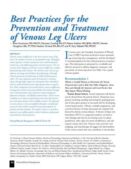
<strong>Best</strong> <strong>Practices</strong> <strong>for</strong> <strong>the</strong><br />
<strong>Prevention</strong> <strong>and</strong> <strong>Treatment</strong><br />
<strong>of</strong> <strong>Venous</strong> <strong>Leg</strong> <strong>Ulcers</strong><br />
– Brian Kunimoto, MD, FRCPC; Maureen Cooling, RN, ET;Wayne Gulliver, MD, BMSc, FRCPC; Pamela<br />
Houghton, BSc, PT, PhD; Hea<strong>the</strong>r Orsted, RN, BN, ET; <strong>and</strong> R. Gary Sibbald, MD, FRCPC<br />
ABSTRACT<br />
Chronic venous insufficiency is <strong>the</strong> most common cause <strong>of</strong> leg<br />
ulcers. Its incidence increases as <strong>the</strong> population ages. Managing<br />
venous leg ulcers involves treating <strong>the</strong> cause, optimizing local<br />
wound care, <strong>and</strong> addressing patient-centered concerns. The cornerstone<br />
<strong>of</strong> <strong>the</strong> diagnosis <strong>of</strong> chronic venous insufficiency includes<br />
demonstrating venous disease. The clinician must rule out significant<br />
coexisting arterial disease by per<strong>for</strong>ming a thorough<br />
clinical assessment <strong>and</strong> obtaining an ankle brachial pressure<br />
index. The most important aspect <strong>of</strong> treatment is resolving<br />
edema through high compression <strong>the</strong>rapy <strong>for</strong> those individuals<br />
with an ankle brachial pressure index greater than or equal to<br />
0.8. O<strong>the</strong>r components <strong>of</strong> successful chronic venous insufficiency<br />
management include increasing mobility <strong>and</strong> medical management.<br />
Selected patients may respond to surgery, biologicals,<br />
adjunctive <strong>the</strong>rapies, <strong>and</strong> lifestyle enhancements. Twelve recommendations<br />
are made incorporating current best clinical practices<br />
<strong>and</strong> expert opinion with available research. The approach<br />
to venous disease is best accomplished through a multidisciplinary<br />
team that revolves around <strong>the</strong> active participation <strong>of</strong><br />
patients <strong>and</strong> <strong>the</strong>ir families. The authors’ intent is to provide a<br />
practical, easy-to-follow guide to allow healthcare pr<strong>of</strong>essionals<br />
to provide best clinical practices.<br />
Ostomy/Wound Management 2001;47(2):34–50<br />
34 OstomyWound Management<br />
In recent years, <strong>the</strong> Canadian Association <strong>of</strong> Wound<br />
Care (CAWC) has been involved in issues surrounding<br />
venous leg ulcer management, <strong>and</strong> has developed<br />
12 recommendations <strong>for</strong> best clinical practices in patient<br />
care. This in<strong>for</strong>mation is presented in a workable <strong>and</strong><br />
effective protocol to address diagnosis, treatment, <strong>and</strong><br />
prevention <strong>of</strong> venous leg ulcers (see Table 1 <strong>for</strong> a quick<br />
reference guide).<br />
Recommendation 1<br />
Obtain a Careful History to Determine <strong>the</strong> <strong>Venous</strong><br />
Characteristics <strong>and</strong> to Rule Out O<strong>the</strong>r Diagnoses. Assess<br />
Pain <strong>and</strong> Identify <strong>the</strong> Systemic <strong>and</strong> Local Factors that<br />
May Impair Wound Healing<br />
<strong>Venous</strong> disease history. Several important risk factors<br />
can be elicited from <strong>the</strong> patient history. Numerous occupations<br />
involving st<strong>and</strong>ing <strong>and</strong> sitting <strong>for</strong> prolonged periods<br />
<strong>of</strong> time place patients at increased risk <strong>for</strong> developing<br />
venous hypertension. 1 Obesity, multiple pregnancies, <strong>and</strong><br />
a previous history <strong>of</strong> major leg trauma are acquired risk<br />
factors <strong>for</strong> chronic venous disease. 2 A history <strong>of</strong> deep vein<br />
thrombosis (DVT) or congenital weakness can lead to<br />
valve damage <strong>and</strong> may be an inciting event in edema<br />
production, o<strong>the</strong>r signs <strong>of</strong> venous insufficiency, <strong>and</strong> subsequent<br />
venous ulcer risk. Varicose veins, previous vein<br />
stripping, <strong>and</strong> sclero<strong>the</strong>rapy all suggest <strong>the</strong> abnormalities<br />
<strong>of</strong> <strong>the</strong> venous system that may contribute to <strong>the</strong> develop-<br />
Dr. Kunimoto is Clinical Assistant Pr<strong>of</strong>essor, Division <strong>of</strong> Dermatology, Department <strong>of</strong> Medicine, at The University <strong>of</strong> British Columbia,<br />
Vancouver, British Columbia, Canada. Ms. Cooling is with <strong>the</strong> Victorian Order <strong>of</strong> Nurses, Toronto, Ontario. Dr. Gulliver is Chairman <strong>of</strong><br />
Dermatology, Memorial University <strong>of</strong> Newfoundl<strong>and</strong>, Chairman <strong>of</strong> Dermatology, St. John’s Health Care Corporation, <strong>and</strong> President/Medical<br />
Director <strong>of</strong> NewLab Clinical Research Inc., St. John’s, Newfoundl<strong>and</strong>. Dr. Houghton is Assistant Pr<strong>of</strong>essor, School <strong>of</strong> Physical Therapy, University<br />
<strong>of</strong> Western Ontario, London, Ontario. Ms. Orsted is a Clinical Specialist, Skin <strong>and</strong> Wound Management, Calgary Wound Care, Calgary,<br />
Alberta. Dr. Sibbald is Director, Dermatology Day Care <strong>and</strong> Wound Healing Clinic, Sunnybrook <strong>and</strong> Women’s College Health Sciences Centre,<br />
Director <strong>of</strong> Continuing Education, Department <strong>of</strong> Medicine, <strong>and</strong> Associate Pr<strong>of</strong>essor <strong>of</strong> Medicine, University <strong>of</strong> Toronto, Toronto, Ontario.<br />
Address correspondence to: Hea<strong>the</strong>r Orsted, 9003 33rd Avenue NW, Calgary, Alberta, T3B 1M2, Canada.
TABLE 1<br />
QUICK REFERENCE GUIDE TO THE 12<br />
RECOMMENDATIONS FOR BEST PRACTICES<br />
IN THE PREVENTION AND TREATMENT OF<br />
VENOUS LEG ULCERS<br />
1. Obtain a careful history to determine <strong>the</strong> venous characteristics<br />
<strong>and</strong> rule out o<strong>the</strong>r diagnoses.Assess pain <strong>and</strong> identify <strong>the</strong> systemic<br />
<strong>and</strong> local factors that may impair wound healing.<br />
2. Determine <strong>the</strong> cause(s) <strong>of</strong> chronic venous insufficiency (CVI)<br />
based on etiology: abnormal valves (reflux), obstruction, or calf<br />
muscle pump failure.<br />
3. Per<strong>for</strong>m <strong>the</strong> ankle-brachial pressure index (ABPI) test on all<br />
patients with venous ulcers to help rule out significant arterial<br />
disease.<br />
4. Implement high compression b<strong>and</strong>aging <strong>for</strong> <strong>the</strong> management <strong>of</strong><br />
venous edema if <strong>the</strong> ABPI is ≥ 0.8.<br />
5. Use graduated compression stockings to manage <strong>and</strong> prevent<br />
venous leg edema.Wearing stockings to decrease <strong>the</strong> frequency<br />
<strong>of</strong> ulcer recurrence is important.<br />
6. Implement intermittent pneumatic compression <strong>the</strong>rapy <strong>and</strong>/or<br />
elevation <strong>of</strong> <strong>the</strong> leg as an added benefit in managing venous<br />
edema <strong>and</strong> venous leg ulcers.<br />
7. Consult with rehabilitation experts to maximize activity <strong>and</strong><br />
mobility. Consider appropriate adjunctive <strong>the</strong>rapies.<br />
8. Assess <strong>for</strong> infection <strong>and</strong> treat if indicated.<br />
9. Optimize <strong>the</strong> local wound healing environment: debridement,<br />
bacterial balance, <strong>and</strong> moisture balance. Use biological agents<br />
when <strong>the</strong> cause has been corrected <strong>and</strong> healing does not proceed<br />
at an expected rate.<br />
10. Implement medical <strong>the</strong>rapy if indicated <strong>for</strong> CVI (superficial <strong>and</strong><br />
deep thrombosis, woody fibrosis).<br />
11. Consider surgical management if significant superficial or per<strong>for</strong>ator<br />
vein disease exists in <strong>the</strong> absence <strong>of</strong> extensive deep disease.<br />
12. Communicate with <strong>the</strong> patient, <strong>the</strong> family, <strong>and</strong> <strong>the</strong> caregivers to<br />
establish realistic expectations <strong>for</strong> (non)healing.The presence or<br />
absence <strong>of</strong> a social support system is important <strong>for</strong> treatment<br />
<strong>and</strong> prevention <strong>of</strong> venous leg ulcers.<br />
ment <strong>of</strong> venous leg ulcers. Varicosities on <strong>the</strong><br />
surface may be due to superficial disease<br />
alone or in combination with per<strong>for</strong>ator<br />
<strong>and</strong>/or deep venous ulceration.<br />
Establishing a chronology <strong>of</strong> events is<br />
important. The onset <strong>of</strong> <strong>the</strong> ulcer may be<br />
traumatic or secondary to edema, infection,<br />
or a combination <strong>of</strong> factors. Asking <strong>the</strong><br />
patient if he or she experiences leg swelling<br />
by <strong>the</strong> end <strong>of</strong> <strong>the</strong> day, <strong>and</strong> <strong>for</strong> how long this<br />
has been occurring, is important. The local<br />
skin is <strong>of</strong>ten extremely itchy <strong>and</strong> may breakdown<br />
with fluid exudation. This may be <strong>the</strong><br />
inciting event <strong>for</strong> local infection. With time,<br />
<strong>the</strong> inner aspect <strong>of</strong> <strong>the</strong> ankle may become<br />
hyperpigmented with leakage <strong>of</strong> red blood<br />
cells, leaving behind hemosiderin <strong>and</strong><br />
melanin deposition. Long-st<strong>and</strong>ing<br />
changes also may include thickening <strong>of</strong><br />
<strong>the</strong> ankle with nonpitting edema (woody<br />
fibrosis) that does not resolve with <strong>the</strong><br />
recumbent position.<br />
The clinician should ask about previous<br />
topical treatments. Patients with venous<br />
disease are very susceptible to irritant <strong>and</strong><br />
allergic reactions. Common allergens<br />
include latex, perfumes, lanolin,<br />
neomycin, <strong>and</strong> o<strong>the</strong>r topical antibiotics, as<br />
well as home remedies (including alternative<br />
medicines). If an ulcer has developed,<br />
a chronology <strong>of</strong> local treatments <strong>and</strong> previous<br />
b<strong>and</strong>aging will help <strong>the</strong> clinician to<br />
determine what aspects <strong>of</strong> care have been<br />
helpful or harmful.<br />
Pain. Pain is associated with <strong>the</strong> venous<br />
ulcer. Pain also may result from phlebitis<br />
(superficial or deep), infection, or local<br />
wound factors (debridement, dressing<br />
changes, or sensitivity to one <strong>of</strong> <strong>the</strong> components<br />
<strong>of</strong> a dressing). C<strong>of</strong>actors such as<br />
arterial disease may be responsible <strong>for</strong><br />
pain. Arterial pain may be aggravated by<br />
limb elevation <strong>and</strong> produce intermittent<br />
claudication <strong>of</strong> <strong>the</strong> calf or a local tightness<br />
with walking. <strong>Venous</strong> disease is <strong>of</strong>ten associated<br />
with pain at <strong>the</strong> end <strong>of</strong> <strong>the</strong> day,<br />
Ostomy/Wound Management 2001;47(2):34–50<br />
KEY POINTS<br />
❏ Chances are that <strong>the</strong> number <strong>of</strong> individuals with peripheral vascular<br />
disease <strong>and</strong> venous ulcers will increase at <strong>the</strong> same rate as <strong>the</strong> proportion<br />
<strong>of</strong> older adults.<br />
❏ This extensive review <strong>of</strong> <strong>the</strong> literature <strong>and</strong> its resultant recommendations<br />
<strong>for</strong> practice serve as a reminder that prompt diagnosis <strong>and</strong> appropriate<br />
care are crucial to preventing unnecessary pain <strong>and</strong> suffering.<br />
❏ Wound care experts familiar with <strong>the</strong> principles <strong>of</strong> venous ulcer prevention<br />
<strong>and</strong> treatment detailed in this article are encouraged to<br />
share <strong>the</strong>m with healthcare pr<strong>of</strong>essionals who are not, <strong>the</strong>reby<br />
reducing <strong>the</strong> incidence <strong>of</strong> persons experiencing a delay in receiving<br />
optimal care.<br />
February 2001 Vol. 47 Issue 2 35
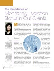
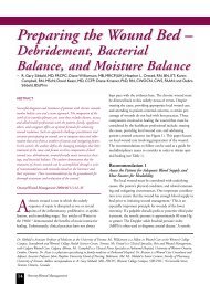
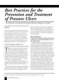
Figure 1<br />
This photo demonstrates skin changes in a patient<br />
with chronic venous insufficiency.<br />
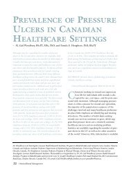
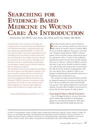
Figure 2<br />
It is important to recognize atrophie blanche, which is<br />
shown above, because <strong>the</strong> skin is fragile <strong>and</strong> ulcers<br />
may develop after only minor trauma.<br />
especially with prolonged st<strong>and</strong>ing or sitting. The resulting<br />
edema <strong>of</strong>ten is reduced by recumbency <strong>and</strong> elevation<br />
<strong>of</strong> <strong>the</strong> limb.<br />
Factors that may affect wound healing. Margolis et al 3<br />
defined several factors that were associated with an increased<br />
incidence <strong>of</strong> nonhealing venous leg ulcers. Four hundred<br />
thirty-three consecutive patients had a number <strong>of</strong> factors<br />
analyzed using a multiple variant logistic regression model.<br />
In order <strong>of</strong> odds ratio, <strong>the</strong> following six risk factors obtained<br />
statistical significance:<br />
1. History <strong>of</strong> venous ligation or stripping – odds<br />
ratio: 4.58<br />
2. Hip or knee surgery – odds ratio: 3.52<br />
3. ABPI < 0.8 – odds ratio: 3.52<br />
4. Fibrin (yellow > 50% <strong>of</strong> <strong>the</strong> ulcer base) – odds<br />
ratio: 3.42<br />
5. Larger size (area) – odds ratio: 1.19<br />
6. Longer duration in months – odds ratio: 1.09.<br />
An odds ratio <strong>of</strong> 4.58 means that a patient with a history<br />
<strong>of</strong> venous ligation or stripping is 4.58 times less likely<br />
to heal by week 24 compared to patients who have not<br />
had this procedure done. In this analysis, diabetes melli-<br />
36 OstomyWound Management<br />
tus, previous deep vein thrombosis, visible varicose veins,<br />
lipodermatosclerosis, <strong>and</strong> undermined wound margin did<br />
not make a significant difference.<br />
Building blocks such as protein, vitamins, <strong>and</strong> trace<br />
metals are required to heal a wound. The functioning <strong>of</strong><br />
<strong>the</strong> wound healing process requires an adequate supply <strong>of</strong><br />
<strong>the</strong>se building blocks. A nutritionist or dietitian should<br />
be consulted if nutritional deficiency is thought to be significant<br />
enough to possibly impair wound healing.<br />
Deficiencies in <strong>the</strong> intake <strong>of</strong> protein <strong>and</strong> vitamin intake<br />
are common in <strong>the</strong> elderly. Managing <strong>the</strong>se deficiencies<br />
may make a difference between a healing <strong>and</strong> a nonhealing<br />
wound even in <strong>the</strong> presence <strong>of</strong> best clinical practices.<br />
Drug history is important. Medications such as prednisone<br />
<strong>and</strong> immunosuppressive agents can have a pr<strong>of</strong>oundly<br />
negative influence. Many patients self-medicate<br />
with antiseptic cleaning agents that have been shown to<br />
be toxic to fibroblast cells. Similarly, patients may be<br />
applying potent topical steroids on <strong>the</strong> wound that are<br />
designed <strong>for</strong> <strong>the</strong> surrounding skin.<br />
Lastly, patients <strong>and</strong> <strong>the</strong>ir caregivers may believe in <strong>the</strong><br />
traditional dogma <strong>of</strong> leaving wounds open to <strong>the</strong> air<br />
allowing <strong>the</strong> <strong>for</strong>mation <strong>of</strong> a dry crust that inhibits healing.<br />
Periulcer assessment. The appearance <strong>of</strong> <strong>the</strong> surrounding<br />
skin is important in <strong>the</strong> clinical diagnosis <strong>of</strong><br />
venous ulcers. Early venous insufficiency presents with<br />
pitting edema that should be distinguished from congestive<br />
heart failure <strong>and</strong> o<strong>the</strong>r systemic diseases. As a result<br />
<strong>of</strong> <strong>the</strong> deposition <strong>of</strong> iron <strong>and</strong> melanin in <strong>the</strong> skin, irregular<br />
pigmentation in <strong>the</strong> gaiter area (lower calf) develops<br />
as <strong>the</strong> disease progresses (see Figure 1).<br />
Lipodermatosclerosis (bound down appearance with atrophy,<br />
telangiectasia, hyperpigmentation, <strong>and</strong> hypopigmentation)<br />
<strong>of</strong>ten occurs later as <strong>the</strong> progressive deposition <strong>of</strong><br />
fibrin in <strong>the</strong> deep dermis <strong>and</strong> fat results in a woody<br />
induration <strong>of</strong> <strong>the</strong> gaiter area <strong>of</strong> <strong>the</strong> calf. This may contribute<br />
to <strong>the</strong> appearance <strong>of</strong> <strong>the</strong> so-called “inverted champagne<br />
bottle leg.” Often accompanying this is <strong>the</strong><br />
appearance <strong>of</strong> atrophie blanche presenting as white areas<br />
<strong>of</strong> extremely thin skin dotted with tiny tortuous blood<br />
vessels. The recognition <strong>of</strong> atrophie blanche (see Figure<br />
2) is important because <strong>the</strong> thin skin is fragile, <strong>and</strong> ulcers<br />
may develop after only minor trauma. Eczema, commonly<br />
known as “stasis dermatitis,” may appear in <strong>the</strong> gaiter<br />
area. The “stasis” is, in fact, a misnomer, as true stasis <strong>of</strong><br />
<strong>the</strong> blood does not exist, but pooling <strong>of</strong> fluid <strong>and</strong><br />
extravasation <strong>of</strong> red blood cells is present in <strong>the</strong> underly-
TABLE 2<br />
CLASSIFICATION OF CVI<br />
Class Type <strong>of</strong> CVI Symptoms<br />
0<br />
1<br />
2<br />
3<br />
Asymptomatic<br />
Mild<br />
Moderate<br />
Severe<br />
Nil<br />
Mild swelling, heaviness, local or generalized<br />
dilatation <strong>of</strong> subcutaneous veins<br />
Hyperpigmentation, moderate brawny edema,<br />
subcutaneous fibrosis<br />
Chronic distal leg pain with ulceration or<br />
pre-ulcerative changes, eczematoid changes,<br />
<strong>and</strong>/or severe edema<br />
From Iafrati M, Welch H, O’Donnell TF, Belkin M, Umphrey S, McLaughlin R..<br />
Correlation <strong>of</strong> venous noninvasive tests with <strong>the</strong> Society <strong>for</strong> Vascular Surgery/International<br />
Society <strong>for</strong> Cardiovascular Surgery. Clinical classification <strong>of</strong> chronic venous insufficiency.<br />
Journal <strong>of</strong> Vascular Surgery; 1994;19(6):1001-1007.<br />
ing dermis that may lead to irritation <strong>of</strong> <strong>the</strong> skin. The<br />
characteristics <strong>of</strong> <strong>the</strong> skin <strong>and</strong> local surrounding structures<br />
may be grouped into four classes based on disease<br />
characteristics that <strong>of</strong>ten help define <strong>the</strong> strength <strong>of</strong> compression<br />
garments (see Table 2).<br />
Recommendation 2<br />
Determine <strong>the</strong> Cause(s) <strong>of</strong> Chronic <strong>Venous</strong> Insufficiency<br />
Based on Etiology: Abnormal Valves (Reflux),<br />
Obstruction, or Calf Muscle Pump Failure<br />
<strong>Venous</strong> hypertension is primarily caused by one <strong>of</strong><br />
three major pathologies, which include:<br />
1. Reflux, known as valve dysfunction<br />
2. Obstruction, which is ei<strong>the</strong>r complete or partial<br />
blockage <strong>of</strong> <strong>the</strong> deep veins. Reflux <strong>and</strong> obstruction<br />
may co-exist.<br />
3. Failure <strong>of</strong> calf muscle pump function related to<br />
decreased activity, paralysis, ankle joint de<strong>for</strong>mity,<br />
or decreased range <strong>of</strong> motion. 4<br />
Chronic venous disease can be congenital or acquired.<br />
Recurrent thrombophlebitis may alert <strong>the</strong> clinician to <strong>the</strong><br />
presence or protein C, S, or Factor 5 (Leiden) deficiency.<br />
Valve dysfunction may be due to a congenital weakness or<br />
acquired secondary to previous episodes <strong>of</strong> thrombophlebitis.<br />
Valves also can be damaged from previous trauma<br />
or infection. Outflow obstruction, such as increased<br />
local pressure, can result from obesity <strong>and</strong> pregnancy.<br />
Damage to <strong>the</strong> venous outflow system, especially in <strong>the</strong><br />
pelvic region, may result from malignancy or radio<strong>the</strong>rapy.<br />
The least reported cause <strong>of</strong> venous<br />
hypertension is musculoskeletal changes<br />
that can lead to calf muscle pump failure.<br />
The dynamics <strong>of</strong> <strong>the</strong> calf muscle pump<br />
are adversely affected by changes that<br />
<strong>of</strong>ten accompany major injuries, neurological<br />
disease, vascular insufficiency,<br />
myositis, <strong>and</strong> bone <strong>and</strong> joint pain. The<br />
calf muscles rapidly waste <strong>and</strong> weaken<br />
with disuse. 5 Even <strong>the</strong> change in gait<br />
related to a painful ulcer can exacerbate<br />
<strong>the</strong> venous hypertension <strong>and</strong> cause calf<br />
muscle disuse atrophy. Back et al 6 stated<br />
that a normal walking motion may be<br />
required <strong>for</strong> full functional activation <strong>of</strong><br />
<strong>the</strong> calf muscle pump <strong>and</strong> that ankle dorsiflexion<br />
past <strong>the</strong> 90-degree position is<br />
needed <strong>for</strong> a normal walking motion.<br />
Investigation <strong>of</strong> <strong>the</strong> venous system should start with a<br />
clinical examination. Many investigations are noninvasive<br />
<strong>and</strong> can assess venous function.<br />
The simplest way to determine venous reflux is to have<br />
<strong>the</strong> patient lie flat <strong>and</strong> raise <strong>the</strong> leg 45 degrees to drain<br />
<strong>the</strong> blood from <strong>the</strong> long saphenous vein (approximately<br />
30 seconds). A rubber tourniquet is <strong>the</strong>n applied above<br />
<strong>the</strong> knee <strong>and</strong> <strong>the</strong> patient is asked to st<strong>and</strong> up. The<br />
tourniquet is released <strong>and</strong> if <strong>the</strong> long saphenous vein fills<br />
in less than 20 seconds, venous insufficiency is present<br />
(superficial or deep). A h<strong>and</strong>-held Doppler can be used<br />
to locate abnormal venous areas. 7 The patient should<br />
st<strong>and</strong> upright with weight on <strong>the</strong> o<strong>the</strong>r leg <strong>and</strong> <strong>the</strong> knee<br />
slightly flexed. The probe is placed over <strong>the</strong> saphen<strong>of</strong>emoral<br />
or saphenopopliteal junction, followed by<br />
manual squeezing <strong>and</strong> release <strong>of</strong> <strong>the</strong> calf muscle. A prolonged<br />
regurgitation indicates reflux. A similar procedure<br />
may be used as a rough indicator <strong>of</strong> per<strong>for</strong>ator incompetence<br />
as well. 7<br />
Color duplex Dopplers are used in <strong>the</strong> vascular lab by<br />
combining pulsed ultrasound systems with real time D<br />
mode to examine flow patterns in precisely defined<br />
areas. 8 This technique can demonstrate <strong>the</strong> failure <strong>of</strong> a<br />
thrombosed vein to collapse under direct compression, as<br />
well as visualize <strong>the</strong> thrombus within <strong>the</strong> vessel wall <strong>and</strong><br />
detect <strong>the</strong> absence <strong>of</strong> or abnormal venous pulsation on<br />
Doppler scanning. This technique is most accurate in<br />
identifying thrombi in <strong>the</strong> common femoral vein to <strong>the</strong><br />
popliteal vein, but less reliable in <strong>the</strong> calf. Duplex systems<br />
February 2001 Vol. 47 Issue 2 37
can be added to color frequency mapping, which<br />
allows instant visualization <strong>of</strong> blood flow <strong>and</strong> its<br />
direction to accurately assess reflux. 8<br />
One <strong>of</strong> <strong>the</strong> most common tests to determine <strong>the</strong><br />
efficacy <strong>of</strong> <strong>the</strong> calf pump is air plethysmography<br />
(APG). Air plethysmography can differentiate<br />
between superficial <strong>and</strong> deep venous disease, assess<br />
<strong>the</strong> degree <strong>of</strong> valvular insufficiency <strong>and</strong> <strong>the</strong> efficiency<br />
<strong>of</strong> <strong>the</strong> calf muscle pump. This technique also can<br />
provide a noninvasive estimate <strong>of</strong> ambulatory<br />
venous pressure. 9<br />
Recommendation 3<br />
Per<strong>for</strong>m <strong>the</strong> Ankle-Brachial Pressure Index Test<br />
on All Patients with <strong>Venous</strong> <strong>Ulcers</strong> to Help Rule<br />
Out Significant Arterial Disease<br />
A physical examination <strong>of</strong> <strong>the</strong> feet <strong>and</strong> legs will<br />
help to detect clinical signs <strong>of</strong> vascular compromise.<br />
Arterial disease is <strong>of</strong>ten marked by a vascular<br />
dilation (flush) that blanches with elevation.<br />
Loss <strong>of</strong> hair <strong>and</strong> thickened nails with decreased<br />
nail luster may be evident. On palpation, <strong>the</strong> foot<br />
is characteristically cold with a loss <strong>of</strong> pulses.<br />
Microcirculatory supply can be tested by pressing<br />
a finger on <strong>the</strong> dorsum <strong>of</strong> <strong>the</strong> dependent foot to<br />
produce a noticeable blanching. Normally, ery<strong>the</strong>ma<br />
should return within 5 seconds, <strong>and</strong> if a<br />
delay is noted, decreased local perfusion (microcirculation<br />
time) is present. Distal gangrene <strong>of</strong><br />
<strong>the</strong> toes with a palpable pulse or adequate circulation<br />
may indicate microemboli from proximal<br />
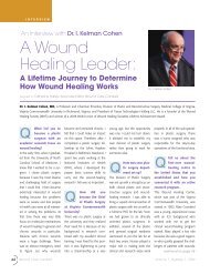
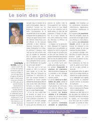
a<strong>the</strong>romatous plaques. The ankle brachial pressure<br />
index (ABPI) is determined by a h<strong>and</strong>-held<br />
Doppler <strong>and</strong> blood pressure cuff (see Figure 3)<br />
<strong>and</strong> is important as part <strong>of</strong> <strong>the</strong> vascular assessment<br />
<strong>of</strong> <strong>the</strong> lower leg. The ABPI is an important component<br />
<strong>of</strong> lower leg assessment <strong>and</strong> should be per<strong>for</strong>med only by<br />
a skilled healthcare practitioner (see Table 3). 10<br />
In <strong>the</strong> vascular lab, reasonable healability potential <strong>for</strong><br />
patients requires a toe pressure <strong>of</strong> 30 mm Hg (in people<br />
with diabetes > 45 mm Hg) or a transcutaneous oxygen<br />
saturation <strong>of</strong> > 30% (see Table 4). 11 A palpable pedal<br />
pulse indicates approximately 80 mm Hg <strong>and</strong> should be<br />
sufficient <strong>for</strong> healing in most patients. Checking sequential<br />
arterial circulation <strong>for</strong> wave<strong>for</strong>ms is important. A<br />
normal Doppler triphasic wave<strong>for</strong>m will become blunted<br />
<strong>and</strong> biphasic/monophasic, <strong>and</strong> pressures will drop dispro-<br />
38 OstomyWound Management<br />
TABLE 3<br />
ABPI PROCEDURE<br />
Step 1<br />
Ensure that <strong>the</strong> patient is lying flat <strong>and</strong> is com<strong>for</strong>table.<br />
Step 2<br />
Secure <strong>the</strong> appropriate size blood pressure cuff around <strong>the</strong><br />
arm (pediatric or oversized cuffs may be indicated).<br />
Apply ultrasound gel over brachial pulse.<br />
Slowly move <strong>the</strong> Doppler probe at a 45-degree angle to<br />
<strong>the</strong> flow over area until a good signal is obtained.<br />
Inflate <strong>the</strong> cuff until Doppler signal disappears, <strong>the</strong>n gradually<br />
release <strong>the</strong> pressure valve until <strong>the</strong> signal returns.<br />
This is <strong>the</strong> brachial systolic pressure.<br />
Step 3<br />
Examine <strong>the</strong> foot <strong>for</strong> posterior tibial <strong>and</strong> dorsalis pedis<br />
pulse using fingers <strong>and</strong>/or Doppler probe.<br />
Step 4<br />
Secure <strong>the</strong> blood pressure cuff just above <strong>the</strong> ankle.<br />
Locate <strong>the</strong> posterior tibial <strong>and</strong> dorsalis pedis pulse using<br />
Doppler probe <strong>and</strong> gel.<br />
Inflate <strong>the</strong> cuff until <strong>the</strong> signal disappears, <strong>the</strong>n gradually<br />
release <strong>the</strong> pressure valve until <strong>the</strong> signal returns.<br />
Repeat with second pulse.This is <strong>the</strong> ankle systolic<br />
pressure<br />
Step 5<br />
To calculate <strong>the</strong> ABPI, divide <strong>the</strong> ankle systolic pressure by<br />
<strong>the</strong> brachial systolic pressure.<br />
Caution: Unusually high readings may be obtained in<br />
elderly or diabetic patients as <strong>the</strong> cuff may not fully compress<br />
<strong>the</strong> calcified vessels.<br />
Adapted from M<strong>of</strong>fatt C, O’Hare L. Ankle pulses are not sufficient to detect<br />
impaired arterial circulation in patients with leg ulcers. Journal <strong>of</strong> Wound<br />
Care.1995;4(3):134–138.<br />
portionately distal to possible bipassable or dilatable<br />
lesions with angioplasty.<br />
Any wound, acute or chronic, affected by ischemia as<br />
a result <strong>of</strong> severe arterial insufficiency, will not heal no<br />
matter what local measures are employed. If <strong>the</strong> arterial disease<br />
is considered uncorrectable or if <strong>the</strong> patient’s general<br />
health precludes surgery, management becomes palliative.<br />
Recommendation 4<br />
Implement High Compression B<strong>and</strong>aging <strong>for</strong> <strong>the</strong><br />
Management <strong>of</strong> <strong>Venous</strong> Edema if <strong>the</strong> ABPI ≥ 0.8<br />
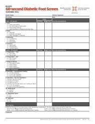
All compression systems must create a pressure gradient<br />
from ankle to knee. The Law <strong>of</strong> Laplace (see Figure 4)
Figure 3<br />
This photo demonstrates how to obtain an ankle<br />
brachial pressure index.<br />
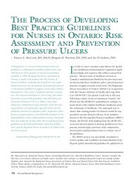
ma<strong>the</strong>matically relates b<strong>and</strong>age tension <strong>and</strong> <strong>the</strong> number <strong>of</strong><br />
layers to inverse <strong>of</strong> <strong>the</strong> radius <strong>of</strong> <strong>the</strong> leg <strong>and</strong> b<strong>and</strong>age width.<br />
Thus, if <strong>the</strong> b<strong>and</strong>age tension is constant as one winds<br />
<strong>the</strong> b<strong>and</strong>age up <strong>the</strong> leg, a compression gradient will naturally<br />
develop because <strong>the</strong> smallest limb radius is at <strong>the</strong><br />
ankle area just proximal to <strong>the</strong> ankle joint. Progressively<br />
larger radii are encountered up <strong>the</strong> leg, resulting in lesser<br />
degrees <strong>of</strong> compression given a constant b<strong>and</strong>age tension.<br />
This gradient <strong>of</strong> pressure provides support against<br />
venous hypertension that is greatest at <strong>the</strong> ankles when<br />
<strong>the</strong> patient is st<strong>and</strong>ing. A compression system must be<br />
capable <strong>of</strong> exerting at least 30 mm Hg <strong>of</strong> pressure at <strong>the</strong><br />
level <strong>of</strong> <strong>the</strong> ankle to reliably prevent fluid exudation. 12–15<br />
The Fletcher, Cullum, <strong>and</strong> Sheldon 16 systematic review<br />
<strong>of</strong> compression treatment <strong>for</strong> venous ulcers states:<br />
1. Compression treatment increases <strong>the</strong> ulcer healing<br />
as compared to no compression.<br />
2. High compression is<br />
more effective than low<br />
compression that should<br />
only be used in <strong>the</strong><br />
absence <strong>of</strong> significant<br />
arterial disease.<br />
3. No clear differences in<br />
effect from different<br />
types <strong>of</strong> compression<br />
system (multi-layer <strong>and</strong><br />
short-stretch b<strong>and</strong>ages,<br />
Unna’s boot) have been<br />
shown. Since <strong>the</strong> publication<br />
<strong>of</strong> this article,<br />
however, <strong>the</strong> updated<br />
Cochrane Library web-<br />
Ankle Brachial<br />
Pressure Index<br />
(ABPI)<br />
> 0.8<br />
> 0.6–0.8<br />
> 0.4-0.6<br />
< 0.4<br />
site(www.updates<strong>of</strong>tware.com/cochrane/cochrane-frame.html) indicates that <strong>the</strong>re may be an advantage to<br />
using elastic systems.<br />
4. Intermittent <strong>and</strong> pneumatic compression<br />
appears to be a useful adjunct to b<strong>and</strong>aging.<br />
5. Ra<strong>the</strong>r than advocate one particular system, <strong>the</strong><br />
increased use <strong>of</strong> any correctly applied high compression<br />
treatment should be promoted.<br />
Compression systems may be classified into three groups:<br />
short-stretch b<strong>and</strong>ages (SSB), long-stretch b<strong>and</strong>ages (LSB),<br />
<strong>and</strong> stockings. If <strong>the</strong> limb affected by <strong>the</strong> ulcer is acutely<br />
edematous, most experts believe that using a SSB system is<br />
preferred. 14,15 The SSB provides little or no elasticity. The<br />
contracting calf muscle exerts a high pressure against <strong>the</strong><br />
fixed resistance <strong>of</strong> <strong>the</strong> SSB. This working pressure drives<br />
blood in <strong>the</strong> deep veins upwards. For <strong>the</strong> same reason, when<br />
<strong>the</strong> calf muscle relaxes, <strong>the</strong> b<strong>and</strong>age does not continue to<br />
exert pressure. This low “resting pressure” facilitates deep<br />
venous filling. 14,15 Short-stretch b<strong>and</strong>age systems require<br />
patients to be ambulatory. Without a calf muscle capable <strong>of</strong><br />
contracting, <strong>the</strong> nonelastic b<strong>and</strong>age becomes less effective,<br />
but edema may fill a fixed volume <strong>and</strong> pressure exerted can<br />
prevent fur<strong>the</strong>r fluid exudation. Patients who tend to shuffle<br />
around need to be trained to walk properly, making sure<br />
<strong>the</strong>y push <strong>of</strong>f with <strong>the</strong>ir toes. Similarly, those patients with<br />
ankle joints stiffened by arthritis or old injuries may not be<br />
good c<strong>and</strong>idates <strong>for</strong> SSB systems.<br />
Elastic or LSBs are more commonly used than SSB<br />
systems in North America <strong>and</strong> <strong>the</strong> United Kingdom. The<br />
“four-layer b<strong>and</strong>age” is popular because <strong>of</strong> its ability to<br />
TABLE 4<br />
VASCULAR SCREENING IN ASSESSMENT<br />
OF ISCHEMIC RISK<br />
Toe<br />
Pressure<br />
(mm Hg)<br />
> 55 mg Hg<br />
> 40 mm Hg<br />
> 20 mm Hg<br />
< 20 mm Hg<br />
Ankle<br />
Doppler<br />
Wave<strong>for</strong>m<br />
Normal<br />
Biphasic or<br />
monophasic<br />
Biphasic or<br />
monophasic<br />
Monophasic<br />
Transcutaneous<br />
Oxygen<br />
Saturation<br />
> 40%<br />
30-40%<br />
20-30%<br />
< 20%<br />
Risk<br />
Low<br />
Low<br />
High<br />
Severe<br />
*Based on personal experience (R.G. Sibbald)<br />
Adapated with permission from Sykes MT, Godsey JB. Vascular evaluation <strong>of</strong> <strong>the</strong> diabetic foot. Clin<br />
Podiatr Med Surg. 1998; 15(1):49–83.<br />
February 2001 Vol. 47 Issue 2 39
Sub-b<strong>and</strong>age = N (number <strong>of</strong> b<strong>and</strong>age layers) x T (b<strong>and</strong>age tension) x Constant<br />
R (radius <strong>of</strong> <strong>the</strong> leg) x B (b<strong>and</strong>age width)<br />
Figure 4<br />
The Law <strong>of</strong> Laplace, demonstrated above, ma<strong>the</strong>matically relates b<strong>and</strong>age tension <strong>and</strong> <strong>the</strong> number <strong>of</strong> layers to inverse <strong>of</strong> <strong>the</strong> radius <strong>of</strong> <strong>the</strong><br />
leg <strong>and</strong> b<strong>and</strong>age width.<br />
maintain high compression over several days up to a<br />
week. This reduces <strong>the</strong> frequency <strong>of</strong> dressing changes, a<br />
great advantage <strong>for</strong> home-care nursing. Because <strong>of</strong> elasticity,<br />
<strong>the</strong> four-layer b<strong>and</strong>age <strong>and</strong> o<strong>the</strong>r LSBs continue to<br />
exert compression even when <strong>the</strong> leg is elevated. This can<br />
be a problem if significant arterial insufficiency exists. As<br />
a result, <strong>the</strong> four-layer b<strong>and</strong>age is not recommended <strong>for</strong><br />
use in patients with an ABPI <strong>of</strong> less than 0.8. All compression<br />
systems require trained personnel <strong>for</strong> application.<br />
O<strong>the</strong>r LSB systems are capable <strong>of</strong> lower levels <strong>of</strong> compression.<br />
These systems may be used with caution in <strong>the</strong><br />
presence <strong>of</strong> moderate arterial insufficiency (ABPI > 0.5).<br />
Like o<strong>the</strong>r b<strong>and</strong>ages, <strong>the</strong>y may be left on <strong>for</strong> a week at a<br />
time. In <strong>the</strong> presence <strong>of</strong> severe arterial disease (ABPI < 0.5),<br />
lower compression systems are contraindicated.<br />
Recommendation 5<br />
Use Graduated Compression Stockings to Manage <strong>and</strong><br />
Prevent <strong>Venous</strong> <strong>Leg</strong> Edema. Wearing Stockings to<br />
Decrease <strong>the</strong> Frequency <strong>of</strong> Ulcer Recurrence is Important<br />
Once <strong>the</strong> venous ulcer has healed, <strong>the</strong> focus must shift<br />
toward prevention. Graduated compression stockings<br />
(GCS) are <strong>of</strong> proven value in managing venous hypertension.<br />
17–19 Adherence to GCS has been shown to decrease<br />
<strong>the</strong> frequency <strong>of</strong> recurrent ulcers. 20,21<br />
Many experts in <strong>the</strong> field suggest compression levels <strong>of</strong><br />
20 mm Hg to 40 mm Hg <strong>for</strong> venous insufficiency.<br />
Evidence suggests that stockings need to extend higher<br />
than <strong>the</strong> knee in <strong>the</strong> majority <strong>of</strong> patients.<br />
Patient compliance with <strong>the</strong> use <strong>of</strong> GCS is a major<br />
issue. Patients should be told that stockings must be<br />
applied first thing in <strong>the</strong> morning <strong>and</strong> removed in <strong>the</strong><br />
evening. Several mechanical devices are available that<br />
facilitate <strong>the</strong> application <strong>of</strong> <strong>the</strong> garments even if a small<br />
ulcer is present. These can aid patients who suffer from<br />
arthritis <strong>of</strong> <strong>the</strong> h<strong>and</strong>s or poor flexibility <strong>of</strong> major joints.<br />
Most stockings have a usable life <strong>of</strong> about 4 to 6 months.<br />
Adequate compression beyond this time cannot be guaranteed.<br />
The cost <strong>of</strong> <strong>the</strong> stockings should be justified to<br />
<strong>the</strong> patient as <strong>the</strong>y <strong>of</strong>fset <strong>the</strong> greater costs <strong>and</strong> decreased<br />
quality <strong>of</strong> life associated with managing recurrences.<br />
40 OstomyWound Management<br />
Some experts on <strong>the</strong> CAWC panel use a nylon or cotton<br />
undersleeve to increase <strong>the</strong> compression <strong>of</strong> lighter<br />
dress support hose (15 mm Hg to 20 mm Hg is effectively<br />
increased to 25 mm Hg to 30 mm Hg).<br />
Alternatively, two dress support hose knee-highs can be<br />
put on top <strong>of</strong> each o<strong>the</strong>r to achieve higher compression<br />
or one pair can be mid-thigh length to decrease edema<br />
above <strong>the</strong> knee. Zippered stockings are helpful to some<br />
patients, while o<strong>the</strong>rs may need a program <strong>of</strong> leg elevation<br />
<strong>and</strong> intermittent pneumatic compression if <strong>the</strong>y cannot<br />
tolerate stockings. Patients must recognize that<br />
venous ulcer prevention means “compression <strong>for</strong> life.”<br />
Recommendation 6<br />
Implement Intermittent Pneumatic Compression Therapy<br />
<strong>and</strong>/or Elevation <strong>of</strong> <strong>the</strong> <strong>Leg</strong> as an Added Benefit in<br />
Managing <strong>Venous</strong> Edema <strong>and</strong> <strong>Venous</strong> <strong>Leg</strong> <strong>Ulcers</strong><br />
More than occasionally, patients may require higher<br />
levels <strong>of</strong> compression than <strong>the</strong>y can com<strong>for</strong>tably tolerate.<br />
Intolerable patient discom<strong>for</strong>t with <strong>the</strong> b<strong>and</strong>age system<br />
makes compliance an issue. The pneumatic compression<br />
(PC) device may be useful as an adjunct to compression<br />
b<strong>and</strong>aging whe<strong>the</strong>r used alone or as an alternative to<br />
compression b<strong>and</strong>aging or stockings in patients who are<br />
relatively immobile <strong>and</strong>, <strong>the</strong>re<strong>for</strong>e, unable to activate <strong>the</strong><br />
calf muscle pump. The elevation <strong>of</strong> <strong>the</strong> affected leg above<br />
<strong>the</strong> level <strong>of</strong> <strong>the</strong> heart also will help reduce edema.<br />
A PC device consists <strong>of</strong> an inflatable sleeve that is<br />
placed around <strong>the</strong> limb <strong>and</strong> inflated to a preset pressure<br />
(30 mm Hg to 60 mm Hg). One type <strong>of</strong> PC, intermittent<br />
pneumatic compression (IPC), involves one chamber<br />
that is inflated intermittently; <strong>the</strong> o<strong>the</strong>r type,<br />
sequential pneumatic compression (SPC), involves a<br />
series <strong>of</strong> chambers that are inflated sequentially in a distal<br />
to proximal direction. This may be used to quickly<br />
reduce <strong>the</strong> volume <strong>of</strong> <strong>the</strong> leg prior to applying <strong>the</strong> compression<br />
b<strong>and</strong>ages or graduated compression stockings.<br />
It is a useful alternative to compression b<strong>and</strong>ages in<br />
patients who lack good mobility <strong>and</strong> cannot walk<br />
around to activate <strong>the</strong> calf muscle pump. It is also useful<br />
in managing lymphedema.
TABLE 5<br />
AN APPROACH TO<br />
CLASSIFICATION AND TREATMENT<br />
OF ANKLE JOINT MOBILITY<br />
1. If ankle joint mobility is present, <strong>and</strong> an SSB system is<br />
being used, <strong>the</strong> patient should be encouraged to raise<br />
both heels <strong>of</strong>f <strong>the</strong> ground while in a st<strong>and</strong>ing position.<br />
The shuffling gait that is so common among <strong>the</strong> elderly<br />
should be discouraged.<br />
2. If joint mobility is reduced but potential <strong>for</strong> improvement<br />
is evident, a physical <strong>the</strong>rapist may be able to<br />
loosen s<strong>of</strong>t-tissue contractures through <strong>the</strong> use <strong>of</strong><br />
physio<strong>the</strong>rapy.<br />
3. In patients with reduced or no ankle joint mobility,<br />
physical <strong>the</strong>rapy <strong>and</strong> intermittent pneumatic compression<br />
should be considered.<br />
One r<strong>and</strong>omized, controlled study compared healing<br />
rates <strong>for</strong> 24 patients using moist occlusive dressings <strong>and</strong><br />
graduated compression stockings (30 mm Hg to 40 mm<br />
Hg) with 21 patients using <strong>the</strong> same treatment plus<br />
sequential PC <strong>for</strong> a total <strong>of</strong> 4 hours per day. The treatment<br />
period lasted 3 months. Only one patient in <strong>the</strong><br />
control group completely healed compared to 10 <strong>of</strong> <strong>the</strong><br />
21 in <strong>the</strong> PC group (P = 0.009, Fischer’s exact probability<br />
test). 22 Pneumatic compression may be a useful adjunct<br />
that complements compression b<strong>and</strong>aging or stocking<br />
<strong>the</strong>rapy <strong>for</strong> difficult-to-control edema. 23 R<strong>and</strong>omized<br />
controlled trials confirm <strong>the</strong> efficacy <strong>of</strong> both SPC or<br />
IPC <strong>the</strong>rapy as an adjunct in managing venous leg ulcers.<br />
Pneumatic compression units are expensive, although<br />
rentals are available. Intermittent pneumatic compression<br />
<strong>the</strong>rapy is contraindicated in <strong>the</strong> presence <strong>of</strong> significant<br />
arterial insufficiency, edema due to congestive heart failure,<br />
active phlebitis, deep vein thrombosis, or <strong>the</strong> presence<br />
<strong>of</strong> localized wound infection or cellulitis.<br />
Recommendation 7<br />
Consult with Rehabilitation Experts to Maximize Activity<br />
<strong>and</strong> Mobility. Consider Appropriate Adjunctive Therapies.<br />
Activity <strong>and</strong> mobility. Altered or inefficient gait patterns,<br />
resulting in a walking disability, occur more frequently<br />
with advancing age. Alex<strong>and</strong>er states that 8% to<br />
19% <strong>of</strong> noninstitutionalized older adults <strong>and</strong> 63% <strong>of</strong><br />
institutionalized older adults have difficulty walking or<br />
require assistance in order to ambulate. 24<br />
The ankle joint is equivalent to <strong>the</strong> hinge component<br />
<strong>of</strong> <strong>the</strong> calf muscle pump, <strong>and</strong> <strong>the</strong>re<strong>for</strong>e, ankle dorsiflexion<br />
<strong>and</strong> plantar flexion are essential <strong>for</strong> <strong>the</strong> muscle pump<br />
to function efficiently. Limited ankle range <strong>of</strong> motion is<br />
known to exacerbate venous congestion <strong>and</strong> edema <strong>for</strong>mation<br />
in patients with chronic venous insufficiency.<br />
Individuals lacking ankle mobility tend to externally<br />
rotate <strong>the</strong> hip <strong>and</strong> shuffle around, barely lifting <strong>the</strong>ir feet<br />
<strong>of</strong>f <strong>the</strong> floor. Fur<strong>the</strong>rmore, <strong>the</strong> ability <strong>of</strong> compression b<strong>and</strong>ages,<br />
such as <strong>the</strong> SSB system, to enhance venous return<br />
is compromised when ankle flexion is restricted. Chronic<br />
venous insufficiency itself may contribute to ankle immobility<br />
through <strong>the</strong> deposition <strong>of</strong> fibrotic tissue.<br />
If ankle joint mobility is normal, <strong>and</strong> an SSB system<br />
is being used, <strong>the</strong> patient should be encouraged to contract<br />
<strong>the</strong> calf muscle while in a st<strong>and</strong>ing position, causing<br />
both heels to raise <strong>of</strong>f <strong>the</strong> ground. Rehabilitation<br />
should include gait re-education that promotes proper<br />
heel-to-toe gait pattern <strong>and</strong> discourages <strong>the</strong> shuffling<br />
gait that is so common in this elderly patient population.<br />
If joint mobility is reduced due to s<strong>of</strong>t tissue contractures,<br />
a physical <strong>the</strong>rapist may per<strong>for</strong>m manual <strong>and</strong><br />
<strong>the</strong>rmal joint mobilization techniques to optimize <strong>the</strong><br />
range <strong>of</strong> motion <strong>of</strong> ankle dorsiflexion <strong>and</strong> plantar flexion<br />
(see Table 5).<br />
Walking involves passive <strong>and</strong> active ankle joint motion<br />
as <strong>the</strong> weight <strong>of</strong> <strong>the</strong> body generates <strong>the</strong> <strong>for</strong>ce that drives<br />
<strong>the</strong> ankle joint pump. The power <strong>of</strong> <strong>the</strong> moving ankle<br />
joint generates <strong>the</strong> pumping <strong>for</strong>ce by virtue <strong>of</strong> <strong>the</strong><br />
anatomic relations <strong>of</strong> <strong>the</strong> leg <strong>and</strong> ankle. 25 The power <strong>of</strong><br />
<strong>the</strong> moving ankle joint <strong>and</strong> <strong>the</strong> competency <strong>of</strong> <strong>the</strong> veins<br />
work toge<strong>the</strong>r <strong>and</strong> <strong>the</strong> calf pump moves venous blood<br />
back up to <strong>the</strong> heart.<br />
Fiatarone et al 26 showed that exercise can increase muscle<br />
strength in frail men <strong>and</strong> women up to 96 years <strong>of</strong><br />
age. The increase in lower extremity strength ranged<br />
from 61% to 374% over baseline. However, <strong>the</strong>se gains<br />
were not maintained in <strong>the</strong> absence <strong>of</strong> continued training.<br />
Certain products (ie, Thera-B<strong>and</strong>, Hygenic<br />
Corporation, Akron, Ohio) promote dorsi <strong>and</strong> plantar<br />
flexion in patients with decreased mobility.<br />
In his editorial in <strong>the</strong> Journal <strong>of</strong> <strong>the</strong> American Geriatric<br />
Society, Ettinger 27 states “a walk a day keeps <strong>the</strong> doctor<br />
away.” He says it is <strong>the</strong> task <strong>of</strong> every physician <strong>and</strong><br />
healthcare pr<strong>of</strong>essional to encourage older patients to be<br />
more physically active to help lower rates <strong>of</strong> adverse<br />
health outcome.<br />
February 2001 Vol. 47 Issue 2 41
Adjunctive <strong>the</strong>rapies. The adjunctive<br />
modalities – ultrasound, pulsed<br />
electromagnetic fields, <strong>and</strong> electrical<br />
stimulation – may be beneficial in<br />
treating chronic venous ulcers that<br />
have failed to close despite good conventional<br />
wound care <strong>and</strong> compression<br />
<strong>the</strong>rapy. The healing effects <strong>of</strong><br />
<strong>the</strong>rapeutic ultrasound on chronic leg<br />
ulcers has been <strong>the</strong> topic <strong>of</strong> at least<br />
seven properly designed clinical studies<br />
that exist in <strong>the</strong> research literature.<br />
28 Three <strong>of</strong> <strong>the</strong>se reports represent<br />
prospective, controlled, clinical trials<br />
that have examined <strong>the</strong> effectiveness<br />
<strong>of</strong> ultrasound specifically on chronic<br />
venous ulcers. 29–31 Although <strong>the</strong>se clinical<br />
studies have produced both positive<br />
<strong>and</strong> negative findings, a recent<br />
meta-analysis that compiled <strong>the</strong><br />
results <strong>of</strong> six well-designed clinical trials<br />
reported a significant beneficial<br />
effect <strong>of</strong> ultrasound on chronic leg<br />
ulcers. 32 This clinical research evidence<br />
is supplemented with extensive<br />
research per<strong>for</strong>med in both animal<br />
<strong>and</strong> cellular experiments demonstrating<br />
that ultrasound stimulates release<br />
<strong>of</strong> inflammatory cell mediators,<br />
which, in turn, activate many important<br />
reparative cell processes such as<br />
fibroplasia <strong>and</strong> angiogenesis. For a<br />
summary <strong>of</strong> cellular <strong>and</strong> molecular<br />
mechanisms <strong>of</strong> ultrasound, consult a<br />
recent review. 33<br />
At least two clinical reports demonstrated<br />
accelerated healing rates <strong>of</strong><br />
venous ulcers treated with pulsed electromagnetic<br />
fields compared to ei<strong>the</strong>r<br />
st<strong>and</strong>ard wound care or placebo treatments.<br />
34,35 Significant improvements<br />
were evident in local blood flow, skin<br />
temperature, <strong>and</strong> subcutaneous tissue<br />
oxygenation, <strong>and</strong> even in human subject<br />
tissue edema. 36,37<br />
Stimulating <strong>the</strong> wound bed using<br />
low level electrical currents has been
shown in numerous clinical reports to accelerate closure<br />
<strong>of</strong> several different types <strong>of</strong> chronic wounds. 28 Although<br />
<strong>the</strong> benefits <strong>of</strong> this modality on chronic pressure ulcers<br />
have clearly been established, few reports have specifically<br />
examined <strong>the</strong> effects <strong>of</strong> this modality on chronic<br />
leg ulcers. A prospective, placebo-controlled clinical<br />
trial that was recently completed by Houghton et al<br />
reported <strong>the</strong> benefits <strong>of</strong> electrical stimulation <strong>of</strong> chronic<br />
venous ulcers. 38<br />
Recommendation 8<br />
Assess <strong>for</strong> Infection <strong>and</strong> Treat If Indicated<br />
Bacteria potentially can become successful competitors<br />
<strong>for</strong> <strong>the</strong> natural resources needed by <strong>the</strong> wound healing<br />
machinery. If a sufficiently large population <strong>of</strong> a pathogenic<br />
species <strong>of</strong> bacteria is multiplying in <strong>the</strong> living tissue<br />
<strong>of</strong> <strong>the</strong> ulcer, healing will be severely impaired. 39 Bacterial<br />
quantitation may not tell <strong>the</strong> whole story. Bacterial virulence<br />
varies depending on <strong>the</strong> bacterial species in <strong>the</strong><br />
wound. If host resistance is deficient, bacteria will thrive<br />
<strong>and</strong> markedly impair <strong>the</strong> healing process. Host resistance<br />
consists <strong>of</strong> systemic <strong>and</strong> local factors. Systemic factors<br />
include immune response <strong>and</strong> wound vascularity. Many<br />
systemic conditions such as diabetes <strong>and</strong> malnutrition<br />
also can contribute to reduced immune responses. Some<br />
examples <strong>of</strong> local factors that may promote bacterial<br />
growth include necrotic debris <strong>and</strong> <strong>for</strong>eign bodies. The<br />
combination <strong>of</strong> <strong>the</strong>se factors determines <strong>the</strong> risk <strong>of</strong> significant<br />
bacterial influence on healing. If bacterial influence<br />
is considered sufficient to abolish good healing,<br />
antimicrobial intervention is necessary.<br />
The decision to use antibiotics becomes important in<br />
several clinical situations. The patient who develops periulcer<br />
ery<strong>the</strong>ma, swelling, cellulitis, purulence, tenderness<br />
<strong>and</strong> pain, <strong>and</strong> sometimes fever <strong>and</strong> malaise, will benefit<br />
from systemic antibiotic <strong>the</strong>rapy. In cases where infection<br />
results in septicemia, intravenous <strong>the</strong>rapy is necessary. In<br />
<strong>the</strong>se situations, taking a culture swab <strong>of</strong> <strong>the</strong> wound base<br />
after cleansing, as well as debriding, is justified. The main<br />
reason to obtain in<strong>for</strong>mation is to make <strong>the</strong> antibiotic<br />
decision-making process more accurate. The patient likely<br />
will be put on antibiotics be<strong>for</strong>e results are available,<br />
<strong>and</strong> changes can be made later if sensitivity results are<br />
not favorable <strong>and</strong> <strong>the</strong> wound is not responding. Care<br />
must be taken in interpreting culture results, especially if<br />
multiple organisms are identified. Deciding which <strong>of</strong> <strong>the</strong><br />
bacteria is <strong>the</strong> pathogen can be very difficult. 39<br />
Infection is only one cause <strong>of</strong> periulcer inflammation.<br />
Cellulitis <strong>and</strong> unusual infections must be differentiated<br />
from o<strong>the</strong>r causes. <strong>Leg</strong> ulcers may result from vasculitis,<br />
pyoderma gangrenosum, <strong>and</strong> malignancies (basal cell carcinoma,<br />
squamous cell carcinoma, <strong>and</strong> <strong>the</strong> like). <strong>Venous</strong><br />
dermatitis occurs much more frequently than wound<br />
infection. Allergic contact dermatitis also must be considered.<br />
This type <strong>of</strong> eczema presents with ery<strong>the</strong>ma, scaling,<br />
erosions, <strong>and</strong> excoriations. Deep swelling characteristic<br />
<strong>of</strong> cellulitis is not seen. Ano<strong>the</strong>r noninfective cause <strong>of</strong><br />
periulcer eczema is <strong>the</strong> irritant contact dermatitis that<br />
occurs under <strong>the</strong> wound dressing if excessive moisture<br />
drains from <strong>the</strong> ulcer. Wound fluid contains many proteolytic<br />
enzymes that can be very irritating to <strong>the</strong> surrounding<br />
skin.<br />
Using topical antibiotics is common in clinical practice,<br />
but <strong>the</strong>ir clinical use needs to be clarified by well-controlled<br />
studies. 39 The choice <strong>of</strong> systemic antibacterials is most <strong>of</strong>ten<br />
empirical <strong>and</strong> occurs prior to bacterial species identification.<br />
Chronic ulcers <strong>of</strong> less than 1-month duration usually<br />
are colonized by Gram-positive organisms (Staphylococcus<br />
aureus, <strong>and</strong> group A streptococcus). Cephalexin is an ideal<br />
choice <strong>of</strong> antibiotic, although cloxacillin is <strong>of</strong>ten used as<br />
well but does not adequately cover streptococcus species.<br />
Gram-negative <strong>and</strong> anerobic organisms may co-exist in<br />
ulcers <strong>of</strong> longer duration, <strong>and</strong> a broad spectrum <strong>of</strong> antibacterial<br />
coverage should be considered.<br />
Distinguishing <strong>the</strong> rapidly healing wound from <strong>the</strong><br />
clinically infected one <strong>of</strong>ten is not difficult. Between<br />
<strong>the</strong>se two extremes lies a gray area where <strong>the</strong> wound<br />
stops healing because <strong>of</strong> significant bacterial numbers, yet<br />
obvious signs <strong>of</strong> infection may not have developed (critical<br />
colonization, increased bacterial burden). In <strong>the</strong>se<br />
cases, despite optimal management <strong>of</strong> venous insufficiency,<br />
using appropriate dressings, <strong>and</strong> appropriate use <strong>of</strong><br />
wound debridement, <strong>the</strong> wound will show no improvement<br />
<strong>for</strong> several weeks. Wound deterioration may be<br />
noted as healthy-looking granulation tissue turns into<br />
dusky dark red, friable (bleeds easily) tissue <strong>and</strong> is <strong>of</strong>ten<br />
hypertrophic or is replaced by yellow slough or necrotic<br />
eschar. The inflammatory response associated with<br />
increased bacteria in <strong>the</strong> wound may cause an increase in<br />
clear exudate prior to frank purulence <strong>and</strong> odor. An<br />
empirical trial <strong>of</strong> topical or oral antibacterials may be<br />
indicated. With <strong>the</strong> emergence <strong>of</strong> resistant bacteria, topical<br />
antimicrobials that have multiple targets <strong>of</strong> action in<br />
<strong>the</strong> bacterial cell have become popular. Two topical agents<br />
February 2001 Vol. 47 Issue 2 43
that have recently received attention are cadexomer iodine<br />
<strong>and</strong> nanocrystalline silver dressings. 40,41 These agents may<br />
support autolytic debridement, decrease surface bacteria,<br />
<strong>and</strong> help achieve moisture balance without excessive toxicity<br />
to <strong>the</strong> cells in <strong>the</strong> wound base.<br />
Recommendation 9<br />
Optimize <strong>the</strong> Local Wound Healing Environment:<br />
Debridement, Bacterial Balance, <strong>and</strong> Moisture Balance. Use<br />
Biological Agents when <strong>the</strong> Cause Has Been Corrected <strong>and</strong><br />
Healing Does Not Proceed at an Expected Rate<br />
Moist wound healing is accepted as <strong>the</strong> best practice by<br />
most advanced wound care practitioners. It is contraindicated<br />
in nonhealable wounds with inadequate vasculature.<br />
The three major components <strong>of</strong> local wound care are<br />
debridement, moisture balance, <strong>and</strong> bacterial balance. 42<br />
Debridement <strong>of</strong> slough (s<strong>of</strong>t, devitalized tissue) <strong>and</strong><br />
necrotic, hard eschar are primarily surgical <strong>and</strong> autolytic<br />
44 OstomyWound Management<br />
TABLE 6<br />
CLASSIFICATION OF WOUND DRESSINGS<br />
AND BIOLOGIC AGENTS<br />
Dressing Type Main Uses<br />
Absorption Contraindications<br />
Plastic films<br />
Hydrogels<br />
amorphous<br />
wafer<br />
Hydrocolloids<br />
Calcium alginates<br />
Hydr<strong>of</strong>ibers<br />
Foams<br />
adhesive<br />
nonadhesive<br />
Biological agents<br />
∆ = 0<br />
+ = small amount<br />
++ = moderate amount<br />
+++ = large amount<br />
• Epi<strong>the</strong>lialization <strong>of</strong> wounds<br />
• Support autolysis<br />
• Protection <strong>of</strong> new<br />
epi<strong>the</strong>lial tissue<br />
• Hydration <strong>of</strong> dry wounds<br />
• Support autolysis<br />
• Donor sites (grafts)<br />
• Epi<strong>the</strong>lialization<br />
• Granulation tissue <strong>for</strong>mation<br />
• Support autolysis<br />
• Absorption <strong>of</strong> exudate<br />
• Hemostasis<br />
• Infected wounds<br />
• Absorption <strong>of</strong> exudate into<br />
<strong>the</strong> fibers (dynamic)<br />
• Infected wounds<br />
• Excessively draining wounds<br />
• Nondynamic absorption<br />
• Difficult cases<br />
• Dormant wounds<br />
∆<br />
+<br />
+<br />
++<br />
+++<br />
+++<br />
∆<br />
• Draining wounds<br />
• Infected wounds<br />
• Poorly granulated wounds<br />
• Excessively draining wounds<br />
• Infected wounds (wafer)<br />
• Infected wounds<br />
• Excessively draining wounds<br />
• Superficial wounds<br />
• Epi<strong>the</strong>lializing wounds<br />
• Epi<strong>the</strong>lializing wounds<br />
• Infected wounds (adhesive)<br />
• Infected wounds<br />
• Necrotic wounds<br />
(hydrocolloids or hydrogels). Moisture balance is achieved<br />
largely through <strong>the</strong> use <strong>of</strong> moist interactive dressings. A<br />
review <strong>of</strong> wound dressings is covered in excellent current<br />
reviews <strong>and</strong> is summarized in Table 6. 42,43<br />
When treating <strong>the</strong> edematous leg, <strong>the</strong> patient should be<br />
warned that <strong>the</strong> ulcer will initially produce a great deal <strong>of</strong><br />
drainage. During this early stage <strong>of</strong> ulcer management, <strong>the</strong><br />
absorbent dressing may have to be changed frequently to<br />
avoid developing contact irritant dermatitis <strong>of</strong> <strong>the</strong> surrounding<br />
skin. Appropriate dressing types <strong>for</strong> this situation include<br />
absorbent foam dressings, hydr<strong>of</strong>ibers, <strong>and</strong> calcium alginates.<br />
As <strong>the</strong> edema subsides, absorbent dressings can be left on <strong>for</strong><br />
up to 1 week with appropriate compression <strong>the</strong>rapy.<br />
Once <strong>the</strong> granulation tissue has filled in <strong>the</strong> defect,<br />
facilitating re-epi<strong>the</strong>lialization becomes important. At<br />
this time, dressing changes should be kept to a minimum.<br />
Keratinocytes begin to migrate across <strong>the</strong> wound<br />
<strong>and</strong> do not anchor to <strong>the</strong> wound bed until it is covered.
Too frequent dressing changes tend to tear <strong>of</strong>f <strong>the</strong><br />
neoepi<strong>the</strong>lium be<strong>for</strong>e it gets a chance to establish itself.<br />
Plastic film dressings (adherent, nonadherent, fenestrated)<br />
may be a good option <strong>for</strong> this stage <strong>of</strong> healing.<br />
A recent advance in managing venous leg ulcers has<br />
been <strong>the</strong> development <strong>of</strong> biological skin equivalents. In<br />
1997, a cultured, allogeneic, bilayered human skin equivalent,<br />
Apligraf TM (Organogenesis, Canton, Mass.;<br />
Novartis Pharmaceuticals Canada Inc., Dorval, Quebec,<br />
Canada), was first released in Canada. This skin equivalent<br />
has made treating some nonhealing venous ulcers<br />
<strong>and</strong> o<strong>the</strong>r difficult-to-treat ulcers possible. By 1999, considerable<br />
Canadian experience had accumulated, resulting<br />
in a consensus paper. 44 The pivotal study that confirmed<br />
<strong>the</strong> efficacy <strong>of</strong> Apligraf TM was a r<strong>and</strong>omized, multicentered,<br />
prospective study involving 275 patients with<br />
venous leg ulcers. 45 Falanga et al concluded that treatment<br />
with human skin equivalent (HSE), combined with<br />
compression <strong>the</strong>rapy, was more effective than compression<br />
<strong>the</strong>rapy alone (63% healed at 6 months versus 49%<br />
in <strong>the</strong> control group). Dermagraft (Advanced Tissue<br />
Science, La Jolla, Calif., <strong>and</strong> Smith <strong>and</strong> Nephew, Largo,<br />
Fla.), living fibroblasts on a vicryl mesh, also has been<br />
successful in healing venous leg ulcers refractory to optimum<br />
four-layer compression <strong>the</strong>rapy alone. 46<br />
Recommendation 10<br />
Implement Medical Therapy If Indicated <strong>for</strong> CVI<br />
(Superficial <strong>and</strong> Deep Thrombosis, Woody Fibrosis)<br />
Deep venous thrombosis commonly presents with an<br />
acute swollen painful leg. Individuals with this condition<br />
have excruciating pain on deep palpation or walking.<br />
Homans sign (exquisite pain with ankle flexion <strong>of</strong> <strong>the</strong><br />
outstretched leg) is <strong>of</strong>ten misleading as a diagnostic test.<br />
In <strong>the</strong> past, clinicians had to rely on invasive venograms;<br />
however, duplex Doppler studies in skilled h<strong>and</strong>s are just<br />
as accurate in making this diagnosis. If DVT occurs in a<br />
young person or is recurrent, investigations should be<br />
carried out to look <strong>for</strong> Factor 5 (Leiden factor), protein<br />
C, or protein S deficiencies. Individuals also may have a<br />
family history <strong>of</strong> recurrent DVTs. The treatment is anticoagulation,<br />
<strong>of</strong>ten with heparin (or low-molecular weight<br />
heparin), followed by oral anticoagulation. Recent evidence<br />
indicates that anticoagulation may need to be continued<br />
<strong>for</strong> initial episodes lasting longer than 6 months<br />
<strong>and</strong> indefinitely <strong>for</strong> recurrence. O<strong>the</strong>r risk factors <strong>for</strong><br />
DVT include smoking <strong>and</strong> using birth control pills.<br />
46 OstomyWound Management<br />
Superficial phlebitis <strong>of</strong>ten is localized, possibly with a<br />
discrete visible red streak with associated pain on palpation<br />
over <strong>the</strong> involved vein. Superficial phlebitis frequently<br />
is aggravated by surrounding edema.<br />
Compression <strong>the</strong>rapy may not be tolerated due to<br />
increased local pain. <strong>Treatment</strong> includes <strong>the</strong> use <strong>of</strong> nonsteroidal<br />
anti-inflammatories or ASA. This treatment<br />
should be tapered as <strong>the</strong> patient improves, because <strong>the</strong>se<br />
agents may impair wound healing. When compression<br />
<strong>the</strong>rapy is used on painful legs, short-stretch compression<br />
(low pressure at rest) is <strong>of</strong>ten better tolerated.<br />
Long-st<strong>and</strong>ing venous disease can be detected in<br />
patients by noting <strong>the</strong> development <strong>of</strong> woody fibrosis<br />
around <strong>the</strong> ankle region. Clinically, loss <strong>of</strong> tissue flexibility<br />
is evident, <strong>and</strong> <strong>the</strong> surface is hard <strong>and</strong> nonmoveable on<br />
palpation. These fibrin deposits may be improved with<br />
long-term pentoxifylline. Many <strong>of</strong> <strong>the</strong> studies have<br />
shown a benefit over compression <strong>the</strong>rapy alone when<br />
used <strong>for</strong> periods <strong>of</strong> 3 to 6 months, 47 but <strong>the</strong>se studies<br />
have been criticized <strong>for</strong> <strong>the</strong>ir lack <strong>of</strong> consistency <strong>of</strong> <strong>the</strong><br />
compression <strong>and</strong> <strong>for</strong> using less than <strong>the</strong> optimal current<br />
gold st<strong>and</strong>ard high compression <strong>the</strong>rapy. Falanga et al 47<br />
documented that 800 mg tid ra<strong>the</strong>r than <strong>the</strong> usual 400<br />
mg tid <strong>of</strong> pentoxyphylline, when combined with <strong>the</strong><br />
Duke boot, provided statistically significant healing over<br />
similar compression combined with oral placebo or lowdose<br />
<strong>the</strong>rapy. However, such doses are likely to be associated<br />
with significant gastrointestinal upset. Pentoxifylline<br />
is a xanthine derivative with many <strong>of</strong> <strong>the</strong> same side<br />
effects as caffeine. Normally, it is not given with anticoagulation<br />
<strong>the</strong>rapy <strong>and</strong> must be used with caution in <strong>the</strong><br />
very elderly as it may potentiate <strong>the</strong> action <strong>of</strong> antihypertensive<br />
agents.<br />
Recommendation 11<br />
Consider Surgical Management if Significant Superficial<br />
or Per<strong>for</strong>ator Vein Disease Exists in <strong>the</strong> Absence <strong>of</strong><br />
Extensive Deep Disease<br />
In some venous ulcer patients, after systemic factors<br />
have been managed, <strong>the</strong> wound bed optimized, <strong>and</strong> compression<br />
<strong>the</strong>rapy instituted, <strong>the</strong> wound stubbornly refuses<br />
to heal. In this situation, <strong>the</strong> venous system should be<br />
examined systematically to consider surgical procedures.<br />
<strong>Venous</strong> incompetence in <strong>the</strong> deep system is unlikely to<br />
be corrected surgically. Per<strong>for</strong>ator incompetence <strong>and</strong> disease<br />
<strong>of</strong> <strong>the</strong> superficial venous system can be managed<br />
using new surgical techniques that are associated with
only mild morbidity. Significant per<strong>for</strong>ator incompetence<br />
may cause severe localized venous hypertension, <strong>and</strong> <strong>the</strong><br />
local edema may not be adequately managed by <strong>the</strong> use<br />
<strong>of</strong> a compression b<strong>and</strong>age system.<br />
Specially trained vascular surgeons may be able to ligate<br />
incompetent per<strong>for</strong>ator veins using fiber-optic instruments<br />
to per<strong>for</strong>m <strong>the</strong> subfascial, endoscopic per<strong>for</strong>ator<br />
surgical procedure. 46 In <strong>the</strong> past, Linton’s procedure, 46 <strong>the</strong><br />
definitive venous surgical procedure, involved opening<br />
<strong>the</strong> entire leg, exposing incompetent per<strong>for</strong>ators. This<br />
was major surgery with marked morbidity <strong>and</strong> prolonged<br />
convalescence.<br />
Historically, venous obstruction was thought to be <strong>the</strong><br />
major etiological factor in CVI. More recently, only a<br />
small minority <strong>of</strong> patients was found to have isolated<br />
venous clot or obstruction. In 90% to 95% <strong>of</strong> venous<br />
ulcer patients, venous insufficiency is caused by valvular<br />
incompetence <strong>and</strong> not by blockage <strong>of</strong> <strong>the</strong> deep veins. 48<br />
Recently, duplex ultrasound scanning has improved <strong>the</strong><br />
measurement <strong>of</strong> reflux in deep, superficial, <strong>and</strong> per<strong>for</strong>ators.<br />
49 Surgical management <strong>of</strong> superficial disease may be<br />
<strong>of</strong> benefit in healing ulcers <strong>and</strong> <strong>for</strong> prevention. More<br />
research needs to be done to determine if any circumstances<br />
exist in which <strong>the</strong> surgery <strong>of</strong> <strong>the</strong> superficial system<br />
might benefit <strong>the</strong> deeper venous circulation. What<br />
still must be determined is <strong>the</strong> long-term sequelae <strong>of</strong> <strong>the</strong><br />
new venous procedures <strong>and</strong> <strong>the</strong> frequency <strong>of</strong> ulcer recurrence<br />
5 to 25 years later. At <strong>the</strong> present time, venous<br />
surgery should be reserved <strong>for</strong> patients who are compliant<br />
<strong>and</strong> do not respond to more conservative treatment<br />
programs.<br />
Recommendation 12<br />
Communicate with <strong>the</strong> Patient, <strong>the</strong> Family, <strong>and</strong> <strong>the</strong><br />
Caregivers to Establish Realistic Expectations <strong>for</strong><br />
(Non)Healing. The Presence or Absence <strong>of</strong> a Social<br />
Support System is Important <strong>for</strong> <strong>Treatment</strong> <strong>and</strong><br />
<strong>Prevention</strong> <strong>of</strong> <strong>Venous</strong> <strong>Leg</strong> <strong>Ulcers</strong><br />
Phillips et al 50 calculated that 68% <strong>of</strong> patients with<br />
venous ulceration reported unpleasant emotions such as<br />
fear, depression, social isolation, anger, anxiety, <strong>and</strong> a<br />
negative self-image. Physical attributes such as <strong>the</strong> ability<br />
to walk, accomplish everyday tasks, <strong>and</strong> maintain mobility<br />
were <strong>of</strong> utmost concern. Chase et al 51 explored four<br />
<strong>the</strong>mes that emerged during <strong>the</strong>ir 1-year study on<br />
patients’ experiences with venous leg ulcers. The first<br />
<strong>the</strong>me was a “<strong>for</strong>ever healing process” that discussed <strong>the</strong><br />
48 OstomyWound Management<br />
patients’ reports <strong>of</strong> <strong>the</strong> lengthy healing process, ulcer<br />
recurrences, <strong>and</strong> chronic pain. The second <strong>the</strong>me was <strong>the</strong><br />
limitations that resulted in a loss <strong>of</strong> mobility, inconveniences<br />
<strong>of</strong> <strong>the</strong>rapy, <strong>the</strong> change in body image, <strong>and</strong> <strong>the</strong><br />
threat <strong>of</strong> amputation. Feeling powerless <strong>and</strong> resigned to<br />
<strong>the</strong> uncertainty <strong>of</strong> healing was <strong>the</strong> third <strong>the</strong>me.<br />
Un<strong>for</strong>tunately, <strong>the</strong> fourth <strong>the</strong>me was “who cares?”<br />
Patients displayed this attitude when <strong>the</strong>y did not have<br />
active involvement or ownership <strong>of</strong> <strong>the</strong> venous ulcer<br />
treatment. These findings are very similar to Krasner’s<br />
report on chronic pain <strong>and</strong> decreased quality <strong>of</strong> life in 14<br />
patients with venous leg ulcers. 52<br />
As Price <strong>and</strong> Harding conclude, “The impact <strong>of</strong> living<br />
with <strong>the</strong> wound <strong>and</strong> its concomitant treatment can<br />
be seen in different ways by different people, but only<br />
<strong>the</strong> patient genuinely underst<strong>and</strong>s <strong>the</strong> significance <strong>of</strong><br />
that experience.” 53 Hayes 54 states <strong>the</strong>re are two issues to<br />
be considered <strong>and</strong> to be addressed: “<strong>the</strong> consequences<br />
<strong>of</strong> doing nothing or <strong>the</strong> consequences <strong>of</strong> intervention.”<br />
Education concerning <strong>the</strong> disease process <strong>and</strong> adherence<br />
to treatment are pivotal to <strong>the</strong> prevention <strong>and</strong><br />
treatment <strong>of</strong> venous leg ulcers. The patient needs to<br />
know why compression is part <strong>of</strong> <strong>the</strong> treatment. A person<br />
with venous ulcers is facing a lifestyle change that<br />
requires mutual goal setting with <strong>the</strong> patient taking<br />
charge <strong>and</strong> maintaining independence. Using an interdisciplinary<br />
approach <strong>and</strong> carefully examining <strong>the</strong><br />
patient’s socioeconomic situation, a support system can<br />
be constructed that is vital to <strong>the</strong> success <strong>of</strong> a treatment<br />
program. The patient should be given <strong>the</strong> opportunity to<br />
verbalize or express his or her concerns <strong>and</strong> frustrations<br />
about how <strong>the</strong> venous ulceration has affected his or her<br />
life. Increased underst<strong>and</strong>ing between <strong>the</strong> patient <strong>and</strong><br />
healthcare team is likely to increase adherence to treatment<br />
protocols <strong>and</strong> improve patient care outcomes.<br />
References<br />
1. Al-Salman MM. <strong>Venous</strong> ulcers in chronic venous insufficiency:<br />
King Khalid University Hospital experience.<br />
Int-Angiol. 1998;17(2):108–112.<br />
2. Elder DM, Greer KE. <strong>Venous</strong> disease: how to heal <strong>and</strong><br />
prevent chronic leg ulcers. Geriatrics. 1995;50(8):30–36.<br />
3. Margolis DJ, et al. Risk factors associated with <strong>the</strong> failure<br />
<strong>of</strong> a venous leg ulcer to heal. Arch Dermatol.<br />
1999;135:920–926.<br />
4. Reinchardt LE. <strong>Venous</strong> ulceration: compression as <strong>the</strong><br />
mainstay <strong>of</strong> <strong>the</strong>rapy. Journal <strong>of</strong> WOCN. 1999;26:39–47.
5. Gross EA, Wood CR, Lazarus GS, Margolis DJ. <strong>Venous</strong><br />
leg ulcers: an analysis <strong>of</strong> underlying venous disease. Br J<br />
Dermatol. 1993;129:270–274.<br />
6. Back TL, Padberg FT, Araki CT, Thompson PN, Hobson<br />
RW. Limited range <strong>of</strong> motion is a significant factor in<br />
venous ulceration. J Vasc Surg. 1995;22(5):519–523.<br />
7. Vowden P. The investigation <strong>and</strong> assessment <strong>of</strong> venous<br />
disease. Journal <strong>of</strong> Wound Care. 1998;7(3)143–147.<br />
8. Donnelly R, Emslie-Smith AM, Gardner ID, Morris AD.<br />
ABC <strong>of</strong> arterial <strong>and</strong> venous disease: vascular complications<br />
<strong>of</strong> diabetes. BMJ. 2000;320(7241):1062–1066.<br />
9. Lunzer MM, Hotvedt PS, Dietz CA, Caldwell MD,<br />
Serfass RC, Hirsch AT. Limb venous hemodynamic<br />
function: relative reproducibility <strong>of</strong> st<strong>and</strong>ard tip-toe<br />
exercise <strong>and</strong> treadmill walking exercise techniques. The<br />
Journal <strong>of</strong> Vascular Technology. 1997;21(1):31–38.<br />
10. M<strong>of</strong>fatt C, O’Hare L. Ankle pulses are not sufficient to<br />
detect impaired arterial circulation in patients with leg<br />
ulcers. Journal <strong>of</strong> Wound Care. 1995;4(3):134–138.<br />
11. Sykes MT, Godsey JB. Vascular evaluation <strong>of</strong> <strong>the</strong> diabetic<br />
foot. Clin Podiatr Med Surg. 1998;15(1):49–83.<br />
12. Partsch H. Do we need firm compression stockings<br />
exerting high pressure? VASA. 1984;13:52–57.<br />
13. Gaylarde PM, Sarkany I, Dodd HJ. The effect <strong>of</strong> compression<br />
on venous stasis. Br J Dermatol. 1993;128:255–258.<br />
14. Zimmet SE. <strong>Venous</strong> leg ulcers: modern evaluation <strong>and</strong><br />
management. Dermatol Surg. 1999;25:236–241.<br />
15. Hampton S. <strong>Venous</strong> leg ulcers: short stretch b<strong>and</strong>age<br />
compression <strong>the</strong>rapy. British Journal <strong>of</strong> Nursing.<br />
1997;6(17):990–992, 994, 996–998.<br />
16. Fletcher A, Cullum N, Sheldon TA. A systematic review<br />
<strong>of</strong> compression treatment <strong>for</strong> venous leg ulcers. BMJ.<br />
1997;315:576–580.<br />
17. Burn<strong>and</strong> KG, Layer GT. Graduated elastic stockings.<br />
BMJ. 1986;293:224–225.<br />
18. Stemmer R, Marescaux J, Furderer C. Compression<br />
treatment <strong>of</strong> <strong>the</strong> lower extremities, particularly with<br />
compression stockings. Dermatologist.<br />
1980;31:355–365.<br />
19. Sigal B, Edelstein AL, Felix WR, et al. Compression <strong>of</strong><br />
<strong>the</strong> deep venous system <strong>of</strong> <strong>the</strong> lower leg during inactive<br />
recumbency. Arch Surg. 1973;106:38–43.<br />
20. Samson RH, Showalter DP. Stockings <strong>and</strong> <strong>the</strong> prevention<br />
<strong>of</strong> recurrent venous ulcers. Dermatol Surg.<br />
1996;22:373-376.<br />
21. Mayberry JC, Moneta GL, Taylor LM Jr, Porter JM.<br />
Fifteen year results <strong>of</strong> ambulatory compression <strong>the</strong>rapy<br />
<strong>for</strong> chronic venous ulcers. Surgery.<br />
1991;109(5):575–581.<br />
22. McCulloch JM, Marler KC, Neal MB, Phifer TJ.<br />
Intermittent pneumatic compression improves venous<br />
ulcer healing. Advances in Wound Care.<br />
1994;7(4):22–26.<br />
23. Coleridge Smith P, Sarin S, Hasty J, Scurr JH.<br />
Sequential gradient pneumatic compression enhances<br />
ulcer healing: a r<strong>and</strong>omized trial. Surgery.<br />
1990;108:871–875.<br />
24. Alex<strong>and</strong>er NB. Gait disorders in older adults. J Am<br />
Geriatr Soc. 1996;44:434–451.<br />
25. Lippman HI, Fishman LM, Farrar RH, Bernstien RK,<br />
Zybert PA. Edema control in <strong>the</strong> management <strong>of</strong><br />
chronic venous insufficiency. Arch Phys Med Rehabil.<br />
1994;75:436–431.<br />
26. Fiatarone MA, Marks EC, Ryan ND, Meredith CN,<br />
Lepsitz LA, Evans WJ. High-intensity strength training<br />
in nonagenarians: effects on skeletal muscle. JAMA.<br />
1990;263(22):3029-3034.<br />
27. Ettinger WH. Physical activity <strong>and</strong> older people: a walk<br />
a day keeps <strong>the</strong> doctor away. J Am Geriatr Soc.<br />
1996;44:207–208<br />
28. Houghton PE, Campbell KE. Choosing an adjunctive<br />
<strong>the</strong>rapy <strong>for</strong> <strong>the</strong> treatment <strong>of</strong> chronic wounds.<br />
Ostomy/Wound Management. 1999;45(8):43–52.<br />
29. Dyson M, Frank C, Suckling J. Stimulation <strong>of</strong> healing<br />
<strong>of</strong> varicose ulcers by ultrasound. Ultrasonics.<br />
1976;14:232–236.<br />
30. Roche C, West J. A controlled trial investigating <strong>the</strong><br />
effect <strong>of</strong> ultrasound on venous ulcers referred from general<br />
practitioners. Physio<strong>the</strong>rapy. 1984;12:475–477.<br />
31. Eriksson SV, Lundenberg T, Malm M. A placebo controlled<br />
trial <strong>of</strong> ultrasound <strong>the</strong>rapy in chronic leg ulceration.<br />
Sc<strong>and</strong> J Rehab Med. 1991;23:211–213.<br />
32. Johannsen F, Nyholm A, <strong>and</strong> Karlsmark T. Ultrasound<br />
<strong>the</strong>rapy in chronic leg ulceration: a meta-analysis.<br />
Wound Repair <strong>and</strong> Regeneration. 1998;6:121–126.<br />
33. Houghton PE. Effects <strong>of</strong> <strong>the</strong>rapeutic modalities on<br />
wound healing: a conservative approach to <strong>the</strong> management<br />
<strong>of</strong> chronic wounds. Physical Therapy Reviews.<br />
1999;4(3):1–25.<br />
34. Ieran M, Zaffuto S, Bagnacani M, Annovi M, Moratti<br />
A, <strong>and</strong> Cadossi R. Effect <strong>of</strong> low frequency pulsing electromagnetic<br />
fields on skin ulcers <strong>of</strong> venous origin in<br />
humans: a double-blind study. J Orthop Res.<br />
1990:276–282.<br />
February 2001 Vol. 47 Issue 2 49
35. Stiller MJ, Pak GH, Shupack JL, Thaler S, Kenny C,<br />
Jondreau L. A portable pulsed electromagnetic field<br />
(PEMF) device to enhance healing <strong>of</strong> recalcitrant<br />
venous ulcers: a double-blind, placebo-controlled clinical<br />
trial. Br J Dermatol. 1992;127:147–154.<br />
36. Mayrovitz H, Larsen P. Effects <strong>of</strong> pulsed electromagnetic<br />
fields on skin microvascular blood perfusion. Wounds.<br />
1992;4(5):197–202.<br />
37. McMeeken JM. Magnetic fields: effects on blood flow<br />
in human subjects. Physio<strong>the</strong>rapy Theory Practice.<br />
1992;8:3–9.<br />
38. Houghton PE, Lovell M, Kincaid CB, Campbell KE,<br />
Woodbury MG, Keast DK, Harris KA. Effect <strong>of</strong> electrical<br />
stimulation on chronic leg wounds due to vascular<br />
insufficiency. Physical Therapy 2000, Abstract <strong>for</strong><br />
Ostomy/Wound Management. (submitted).<br />
39. Dow G, Browne A, Sibbald RG. Infection in chronic<br />
wounds: controversies in diagnosis <strong>and</strong> treatment.<br />
Ostomy/Wound Management. 1999;45(8):23–40.<br />
40. Sundberg J, Meller R. A retrospective review <strong>of</strong> <strong>the</strong> use<br />
<strong>of</strong> cadexomer iodine in <strong>the</strong> treatment <strong>of</strong> chronic<br />
wounds. Wounds. 1997;9(3):68–86.<br />
41. Wright JB, Lam K, Burrell RE. Wound management in<br />
an era <strong>of</strong> increasing bacterial antibiotic resistance: a role<br />
<strong>for</strong> topical silver treatment. American Journal <strong>for</strong><br />
Infection Control. 1998;26(6):572–577.<br />
42. Sibbald RG, Williamson D, Orsted HL, et al. Preparing<br />
<strong>the</strong> wound bed. Ostomy/Wound Management.<br />
2000;46(11);14–35.<br />
43. Ovington LG. Dressings <strong>and</strong> adjunctive <strong>the</strong>rapies:<br />
AHCPR guidelines revisited. Ostomy/Wound<br />
Management. 1999;45(1A):94S–106S.<br />
44. Dolynchuk K, Hull P, Guen<strong>the</strong>r L, et al. The role <strong>of</strong><br />
Apligraf TM in <strong>the</strong> treatment <strong>of</strong> venous leg ulcers.<br />
Ostomy/Wound Management 1999;45(1):34–43.<br />
50 OstomyWound Management<br />
45. Falanga V, Margolis D, et al. Rapid healing <strong>of</strong> venous<br />
leg ulcers <strong>and</strong> lack <strong>of</strong> clinical rejection with an allogeneic<br />
cultured human skin equivalent. Arch Dermatol.<br />
1998;134:293–300.<br />
46. Sparks SR, Ballard JL, Bergan JJ, Killeen JD. Early benefits<br />
<strong>of</strong> subfascial endoscopic per<strong>for</strong>ator surgery (SEPS)<br />
in healing venous ulcers. Ann Vasc Surg.<br />
1997;11(4):367–373.<br />
47. Falanga V, Fujitani RM, et al. Systemic treatment <strong>of</strong><br />
venous leg ulcers with high doses <strong>of</strong> pentoxifylline: efficacy<br />
in a r<strong>and</strong>omized placebo-controlled trial. Wound<br />
Repair <strong>and</strong> Regeneration. 1999;7(4):208–213.<br />
48. McEnroe CS, O’Donnel TF, MacKey W. Correlation <strong>of</strong><br />
clinical findings with venous hemodynamics in 386<br />
patients with chronic venous insufficiency. Am J Surg.<br />
1988;156:148–152.<br />
49. Cassina PC, Brunner U, Kessler W. Surgical management<br />
<strong>of</strong> varicose veins in advanced chronic venous<br />
insufficiency. Curr Probl Dermatol. 1999;27:174–181.<br />
50. Phillips T, Stanton B, Provan A, Lew R. A study on <strong>the</strong><br />
impact <strong>of</strong> leg ulcers on quality <strong>of</strong> life: financial, social,<br />
<strong>and</strong> psychologic implications. J Am Acad Dermatol.<br />
1994;31(1):49–53.<br />
51. Chase SK, Melloni M, Savage A. A <strong>for</strong>ever healing: <strong>the</strong><br />
lived experience <strong>of</strong> venous ulcer disease. Journal <strong>of</strong><br />
Vascular Nursing. 1997;June;15:73–78.<br />
52. Krasner D. The chronic wound pain experience: a conceptual<br />
model. Ostomy/Wound Management.<br />
1995;41(3):20–25.<br />
53. Price PE, Harding KG. Acute <strong>and</strong> chronic wounds: differences<br />
in self-reported health-related quality <strong>of</strong> life.<br />
2000;9(2): 93–95.<br />
54. Hayes M. Quality <strong>of</strong> life in patients with chronic leg ulceration.<br />
Journal <strong>of</strong> Wound Care. 1997;July 6(7):348–349.