Nile red fluorescence demonstrates lipid in the envelope of vesicles ...
Nile red fluorescence demonstrates lipid in the envelope of vesicles ...
Nile red fluorescence demonstrates lipid in the envelope of vesicles ...
Create successful ePaper yourself
Turn your PDF publications into a flip-book with our unique Google optimized e-Paper software.
Can. J. Microbiol. Downloaded from www.nrcresearchpress.com by HARVARD UNIVERSITY HERBARIA on 04/04/12<br />
For personal use only.<br />
<strong>Nile</strong> <strong>red</strong> <strong>fluorescence</strong> <strong>demonstrates</strong> <strong>lipid</strong> <strong>in</strong> <strong>the</strong> <strong>envelope</strong> <strong>of</strong> <strong>vesicles</strong> from N2-fix<strong>in</strong>g<br />
cultures <strong>of</strong> Frankia<br />
HAYES C. LAMONT, WARWICK B. SILVESTER,~<br />
Harvard Forest, Harvard University, Petersham, MA 01366, U.S.A.<br />
Received August 13, 1987<br />
Accepted November 30, 1987<br />
AND JOHN G. TORREY~<br />
LAMONT, H. C., SILVESTER, W. B., and TORREY, J. G. 1988. <strong>Nile</strong> <strong>red</strong> <strong>fluorescence</strong> <strong>demonstrates</strong> <strong>lipid</strong> <strong>in</strong> <strong>the</strong> <strong>envelope</strong> <strong>of</strong><br />
<strong>vesicles</strong> from N2-fix<strong>in</strong>g cultures <strong>of</strong> Frankia. Can. J. Microbiol. 34: 656-660.<br />
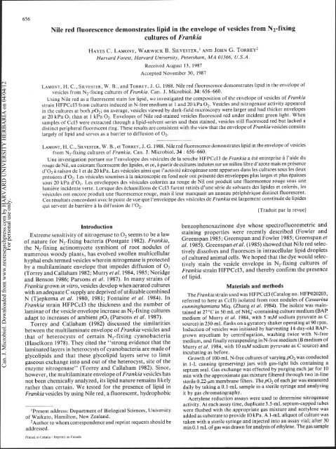
Us<strong>in</strong>g <strong>Nile</strong> <strong>red</strong> as a fluorescent sta<strong>in</strong> for <strong>lipid</strong>, we <strong>in</strong>vestigated <strong>the</strong> composition <strong>of</strong> <strong>the</strong> <strong>envelope</strong> <strong>of</strong> <strong>vesicles</strong> <strong>of</strong> Frankia<br />
stra<strong>in</strong> HFPCcI3 from cultures <strong>in</strong>duced <strong>in</strong> N-free medium at 1 and 20 kPa 02. Vesicles and nitrogenase activity appea<strong>red</strong><br />
<strong>in</strong> <strong>the</strong> cultures at both p02; on average, <strong>vesicles</strong> viewed by dark-field microscopy were larger and had thicker <strong>envelope</strong>s<br />
at 20 kPa O2 than at 1 kPa 02. Envelopes <strong>of</strong> <strong>Nile</strong> <strong>red</strong>-sta<strong>in</strong>ed <strong>vesicles</strong> fluoresced <strong>red</strong> under <strong>in</strong>cident green light. When<br />
samples <strong>of</strong> CcI3 were extracted through a <strong>lipid</strong>-solvent series and <strong>the</strong>n sta<strong>in</strong>ed, <strong>vesicles</strong> still fluoresced <strong>red</strong> but lacked a<br />
dist<strong>in</strong>ct peripheral fluorescent r<strong>in</strong>g. These results are consistent with <strong>the</strong> view that <strong>the</strong> <strong>envelope</strong> <strong>of</strong> Frankia <strong>vesicles</strong> consists<br />
largely <strong>of</strong> <strong>lipid</strong> and serves as a barrier to diffusion <strong>of</strong> 02.<br />
LAMONT, H. C., SILVESTER, W. B., et TORREY, J. G. 1988. <strong>Nile</strong> <strong>red</strong> <strong>fluorescence</strong> <strong>demonstrates</strong> <strong>lipid</strong> <strong>in</strong> <strong>the</strong> <strong>envelope</strong> <strong>of</strong> <strong>vesicles</strong><br />
from N2-fix<strong>in</strong>g cultures <strong>of</strong> Frankia. Can. J. Microbiol. 34 : 656-660.<br />
Une <strong>in</strong>vestigation portant sur I'enveloppe des vesicules de la souche HFPCcI3 de Frankia a Cte entreprise a l'aide du<br />
rouge de Nil, un colorant fluorescent des <strong>lipid</strong>es, et ce, a partir de cultures <strong>in</strong>duites sur un milieu libre d'azote mais en presence<br />
d'02 a raison de 1 et de 20 kPa. Les vesicules a<strong>in</strong>si que I'activite nitrogenase sont apparues dans les cultures sous les deux<br />
pressions d'02. Les vesicules soumises a la microscopic en fond noir ont presente des enveloppes plus larges et plus Cpaisses<br />
sous 20 kPa d'02. Les enveloppes des vesicules colorees au rouge de Nil ont produit une <strong>fluorescence</strong> rouge sous une<br />
lumikre <strong>in</strong>cidente verte. Lorsque des echantillons de CcI3 furent retires d'une serie de solvants des <strong>lipid</strong>es et colores, les<br />
vesicules ont encore produit une <strong>fluorescence</strong> rouge, mais il leur manquait un anneau peripherique dist<strong>in</strong>ct fluorescent.<br />
Ces resultats concordent avec le po<strong>in</strong>t de vue que l'enveloppe des vesicules de Frankia est largement constituee de <strong>lipid</strong>es<br />
qui servent de barrikre a la diffusion de 1'02.<br />
[Traduit par la revue]<br />
Introduction<br />
Extreme sensitivity <strong>of</strong> nitrogenase to 0, seems to be a law<br />
<strong>of</strong> nature for N2-fix<strong>in</strong>g bacteria (Postgate 1982). Frankia,<br />
<strong>the</strong> N2-fix<strong>in</strong>g act<strong>in</strong>omycete symbiont <strong>of</strong> root nodules <strong>of</strong><br />
numerous woody plants, has evolved swollen multicellular<br />
hyphal ends termed <strong>vesicles</strong> where<strong>in</strong> nitrogenase is protected<br />
by a multilam<strong>in</strong>ate <strong>envelope</strong> that impedes diffusion <strong>of</strong> 0,<br />
(Torrey and Callaham 1982; Murry et a[. 1984,1985; Noridge<br />
and Benson 1986; Parsons et al. 1987). In many stra<strong>in</strong>s <strong>of</strong><br />
benzophenoxaz<strong>in</strong>one dye whose spectr<strong>of</strong>luorometric and<br />
sta<strong>in</strong><strong>in</strong>g properties were recently described (Fowler and<br />
Greenspan 1985; Greenspan and Fowler 1985; Greenspan et<br />
al. 1985). Greenspan et al. (1985) showed that <strong>Nile</strong> <strong>red</strong> selec-<br />
tively dissolves and fluoresces <strong>in</strong> <strong>in</strong>tracellular <strong>lipid</strong> droplets<br />
<strong>of</strong> cultu<strong>red</strong> animal cells. We hoped that <strong>the</strong> dye would selec-<br />
tively sta<strong>in</strong> <strong>the</strong> vesicle <strong>envelope</strong> <strong>in</strong> N2-fix<strong>in</strong>g cultures <strong>of</strong><br />
Frankia stra<strong>in</strong> HFPCcI3, and <strong>the</strong>reby confirm <strong>the</strong> presence<br />
<strong>of</strong> <strong>lipid</strong>.<br />
Frankia grown <strong>in</strong> vitro, <strong>vesicles</strong> develop when aerated cultures<br />
with an adequate C supply are deprived <strong>of</strong> utilizable comb<strong>in</strong>ed<br />
(TJepkema et a'' 19809 1981; Fonta<strong>in</strong>e et a'' 1984)' In<br />
~rankia stra<strong>in</strong> HFPCCI~ <strong>the</strong> thickness and <strong>the</strong> number <strong>of</strong><br />
lam<strong>in</strong>ae <strong>of</strong> <strong>the</strong> vesicle <strong>envelope</strong> iXl~rease as N2-fix<strong>in</strong>g cultures<br />
adapt to <strong>in</strong>creases <strong>of</strong> ambient p02 (Parsons et al. 1987).<br />
Torrey and Callaham (1982) discussed <strong>the</strong> similarities<br />
between <strong>the</strong> multilam<strong>in</strong>ate <strong>envelope</strong> <strong>of</strong> Frankia <strong>vesicles</strong> and<br />
Materials and methods<br />
The Frankia stra<strong>in</strong> used was HFPCcI3 (Catalog no. HFP020203,<br />
refer<strong>red</strong> to here as CcI3) isolated from root nodules <strong>of</strong> Casuar<strong>in</strong>a<br />
cunn<strong>in</strong>ghamiana Miq. (Zhang et a/. 1984). The isolate was ma<strong>in</strong>ta<strong>in</strong>ed<br />
at 270~ <strong>in</strong> 50 mL <strong>of</strong> NH$-conta<strong>in</strong><strong>in</strong>g culture medium (BAP<br />
medium <strong>of</strong> Murry et al. 1984, with 5 mM sodium pyruvate as C<br />
source) <strong>in</strong> 250-m~ flasks on a gyratory shaker operat<strong>in</strong>g at 90 rpm.<br />
Induction <strong>of</strong> <strong>vesicles</strong> was <strong>in</strong>itiated by harvest<strong>in</strong>g 14-day-old BAPthat<br />
<strong>of</strong> heterocysts <strong>of</strong> certa<strong>in</strong> N2-fix<strong>in</strong>g cyanobacteria grown mycelium by centrifugation, wash<strong>in</strong>g twice with N-free<br />
(Haselkorn 1978). They cited <strong>the</strong> "strong evidence that <strong>the</strong><br />
lam<strong>in</strong>ated layers <strong>in</strong> heterocysts <strong>of</strong> cyanobacteria are made <strong>of</strong><br />
glyco<strong>lipid</strong>s and that <strong>the</strong>se glyco<strong>lipid</strong> layers serve to limit<br />
gaseous exchange <strong>in</strong>to and out <strong>of</strong> <strong>the</strong> heterocyst, site <strong>of</strong> <strong>the</strong><br />
enzyme nitrogenase" (Torrey and Callaham 1982). S<strong>in</strong>ce,<br />
however, <strong>the</strong> multilam<strong>in</strong>ate <strong>envelope</strong> <strong>of</strong> Frankia <strong>vesicles</strong> has<br />
not been chemically analyzed, its <strong>lipid</strong> nature rema<strong>in</strong>s likely<br />
ra<strong>the</strong>r than certa<strong>in</strong>. We tested for <strong>the</strong> presence <strong>of</strong> <strong>lipid</strong> <strong>in</strong><br />
Frankia <strong>vesicles</strong> by us<strong>in</strong>g <strong>Nile</strong> <strong>red</strong>, a fluorescent, hydrophobic<br />
medium, and f<strong>in</strong>ally resuspend<strong>in</strong>g <strong>in</strong> N-free medium (B medium <strong>of</strong><br />
Murry et al. 1984, with 10 mMsodium pyruvate as C source) and<br />
<strong>in</strong>cubat<strong>in</strong>g as before.<br />
Growth <strong>of</strong> 100-mL N-free cultures <strong>of</strong> vary<strong>in</strong>gp02 was conducted<br />
<strong>in</strong> I-L cann<strong>in</strong>g (preserv<strong>in</strong>g) jars with gas-tight lids conta<strong>in</strong><strong>in</strong>g a<br />
septum seal. Gas exchange was effected by purg<strong>in</strong>g each jar for 10<br />
m<strong>in</strong> with <strong>the</strong> approximate gas mixture filte<strong>red</strong> through two <strong>in</strong>-l<strong>in</strong>e<br />
sterile 0.22-pm membrane filters. Thep02 <strong>of</strong> each jar was measu<strong>red</strong><br />
daily by tak<strong>in</strong>g a 0.1-mL sample <strong>in</strong> a sterile syr<strong>in</strong>ge and analyz<strong>in</strong>g<br />
it by gas chromatography.<br />
Acetylene <strong>red</strong>uction assays were used to determ<strong>in</strong>e nitrogenase<br />
activity. At each assay time, duplicate 3.5-mL septum-capped tubes<br />
'Present address: Department <strong>of</strong> Biological Sciences, University were flushed with <strong>the</strong> appropriate gas mixture and acetylene was<br />
<strong>of</strong> Waikato, Hamilton, New Zealand.<br />
added as substrate to provide 10 kPa. A 1-mL aliquot <strong>of</strong> culture was<br />
2Author to whom correspondence and repr<strong>in</strong>t requests should be taken with a sterile syr<strong>in</strong>ge and <strong>in</strong>jected <strong>in</strong>to an assay vial; after 30<br />
addressed.<br />
m<strong>in</strong> 0.1 mL <strong>of</strong> gas was drawn for analysis <strong>of</strong> ethylene. Thegas sample<br />
Prlnled In Canada / ImDrlme au Canada
Can. J. Microbiol. Downloaded from www.nrcresearchpress.com by HARVARD UNIVERSITY HERBARIA on 04/04/12<br />
For personal use only.<br />
Culture age, h<br />
FIG. 1. Time dependence <strong>of</strong> acetylene <strong>red</strong>uction rate (nitrogenase<br />
activity) <strong>of</strong> Frankia stra<strong>in</strong> HFPCcI3 follow<strong>in</strong>g transfer from NHI -<br />
conta<strong>in</strong><strong>in</strong>g medium to N-free medium at 1 k& 0, (0) or at 20 k6a<br />
0 2 (0).<br />
was <strong>in</strong>jected <strong>in</strong>to a Carle 9500 gas chromatograph equipped with a<br />
1 .O-m Porapak T column and a flame ionization detector. All assay<br />
results are expressed as pmol C2H4 mL- m<strong>in</strong>-'.<br />
Culture samples were fixed, follow<strong>in</strong>g centrifugation, by add<strong>in</strong>g<br />
to <strong>the</strong> pellet 1 mL <strong>of</strong> 3% glutaraldehyde (from 50% glutaraldehyde,<br />
Fisher Scientific) <strong>in</strong> 25 mM potassium phosphate, pH 6.9. Fixation<br />
times varied from 2 h to 10 weeks; after 24 h, fix<strong>in</strong>g vials were trans-<br />
fer<strong>red</strong> from room temperature to 4OC. For observation or fur<strong>the</strong>r<br />
process<strong>in</strong>g <strong>the</strong> fixed mycelium was washed twice with distilled water.<br />
<strong>Nile</strong> <strong>red</strong> (Eastman Kodak Co., Rochester, NY) was dissolved <strong>in</strong><br />
acetone at 1 mg mL-' to make a stock solution that could be<br />
refrigerated or frozen. The sta<strong>in</strong><strong>in</strong>g solution proper conta<strong>in</strong>ed <strong>Nile</strong><br />
<strong>red</strong>, 5 pg mL-', <strong>in</strong> 50% aqueous glycerol. Follow<strong>in</strong>g centrifuga-<br />
tion, a pellet <strong>of</strong> fixed Frankia was resuspended <strong>in</strong> l mL <strong>of</strong> <strong>Nile</strong> <strong>red</strong><br />
sta<strong>in</strong><strong>in</strong>g solution. After 1 h <strong>the</strong> suspension was concentrated to about<br />
0.1 mL by centrifugation, and a drop <strong>of</strong> <strong>the</strong> concentrate, still <strong>in</strong><br />
sta<strong>in</strong><strong>in</strong>g solution, was mounted between alcohol-cleaned slide and<br />
cover slip. Photomicrography was done no less than 12 h later to<br />
permit substantial evaporation <strong>of</strong> water from <strong>the</strong> mount<strong>in</strong>g solution.<br />
Half <strong>of</strong> <strong>the</strong> 118-h sample from 20 kPa O2 was extracted with <strong>lipid</strong><br />
solvents before be<strong>in</strong>g sta<strong>in</strong>ed. The solvent sequence was 50%<br />
isopropanol (aqueous), 100% isopropanol (1 h), hexane/<br />
isopropanol::3/2 (v/v, I h), 100% isopropanol, 50% isopropanol<br />
(aqueous), distilled water. For part <strong>of</strong> <strong>the</strong> material hexane/<br />
isopropanol was omitted from <strong>the</strong> sequence so that <strong>the</strong> least polar<br />
solvent was 100% isopropanol. The extracted mycelium was sta<strong>in</strong>ed<br />
with <strong>Nile</strong> <strong>red</strong> and mounted as described above.<br />
The sta<strong>in</strong>ed mycelium was exam<strong>in</strong>ed and photographed both by<br />
transmitted-light dark-field microscopy and by <strong>in</strong>cident-light fluores-<br />
cence microscopy, us<strong>in</strong>g a Leitz Ortholux equipped with a dark-field<br />
condenser, a Ploem-type vertical illum<strong>in</strong>ator, and a Zeiss oil-<br />
immersion objective. For <strong>fluorescence</strong> microscopy an HBO 100 W<br />
mercury lamp was used with excitation filters BG 38, Baird <strong>in</strong>terfer-<br />
ence 377-548 nm, and Zeiss wide-band <strong>in</strong>terference 535 nm, <strong>the</strong> 580-<br />
nm dichroic beam splitter, and <strong>the</strong> 610-nm barrier filter.<br />
Results<br />
Nitrogenase activity, measu<strong>red</strong> as <strong>the</strong> rate <strong>of</strong> acetylene<br />
<strong>red</strong>uction, was observed <strong>in</strong> CcI3 cultuges grown without<br />
comb<strong>in</strong>ed N at both 1 and 20 kPa 02, but arose first <strong>in</strong> <strong>the</strong> 1<br />
kPa 0, culture (Fig. 1) when <strong>vesicles</strong> were still small and only<br />
th<strong>in</strong>ly <strong>envelope</strong>d (Fig. 2). Activity appea<strong>red</strong> 40 to 60 h after<br />
LAMONT ET AL. 657<br />
<strong>in</strong>troduction <strong>of</strong> N-free medium (Fig. 1). Vesicles exam<strong>in</strong>ed at<br />
any given sampl<strong>in</strong>g time were larger and had thicker light-<br />
scatter<strong>in</strong>g <strong>envelope</strong>s, on <strong>the</strong> average at 20 kPa 0, (Figs. 8,<br />
10, 12) than at 1 kPa 0, (Figs. 2, 4, 6), confirm<strong>in</strong>g <strong>the</strong><br />
f<strong>in</strong>d<strong>in</strong>gs <strong>of</strong> Parsons et al. (1987). The light-scatter<strong>in</strong>g enve-<br />
lope extended around each vesicle and down its stalk, at least<br />
to <strong>the</strong> first septum.<br />
<strong>Nile</strong> <strong>red</strong>-sta<strong>in</strong>ed <strong>vesicles</strong>, when excited with <strong>in</strong>cident green<br />
light, fluoresced <strong>red</strong> (Figs. 3, 5,7,9, 11, 13, 15, 17). Mature<br />
<strong>vesicles</strong> were most <strong>in</strong>tensely fluorescent, a peripheral r<strong>in</strong>g up<br />
to 0.5 pm thick usually be<strong>in</strong>g somewhat brighter than <strong>the</strong><br />
rema<strong>in</strong><strong>in</strong>g central area. The r<strong>in</strong>g <strong>of</strong> <strong>fluorescence</strong> occupied <strong>the</strong><br />
same position as, but was always th<strong>in</strong>ner than, <strong>the</strong> scatte<strong>red</strong>-<br />
light r<strong>in</strong>g observed by transmitted-light dark-field microscopy<br />
(Figs. 8-13). Fluorescence usually extended <strong>in</strong>to <strong>the</strong> light-<br />
scatter<strong>in</strong>g vesicle stalk (Figs. 7, 11). There was no <strong>red</strong><br />
auto<strong>fluorescence</strong> <strong>of</strong> unsta<strong>in</strong>ed <strong>vesicles</strong> excited with green light<br />
(but substantial yellow auto-<strong>fluorescence</strong> was produced with<br />
blue excitation).<br />
When <strong>the</strong> fixed 118-h CcI3 samples from 20 kPa 0, were<br />
extracted through a solvent series term<strong>in</strong>at<strong>in</strong>g with ei<strong>the</strong>r<br />
isopropanol or hexane/isopropanol::3/2, and <strong>the</strong>n sta<strong>in</strong>ed<br />
with <strong>Nile</strong> <strong>red</strong>, mature <strong>vesicles</strong> still fluoresced <strong>red</strong> but lacked<br />
a dist<strong>in</strong>ct peripheral fluorescent r<strong>in</strong>g and fluorescent stalk<br />
(Figs. 15, 17). In dark-field images <strong>of</strong> extracted mature<br />
<strong>vesicles</strong> a ragged scatte<strong>red</strong>-light r<strong>in</strong>g rema<strong>in</strong>ed, borne, as <strong>in</strong><br />
<strong>the</strong> <strong>fluorescence</strong> images, on an almost <strong>in</strong>visible stalk (Figs. 14,<br />
16). Extraction with acetone produced essentially identical<br />
changes (not shown).<br />
A major source <strong>of</strong> uncerta<strong>in</strong>ty <strong>in</strong> <strong>in</strong>terpretation <strong>of</strong> <strong>the</strong><br />
microscopic images is <strong>the</strong> fact that <strong>the</strong> fixed mycelium was<br />
whole mounted. The scatte<strong>red</strong>-light image <strong>of</strong> <strong>the</strong> vesicle enve-<br />
lope, be<strong>in</strong>g generated by a partially out-<strong>of</strong>-focus, vertically<br />
curved shell <strong>of</strong> (presumably) high refractive <strong>in</strong>dex, was<br />
artifactually thick and varied <strong>in</strong> thickness depend<strong>in</strong>g upon<br />
which dark-field condenser, which objective, and which<br />
objective aperture was chosen; once such a choice was made,<br />
and <strong>the</strong>n strictly adhe<strong>red</strong> to, all <strong>the</strong> scatte<strong>red</strong>-light images<br />
based on that particular choice were consistent with each<br />
o<strong>the</strong>r. By comparison, <strong>the</strong> <strong>fluorescence</strong> image <strong>of</strong> <strong>Nile</strong> <strong>red</strong>-<br />
sta<strong>in</strong>ed <strong>vesicles</strong> was quite sharp and <strong>in</strong>sensitive (<strong>in</strong> quality, not<br />
<strong>in</strong>tensity) to changes <strong>of</strong> oil-immersion objective and objec-<br />
tive aperture; however, s<strong>in</strong>ce <strong>the</strong> vertical position <strong>of</strong> fluores-<br />
cent material could not be def<strong>in</strong>ed, it is possible that <strong>Nile</strong> <strong>red</strong><br />
<strong>in</strong> <strong>the</strong> <strong>envelope</strong> generated at least some <strong>of</strong> <strong>the</strong> overall fluores-<br />
cence <strong>of</strong> each vesicle.<br />
Discussion<br />
Members <strong>of</strong> <strong>the</strong> Act<strong>in</strong>omycetales are characterized <strong>in</strong> part<br />
by <strong>the</strong>ir unique <strong>lipid</strong>, especially phospho<strong>lipid</strong>, constituents<br />
(Lechevalier et al. 1982). Frankia is characterized by <strong>the</strong><br />
presence <strong>of</strong> type I phospho<strong>lipid</strong>s (Lechevalier et al. 1982;<br />
Lopez et al. 1983). The phospho<strong>lipid</strong>s found <strong>in</strong> Frankia<br />
cultures are remarkably constant with respect to variations<br />
<strong>of</strong> nutrient medium, culture age, and morphogenetic state<br />
(Lopez et al. 1983). These <strong>lipid</strong>s are presumed to be compo-<br />
nents <strong>of</strong> bacterial cell membranes or <strong>of</strong> storage <strong>in</strong>clusions. In<br />
<strong>the</strong> <strong>Nile</strong> <strong>red</strong>-sta<strong>in</strong>ed preparations <strong>of</strong> Frankia, both diffuse and<br />
particulate fluorescent material <strong>in</strong> vegetative hyphae (e.g.,<br />
Figs. 3, 5) presumably represents <strong>the</strong> typical polar <strong>lipid</strong>s<br />
refer<strong>red</strong> to above. An important question is whe<strong>the</strong>r <strong>the</strong> <strong>Nile</strong><br />
<strong>red</strong>-sta<strong>in</strong>ed material associated with <strong>the</strong> vesicle <strong>envelope</strong> is an<br />
unusual <strong>lipid</strong> or belongs to <strong>the</strong> type I phospho<strong>lipid</strong>s charac-
Can. J. Microbiol. Downloaded from www.nrcresearchpress.com by HARVARD UNIVERSITY HERBARIA on 04/04/12<br />
For personal use only.<br />
658 CAN. J. MICROBIOL. VOL. 34, 1988<br />
FIGS. 2-7. Pai<strong>red</strong> dark-field (left) and <strong>Nile</strong> <strong>red</strong> <strong>fluorescence</strong> (right) images <strong>of</strong> Frankia stra<strong>in</strong> HFPCcI3 at different times follow<strong>in</strong>g transfer<br />
from NHJ-conta<strong>in</strong><strong>in</strong>g medium to N-free medium at 1 kPa 0,. Figs. 2, 3: 47 h. Figs. 4, 5: 67 h. Figs. 6,7: 118 h. In Fig. 6 <strong>the</strong>re is a strongly<br />
light-scatter<strong>in</strong>g hyphal wall segment (arrow) subjacent to a develop<strong>in</strong>g vesicle; <strong>in</strong> Fig. 7 <strong>the</strong> same wall segment (arrow) exhibits strong <strong>Nile</strong><br />
<strong>red</strong> <strong>fluorescence</strong>.<br />
teristic <strong>of</strong> <strong>the</strong> genus. Chemical analysis <strong>of</strong> <strong>lipid</strong>s extracted<br />
from vesicle-enriched cultures grown under raised p02<br />
should make possible <strong>the</strong> dist<strong>in</strong>ction between <strong>the</strong> constitutive<br />
<strong>lipid</strong>s and specialized <strong>lipid</strong>s associated with <strong>the</strong> vesicle enve-<br />
lope (analogous to those <strong>of</strong> cyanobacterial heterocysts).<br />
The results reported here, based on <strong>the</strong> use <strong>of</strong> <strong>the</strong> dye <strong>Nile</strong><br />
<strong>red</strong> as a fluorescent hydrophobic probe for <strong>lipid</strong>, are consis-<br />
tent with <strong>the</strong> view <strong>of</strong> Torrey and Callaham (1982) that <strong>the</strong><br />
<strong>envelope</strong> <strong>of</strong> Frankia <strong>vesicles</strong> consists largely <strong>of</strong> <strong>lipid</strong> and serves<br />
as a diffusion barrier protect<strong>in</strong>g nitrogenase aga<strong>in</strong>st 0,.<br />
Shown by Parsons et al. (1987) to be light-scatter<strong>in</strong>g under<br />
dark-field illum<strong>in</strong>ation, <strong>the</strong> vesicle <strong>envelope</strong> proved also to<br />
be sta<strong>in</strong>able with <strong>Nile</strong> <strong>red</strong>. We observed that <strong>the</strong> peripheral<br />
fluorescent r<strong>in</strong>g <strong>of</strong> <strong>Nile</strong> <strong>red</strong>-sta<strong>in</strong>ed mature <strong>vesicles</strong> was no<br />
longer visible after exposure <strong>of</strong> fixed mycelium to even a rela-<br />
tively polar solvent, namely isopropanol.<br />
In mature <strong>vesicles</strong> <strong>Nile</strong> <strong>red</strong> sta<strong>in</strong><strong>in</strong>g was not restricted to <strong>the</strong><br />
peripheral r<strong>in</strong>g; <strong>the</strong> central area was also sta<strong>in</strong>ed, usually less<br />
<strong>in</strong>tensely than <strong>the</strong> periphery. Moreover, <strong>the</strong> central area<br />
rema<strong>in</strong>ed sta<strong>in</strong>able after extraction with <strong>lipid</strong> solvents. Our<br />
evidence does not exclude <strong>the</strong> possibility that <strong>the</strong> <strong>lipid</strong> solvents<br />
caused <strong>the</strong> <strong>envelope</strong> to collapse <strong>in</strong>ward ra<strong>the</strong>r than to dissolve.<br />
<strong>Nile</strong> <strong>red</strong> be<strong>in</strong>g a hydrophobic ra<strong>the</strong>r than a <strong>lipid</strong>-specific dye<br />
(Greenspan and Fowler 1985, Table 4), we suggest that <strong>the</strong><br />
solvent-resistant, sta<strong>in</strong>able components <strong>of</strong> <strong>the</strong> vesicle are<br />
hydrophobic doma<strong>in</strong>s <strong>of</strong> <strong>in</strong>tracellular prote<strong>in</strong>s and <strong>in</strong>clusions.<br />
Although <strong>in</strong> Casuar<strong>in</strong>a root nodules CcI3 fixes N, without<br />
form<strong>in</strong>g <strong>vesicles</strong> (Zhang and Torrey 1985), <strong>in</strong> pure culture its<br />
capacity to fix N, at p02 greater than 0.3 kPa depends upon<br />
<strong>in</strong>duction <strong>of</strong> <strong>vesicles</strong> (Murry et al. 1985). Protection <strong>of</strong> vesicle<br />
nitrogenase aga<strong>in</strong>st 0, <strong>in</strong> pure culture has been attributed to<br />
<strong>the</strong> lam<strong>in</strong>ate <strong>envelope</strong> act<strong>in</strong>g as a passive barrier to diffusion<br />
<strong>of</strong> dissolved 0, (Torrey and Callaham 1982; Murry et al.<br />
1985; Parsons et al. 1987). The evidence for <strong>lipid</strong> be<strong>in</strong>g a<br />
major component <strong>of</strong> <strong>the</strong> <strong>envelope</strong> is not merely <strong>the</strong> <strong>envelope</strong>'s<br />
lam<strong>in</strong>ate structure <strong>in</strong> frozen-fractu<strong>red</strong> material, but also its<br />
FIGS. 8-17. Pai<strong>red</strong> dark-field (left) and <strong>Nile</strong> <strong>red</strong> <strong>fluorescence</strong> (right) images <strong>of</strong> Frankia stra<strong>in</strong> HFPCcI3 at different times follow<strong>in</strong>g transfer<br />
from NH2-conta<strong>in</strong><strong>in</strong>g medium to N-free medium at 20 kPa 0,. Figs. 8,9: 47 h. Figs. 10, 11: 67 h. The uppermost vesicle <strong>of</strong> Figs. 10 and<br />
11 illustrates close correspondence between <strong>the</strong> scatte<strong>red</strong>-light and <strong>Nile</strong> <strong>red</strong> images <strong>of</strong> <strong>the</strong> outer wall, <strong>in</strong> both <strong>the</strong> vesicle body and <strong>the</strong> stalk<br />
(arrows). Figs. 12-17: 118 h. Figs. 14,15: mycelium extracted with isopropanol. Figs. 16,17: mycelium extracted with hexane-isopropanol<br />
(3:2). After extraction, <strong>the</strong> vesicle stalk no longer scatters light, <strong>the</strong> peripheral scatte<strong>red</strong>-light r<strong>in</strong>g <strong>of</strong> <strong>the</strong> vesicle body has become patchy<br />
<strong>in</strong> form, and <strong>the</strong> peripheral <strong>Nile</strong> <strong>red</strong> <strong>fluorescence</strong> r<strong>in</strong>g <strong>of</strong> <strong>the</strong> vesicle body is no longer dist<strong>in</strong>guishable.
Can. J. Microbiol. Downloaded from www.nrcresearchpress.com by HARVARD UNIVERSITY HERBARIA on 04/04/12<br />
For personal use only.<br />
L.AhlOKT ET AL.
Can. J. Microbiol. Downloaded from www.nrcresearchpress.com by HARVARD UNIVERSITY HERBARIA on 04/04/12<br />
For personal use only.<br />
660<br />
apparent dissolution by <strong>lipid</strong> solvents commonly used to<br />
dehydrate fixed specimens for embedd<strong>in</strong>g and section<strong>in</strong>g<br />
(Torrey and Callaham 1982; Lancelle et a/. 1985). A<br />
p<strong>red</strong>om<strong>in</strong>antly <strong>lipid</strong> <strong>envelope</strong> could be expected to have a<br />
refractive <strong>in</strong>dex above 1.43 (Lange 1956), which toge<strong>the</strong>r with<br />
<strong>the</strong> <strong>envelope</strong>'s lam<strong>in</strong>ar <strong>in</strong>homogeneity would cause it to<br />
scatter light <strong>in</strong> aqueous media, as has been observed (Parsons<br />
et a/. 1987).<br />
In any case, <strong>the</strong> present evidence strongly supports <strong>the</strong> view<br />
that <strong>the</strong> vesicle <strong>envelope</strong>, which is progressively elaborated <strong>in</strong><br />
N,-fix<strong>in</strong>g Frankia grown under <strong>in</strong>creas<strong>in</strong>g PO,, consists <strong>of</strong><br />
<strong>lipid</strong>s arranged <strong>in</strong> a multilam<strong>in</strong>ate structure. Its chemical<br />
nature and f<strong>in</strong>al pro<strong>of</strong> <strong>of</strong> its role as a physical barrier to direct<br />
entry <strong>of</strong> 0, <strong>in</strong>to <strong>the</strong> vesicle rema<strong>in</strong> to be demonstrated.<br />
Acknowledgements<br />
This research was supported by U.S. Department <strong>of</strong> Energy<br />
grant DE-FG02-84ER13 198 and by <strong>the</strong> Maria Moors Cabot<br />
Foundation for Botanical Research <strong>of</strong> Harvard University.<br />
We are grateful to Ela<strong>in</strong>e Doughty and Julie Withbeck for<br />
technical assistance.<br />
CAN. J. MICROBIOL. VOL. 34, 1988<br />
FONTAINE, M. S., LANCELLE, S. A., and TORREY, J. G. 1984. Initiation<br />
and ontogeny <strong>of</strong> <strong>vesicles</strong> <strong>in</strong> cultu<strong>red</strong> Frankia sp. stra<strong>in</strong><br />
HFPArI3. J. Bacteriol. 160: 921-927.<br />
FOWLER, S. D., and GREENSPAN, P. 1985. Application <strong>of</strong> <strong>Nile</strong> <strong>red</strong>,<br />
a fluorescent hydrophobic probe, for <strong>the</strong> detection <strong>of</strong> neutral <strong>lipid</strong><br />
deposits <strong>in</strong> tissue sections: comparison with oil <strong>red</strong> 0. J.<br />
Histochem. Cytochem. 33: 833-836.<br />
GREENSPAN, P., and FOWLER, S. D. 1985. Spectr<strong>of</strong>luorometric<br />
studies <strong>of</strong> <strong>the</strong> <strong>lipid</strong> probe, <strong>Nile</strong> <strong>red</strong>. J. Lipid Res. 26: 781-789.<br />
GREENSPAN, P., MAYER, E. P., and FOWLER, S. D. 1985. <strong>Nile</strong> <strong>red</strong>:<br />
a selective fluorescent sta<strong>in</strong> for <strong>in</strong>tracellular <strong>lipid</strong> droplets. J. Cell<br />
Biol. 100: 965-973.<br />
HASELKORN, R. 1978. Heterocysts. Annu. Rev. Plant Physiol. 29:<br />
319-344.<br />
LANCELLE, S. A., TORREY, J. G., HEPLER, P. K., and CALLAHAM,<br />
D. A. 1985. Ultrastructure <strong>of</strong> freeze-substituted Frankia stra<strong>in</strong><br />
HFPCcI3, <strong>the</strong> act<strong>in</strong>omycete isolated from root nodules <strong>of</strong> Casuar<strong>in</strong>a<br />
cunn<strong>in</strong>ghamiana. Protoplasma, 127: 64-72.<br />
LANCE, N. A. 1956. Handbook <strong>of</strong> chemistry. 9th ed. Handbook<br />
Publishers, Sandusky, OH. pp. 760-763.<br />
LECHEVALIER, M. P., HORRI~RE, F., and LECHEVALIER, H. 1982. The<br />
biology <strong>of</strong> Frankia and related organisms. Dev. Ind. Micmbiol.<br />
23: 51-60.<br />
LOPEZ, M. F., WHALING, C. S., and TORREY, J. G. 1983. The polar<br />
<strong>lipid</strong>s and free sugars <strong>of</strong> Frankia <strong>in</strong> culture. Can. J. Bot. 61:<br />
2834-2842.<br />
MURRY, M. A., FONTAINE, M. S., and TJEPKEMA, J. D. 1984.<br />
Oxygen protection <strong>of</strong> nitrogenase <strong>in</strong> Frankia sp. HFPArI3. Arch.<br />
Microbiol. 139: 162-166.<br />
MURRY, M. A,, ZHANG, Z., and TORREY, J. G. 1985. Effect <strong>of</strong> O2<br />
on vesicle formation, acetylene <strong>red</strong>uction, and 02-uptake k<strong>in</strong>etics<br />
<strong>in</strong> Frankia sp. HFPCcI3 isolated from Casuar<strong>in</strong>a cunn<strong>in</strong>ghamiana.<br />
Can. J. Microbiol. 31: 804-809.<br />
NORIDGE, N. A., and BENSON, D. R. 1986. Isolation and nitrogenfix<strong>in</strong>g<br />
activity <strong>of</strong> Frankia sp. stra<strong>in</strong> CpIl <strong>vesicles</strong>. J. Bacteriol. 166:<br />
301-305.<br />
PARSONS, R., SILVESTER, W. B., HARRIS, S., GRUIJTERS, W. T. M.,<br />
and BULLIVANT, S. 1987. Frankia <strong>vesicles</strong> provide <strong>in</strong>ducible and<br />
absolute oxygen protection for nitrogenase. Plant Physiol. 83:<br />
728-73 1.<br />
POSTGATE, J. R. 1982. The fundamentals <strong>of</strong> nitrogen fixation<br />
Cambridge University Press, Cambridge.<br />
TJEPKEMA, J. D., ORMEROD, W., and TORREY, J. G. 1980. Vesicle<br />
formation and acetylene <strong>red</strong>uction activity <strong>in</strong> Frankia sp. CpIl<br />
cultu<strong>red</strong> <strong>in</strong> def<strong>in</strong>ed nutrient media. Nature (London), 287:<br />
633-635.<br />
TJEPKEMA, J. D., ORMEROD, W., and TORREY, J. G. 1981. Factors<br />
affect<strong>in</strong>g vesicle formation and acetylene <strong>red</strong>uction (nitrogenase<br />
activity) <strong>in</strong> Frankia sp. CpIl. Can. J. Microbiol. 27: 815-823.<br />
TORREY, J. G., and CALLAHAM, D. 1982. Structural features <strong>of</strong> <strong>the</strong><br />
vesicle <strong>of</strong> Frankia sp. CpIl <strong>in</strong> culture. Can. J. Microbiol. 28:<br />
749-757.<br />
ZHANG, Z., and TORREY, J. G. 1985. Biological and cultural characteristics<br />
<strong>of</strong> <strong>the</strong> effective Frankia stra<strong>in</strong> HFPCcI3 (Act<strong>in</strong>omycetales)<br />
from Casuar<strong>in</strong>a cunn<strong>in</strong>ghamiana (Casuar<strong>in</strong>aceae). Ann.<br />
Bot. 56: 367-378.<br />
ZHANG, Z., LOPEZ, M. F., and TORREY, J. G. 1984. A comparison<br />
<strong>of</strong> cultural characteristics and <strong>in</strong>fectivity <strong>of</strong> Frankia isolates from<br />
root nodules <strong>of</strong> Casuar<strong>in</strong>a species. Plant Soil, 78: 79-90.


