Goussia Labbe´ , 1896 (Apicomplexa, Eimeriorina) - Institute of ...
Goussia Labbe´ , 1896 (Apicomplexa, Eimeriorina) - Institute of ...
Goussia Labbe´ , 1896 (Apicomplexa, Eimeriorina) - Institute of ...

Create successful ePaper yourself
Turn your PDF publications into a flip-book with our unique Google optimized e-Paper software.
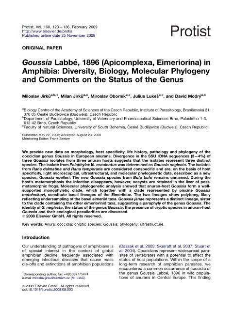
Protist, Vol. 160, 123—136, February 2009<br />
http://www.elsevier.de/protis<br />
Published online date 25 November 2008<br />
ORIGINAL PAPER<br />
<strong>Goussia</strong> Labbé, <strong>1896</strong> (<strong>Apicomplexa</strong>, <strong>Eimeriorina</strong>) in<br />
Amphibia: Diversity, Biology, Molecular Phylogeny<br />
and Comments on the Status <strong>of</strong> the Genus<br />
Miloslav Jirku˚ a,b,1 , Milan Jirku˚ a,c , Miroslav Oborník a,c , Julius Lukesˇ a,c , and David Modry´ a,b<br />
a<br />
Biology Centre <strong>of</strong> the Academy <strong>of</strong> Sciences <strong>of</strong> the Czech Republic, <strong>Institute</strong> <strong>of</strong> Parasitology, Branisˇ ovská 31,<br />
370 05 České Budějovice (Budweis), Czech Republic<br />
b<br />
Department <strong>of</strong> Parasitology, University <strong>of</strong> Veterinary and Pharmaceutical Sciences Brno, Palackého 1-3,<br />
612 42 Brno, Czech Republic<br />
c<br />
Faculty <strong>of</strong> Natural Sciences, University <strong>of</strong> South Bohemia, České Budějovice (Budweis), Czech Republic<br />
Submitted May 22, 2008; Accepted August 23, 2008<br />
Monitoring Editor: Frank Seeber<br />
We provide new data on morphology, host specificity, life history, pathology and phylogeny <strong>of</strong> the<br />
coccidian genus <strong>Goussia</strong> in European anurans. Divergence in the SSU rDNA sequences (3—4%) <strong>of</strong><br />
three <strong>Goussia</strong> isolates from three anuran hosts suggests that the isolates represent three distinct<br />
species. The isolate from Pelophylax kl. esculentus was determined as <strong>Goussia</strong> neglecta. The isolates<br />
from Rana dalmatina and Rana temporaria are considered conspecific and are, on the basis <strong>of</strong> host<br />
specificity, light microscopical, ultrastructural, and molecular phylogenetic data, described as a new<br />
species, <strong>Goussia</strong> noelleri. The new <strong>Goussia</strong> species from Bufo bufo remains unnamed. During the<br />
host’s metamorphosis the infection disappears, however, oocysts are retained in the liver <strong>of</strong> postmetamorphic<br />
frogs. Molecular phylogenetic analysis showed that anuran-host <strong>Goussia</strong> form a wellsupported<br />
monophyletic clade, which together with a clade represented by piscine <strong>Goussia</strong><br />
metchnikovi, constitute basal lineages <strong>of</strong> the Eimeriidae. The two lineages show polytomy, likely<br />
reflecting undersampling <strong>of</strong> the basal eimeriid taxa. <strong>Goussia</strong> janae represents a distinct lineage, sister<br />
to the clade containing the other eimeriorinid taxa, suggesting a paraphyly <strong>of</strong> the genus <strong>Goussia</strong>. The<br />
identity <strong>of</strong> G. neglecta, the status <strong>of</strong> the genus <strong>Goussia</strong>, the presence <strong>of</strong> cryptic species in anuran-host<br />
<strong>Goussia</strong> and their ecological peculiarities are discussed.<br />
& 2008 Elsevier GmbH. All rights reserved.<br />
Key words: Anura; coccidia; cryptic species; <strong>Goussia</strong>; phylogeny; ultrastructure.<br />
Introduction<br />
Our understanding <strong>of</strong> pathogens <strong>of</strong> amphibians is<br />
<strong>of</strong> special interest in the context <strong>of</strong> global<br />
amphibian decline, frequently associated with<br />
emerging infectious diseases that cause mass<br />
die-<strong>of</strong>fs and extinctions <strong>of</strong> amphibian populations<br />
1 Corresponding author; fax +420 387775474<br />
e-mail miloslav.jirku@seznam.cz (M. Jirku˚ ).<br />
& 2008 Elsevier GmbH. All rights reserved.<br />
doi:10.1016/j.protis.2008.08.003<br />
ARTICLE IN PRESS<br />
(Daszak et al. 2003; Skerratt et al. 2007; Stuart et<br />
al. 2004). Coccidians represent widespread parasites<br />
<strong>of</strong> vertebrates with a potential to affect the<br />
status <strong>of</strong> host populations. Within the scope <strong>of</strong> a<br />
long-term research <strong>of</strong> amphibian parasites, we<br />
encountered a common occurrence <strong>of</strong> coccidia <strong>of</strong><br />
the genus <strong>Goussia</strong> Labbé, <strong>1896</strong> in wild populations<br />
<strong>of</strong> anurans in Central Europe. This finding
124 M. Jirku˚ et al.<br />
allowed us to conduct an extensive study <strong>of</strong><br />
members <strong>of</strong> this elusive and overlooked coccidian<br />
genus with consequences for understanding <strong>of</strong><br />
thus far unrecognized taxonomic diversity and<br />
phylogenetic patterns <strong>of</strong> poikilotherm coccidians<br />
in general (Dyková and Lom 1981; Jirku˚ et al.<br />
2002).<br />
The genus <strong>Goussia</strong> was erected to accommodate<br />
piscine coccidia with oocysts possessing<br />
four dizoic sporocysts composed <strong>of</strong> two valves<br />
joined by a longitudinal suture (bivalved sporocysts).<br />
The fine, <strong>of</strong>ten elastic oocyst wall and the<br />
absence <strong>of</strong> an oocyst residuum are other typical<br />
features <strong>of</strong> the genus. Most species are probably<br />
homoxenous, but facultatively a heteroxenous life<br />
cycle was confirmed experimentally for <strong>Goussia</strong><br />
carpelli (Steinhagen and Körting 1990).<br />
Approximately 50 nominal species <strong>of</strong> <strong>Goussia</strong><br />
have been described from freshwater or marine<br />
fish and amphibians. The generic name <strong>Goussia</strong><br />
has a complicated history (Dyková and Lom 1981;<br />
Levine 1983). Although the genus name was<br />
revived by Dyková and Lom (1981), the most<br />
recent taxonomical review <strong>of</strong> the suborder <strong>Eimeriorina</strong><br />
(Upton 2000) lists <strong>Goussia</strong> as a synonym<br />
<strong>of</strong> Eimeria without further comments. However,<br />
the generic name remains recognized as valid by<br />
some authors and new species are being continuously<br />
described assigned to it. Virtually nothing<br />
is known about the relationships <strong>of</strong> <strong>Goussia</strong> to<br />
other coccidian taxa except that the welldescribed<br />
piscine <strong>Goussia</strong> janae Lukesˇ et Dyková,<br />
1990 is not closely related to Eimeria (Jirku˚ et al.<br />
2002; Lukesˇ and Stary´ 1992).<br />
Among amphibian hosts, only larvae <strong>of</strong> anurans<br />
(tadpoles) were found to be parasitized by<br />
<strong>Goussia</strong>. Two intestinal species are known from<br />
anurans. Nöller (1920) described Eimeria neglecta<br />
based on oocysts recovered from tadpoles <strong>of</strong><br />
European Ranids. In 1995, Molnár redescribed<br />
this coccidium, recognized its affinity to fish-host<br />
coccidia and emended its taxonomic status to<br />
<strong>Goussia</strong> neglecta. Later, Paperna et al. (1997)<br />
described <strong>Goussia</strong> hyperolisi from tadpoles <strong>of</strong> the<br />
reed frog Hyperolius viridiflavus from Kenya.<br />
Although <strong>Goussia</strong> infections in the anuran hosts<br />
seem to be restricted to tadpoles and the<br />
disappearance <strong>of</strong> infection during the host’s<br />
metamorphosis was recorded repeatedly (Molnár<br />
1995; Nöller 1920; Paperna et al. 1997), retention<br />
<strong>of</strong> oocysts in liver sinusoids <strong>of</strong> post-metamorphic<br />
frogs was documented only recently (Jirku˚ and<br />
Modry´ 2006).<br />
Herein, we provide data on morphology, biology<br />
and phylogenetic affinities <strong>of</strong> anuran <strong>Goussia</strong>, one<br />
ARTICLE IN PRESS<br />
<strong>of</strong> which is described as a new species, and<br />
describe the relationships <strong>of</strong> European anurans<br />
with their coccidian parasites using several<br />
approaches. Small subunit ribosomal DNA (SSU<br />
rDNA)-based phylogenetic analysis <strong>of</strong> three <strong>Goussia</strong><br />
species from different European anuran taxa<br />
and two fish-host <strong>Goussia</strong> provide novel insight<br />
into the taxonomy and phylogenetic affinities <strong>of</strong><br />
this genus.<br />
Results<br />
Of all examined tadpoles representing five anuran<br />
species, <strong>Goussia</strong> infections were recorded in<br />
tadpoles <strong>of</strong> Rana dalmatina (Loc1), Rana temporaria<br />
(Loc1, Loc2), Pelophylax kl. esculentus (Loc4)<br />
and Bufo bufo (Loc1). Hyla arborea was the only<br />
negative host. In contrast to Locality (Loc) 1 and<br />
Loc3, no infection was recorded in B. bufo<br />
tadpoles from Loc2. Similarly, P. kl. esculentus<br />
tadpoles were infected at Loc4, but not at Loc2.<br />
Free oocysts (Fig. 1) were detected in fresh faecal<br />
samples <strong>of</strong> wild tadpoles examined by the flotation<br />
method. Upon dissections, fully sporulated<br />
oocysts were observed either freely or enclosed<br />
within the yellow bodies in squash preparations <strong>of</strong><br />
intestinal mucosa, intestinal contents and livers<br />
(Figs 2—5) <strong>of</strong> all tadpoles, including those<br />
appearing to be negative based on the examination<br />
<strong>of</strong> faeces by flotation.<br />
Identity <strong>of</strong> Anuran Isolates<br />
The isolate from P. kl. esculentus is treated as<br />
G. neglecta based on morphology and origin from<br />
the type host, and is further characterized by SSU<br />
rDNA sequence. The isolate from B. bufo is<br />
characterized by sporocyst morphology and partial<br />
SSU rDNA sequence and is conservatively<br />
treated as <strong>Goussia</strong> sp. due to the absence <strong>of</strong><br />
ultrastructural and reliable host specificity data.<br />
Based on transmission experiments, <strong>Goussia</strong> from<br />
R. dalmatina and R. temporaria are considered<br />
conspecific. They are morphologically characterized<br />
using the R. dalmatina isolate, and according<br />
to the SSU rDNA sequence divergence, a new<br />
species is proposed, the description <strong>of</strong> which<br />
follows.<br />
<strong>Goussia</strong> noelleri sp. nov.<br />
Oocyst morphology: Shape <strong>of</strong> oocysts variable,<br />
depending on position <strong>of</strong> sporocysts tightly<br />
enclosed by a fine, elastic, colourless oocyst wall
(Figs 1, 6, 7). Oocyst measurements: 10.6<br />
(10.0—11.5) 10.0 (8.0—11.0) mm, length—width<br />
ratio (L/W) 1.07 (1.00—1.25). Oocyst residuum,<br />
micropyle and polar granule absent. Sporocysts<br />
dizoic, elliptical, <strong>of</strong>ten assymetrical with somewhat<br />
pointed pole(s) and/or one side being slightly<br />
flattened (Figs 1—3). Occasionally, atypical triangular<br />
sporocysts were observed (Fig. 4). The sporocyst<br />
measured 7.6 (7.0—8.5) 4.8 (4.0—5.5) mm,<br />
L/W 1.60 (1.40—1.78) and contained a usually<br />
compact and elliptical sporocyst residuum<br />
(4.0—6.0 2.5—4.0 mm) rarely scattered among<br />
sporozoites, composed <strong>of</strong> granules 0.5—1.0 mm in<br />
diameter (Figs 1—4). Sporozoites were arranged<br />
in parallel within the sporocyst partly encircling the<br />
sporocyst residuum (Fig. 7). A transversal striation<br />
was sometimes observed in sporozoites (Fig. 3).<br />
Sporozoites longer than sporocyst, arranged head<br />
to tail, each curved at opposite pole <strong>of</strong> sporocyst<br />
(Fig. 7). In the light microscope, using Nomarski<br />
ARTICLE IN PRESS<br />
<strong>Goussia</strong>: Diversity, Biology, Molecular Phylogeny<br />
Figures 1—6. Light (1—5) and scanning electron microscopy (6) <strong>of</strong> oocysts and sporocysts <strong>of</strong> <strong>Goussia</strong><br />
noelleri sp. nov. from Rana dalmatina tadpoles. 1. Oocyst isolated from faeces by flotation showing fine<br />
elastic oocyst wall (arrow). 2. Ruptured oocyst in squash preparation <strong>of</strong> intestinal mucosa. Halo surrounding<br />
the four sporocysts is collapsed oocyst wall. 3. Mass <strong>of</strong> sporocysts in squash preparation <strong>of</strong> liver. Striation is<br />
only barely visible in some sporozoites (arrows). Arrowheads show the two sporozoite refractile bodies. 4.<br />
Comparision <strong>of</strong> atypically (arrow) and typically formed sporocyst. 5. Three yellow bodies containing oocysts<br />
in squash preparation <strong>of</strong> intestinal mucosa. 6. Within the oocyst, sporocysts are tightly encloced by a thin<br />
oocyst wall. Note the longitudinal suture clearly visible in the sporocyst wall. Scale bar ¼ 5 mm (1—4, 6);<br />
10 mm (5). Nomarski interference contrast (1—5).<br />
125<br />
Figure 7. Composite line drawing <strong>of</strong> <strong>Goussia</strong> noelleri<br />
sp. nov. oocyst. Scheme on the right shows<br />
arrangement <strong>of</strong> sporozoites within sporocyst (not in<br />
scale). Scale bar ¼ 5 mm.
Table 1. Morphometrical comparision <strong>of</strong> <strong>Goussia</strong> spp. parasitizing tadpoles based on literature and original data. L/W—length/width ratio; *—<br />
type hosts and type localities; hyphens indicate missing data. Measurements are in mm.<br />
Species or isolate Host(s) Oocyst Sporocyst Locality Reference<br />
<strong>Goussia</strong> neglecta Pelophylax sp., Rana 9.0—10.0, spherical or irregular Hamburg and<br />
sp.<br />
Thüringen, Germany<br />
(Nöller, 1920) Molnár<br />
(1995)<br />
7.0 3.5—4.0<br />
<strong>Goussia</strong> neglecta Pelophylax kl.<br />
10.6 (8.5—12.5), spherical Százhalombatta,<br />
esculentus*, Pelophylax<br />
ridibundus<br />
Hungary*<br />
(Nöller, 1920) Molnár<br />
8.8 (6.5—10.2) 4.8 (4.0—7.7), L/W not<br />
(1995)<br />
<strong>Goussia</strong> neglecta Pelophylax kl.<br />
defined<br />
— Sˇnejdlík pond, Czech<br />
esculentus*<br />
Republic<br />
9.4 (8.0—10.0)<br />
(1.73—2.00)<br />
5.1 (5.0—5.5), L/W 1.82<br />
<strong>Goussia</strong> noelleri sp. Rana dalmatina* 10.6 (10.0—11.5) 10.0 (8.0—11.0), L/W Zaječí potok, Czech<br />
nov.<br />
1.07 (1.00—1.25), spherical or irregular Republic*<br />
7.6 (7.0—8.5)<br />
(1.40—1.78)<br />
4.8 (4.0—5.5), L/W 1.60<br />
<strong>Goussia</strong> noelleri sp. Rana temporaria — Zaječí potok, Czech<br />
nov.<br />
Republic*<br />
7.8 (7.0—8.5)<br />
(1.45—1.78)<br />
4.8 (4.5—5.5), L/W 1.63<br />
<strong>Goussia</strong> sp. Bufo bufo — Zaječí potok, Czech<br />
Republic<br />
7.9 (7.0—9.0)<br />
(1.50—2.25)<br />
4.5 (4.0—5.0), L/W 1.74<br />
<strong>Goussia</strong> hyperolisi Hyperolius viridiflavus* 7.7 (7.0—9.8), spherical Sagana fish ponds,<br />
Kenya*<br />
Paperna, Ogara and<br />
7.2 (5.6—7.7) 4.9 (4.2—5.6), L/W not<br />
Schein (1997)<br />
defined<br />
Nöller (1920)<br />
Molnár (1995)<br />
This study<br />
This study<br />
This study<br />
This study<br />
Paperna et al.<br />
(1997)<br />
126 M. Jirku˚ et al.<br />
ARTICLE IN PRESS
interference contrast (NIC), two refractile bodies<br />
were observed. The bigger refractile body (1.5 mm<br />
in diameter) was located in the middle <strong>of</strong> the<br />
sporozoite, the smaller refractile body (1 mm in<br />
diameter) was located at the very end <strong>of</strong> its curved<br />
part (Fig. 3). A longitudinal suture was discernible<br />
in the sporocyst wall using NIC.<br />
The sporocysts <strong>of</strong> G. noelleri (hosts R. temporaria<br />
and R. dalmatina) and the isolate from B. bufo<br />
were smaller, compared to the G. neglecta isolate<br />
from P. kl. esculentus, but the size ranges overlapped<br />
and the isolates could not be distinguished<br />
reliably (Table 1). Morphometry <strong>of</strong> the four anuran<br />
isolates matches the original description and<br />
redescription <strong>of</strong> G. neglecta (Molnár 1995; Nöller<br />
1920).<br />
Endogenous development: Endogenous stages<br />
develop in the cytoplasm <strong>of</strong> enterocytes, usually<br />
in the region below the host cell nucleus (Fig. 11)<br />
throughout the entire intestine <strong>of</strong> R. dalmatina<br />
tadpoles. Measurements in the following text refer<br />
ARTICLE IN PRESS<br />
<strong>Goussia</strong>: Diversity, Biology, Molecular Phylogeny<br />
to the R. dalmatina isolate. Merogonic and<br />
gamogonic stages were randomly distributed,<br />
and were rarely observed in histological sections<br />
compared to sporogonic stages. Meronts located<br />
within a distinct parasitophorous vacuole just<br />
above the basal membrane <strong>of</strong> the enterocytes<br />
measured 6.5—8.0 4.5—5.5 mm and contained<br />
approximately 15 merozoites arranged in parallel<br />
(6.0—7.0 1.5—2.0 mm), associated with a residual<br />
body (Figs 8, 10). Mature microgamonts<br />
(5.0—9.0 mm in diameter) were irregular in shape<br />
and contained fine, spirally arranged microgametes.<br />
Spherical to subspherical macrogamonts<br />
(8.0—9.5 mm in diameter) possess a large nucleus<br />
(Fig. 11). Endogenously sporulating oocysts were<br />
observed in various stages <strong>of</strong> sporogony in<br />
histological sections, varying 6.0—9.0 mm in diameter<br />
(Fig. 12), and usually forming aggregations<br />
in the basal part <strong>of</strong> the epithelial layer.<br />
These aggregations were formed by groups <strong>of</strong><br />
2—6 oocysts located within the yellow bodies<br />
Figures 8—14. Light microscopy <strong>of</strong> haematoxylin and eosin-stained sections (8, 9, 11, 13) or toluidinestained<br />
sections (12, 14) <strong>of</strong> the endogenous stages <strong>of</strong> <strong>Goussia</strong> noelleri sp. nov. from Rana dalmatina tadpoles.<br />
8. Meront within a distinct parasitophorous vacuole. 9. Early meront. 10. Meront in a squash preparation <strong>of</strong><br />
the intestinal mucosa. Note the well-visible parasitophorous vacuole. 11. Three macrogamonts (arrows)<br />
showing typical localisation <strong>of</strong> the endogenous stages <strong>of</strong> G. noelleri below nuclei <strong>of</strong> the host cells. 12. Two<br />
yellow bodies (arrows) within the host intestinal epithelium. Each yellow body contains several oocysts as<br />
indicated by the number <strong>of</strong> deeply stained sporocysts (arrowhead). Note the compressed epithelial cells<br />
surrounding the yellow bodies. 13. Heavily infected intestinal epithelium. Note that most <strong>of</strong> the volume <strong>of</strong> the<br />
epithelial cells is replaced by the yellow bodies (*) containing oocysts (arrows). 14. Cross section <strong>of</strong> liver<br />
sinusoid containing erythrocytes (e) and melanomacrophage (arrow) with a phagocytosed oocyst <strong>of</strong><br />
G. noelleri (arrowhead). Scale bar ¼ 10 mm (8—11), 10 mm (12—14).<br />
127
128 M. Jirku˚ et al.<br />
(Figs 5, 12, 13). No morphological and morphometrical<br />
differences could be found between the four<br />
anuran isolates.<br />
In both squash and histological preparations,<br />
mature oocysts were frequently observed inside<br />
melanomacrophages in the lumina <strong>of</strong> liver sinusoids<br />
(Fig. 14) <strong>of</strong> all dissected tadpoles. The<br />
oocysts are transported to this extraintestinal<br />
location by the macrophages (Jirku˚ and Modry´<br />
2006). In addition, coprological, squash and<br />
histological examinations <strong>of</strong> gastrointestinal tracts<br />
<strong>of</strong> frogletts <strong>of</strong> R. dalmatina, previously experimentally<br />
infected as tadpoles were consistently negative<br />
up to 15 months post-metamorphosis. In<br />
contrast, presence <strong>of</strong> morphologically intact<br />
oocysts was observed in squash preparations <strong>of</strong><br />
liver <strong>of</strong> all these animals till the end <strong>of</strong> the<br />
experiment (15 months post-metamorphosis).<br />
Ultrastructure: Macrogamonts and sporogonic<br />
stages were observed in ultrathin sections<br />
<strong>of</strong> the intestines <strong>of</strong> R. dalmatina. The cytoplasm<br />
<strong>of</strong> macrogamonts (7.2—8.5 7.2—7.8 mm) contained<br />
numerous amylopectin granules and lipid<br />
inclusions (Fig. 15), and lacked wall-forming<br />
bodies. No parasitophorous vacuole enclosed<br />
the macrogamonts, the unit membrane <strong>of</strong> which<br />
seemed to communicate directly with the host cell<br />
cytoplasm. Pellicular projections into the host cell<br />
cytoplasm could be observed on the surface <strong>of</strong><br />
macrogamonts (Fig. 15). Zygotes or early oocysts<br />
(9.0—10.1 6.0—9.9 mm) (Fig. 16) differ from<br />
macrogamonts by the oocyst wall, which is in<br />
direct contact with host cell cytoplasm. No signs<br />
<strong>of</strong> an intervening membrane <strong>of</strong> the parasitophorous<br />
vacuole or an empty vacuolar space were<br />
observed. The oocyst wall is bilayered, composed<br />
<strong>of</strong> a thinner inner layer ( 7 nm) and a thicker outer<br />
layer ( 13 nm). In advanced oocysts, four sporoblasts<br />
bound by a unit membrane were formed,<br />
each enveloped by an additional loose membrane<br />
(Fig. 17). The space outside and inside this loose<br />
membrane(s) was filled with a sparse, foamy<br />
substance (Fig. 17). Next, advanced sporoblasts<br />
were covered by a bilayered membrane which<br />
eventually transformed into a thick sporocyst wall.<br />
The wall <strong>of</strong> fully developed sporocysts was<br />
apparently unilayered (28—30 nm thick) without<br />
any transverse striation (Fig. 19). The sporocyst<br />
wall widened up to 100 nm along the sutural line,<br />
which lacked any attached structures (Fig. 19).<br />
Sporocysts observed in SEM preparations had a<br />
smooth surface and a distinct curved longitudinal<br />
suture (Fig. 6). In mature oocysts, the space<br />
between sporocysts was filled with amorphous<br />
foamy material <strong>of</strong> variable density (Fig. 18). In all<br />
ARTICLE IN PRESS<br />
infections, aggregations <strong>of</strong> amylopectin granules<br />
representing deteriorated zygotes were observed<br />
together with mature oocysts within infected<br />
epithelial cells and yellow bodies (Fig. 22).<br />
The only discernible structures in the poorly preserved<br />
sporozoites were micronemes, a bilayered<br />
pellicle and two refractile bodies. The refractile<br />
bodies were <strong>of</strong> homogenous consistence, each<br />
surrounded by a distinct layer <strong>of</strong> fine amylopectin<br />
granules (Figs 20, 21). Sporozoites partly encircled<br />
the sporocyst residuum composed <strong>of</strong> amylopectin<br />
granules and lipid inclusions with an intervening<br />
cement (Fig. 20).<br />
Pathology: On the ultrastructural level, host<br />
cells <strong>of</strong> R. dalmatina containing the sporogonic<br />
stages demonstrated progressive cytoplasmic<br />
degradation characterized by cytoplasm homogenisation,<br />
degeneration <strong>of</strong> organelles and<br />
appearance <strong>of</strong> a multilaminated matrix. Such<br />
deteriorated host cells gradually lost integrity,<br />
<strong>of</strong>ten fused together and became the yellow<br />
bodies usually containing several oocysts or<br />
zygotes (Fig. 22). The yellow bodies were subsequently<br />
released from the epithelial layer into the<br />
intestinal lumen (Fig. 23).<br />
Despite heavy infections, no inflammatory<br />
response was observed in histological sections<br />
<strong>of</strong> infected tissues, but significant histopathological<br />
changes were present during culmination <strong>of</strong><br />
the infection, indicated by a presence <strong>of</strong> sporogonic<br />
stages. In such cases, the intestinal<br />
epithelium appeared to be disintegrated, with<br />
intervening foci <strong>of</strong> sporogonic stages associated<br />
with the yellow bodies (Figs 12, 13) or empty<br />
spaces left after their release. Most <strong>of</strong> the volume<br />
<strong>of</strong> affected enterocytes was thus occupied by<br />
sporogonic stages and the yellow bodies (Fig. 13).<br />
Despite such obvious pathology, animals which<br />
were not dissected and shed comparable quantities<br />
<strong>of</strong> oocysts as the histologically examined<br />
ones, showed no mortality and successfully<br />
completed the metamorphosis. At the level <strong>of</strong><br />
histopathology, no differences were observed<br />
between the four isolates.<br />
Transmission experiments: Most experimental<br />
trials (Table 2) suffered from the failure <strong>of</strong> negative<br />
controls, probably as a result <strong>of</strong> contamination,<br />
and cannot be classified as reliable (indicated by<br />
stars in Table 2). Of 25 trials involving European<br />
anurans, 16 resulted in infections, and only the<br />
infections <strong>of</strong> R. dalmatina tadpoles with isolates<br />
<strong>of</strong> G. noelleri originating from R. dalmatina and<br />
R. temporaria had proper negative controls and<br />
can be regarded as reliable. Tadpoles or adults<br />
<strong>of</strong> Xenopus laevis and Pleurodeles waltl never
ecame infected. Coprological examination <strong>of</strong><br />
all experimentally infected R. dalmatina tadpoles,<br />
together with histological examination <strong>of</strong> 10<br />
<strong>of</strong> them showed disappearance <strong>of</strong> the infection<br />
from the gastrointestinal tract during metamorphosis.<br />
Fresh preparations made from these<br />
ARTICLE IN PRESS<br />
<strong>Goussia</strong>: Diversity, Biology, Molecular Phylogeny<br />
Figures 15—23. Transmission electron microscopy <strong>of</strong> <strong>Goussia</strong> noelleri sp. nov. from Rana dalmatina tadpole.<br />
15. Macrogamont showing projections <strong>of</strong> its membrane into the host cell cytoplasm (arrow). 16. Zygote. 17.<br />
Immature oocyst containing four sporoblasts enveloped by loose membranes (arrow). 18. Mature oocysts<br />
within detached host cell. Note the foamy substance filling the space between sporocysts (*) and deformed<br />
host cell nucleus (n). 19. Cross section <strong>of</strong> the sporocyst wall thickening along the suture. The sporocyst wall is<br />
covered by bilayered oocyst wall (arrow); the electron dense layer below the sporocyst wall is the bilayered<br />
sporozoite pellicle. 20. Longitudinal section <strong>of</strong> sporocyst showing sporocyst residuum (sr) and sporozoite<br />
with refractile body (rb) covered by a thick layer <strong>of</strong> fine amylopectin granules. 21. Detail <strong>of</strong> a sporozoite<br />
containing two refractile bodies (rb). 22. Cross section <strong>of</strong> a heavily infected intestinal mucosa containing<br />
group <strong>of</strong> yellow bodies with retained nuclei (n) <strong>of</strong> former host cells. Note the aggregation <strong>of</strong> amylopectin<br />
granules (a) indicating remnants <strong>of</strong> a deteriorated zygote. 23. Yellow body protruding above the surface <strong>of</strong><br />
epithelium just before its release into the intestinal lumen. Scale bars ¼ 2 mm(15—17); 5 mm(18, 23); 1 mm(20,<br />
21); 10 mm (22).<br />
129<br />
tadpoles, however, revealed oocysts in intestinal<br />
contents up to 3 days post-metamorphosis.<br />
Presence <strong>of</strong> oocysts in liver was recorded in all<br />
experimentally infected R. dalmatina tadpoles<br />
processed for histology before, during and after<br />
metamorphosis.
130 M. Jirku˚ et al.<br />
Experimentally infected R. dalmatina tadpoles<br />
invariably shed low numbers <strong>of</strong> oocysts 25—35<br />
days post infection. Both fresh and histological<br />
preparations from tadpoles at the onset <strong>of</strong> oocyst<br />
shedding revealed terminating infections manifested<br />
by high numbers <strong>of</strong> sporogonic stages and<br />
significant histopathological changes <strong>of</strong> intestinal<br />
epithelia, suggesting delay <strong>of</strong> oocyst shedding,<br />
which hampered evaluation <strong>of</strong> the length <strong>of</strong><br />
prepatent period.<br />
Phylogenetic analysis: Comparison between<br />
G. noelleri, <strong>Goussia</strong> sp. from B. bufo and G.<br />
neglecta showed 96—97% similarity <strong>of</strong> SSU rDNA<br />
sequences. In our analysis (Fig. 24), three main<br />
coccidian clades — eimeriid, sarcocystid and<br />
adeleid — were recognized. Within Eimeriidae<br />
sensu lato (s.l.) (the sister clade <strong>of</strong> the sarcocystid<br />
lineage), the basal clades were invariably formed<br />
by taxa from the poikilotherm hosts, i.e. an<br />
unspecified intranuclear coccidium, Choleoeimeria<br />
sp., <strong>Goussia</strong> metchnikovi, anuran <strong>Goussia</strong><br />
isolates, Eimeria tropidura, Eimeria arnyi and<br />
E. ranae. The only exceptions from this pattern<br />
are Lankesterella minima and Caryospora bigenetica<br />
with unstable position(s) in different analyses,<br />
both also from poikilotherms. E. tropidura<br />
clustered together with Choleoeimeria sp. and<br />
formed a well-supported lineage, which is sister to<br />
the clade comprising all Stieda body-bearing<br />
eimeriid coccidia. All <strong>Goussia</strong> sequences, except<br />
that <strong>of</strong> G. janae, appeared in all analyses at the<br />
base <strong>of</strong> the eimeriid clade. All three anuran<br />
<strong>Goussia</strong> isolates formed a distinct clade supported<br />
by high bootstrap values, with G. noelleri<br />
and <strong>Goussia</strong> sp. from B. bufo being closely related.<br />
Although the anuran <strong>Goussia</strong> clade together<br />
ARTICLE IN PRESS<br />
Table 2. Results <strong>of</strong> transmission experiments with <strong>Goussia</strong> isolates.<br />
Experimental animals Origin <strong>of</strong> infectious material<br />
Rana dalmatina Rana temporaria Bufo bufo<br />
Rana dalmatina 2/2 1/2 1/2*<br />
Rana temporaria 2/2 2/2* 0/2*<br />
Pelophylax kl. esculentus 1/2* 2/2* 1/1*<br />
Bufo bufo 2/4* 1/2* 1/2*<br />
Hyla arborea 0/1 — —<br />
Xenopus laevis tadpoles 0/4 0/2 0/1<br />
Xenopus laevis adults 0/4 adults — —<br />
Pleurodeles waltl larvae 0/1 0/1 —<br />
Pleurodeles waltl adults 0/4 adults — —<br />
No. <strong>of</strong> experimental trials resulting in infections/No. <strong>of</strong> experimental trials conducted. Each trial involved 50<br />
experimental tadpoles, except for H. arborea and P. waltl when 20 individuals were used in each trial. Stars (*)<br />
indicate trials in which negative controls became infected.<br />
with a lineage represented by G. metchnikovi form<br />
a polytomy, their position at the base <strong>of</strong> the clade<br />
comprising all remaining eimeriids was stable in all<br />
performed analyses. G. janae represents a distinct<br />
lineage sister to the clade comprising all the<br />
other eimeriorinid coccidia (Sarcocystidae and<br />
Eimeriidae) which parasitize vertebrate hosts.<br />
Discussion<br />
Identity <strong>of</strong> G. neglecta<br />
Our data showed that the concept <strong>of</strong> a single<br />
<strong>Goussia</strong> sp. in European anurans is unsustainable.<br />
G. neglecta was described from Hamburg in<br />
Germany from insufficiently determined anurans<br />
(Nöller 1920). In 1995, Molnár redescribed<br />
G. neglecta based on light-microscopic observations<br />
<strong>of</strong> material from P. kl. esculentus and<br />
Pelophylax ridibundus, without proposing a type<br />
host. Recently, Duszynski et al. (2007) justifiably<br />
proposed P. kl. esculentus as the type host <strong>of</strong><br />
G. neglecta, but provided Hamburg as the type<br />
locality. This act erroneously blends together<br />
the incomplete description by Nöller (1920) with<br />
Molnár’s (1995) redescription. To correct this<br />
confusion, we suggest to fully accept the redescription,<br />
and propose P. kl. esculentus as the<br />
type host, and Százhalombatta in Hungary as the<br />
type locality <strong>of</strong> G. neglecta.<br />
Unmasked Diversity <strong>of</strong> Anuran <strong>Goussia</strong><br />
The distribution pattern <strong>of</strong> <strong>Goussia</strong> infections at different<br />
localities with similar amphibian communities
0.1<br />
0.97/86<br />
Babesia orientalis<br />
Babesia motasi<br />
seems confusing. At one locality, both Rana and<br />
B. bufo tadpoles harbour <strong>Goussia</strong> infections,<br />
whereas at another locality, only Rana tadpoles<br />
ARTICLE IN PRESS<br />
Eimeria praecox<br />
1.00/73 Eimeria maxima<br />
Eimeria mitis<br />
Eimeria mivati<br />
Eimeria acervulina<br />
Eimeria tenella<br />
0.99/84 Eimeria necatrix<br />
Cyclospora colobi<br />
0.95/98 Cyclospora papionis<br />
0.99/63 Cyclospora cayetanensis<br />
0.84/- Eimeria alabamensis<br />
Eimeria bovis<br />
0.76/59 Isospora robini<br />
Atoxoplasma sp.<br />
0.99/67<br />
Eimeria albigulae<br />
0.98/74 Eimeria chaetopidi<br />
0.83/65 Eimeria peromysci<br />
0.74/- Eimeria reedii<br />
Eimeria chobotarii<br />
0.96/- Eimeria dipodomysis<br />
0.96/- Eimeria catronensis<br />
Eimeria pilarensis<br />
0.99/92 Eimeria falciformis<br />
0.99/79 Eimeria nieschulzi<br />
0.99/59 Eimeria langebartelii<br />
0.97/100 Eimeria separata<br />
0.83/- Eimeria telekii<br />
Caryospora bigenetica<br />
-/97<br />
100/100<br />
Lankesterella minima<br />
Eimeria reichenowi<br />
0.92/55<br />
Eimeria gruis<br />
Eimeria ranae<br />
Eimeria arnyi<br />
poikilothermic<br />
Choleoeimeria sp.<br />
hosts<br />
1.00/86<br />
Eimeria tropidura<br />
<strong>Goussia</strong> noelleri sp. nov.<br />
<strong>Goussia</strong> sp. from Bufo bufo<br />
E<br />
<strong>Goussia</strong> neglecta<br />
<strong>Goussia</strong> metchnikovi<br />
Intranuclear coccidium<br />
Besnoitia bennetti<br />
1.00/83<br />
Besnoitia besnoiti<br />
0.81/65 0.86/64 Besnoitia jellisonii<br />
Hammondia hammondi<br />
0.99/95<br />
Toxoplasma gondii<br />
1.00/87<br />
Neospora caninum<br />
0.90/60<br />
Isospora beli<br />
Isospora orlovi<br />
0.99/87 Cystoisospora timoni<br />
Isospora felis<br />
Cystoisospora ohioensis<br />
S Hyaloklossia lueberkuehni<br />
Sarcocystis muris<br />
Sarcocystis rodentifelis<br />
Sarcocystis neurona<br />
Sarcocystis gallotiae<br />
<strong>Goussia</strong> janae<br />
Hepatozoon sp.<br />
0.94/87 Hepatozoon sp.<br />
Hepatozoon catesbianae<br />
1.00/87<br />
Hepatozoon americanum<br />
Adelina grylli<br />
Adelina bambarooniae<br />
Adelina bambarooniae<br />
Adelina dimidiata<br />
Theileria sergenti<br />
Theileria bufeli<br />
Theileria annulata<br />
Cytauxzoon felis<br />
Eimeriidae<br />
0.99/86<br />
Sarcocystidae<br />
<strong>Goussia</strong>: Diversity, Biology, Molecular Phylogeny<br />
Figure 24. Bayesian phylogenetic tree as inferred from partial SSU rDNA sequences. Numbers above<br />
branches indicate Bayesian posterior probability/maximum likelihood boostraps/maximum parsimony<br />
bootstraps. Black stars show nodes supported by pp equal to 1.00 and both bootsraps over 90%. Brackets<br />
indicate character <strong>of</strong> excystation structures.<br />
are infected. High population densities <strong>of</strong> tadpoles,<br />
coupled with up to 100% prevalence and<br />
heavy infections, all resulting in high oocyst<br />
homeothermic hosts<br />
Stieda bodies<br />
Valvular sutures<br />
131
132 M. Jirku˚ et al.<br />
output, necessarily lead to contamination <strong>of</strong> the<br />
aquatic environment, with oocysts being immediately<br />
infectious for new hosts due to their<br />
endogenous sporulation. In general, the tadpoles<br />
<strong>of</strong> anuran species involved in this study <strong>of</strong>ten<br />
occur syntopically, and it is unlikely that receptive<br />
hosts could remain uninfected in such conditions<br />
if the host range <strong>of</strong> <strong>Goussia</strong> found involved hosts<br />
from different genera. While the anuran <strong>Goussia</strong><br />
isolates cannot be distinguished from each other<br />
and from G. neglecta by oocyst/sporocyst morphology,<br />
the 3—4% divergence <strong>of</strong> their SSU rDNA<br />
sequences indicates that each anuran genus<br />
hosts a distinct <strong>Goussia</strong> species. Infection experiments<br />
suggest that the closely related R. dalmatina<br />
and R. temporaria (Veith et al. 2003) are<br />
parasitized by G. noelleri. The most plausible<br />
explanation for the peculiar pattern <strong>of</strong> distribution<br />
in different localities, absence <strong>of</strong> morphological<br />
differences and sequence divergence among the<br />
anuran isolates is that three cryptic <strong>Goussia</strong> spp.<br />
parasitize Rana spp., P. kl. esculentus and B. bufo,<br />
respectively. This conclusion conforms with Molnár<br />
et al. (2005), who showed that morphologically<br />
indistinguishable <strong>Goussia</strong> carpelli isolates from<br />
different cyprinid fish represent distinct species<br />
with a narrow host specificity.<br />
Interestingly, post mortem examination showed<br />
that the coprological results obtained by flotation<br />
do not reflect the real prevalence <strong>of</strong> <strong>Goussia</strong><br />
in tapoles, possibly as a result <strong>of</strong> intermittent<br />
oocyst shedding, rendering coprology unsuitable<br />
for prevalence assessment <strong>of</strong> <strong>Goussia</strong> species.<br />
In agreement with others (Paperna et al. 1997)<br />
we found that <strong>Goussia</strong> infections are common<br />
among anuran tadpoles. Given their narrow host<br />
specificity, the diversity <strong>of</strong> anuran <strong>Goussia</strong> might<br />
thus be remarkable. However, the morphology<br />
<strong>of</strong> oocysts/sporocysts as well as their size<br />
ranges overlap in known anuran <strong>Goussia</strong> spp.<br />
and should not be considered a primary taxonomic<br />
criterion.<br />
While oocysts/sporocysts <strong>of</strong> anuran <strong>Goussia</strong><br />
are morphologically rather uniform, possibly as a<br />
consequence <strong>of</strong> being subject to similar selection<br />
forces, the situation may be different for the<br />
endogenous stages, which develop in the digestive<br />
tract <strong>of</strong> tadpoles. Different physiological environments<br />
<strong>of</strong> their hosts might therefore represent major<br />
selection mechanisms leading to species-specific<br />
morphological adaptations. Information on endogenous<br />
development should thus become regular<br />
part <strong>of</strong> future studies, that should incluce also<br />
molecular data. Following the proposed taxonomic<br />
requirements, we consider differences in the SSU<br />
ARTICLE IN PRESS<br />
rDNA sequence <strong>of</strong> the B. bufo isolate insufficient for<br />
the description <strong>of</strong> a new species.<br />
Differential Diagnosis <strong>of</strong> G. noelleri<br />
At the ultrastructural level, G. noelleri is characterized<br />
by the absence <strong>of</strong> wall-forming bodies and<br />
parasitophorous vacuole in the gamogonic and<br />
sporogonic stages, presence <strong>of</strong> pellicular projections<br />
in macrogamonts, two refractile bodies per<br />
sporozoite, each surrounded by fine amylopectin<br />
granules, an unilayered sporocyst wall without<br />
transverse striation and a simple sporocyst suture.<br />
The lack <strong>of</strong> wall-forming bodies is a common trait<br />
<strong>of</strong> fish and amphibian coccidia (Paperna and<br />
Lainson 1995; Paperna et al. 1997), while the<br />
absence <strong>of</strong> a parasitophorous vacuole seems to<br />
be specific for G. noelleri. Another feature differentiating<br />
the anuran G. hyperolisi and G. noelleri<br />
from most piscine congeners is the absence <strong>of</strong> the<br />
sporocyst wall striation (Lom and Dyková 1992).<br />
Refractile bodies were observed in G. hyperolisi,<br />
yet in contrast to G. noelleri, no amylopectin<br />
granules were observed on their surface (Paperna<br />
et al. 1997). However, a layer <strong>of</strong> amylopectin<br />
granules reminiscent <strong>of</strong> that observed around<br />
refractile bodies <strong>of</strong> G. noelleri was described from<br />
anuran Eimeria bufomarini and piscine <strong>Goussia</strong><br />
sinensis (Baska and Molnár 1989; Paperna and<br />
Lainson 1995).<br />
Status <strong>of</strong> the genus <strong>Goussia</strong>: The solitary position<br />
<strong>of</strong> G. janae at the base <strong>of</strong> the eimeriorinid<br />
coccidia, along with the branching <strong>of</strong> the anuran<br />
<strong>Goussia</strong> spp. and the piscine G. metchnikovi<br />
within the Eimeriidae s.l. clade suggest that the<br />
genus <strong>Goussia</strong> is paraphyletic, characterized by<br />
non-unique features such as four bivalved dizoic<br />
sporocysts per oocyst. Moreover, this feature<br />
is shared with the reptile-host coccidian genera<br />
Acroeimeria and Choleoeimeria (Paperna and<br />
Landsberg 1989). Importantly, the polytomy <strong>of</strong><br />
the anuran <strong>Goussia</strong> spp. and G. metchnikovi<br />
shows that basal eimeriid lineages are undersampled<br />
and taxonomic rearrangements using the<br />
limited available dataset would be unstable.<br />
As there are two poorly characterized type<br />
species <strong>of</strong> the genus <strong>Goussia</strong> (Levine 1983),<br />
topotypic material is needed to redefine them,<br />
and re-evaluate the taxonomy <strong>of</strong> the <strong>Goussia</strong>-like<br />
piscine and amphibian coccidia. The genus<br />
should be redescribed by combination <strong>of</strong> oocyst/<br />
sporocyst morphological features, character <strong>of</strong><br />
the endogenous development and phylogenetic<br />
affinity <strong>of</strong> the type species. Paperna and Landsberg<br />
(1989) erected the genera Acroeimeria and
Choleoeimeria to accommodate epicellular intestinal<br />
and biliary species parasitizing reptiles,<br />
respectively. However, both genera possess four<br />
bivalved dizoic sporocysts per oocyst, and thus fit<br />
the definition <strong>of</strong> the genus <strong>Goussia</strong>, if the type <strong>of</strong><br />
the endogenous development and additional<br />
oocyst characteristics were not considered.<br />
Indeed, analyses <strong>of</strong> the available coccidian<br />
sequences confirm the position <strong>of</strong> Choleoeimeria<br />
as a distinct genus (Jirku˚ et al. 2002; this study).<br />
The genus <strong>Goussia</strong> s.l. currently accommodates<br />
coccidia that share oocyst/sporocyst morphology,<br />
but vary significantly in localisation and morphology<br />
<strong>of</strong> their endogenous stages (Dyková and Lom<br />
1981, 1983). While G. metchnikovi is an extraintestinal<br />
species, both anuran <strong>Goussia</strong> spp. and<br />
G. janae included in our phylogenetic analysis are<br />
intestinal species. On the other hand, anuran<br />
<strong>Goussia</strong> spp. and G. metchnikovi are intracellular,<br />
whereas G. janae is epicellular. Although the<br />
dataset <strong>of</strong> <strong>Goussia</strong> s.l. available for phylogenetic<br />
analyses remains very limited, it already reveals<br />
that the genus includes several unrelated lineages.<br />
Despite its likely artificial nature, we suggest to<br />
retain the genus <strong>Goussia</strong> s.l. for fish- and<br />
amphibian-host coccidia possessing four bivalved<br />
dizoic sporocysts per oocyst and a fine elastic<br />
oocyst wall. This definition allows to avoid the<br />
premature introduction <strong>of</strong> new names and to<br />
distinguish them from reptile-host coccidia with<br />
similar oocyst/sporocyst characteristics, but<br />
invariably having solid non-elastic oocyst wall.<br />
Ecological implications for anuran <strong>Goussia</strong><br />
populations: So far, the model species <strong>Goussia</strong><br />
carpelli was experimentally shown to be transmitted<br />
directly via oocysts in non-dessicating<br />
sediments (infective for up to 18 months) and via<br />
tubificid oligochaetes or chironomid larvae that<br />
serve as paratenic hosts (Steinhagen and Hespe<br />
1998). Such patterns <strong>of</strong> transmission, however,<br />
do not seem operational for anuran coccidia for<br />
the following reasons: (i) <strong>Goussia</strong> oocysts do not<br />
survive even short periods in a dry environment<br />
and tadpoles <strong>of</strong>ten live in water bodies that dry up<br />
anually; (ii) live prey (potential paratenic host) is<br />
not consumed by tadpoles <strong>of</strong> most anurans; (iii)<br />
generations <strong>of</strong> tadpoles in anurans from temperate<br />
regions are separated by more than 6 months<br />
(absence <strong>of</strong> a suitable host) and (iv) tadpoles<br />
spend their entire larval development within<br />
isolated aquatic systems. Thus, coincidence <strong>of</strong><br />
drought and an absence <strong>of</strong> suitable hosts might<br />
easily cause a collapse <strong>of</strong> anuran <strong>Goussia</strong><br />
populations at a particular locality, with rapid<br />
(re)colonization being unlikely due to breeding site<br />
ARTICLE IN PRESS<br />
<strong>Goussia</strong>: Diversity, Biology, Molecular Phylogeny<br />
fidelity <strong>of</strong> neighbouring host populations (Duellman<br />
and Trueb 1994).<br />
Despite these apparent constraints, G. noelleri<br />
was recorded at localities that periodically dry<br />
out. We propose that oocysts retained inside the<br />
metamorphed frogs (Jirku˚ and Modry´ 2006; this<br />
study) provide an alternative way for the transmission<br />
<strong>of</strong> anuran <strong>Goussia</strong> between consecutive<br />
tadpole populations. Anurans <strong>of</strong>ten hibernate in<br />
breeding water bodies, and oocysts trapped within<br />
their liver sinusoids can be released into the<br />
environment either when the host dies during<br />
hibernation and the cadaver decomposes, or is<br />
consumed by tadpoles and possibly also by<br />
scavengers or predators. Finally, the reservoir host<br />
anurans might transport the <strong>Goussia</strong> oocysts to a<br />
new locality when a new breeding site is colonized.<br />
Based on the presented results, we conclude that<br />
<strong>Goussia</strong> spp. are diversified and common parasites<br />
<strong>of</strong> tadpoles. Importantly, our observations suggest<br />
that despite pathological processes associated with<br />
infections, <strong>Goussia</strong> are unlikely to have negative<br />
influence on tadpole populations <strong>of</strong> the host<br />
species involved in the study.<br />
Taxonomic summary<br />
<strong>Goussia</strong> noelleri sp. nov.<br />
133<br />
Type host: Tadpoles <strong>of</strong> agile frog Rana dalmatina<br />
Fitzinger in Bonaparte (Anura: Ranidae).<br />
Other hosts: Tadpoles <strong>of</strong> common frog Rana<br />
temporaria (L.) (Anura: Ranidae).<br />
Type locality: Zaječí potok, vicinity <strong>of</strong> Brno,<br />
16136 0 23 00 E, 49114 0 15 00 N, Czech Republic. Both R.<br />
dalmatina and R. temporaria occur at the locality.<br />
Other localities: Raduň-Zámecky´ rybník, vicinity<br />
<strong>of</strong> Opava, 49153 0 23 00 N, 17156 0 38 00 E, Czech<br />
Republic. Only R. temporaria occurs at the locality.<br />
Site <strong>of</strong> infection: Epithelial cells <strong>of</strong> the whole<br />
intestine <strong>of</strong> tadpoles. Oocysts are present within<br />
liver sinusoids <strong>of</strong> tadpoles and metamorphosed<br />
frogs (confirmed up to 15 months post-metamorphosis).<br />
Prevalence: 100% <strong>of</strong> R. dalmatina tadpoles at<br />
the type locality.<br />
Type material/hapantotype: Histological sections<br />
<strong>of</strong> infected intestine, gold-coated oocysts<br />
for SEM, infected intestine in absolute ethanol,<br />
digital photomicrographs, and infected symbiotype<br />
R. dalmatina tadpole specimen in absolute<br />
ethanol deposited at the parasitological collection<br />
<strong>of</strong> the <strong>Institute</strong> <strong>of</strong> Parasitology, Biology Centre,
134 M. Jirku˚ et al.<br />
Academy <strong>of</strong> Sciences <strong>of</strong> the Czech Republic,<br />
České Budějovice, no. IPASCR Prot. Coll.: P-2.<br />
DNA sequences: SSU rDNA available at Gen-<br />
Bank TM under the accession number ‘‘submitted’’.<br />
Etymology: The species is named after the<br />
German parasitologist Pr<strong>of</strong>. Dr. med. vet. Wilhelm<br />
Nöller (1890—1964), who had discovered <strong>Goussia</strong><br />
in anuran tadpoles for the first time, as a tribute to<br />
his significant contribution to the knowledge <strong>of</strong><br />
protistan parasites <strong>of</strong> amphibians. Nöller’s name is<br />
latinized to ‘‘Noeller’’ and the species name itself<br />
is created as its genitive form ‘‘noelleri’’.<br />
Remarks: Up to date there are two <strong>Goussia</strong><br />
spp. described from amphibians; G. hyperolisi<br />
from tadpoles <strong>of</strong> reed frogs H. viridiflavus in Kenya<br />
and G. neglecta from tadpoles <strong>of</strong> Pelophylax spp.<br />
in Europe. Morphological and morphometrical<br />
features <strong>of</strong> oocysts and sporocysts <strong>of</strong> G. noelleri<br />
and G. neglecta overlap and the two species<br />
cannot be distinguished from each other based<br />
on oocyst/sporocyst morphology (Table 1). The<br />
only differentiating trait is the divergence <strong>of</strong> SSU<br />
rDNA sequences. Sporocysts <strong>of</strong> G. hyperolisi<br />
(5.6—7.7 4.2—5.6) are somewhat smaller compared<br />
to G. noelleri (7.0—8.5 4.0—5.5). On<br />
the ultrastructural level, G. noelleri differs from<br />
G. hyperolisi by the apparent absence <strong>of</strong> a<br />
parasitophorous vacuole and presence <strong>of</strong> two<br />
refractile bodies per sporozoite, each surrounded<br />
by the layer <strong>of</strong> fine amylopectin granules.<br />
Methods<br />
Collection, handling and examination <strong>of</strong> hosts: During<br />
2001—2005, a total <strong>of</strong> 3703 tadpoles representing 5<br />
anuran species were examined: Rana temporaria Linnaeus,<br />
1758 (n ¼ 1421), Rana dalmatina Fitzinger in Bonaparte,<br />
1840 (n ¼ 1270), Pelophylax kl. esculentus (Linnaeus, 1758)<br />
(formerly Rana kl. esculenta) (n ¼ 100), Bufo bufo Linnaeus,<br />
1758 (n ¼ 865) and Hyla arborea Linnaeus, 1758 (n ¼ 47).<br />
We use the recently established amphibian nomenclature<br />
following Frost (2007).<br />
Upon collection, tadpoles were placed individually into<br />
100 ml vials with dechlorinated tap water and kept in open<br />
vials for 24 h at room temperature. Faecal debris from<br />
each vial was collected after 24 h with a Pasteur pipette.<br />
The faeces were homogenized, sieved, and 1/4—1/3 <strong>of</strong><br />
each faecal sample was examined by a flotation method<br />
using sucrose solution (s.g. 1.3), the remaining faecal debris/<br />
oocyst suspensions being used for experimental infections<br />
(see below). Oocysts within yellow bodies were observed only<br />
in squash preparations <strong>of</strong> tadpole intestines because yellow<br />
bodies cannot be concentrated by flotation method.<br />
Selected R. dalmatina tadpoles were pithed, dissected in<br />
10% buffered formalin bath, various tissues examined in fresh<br />
preparations, and the gastrointestinal tract and liver were<br />
processed for histology or electron microscopy. One week<br />
after collection, non-dissected tadpoles were released at a<br />
ARTICLE IN PRESS<br />
part <strong>of</strong> the original locality from which they could not return to<br />
the original population. Specimens <strong>of</strong> gudgeon (Gobio gobio<br />
Linnaeus, 1758) were collected using electr<strong>of</strong>ishing. Upon<br />
dissection, squash preparations <strong>of</strong> splees were examined in<br />
light microscopy and spleen infected with sporogonic stages<br />
<strong>of</strong> <strong>Goussia</strong> metchnikovi (Laveran, 1897) were used for DNA<br />
isolation.<br />
Localities: Studies were conducted at two principal<br />
localities in the Czech Republic: Locality (Loc) 1. Zaječí<br />
[zayetchee] potok, vicinity <strong>of</strong> Brno, 16136 0 23 00 E, 49114 0 15 00 N,<br />
303 m above sea level (asl.): (examined tadpoles) R. dalmatina<br />
n ¼ 1270, R. temporaria n ¼ 667, H. arborea n ¼ 47, B. bufo<br />
n ¼ 420. Loc 2. Raduň-Zámecky´ rybník, vicinity <strong>of</strong> Opava,<br />
17156 0 38 00 E, 49153 0 23 00 N, 301 m asl.: R. temporaria n ¼ 754,<br />
P. kl. esculentus n ¼ 50, B. bufo n ¼ 396. Additional material<br />
was collected at the following localities: Loc 3. Babí doly,<br />
vicinity <strong>of</strong> Brno, 16136 0 12 00 E, 49117 0 22 00 N, 390 m asl.: B. bufo<br />
n ¼ 49; Loc 4. Sˇ nejdlík Pond, vicinity <strong>of</strong> České Budějovice,<br />
14125 0 04 00 E, 49100 0 20 00 N, 380 m asl: P kl. esculentus n ¼ 50.<br />
Gobio gobio specimens were collected in Bohuslavice nad<br />
Vlárˇí, 17155 0 39 00 E, 49105 0 26 00 N, 330 m asl.<br />
Microscopy: Squash preparations <strong>of</strong> various viscera,<br />
oocysts concentrated by flotation, and histological sections<br />
were examined by light microscopy using an Olympus AX 70<br />
microscope equipped with Nomarski interference contrast<br />
(NIC) optics. Use <strong>of</strong> NIC for examination <strong>of</strong> squash preparations<br />
proved to be necessary, as it greatly enhanced the<br />
visibility <strong>of</strong> the relatively small <strong>Goussia</strong> oocysts/sporocysts. In<br />
squash preparations, intact oocysts were seen rarely, as they<br />
do not withstand pressure during sample processing. For<br />
histology, tissues were fixed in 10% formalin, processed<br />
routinely, embedded in paraffin and sections were stained<br />
with haematoxylin and eosin. Measurements were obtained<br />
using a calibrated ocular micrometer on at least 30 individuals<br />
<strong>of</strong> each developmental stage.<br />
For transmission electron microscopy (TEM), tissues from<br />
R. dalmatina tadpoles were fixed overnight with 2.5%<br />
glutaraldehyde in 0.1 M sodium cacodylate buffer (pH 7.2),<br />
postfixed for 2 h at 4 1C in 1% osmium tetroxide, and<br />
embedded in Durcupan. Ultrathin sections were viewed in a<br />
JEOL 1010 transmission electron microscope. For scanning<br />
electron microscopy (SEM), oocysts were concentrated by<br />
flotation from faeces <strong>of</strong> R. dalmatina tadpoles, washed three<br />
times with tap water to remove flotation solution and stored<br />
for 2 weeks in 4% formalin. Oocysts in formalin were allowed<br />
to settle on poly-lysine coated coverslips for 20 min. Coverslips<br />
with oocysts were then fixed in 2.5% glutaraldehyde in<br />
0.2 M cacodylate buffer (CB) for 30 min and washed in CB<br />
(3 10 min). Coverslips with oocysts were then postfixed for<br />
30 min in 4% osmium tetroxide in CB (1:1 ratio), washed with<br />
CB (3 10 min), dehydrated, critical-point dried, coated with<br />
gold, and examined with a JEOL 6300 scanning electron<br />
microscope.<br />
Transmission experiments: For all experiments a suspension<br />
<strong>of</strong> sieved faecal debris containing oocysts from tadpoles<br />
from Loc1 was used. The infectious material from tadpoles <strong>of</strong><br />
particular host species were pooled and kept in dechlorinated<br />
tap water in 0.5 l containers without preservatives to avoid the<br />
intoxication <strong>of</strong> experimental animals. Every 3 days, the<br />
suspension was stirred, left to sediment and water was<br />
exchanged.<br />
Experimental animals were kept at 20 1C, with artificial<br />
illumination simulating the actual outdoor photoperiod.<br />
Coccidia-free tadpoles <strong>of</strong> R. dalmatina, R. temporaria, P. kl.<br />
esculentus and B. bufo were raised from eggs collected from<br />
Locs 1 and 2. Tadpoles <strong>of</strong> each species were kept together in
aerated dechlorinated tap water and fed ad-libidum with a<br />
universal granulated fish food, Lon Mix (Aqua Tropic Lonsky´,<br />
Prague) and chopped lettuce till the start <strong>of</strong> experimental<br />
trials. Xenopus laevis (Daudin, 1802) and Pleurodeles waltl<br />
Michahelles, 1830 were obtained from laboratory colonies <strong>of</strong><br />
the University <strong>of</strong> Veterinary and Pharmaceutical Sciences<br />
Brno, Czech Republic. Post-metamorphic juveniles and adults<br />
were kept in plastic vivaria containing wet coco substrate and<br />
plastic shelters, fed ad-libidum with wingless Drosophila<br />
melanogaster and Gryllus assimilis supplemented with Reptivite<br />
(Zoo Med Laboratories Inc., San Luis Obispo, CA).<br />
In each experiment, a group <strong>of</strong> 50 tadpoles was used. After a<br />
day <strong>of</strong> starvation, each experimental group was placed into a 4 l<br />
tank and fed with a mixture <strong>of</strong> faecal debris/oocyst suspension<br />
and granulated fish food. After 12 h exposure, the tadpoles were<br />
relocated into a coccidia-free 25 l tank. As a test <strong>of</strong> the infectivity<br />
<strong>of</strong> oocysts, 50 tadpoles <strong>of</strong> R. dalmatina, R. temporaria and<br />
B. bufo, each in two separate experiments, were exposed to<br />
oocysts originating from wild conspecific tadpoles as positive<br />
controls. Tanks with 200—300 tadpoles <strong>of</strong> each amphibian<br />
species used in experiments were annually kept till metamorphosis<br />
as negative controls to ensure that the experimental<br />
animals were coccidia-free at the outset <strong>of</strong> the trials. All faecal<br />
sediments from tanks with tadpoles were removed using a<br />
rubber hose, concentrated by repeated sedimentation, homogenized<br />
and examined using the flotation method every second<br />
day in exposed (including positive controls) tadpoles and once<br />
weekly in negative control tadpoles.<br />
In order to test the host specificity <strong>of</strong> the <strong>Goussia</strong> isolates,<br />
tadpoles <strong>of</strong> seven different anuran species were exposed<br />
to oocysts originating from naturally infected tadpoles <strong>of</strong><br />
R. dalmatina, R. temporaria or B. bufo (see Table 2). In<br />
addition, four adult X. laevis and P. waltl each were orally<br />
inoculated with 0.25—0.50 ml <strong>of</strong> faecal debris/oocyst suspension<br />
originating from R. dalmatina.<br />
To assess the fate <strong>of</strong> infection during and after metamorphosis,<br />
20 experimentally infected tadpoles <strong>of</strong> R. dalmatina<br />
were kept beyond metamorphosis. Frogs were dissected and<br />
processed for histology at 2 week intervals for the first 2<br />
months, at 4 week intervals for the third and fourth month, and<br />
at 12 week intervals until 15 months <strong>of</strong> age. All faeces<br />
continuously collected from vivaria with metamorphosed frogs<br />
were examined by flotation.<br />
DNA extraction, PCR amplification and sequencing:<br />
Total DNA <strong>of</strong> anuran <strong>Goussia</strong> isolates was isolated from the<br />
mashed intestines <strong>of</strong> tadpoles heavily infected with gamogonic<br />
and sporogonic stages, as described elsewhere (Maslov<br />
et al. 1996). Total DNA <strong>of</strong> G. metchnikovi was isolated from<br />
mashed cysts from spleen <strong>of</strong> a gudgeon G. gobio containing<br />
sporogonial stages. The SSU rDNA genes from anuran<br />
isolates were amplified using universal eukaryotic primers<br />
(Medlin et al. 1988). The SSU rDNA <strong>of</strong> G. metchnikovi was<br />
amplified using specific primers 5 0 GAAACTGCGAATGGCT-<br />
CATT 3 0 and 5 0 CTTGCGCCTACTAGGCATTC 3 0 . For PCR, a<br />
program consisting <strong>of</strong> 30 cycles <strong>of</strong> 95 1C for 1 min, 48 1C for<br />
1 min, and 72 1C for 1 min was used. PCR products were gelpurified,<br />
cloned into the TOPO TA vector (Invitrogen), and<br />
sequenced.<br />
Phylogenetic analysis: Sequences were aligned together<br />
with relevant publicly available homologues using ClustalX<br />
(Thompson et al. 1997), alignment was manually checked and<br />
gaps and ambiguously aligned regions were excluded from<br />
the analysis. Maximum parsimony (MP) trees were computed<br />
using PAUP 4b10 (Sw<strong>of</strong>ford 2000) with MP bootstraps<br />
computed from 1000 replicates. Maximum likelihood trees<br />
were computed with the model chosen by Modeltest 3.7<br />
ARTICLE IN PRESS<br />
<strong>Goussia</strong>: Diversity, Biology, Molecular Phylogeny<br />
(Posada and Crandall 1998) and discrete gamma distribution<br />
in four categories and estimated proportion <strong>of</strong> variable sites as<br />
implemented in PhyML (Guidon and Gasquel 2003). ML<br />
bootstrap support was inferred from 300 replicates using the<br />
same model. The Bayesian tree and posterior probabilities<br />
were computed using MrBayes with priors, chain number, and<br />
temperature set to default. Two parallel Markov chains were<br />
run for 2 10 6 generations, every 100th tree was sampled,<br />
and first 5 10 5 generations were omitted from topology and<br />
probability reconstruction.<br />
The following GenBank TM sequences were included in the<br />
SSU rDNA analysis: Atoxoplasma sp. AY331571, Caryospora<br />
bigenetica AF060975, Choleoeimeria sp. AY043207, Cyclospora<br />
cayetanensis AF111183, Cyclospora colobi AF111186,<br />
Cyclospora papionis AF111187, Eimeria acervulina U67115,<br />
Eimeria alabamensis AF291427, Eimeria albigulae AF307880,<br />
Eimeria arnyi AY613853, Eimeria bovis U77084, Eimeria<br />
catronensis AF324213, Eimeria chaetopidi AF339489, Eimeria<br />
chobotarii AF324214, Eimeria dipodomysis AF339490,<br />
Eimeria falciformis AF080614, Eimeria langebartelii<br />
AF311640, Eimeria maxima U67117, Eimeria mitis U40262,<br />
Eimeria mivati U76748, Eimeria necatrix U67119, Eimeria<br />
nieschulzi U40263, Eimeria peromysci AF339492, Eimeria<br />
pilarensis AF324215, Eimeria praecox U67120, Eimeria ranae<br />
EU717219, Eimeria reedi AF311642, Eimeria separata<br />
AF311643, Eimeria telekii AF246717, Eimeria tenella U40264,<br />
Eimeria tropidura AF324217, <strong>Goussia</strong> noelleri FJ009241;<br />
<strong>Goussia</strong> neglecta FJ009242; <strong>Goussia</strong> sp. from Bufo bufo<br />
FJ009243; <strong>Goussia</strong> metchnikovi FJ009244; Intranuclear coccidium<br />
AY728896, Isospora robini AF080612, Lankesterella<br />
minima AF080611; Sarcocystidae: Besnoitia bennetti<br />
AY665399, Besnoitia besnoiti AF109678, Besnoitia jellisonii<br />
AF291426, Cystoisospora timoni AY279205, Cystoisospora<br />
ohioensis AY618555, Hammondia hammondi AF096498, Hyaloklossia<br />
lueberkuehni AF298623, Isospora beli AF106935,<br />
Isospora felis L76471, Isospora orlovi AY365026, Neospora<br />
caninum U17346, Sarcocystis gallotiae AY015112, Sarcocystis<br />
muris M34846, Sarcocystis neurona U07812, Sarcocystis<br />
rodentifelis AY015111, Toxoplasma gondii U12138; others:<br />
Adelina bambarooniae AF494059, Adelina bambarooniae<br />
AF494058, Adelina dimidiata DQ096835, Adelina grylli<br />
DQ096836, Babesia motasi AY533147, Babesia orientalis<br />
AY596279, Cytauxzoon felis AY679105, <strong>Goussia</strong> janae<br />
AY043206, Hepatozoon americanum AF176836, Hepatozoon<br />
catesbianae AF130361, Hepatozoon sp. AF297085, Hepatozoon<br />
sp. AB181504, Theileria annulata AY524666, Theileria<br />
bufeli AY661513, Theileria sergenti AY661515.<br />
Acknowledgements<br />
135<br />
This study was supported by grant <strong>of</strong> the Grant<br />
Agency <strong>of</strong> the Czech Republic no. 206/03/1544.<br />
Permits (nos. MZˇ P 30446/03-620/6060/03, OOP/<br />
5871/01-V853, OOP/6418/99-V556) for work with<br />
amphibians were issued by the Czech Ministry<br />
<strong>of</strong> Environment. We thank Věra Kučerová, Blanka<br />
Cikánová, Veronika Schacherlová and Marie Flasˇková<br />
(<strong>Institute</strong> <strong>of</strong> Parasitology) for help with histology<br />
and electron microscopy, Markéta Ondračková<br />
(<strong>Institute</strong> <strong>of</strong> Vertebrate Biology) for providing<br />
G. gobio, and Pr<strong>of</strong>. Klaus Hausmann (Freie<br />
Universität Berlin) for information on W. Nöller.
136 M. Jirku˚ et al.<br />
References<br />
Baska F, Molnár K (1989) Ultrastructural observations<br />
on different developmental stages <strong>of</strong> <strong>Goussia</strong> sinensis<br />
(Chen, 1955), a parasite <strong>of</strong> the silver carp (Hypophthalmichthys<br />
molitrix Valenciennes, 1844). Acta Vet Hung 37:<br />
81—87<br />
Daszak P, Cunningham AA, Hyatt AD (2003) Infectious<br />
disease and amphibian population declines. Divers Distrib 9:<br />
141—150<br />
Duellman WE, Trueb L (1994) Biology <strong>of</strong> Amphibians. The<br />
John Hopkins University Press, Baltimore and London pp<br />
493—553<br />
Duszynski DW, Bolek MG, Upton SJ (2007) Coccidia<br />
(Apicomplex: Eimeriidae) <strong>of</strong> amphibians <strong>of</strong> the world. Zootaxa<br />
1667: 3—77<br />
Dyková I, Lom J (1981) Fish coccidia: critical notes on<br />
life cycles, classification and pathogenicity. J Fish Dis 4:<br />
487—505<br />
Dyková I, Lom J (1983) Fish coccidia: An annotated list <strong>of</strong><br />
described species. Folia Parasitol 30: 193—208<br />
Frost DR (2007) Amphibian Species <strong>of</strong> the World: an Online<br />
Reference. Version 5.0. Electronic Database accessible at:<br />
/http://research.amnh.org/herpetology/amphibia/index.phpS.<br />
American Museum <strong>of</strong> Natural History, New York, USA<br />
Guidon S, Gasquel O (2003) A simple, fast, and accurate<br />
algorithm to estimate large phylogenies by maximum likelihood.<br />
Syst Biol 52: 696—704<br />
Jirku˚ M, Modry´ D (2006) Extra-intestinal localisation <strong>of</strong><br />
<strong>Goussia</strong> sp. (<strong>Apicomplexa</strong>) oocysts in Rana dalmatina (Anura:<br />
Ranidae), and the fate <strong>of</strong> infection after metamorphosis. Dis<br />
Aquat Org 70: 237—241<br />
Jirku˚ M, Modry´ D, Sˇ lapeta JR, Koudela B, Lukesˇ J (2002)<br />
The phylogeny <strong>of</strong> <strong>Goussia</strong> and Choleoeimeria (<strong>Apicomplexa</strong>;<br />
<strong>Eimeriorina</strong>) and the evolution <strong>of</strong> excystation structures in<br />
coccidia. Protist 153: 379—390<br />
Levine ND (1983) The Genera Barrouxia, Defretinella, and<br />
<strong>Goussia</strong> <strong>of</strong> the coccidian family Barrouxiidae (Protozoa,<br />
<strong>Apicomplexa</strong>). J Protozool 30: 542—547<br />
Lom J, Dyková I (1992) Protozoan Parasites <strong>of</strong> Fishes.<br />
Elsevier, New York pp 99<br />
Lukesˇ J, Stary´ V (1992) Ultrastructure <strong>of</strong> the life cycle stages<br />
<strong>of</strong> <strong>Goussia</strong> janae (<strong>Apicomplexa</strong>, Eimeriidae), with X-ray<br />
microanalysis <strong>of</strong> accompanying precipitates. Can J Zool 70:<br />
2382—2397<br />
Maslov DA, Lukesˇ J, Jirku˚ M, Simpson L (1996) Phylogeny<br />
<strong>of</strong> trypanosomes as inferred from the small and large subunit<br />
rRNAs: Implications for the evolution <strong>of</strong> parasitism in the<br />
trypanosomatid protozoa. Mol Biochem Parasitol 75:<br />
197—205<br />
Medlin L, Elwood HJ, Stickel S, Sogin ML (1988) The<br />
characterisation <strong>of</strong> enzymatically amplified eukaryotic 16Slike<br />
rRNA-coding regions. Gene 71: 491—499<br />
ARTICLE IN PRESS<br />
Molnár K(1995) Redescription <strong>of</strong> <strong>Goussia</strong> neglecta n. comb.<br />
(Nöller, 1920) (<strong>Apicomplexa</strong>; Coccidia) and notes on its<br />
occurrence in the gut <strong>of</strong> tadpoles. Acta Vet Hung 43:<br />
269—275<br />
Molnár K, Ostoros G, Baska F (2005) Cross-infection<br />
experiments confirm the host specificity <strong>of</strong> <strong>Goussia</strong> spp<br />
(Eimeriidae: <strong>Apicomplexa</strong>) parasitizing cyprinid fish. Acta<br />
Protozool 44: 43—49<br />
Nöller W (1920) Zur Kenntnis der Coccidien des Wasserfrosches<br />
(Eimeria neglecta nov. spec.) (Befruchtung und<br />
Sporogonie von Lankesterella). Arch Protistenkd 41: 176—180<br />
Paperna I, Lainson R (1995) Life history and ultrastructure <strong>of</strong><br />
Eimeria bufomarini n.sp. (<strong>Apicomplexa</strong>: Eimeriidae) <strong>of</strong> the<br />
giant toad, Bufo marinus (Amphibia: Anura) from Amazonian<br />
Brazil. Parasite 2: 141—148<br />
Paperna I, Landsberg JH (1989) Description and taxonomic<br />
discussion <strong>of</strong> eimerian coccidian from African and Levantine<br />
geckoes. S Afr J Zool 24: 345—355<br />
Paperna I, Ogara W, Schein M (1997) <strong>Goussia</strong> hyperolisi n.<br />
sp.: a coccidian infection in reed frog Hyperolis viridiflavus<br />
tadpoles which expires towards metamorphosis. Dis Aquat<br />
Org 31: 79—88<br />
Posada D, Crandall KA (1998) Modeltest: testing the model<br />
<strong>of</strong> DNA substitution. Bioinformatics 14: 817—818<br />
Skerratt LF, Berger L, Speare R, Cashins S, McDonald KR,<br />
Phillott AD, Hines HB, Kenyon N (2007) Spread <strong>of</strong><br />
chytridiomycosis has caused the rapid global decline and<br />
extinction <strong>of</strong> frogs. Ecohealth 4: 125—134<br />
Steinhagen D, Hespe K (1998) Carp coccidiosis: longevity<br />
and transmission <strong>of</strong> <strong>Goussia</strong> carpelli (<strong>Apicomplexa</strong>: Coccidia)<br />
in the pond environment. Folia Parasitol 45: 326—328<br />
Steinhagen D, Körting W (1990) The role <strong>of</strong> tubificid<br />
oligochaetes in the transmission <strong>of</strong> <strong>Goussia</strong> carpelli.<br />
J Parasitol 76: 104—107<br />
Stuart SN, Chanson JS, Cox NA, Young BE, Rodrigues<br />
ASL, Fischman DL, Waller RW (2004) Status and trends <strong>of</strong><br />
amphibian declines and extinctions worldwide. Science 306:<br />
1783—1786<br />
Sw<strong>of</strong>ford DL (2000) PAUP* Phylogenetic Analysis Using<br />
Parsimony (*and other Methods). Sinauer Associates, Sunderland,<br />
MA<br />
Thompson JD, Gibson TJ, Plesniak F, Jeanmougin F,<br />
Higgins DG (1997) The ClustalX windows interface: flexible<br />
strategies for multiple sequence alignment aided by quality<br />
analysis tools. Nucleic Acids Res 24: 4876—4882<br />
Upton SJ (2000) Suborder <strong>Eimeriorina</strong> Léger, 1911. In Lee JJ,<br />
Leedale GF, Bradbury P (eds) An Illustrated Guide to the<br />
Protozoa. 2nd edition. Society <strong>of</strong> Protozoologists, Lawrence,<br />
pp 318—339<br />
Veith M, Kosuch J, Vences M (2003) Climatic oscillations<br />
triggered post-Messinian speciation <strong>of</strong> Western Palearctic<br />
brown frogs (Amphibia, Ranidae). Mol Phylogenet Evol 26:<br />
310—327