

Docteur de l'université Automatic Segmentation and Shape Analysis ...
Docteur de l'université Automatic Segmentation and Shape Analysis ...
Docteur de l'université Automatic Segmentation and Shape Analysis ...

Create successful ePaper yourself
Turn your PDF publications into a flip-book with our unique Google optimized e-Paper software.
20 Chapter 2 Literature Review<br />
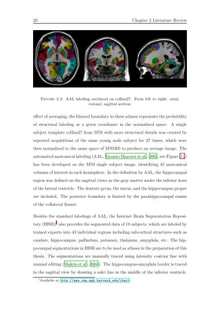
Figure 2.2: AAL labeling overlayed on collins27. From left to right: axial,<br />
coronal, sagittal section.<br />
effect of averaging, the blurred boundary in these atlases represents the probability<br />
of structural labeling at a given coordinate in the normalized space. A single<br />
subject template collins27 from MNI with more structural <strong>de</strong>tails was created by<br />
repeated acquisitions of the same young male subject for 27 times, which were<br />
then normalized to the same space of MNI305 to produce an average image. The<br />
automated anatomical labeling (AAL, Tzourio-Mazoyer et al., 2002, see Figure 2.2)<br />
has been <strong>de</strong>veloped on the MNI single subject image, i<strong>de</strong>ntifying 45 anatomical<br />
volumes of interest in each hemisphere. In the <strong>de</strong>finition by AAL, the hippocampal<br />
region was <strong>de</strong>fined on the sagittal views as the gray matter un<strong>de</strong>r the inferior horn<br />
of the lateral ventricle. The <strong>de</strong>ntate gyrus, the uncus, <strong>and</strong> the hippocampus proper<br />
are inclu<strong>de</strong>d. The posterior boundary is limited by the parahippocampal ramus<br />
of the collateral fissure.<br />
Besi<strong>de</strong>s the st<strong>and</strong>ard labelings of AAL, the Internet Brain <strong>Segmentation</strong> Reposi-<br />
tory (IBSR) 1 also provi<strong>de</strong>s the segmented data of 18 subjects, which are labeled by<br />
trained experts into 43 individual regions including subcortical structures such as<br />
caudate, hippocampus, pallindum, putamen, thalamus, amygdala, etc. The hip-<br />
pocampal segmentations in IBSR are to be used as atlases in the preparation of this<br />
thesis. The segmentations are manually traced using intensity contour line with<br />
manual editing (Makris et al., 2004). The hippocampus-amygdala bor<strong>de</strong>r is traced<br />
in the sagittal view by drawing a sulci line in the middle of the inferior ventricle.<br />
1 Available at http://www.cma.mgh.harvard.edu/ibsr/.


