PDF(575K) - Wiley Online Library
PDF(575K) - Wiley Online Library
PDF(575K) - Wiley Online Library

You also want an ePaper? Increase the reach of your titles
YUMPU automatically turns print PDFs into web optimized ePapers that Google loves.
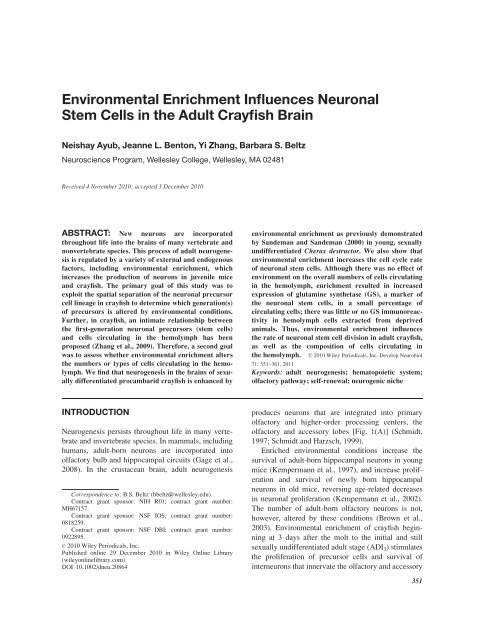
Environmental Enrichment Influences Neuronal<br />
Stem Cells in the Adult Crayfish Brain<br />
Neishay Ayub, Jeanne L. Benton, Yi Zhang, Barbara S. Beltz<br />
Neuroscience Program, Wellesley College, Wellesley, MA 02481<br />
Received 4 November 2010; accepted 3 December 2010<br />
ABSTRACT: New neurons are incorporated<br />
throughout life into the brains of many vertebrate and<br />
nonvertebrate species. This process of adult neurogenesis<br />
is regulated by a variety of external and endogenous<br />
factors, including environmental enrichment, which<br />
increases the production of neurons in juvenile mice<br />
and crayfish. The primary goal of this study was to<br />
exploit the spatial separation of the neuronal precursor<br />
cell lineage in crayfish to determine which generation(s)<br />
of precursors is altered by environmental conditions.<br />
Further, in crayfish, an intimate relationship between<br />
the first-generation neuronal precursors (stem cells)<br />
and cells circulating in the hemolymph has been<br />
proposed (Zhang et al., 2009). Therefore, a second goal<br />
was to assess whether environmental enrichment alters<br />
the numbers or types of cells circulating in the hemolymph.<br />
We find that neurogenesis in the brains of sexually<br />
differentiated procambarid crayfish is enhanced by<br />
INTRODUCTION<br />
Neurogenesis persists throughout life in many vertebrate<br />
and invertebrate species. In mammals, including<br />
humans, adult-born neurons are incorporated into<br />
olfactory bulb and hippocampal circuits (Gage et al.,<br />
2008). In the crustacean brain, adult neurogenesis<br />
Correspondence to: B.S. Beltz (bbeltz@wellesley.edu).<br />
Contract grant sponsor: NIH R01; contract grant number:<br />
MH67157.<br />
Contract grant sponsor: NSF IOS; contract grant number:<br />
0818259.<br />
Contract grant sponsor: NSF DBI; contract grant number:<br />
0922895.<br />
' 2010 <strong>Wiley</strong> Periodicals, Inc.<br />
Published online 29 December 2010 in <strong>Wiley</strong> <strong>Online</strong> <strong>Library</strong><br />
(wileyonlinelibrary.com)<br />
DOI 10.1002/dneu.20864<br />
environmental enrichment as previously demonstrated<br />
by Sandeman and Sandeman (2000) in young, sexually<br />
undifferentiated Cherax destructor. We also show that<br />
environmental enrichment increases the cell cycle rate<br />
of neuronal stem cells. Although there was no effect of<br />
environment on the overall numbers of cells circulating<br />
in the hemolymph, enrichment resulted in increased<br />
expression of glutamine synthetase (GS), a marker of<br />
the neuronal stem cells, in a small percentage of<br />
circulating cells; there was little or no GS immunoreactivity<br />
in hemolymph cells extracted from deprived<br />
animals. Thus, environmental enrichment influences<br />
the rate of neuronal stem cell division in adult crayfish,<br />
as well as the composition of cells circulating in<br />
the hemolymph. ' 2010 <strong>Wiley</strong> Periodicals, Inc. Develop Neurobiol<br />
71: 351–361, 2011<br />
Keywords: adult neurogenesis; hematopoietic system;<br />
olfactory pathway; self-renewal; neurogenic niche<br />
produces neurons that are integrated into primary<br />
olfactory and higher-order processing centers, the<br />
olfactory and accessory lobes [Fig. 1(A)] (Schmidt,<br />
1997; Schmidt and Harzsch, 1999).<br />
Enriched environmental conditions increase the<br />
survival of adult-born hippocampal neurons in young<br />
mice (Kempermann et al., 1997), and increase proliferation<br />
and survival of newly born hippocampal<br />
neurons in old mice, reversing age-related decreases<br />
in neuronal proliferation (Kempermann et al., 2002).<br />
The number of adult-born olfactory neurons is not,<br />
however, altered by these conditions (Brown et al.,<br />
2003). Environmental enrichment of crayfish beginning<br />
at 3 days after the molt to the initial and still<br />
sexually undifferentiated adult stage (ADI3) stimulates<br />
the proliferation of precursor cells and survival of<br />
interneurons that innervate the olfactory and accessory<br />
351
352 Ayub et al.<br />
Figure 1 A: Diagram of a crayfish brain. Cell Clusters 9 and 10, which are composed of interneurons<br />
in the olfactory pathway, are the sites where adult-born neurons are integrated. B:<br />
Olfactory lobe, accessory lobe, neurogenic niche and streams in the P. clarkii brain labeled for<br />
5-bromo-2-deoxyuridine (BrdU) (green, S-phase marker), GS (blue, glial marker), and propidium<br />
iodide (red, nucleic acid marker). The neurogenic niche is outlined by a white box. C: The neurogenic<br />
niche, labeled for GS (blue), propidium iodide (red), and BrdU (green). The asterisk marks<br />
the vascular cavity in the niche that is confluent with the circulation (Sullivan et al., 2007a; Sandeman<br />
et al., 2009). Arrowheads point to BrdU-labeled cells. D: Dextran Micro-Ruby (red) injection<br />
into the dorsal artery fills blood vessels in the brain (inset) and vascular cavity of the niche (outlined<br />
by anti –Elav [green; a neuronal RNA binding protein marker] and propidium iodide [blue]).<br />
The niche lies on a blood vessel, also filled with dextran (arrows). E: Model illustrating our current<br />
hypothesis regarding the generations of precursors responsible for the production of adult-born neurons.<br />
The first-generation (stem) cells reside in the niche. They divide symmetrically and their<br />
daughters (second-generation cells) migrate along a fibrous tract created by the niche cells, to either<br />
cell Cluster 9 or 10. At least one more division occurs in the proliferation zone before the descendants<br />
(third and later generations) differentiate into neurons. AL, accessory lobe; LPZ, lateral<br />
proliferation zone; MPZ, medial proliferation zone; OL, olfactory lobe; S, G2 and M-labeled<br />
arrows indicate cells that are actively in the cell cycle. Scale Bars: 200 lm inB;25lm inC;25<br />
lm inD;100lm in inset. [Fig. 1(D) from Sullivan et al., 2007a; E from Zhang et al., 2010].<br />
lobes (Sandeman and Sandeman, 2000). It is not<br />
known whether environmental enrichment is an<br />
effective regulator of neuronal production in crayfish<br />
after the initial adult stage (ADI).<br />
Our understanding of environmental regulation of<br />
neurogenesis is incomplete without a knowledge of<br />
which generations in the neuronal precursor lineage<br />
are influenced by enriched conditions. Changes in the<br />
numbers of first-generation precursors (stem cells) or<br />
their cell cycle rate have a much greater potential<br />
to alter neuronal proliferation than influences exerted<br />
on later generations in the lineage. However, this<br />
Developmental Neurobiology<br />
knowledge is not available in either mammals or<br />
crustaceans. In mammals, defining the influence of<br />
environment on specific neuronal precursor generations<br />
is particularly challenging, because several<br />
types of progenitor cells coexist in the neurogenic<br />
niches producing adult-born neurons (García-<br />
Verdugo et al., 1998; Seri et al., 2004; Zhao et al.,<br />
2008), and the lineage relationships among these cell<br />
types have not been directly demonstrated (Kan et al.,<br />
2010). Therefore, to detect changes in the mitotic<br />
index in specific classes of precursors, multiple<br />
markers that characterize stages in this lineage must
e assessed in conjunction with cell cycle indicators<br />
(Kuhn and Peterson, 2008).<br />
In contrast, adult neurogenesis in the crayfish brain<br />
involves generations of precursor cells that are<br />
spatially segregated from each other, except for a<br />
small transitional region between each compartment<br />
[Fig. 1(B,E)] (Sullivan et al., 2007a,b). The neuronal<br />
stem cells (first-generation precursors) comprise a vascularized<br />
niche [Fig. 1(B–D)]; these bipolar cells also<br />
provide a tract along which their progeny (the secondgeneration<br />
precursors) migrate (Sullivan et al., 2005,<br />
2007a,b). These migratory precursors move toward<br />
proliferation zones in cell Clusters 9 and 10<br />
[Fig. 1(A)] (terminology of Sandeman et al., 1992),<br />
where they divide at least once more. Their progeny<br />
differentiate into Clusters 9 (local) and 10 (projection)<br />
interneurons that innervate the primary olfactory processing<br />
areas (the olfactory lobes) and higher-order<br />
processing centers (the accessory lobes) (Sullivan and<br />
Beltz, 2005). Because of the spatial segregation of the<br />
first, second, and third and later generations of neuronal<br />
precursors in crayfish, influences of environmental<br />
enrichment on different parts of the lineage are easily<br />
assessed.<br />
To define the influence of environmental enrichment<br />
on adult neurogenesis in crayfish, Sandeman<br />
and Sandeman (2000) gauged the incorporation of<br />
5-bromo-2-deoxyuridine (BrdU) into the third-generation<br />
neuronal precursors and their descendants in the<br />
proliferation zones of Clusters 9 and 10, where they<br />
found increased cell proliferation and survival. The<br />
neuronal precursor cell lineage in crayfish [see Fig.<br />
1(E)] had not yet been discovered, and so influences<br />
on the first and second-generation precursors were<br />
not determined. The primary goal this study, therefore,<br />
was to exploit the spatial separation of this<br />
lineage to examine the influence of environmental<br />
enrichment on these precursors. The second goal was<br />
to test whether environmental enrichment alters adult<br />
neurogenesis in older crayfish, or whether these<br />
effects are confined to very young (ADI) crayfish.<br />
Finally, experiments in crayfish have indicated<br />
that the neuronal stem cells are not a self-renewing<br />
population (Zhang et al., 2009; Benton et al., 2010).<br />
Given the close relationship between the vasculature<br />
and the niche where these first-generation precursors<br />
reside, we have hypothesized that the hematopoietic<br />
system is a potential source of neuronal stem cells in<br />
crayfish (Zhang et al., 2009). If hematopoietic stem<br />
cells indeed contribute to the first-generation precursor<br />
pool, environmental conditions may also affect<br />
their numbers or properties. The final goal of the<br />
present studies, therefore, was to ask if living conditions<br />
influence the numbers or specific composition<br />
of cells circulating in the hemolymph, which would<br />
be products of the hematopoietic system.<br />
These studies show that neurogenesis in the brains<br />
of sexually differentiated (but not yet sexually active)<br />
mid-life crayfish (Procambarus spp.) is enhanced by<br />
environmental enrichment as previously demonstrated<br />
in young ADI 3 Cherax destructor (Sandeman<br />
and Sandeman, 2000). In addition, we show that environmental<br />
enrichment increases the rate of BrdU<br />
incorporation into the first-generation neuronal<br />
precursors, indicating that the cell cycle time of these<br />
stem cells decreases. In small adult crayfish (carapace<br />
length 4 mm, CL4), enrichment also increases the<br />
number of first-generation precursor cells residing in<br />
the niche, although this effect was not seen in larger<br />
animals (carapace length 10 mm, CL10). Although<br />
there was no effect of environment on the overall<br />
numbers of cells circulating in the hemolymph,<br />
enrichment resulted in immunoreactivity for glutamine<br />
synthetase (GS), a marker of the neuronal stem<br />
cells residing in the niche [Fig. 1(B,C)], in a small<br />
percentage of circulating cells; there was little or no<br />
expression of this enzyme in hemolymph cells<br />
extracted from animals maintained in the deprived<br />
environment. Thus, enrichment influences the firstgeneration<br />
neuronal precursors (stem cells) as well as<br />
the composition of cells circulating in the hemolymph<br />
in adult crayfish. These findings have been previously<br />
published in abstract form (Ayub et al., 2010).<br />
MATERIALS AND METHODS<br />
Animals<br />
Environment Affects Neuronal Stem Cells 353<br />
Experiments were conducted using two closely related<br />
sympatric species of freshwater crayfish (Procambarus<br />
clarkii and Procambarus acutus) obtained from a commercial<br />
supplier (Carolina Biological Supply Company,<br />
Burlington, NC). Crayfish were housed and maintained in<br />
the Wellesley College Animal Care Facility, where they<br />
were kept at room temperature on a 12/12 light/dark cycle.<br />
Two sizes of crayfish with starting carapace lengths (CL) of<br />
4 mm and 10 mm (*ADII and ADVII, respectively) were<br />
used in these experiments. CL4 crayfish are sexually undifferentiated,<br />
whereas the CL10 crayfish are sexually differentiated<br />
but not yet sexually active. Mixed groups of male<br />
and female crayfish of both sizes were therefore used in<br />
these studies.<br />
Enriched and Deprived Environments<br />
Four groups of crayfish (n ¼ 10 per group) were maintained<br />
in a 20-gallon aquarium with recirculating, filtered artificial<br />
pond water: (1) enriched CL4, (2) enriched CL10, (3)<br />
Developmental Neurobiology
354 Ayub et al.<br />
deprived CL4, and (4) deprived CL10. Each group consisted<br />
of 10 crayfish at the beginning of the experiment, a<br />
number small enough that they could establish territories<br />
but be freely active. The enriched environment consisted of<br />
group housing in a 20-gallon tank (30@ 3 12.5@ 3 12.5@)<br />
filled with gravel, foliage, tunnels, a tree-like structure, and<br />
a rock; CL4 and CL10 crayfish were separated by a large<br />
mesh divider, which divided the tank in half. In the<br />
deprived environments, crayfish were individually housed<br />
in containers that floated on top of the tank. Ice cube trays<br />
in which the bottom was replaced with nylon mesh housed<br />
the CL10 crayfish; CL4 animals were maintained in conical<br />
tubes that were cut down and covered with nylon mesh. For<br />
both groups, the deprived crayfish had 1.8 cm 3 space/mm<br />
carapace length. Water flowed freely through the mesh bottoms<br />
in these containers, insuring that the deprived animals<br />
shared the same water as the enriched animals. To increase<br />
the sample size and to replicate initial findings, two sequential<br />
trials were conducted; as there were no statistically<br />
significant differences in the results from the two trials,<br />
data were combined for the final results.<br />
Hemolymph Analysis<br />
After *3 weeks exposed to these environmental conditions,<br />
hemolymph samples were taken between 11:00 and<br />
13:00 h from four CL10 animals from both enriched and<br />
deprived groups. This time frame was chosen based on the<br />
need for a minimum of 2 weeks to observe the influence of<br />
environmental enrichment in ADI crayfish (Sandeman and<br />
Sandeman, 2000). Hemolymph was drawn from the dorsal<br />
sinus of the animal with a 1 mL syringe and 25 5/8 gauge<br />
needle, and mixed 1:2 with anticoagulant buffer (Söderhäll<br />
et al., 2003). This mixture was combined with Trypan<br />
blue (Sigma) (1:1), and 10 lL of this solution placed on<br />
the counting grid of a hemocytometer for quantification of<br />
hemolymph cells. Average counts were calculated from<br />
2 to 3 separate samples taken from each blood draw. Hemolymph<br />
from CL4 crayfish was not evaluated because the<br />
blood volume is too small to provide adequate samples for<br />
analysis. One factor that may influence hemolymph cell<br />
counts is time of day; hemolymph counts in the mole-crab<br />
(Emerita asiatica) were found to be higher in the afternoon<br />
than in the morning (Ravindranath, 1977). In the present<br />
studies, to control for time of day, counts were conducted<br />
in a random order and they were completed within 2 h on<br />
the same day. Therefore, circadian factors are not likely to<br />
have influenced the results of the present studies.<br />
BrdU Exposure and<br />
Immunohistochemistry<br />
Beginning at 19:00 h on the day hemolymph samples were<br />
taken, crayfish were incubated in the S-phase marker BrdU<br />
(Sigma; 2 mg/mL of pond water). Crayfish from the<br />
enriched environment were incubated in BrdU together,<br />
while those from the deprived environments were incubated<br />
individually, thus replicating the social environment of their<br />
Developmental Neurobiology<br />
living conditions. After 18 h in BrdU, they were sacrificed.<br />
Brains were dissected in cold crayfish saline and fixed overnight<br />
in 4% paraformaldehyde in 0.1M phosphate buffer<br />
(PB) and processed immunohistochemically as in prior<br />
studies (Zhang et al., 2009). Briefly, brains were rinsed<br />
multiple times in PB containing 0.3% Triton X-100<br />
(PBTx), then incubated in 0.2 N HCl and rinsed in PBTx.<br />
Brains were incubated with rat anti-BrdU (Accurate Chemical<br />
and Scientific, Westbury, NY; 1:50 dilution) at room<br />
temperature for 3 h. Subsequently, brains were rinsed and<br />
incubated for 12 h at 48C with mouse anti-GS (Becton<br />
Dickinson; 1:100), rinsed again and incubated with secondary<br />
antibodies (Cy2-conjugated goat anti-rat IgG and Cy5conjugated<br />
goat anti-mouse IgG; Jackson Immunoresearch;<br />
1:100; 12 h at 48C). Brains were rinsed in PBTx, incubated<br />
in the nuclear marker propidium iodide (10 lg/mL in PB;<br />
Sigma) for 10 min, and rinsed again in PB. The brains were<br />
mounted on slides with Gel/Mount TM (Electron Microscopy<br />
Sciences, Hatfield, PA).<br />
At the time of dissection, hemolymph was taken from<br />
four CL10 crayfish from each environmental condition and<br />
smeared on slides prepared with poly-L-lysine to promote<br />
cell adhesion. Hemolymph cells were fixed with 4%<br />
paraformaldehyde for 15 min, rinsed for 1 h with PBTx,<br />
and incubated with donkey anti-mouse IgG FAB (Jackson<br />
Immunoresearch; 1:10) and donkey anti-rabbit IgG<br />
(Jackson Immunoresearch; 1:100); samples were fixed<br />
again with 4% paraformaldehyde. These treatments were<br />
done to prevent nonspecific binding of subsequent antibodies.<br />
Hemolymph smears were then rinsed for another hour,<br />
and all but one of the samples from the deprived animals<br />
was incubated overnight at 48C with mouse anti-GS; the<br />
slide without this antibody served as a no-primary control<br />
to test the specificity of the secondary antibody. Slides were<br />
rinsed for 1 h with PBTx, incubated with donkey antimouse<br />
IgG Cy5 (1:500) for 30 min at room temperature,<br />
and rinsed again with PBTx. Finally, the hemolymph<br />
smears were incubated with propidium iodide for 5 min<br />
at room temperature, rinsed with PB and mounted with<br />
Gel/Mount TM .<br />
Confocal Microscopy<br />
Brains and hemolymph smears were imaged with a Leica<br />
TCS SP5 laser scanning confocal microscope using argon,<br />
krypton, and helium-neon lasers. Serial optical sections<br />
were taken at 1–2 lm intervals and saved as stacks.<br />
Data Analysis and Statistics<br />
GS labeling of neuronal precursors provided a means of<br />
visualizing the neurogenic niche and migratory streams, and<br />
BrdU allowed quantification of the number of S-phase cells<br />
in these regions. Propidium iodide, a nucleic acid marker<br />
that labels all nuclei, permitted quantification of total numbers<br />
of niche cells as well as the numbers of blood cells in<br />
smears. The numbers of BrdU-labeled cells in the niche,<br />
streams and Clusters 9 and 10 were used as a measure of the
proliferation rate of neuronal precursors, and hence an indication<br />
of the cell cycle time. Labeled neuronal precursors<br />
and cells in the hemolymph were counted by projecting<br />
each image in the stack onto a computer screen, and tracing<br />
the labeled cells onto a transparent plastic sheet. The traced<br />
cells were counted, and independent samples t-tests and<br />
analysis of variance (ANOVA) with a post hoc Tukey were<br />
run with JMP7 (SAS, Cary, NC).<br />
RESULTS<br />
Environmental Enrichment Influences<br />
Third-Generation Neuronal Precursors<br />
and Their Descendants in Clusters 9 and<br />
10 in CL4 and CL10 Crayfish<br />
The findings of Sandeman and Sandeman (2000) demonstrated<br />
that environmental enrichment enhances cell<br />
proliferation of third-generation precursors and their<br />
descendants in Clusters 9 and 10 in the brains of ADI<br />
crayfish. Our studies tested whether environment has<br />
a similar influence in crayfish at later stages of development.<br />
An independent samples t-test assessed the<br />
effect of environmental enrichment and deprivation<br />
on the mean number of BrdU cells in Clusters 9 and<br />
10 for crayfish of CL4 and CL10. For the younger/<br />
smaller CL4 animals, enrichment significantly<br />
enhanced cell proliferation in Cluster 9 (t(18) ¼<br />
3.504, p ¼ 0.003) [Fig. 2(A)] and Cluster 10<br />
(t(17.104) ¼ 8.217, p < 0.001) [Fig. 2(B)]. Similar<br />
effects were observed in the older/larger crayfish<br />
(CL10): Cluster 9 (t(39.911) ¼ 3.710, p ¼ 0.001),<br />
and Cluster 10 (t(43.999) ¼ 4.868, p < 0.001).<br />
Mean numbers of BrdU-labeled cells in Clusters 9 and<br />
10 of all groups also were compared using ANOVA<br />
and post hoc Tukey tests to ask whether the degree of<br />
environmental influence on neurogenesis was size/<br />
age-dependent. The ANOVA analysis confirmed the<br />
effects of environmental conditions on neurogenesis<br />
in these regions and showed that size did not influence<br />
the magnitude of these effects, over the range tested.<br />
The influence of environment on neurogenesis is therefore<br />
not restricted to ADI animals (*CL2) (Sandeman<br />
and Sandeman, 2000), but persists in larger/older sexually<br />
differentiated crayfish (CL4–CL10).<br />
Influence of Environmental Enrichment<br />
on the First-Generation Precursors<br />
(Neuronal Stem Cells)<br />
The primary goal of this study was to determine<br />
whether the first- and second-generation neuronal<br />
precursors are also influenced by environmental conditions.<br />
In both CL4 and CL10 crayfish, environmen-<br />
Environment Affects Neuronal Stem Cells 355<br />
Figure 2 Environmental enrichment has a significant<br />
effect on cell proliferation in Cluster 9 (A) and Cluster 10<br />
(B), as indicated by increased incorporation of BrdU in<br />
crayfish housed in enriched compared to deprived conditions.<br />
Histograms represent mean values for each group of<br />
crayfish, with standard deviations indicated. N ¼ number of<br />
cell clusters. Asterisks represent t-test significance values:<br />
** indicates a p value
356 Ayub et al.<br />
Figure 3 Environmental enrichment has a significant effect<br />
on the cell cycle rate of first-generation neuronal precursors in<br />
the niche (A) and second-generation precursors in the migratory<br />
streams (C), as indicated by the mean numbers (6S.D.)<br />
of BrdU-labeled cells in these areas. The mean numbers of<br />
cells in the niche of crayfish housed in enriched conditions is<br />
increased relative to crayfish maintained in deprived conditions<br />
(B). N ¼ number of niches (A, B) or streams (C), and<br />
asterisks represent t-test significance values: ** signifies a p<br />
value
cells (F(1,6) ¼ 3.576, p > 0.05) circulating in the<br />
hemolymph. As reported in the literature (Ravindranath,<br />
1977; Owens and O’Neill, 1997), total counts<br />
of circulating cells are highly variable in decapod<br />
crustaceans, both between animals and for an individual<br />
animal on different days. In our studies, total<br />
hemocyte counts for individual animals ranged from<br />
0.6 to 13.8 3 10 6 cells per milliliter of hemolymph.<br />
In spite of this variability, however, there is a significantly<br />
greater expression of the niche precursor cell<br />
marker GS in circulating cells from animals maintained<br />
in an enriched environment, relative to the<br />
deprived environment (t(10) ¼ 2.228, p < 0.001). In<br />
the environmentally deprived animals, GS is<br />
expressed in 1.1% of circulating hemolymph cells;<br />
3.7% of circulating cells in crayfish housed in<br />
enriched conditions expressed GS (Fig. 4). These<br />
percentages were calculated based on counting a<br />
total of 12,851 hemocytes from 27 blood samples<br />
taken from enriched animals, and 7,148 hemocytes<br />
from 21 blood samples from deprived crayfish.<br />
These data indicate that enrichment enhances the<br />
expression of this enzyme in cells circulating in the<br />
hemolymph, which are derived from hematopoietic<br />
tissues.<br />
DISCUSSION<br />
The current experiments exploited the spatial separation<br />
of the first-, second-, and third-generations of<br />
neuronal precursor cells (Sullivan et al., 2007a, b) in<br />
the crayfish brain, to determine which of these may<br />
be altered by environmental conditions. Two sizes of<br />
crayfish were utilized in the present studies, to ask<br />
whether the influence of environment is size/age-dependent.<br />
Finally, the numbers and types of circulating<br />
cells were assessed in the CL10 crayfish, to test<br />
whether environmental enrichment alters the blood<br />
cell count or composition of the hemolymph.<br />
Influences of Environmental Enrichment<br />
on the Precursor Cell Lineage that<br />
Produces Adult-Born Neurons<br />
The most compelling finding of this study is that<br />
environmental factors influence the cell cycle rate of<br />
first-generation neuronal precursors in the neurogenic<br />
niche. In both CL4 and CL10 crayfish, significantly<br />
more BrdU-labeled first-generation cells were<br />
detected in the brains of animals maintained in<br />
enriched, relative to deprived, environments. This is<br />
the first demonstration that the proliferative potential<br />
Environment Affects Neuronal Stem Cells 357<br />
Figure 4 Enrichment has a significant effect on the<br />
percentage of GS-labeled cells in the hemolymph. In the<br />
environmentally deprived animals, GS is expressed in 1.1%<br />
of circulating hemolymph cells; 3.7% of circulating cells in<br />
crayfish housed in enriched conditions expressed GS (A).<br />
B: A small percentage of all circulating cells (PI, red) from<br />
enriched CL10 crayfish label with GS (blue). N ¼ number<br />
of animals from which hemolymph was sampled. Asterisks<br />
represent t-test values: ** signifies a p value
358 Ayub et al.<br />
Kempermann et al. (1997) investigated the effect of<br />
environmental enrichment on neurogenesis and neuronal<br />
progenitor cells in young mice (*postnatal day<br />
21). The enriched environment consisted of mice living<br />
communally in a large cage with a set of tunnels,<br />
running wheels, and toys to increase the complexity of<br />
living conditions; these environments were rearranged<br />
daily, to create novelty. The control environment<br />
included three mice living in a standard cage. After<br />
the 40-day environmental exposure, levels of BrdU<br />
incorporation in hippocampal cells were compared in<br />
short- and long-survival time studies, to determine differences<br />
in cell proliferation and survival, respectively.<br />
Contrary to the findings presented here, neuronal<br />
proliferation was unaffected by environmental<br />
enrichment, but cell survival (measured 4 weeks after<br />
BrdU administration) increased. However, in older<br />
mice long-term exposure (10 months) to enrichment<br />
did result in increased precursor proliferation and neuronal<br />
survival (Kempermann et al., 2002). Our results<br />
and those of Sandeman and Sandeman (2000) showing<br />
an influence of short-term environmental exposure on<br />
both proliferation and survival in crayfish may represent<br />
species differences in the regulation of neurogenesis<br />
by environment, or could be a reflection of the<br />
very distinct environments to which the crayfish were<br />
exposed. The control environment in the mouse studies<br />
was equivalent to standard group housing for mice.<br />
However, in the current series of experiments in crayfish,<br />
the deprived condition included space limitations<br />
that limited physical activity, social isolation and a<br />
barren landscape, replicating the conditions of the<br />
Sandeman and Sandeman (2000) study.<br />
Previous studies in mice and crustaceans have<br />
attempted to determine the individual contributions<br />
of the various environmental components to the<br />
regulation of neurogenesis. For example, voluntary<br />
physical activity and exercise have been shown to<br />
increase neurogenesis in mice as well as to reverse<br />
the decline in neurogenesis experienced by older animals<br />
(Van Praag et al., 1999; 2005). The enriched<br />
conditions used in the current studies provided a<br />
complex environment that included social interaction,<br />
a large space with interesting features such as plants<br />
and burrows, and the opportunity for exploration and<br />
physical activity. Our study did not clarify which of<br />
these elements may mediate the beneficial effects of<br />
enrichment. To establish the enriched and deprived<br />
conditions, however, several other environmental<br />
arrangements were tested. We found that the space<br />
limitations were a critical aspect of the deprived condition,<br />
and that use of a larger deprived environment<br />
eliminated the overall influence on proliferation,<br />
measured by BrdU incorporation, between the two<br />
Developmental Neurobiology<br />
groups of animals (data not shown). Space restrictions<br />
have been shown to inhibit animals’ growth<br />
(Rugh, 1934), and it is therefore possible that limitations<br />
in space and physical activity may be primary<br />
determinants of neurogenic potential in crayfish in<br />
these studies. Indeed, locomotory studies in large<br />
crayfish do show a weak correlation between the level<br />
of physical activity and the survival of adult-born<br />
neurons in large crayfish (CL > 25 mm), but no effect<br />
on proliferation (Sandeman, unpublished results).<br />
Locomotion therefore may be one critical element<br />
that differed between the two conditions in our study.<br />
Isolation versus social interaction did not appear to<br />
play a role in determining levels of neurogenesis in<br />
our study, as crayfish isolated in larger environments<br />
while developing the environmental conditions for<br />
these experiments did not have reduced levels of neurogenesis<br />
compared to those maintained in groups in<br />
the enriched environments (data not shown). Song et<br />
al. (2007) also studied the relationship between social<br />
interactions and neurogenesis in young adult crayfish,<br />
and found no effect of social dominance or social<br />
isolation on cell proliferation in Clusters 9 and 10,<br />
although social domination enhanced the survival of<br />
BrdU-labeled cells when compared to social subordination.<br />
Additional experiments are necessary to<br />
define which specific components of the enriched<br />
environment are crucial for the influences observed in<br />
the present study.<br />
Concomitant with the increase in BrdU incorporation<br />
into first-generation precursors in CL4 crayfish,<br />
there was a decrease in the total numbers of niche<br />
cells in the enriched animals compared with deprived;<br />
these data were acquired by counting the numbers of<br />
propidium iodide labeled nuclei in the niche. These<br />
findings are logical in the sense that an increased cell<br />
cycle rate among the niche precursors would result in<br />
more cells migrating away from the niche; this might<br />
result in fewer total numbers of niche precursors<br />
remaining in the niche. If the cell cycle rate slows, as<br />
in the deprived environment, niche precursors will be<br />
retained in the niche, causing their numbers to be<br />
larger relative to the enriched environment. Although<br />
these relationships are potentially interesting, this<br />
result was not replicated in the older CL10 crayfish,<br />
and so this effect may be specific to younger animals.<br />
Further, in CL10 animals, enrichment enhanced BrdU<br />
incorporation into the migratory second-generation<br />
neuronal precursors; a trend in this direction was also<br />
seen in CL4 crayfish, but this result was not statistically<br />
significant. Finally, our results confirm the influence<br />
of environment on BrdU incorporation among<br />
the third-generation precursors and their descendants<br />
in Clusters 9 and 10 previously reported by Sandeman
and Sandeman (2000) in ADI Cherax destructor. The<br />
present studies utilized larger/older crayfish of two<br />
species, P. clarkii and P. acutus, demonstrating that<br />
environmental effects on adult neurogenesis in crayfish<br />
persist beyond the first adult (ADI) stage.<br />
Effects of Environmental Enrichment<br />
on Cells Circulating in the Hemolymph<br />
The results of Zhang et al. (2009) indicated that niche<br />
precursor cells are not a self-renewing pool of cells, a<br />
suggestion that has now been directly demonstrated<br />
using double nucleoside labeling (Benton et al.,<br />
2010). It has been hypothesized that the hematopoietic<br />
system may be a source of first-generation neuronal<br />
progenitors (neuronal stem cells); the vasculature<br />
and the niche are closely associated physically, and<br />
the niche precursors and cells in nearby blood vessels<br />
have similar morphologies (Zhang et al., 2009).<br />
Another goal of this study, therefore, was to determine<br />
if environmental conditions influence the numbers<br />
or types of circulating cells. Counts of cells in<br />
hemolymph samples from enriched and deprived<br />
CL10 crayfish showed no differences in the numbers<br />
of circulating live, dead or total cells in the two<br />
environmental conditions.<br />
GS is an enzyme that converts glutamate to glutamine,<br />
and is a marker of astrocytes and early<br />
stem cells (Wen et al., 2007; 2009). Antibodies<br />
generated against GS label the neuronal stem cells<br />
in crustacean brain [e.g., see Fig. 1(B–D)], as well<br />
as a small percentage of hemolymph cells (Benton<br />
et al., 2010). GS expression was investigated in<br />
circulating cells of crayfish (CL10) maintained in<br />
enriched and deprived environments, to determine<br />
if the presence of GS in circulating cells in the<br />
hemolymph is altered by environmental conditions.<br />
The results indicate that although percentages of<br />
GS-labeled cells in hemolymph samples are variable,<br />
on average 3.7% of the circulating cells in<br />
enriched crayfish express GS; an average of 1.1%<br />
of circulating cells in crayfish maintained in<br />
deprived conditions expressed this niche cell<br />
marker. In some of the environmentally deprived<br />
animals no GS-labeled cells were observed in<br />
blood samples, a situation that was never encountered<br />
in the environmentally enriched crayfish.<br />
These results indicate that environmental enrichment<br />
enhances GS expression in circulating hemolymph<br />
cells of crayfish. These findings are consistent<br />
with a relationship between the hematopoietic<br />
and neurogenic systems, and this possibility is<br />
being carefully examined.<br />
Environment Affects Neuronal Stem Cells 359<br />
The fact that antibodies generated against GS bind<br />
to both the neuronal stem cells and to a small proportion<br />
of circulating cells is intriguing. A characteristic<br />
feature of neurogenic niches in both vertebrate and<br />
invertebrate organisms is vascularization (Tavazoie<br />
et al., 2008). Supportive (e.g., nutrients) and instructive<br />
(e.g., hormones, cytokines and other circulating<br />
factors) roles for the vascular input have been widely<br />
acknowledged in the vertebrates (Riquelme et al.,<br />
2008; Kan et al., 2010). There are also suggestions<br />
that the vascular contribution may be even more central<br />
to the adult neurogenesis story. Cells derived<br />
from mammalian bone marrow have a tendency to<br />
migrate to the brain when infused into a host animal<br />
(Eglitis and Mezey, 1997; Kopen et al., 1999; Brazelton<br />
et al., 2000), including into areas undergoing<br />
active postnatal neurogenesis. The descendants of<br />
these cells express a variety of glial and neuronal<br />
markers (Eglitis and Mezey, 1997; Mezey et al.,<br />
2000; Chen et al., 2001), and in one study these<br />
developed the characteristics of astrocytes (Kopen<br />
et al, 1999). In vitro, bone marrow cells have been<br />
induced by various means to form neurons (Sanchez-<br />
Ramos et al., 2000, 2001, 2002; Kohyama et al.,<br />
2001), and in one case the bone marrow-derived<br />
neurons responded to depolarizing stimuli, showing a<br />
rapid and reversible calcium increase in response to<br />
acetylcholine, a response characteristic of neurons<br />
(Kohyama et al., 2001). Other studies, however, suggest<br />
that cell fusion may account for the acquisition<br />
of such broad properties by stem cells (Morshead<br />
et al., 2002; Wagers et al., 2002; Wells, 2002; Coyne<br />
et al., 2006), casting doubt on the significance of prior<br />
studies. Nevertheless, the idea that cells derived from<br />
bone marrow or umbilical cord blood can transdifferentiate<br />
into neurons and glia in response to signals in<br />
the brain persists in the literature. Direct tests of the<br />
potential relationships between cells circulating in the<br />
vasculature and mechanisms of adult neurogenesis<br />
are possible in the crayfish, and these experiments are<br />
currently underway in our laboratory.<br />
The authors thank P. Carey and V. LePage for care of<br />
the animals used in these studies, and D.C. Sandeman for<br />
his critical reading of the manuscript. The work was supported<br />
by NIH R01 MH67157, NSF IOS 0818259 and NSF<br />
DBI 0922895.<br />
REFERENCES<br />
Ayub N, Benton JL, Zhang Y, Beltz BS. 2010. Environmental<br />
enrichment influences the cell cycle rate and<br />
number of neuronal stem cells in the crayfish brain. Soc<br />
Neurosci Abstr 36:435.3.<br />
Developmental Neurobiology
360 Ayub et al.<br />
Benton JL, Zhang Y, Kirkhart CR, Sandeman DC, Beltz<br />
BS. 2010. Primary neuronal precursors in the crayfish<br />
brain: self-renewal or replenishment from another<br />
source? Soc Neurosci Abstr 36:233.1.<br />
Brazelton TR, Rossi FM, Keshet GI, Blau HM. 2000. From<br />
marrow to brain: expression of neuronal phenotypes in<br />
adult mice. Science 290:1775–1779.<br />
Brown J, Cooper-Kuhn CM, Kempermann G, van Praag H,<br />
Winkler J, Gage FH, Kuhn HG. 2003. Enriched environment<br />
and physical activity stimulate hippocampal but<br />
not olfactory bulb neurogenesis. Eur J Neurosci 17:<br />
2042–2046.<br />
Chen J, Sanberg PR, Li Y, Wang L, Lu M, Willing AE,<br />
Sanchez-Ramos J, et al. 2001. Intravenous administration<br />
of human umbilical cord blood reduces behavioral deficits<br />
after stroke in rats. Stroke 32:2682–2688.<br />
Coyne TM, Marcus AJ, Woodbury D, Black IB. 2006.<br />
Marrow stromal cells transplanted to the adult brain<br />
are rejected by an inflammatory response and transfer<br />
donor labels to host neurons and glia. Stem Cells<br />
24:2483–2492.<br />
Eglitis MA, Mezey E. 1997. Hematopoietic cells differentiate<br />
into both microglia and macroglia in the brains of<br />
adult mice. Proc Natl Acad Sci USA 94:4080–4085.<br />
Gage FH, Kempermann G, Song H, editors. 2008. Adult<br />
Neurogenesis. Cold Spring Harbor, NY: Cold Spring<br />
Harbor Laboratory Press, p 673.<br />
García-Verdugo JM, Doetsch F, Wichterle H, Lim DA,<br />
Alvarez-Buylla A. 1998. Architecture and cell types of<br />
the adult subventricular zone: In search of the stem cells.<br />
J Neurobiol 36:234–248.<br />
Kan I, Barhum Y, Melamed E, Offen D. 2010. Mesenchymal<br />
stem cells stimulate endogenous neurogenesis in the<br />
subventricular zone of adult mice. Stem Cell Rev<br />
PMID:20830611.<br />
Kempermann G, Gast D, Gage, FH. 2002. Neuroplasticity<br />
in old age: sustained fivefold induction of hippocampal<br />
neurogenesis by long-term environmental enrichment.<br />
Ann Neurol 52:135–143.<br />
Kempermann G, Kuhn HG, Gage FH. 1997. More hippocampal<br />
neurons in adult mice living in an enriched<br />
environment. Nature 386:493–495.<br />
Kohyama J, Abe H, Shimazaki T, Koizumi A, Nakashima<br />
K, Gojo S, Taga, T, et al. 2001. Brain from bone: efficient<br />
\meta-differentiation" of marrow stroma-derived<br />
mature osteoblasts to neurons with Noggin or a demethylating<br />
agent. Differentiation 68:235–44.<br />
Kopen GC, Prockop DJ, Phinney DG. 1999. Marrow stromal<br />
cells migrate throughout forebrain and cerebellum,<br />
and they differentiate into astrocytes after injection into<br />
neonatal mouse brains. Proc Natl Acad Sci USA<br />
96:10711–10716.<br />
Kuhn HG, Peterson DA. 2008. Detection and phenotypic<br />
characterization of adult neurogenesis. In: Gage FH,<br />
Kempermann G, Song H, editors. Adult Neurogenesis.<br />
Cold Spring Harbor, NY: Cold Spring Harbor Laboratory<br />
Press, pp 25–47.<br />
Mezey E, Chandross KJ, Harta G, Maki RA, McKercher<br />
SR. 2000 Turning blood into brain: cells bearing neuro-<br />
Developmental Neurobiology<br />
nal antigens generated in vivo from bone marrow.<br />
Science 290:1779–1782.<br />
Morshead CM, Benveniste P, Iscove NN, van der Kooy D.<br />
2002. Hematopoietic competence is a rare property of<br />
neural stem cells that may depend on genetic and epigenetic<br />
alterations. Nat Med 8:268–273.<br />
Owens L, O’Neill A. 1997. Use of a clinical cell flow<br />
cytometer for differential counts of prawn Penaeus<br />
monodon haemocytes. Dis Aquat Org 31:147–153.<br />
Ravindranath MH. 1977. Circulating hemocyte population<br />
of mole-crab Emerita Asiatica. Biol Bull<br />
152:415–423.<br />
Riquelme PA, Drapeau E, Doetsch F. 2008. Brain microecologies:<br />
neural stem cell niches in the adult mammalian<br />
brain. Philos Trans R Soc Lond B Biol Sci 363:123–137.<br />
Rugh R. 1934. The space factor in the growth rate of<br />
tadpoles. Ecology 15:407–411.<br />
Sanchez-Ramos JR. 2002. Neural cells derived from adult<br />
bone marrow and umbilical cord blood. J Neurosci Res<br />
69:880–893.<br />
Sanchez-Ramos J, Song S, Cardozo-Pelaez F, Hazzi C, Stedeford<br />
T, Willing A, Freeman TB, et al. 2000. Adult<br />
bone marrow stromal cells differentiate into neural cells<br />
in vitro. Exp Neurol 164:247–256.<br />
Sanchez-Ramos JR, Song S, Kamath SG, Zigova T, Willing<br />
A, Cardozo-Pelaez F, Stedeford T, et al. 2001. Expression<br />
of neural markers in human umbilical cord blood.<br />
Exp Neurol 171:109–115.<br />
Sandeman D, Sandeman R, Derby C, Schmidt M. 1992.<br />
Morphology of the brain of crayfish, crabs, and spiny<br />
lobsters: common nomenclature for homologous structures.<br />
Biol Bull 183:304–326.<br />
Sandeman DC, Benton JL, Beltz BS. 2009. An identified<br />
serotonergic neuron regulates neurogenesis in the crayfish<br />
brain. Dev Neurobiol 69:530–545.<br />
Sandeman R, Sandeman D. 2000. \Impoverished" and<br />
\Enriched" living conditions influence the proliferation<br />
and survival of neurons in crayfish brain. J Neurobiol<br />
45:215–226.<br />
Schmidt M. 1997. Continuous neurogenesis in the olfactory<br />
brain of adult shore crabs, Carcinus maenas. Brain Res<br />
762:131–143.<br />
Schmidt M, Harzsch S. 1999. Comparative analysis of<br />
neurogenesis in the central olfactory pathway of the adult<br />
decapod crustaceans by in vivo BrdU-labeling. Biol Bull<br />
196:127–136.<br />
Seri B, García-Verdugo JM, Collado-Morente L, McEwen<br />
BS, Alvarez-Buylla A. 2004. Cell types, lineage, and<br />
architecture of the germinal zone in the adult dentate<br />
gyrus. J Comp Neurol 478:359–378.<br />
Söderhäll I, Bangyeekhun E, Mayo S, Söderhäll K. 2003.<br />
Hemocyte production and maturation in an invertebrate<br />
animal; proliferation and gene expression in hematopoietic<br />
stem cells of Pacifastacus leniusculus. Dev Comp<br />
Immunol 27:661–672.<br />
Song CK, Johnstone LM, Schmidt M, Derby CD, Edwards<br />
DH. 2007. Social domination increases neuronal survival<br />
in the brain of juvenile crayfish Procambarus clarkii.<br />
J Exp Biol 210:1311–1324.
Sullivan JM, Beltz BS. 2005. Newborn cells in the adult<br />
crayfish brain differentiate into distinct neuronal types.<br />
J Neurobiol 65:157–170.<br />
Sullivan JM, Benton JL, Sandeman DC, Beltz, BS. 2007a.<br />
Adult neurogenesis: A common strategy across diverse<br />
species. J Comp Neurol 500:574–584.<br />
Sullivan JM, Sandeman DC, Benton JL, Beltz BS.<br />
2005. Characterization of a putative stem/progenitor<br />
cell niche in the brain of an adult invertebrate, the<br />
crayfish Procambarus clarkii. Soc Neurosci Abstr<br />
31:366.4.<br />
Sullivan JM, Sandeman DC, Benton JL, Beltz BS. 2007b.<br />
Adult neurogenesis and cell cycle regulation in the<br />
crustacean olfactory pathway: from glial precursors to<br />
differentiated neurons. J Mol Hist 38:527–542.<br />
Tavazoie M, Van der Veken L, Silva-Vargas V, Louissaint<br />
M, Colonna L, Zaidi B, Garcia-Verdugo JM, Doetsch F.<br />
2008. A specialized vascular niche for adult neural stem<br />
cells. Cell Stem Cell 3:279–288.<br />
Van Praag H, Kempermann G, Gage FH. 1999. Running<br />
increases cell proliferation and neurogenesis in adult<br />
mouse dentate gyrus. Nat Neurosci 2:266–270.<br />
Van Praag H, Shubert T, Zhao CM, Gage FH. 2005. Exercise<br />
enhances learning and hippocampal neurogenesis in<br />
aged mice. J Neurosci 25:8680–8685.<br />
Environment Affects Neuronal Stem Cells 361<br />
Wagers AJ, Sherwood RI, Christensen JL, Weissman<br />
IL. 2002. Little evidence for developmental plasticity<br />
of adult hematopoietic stem cells. Science 297:2256–<br />
2259.<br />
Wells WA. 2002. Is transdifferentiation in trouble? J Cell<br />
Biol 157:15–18.<br />
Wen C-M, Cheng Y-H, Huang Y-F, Wang C-S. 2007. Isolation<br />
and characterization of a neural progenitor cell line<br />
from tilapia brain. Comp Biochem Phys A: Mol Integ<br />
Phys 149:167–180.<br />
Wen C-M, Huang J-Y, Ciou J-H, Kao Y-L, Cheng Y-<br />
H. 2009. Immunocytochemical and molecular characterization<br />
of GBC4 as a tanycyte-like cell line<br />
derived from grouper brain. Comp Biochem Phys A<br />
153:191–201.<br />
Zhang Y, Allodi S, Sandeman DC, Beltz BS. 2009. Adult<br />
Neurogenesis in the crayfish brain: proliferation, migration,<br />
and possible origin of precursor cells. Dev Neurobiol<br />
69:415–436.<br />
Zhang Y, Benton JL, Beltz BS. 2011. 5-HT receptors<br />
mediate lineage-dependent effects of serotonin on adult<br />
neurogenesis in Procambarus clarkii. Neur Dev 6:2.<br />
Zhao C, Deng W, Gage FH, 2008. Mechanisms and<br />
functional implications of adult neurogenesis. Cell<br />
132:645–660.<br />
Developmental Neurobiology