27. Kocam?s H, Sar? EK, Nazl? M, Gulmez N, Aslan S, Deprem T
27. Kocam?s H, Sar? EK, Nazl? M, Gulmez N, Aslan S, Deprem T
27. Kocam?s H, Sar? EK, Nazl? M, Gulmez N, Aslan S, Deprem T
You also want an ePaper? Increase the reach of your titles
YUMPU automatically turns print PDFs into web optimized ePapers that Google loves.
Kafkas Univ Vet Fak Derg<br />
15 (4): 611-614, 2009<br />
Pancreas in the vertebrates is subdivided into two<br />
parts: One of them is exocrine part where digestive<br />
enzymes are released and the other one is endocrine<br />
part where regulatory hormones are released into<br />
blood circulation 1 . The endocrine part, formed by the<br />
islets of Langerhans, is multihormonal unit composed<br />
RESEARCH ARTICLE<br />
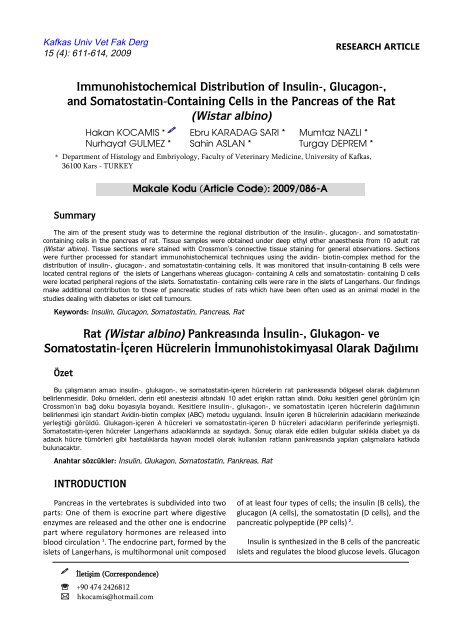
Immunohistochemical Distribution of Insulin-, Glucagon-,<br />
and Somatostatin-Containing Cells in the Pancreas of the Rat<br />
(Wistar albino)<br />
Hakan KOCAMIS * Ebru KARADAG SARI * Mumtaz NAZLI *<br />
Nurhayat GULMEZ * Sahin ASLAN * Turgay DEPREM *<br />
*<br />
Department of Histology and Embriyology, Faculty of Veterinary Medicine, University of Kafkas,<br />
36100 Kars - TURKEY<br />
Summary<br />
İletişim (Correspondence)<br />
℡ +90 474 2426812<br />
hkocamis@hotmail.com<br />
Makale Kodu (Article Code): 2009/086-A<br />
The aim of the present study was to determine the regional distribution of the insulin-, glucagon-, and somatostatincontaining<br />
cells in the pancreas of rat. Tissue samples were obtained under deep ethyl ether anaesthesia from 10 adult rat<br />
(Wistar albino). Tissue sections were stained with Crossmon’s connective tissue staining for general observations. Sections<br />
were further processed for standart immunohistochemical techniques using the avidin- biotin-complex method for the<br />
distribution of insulin-, glucagon-, and somatostatin-containing cells. It was monitored that insulin-containing B cells were<br />
located central regions of the islets of Langerhans whereas glucagon- containing A cells and somatostatin- containing D cells<br />
were located peripheral regions of the islets. Somatostatin- containing cells were rare in the islets of Langerhans. Our findings<br />
make additional contribution to those of pancreatic studies of rats which have been often used as an animal model in the<br />
studies dealing with diabetes or islet cell tumours.<br />
Keywords: Insulin, Glucagon, Somatostatin, Pancreas, Rat<br />
Rat (Wistar albino) Pankreasında İnsulin-, Glukagon- ve<br />
Somatostatin-İçeren Hücrelerin İmmunohistokimyasal Olarak Dağılımı<br />
Özet<br />
Bu çalışmanın amacı insulin-, glukagon-, ve somatostatin-içeren hücrelerin rat pankreasında bölgesel olarak dağılımının<br />
belirlenmesidir. Doku örnekleri, derin etil anestezisi altındaki 10 adet erişkin rattan alındı. Doku kesitleri genel görünüm için<br />
Crossmon’ın bağ doku boyasıyla boyandı. Kesitlere insulin-, glukagon-, ve somatostatin içeren hücrelerin dağılımının<br />
belirlenmesi için standart Avidin-biotin complex (ABC) metodu uygulandı. İnsulin içeren B hücrelerinin adacıkların merkezinde<br />
yerleştiği görüldü. Glukagon-içeren A hücreleri ve somatostatin-içeren D hücreleri adacıkların periferinde yerleşmişti.<br />
Somatostatin-içeren hücreler Langerhans adacıklarında az sayıdaydı. Sonuç olarak elde edilen bulgular sıklıkla diabet ya da<br />
adacık hücre tümörleri gibi hastalıklarda hayvan modeli olarak kullanılan ratların pankreasında yapılan çalışmalara katkıda<br />
bulunacaktır.<br />
Anahtar sözcükler: İnsulin, Glukagon, Somatostatin, Pankreas, Rat<br />
INTRODUCTION<br />
of at least four types of cells; the insulin (B cells), the<br />
glucagon (A cells), the somatostatin (D cells), and the<br />
pancreatic polypeptide (PP cells) 2 .<br />
Insulin is synthesized in the B cells of the pancreatic<br />
islets and regulates the blood glucose levels. Glucagon
612<br />
Immunohistochemical Distribution of...<br />
is synthesized in the A cells of the pancreas and<br />
regulates blood glucose levels as well 3 . Somatostatin<br />
is a polypeptid hormone 4 which is also known as a<br />
somatotropin release-inhibiting factor (SRIF) and<br />
secreted by pancreatic δ (D) cells 5,6 . Somatostatin is<br />
known to be a multifunctional hormone that inhibits<br />
the secretion of a large number of hormones including<br />
insulin, glucagon, gastrin and cholecystokinin 4 .<br />
The purpose of this study was to determine the<br />
regional distribution of the insulin-, glucagon-, and<br />
somatostatin-containing cells in the pancreas of the<br />
rat by immunohistochemistry using specific antisera<br />
against insulin, glucagon, and somatostatin.<br />
MATERIAL and METHODS<br />
In the present study, ten adult rats (Wistar albino)<br />
were used without any gender distinction. Tissue<br />
specimens were dissected and obtained under deep<br />
ethyl ether anaesthesia from the lobes of the pancreas.<br />
Samples from the pancreas were fixed in Bouin’s<br />
solution for 12 h at 4ºC. After paraffin embedding,<br />
serial tissue sections were cut at 5-6 µm in thickness.<br />
Sections of each tissue were stained with Crossmon’s<br />
connective tissue stain 7 for general observations.<br />
Immunohistochemistry<br />
Each section was deparaffinized, rehydrated and<br />
then immunostained with the avidin-biotin-complex<br />
(ABC) method 8 . The endogeneous peroxidase and<br />
non-specific binding sites for andibodies were<br />
supressed by treating sections with 0.5% hydrogen<br />
peroxide for 30 min and 10% normal rabbit serum for<br />
10 min at room temperature, respectively. Furthermore,<br />
sections were processed for standart immunohistochemical<br />
techniques. The working dilutions and the<br />
sources of antibodies used are listed in the Table 1.<br />
Peptide specific antibodies isolated from mammalian<br />
species were used. Negative controls were carried out<br />
by incubating sections with phosphate-buffered<br />
saline (PBS) instead of the primer antiserum. Negative<br />
controls were also conducted with tissue sections<br />
from the gastrointestinal tract of rabbits known to<br />
contain the hormones studied. The sections were<br />
incubated in primary antisera in PBS-containing<br />
bovine serum albumin (2.5%) and Triton X-100 (0.2%)<br />
for 1 h at room temperature. Subsequently, the<br />
binding of primary antisera was detected using rabbitantimouse<br />
antisera and Strept ABC. Finally, the<br />
chromogen protocol was used the reveal the distribution<br />
of bound peroxidase activity 9 .<br />
Table 1. The primary and secodary antibodies and their dilutions<br />
Tablo 1. Primer ve sekonder antikorlar ve dilusyonları<br />
Antisera Working Dilutions Sources<br />
Insulin<br />
Glucagon<br />
Somatostatin<br />
Goat-anti-rabbit Ig G<br />
Rabbit anti-mouse Ig G<br />
Strept ABC<br />
RESULTS<br />
1: 40<br />
1: 40<br />
1: 100<br />
1: 100<br />
1: 100<br />
1: 50<br />
Rat pancreas was found (as expected) to be consisted<br />
of exocrine and endocrine (islets of Langerhans) parts.<br />
The endocrine parts of the pancreas were scattered<br />
singly or in small groups of islets of various shapes<br />
and size in the interstitium of the exocrine parts.<br />
The islets of Langerhans were composed of two<br />
different regions, which were central and peripheral<br />
regions. Positive immunohistochemical reactions were<br />
observed in the pancreatic islets of rat against insulin-,<br />
glucagon-, and somatostatin containing cells. No specific<br />
staining was found in the exocrine part of pancreas.<br />
Insulin-containing cells (B cells)<br />
Insulin-containing cells were abundant in the<br />
whole islets of Langerhans. Those cells were located<br />
in the central regions of the islets. B cells were found<br />
to have round shape (Fig 1).<br />
Glucagon-containing cells (A cells)<br />
Signet<br />
Signet<br />
Dako<br />
Zymed<br />
Dako<br />
Dako<br />
Fig 1. Insulin-containig cells (B cells) in islets of Langerhans. Bar: 100 µm<br />
Şekil 1. Langerhans adacıklarında insulin-içeren hücreler (B<br />
hücreleri). Bar: 100 µm<br />
Glucagon-containing cells were located in peripheral<br />
regions of the islets. They were surrounded by the B<br />
cells and had round shape (Fig 2).
Fig 2. Glucagon-containing cells (A cells) in islets of Langerhans.<br />
Bar: 100 µm<br />
Şekil 2. Langerhans adacıklarında glukagon-içeren hücreler (A<br />
hücreleri). Bar: 100 µm<br />
Somatostatin-containing cells (D cells)<br />
Somatostatin-containing cells were found rarely in<br />
the islets of Langerhans. D cells were localised<br />
especially in the peripheral regions of the islets. They<br />
were found to have irregular shape (Fig 3).<br />
Fig 3. Somatostatin-containig cells (D cells) in islets of Langerhans.<br />
Bar: 100 µm<br />
Şekil 3. Langerhans adacıklarında somatostatin-içeren hücreler (D<br />
hücreleri). Bar: 100 µm<br />
DISCUSSION<br />
The pancreas is composed of two secretory parts,<br />
which are the insular or endocrine part and the acinar<br />
or the exocrine part 2 . In this study, pancreas was<br />
found to be consisted of exocrine and endocrine<br />
(islets of Langerhans) parts, as explained in literature.<br />
It was reported that insulin-immunoreactive cells<br />
were arranged in the central region of pancreas islets<br />
613<br />
KOCAMIŞ, KARADAĞ SARI, NAZLI<br />
GÜLMEZ, ASLAN, DEPREM<br />
in wood mouse 10 and hamster 11 . However, it was<br />
described that insulin-immunoreactive B cells occupied<br />
the majority of the periphery regions of islets in<br />
monkey pancreas 12 . In the present study, insulincontaining<br />
cells were observed in the central regions<br />
of islets of Langerhans, as in agreement with previous<br />
studies 10,11,13 .<br />
In monkey 12 and equine 14 , glucagon-immunoreactive<br />
cells were found in the central regions of<br />
pancreatic islets where insulin-immunoreactive cells<br />
were numerously located in most of the domestic<br />
animals. Glucagon-containing cells were seen peripheral<br />
regions of pancreatic islets in our study. Sujatha et<br />
al. 12 and Helmstaedter et al. 14 reported that somatostatin-immunoreactive<br />
cells were found mostly with<br />
glucagon- immunoreactive cells in the center of<br />
pancreatic islets. However, somatostatin-containing<br />
cells were located peripheral regions of the islets of<br />
Langerhans in the current study as described in some<br />
of the studies 10,11,13 . Taken together, cellular pattern of<br />
islets has a species difference that may eventually<br />
give rise to functional significance.<br />
There were several immunohistochemical and<br />
morphometric studies on the glucagon-, insulin-, and<br />
somatostatin-immunoreactive cells in the pancreas 15-21 .<br />
It has been indicated that in diabetic state, pancreatic<br />
beta-cells showed a weak immunostaining for insulin,<br />
appears to suggest that induction of regenerative<br />
stimulus in diabetic state triggers pancreatic regenerative<br />
processes, thereby restoring functional activities of<br />
the pancreas 15 . It was demonstrated that significantly<br />
less B-cell area and markedly larger A- and D- cell<br />
areas within the diabetic islets were found when<br />
compared with the non-diabetic and pre-diabetic<br />
islets 19 . In another study, they have observed a direct<br />
relationship between total hormone area and<br />
hormone content, in islets from normal and hyperglycemic<br />
diabetic dogs 20 . This findings suggest that<br />
there are acute effects of insulin or of normalization<br />
of glycemia on the A- and D-cells, to reduce hormone<br />
content in diabetes. Badawoud 21 demonstrated that<br />
the B cell volume density was significantly lower in<br />
the diabetic group, while islet volume density, islet<br />
diameter, islet volume and absolute islet cell numbers<br />
were significantly greater in the diabetic group. The B<br />
cell nuclear diameter and volume were not significantly<br />
different in the diabetic group. This study indicated<br />
that maternal diabetes induced foetal islet hypertrophy<br />
and caused an increase in the total islet cell<br />
number 21 .
614<br />
Immunohistochemical Distribution of...<br />
In conclusion, it was monitored that insulincontaining<br />
B cells were located central regions of the<br />
islets whereas glucagon- containing A cells and<br />
somatostatin- containing D cells were located peripheral<br />
regions of the islets. Somatostatin- containing cells<br />
were rare in the islets of Langerhans. Therefore, the<br />
findings make additional contribution to those of<br />
pancreatic studies of rats which have been often used<br />
as an animal model in the studies dealing with<br />
diabetes or islet cell tumours.<br />
REFERENCES<br />
1. Kobayashi K, Syed Ali S: Cell types of the endocrine<br />
pancreas in the shark, Scylliorhinus stellaris as revealed by<br />
correlative light and electron microscopy. Cell Tissue Res,<br />
215, 475-490, 1981.<br />
2. Bendayan M, Ito S: Immunohistochemical localization of<br />
exocrine enzymes in normal rat pancreas. J Histochem<br />
Cytochem, 27, 1029-1034, 1979.<br />
3. Hsu SM, Raine L, Fanger H: Use of avidin-biotin<br />
peroxidase complex (ABC) in immunoperoxidase techniques:<br />
A comparison between ABC and unlabeled antibody (PAP)<br />
prosedures. J Histochem Cytochem, 29, 577-580, 1981.<br />
4. Reisine T, Bell GL: Molecular proporties of somatostatin<br />
receptors. Neurosci, 67, 777-790, 1995.<br />
5. Patel YC, Greenwood M, Panetta R, Hukovic N, Grigorakis<br />
S, Robertson LA, Srikant CB: Molecular biology of<br />
somatostatin receptor subtypes. Metabolism, 45, 31-38, 1996.<br />
6. Reichlin S: Somatostatin. N Engl J Med, 309, 1495-<br />
1501,1983.<br />
7. Crossmon G: A modification of Mallory’s connective tissue<br />
stain with a discussion of the principles involved. Anat Rec,<br />
69, 33-38, 1937.<br />
8. Hsu WH, Crump MH: The Adrenal Gland. In, Mc Donald<br />
LE, Pineda MH (Eds): Veterinary Endocrinology and<br />
Reproduction. Philedelphia, Lea&Febiger, pp. 186-201, 1989.<br />
9. Shu S, Ju G, Fan L: The glucose oxidase-DAB-nickel in<br />
peroxidase histochemistry of the nervous system. Neurosci<br />
Lett, 85, 169-171, 1988.<br />
10. Yukawa M, Takeuchi T, Watanabe T, Kitamura S:<br />
Proportions of various endocrine cells in the pancreatic islets<br />
of wood mice (Apodemus speciosus). Anat Histol Embryol,<br />
28, 13-16, 1999.<br />
11. Camihort G, Del Zotto H, Gomez-Dumm CL, Gagliardino<br />
JJ: Quantitative ultrastructural changes inducted by sucrose<br />
administration in the pancreatic B cells of normal hamsters.<br />
Biocell, 24, 31-37, 2000.<br />
12. Sujatha SR, Pulimood A, Gunasekaran S: Comparative<br />
immunohistochemistry of isolated rat & monkey pancreatic<br />
islets cell types. Indian J Med Res, 119, 38-44, 2004.<br />
13. Wieczorec G, Pospischil A, Perentes EA: Comparative<br />
immunohistochemical study of pancreatic islets in laboratory<br />
animals (rats, dogs, minipigs, nonhuman primats). Exp Toxicol<br />
Pathol, 50, 151-172, 1998.<br />
14. Helmstaedter V, Feurle GE, Forsmann WG: Insulin-,<br />
glucagon-, and somatostatin-immunoreactive cells in the<br />
equine pancreas. Cell Tissue Res, 172, 447-454, 1976.<br />
15. Adewole SO, Ojewole JA: Insulin-induced immunohistochemical<br />
and morphological changes in pancreatic beta-cells<br />
of streptozotocin-treated diabetic rats. Methods Find Exp Clin<br />
Pharmacol, 29, 447-55, 2007.<br />
16. Calvo RM, Forcen R, Obregon MJ, Escobar del Rey F,<br />
Morreale de Escobar G, Regadera J: Immunohistochemical<br />
and morphometric studies of the fetal pancreas in diabetic<br />
pregnant rats. Effects of insulin administration. Anat Rec, 251,<br />
173-80, 1998.<br />
17. Fu Q, Honda M, Ohgawara H, Igarashi N, Toyada C,<br />
Omori Y, Kobayashi M: Morphological analysis of pancreatic<br />
endocrine cells in newborn animals delivered by experimental<br />
diabetic rats. Diabetes Res Clin Pract, 31, 57-62, 1996.<br />
18. <strong>Gulmez</strong> N, <strong>Kocam</strong>is H, <strong>Aslan</strong> S, <strong>Nazl</strong>i M: Immunohistochemical<br />
distribution of cells containing insulin, glucagon<br />
and somatostatin in the goose (Anser anser) pancreas. Turk J<br />
Vet Anim Sci, 28, 403-407, 2004.<br />
19. Iwashima Y, Watanabe K, Makino I: Changes in the<br />
pancreatic A-, B- and D-cell populations during development<br />
of diabetes in spontaneously diabetic Chinese hamsters of the<br />
Asahikawa colony (CHAD). Diabetes Res Clin Pract, 8, 201-<br />
14, 1990.<br />
20. Rastogi KS, Brubaker PL, Kawasaki A, Efendic S, Vranic<br />
M: Increase in somatostatin to glucagon ratio in islets of<br />
alloxan-diabetic dogs: Effect of insulin-induced euglycemia.<br />
Can J Physiol Pharmacol, 71, 512-7, 1993.<br />
21. Badawoud MH: Immunohistochemical and morphometric<br />
study of the effect of maternal diabetes on rat foetal<br />
pancreatic islets. Folia Morphol (Warsz), 62, 19-24, 2003.


