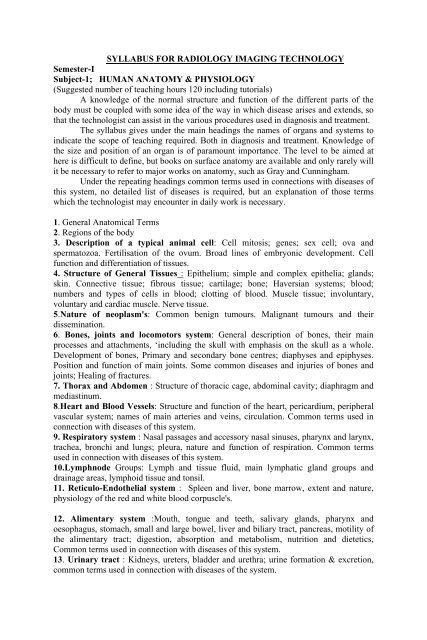
SYLLABUS FOR RADIOLOGY IMAGING TECHNOLOGY Semester-I ...
SYLLABUS FOR RADIOLOGY IMAGING TECHNOLOGY Semester-I ...
SYLLABUS FOR RADIOLOGY IMAGING TECHNOLOGY Semester-I ...

Create successful ePaper yourself
Turn your PDF publications into a flip-book with our unique Google optimized e-Paper software.
<strong>SYLLABUS</strong> <strong>FOR</strong> <strong>RADIOLOGY</strong> <strong>IMAGING</strong> <strong>TECHNOLOGY</strong><br />
<strong>Semester</strong>-I<br />
Subject-1; HUMAN ANATOMY & PHYSIOLOGY<br />
(Suggested number of teaching hours 120 including tutorials)<br />
A knowledge of the normal structure and function of the different parts of the<br />
body must be coupled with some idea of the way in which disease arises and extends, so<br />
that the technologist can assist in the various procedures used in diagnosis and treatment.<br />
The syllabus gives under the main headings the names of organs and systems to<br />
indicate the scope of teaching required. Both in diagnosis and treatment. Knowledge of<br />
the size and position of an organ is of paramount importance. The level to be aimed at<br />
here is difficult to define, but books on surface anatomy are available and only rarely will<br />
it be necessary to refer to major works on anatomy, such as Gray and Cunningham.<br />
Under the repeating headings common terms used in connections with diseases of<br />
this system, no detailed list of diseases is required, but an explanation of those terms<br />
which the technologist may encounter in daily work is necessary.<br />
1. General Anatomical Terms<br />
2. Regions of the body<br />
3. Description of a typical animal cell: Cell mitosis; genes; sex cell; ova and<br />
spermatozoa. Fertilisation of the ovum. Broad lines of embryonic development. Cell<br />
function and differentiation of tissues.<br />
4. Structure of General Tissues : Epithelium; simple and complex epithelia; glands;<br />
skin. Connective tissue; fibrous tissue; cartilage; bone; Haversian systems; blood;<br />
numbers and types of cells in blood; clotting of blood. Muscle tissue; involuntary,<br />
voluntary and cardiac muscle. Nerve tissue.<br />
5.Nature of neoplasm's: Common benign tumours. Malignant tumours and their<br />
dissemination.<br />
6. Bones, joints and locomotors system: General description of bones, their main<br />
processes and attachments, ‘including the skull with emphasis on the skull as a whole.<br />
Development of bones, Primary and secondary bone centres; diaphyses and epiphyses.<br />
Position and function of main joints. Some common diseases and injuries of bones and<br />
joints; Healing of fractures.<br />
7. Thorax and Abdomen : Structure of thoracic cage, abdominal cavity; diaphragm and<br />
mediastinum.<br />
8.Heart and Blood Vessels: Structure and function of the heart, pericardium, peripheral<br />
vascular system; names of main arteries and veins, circulation. Common terms used in<br />
connection with diseases of this system.<br />
9. Respiratory system : Nasal passages and accessory nasal sinuses, pharynx and larynx,<br />
trachea, bronchi and lungs; pleura, nature and function of respiration. Common terms<br />
used in connection with diseases of this system.<br />
10.Lymphnode Groups: Lymph and tissue fluid, main lymphatic gland groups and<br />
drainage areas, lymphoid tissue and tonsil.<br />
11. Reticulo-Endothelial system : Spleen and liver, bone marrow, extent and nature,<br />
physiology of the red and white blood corpuscle's.<br />
12. Alimentary system :Mouth, tongue and teeth, salivary glands, pharynx and<br />
oesophagus, stomach, small and large bowel, liver and biliary tract, pancreas, motility of<br />
the alimentary tract; digestion, absorption and metabolism, nutrition and dietetics,<br />
Common terms used in connection with diseases of this system.<br />
13. Urinary tract : Kidneys, ureters, bladder and urethra; urine formation & excretion,<br />
common terms used in connection with diseases of the system.
2<br />
- 2 -<br />
14. Reproductive system : Male genital tract; testes, epidedymis, seminal vesicle and<br />
prostate; female genital tract; uterine tubes, ovaries, uterus, vagina and vulva, the<br />
mammary glands; menstruation, pregnancy and lactation; common terms used in<br />
connection with diseases of this system.<br />
15. Endocrine glands; Anatomy and function of pituitary, thyroid, para thyroids,<br />
adrenal, thymus, pancreas and gonads as endocrine organs; common terms used in<br />
connection with diseases of this system.<br />
16. Nervous system: Brain; main subdivisions and lobes; ventricular system, spinal cord,<br />
concept of motor, sensory and reflex pathways; meninges and cerebrospinal fluid; its<br />
circulation; autonomic nervous system; common terms used in connection with diseases<br />
of this system.<br />
17. Special sensory organs: Structure and function of the eye; structure and function of<br />
the ear; structure and function of the skin.<br />
18. Surface markings and topographical relations; radiographic anatomy.<br />
BOOKS <strong>FOR</strong> STUDY<br />
Text book<br />
1.Anatomy and Physiology for Radiographers - C.A. Warrick<br />
Reference books<br />
2.Gray's anatomy Descriptive and applied - T.B. Johnstor.<br />
3.Foundation of Anatomy and Physiology - Ross and Wilson.<br />
4.An Atlas of Normal Radiographic Anatomy - Richard & Alvin<br />
5.Essentials of Human Anatomy - Russell<br />
6.Best and Taylor : The Human Body – its anatomy and physiology ( Chapman and Hall)<br />
7.Blewett and Rackow : Anatomy and Physiology for Radiographers ( Butterworth )<br />
8.Dean : Basic Anatomy and Physiology for Radiographers ( Blackwell )<br />
9.Fitzgerald : Anatomy 1600 multiple choice question ( Butterworth )<br />
10.Hamilton et al : Surface and Radiological Anatomy ( Heffer )<br />
SEMESTER - I<br />
SUBJECT 2 : BASIC PHYSICS & RADIATION PHYSICS<br />
This syllabus should be augmented by as much of practical and demonstration classes as<br />
possible. Suggested number of minimum teaching hours: 120<br />
1.Basic concepts: Units and measurements-Force, work, power and energy-Temperature<br />
and heat-SI units of above parameters. Atomic structure-atom model-Nucleus-electronic<br />
configuration-periodic table-Isotopes-Ionization-excitation-Binding energy-electron volt-<br />
Electro magnetic radiation-Quantum nature of radiation-mass energy equivalence-<br />
Fluorescence-electromagnetic spectrum<br />
2.Electricity and magnetism: Electric charges, Coulomb’s law-Unit of charge-Electric<br />
potential, unit of potential-Electric induction, capacitance and capacitors, series and parallel<br />
connection-electric current, unit, resistance, ohm’s law, electric power, Joule’s law<br />
Magnetism: Magnetic induction-magnetic properties-Hysteresis-magnetic effect of<br />
current-Electrical instruments, Galvanometer, voltmeter, ammeter and multimeter.
3<br />
- 3 -<br />
3.Electromagnetic Induction: Induced electro motive force-Faradays experiments- laws of<br />
electro magnetic induction-Self and mutual induction-Alternating current- Ac generator-<br />
Peak and RMS values- AC circuits with resistance-capacitance and inductance- Choke<br />
coil- eddy current. Transformer-theory, design, losses- auto transformer- high voltage<br />
transformer- electric power transmission<br />
4.X-rays: Discovery of x-rays- properties-production- x-ray spectrum- bremsstrahlung and<br />
characteristic x-rays- X-ray tube; Coolidge tube, tube design, line focus principle-space<br />
charge effect, tube cooling- Modern x-ray tubes-stationary anode, rotating anode, grid<br />
controlled x-ray tubes, heel effect, off focus radiation, tube insert and housing-Tube<br />
rating-Quality and intensity of x-rays-,factors influencing them.<br />
5.X-ray generator circuits: Vacuum tube diodes-semi conductor diodes-transister-<br />
rectification, half and full wave-self rectification – X-ray generator; filament circuit-kilo<br />
voltage circuit-single phase generator-three phase generator-constant potential generator<br />
Fuses, switches and interlocks-Exposure switching and timers-HT cables-earthing<br />
6.Radioactivity: Discovery of radioactivity, natural radioactivity-activity units- radium,<br />
thorium and uranium series- alpha, beta decay and gamma rays - radioactive disintegrationexponential<br />
decay, half life period, decay constant. Artificial radioactivity –production of<br />
radioisotopes-cyclotron-neutron-fission and fusion-chain reaction-atom bomb-nuclear<br />
reactor<br />
7.Interaction of X and gamma rays: Transmission through matter, law of exponential<br />
attenuation, half value layer, linear attenuation coefficient-coherent scattering-photoelectric<br />
effect- compton scattering-pair production-photonuclear disintegration-Particle interactions.<br />
Interactions of x and gamma rays in the body; fat-soft tissue-bone-contrast media-total<br />
attenuation coefficient-relative clinical importance<br />
8.Radiation quantities and units: Radiation intensity-exposure, roentgen, its limitationskerma<br />
and absorbed dose-electronic equilibrium-rad, gray, conversion factor for roentgen to<br />
rad-RBE-LET-quality factor-dose equivalent-rem, sievert.<br />
9.Radiation detection and measurements: Principle of radiation detection-Ionization<br />
chamber-proportional counter-GM tubes-scintillation detectors-semiconductor detector-<br />
Gamma ray spectrometer. Measuring system: free ionization chamber-thimble ion chambercondenser<br />
chamber-victoreen electrometer-secondary standard dosimeter-film dosimeterchemical<br />
dosimeter-thermoluminecent dosimeter-Pocket dosimeter. Radiation survey meterzone<br />
monitor-contamination monitor, their function use and maintenance.<br />
BOOKS <strong>FOR</strong> STUDY<br />
Text book<br />
1.First year Physics for Radiographers - Hay & Hughes.<br />
Reference books<br />
1.Basic radiological physics-K.Thayalan, Jaypee publishers (P) Ltd, New Delhi(2001)<br />
2.Fundamental of X-ray and Radium Physics - Joseph Selman<br />
3.Basic Medical Radiation Physics - Stanton.<br />
4.Chrtistensen's Physics of Diagnostic Radiology - Christesen.
4<br />
- 4 -<br />
<strong>Semester</strong> II<br />
Subject : 3 : RADIOGRAPHIC PHOTOGRAPHY:<br />
(Suggested number of teaching hours is 120, including tutorials and practical<br />
demonstration).<br />
This Radiographic photography syllabus is intended as a guide to the theory and<br />
practical knowledge required by the students. Appreciation and application of all the factors<br />
listed below will enable the technologist to produce x-ray films of good quality and<br />
diagnostic value. The lectures should be linked with practical demonstration to illustrate the<br />
importance of all that goes to make up correct exposure conditions.<br />
1.X-ray film materials: Structure of film emulsion-Grain technology-Geletine-Basic film<br />
types-Film formats and packing-Direct exposure duplitised films-Single coated emultions-<br />
Films for specialised use-manufacturing process.<br />
Sensitometry :Photographic density—characteristic curve –information from the<br />
characteristic curve-speed Vs definition<br />
Storage of x-ray film-unprocessed film-radiographs<br />
2. Intensifying screens and cassettes: Intensifying screen- phosphor- Construction-<br />
Intensifying factor-speed and detail-crossover effect-resolution-mottle-reciprocity-screen<br />
asymmetry- screen-film contact- screen types and cleaning.<br />
New phosphor technology-influence of kilo voltage. Photostimulable phosphor<br />
imaging<br />
x-ray cassette-design-types- Identification of cassettes- General care of cassettes and<br />
storage.<br />
3.Photochemistry: Film processing-latent image formation-Mechanism-theory-Developernature<br />
of development-pH scale-constitution of developer-development time-factors in the<br />
use of developer. Fixers-constitution of fixing solution-factors affecting the fixerreplenishment<br />
of fixer--silver conservation-Drying –developer and fixer for automatic film<br />
processor-rinsing-washing and drying.<br />
4.processing equipment: Materials for processing equipment-manual processor-care of<br />
processing equipment-automatic processor-manual VS automatic-principles and typical<br />
equipment Microprocessor control-Cine processing-Daylight systems-Processing faultsmaintenance<br />
5.Processing room: Day light processing-location of the dark room-dark room illuminationequipment<br />
and layout-x-ray viewing room.<br />
Daylight handling-daylight systems with cassettes-without cassettes.<br />
6. Radiographic image-components of image quality-unsharpness in radiographic imagecontrast<br />
of the radiographic image-distinctness of the radiographic image-size, shape and<br />
spatial relationships.<br />
Presentation of radiographs-opaque letters and legents-perporating devices-actinic<br />
markers-Identification of dental films-preparation of stereo radiographs-viewing conditions<br />
7.Monitor photography- Characteristics of the video image-television camera-imaging<br />
camera—imaging film-sensitometric characteristics-processing-final image.<br />
Laser-light and laser-laser imaging-laser imagers—imaging plates-principle of photo<br />
stimulated luminescence
5<br />
- 5 -<br />
BOOKS <strong>FOR</strong> STUDY<br />
Text book<br />
1.Radiographic Imaging - Chesney & Chesney, Blakwell scientific publications, oxford<br />
(1981)<br />
Reference books<br />
1.Radiographic imaging-Derrick P. Roberts and Nigel L. Smith. Churchill<br />
Livingstone, Edinburgh (1994)<br />
2.Radiographic Latent image processing - W.E.J. Mckinney<br />
3.Photographic processing, quality control and evaluation of photographic material -J.E.<br />
Gray<br />
4.Photographic processing Chemistry - L.F.A. Mason.<br />
5.Physical and photography principles of Medical Radiography -<br />
Seeman & Herman.<br />
<strong>Semester</strong> II<br />
Subject – 4; GENERAL PRINCIPLES OF HOSPITAL PRACTICE AND<br />
PATIENT CARE<br />
Suggested number of teaching hours 100 including tutorials and demonstrations.<br />
This section is intended to emphasise to the student technologist the importance of patient<br />
welfare. Many of the points included in this section may be considered during the<br />
teaching of other subjects also; but it is strongly urged that specific teaching and as much<br />
practical demonstration and instruction as possible should be given in this section.<br />
Modern hospital treatment is based on team work, it is essential that the student<br />
should appreciate the technologists role and that the importance of co-operation with<br />
wards and other departments.<br />
The students should be attached to wards or the accident and emergency<br />
department for a definite training period, the length of time being suited to the individual<br />
hospital.<br />
1. Hospital procedure: Hospital staffing and organisation; records relating to patients<br />
and departmental statistics; professional attitude of the technologist to patients and other<br />
members of the staff; medico- legal aspects; accidents in the departments, appointments,<br />
organisation; minimising waiting time; out-patient and follow-up clinics; stock-taking<br />
and stock keeping.<br />
2. Care of the patient : FIRST contact with patients in the department; management of<br />
chair and stretcher patients and aids for this, management of the unconscious patient;<br />
elementary hygiene; personal cleanliness; hygiene in relation to patients (for example<br />
clean linen and receptacles , nursing care; temperature pulse and respiration; essential<br />
care of the patient who has a tracheotomy; essential care of the patient who has a<br />
colostomy; bedpans and urinals; simple application of a sterile dressing.<br />
3. First aid : Aims and objectives of first aid; wounds and bleeding, dressing and<br />
bandages; pressure and splints, supports etc. Shock; insensibility; asphyxia; convulsions;<br />
resuscitation, use of suction apparatus, drug reactions; prophylactic measures;<br />
administration of oxygen; electric shock; burns; scalds; haemorrhage; pressure points;<br />
compression band. Fractures; splints, bandaging; dressing, foreign bodies ; poisons.
6<br />
- 6 -<br />
4. Infection : Bacteria, their nature and appearance ; spread of infections; auto-infection<br />
or cross-infection; the inflammatory process; local tissue reaction, general body reaction;<br />
ulceration; asepsis and antisepsis.<br />
5. Principles of asepsis: Sterilisation - methods of sterilisation; use of central sterile<br />
supply department; care of identification of instruments, surgical dressings in common<br />
use, including filamented swabs, elementary operating theatre procedure; setting of trays<br />
and trolleys in the radiotherapy department (for study by radiotherapy students only)<br />
6. Departmental procedures: Department staffing and organisation; records relating to<br />
patients and departmental statistics; professional attitudes of the technologist to patients<br />
and other members of the staff, medico-legal aspects accidents in the department;<br />
appointments; organisation; minimising waiting time; out-patient and follow-up clinics;<br />
stock taking and stock keeping.<br />
7. Drugs in the department : Storage : classification; labelling and checking, regulations<br />
regarding dangerous and other drugs; units of measurement, special drugs, antidepressive,<br />
anti-hypertensive etc.<br />
BOOKS <strong>FOR</strong> STUDY<br />
Text book<br />
1.Deeley – A guide to Radiotherapy nursing -Livingstone<br />
Reference books<br />
1.Care of patient in diagnostic Radiography - Chesney & Chesney.<br />
2.Chesney's Care of the patient in Diagnostic Radiography - Pauline J . Culmer.<br />
3.Aid to Tray and Trolley Setting - Marjorie Hougton<br />
4. First Aid - Haugher & Gardner<br />
5.A guide to Oncology nursing (Livingstone) - Deeley<br />
6. Practical nursing and first- aid - Ross and Wilson. Livingstone.<br />
<strong>Semester</strong> - III<br />
Subject – 5; PHYSICS OF DIAGNOSTIC <strong>RADIOLOGY</strong> AND EQUIPMENT<br />
(Suggested number of teaching hours 120 including tutorials and practical<br />
demonstration).<br />
1.Radiography: Primary radiological iamge-Image produced by contrast media-<br />
Attenuation-Linear and mass attenuation coefficient-Factors affecting attenuationapplication<br />
in radiology-Filters-inherent and added filters-Heavy metal filters-X-ray<br />
beam restrictors-aperture diaphragm-cones and cylinders-collimators-function of<br />
restrictor<br />
2.Scattered radiation: significance of scatter-Grid ,principle, design and type-evaluation<br />
of grid performance-lead content-Grid cut off-moving girds-Grid selection-air gap<br />
technique<br />
3.Fluoroscopy: Direct fluoroscope-Image intensifier design—brightness gain-Imaging<br />
characteristics—multi field image intensifiers-Close circuit television-television<br />
scanning- television image quality-Fluoroscopic image recorders-TV image recorders.
7<br />
- 7 -<br />
4.Radiographic image: Image clarity-contrast- factors affecting contrast-Image qualitymottle,<br />
sharpness and resolution-Line spread function-Modulation transfer function-<br />
Noise and wiener spectrum.<br />
Magnification-distortion-penumbra-un sharpness-inverse square law-evaluation of<br />
resolution- quantum mottle-patient exposure<br />
5.Body section radiography: Basic method of tomography-terminology-blurring-section<br />
thickness-narrow and wide angle tomography-circular tomography-tomographic motionsphantom<br />
images-tomographic angle determination-phantomography.<br />
Stereoscopy- physiology of depth perception-stereoscopic filming—viewingmerits<br />
and demerits<br />
6.Mammography: Technical aspects of Mammograhy, generator, x-ray tubes,<br />
Accessories, Resolution, quality control. Application and role in medicine.<br />
Xeroradiography-principles-xeroradiographic plate powder development-image<br />
development- image quality-liquid toner xeroradiography.<br />
7.Ultrasound: Physical characteristics of sound--transducer- characteristics of ultrasound<br />
beam- interaction of ultrasound and matter-quarter wave matching-ultrosonic displayimaging<br />
principles-Doppler techniques-real time ultrasound- ultrasound instrumentation-<br />
bio effects and safety considerations.<br />
Books for study.<br />
Text book<br />
1. "Christensen’s Physics of diagnostic Radiology" ( Lea & Febiger )<br />
Reference books<br />
2." First year Physics for Radiographers Hay & Hughes ( ELBS )<br />
3."Basic Medical Radiation Physics Stantor ( Appleton- Century & Crofts)<br />
4 X- ray Equipment for student Radiographers" By: Chesney & Chesney (<br />
Blackwell )<br />
5.A Manual of Radiographic equipment. By: Sybil M. Stockley (Churchill Livingstone)<br />
6."Principles of Diagnostic X-ray apparatus" by : Hill ( Macmillan.)<br />
7.. “Radiologic science for Technologist” Stewart C. Bushong , ( M Mosby.)<br />
<strong>Semester</strong> -III<br />
Subject- 6; PRINCIPLES OF RADIODIAGNOSIS & RADIOGRAPHIC<br />
TECHNIQUES<br />
(Suggested number of teaching hours 120 including tutorials and demonstration)<br />
The term technique in the text implies a full knowledge of the procedure for X-ray<br />
examination; preparation of the room, apparatus and instruments; position of the patient<br />
for at least two projections; at (right angles) relative positions of the X-ray tube and<br />
patients; relevant exposure factors; use of accessories, such as radiographic cones, grids<br />
and position aids.<br />
Throughout the course attention should be given toa). The close association of<br />
theory with practical work. .(b). The anatomical and physiological basis of radiographic<br />
procedure.<br />
The student should be made familiar with radiographic appearance both of the<br />
normal subject and of common abnormal conditions where elementary knowledge of the<br />
pathology involved will ensure the application of the appropriate radiographic technique
8<br />
- 8 -<br />
which may be necessary for various disabilities or types of subject. The need for radiation<br />
precautions should be emphasised, as they apply to both patients and all hospital staff<br />
In this text the projections tested are considered to be university applicable. For<br />
each area studied, the topics will be presented under the following headings:<br />
a. Anatomy (review), b. Clinical indications.<br />
c. Preparation of the room. d. Accessory equipment.<br />
e. Preparation of patient., f. Routine views.<br />
g. Supplementary views: + modifications in cases of trauma.<br />
h. Radiation protection., i. Care of patient.<br />
For each view studied will be presented as follows.<br />
a. Positioning of patient. b. Immobilisation.<br />
c. Identification. d. Centring point.<br />
e. Direction of central X-ray relative to the film.<br />
f. Parts demonstrated.<br />
g. Exposure factor - KVP MA sec. (MAS) FFD grid/ Non-grid screen<br />
Specialist problems: Where appropriate tutors should refer to the following list of<br />
specialist problems which may arise necessitating technique variations.<br />
a. Children and neonates b. Seriously ill or injured patients.<br />
c. Elder patients. d. Deaf and blind patients.<br />
e. Language difficulties. f. Unconscious patients or Anaesthetised patients.<br />
Radiographic technique: Skeletal system<br />
1. Upper limb: Technique for hand, fingers, thumb, wrist joint carpal bones, forearm,<br />
elbow joint, radio ulnar joints and humerus supplementary techniques for the above. eg.<br />
carpal tunnel view, ulnar groove, head of the radius, supracondylar projections.<br />
2. Lower limb: Technique for foot, toes, great toe, tarsal bones, calcaneum, ankle joint,<br />
lower leg, knee, patella & femur.<br />
Supplementary technique: Stress view for torn ligaments -- Subtalar joint and talo<br />
calcaneal joint.- Inter condylar projection of the knee-- Tibial tubercle-- Length<br />
measurement technique.<br />
3.Shoulder girdle and thorax: Technique for shoulder joint, scapular, calvicle, acromio<br />
clavicular joints, sternum, ribs, sterno-clavicular joint.<br />
Supplementary projections and technique –(i). Recurrent dislocation of shoulder.<br />
(ii). Traumatic dislocation of shoulder.( iii). cervical ribs.<br />
4.Vertebral column: Technique for Atlanta-occipital joint, cervical spine, cervico<br />
thoracic spine, thoracic spine, thoraco- lumber spine, lumbo sacral spine, sacrum and<br />
coccyx.<br />
Supplementary technique to demonstrate( i.) Scoliosis (ii). Kyphosis (iii).<br />
Spondylolisthesis (iv). disc lesion (v). Union of spinal graft. Adaptation of techniques<br />
to demonstrate specific pathologies.<br />
5. Pelvic girdle and hip region: Technique for whole pelvis. Ileum, ischium, pubic<br />
bones, sacro iliac joint, symphysis pubis, hip joint, acetabulum neck of femur, greater and<br />
lesser trochanter.<br />
Supplimentary technique- Congenital dislocation of hips:<br />
Epiphysis of femur:-Lateral projections for hip joints to show femoral head and neck<br />
relationship.
9<br />
- 9 -<br />
6. Skeletal survey: Skeletal survey for metabolic bone disease, metastases, hormonal<br />
disorder, renal disorders.<br />
7. Skull: Basic projections for cranium, facial bones, nasal bones and mandible.<br />
- Technique for- petrous temporals for mastoids - Internal auditory canal - Accessory<br />
nasal sinuses- Tempero - mandibular joint - Orbits and optic foramen - Zygomatic<br />
arches.- Styloid process. - Pituitary fossa - Jugular foramen.<br />
8. Dental radiography: Technique for intra oral full mouth - Occlusal projections-<br />
Extra oral projections including orthopan tomography - Supplimentary techniques.<br />
9. Cardiovascular system: Routine projections for heart and vessels (without the uses of<br />
contrast agent) Supplementary views for above.<br />
10. Upper respiratory system: Technique for post nasal air ways, larynx, trachea,<br />
thoracic inlet, thyroid gland. 1. Valsalva Manoeuvres. 2. Phonation.<br />
Lungs and mediasstinum: Technique for routine projections:- Supplementary<br />
projections - antero posterior, obliques, lordotic and apical projection. Use of penetrated<br />
postero - anterior projection - Expiration technique - Technique for pleural fluid levels<br />
and adhesions.<br />
Diaphragm: Inclusion of diaphragm on chest and abdominal films.<br />
11. Abdominal viscera: Technique for plain film examination - Projection for acute<br />
abdomen patients - Technique to demonstrate (i). foreign bodies (ii). imperforate anus.<br />
12. Radiography using mobile x-ray unit: Radiography in the ward - Radiography in<br />
the specialised unit. eg. - Intensive care unit - Coronary care.- Neonatal unit -<br />
Radiography in the operating theatre.<br />
Books for study<br />
Text book<br />
1."Diagnostic Radiography" Glenda.J. Bryan ( ELBS )<br />
2."Positioning in Radiography" Clarks ( CBS Publishers, New Dilhi.)<br />
Reference books<br />
1."Radiographic positions and Radiological procedures" Vinita Merrill ( Jaypee<br />
Brothers,<br />
New Delhi )<br />
2."Manual of Radiographic Technique" T. Holn & P.E.S. Palmer ( World Health<br />
Organisation )<br />
3." Text book of Radiologic -Technology" Jacoby and Paris ( Mosby )<br />
4."Contrast Radiography" Scarrow ( Schering Chemical )<br />
5 " A manual of Radiographic positioning" Greenfield and Cooper (Lipincott )<br />
6."Illustrated guide to X-ray Techniques" Culliman ( Blackwell )<br />
7.A Guide to Radiological Procedures" Stephen Chapman & Richard Nakielny.<br />
( A Prism books (P) Ltd., Bangalore )<br />
8. Applied angiography for Radiographers Paul & Douglas (W.B. Saunder company<br />
)<br />
SEMESTER- IV<br />
Subject – 7; RADIOGRAPHIC TECHNIQUES &SPECIAL PROCEDURES<br />
(The suggested number of teaching hours 120 including tutorials and demonstration )
10<br />
- 10 -<br />
For each of the examination the points listed below should be included.<br />
Review the anatomy of the area - State the clinical indication for the examination- State<br />
contra indication if any for the examination - Describe the preparation of the patient<br />
including the pre medication if appropriate - Specify the type and quantity of contrast<br />
agent used - Describe the method of introduction of the contrast agent - Describe the<br />
series of projections taken during the examination - Indicate the timings of the<br />
radiographs in relation to the administration of contrast agent - Outline the practical<br />
problems and the way in which they may be overcome - Explain the choice of exposure<br />
factor - Detail the measures that should be taken for radiation protection - Explain the<br />
after care of the patient.<br />
1. Contrast media : Terms used to describe contrast media - Structure of compounds -<br />
.Types of contrast media - General principles governing the uses of contrast agents-<br />
Strength and quantity of the contrast agents - Method of introduction of the contrast<br />
agents -. Discuss the records which should be kept regarding contrast media.<br />
2. Emergencies in the x-ray department : Reactions to contrast media - Preventive<br />
measures -. Treatment of reaction - Basic emergency equipment and Emergency drugs.<br />
3. Gastrointestinal tract : Fluoroscopy, general considerations, responsibility of<br />
radiographers-- Barium swallow, pharynx and oesophagus -- Barium meal and follow<br />
through -- Hypo tonic duodenography -- Small bowel enema -- Barium Enema routine<br />
projections for colon and rectum, colonic activators; double contrast studies; colostomy.<br />
special techniques for specific disease to be examined -- Water soluble contrast media -<br />
eg. gastrograffin studies.<br />
4. Salivary system :Routine technique, procedure - sialography.<br />
5. Billary system: Plain film radiography - Oral cholecystography -- Intravenous<br />
cholangiography -- Percutaneous cholangiography --Endoscopic retrograde cholangiopancreatography.<br />
(ERCP)-- Operative cholangiography --Post-Operative<br />
cholangiography ( T - tube Cholangiography).<br />
6.Urinary system: Intravenous urography - Retrograde pyelography -- Antegrade<br />
pyelography -- Cystography and micturating cystography – Urethrography -- Renal<br />
puncture – Vesiculography -- Cavernosography.<br />
7. Female reprotective system-- HysterosalpinGography.<br />
8. Mammography: Mammography : cyst puncture, mammary duct injection.<br />
9.Respiratory system : Nasopharyngography – Layngography - Bronchography.<br />
10.Ceneral nervous system – Myelography -- Cerebral studies – Ventriculography --<br />
Encephalography.<br />
11. Arthrography-- Shoulder: hip, knee, elbow<br />
12. Discography-Technique and procedures
11<br />
- 11 -<br />
13. Angiography-- Carotid Angiography (4 Vessels angiography)-- Thoracic and Arch<br />
Aortography.-- Selective studies (Renal; inferior and superior) Coeliac axis -- Vertebral<br />
angiography-- Femoral arteriography—Angiocardiography<br />
14.Venography-- percutaneous transplenic portal venography -- Peripheral venography--<br />
Intra - osseous venography - Frontal venography -- Jugular venography -- Inferior and<br />
superior venocavography-- Renal and adrenal phlebography.<br />
15.Lymphatic system : Techniques for routine projection-- Soft tissues differentiation<br />
for region concerned- Lymphography<br />
16. Dacrocrocystography-- Techniques and procedures<br />
17 Sinussography –Routine Technique and procedure.<br />
18. Tomography:- General Principles-- Estimation, selection of depth of layer-- Layer<br />
thickness required for different examination -- Spacing of layers-- Types and advantages<br />
of various movements -- Choice of tomographic movement- exposure factor-- Sequential,<br />
Horizontal and multi section tomography -- Application of Tomography to specific<br />
regions.<br />
19. Macroradiography : General principles –Requirement –Equipment - Technique -<br />
use<br />
20. Stereography :- Principles involved - tube shift in relation to patient.—Technique -<br />
Stereoscopic apparatus for viewing and orientation of films-- Correct marking of<br />
stereographs -- Application to specific regions.<br />
21. Subtraction: - Photographic subtraction -- Colour subtraction -- Electronic<br />
subtraction<br />
22. Soft tissue radiography : - High and low kilo voltage technique; differential<br />
filtration-- Non - screen technique - simultaneous screen and non -screen technique --<br />
Multiple radiography -- Uses of soft tissue radiography.<br />
23. High KV radiography : General principles -- Relation to patient dose -- Change in<br />
radiographic contrast- Scatter elimination; beam collimation; grid ratio -- Speed and type<br />
of grid movement -- Radiographic factor; application and uses.<br />
24. Localization of foreign bodies : General location principles -- Ingested; inhaled;<br />
inserted; embedded foreign bodies -- Foreign bodies in eye -- Preparation of the area to<br />
be investigated -- Appropriate projection for all regions -Techniques to locate nonopaque<br />
foreign body.<br />
Books for study<br />
Text book<br />
1. "Diagnostic Radiography" Glenda.J. Bryan ( ELBS )<br />
2. "Positioning in Radiography" Clarks ( CBS Publishers, New Dilhi.)<br />
Reference book<br />
1.."Radiographic positions and Radiological procedures" Vinita Merrill ( Jaypee<br />
Brothers, New Delhi )<br />
2."Manual of Radiographic Technique" T. Holn & P.E.S. Palmer ( World Health
12<br />
- 12 -<br />
Organisation )<br />
3." Text book of Radiologic -Technology" Jacoby and Paris ( Mosby )<br />
4."Contrast Radiography" Scarrow ( Schering Chemical )<br />
5." A manual of Radiographic positioning" Greenfield and Cooper (Lipincott )<br />
6."Illustrated guide to X-ray Techniques" Culliman ( Blackwell )<br />
7." A Guide to Radiological Procedures" Stephen Chapman & Richard Nakielny.<br />
( A Prism books (P) Ltd., Bangalore )<br />
8. Applied angiography for Radiographers " Paul & Douglas (W.B. Saunder Company)<br />
SEMESTER- IV<br />
Subject-8; CARE OF THE PATIENTS RELEVANT TO DIAGNOSTIC<br />
<strong>RADIOLOGY</strong><br />
(Suggested number of teaching hours 100, including tutorials and demonstrations).<br />
The aim of this subject is primarily to develop and ensure the successful interaction and<br />
manipulation of caring and communication skill which radiographers need to practice on<br />
a daily basis. This subject provides the students with a clear understanding of their role<br />
and responsibilities relevant to special diagnostic procedures, how the hospital<br />
organisation exists to serve the patient. It deals with the preparation of the patient before,<br />
during and after various diagnostic procedures. It also deals with various contrast agents<br />
used for different radiological procedures, their side effects and resuscitation are dealt<br />
here with.<br />
1. Preparation of patients for general radiological procedures : Departmental<br />
instructions to out-patients or ward staff; use of aperients, enemas and colonic irrigations,<br />
flatulence and flatus; causes and methods of relief; principles of catheterisation and<br />
intubation, pre medication; its uses and methods; anaesthetised patients, nursing care<br />
before and after special x-ray examination (for example in neurological, vascular and<br />
respiratory conditions); diabetic patients special attention to food; hazards of trauma.<br />
2 Radiological contrast agents : Opaque agents and gases. Relationship of x-ray<br />
transmission to density and atomic number of the elements of contrast medium.<br />
Types of Barium sulphate solutions, concentration and its particular uses, flavouring<br />
agents.<br />
Iodine preparation : Organic compounds, water - soluble group; significance of<br />
iodine content, proprietary preparations, iodised oil, Application of various systems of<br />
human body , Volume, contra-indications, methods of administration and route.<br />
Sensitivity test, side effects and management, elimination from the body.<br />
Gases : Air, Oxygen and carbon di-oxide application and dangers.<br />
3 Emergencies in the x - ray department and management: External defibrillation,<br />
direct cardic massage, internal defibrillation, complications; cardiac arrest, respiratory<br />
arrest. bronchography, local anaesthetics; reactions, treatment.<br />
4. Special procedures in diagnosis Radiology: a). The Gastro intestinal tract : Barium<br />
meal, Barium swallow, Small bowel enema, Barium enema. . The renal tract :<br />
Intravenous urography, retrograde pyelography, cysto urethrography. The biliary tract.<br />
Oral cholecystography, Intravenous cholangiography, operative and post-operative<br />
cholangiography, percutanous transhepatic cholangiography
13<br />
- 13 -<br />
The respiratory tract ;Bronchography. Gynaecology. Hysterosalpingography.<br />
Cardio vascular system .Angiography, aortography-cerebral angiography, Splenoportovenography.The<br />
Lymphatic system. Lymphangiography. Central nervous system.<br />
Myelography. Sialography ; Ultrasound + Guided procedures. General preparation, care<br />
and CT scan + guided procedures safety measures. MRI<br />
Books for study<br />
Text book<br />
1." Care of patient in diagnostic Radiography" Chesney & Chesney ( Blackwell<br />
Scientific )<br />
Reference books:<br />
2. " Chesney's Care of the patient in Diagnostic Radiography" Pauline J . Culmer.<br />
( Blackwell Scientific )<br />
3. " Aid to Tray and Trolley Setting" Marjorie Hougton ( Bacilliere )<br />
4. "First Aid' Haugher & Gardner ( Hamlyn. )<br />
5."Practical nursing and first- aid" Ross and Wilson ( Livingstone<br />
SEMESTER-V<br />
SUBJECT -9 : QUALITY ASSURANCE IN DIAGNOSTIC <strong>RADIOLOGY</strong><br />
(Suggested No. of teaching hours 100 including tutorials and practicals).<br />
The provision of high quality health care in the goal of all medical service.<br />
Diagnostic radiology provides a valuable input into health care delivery system. Efficient<br />
utilization of the technology can be assured only through planned systematic and<br />
organized quality assurance procedures. Good diagnostic images would lead to accurate<br />
diagnosis and better management of health problem.<br />
1. Objectives: Improve the quality of imaging thereby increasing the diagnostic<br />
value ;To reduce the radiation exposure ; Reduction of film wastage and repeat<br />
examination ; To maintain the various diagnostic and imaging units at their optimal<br />
performance.<br />
2.QA activities. Equipment selection phase; Equipment installation and acceptance<br />
phase; Operational phase; Preventive maintenance.<br />
3.QA programme at radiological faculty level :Responsibility; Purchase ;<br />
Specifications ; Acceptance ; Routine testing ; Evaluation of results of routine testing ;<br />
Record keeping; Quality assurance practical exercise in the X ray generator and tube ;<br />
Image receptors from processing ; Radiographic equipment; Fluoroscopic equipment;<br />
Mammographic equipment ; Conventional tomography; Computed tomography ; Film<br />
processing, manual and automatic ;Consideration for storage of film and chemicals ;<br />
Faults tracing ; Accuracy of imaging- image distortion for digital imaging devices.<br />
4. QA Programmed tests: Light beam alignment ; X-ray out-put and beam quality check<br />
; KVp check ; Focal spot size and angle measurement ; Timer check ; MAs test ; Grid<br />
alignment test ; High and low contrast resolutions ; Mechanical and electrical checks ;<br />
Cassette leak check ; Proper screen- film contact test ; Safe light test; Radiation proof<br />
test ;Field alignment test for fluoroscopic device ; Resolution test; Phantom<br />
measurements - CT, US and MRI
14<br />
- 14 -<br />
5.QA of film and image recording devices: Sensitometry ; Characteristic curve ; Film<br />
latitude ; Film contrast ;Film speed Resolution, distortion, artifacts of films and image<br />
recording<br />
6.Maintanance and care of equipment. Safe operation of equipment -<br />
Routine cleaning of equipment and instruments - Cassette, screen maintenance of<br />
automatic processor and manual processing units. Routine maintenance of quipment's,<br />
record keeping and log book ; maintenance ; Reject analysis and objectives of reject<br />
analysis programme.<br />
Books for study<br />
Text book<br />
1."Quality assurance in Diagnostic Radiology" By: J.M. Mclemore ( Year book of<br />
Medical publishers )<br />
Reference book<br />
2."Quality Control in diagnostic imaging" By: J.E. GRAY ( University Park Press )<br />
3."Processing and Quality Control" By: William, E.J. Mckinney (J.B. Lippincott<br />
Company )<br />
4."Concepts in Medical Radiographic imaging" By: Marianne Tortoice (W.B.<br />
Saunders Company )<br />
5."Quality assurance Management" By: G.E. Hayes ( Charger production )<br />
6.Diagnostic Imaging: Quality Assurance By: M.M. Rehani ( Jaypee Bros Medical<br />
Publishers )<br />
SEMESTER:VI<br />
Subject- 10; RECENT ADVANCES IN DIAGNOSTIC <strong>IMAGING</strong><br />
(Suggested No. of teaching hours 100).<br />
This subject enables the student Technologist to learn and understand the advancement in<br />
Radio Diagnostic Technology, Imaging equipment and imaging modalities developed in<br />
recent years. This subject also enables the student to learn and understand the basic<br />
concept of computer applications in diagnostic radiology and imaging.<br />
1. Introduction to computer :History and development of computer-basics-storage and<br />
transfer of data- operation of computers-performance of computer systems- computer<br />
software and hardware- storage acquisition processing and display of digital images -<br />
Care and preventive maintenance of the computer system.<br />
2. Computed tomography: Basic principle-data accumulation-image reconstructionstoring<br />
the image-viewing the image-evaluation of image - equipment for tomography-<br />
table-gantry-x-ray generator-different generations--image quality-patient exposureartefacts.<br />
3. Magnetic resonance imaging. Magnetic resonance imaging- basic principle-<br />
Instrumentation-Magnetic field gradient coils-Spin echo imaging sequence-multi slice
15<br />
- 15 -<br />
imaging-multi echo imaging-contrast-multi planar imaging-inversion recovery pulse<br />
sequence-signal to noise ratio-fast imaging techniques-safety considerations.<br />
4. Digital radiographic imaging:<br />
History and development ; Theory and Principle Digital fluoroscopy system-<br />
digitized image- digital subtraction techniques- digital image processing- future<br />
equipment developments- Clinical application - PACS ( Picture Archival and<br />
Communication System )-Digital Image image quality-: Laser film printers.<br />
5 .Interventional procedures. C.T. Guided procedures: Fine needle aspiration cytology ;<br />
Fine needle aspiration Biopsy Stereo tactic biopsy ; Radio surgery.<br />
Ultrasound guided procedures: Fine needle aspiration cytology ; Fine needle<br />
aspiration -Fine needle aspiration Biopsy.<br />
Fluoroscopy guided procedures: Endoscopic Retrograde choledocho-<br />
pancreatography ;Percutaneous nephrolithotomy ; Percutaneous nephrostomy ;<br />
Percutaneous transhepatic biliary drainage ; Angioplasty ; Embolistion -Transjugular<br />
liver biopsy<br />
Books for study<br />
Text book<br />
1."Recent advances in Radiology and Medical Imaging" Lodge & Steiner<br />
( Churchill Livingstone )<br />
Reference books<br />
1."MRI for Technologists" Peggy Woodward & Roger F. Freimark ( McGraw Hill )<br />
2."Imaging for Students" David A. Lisle ( Arnold )<br />
3."Digittal subtraction ateriography" Charles, Andrew, Joseph ( Year book<br />
Medical Publishers)<br />
4."Atlas of Interventional Radiology" Constantin Cope ( J.P. Lipincott. )<br />
5. “Principles of Radiographic Imaging” Richard R. Carlton ( Arlene M. Alder )<br />
6. “Radiologic Science for Technologist” Stewart C. Bushong ( Mosby )<br />
SEMESTER- VI<br />
SUBJECT-11; RADIATION HAZARDS,CONTROL & SAFETY<br />
(Suggested number of teaching hours 80 including tutorials and demonstrations).<br />
1.Radiation protection; principle, history& development-National & international<br />
agencies; AERB, BARC, ICRP,WHO,IAEA and their role. Equivalent dose-effective dosesievert-rem.<br />
Sources of radiation-natural-man made & internal exposures.<br />
2. Biological effects of radiation; effects on cell-stochastic & deterministic effects-radiation<br />
risk-tissues at risk-genetic, somatic & fetus risk-risk at other industries. Dose equivalent<br />
limits-philosophy-ICRP(60) concepts-AERB guidelines.<br />
3.Planning of radiation installation-protection from primary, leakage and scattered<br />
radiation. Concepts of workload, use factor, occupancy factor & distance. Barrier designbarrier<br />
materials-concrete, brick& lead. Primary & secondary barrier design calculations.<br />
Design of doors. Control of radiation-effects of time, distance and shielding.
16<br />
- 16 -<br />
4.Personnel monitoring systems; principle and objective-film badge-guidelines for usethermoluminecent<br />
dosimeter badge-pocket dosimeter. Area monitoring and radiation<br />
survey, practical use of survey meter, zone monitors and phantoms. Survey in x-ray,<br />
fluoroscopy and CT scan units.<br />
5.AERB safety code and ethics; Built in safety specification for diagnostic x-ray ,<br />
fluoroscopy and CT units. Specification for radiation protection devices-room layout.<br />
Operational safety-Radiation protection programme-Personnel requirements and<br />
responsibilities-regulatory controls.<br />
6. Patient protection; Safe work practice in diagnostic radiology-Radiation absorbed dose<br />
from general, dental, fluoroscopy x-ray and CT examinations-X-ray examinations during<br />
pregnancy-x-ray examinations associated with illness, not associated with illness-medicolegal<br />
or insurance purpose x-ray examinations-medical research –x-ray-avoidence of<br />
unnecessary radiation dose.<br />
Radiation emergencies-situation preparedness, safety and prevention-legal<br />
requirements. Recent developments in radiation safety related topics.<br />
Books for study<br />
Text book<br />
Radiation Protection in Hospitals. Richard F.Mould<br />
Reference book<br />
1.Basic radiological physics. Jaypee bothers pvt ltd, New delhi<br />
2.An Introduction to Radiation Protection. Allen Martin & Samuel<br />
3.Radiation safety in Medical practice. M.M. Rchami.<br />
4.Radiation Protection. Ronald L. Kathren<br />
5.AERB safety code and manuals,
17<br />
- 17 -