Identification of Tilletia species using rep-PCR fingerprinting technique
Identification of Tilletia species using rep-PCR fingerprinting technique
Identification of Tilletia species using rep-PCR fingerprinting technique

You also want an ePaper? Increase the reach of your titles
YUMPU automatically turns print PDFs into web optimized ePapers that Google loves.
____________________________<br />
Corresponding author: Dragana Ignjatović-Micić, Maize Research Institute,<br />
S. Bajica 1, 11185 Belgrade, Serbia, e-mail: idragana@mrizp.rs<br />
UDC 575<br />
DOI: 10.2298/GENSR1101183Z<br />
IDENTIFICATION OF <strong>Tilletia</strong> SPECIES USING REP-<strong>PCR</strong><br />
FINGERPRINTING TECHNIQUE<br />
Original scientific paper<br />
Vesna ŽUPUNSKI 1 , Dragana IGNJATOVIĆ-MICIĆ 1 , Ana NIKOLIĆ 1 , Slavica<br />
STANKOVIĆ 1 , Radivoje JEVTIĆ 2 , Jelena LEVIĆ 1 , Dragica IVANOVIĆ 1<br />
1 Maize Research Institute, Belgrade, Serbia<br />
2 Institute for Field and Vegetable Crops, Novi Sad, Serbia<br />
Župunski V., D. Ignjatović-Micić, A. Nikolić, S.Stanković,<br />
R.Jevtić, J.Lević, and D. Ivanović (2011): <strong>Identification</strong> <strong>of</strong> <strong>Tilletia</strong> <strong>species</strong><br />
<strong>using</strong> <strong>rep</strong>-<strong>PCR</strong> <strong>fingerprinting</strong> <strong>technique</strong>. - Genetika, Vol 43, No. 1, 183 -<br />
195.<br />
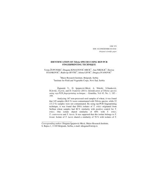
Analyzing 167 non-processed seed samples <strong>of</strong> wheat, it was found<br />
that 145 samples (86.8 %) were contaminated with <strong>Tilletia</strong> <strong>species</strong>, while 22<br />
(13.2 %) samples were not contaminated. By <strong>using</strong> <strong>rep</strong>-<strong>PCR</strong> <strong>fingerprinting</strong><br />
<strong>technique</strong>, it was found that DNA isolates <strong>of</strong> T. tritici originated from<br />
Serbian wheat samples had 80 % similarity with positive control for T.<br />
tritici. One isolate shared similarity <strong>of</strong> 60% with T. tritici,<br />
T. controversa and T. laevis. It was supposed that this isolate belongs to T.<br />
bromi. Isolate <strong>of</strong> T. laevis shared a similarity <strong>of</strong> 70 % with isolates <strong>of</strong> T.
184 GENETIKA, Vol. 43, No. 1, 183-195, 2011<br />
tritici and T. controversa, while T. walkeri was more than 10 % similar with<br />
T. tritici, T. controversa and T. laevis. Although T. controversa and T. tritici<br />
had high percent <strong>of</strong> genetic similarity, they were clustered separately. Our<br />
results suggest that <strong>rep</strong>-<strong>PCR</strong> <strong>fingerprinting</strong> could be a useful tool for<br />
monitoring presence <strong>of</strong> morphologically similar <strong>Tilletia</strong> <strong>species</strong> in wheat<br />
production areas.<br />
Key words: identification, <strong>rep</strong>-<strong>PCR</strong> <strong>fingerprinting</strong>, <strong>Tilletia</strong> tritici,<br />
<strong>Tilletia</strong> controversa, <strong>Tilletia</strong> laevis, <strong>Tilletia</strong> bromi, <strong>Tilletia</strong> walkeri, wheat<br />
INTRODUCTION<br />
Bunt <strong>of</strong> wheat is a worldwide distributed disease caused by <strong>Tilletia</strong> <strong>species</strong>.<br />
Except wheat, hosts <strong>of</strong> <strong>Tilletia</strong> <strong>species</strong> are rye, barley, triticale and many other<br />
<strong>species</strong> <strong>of</strong> family Poaceae. <strong>Tilletia</strong> tritici (Bjerk.) G. Wint. and T. laevis J.G. Kühn,<br />
which incite common bunt, are the most <strong>rep</strong>orted Tilleta <strong>species</strong> in Serbia (JEVTIĆ et<br />
al., 1997). KOSTIĆ and SMILJKOVIĆ (1968) <strong>rep</strong>orted T. triticoides Savalescu and<br />
T. intermedia Grassner in northeastern part <strong>of</strong> the country. Both T. controversa J.G.<br />
Kühn which incites dwarf bunt and T. indica Mitra which causes Karnal bunt <strong>of</strong><br />
wheat are quarantine <strong>species</strong> for Serbia. T. indica is also listed as an A1 quarantine<br />
pest for the EPPO region (European and Mediterranean Plant Protection<br />
Organization).<br />
No researches on diversity and distribution <strong>of</strong> <strong>Tilletia</strong> <strong>species</strong> in Serbia has<br />
been undertaken for the last 10 years. Bearing in mind that characteristics <strong>of</strong><br />
pathogens can be changed, as well as their virulence, it is important to monitor the<br />
presence <strong>of</strong> <strong>Tilletia</strong> <strong>species</strong>, varieties and races, in wheat production areas (JEVTIĆ,<br />
1998).<br />
<strong>Identification</strong> <strong>of</strong> <strong>Tilletia</strong> <strong>species</strong> on the basis <strong>of</strong> teliospores morphological<br />
characteristics is very unreliable. Teliospores isolated from individual sort, different<br />
races, as well as from geographical area can vary in the depth <strong>of</strong> reticulation.<br />
According to GOATES et al. (1996), there is an overlap <strong>of</strong> about 10% in the<br />
morphology <strong>of</strong> <strong>Tilletia</strong> <strong>species</strong> with reticulate episporium. This makes identification<br />
<strong>of</strong> some individual teliospores <strong>of</strong> quarantine T. controversa and non-quarantine<br />
T. tritici, difficult or impossible. There is also overlap in teliospore morphology<br />
between T. controversa and certain members <strong>of</strong> T. fusca Ellis & Everh complex<br />
which causes bunt <strong>of</strong> grass (MATHRE, 1996). Members <strong>of</strong> T. fusca. complex are T.<br />
fusca var. bromi-tectorum, T. fusca var. guyotiana and T. fusca var. fusca. The hosts<br />
<strong>of</strong> T. fusca var. bromi-tectorum and T. fusca var. guyotiana are Bromus sp, so some<br />
authors consider these two varieties as T. bromi (Brockm.) Brockm. (BOYD and<br />
CARRIS, 1997) The hosts <strong>of</strong> T. fusca var. fusca are Vulpia sp. Given that Bromus and<br />
Vulpia sp. are found in and around wheat fields, the harvested grain can be<br />
contaminated with teliospores <strong>of</strong> T. fusca, so it could make quarantine decisions<br />
difficult (MATHRE, 1996).<br />
Biochemical <strong>technique</strong>s played a significant role in identification <strong>of</strong><br />
different plant pathogens, thus improving morphological identification (LEVIĆ and<br />
PENČIĆ, 1993; IVANOVIĆ et al., 1995; STOJKOV et al., 1996; STOJKOV and
V.ZUPUNSKI et al. <strong>Tilletia</strong> SPECIES FINGERPRINT 185<br />
IGNJATOVIC, 1998; MATHRE, 1996). However, the most accurate results can be<br />
achieved by the use <strong>of</strong> molecular <strong>technique</strong>s. These <strong>technique</strong>s are unavoidable in<br />
identification <strong>of</strong> different plant (IGNJATOVIĆ-MICIĆ et al., 2007, 2009; PEJIĆ et al.,<br />
1998; VERMA et al., 1999; MIAN et al., 2002) and pathogen (MCDONALD et al., 2000;<br />
FERNANDEZ – ORTUNO et al., 2011; LADHALAKSHMI et al., 2009) genotypes.<br />
Initially, <strong>PCR</strong> (polymerase chain reaction) based genomic <strong>fingerprinting</strong><br />
<strong>technique</strong>s with markers such are RAPD (random amplified polymorphic DNA),<br />
AFLP (amplified fragment length polymorphism) and <strong>PCR</strong>-RFLP (restriction<br />
fragment length polymorphism) <strong>of</strong> ITS rDNA region were applied, in order to detect<br />
polymorphism between T. controversa, T. tritici and grass bunt fungi (BOYD et al.,<br />
1998, PIMENTEL et al., 1998; SHI et al., 1996). In general, these methods have not<br />
been precise in distinguishing T. controversa from T. trtici, but they could make<br />
clear difference between wheat and grass bunt fungi. Although these <strong>technique</strong>s did<br />
not make clear difference between T. controversa and T. tritici, significant divisions<br />
between members <strong>of</strong> T. fusca comlex, based upon host specificity, were obtained<br />
(MCDONALD et al., 2000). One such division was obtained in T. fusca var. fusca,<br />
where RAPD pr<strong>of</strong>iles <strong>of</strong> isolates from Vulpia oct<strong>of</strong>lora (Walt.) Rydb. shared only 20<br />
to 35% similarity with isolates from V. microstachys (Nutt.) Benth. (BOYD et al.,<br />
1998, BOYD and CARRIS, 1998). Using RAPD markers it was also obtained that T.<br />
fusca var. bromi-tectorum and T. fusca var. guyotiana are similar <strong>of</strong> about 55%<br />
(BOYD and CARRIS, 1998).<br />
Repetitive-sequence-based polymerase chain reaction (<strong>rep</strong>-<strong>PCR</strong>)<br />
<strong>fingerprinting</strong> <strong>technique</strong> was shown to be a molecular <strong>technique</strong> which is successful<br />
in distinguishing T. controversa from T. tritici (MCDONALD et al., 2000). This<br />
<strong>technique</strong> is based on <strong>PCR</strong>-mediated amplification <strong>of</strong> DNA sequences located<br />
between <strong>rep</strong>eated elements which are termed BOX, REP (<strong>rep</strong>etitive extragenic<br />
palindromic), and ERIC (enterobacterial <strong>rep</strong>etitive intergenic consensus) elements.<br />
These <strong>rep</strong>eated elements were first detected in prokaryotic genome, but they were<br />
also found very useful for genome variability characterization <strong>of</strong> several fungal<br />
genera, such are Verticillium (ARORA et al., 1996), Fusarium (EDEL et al., 1995),<br />
Stagonospora (CZEMBOR et al., 1999), Septoria (CZEMBOR et al., 1999), and<br />
Leptosphaeria (JEDRYCZKA, et al., 1999). Rep-<strong>PCR</strong> products separated on agarose<br />
gels yield DNA fingerprints that can be either visually compared or subjected to<br />
computer assisted pattern analysis (RADEMAKER et al., 1997). The main advantages<br />
<strong>of</strong> <strong>rep</strong>-<strong>PCR</strong> <strong>fingerprinting</strong> over RAPD <strong>technique</strong> are simplicity, tolerance to wider<br />
range <strong>of</strong> DNA concentrations and generation <strong>of</strong> <strong>rep</strong>roducible results (MCDONALD et<br />
al., 2000).<br />
QING et al. (2009) and LIU et al. (2009) found highly selective primers for<br />
distinguishing T. controversa from T. tritici, <strong>using</strong> a random amplified polymorphic<br />
DNA (RAPD) primer-mediated asymmetric-<strong>PCR</strong> (RM-<strong>PCR</strong>) <strong>technique</strong> and AFLP<br />
<strong>technique</strong>s, respectively. Even though highly selective primers for economically<br />
important <strong>Tilletia</strong> <strong>species</strong> were found, their use is not suitable for distinguishing<br />
varieties within <strong>Tilletia</strong> <strong>species</strong>. Therefore, at present <strong>rep</strong>-<strong>PCR</strong> <strong>fingerprinting</strong><br />
<strong>technique</strong> is valuable not only for studying taxonomic diversity <strong>of</strong> pathogens, but
186 GENETIKA, Vol. 43, No. 1, 183-195, 2011<br />
also for monitoring the presence <strong>of</strong> <strong>Tilletia</strong> <strong>species</strong> and their varieties in wheat<br />
production areas.<br />
The aim <strong>of</strong> this study was to identify <strong>Tilletia</strong> <strong>species</strong> found in seed samples<br />
collected in Serbia <strong>using</strong> <strong>rep</strong>-<strong>PCR</strong> <strong>fingerprinting</strong> <strong>technique</strong>s. Considering the fact<br />
that the resistance breakdown is the constant threat, precise identification <strong>of</strong> <strong>Tilletia</strong><br />
<strong>species</strong> would be <strong>of</strong> great importance, in order to make plant protection more<br />
efficient and reliable. MCDONALD et al. (2000) found this <strong>technique</strong> very useful for<br />
distinguishing T. indica from T. walkeri Castlebury & Carris, as well as T.<br />
controversa from T. tritici, and they suggested that <strong>rep</strong>-<strong>PCR</strong> <strong>fingerprinting</strong><br />
<strong>technique</strong>s could be used for diagnostic purposes.<br />
MATERIALS AND METHODS<br />
Collection <strong>of</strong> wheat seed samples<br />
One hundred and sixty seven non-processed seed samples <strong>of</strong> wheat used in<br />
this study were collected in cooperation with regional phytosanitary laboratory<br />
Agroinstitut-Sombor during the 2007-2008 harvest season. Wheat grain was<br />
collected from 39 municipalities in 7 districts <strong>of</strong> Vojvodina and 6 municipalities in 5<br />
districts <strong>of</strong> Central Serbia. For teliospore extraction, 50 g <strong>of</strong> subsamples were used<br />
(OEPP/ EPPO, 2007)<br />
Teliospore extraction<br />
Teliospores were extracted from 50 g subsamples <strong>of</strong> each grain sample<br />
<strong>using</strong> the size-selective sieving wash method (PETERSON et al., 2000) and<br />
OEPP/EPPO diagnostic protocol for T. indica (2007). Except that a 10-µm mesh<br />
nylon sieve was used instead <strong>of</strong> 20-µm nylon sieve. Extracted teliospores were<br />
mounted in 15% glycerol and examined by light microscopy at 250x magnification.<br />
Method for isolation and germination <strong>of</strong> teliospores<br />
The protocol for isolation <strong>of</strong> teliospores from both the microscope slide and<br />
the cover slip was a modification <strong>of</strong> that described in OEPP/EPPO diagnostic<br />
protocol for T. indica (2007). Except that a 10 µm mesh nylon sieve was used<br />
instead <strong>of</strong> 20 µm mesh nylon sieve and teliospores were surface-sterilized with 5%<br />
commercial bleach solution (approximately 0.2% active NaOCl) instead <strong>of</strong> 10%<br />
commercial bleach solution (approximately 0.4% active NaOCl). One part <strong>of</strong><br />
teliospore suspension was plated on 2% water agar with antibiotics and incubated at<br />
16°C from 10 to 15 days, whereas the other part <strong>of</strong> suspension, which was also<br />
plated on 2% antibiotic water agar, was incubated at 5°C in the presence <strong>of</strong> light<br />
from 3 to 6 weeks. These are environmental conditions required for teliospore<br />
germination and production <strong>of</strong> primary sporidia and small colonies with secondary<br />
sporidia <strong>of</strong> T. tritici, T. laevis and T. controversa (GOATES, 1996) Mycelial mat for<br />
DNA isolation was produced according the OEPP/EPPO diagnostic protocol for T.<br />
indica (2007), but the cuts <strong>of</strong> small blocks <strong>of</strong> agar bearing germinated teliospores or<br />
colonies were incubated at 16-20°C in the dark instead <strong>of</strong> at 21°C in the light.
V.ZUPUNSKI et al. <strong>Tilletia</strong> SPECIES FINGERPRINT 187<br />
Isolation <strong>of</strong> DNA and <strong>rep</strong>-<strong>PCR</strong> amplification<br />
DNA was isolated from mycelial mats <strong>using</strong> the procedure <strong>of</strong> MÖLLER et al.<br />
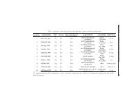
(1992). Rep-<strong>PCR</strong> reactions were performed <strong>using</strong> 19 DNA isolates <strong>of</strong> <strong>Tilletia</strong><br />
<strong>species</strong>. Fifteen DNA isolates were obtained from nine samples <strong>of</strong> wheat seed, while<br />
four DNA isolates were positive controls for T. tritici, T. controversa, T. laevis and<br />
T. walkeri (Table 1). The collection <strong>of</strong> T. tritici, used as positive control, was<br />
obtained from the Institute for Field and Vegetable Crops, Novi Sad DNA isolates<br />
used as positive controls for T. controversa, T. laevis and T. walkeri were obtained<br />
from strains that originate from the CBS collection (Centraalbureau voor<br />
Schimmelcultures, Utrecht, the Netherlands).<br />
The <strong>rep</strong>-<strong>PCR</strong> protocol was carried out <strong>using</strong> a combination <strong>of</strong> two<br />
procedures. One was described by MCDONALD et al. (2000) and the other was<br />
OEPP/EPPO diagnostic protocol for T. indica (2007). Rep-<strong>PCR</strong> fingerprints were<br />
obtained with the use <strong>of</strong> the following primers: BOX 1A1R (5’-CTACGGCAA GGC<br />
GACGCTGACG 3’); ERIC 1R (5’ATGTAAGCTCCTGGGGATTCAC3’) and 2I<br />
(5’-AAGTAAGTGACTGGGGTGAGCG-3’); and REP 1R (5’-IIIICGICGICATCI<br />
GGC-3’) and 2I (5’-ICGICTTATCIGGCCTAC-3’). Briefly, 100ng <strong>of</strong> purified DNA<br />
was used as template in 50 µl reaction mixture containing 0.5 µM <strong>of</strong> each primer, 0.2<br />
mM <strong>of</strong> each deoxynucleotide triphosphates, 6.25 units <strong>of</strong> Taq DNA polymerase, 1x<br />
<strong>PCR</strong> buffer and 1.5 mM MgCl2. <strong>PCR</strong> cycling parameters were: an initial<br />
denaturation at 95°C for 7 min; 35 cycles consisting <strong>of</strong> 94°C for 3 s, 92°C for 30 s,<br />
then either 40°C (REP primers) or 50°C (ERIC/BOX primers) for 1 min; extension<br />
at 65°C for 8 min; and a single final extension at 65°C for 8 min, followed by<br />
cooling at 4°C. Amplified <strong>PCR</strong> products (9 µl per sample) were separated on 1.5%<br />
agarose gels in 1x TAE (0.04 M Tris-acetate and 1 mM EDTA, pH 8.0) buffer for 18<br />
to 19 h at 2 V/cm, at 4°C. DNA bands were stained with ethidium bromide,<br />
visualized under UV transilluminator and photographed with digital camera. Gels<br />
were normalized <strong>using</strong> 1-kb DNA ladder (Fermentas) loaded on both sides and in the<br />
center <strong>of</strong> 20-lane gels. Positions <strong>of</strong> DNA fragments on agarose gel were scored<br />
visually. Using a band-based method, a matrix <strong>of</strong> binary variables (band present - 1<br />
and band absent - 0) was generated for each genotype. Genetic similarities were<br />
estimated <strong>using</strong> the similarity coefficient defined by JACCARD (1908). Cluster<br />
analysis was carried out on the matrix <strong>of</strong> genetic similarities <strong>using</strong> the unweighted<br />
pair group method with arithmetic averages (UPGMA). The computing <strong>of</strong> binary<br />
data including coefficients <strong>of</strong> similarities and UPGMA clustering were performed<br />
<strong>using</strong> NTSYS-pc s<strong>of</strong>tware (ROHLF, 2000).
188 GENETIKA, Vol. 43, No. 1, 183-195, 2011<br />
Table 1 DNA isolates designated by isolate number and supplier<br />
sample<br />
number<br />
Sources Supplier<br />
1 positive control for<br />
<strong>Tilletia</strong> walkeri<br />
CBS<br />
2 positive control for <strong>Tilletia</strong> laevis CBS<br />
3 positiv control for <strong>Tilletia</strong> controversa CBS<br />
4 mercantile wheat 1 Agroinstitut, Sombor<br />
5 mercantile wheat 1 Agroinstitut, Sombor<br />
6 mecantile wheat 1 Agroinstitut, Sombor<br />
7 mercantile wheat 4 Agroinstitut, Sombor<br />
8 mercantile wheat 4 Agroinstitut, Sombor<br />
9 mercantile wheat 2 Agroinstitut, Sombor<br />
10 mercantile wheat 2 Agroinstitut, Sombor<br />
11 mercantile wheat 11 Agroinstitut, Sombor<br />
12 mercantile wheat 11 Agroinstitut, Sombor<br />
13 cultivar Ljiljana Agroinstitut, Sombor<br />
14 cultivar Ljiljana Agroinstitut, Sombor<br />
15 mercantile wheat 6 Agroinstitut, Sombor<br />
16 mercantile wheat 7 Agroinstitut, Sombor<br />
17 positive control for<br />
Institute for Field and<br />
<strong>Tilletia</strong> tritici<br />
Vegetable Crops, Novi Sad<br />
18 artificialy contaminated wheat seed Institute for Field and<br />
with <strong>Tilletia</strong> tritici<br />
Vegetable Crops, Novi Sad<br />
19 mercantile wheat 14 Agroinstitut, Sombor<br />
RESULTS<br />
Trees with early growing season onset were predominant in the studied<br />
walnut Examination <strong>of</strong> 167 seed samples collected from Vojvodina and Central<br />
Serbia, showed that 145 (86.83%) samples were contaminated with teliospores <strong>of</strong><br />
<strong>Tilletia</strong> <strong>species</strong>, while 22 (13.17%) samples were not contaminated.<br />
<strong>Identification</strong> <strong>of</strong> <strong>Tilletia</strong> <strong>species</strong> by <strong>rep</strong>-<strong>PCR</strong> genomic <strong>fingerprinting</strong><br />
Rep-<strong>PCR</strong> fingerprint patterns were obtained from 19 DNA isolates <strong>of</strong><br />
<strong>Tilletia</strong> <strong>species</strong>. Each primer pair generated ~25 to 30 bands visible on agarose gel.<br />
The size <strong>of</strong> the amplification products ranged from 500 bp to 3500 bp (Figure 1).<br />
Four DNA isolates numbered as 4, 10, 11 and 13 were not analyzed because bands<br />
were not clearly visible when REP primers were used. Dendrogram obtained from<br />
similarity matrix is presented in Figure 2.Positive control for T. tritici and ten out <strong>of</strong><br />
eleven DNA isolates obtained from Serbian wheat seed samples were clustered<br />
together with more than 80% similarity, and thus were confirmed as T. tritici (Figure<br />
2). Isolate 14 and isolates <strong>of</strong> T. tritici,T. controversa and T. laevis shared a similarity<br />
<strong>of</strong> 60%, although isolate 14 was obtained from the seed sample where only
V.ZUPUNSKI et al. <strong>Tilletia</strong> SPECIES FINGERPRINT 189<br />
teliospores <strong>of</strong> T. tritici were found <strong>using</strong> microscopy method. Positive controls for T.<br />
controversa and T. laevis were not clustered together with isolates <strong>of</strong> T. tritici.<br />
Isolate <strong>of</strong> T. controversa shared a similarity <strong>of</strong> 75% with isolates <strong>of</strong> T. tritici, while<br />
isolate <strong>of</strong> T. laevis shared a similarity <strong>of</strong> 70% with isolates <strong>of</strong> T. tritici and T.<br />
controversa (Figure 2). Similarity between T. walkeri and T. tritici, T. controversa<br />
and T. laevis was approximately 10% (Figure 2).<br />
Figure 1 DNA fingerprints <strong>of</strong> 19 <strong>Tilletia</strong> genotypes obtained by <strong>rep</strong>-<strong>PCR</strong> <strong>technique</strong>.<br />
(A) REP-<strong>PCR</strong>, (B) ERIC-<strong>PCR</strong>, (C) BOX-<strong>PCR</strong>. Lane 1=positive control for <strong>Tilletia</strong> walkeri; Lane<br />
2=positive control for <strong>Tilletia</strong> laevis; Lane 3=positive control for <strong>Tilletia</strong> controversa; Lane 4=mercantile<br />
wheat 1.1; Lane 5= mercantile wheat 1.2; Lane 6=mercantile wheat 1.3; Lane 7=mercantile wheat 4.1;<br />
Lane 8=mercantile wheat 4.2; Lane 9= mercantile 2.1; Lane 10=mercantile wheat 2.2; Lane<br />
11=mercantile wheat 11.1; Lane 12=mercantile wheat 11.2; Lane 13=cultivar Ljiljana 1; Lane 14=cultivar<br />
Ljiljana 2; Lane 15=mercantile wheat 6; Lane 16=mercantile wheat 7;Lane 17 = positive control for<br />
<strong>Tilletia</strong> tritici; Lane 18= artificialy contaminated wheat seed with <strong>Tilletia</strong> tritici; Lane 19=mercantile<br />
wheat 14; Lane M= DNA molecular size markers (1 kbp ladder); sizes in base pairs.
190 GENETIKA, Vol. 43, No. 1, 183-195, 2011<br />
Figure 2 Dendrogram <strong>of</strong> 19 <strong>Tilletia</strong> genotypes derived from UPGMA analysis.<br />
TW=positive control for <strong>Tilletia</strong> walkeri; TL=positive control for <strong>Tilletia</strong> laevis; TC=positive control for<br />
<strong>Tilletia</strong> controversa; TT = positive control for <strong>Tilletia</strong> tritici; 5= mercantile wheat 1.1; 6=mercantile<br />
wheat 1.2; 7=mercantile wheat 4.1; 8=mercantile wheat 4.2; 9= mercantile wheat 2.1; 12=mercantile<br />
wheat 11.2; 14=cultivar Ljiljana 2; 15=mercantile wheat 6; 16=mercantile wheat 7; 18= artificialy<br />
contaminated wheat seed with <strong>Tilletia</strong> tritici; 19=mercantile wheat 14<br />
DISCUSSION<br />
Examination <strong>of</strong> 167 seed samples <strong>of</strong> wheat from Vojvodina and Central Serbia,<br />
showed that 145 (86.83%) samples were contaminated with teliospores <strong>of</strong> <strong>Tilletia</strong><br />
<strong>species</strong>. These results were in line with results obtained by STOJANOVIĆ et al. (1993)<br />
and JEVTIĆ et al. (1997). STOJANOVIĆ et al. (1993) examined 179 seed samples <strong>of</strong><br />
wheat from 43 locations in Serbia, and found that 73.7% samples were contaminated<br />
with T. tritici. JEVTIĆ et al. (1997) examined 120 seed samples collected from Srem<br />
District, south part <strong>of</strong> Vojvodina, and revealed that 85.3% samples were<br />
contaminated with T. tritici.<br />
<strong>Identification</strong> <strong>of</strong> <strong>Tilletia</strong> <strong>species</strong>, based on morphological characteristics <strong>of</strong><br />
teliospores, is very difficult or in some cases impossible. Deep reticulations and<br />
prominent sheath are the best characteristics for distinguishing T. controversa from<br />
T. tritici, but there is an overlap <strong>of</strong> about 10% in the morphology <strong>of</strong> <strong>Tilletia</strong> <strong>species</strong><br />
with reticulate exosporium, so individual teliospores <strong>of</strong> T. controversa with short<br />
reticulations and sheath thickness <strong>of</strong> 1.5 µm can be wrongly identified as T. tritici<br />
(GOATES, 1996). LIANG et al. (1982) tried to determine T. controversa and T. tritici<br />
based on depth <strong>of</strong> reticulations, thickness <strong>of</strong> gelatinous sheath, and germination test,<br />
with a success rate <strong>of</strong> 70%.
V.ZUPUNSKI et al. <strong>Tilletia</strong> SPECIES FINGERPRINT 191<br />
The similarity in morphology between T. controversa and certain members<br />
<strong>of</strong> T. fusca (T. bromi) complex is another problem for distinguishing T. controversa<br />
in a survey <strong>of</strong> the health <strong>of</strong> cereal seed. PETERSON et al. (2009) developed risk<br />
assessment model for importation <strong>of</strong> United States milling wheat containing T.<br />
controversa to People’s Republic <strong>of</strong> China. They analyzed presence <strong>of</strong> T.<br />
controversa in samples <strong>of</strong> wheat export shipments. T. controversa was distinguished<br />
from T. tritici based on reticulation depth. If reticulation depth was ≥ 0.95 µm,<br />
teliospores were identified as T. controversa. This method does not differentiate T.<br />
controversa from T. bromi, so there is the risk <strong>of</strong> possible misidentifying grass smut<br />
which leads to risk for possible commodity rejection. Grass smuts are commonly<br />
present in wheat export shipments but in the low number. Although they are not<br />
pathogens <strong>of</strong> wheat, their hosts, Bromus and Festuca sp, can be found in and around<br />
wheat fields.<br />
The aim <strong>of</strong> this study was to identify <strong>Tilletia</strong> <strong>species</strong> which were found in<br />
seed samples <strong>of</strong> wheat, <strong>using</strong> <strong>rep</strong>-<strong>PCR</strong> <strong>fingerprinting</strong> <strong>technique</strong>. Analysis <strong>of</strong> eleven<br />
DNA isolates <strong>of</strong> <strong>Tilletia</strong> <strong>species</strong>, obtained from nine seed samples, revealed that ten<br />
out <strong>of</strong> eleven isolates belong to T. tritici. These isolates and the positive control for<br />
T. tritici had more than 80% similarity, which was consistent with result obtained by<br />
MCDONALD et al. (2000). In our study only one isolate <strong>of</strong> T. laevis and one isolate <strong>of</strong><br />
T. controversa were used and they shared similarity <strong>of</strong> about 70% with isolates <strong>of</strong><br />
T. tritici. MCDONALD et al. (2000) analyzed large number <strong>of</strong> T. controversa, T. tritci<br />
and T. laevis isolates and they also shared 70% similarity. Isolate 14 obtained from<br />
the seed sample contaminated with teliospores <strong>of</strong> T. tritici had 60% similarity with<br />
isolates <strong>of</strong> T. tritici, T. laevis and T. controversa. Positive control for T. bromi was<br />
not used in this study, but on the basis <strong>of</strong> similarity <strong>of</strong> our results and these obtained<br />
by MCDONALD et al. (2000), it might be assumed that isolate 14 belongs to T. bromi.<br />
This assumption is supported by the fact that teliospores <strong>of</strong> T. tritici and T. bromi can<br />
be morphologically similar, thus they could have been misidentified. According the<br />
MCDONALD et al. (2000) T. bromi shared >55% similarity with T. tritici, T. laevis<br />
and T. controversa. Similarity between T. walkeri and T. tritici, T. controversa and<br />
T. laevis was approximately 10%, which was in line with result obtained by<br />
MCDONALD (2000).<br />
The results <strong>of</strong> our study indicate the high sensitivity <strong>of</strong> size selective sieving<br />
method as well as the study <strong>of</strong> PETERSON et al. (2000). By <strong>using</strong> this method it was<br />
possible to detect ≤5 teliospores in 50g seed samples. However there is still problem<br />
<strong>of</strong> producing mycelial mats for DNA isolation if the seed samples are contaminated<br />
with one or several teliospores. This is the reason why we have successfully isolated<br />
DNA from only nine out <strong>of</strong> 167 examined seed samples. It is important to say that<br />
these nine samples were highly contaminated with teliospores <strong>of</strong> <strong>Tilletia</strong> <strong>species</strong>,<br />
unlike the rest <strong>of</strong> seed samples which were contaminated with about 5 teliospores <strong>of</strong><br />
<strong>Tilletia</strong> <strong>species</strong>.<br />
Regardless <strong>of</strong> the difficulties in obtaining mycelial mat, MCDONALD et al.<br />
(2000) suggested that <strong>rep</strong>-<strong>PCR</strong> has potential as a diagnostic method when it’s<br />
important to distinguish quarantined T. indica from T. indica-like teliospores.
192 GENETIKA, Vol. 43, No. 1, 183-195, 2011<br />
Furthermore, T. tritici, T. controversa and T. laevis were clustered separately despite<br />
the high genetic similarity <strong>of</strong> 70%. T. bromi which has morphologically similar<br />
teliospores to those <strong>of</strong> T. controversa also clustered separately, providing<br />
differentiation <strong>of</strong> these two <strong>species</strong>. T. bromi can be distinguished from<br />
T. controversa <strong>using</strong> RAPD markers (SHI et al., 1996; BOYD et al., 1998), but<br />
according the MCDONALD et al. (2000) <strong>rep</strong>-<strong>PCR</strong> <strong>fingerprinting</strong> <strong>technique</strong> has<br />
advantages due to its simplicity, the universality <strong>of</strong> <strong>PCR</strong> primers and tolerance <strong>of</strong> a<br />
wider range <strong>of</strong> DNA concentrations in generating <strong>rep</strong>roducible results. QING et al.<br />
(2009) and LIU et al. (2009) developed high selective primers for identification <strong>of</strong> T.<br />
controversa, and for the first time it was possible to distinguish T. controversa from<br />
T. tritici on the basis <strong>of</strong> the presence <strong>of</strong> one unique DNA fragment. This is a very<br />
important discovery that could provide accurate identification <strong>of</strong> T. controversa,<br />
avoiding confusion during identification <strong>of</strong> this quarantine <strong>species</strong>. Nevertheless,<br />
<strong>rep</strong>-<strong>PCR</strong> <strong>fingerprinting</strong> is a useful <strong>technique</strong> for studying, diversity, phylogenetic<br />
relationships and taxonomic status <strong>of</strong> wheat bunt <strong>species</strong>, considering the fact that<br />
different isolates <strong>of</strong> the same variety, such as T. fusca var. fusca isolates on V.<br />
microstachys and V. oct<strong>of</strong>lora, could share only 20 to 35% <strong>of</strong> similarity. (BOYD et<br />
al., 1998; MCDONALD et al., 2000). These findings could be <strong>of</strong> a great importance not<br />
only for studying diversity <strong>of</strong> <strong>Tilletia</strong> <strong>species</strong>, but also for understanding<br />
relationships between a pathogen and its hosts.<br />
Received, November 11 th 2010<br />
REFERENCES<br />
Accepted, March 14 h 2011<br />
ARORA, D. K., P. R. HIRSCH, and B. R. KERRY (1996): <strong>PCR</strong>-based molecular discrimination <strong>of</strong> Verticillium<br />
chlamydosporum isolates. Mycol. Res. 100:801-809.<br />
BOYD, M. L., and L. M. CARRIS (1997): Molecular relationships among varieties <strong>of</strong> the <strong>Tilletia</strong> fusca<br />
(T. bromi) complex and related <strong>species</strong>. Mycol. Res. 101:269-277.<br />
BOYD, M. L., and L. M. CARRIS (1998): Evidence supporting the separation <strong>of</strong> Vulpia- and Bromus -<br />
infectiong isolates in the <strong>Tilletia</strong> fusca (T. bromi) complex. Mycologia 90: 1031-1039.<br />
BOYD, M. L., L. M. CARRIS, and P. M. GRAY (1998): Characterization <strong>of</strong> <strong>Tilletia</strong> goloskokovii and allied<br />
<strong>species</strong>. Mycologia 90:310-322.<br />
CZEMBOR, P. C., and E. ARSENIUK (1999): Study <strong>of</strong> genetic variability among monopycnidial and<br />
monopycnidiospore isolates derived from single pycnidia <strong>of</strong> Stagonospora spp. and Septoria<br />
tritici with the use <strong>of</strong> RAPD-<strong>PCR</strong>, MP-<strong>PCR</strong> and <strong>rep</strong>-<strong>PCR</strong> <strong>technique</strong>s. J. Phytopathol. 147:539-<br />
546.<br />
EDEL, V., C. STEINBERG, I. AVELANGE, G. LAGUERRE, and C. ALABOUVETTE (1995): Comparison <strong>of</strong> three<br />
molecular methods for the characterization <strong>of</strong> Fusarium oxysporum strains. Phytopathology<br />
85:579-585.<br />
FERNANDEZ-ORTUÑO D., S. L. ATKINS, B. A., FRAAIJE (2011): The use <strong>of</strong> CYP51C gene based <strong>PCR</strong>-RFLP<br />
assay for simultaneous detection and identification <strong>of</strong> Fusarium avenaceum and F. tricinctum<br />
in wheat. International Journal <strong>of</strong> Food Microbiology 145(1): 370-374.
V.ZUPUNSKI et al. <strong>Tilletia</strong> SPECIES FINGERPRINT 193<br />
GOATES, B. J. (1996): Common Bunt and Dwarf Bunt. (In R. D. Wilcoxson, E. E. Saari, G. P. Hettel & A.<br />
McNab (Eds), Bunt and Smut Diseases <strong>of</strong> Wheat- Concepts and Methods <strong>of</strong> Disease<br />
Management, (pp:12-25), Mexico: CIMMYT)<br />
IGNJATOVIĆ-MICIĆ, D., S. MLADENOVIC DRINIĆ, A. NIKOLIĆ, V. LAZIĆ-JANČIĆ (2007): Comparison <strong>of</strong> AFLP<br />
and SSR markers for genetic diversity studies in maize populations. Maydica 52: 399-406.<br />
IGNJATOVIĆ-MICIĆ, D. K. MARKOVIĆ, D. RISTIĆ, S. MLADENOVIĆ DRINIĆ, S. STANKOVIĆ, V.LAZIĆ-JANČIĆ, M.<br />
DENIĆ (2009): Variability analysis <strong>of</strong> normal and opaque2 maize inbred lines. Genetika 41(1):<br />
81 -93.<br />
IVANOVIĆ, D., R. OSLER, N. KATIS, M. S. IVANOVIĆ and D. IGNJATOVIĆ (1995): Principal maize viruses in Mediterranean<br />
countries. Agronomie 15: 443-446.<br />
JACCARD, P. (1908): New researches on plant distribution. Nouvelles rescherches sur la distribution<br />
florale. Bull. Soc. Vaud. Sci. Nat. 44: 223-270<br />
JEDRYCZKA, M., T. ROUXEL, and M-H. BALESDENT (1999): Rep-<strong>PCR</strong> based genomic <strong>fingerprinting</strong> <strong>of</strong><br />
isolates <strong>of</strong> Leptosphaeria maculans from Poland. Eur. J. Plant Pathol. 105:813-823.<br />
JEVTIĆ, R., S. STOJANOVIĆ, M. DOPUĐA, D. MATIJEVIĆ, and M. MILOŠEVIĆ (1997): Occurrence <strong>of</strong> bunt and<br />
smuth in Serbia. XXXI Agronomy Conference. Book <strong>of</strong> abstracts 29: 217-223. (original text in<br />
Serbian)<br />
JEVTIĆ, R. (1998): Races, gene identification and resistance sources; Resistance <strong>of</strong> varieties and cropping<br />
practice defense. Chapters in book: Wheat blunt, Milošević, M., Stojanović, S., Jevtić, R.,<br />
Matijević, D., Rajković, Snežana. Institute <strong>of</strong> Field and Vegetable Crops, Novi Sad; Institute <strong>of</strong><br />
Agricultural Research SERBIA, Belgrade; Institute for Plant Protection and Environment,<br />
Belgrade. Feljton, Novi Sad, pp.: 47-78. (original text in Serbian)<br />
KOSTIĆ, B., and H. SMILJAKOVIĆ (1968): Wheat diseases under intensive production and their suppression.<br />
Agrohemija 7-8: 331-342. (original text in Serbian)<br />
LADHALAKSHMI D., A. VIJAYASAMUNDEESWARI, V. PARANIDHARAN, R. SAMIYAPPAN, R. VELAZHALAN<br />
(2009): Molecular identification <strong>of</strong> isolates <strong>of</strong> Peronosclerospora sorghi from maize <strong>using</strong><br />
<strong>PCR</strong>-based SCAR marker. World Journal <strong>of</strong> Microbiology and Biotechnology 25(12): 2129-<br />
2135.<br />
LEVIĆ, J., V. PENČIĆ (1993): Morphology <strong>of</strong> a new pathotype <strong>of</strong> Bipolaris zeicola (Stout) Shoemaker. J.<br />
Phytopathology 139: 339-346.<br />
LIANG, Z., Y. GUO, Y. ZHU, and Y. SONG (1982): Statistic analysis based on the teliospores morphological<br />
characters in differentiating dwarf smut from stinking smut. Acta Phytophylacica Sin. 9: 243-<br />
250.<br />
LIU, J.H., L. GAO, T.G. LIU, and W.Q. CHEN (2009): Development <strong>of</strong> a sequence-characterized amplified<br />
region marker for diagnosis <strong>of</strong> dwarf bunt <strong>of</strong> wheat and detection <strong>of</strong> <strong>Tilletia</strong> controversa Kühn.<br />
Letters in Applied Microbiology 49: 235-240.<br />
MATHRE, D. E. (1996): Dwarf Bunt: Politics, <strong>Identification</strong>, and Biology. Annu. Rev. Phytopathol, 34: 67-<br />
85<br />
MCDONALD, J.G., E. WONG, and G. P. WHITE (2000): Differentiation <strong>of</strong> <strong>Tilletia</strong> Species by <strong>rep</strong>-<strong>PCR</strong><br />
Genomic Fingerprinting. Plant Dis. 84 (10): 1121-1125.<br />
MIAN ROUF M.A., A.A. HOPKINS, J.C. ZWONITZER (2002): Determination <strong>of</strong> genetic diversity in tall fescue<br />
with AFLP markers. Crop Sci. 42: 944-950.
194 GENETIKA, Vol. 43, No. 1, 183-195, 2011<br />
MÖLLER E.M., G. BAHNWEG, H. SANDERMANN, and H. H. GEIGER (1992): A simple and efficient protocol<br />
for isolation <strong>of</strong> high molecular weight DNA from filamentous fungi, fruit bodies, and infected<br />
tissues. Nucleic Acids Research. 20 (22) 6115-6116.<br />
OEPP/EPPO (2007): Diagnostic Protocols for Regulated Pests. EPPO Standards PM 7/29 (2) <strong>Tilletia</strong> indica.<br />
Bulletin OEPP/EPPO Bulletin 37, 503-520.OEPP/EPPO (Paris).<br />
PEJIĆ, I., P. AJMONE-MARSAN, M. MORGANTE, V. KOZUMPLICK, P. CASTIGLIONI, G. TARAMINO, M. MOTTO<br />
(1998): Comparative analysis <strong>of</strong> genetic similarity among maize inbred lines detected by<br />
RFLPs, RAPDs, SSRs, and AFLPs. Theor. Appl. Genet. 97:1248-1255.<br />
PETERSON, G. L., M. R. BONDE, and J. G. PHILLIPS (2000): Size-Selective Sieving for Detecting Teliospores<br />
<strong>of</strong> <strong>Tilletia</strong> indica in Wheat Seed Samples. Plant Dis. 84: 999-1007<br />
PETROSON, G. L., T. B. WHITAKER, R. J. STEFANSKI, E. V. PODLECKIS, J. G. PHILLIPS, J. S. WU, and W. H.<br />
MARTINEZ (2009): A Risk Assessment Model for Importation <strong>of</strong> United States Milling Wheat<br />
Containing <strong>Tilletia</strong> contraversa. Plant Dis. 93 (6): 560-573.<br />
PIMENTEL, G., L. M. CARRIS, and T. L. PEEVER (1998): Characterization <strong>of</strong> interspecific hybrids between<br />
<strong>Tilletia</strong> controversa and <strong>Tilletia</strong> bromi. Mycologia 92 (3): 411-420.<br />
RADEMAKER, J. L. W., and F. J. DE BRUIJN (1997): Characterization and classification <strong>of</strong> microbes by <strong>rep</strong>-<br />
<strong>PCR</strong> geno mic <strong>fingerprinting</strong> and computer-assisted pattern analysis (In: G. Caetano-Anollés<br />
and P. M. Gressh<strong>of</strong>f (Eds), DNA Markers: Protocols, Applications and Overviews (pp:151-<br />
171) John Wiley & Sons, New York).<br />
ROHLF, F.J. (2000): NTSYS-pc. Numerical taxonomy and multivariate analysis system. Version 2.0<br />
Exeter S<strong>of</strong>tware, Setaket, N.Y<br />
STOJKOV, S., J. LEVIĆ, , D. IGNJATOVIĆ, D. IVANOVIĆ, V. PENČIĆ (1996): Periconia pathogenes on maize<br />
root and stalk.. Zaštita bilja, vol. 47(3), 217: 229-240. (original text in Serbian).<br />
STOJKOV, S., D. IGNJATOVIĆ (1998): Variability <strong>of</strong> fungus Microdochium bolleyi isolate - maize root and<br />
stalk pathogen. 7 th International Congress <strong>of</strong> Plant Pathology. 9-16. Avgust, Edinburgh.<br />
VERMA, K.S., V. KHANNA, N. SINGH (1999): Random Amplified Polymorphic DNA Analysis <strong>of</strong> Indian<br />
Scented Basmati Rice (Oryza sativa L.) Germplasm for <strong>Identification</strong> <strong>of</strong> Variability and<br />
Duplicate Accessions, if any. Electrophoresis, 20: 1786–1789.<br />
QING, Y., N. SIJI, Y. YOUPING, L.MINHUI, C. JUN, and W. ZHONGKANG (2009): Development <strong>of</strong> a <strong>PCR</strong>-<br />
based diagnostic tool specific to wheat dwarf bunt, caused by <strong>Tilletia</strong> controversa. Eur. J. Plant<br />
Pathol. 124: 585-594.<br />
SHI, Y. L., P.LOOMIS, D. CHRISTIAN, L. M. CARRIS, and H. LEUNG (1996): Analysis <strong>of</strong> the genetic<br />
relationships among the wheat bunt fungi <strong>using</strong> RAPD and ribosomal DNA markers.<br />
Phytopathology 86: 311-318.<br />
STOJANOVIĆ, S., M. DOPUĐA, J. STOJANOVIĆ, and S. GUDŽIĆ (1993): Bunt as a frequent wheat disease in<br />
Serbia. I Yugoslav Conference on Plant Protection. Vrnjačka Banja. Book <strong>of</strong> Abstracts, p. 18<br />
(original text in Serbian)
V.ZUPUNSKI et al. <strong>Tilletia</strong> SPECIES FINGERPRINT 195<br />
IDENTIFIKACIJA VRSTA RODA <strong>Tilletia</strong> <strong>rep</strong>-<strong>PCR</strong> FINGERPRINTING<br />
TEHNIKOM<br />
Vesna ŽUPUNSKI 1 , Dragana IGNJATOVIĆ-MICIĆ 1 , Ana NIKOLIĆ 1 ,Slavica<br />
STANKOVIĆ 1 , Radivoje JEVTIĆ 2 , Jelena LEVIĆ 1 Dragica IVANOVIĆ 1<br />
1 Institut za kukuruz, Beograd, Srbija<br />
2 Institut za ratarstvo i povrtarstvo, Novi Sad, Srbija<br />
I z v o d<br />
Istraživanjem kontaminiranosti uzoraka semena pšenice teleutosporama<br />
<strong>Tilletia</strong> vrsta, utvrđeno je da je od 167 uzoraka nedorađenog semena pšenice, bilo<br />
kontaminirano 145 (86,8%), dok su 22 uzorka (13,2%) smatrana nekontaminiranim.<br />
Identifikacija <strong>Tilletia</strong> vrsta izvršena je <strong>rep</strong>-<strong>PCR</strong> <strong>fingerprinting</strong> tehnikom. Izolati<br />
prikupljeni na teritoriji Republike Srbije identifikovani su kao T. tritici, s obzirom da<br />
su sa pozitivnom kontrolom imali genetičku sličnost veću od 80%. Jedan od izolata<br />
koji je vodio poreklo iz opštine Apatin bio je oko 60% genetički sličan sa izolatima<br />
T. tritici, T. controversa i T. laevis. Pretpostavljeno je da pripada vrsti T. bromi.<br />
Genetička sličnost izolata T. walkeri i vrsta: T. tritici, T. laevis i T. controversa,<br />
iznosila je nešto više od 10%. Genetička sličnost T. tritici, T. controversa i T. laevis<br />
bila je oko 70 %. I pored visokog procenta genetičke sličnosti između T. controversa<br />
i T. tritici, napravljena je razlika među njima, što <strong>rep</strong>-<strong>PCR</strong> <strong>fingerprinting</strong> tehniku čini<br />
veoma podesnom za praćenje prisustva morfološki sličnih Tilleria vrsta prilikom<br />
kontrole kvaliteta semena pšenice.<br />
Primljeno 11. XI. 2010.<br />
Odobreno 14. III. 2011.