Abstract book - ESPRAS
Abstract book - ESPRAS
Abstract book - ESPRAS

You also want an ePaper? Increase the reach of your titles
YUMPU automatically turns print PDFs into web optimized ePapers that Google loves.
Croatian Medical Association<br />
Croatian Society of Plastic, Reconstructive and Aesthetic Surgery<br />
University Hospital Dubrava, Zagreb, Croatia<br />
Department for Plastic, Reconstructive and Aesthetic Surgery<br />
<strong>Abstract</strong> <strong>book</strong><br />
5th CROATIAN CONGRESS FOR PLASTIC,<br />
RECONSTRUCTIVE AND AESTHETIC<br />
SURGERY<br />
<strong>ESPRAS</strong> appointed Congress for 2004.<br />
Editors: Sanda Stanec, Zlatko Vlajčić,<br />
Krešimir Martić, Franjo Rudman ml.<br />
Dubrovnik-Cavtat, Croatia<br />
15th-20th October, 2004.
Naslovna strana – reklama ROZISTEP
5th CROATIAN CONGRESS FOR PLASTIC, RECONSTRUCTIVE AND<br />
AESTHETIC SURGERY<br />
<strong>ESPRAS</strong> appointed Congress for 2004.<br />
Dubrovnik-Cavtat, Croatia<br />
15th-20th October, 2004<br />
<strong>Abstract</strong> <strong>book</strong><br />
Organizer : Croatian Society of Plastic, Reconstructive<br />
and Aesthetic Surgery, Croatian Medical Association<br />
Publisher : Croatian Society of P lastic, Reconstructive<br />
and Aesthetic Surgery and Znanje d.d., Zagreb, Croatia<br />
Editors: Sanda Stanec, MD, PhD, Editor-in-Chief<br />
Zlatko Vlajčić, MD, Associate Editor<br />
Krešimir Martić, MD, Associate Editor<br />
Franjo Rudman ml., MD, Associate Editor<br />
Printed by: Znanje d.d., Zagreb, Croatia<br />
All rights reserved. No part of this publication may be reproduced, or transmitted in any form or by<br />
any means without prior written permission from the publisher.
5th CROATIAN CONGRESS FOR PLASTIC, RECONSTRUCTIVE AND<br />
AESTHETIC SURGERY<br />
<strong>ESPRAS</strong> appointed Congress for 2004.<br />
Croatian Medical Association<br />
organized by<br />
Croatian Society of Plastic,<br />
Reconstructive and Aesthetic Surgery<br />
University Hospital Dubrava, Zagreb, Croatia<br />
Department for Plastic, Reconstructive<br />
and Aesthetic Surgery<br />
Local co-organizer<br />
County Hospital Dubrovnik<br />
Under the auspicies of<br />
Dubrovnik – Neretva County
ORGANIZING COMMITTEE<br />
CONGRESS PRESIDENT<br />
Zdenko Stanec<br />
President of the Croatian Society of Plastic, Reconstructive and Aesthetic<br />
Surgery (CSPRAS)<br />
VICE PRESIDENTS<br />
Marko Margaritoni<br />
Zdravko Roje<br />
GENERAL SECRETARIES<br />
Sanda Stanec<br />
Rado Žic<br />
TREASURER<br />
Rudolf Milanović<br />
ORGANISING COMMITTEE<br />
Zdenko Stanec, Marko Margaritoni, Zdravko Roje, Rado Žic, Sanda Stanec,<br />
Rudolf Milanović, Srećko Budi, Franjo Rudman, Krešimir Martić, Zlatko Vlajčić, Marina<br />
Mamić-Pavelić<br />
SCIENTIFIC COMMITTEE<br />
Josip Unušić, Mišo Virag, Neven Olivari, Milomir Ninković, Radojko Ivrlač,<br />
Vedran Uglešić, Mario Zambelli, Sanda Stanec, Rado Žic, Ivo Džepina<br />
HONORARY COMMITTEE<br />
Ivan Prpić, Đorđe Montani, Ivo Padovan
PAST CONGRESES ORGANIZED BY CSPRAS<br />
1st Croatian Congress for Plastic, Reconstructive and Aesthetic Surgery, with<br />
international participation, Dubrovnik, 19. - 23. rujan 1998.<br />
2nd Croatian Congress for Plastic, Reconstructive and Aesthetic Surgery, with<br />
international participation, Opatija , 4. - 8. rujan 1999.<br />
3rd Croatian Congress for Plastic, Reconstructive and Aesthetic Surgery, with<br />
international participation, Split, 20.-24. rujan, 2000.<br />
4th Croatian Congress for Plastic, Reconstructive and Aesthetic Surgery, with<br />
international participation, Zagreb, 25. – 28. rujan 2002.<br />
PAST <strong>ESPRAS</strong> APPOINTED MEETINGS<br />
1st European Appointed National Meeting: September, 3- 6, 1998, Istambul, Turkey<br />
2nd European Appointed National Meeting: September, 2 – 4, 1999, St.Gallen,<br />
Switzerland<br />
3rd European Appointed National Meeting: July, 5 – 7, 2000, Birmingham, UK<br />
4th European Appointed National Meeting: October 1 – 5, 2003, Athens, Greece<br />
CONGRESS SECRETARIAT:<br />
Department for Plastic, Reconstructive and Aesthetic Surgery<br />
University Hospital Dubrava<br />
Avenija Gojka Šuška 6, 10000 Zagreb, Croatia<br />
Tel.: +385 1 290 2569/ Fax.:+385 1 290 2451/ E-mail: plkir@kbd.hr<br />
From October 11, for all necessary informations regarding<br />
Congress, please contact the Congress Office:<br />
CROATIA HOTEL<br />
20210 Cavtat, Croatia<br />
Salon 5<br />
Tel: +385/(0)20/ 475-896 / Tel: 1896 (for calls from the hotel)<br />
Congress web site: www.kbd.hr/plastkir
CONTENTS<br />
Lectures 1<br />
SESSION A :<br />
Head and Neck Reconstructive Surgery<br />
SESSION B :<br />
Head and Neck Aesthetic Surgery<br />
SESSION C :<br />
Breast Reconstructive Surgery<br />
SESSION D :<br />
Breast Aesthetic Surgery<br />
SESSION E :<br />
Body Contouring<br />
SESSION F :<br />
Upper Extremity Reconstructions<br />
SESSION G :<br />
Microsurgery<br />
SESSION H :<br />
Lower Extremity Reconstructions<br />
SESSION I :<br />
Miscellaneous
Scientific Posters<br />
Sponsors & Exhibitors<br />
List of Participants
LECTURES
REKLAMA FOTONA
L1. Craniofacial deformity<br />
Prof. Ian T. Jackson<br />
Institute for Craniofacial and Reconstructive Surgery<br />
Southfield, USA<br />
Craniofacial deformity can be relatively minor or it can be devastating. It can be congenital or it<br />
may result from trauma or tumor.<br />
Craniosynostosis<br />
A single suture involvement can be treated at any time; multiple suture involvement is associated<br />
with raised intracranial pressure and suture release with skull fragment repositioning should be<br />
performed. Any suture may be affected. Measurement of head size is important in decision-<br />
making. Postural skull deformity should be diagnosed correctly, and in most cases surgical<br />
treatment is not required. Molding helmets are employed if the deformity does not self-correct.<br />
Crouzon and Apert Syndromes<br />
Rarely is early treatment necessary, but with the advent of distraction osteogenesis there is a<br />
move towards earlier correction. The long-term follow-up of this treatment is not, as yet,<br />
available - it may only be an Aevent in time@ and further distractions may be necessary. The<br />
surgical management consists of frontosupraorbital advancement as required, with midface an<br />
advancement at the LeFort III level.<br />
Hypertelorism<br />
The basic mechanism is that of a midline cleft and the severity is variable. Severe cases require<br />
early treatment to bring the orbits into their correct position and hopefully establish binocular<br />
vision. Secondary procedures are frequent, e.g. further osteotomies, nasal reconstruction, and<br />
palatal procedures. Facial bipartition is frequently indicated, this gives very stable and<br />
satisfactory correction. If there is a frontal encephalocele, this is resected and repaired at the<br />
same time as the bony correction.<br />
Hypotelorism<br />
This is a rare condition, however if it occurs orbital osteotomies are performed. The orbits are<br />
moved laterally and the midline defect is bone grafted.<br />
Hemifacial Microsomia<br />
This occurs with varying severity, in the most severe cases all levels of the face are involved -<br />
skull, orbit, maxilla, mandible and soft tissue. The bony problems are dealt with by osteotomies<br />
and/or bone grafts. The soft tissue is augmented with free tissue transfer. Distraction<br />
osteogenesis will be used when indicated.<br />
Facial Clefts<br />
These can involve specific cases or multiple facial regions. The minor clefts are treated in a<br />
relatively standard fashion by excision, local flaps, and suture, the more severe cases may need<br />
the addition of bone grafts, osteotomies or free tissue transfers, as indicated.<br />
Team Approach in Craniofacial Surgery<br />
These complex anomalies require a multi-specialist approach with an experienced and dedicated<br />
team - a plastic surgeon experienced in the craniofacial area, neurosurgeon, maxillofacial<br />
surgeon, orthodontist, prosthedontist, pediatrician, social worker, experienced nursing staff, and a<br />
good anesthetic team.
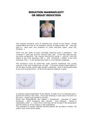
L2. New techniques in breast reduction<br />
Prof. Phillip Blondeel<br />
Universitair Ziekenhuis, Gent, Belgium<br />
L3. Emergency free flap reconstruction of lower leg trauma<br />
Prof. Zoran Arnež<br />
Department for Plastic, Reconstructive Surgery and Burns, Clinical Center,<br />
Ljubljana, Slovenia<br />
L4. Extended composite facelift<br />
Prof. Edgar Biemer<br />
Abteilung fur Plastische und Wiederherstellungschirurgie<br />
Klinikum rechts der Isar, Munchen, Germany<br />
L5. Perspectives in corset platysmaplasty<br />
Prof. Richard C. Sadove<br />
The Sadove Clinic, Tel Aviv, Israel<br />
L6. Breast augmentation: possibilities and impossibilities<br />
Prof. Rolf R. Olbrisch<br />
Klinik fur Plastische Chirurgie<br />
Diakonie-Krankenhaus, Dusseldorf, Germany<br />
L7. Submucous cleft palate, methods of repair,<br />
our philosophy<br />
Prof. John Boorman<br />
Guy’s Hospital, London & Queen Victoria Hospital, East Grinstead, UK<br />
Classic SMCP - Triad of features<br />
Notched posterior nasal spine<br />
Bifid uvula<br />
Lucent midline strip<br />
Occult SMCP<br />
No Intraoral signs<br />
Small midline defect on endoscopy<br />
?Musculus uvulae absence<br />
Clinical situation is more complex<br />
Spectrum of severity<br />
Key is abnormal levator insertions<br />
Examination important
Lateral videofluoroscopy most useful<br />
Presentation is at various stages<br />
Neonatal examination<br />
Feeding difficulties<br />
Failure to thrive<br />
Glue Ear<br />
Speech problems – Velopharyngeal insufficiency<br />
May be part of Velocardiofacial Syndrome (22q11 deletion)<br />
Not all SMCP need treatment ~50% will speak normally<br />
Treat when there is evidence of palatal dysfunction (feeding/speech)<br />
Surgical options<br />
Correction of muscle abnormality (Sommerlad / Furlow etc.)<br />
Pharyngoplasty if that fails<br />
L8. Radical sternectomy and primary musculocutaneous flap<br />
reconstruction to control sternal osteitis<br />
Prof. Andrej Banic<br />
Department of Plastic, Reconstructive and Aesthetic Surgery,<br />
University Hospital Inselspital, Berne, Switzerland<br />
Objective: Sternal osteitis after median sternotomy causes considerable<br />
morbidity and mortality. The use of muscle and omentum flaps has been<br />
proved as valid adjunct to combat these severe infections. In this study we<br />
present our experience with a more radical approach.<br />
Methods: Sternectomy consisted of the resection of the entire sternum<br />
including the costochondral arches and the sternoclavicular joints, and was<br />
followed by the repair of the defect with musculocutaneous flaps without any<br />
re-stabilization of the thoracic wall. 13 patients received a vertical rectus<br />
abdominis musculocutaneous (VRAM) flap, 14 patients a pedicled latissimus<br />
dorsi musculocutaneous (pLDM) flap and 12 patients a free LDM flap (total<br />
of 40 flaps in 39 patients out of 66 patients who required surgical revision<br />
due to sternal osteitis out of 6'078 sternotomy patients).<br />
Results: Two patients died within 30 days postoperatively (early mortality<br />
5.1%), however, not due to sternal infection, which was cured without any<br />
recurrence in all patients. 17 patients (44%) required secondary, mostly<br />
minor operations due to local complications. Despite some paradoxical chest<br />
movements, the patient satisfaction was unanimously high at the long-term<br />
follow-up (0.4 to 8.5 years, median 2.3). The short- and long-term<br />
complication rates were similar in all three groups.<br />
Conclusion: We conclude that radical sternectomy and immediate<br />
musculocutaneous flap repair provided definitive control of sternal infection<br />
even in the most severe cases, thus reducing infection-related mortality. The<br />
tradeoff was a substantial rate of local complications, which, however, did<br />
not cause any relevant morbidity.
L9. Malignant melanoma. Contemporary diagnostic<br />
procedures,treatment, and perspectives for the future<br />
Prof. Krzysztof Drzewiecki<br />
Department of Plastic Surgery, Rigshospitalet University Hospital<br />
Copenhagen, Denmark<br />
Cutaneous Malignant Melanoma (CMM) comprises 3% of all malignancies.<br />
The incidence rate has tripled during the last 40 years. Middle-aged persons<br />
are most often hit by this tumour. Females are slightly overrepresented. UVspectrum<br />
of sunlight is the most important external ethiological factor.<br />
Constitutional predisposing factors are freckled persons, who are sensitive to<br />
sunshine, persons with many naevi and congenital naevi. 4-10% of patients<br />
with CMM report about other single cases of CMM in their families.<br />
However, in less than 1% of the melanoma cases a gene defect can be<br />
detected. Dermatoscopy and SIA-scopy enhance the probability of a correct<br />
clinical diagnosis, provided the doctor is accustomed with this method.<br />
Clinical examination, sentinel node procedure, and microscopy are necessary<br />
to classify a CMM correctly – before the treatment. The American Joint<br />
Committee on Cancer (AJCC) has a staging system based on a TNM<br />
assessment, which should be used<br />
The standard treatment of CMM is surgery for primary lesion, surgery for<br />
secondary regional lymph node deposits and surgery when ever technically<br />
possible for distant metastases. Hypertherm Regional Perfusion treatment for<br />
regional metastases on the extremities is widely accepted. X-ray treatment is<br />
used for CMM metastases that are not accessible for surgery. Chemotherapy,<br />
biological modifiers and vaccines should only be used as part of a clinically<br />
controlled study, because they are experimental treatments. Interferons and<br />
vaccines are used in a clinical experimental setting as an adjuvant therapy<br />
following surgery in suitable patients.<br />
L10. Functional free flaps reconstruction<br />
Prof. Milomir Ninković<br />
Department of Plastic, Reconstructive and Hand Surgery, Burn Centre,<br />
Technical University - Hospital Bogenhausen, Munchen, Germany<br />
The transfer of a free microvascular flap is a well-established method in the<br />
reconstructive surgery. It provides tissue with a rich blood supply, which<br />
improves healing process, its resistance to infections, and quality of<br />
reconstruction. The technique settles freedom in flap design for optimal<br />
contour in accordance with size and shape of the defect.<br />
The timing of free tissue transfer after upper extremities injury seems to be a<br />
very important issue. The free flap closure can be divided according to time of<br />
reconstruction into three categories: primary free flap closure (within 24h),<br />
delayed primary free flap closure (2-7 days, and secondary free flap closure<br />
(after one week).<br />
Many factors besides timing of closure and the success of flap survival alone<br />
influence the functional outcome of the upper extremity reconstruction. For<br />
instance severity of injury, particular the nerve and muscle damaged, as well as<br />
length of bone loss etc.<br />
In this presentation all factors which influence the final result of upper<br />
extremity are discussed and principles of treatment are defined.
L11. Replantation<br />
Prof. Aurelio Portincasa<br />
Plastic and Reconstrucive Surgery Department<br />
University of Foggia, Foggia (Italy)<br />
Definitions:<br />
Replantation: if a part is completely amputated, i.e. cut off without any<br />
attaching structures (vessels, nerves, tendons, bone) whatsoever, it will<br />
be replanted<br />
Revascularization: if a part has been deprived of its main blood supply,<br />
but there are connecting structures remaining, such as tendon or nerve, the<br />
part has been incompletely amputated and will be revascularized i .<br />
Currently the ultimate success of a replantation attempt is judged by<br />
functional as well as cosmetic parameters.Meyer ii state that patients with<br />
amputations proximal to the wrist joint but close to it are good candidates<br />
for replantation, as evidenced by Chen Grade I or II recovery in 80%.In<br />
general, upper extremities amputated proximal to the midforearm should<br />
not be replanted if the warm ischemia time exceeds 6 hours. The<br />
following are universal contraindications to replantation:<br />
- concomitant life-threatening injury<br />
- multiple segmental injuries in the amputated part<br />
- severe crushing or avulsion of the tissues<br />
- extreme contamination<br />
- inhibiting systemic illness (small vessel disease,<br />
diabetes mellitus, etc)<br />
- prior surgery or trauma to the amputated part precluding replantation<br />
Return of function in forearm replantations depends largely on two factors:<br />
(a) the degree of nerve regeneration, and<br />
(b) the hand rehabilitation program iii .<br />
The most frequent complications in upper limb replantation and<br />
revascularization are: infection (30%); inadequate debridement;<br />
nonunion and intrinsic muscle function weak or absent.<br />
Excellent or good results are noted in patients with clean, guillotine-type<br />
distal amputations or incomplete proximal amputations with intact nerves.<br />
Fair and poor results are associated with crush or avulsion injuries. The<br />
potential for functional recovery is directly proportional to the amount of<br />
viable tissue remaining iv v .<br />
In the hand, all other criteria being favorable few surgeons would argue<br />
against replantation in the following circumstances:<br />
- multiple finger amputations<br />
- thumb amputations<br />
- complete amputations of the hand at the palm or wrist<br />
- all amputations in children<br />
Replantation is still controversial in the following clinical situations:<br />
- loss of a single digit other than the thumb, especially the index<br />
and digitorum superficialis (FDS) tendon insertion<br />
- single-digit amputations distal to the FDS insertion<br />
- ring finger avulsion injuries<br />
Other contraindications to replantation have been listed above.
As regard the level of amputation, scapulothoracic amputation with brachial<br />
plexus root avulsions represented the only real absolute contra-indication to<br />
replantation. Amputations through the arm to the wrist have the potential for<br />
recovery of useful functions, and should be attempted in selected cases after<br />
careful evaluation. The same evaluation and case selection is recommended<br />
for single digit and distal digital amputations.<br />
There is general agreement on the absolute indication for replantation of the<br />
thumb, mid-palm, wrist, and forearm level in children so long as the part is<br />
not severely crushed.<br />
Microsurgical repair of the tiny vessels of infants makes the operation<br />
technically difficult; on the other hand, functional return after replantation<br />
of digits in small children is often quite good. In the elderly, useful<br />
functional recovery cannot be expected with any reliability, thus any<br />
attempt at replantation in elderly patients should be carefully weighed<br />
against the potential systemic insult from the anesthesia and operation.<br />
Clean, minimally crushed amputations yield the best results after replantation.<br />
Avulsion injuries, severely contaminated wounds, and amputations with<br />
multiple levels of injury are poor choices for replantation. Microsurgical repair<br />
in cases where the entire finger has been degloved does not result in good<br />
function.<br />
Kleinert vi believes that 12 hours or more of warm ischemia is a relative<br />
contraindication to digital replantation. Prompt cooling of the amputated digit<br />
to 4°C prolongs the acceptable ischemic period to over 24 hours, with a good<br />
chance of complete survival and full functional return.<br />
Regarding the patient selection, his/her occupation, economic and social<br />
status, nationality, mental health and cooperativeness must all be taken<br />
into account when deciding whether to attempt replantation or not.<br />
When performing a replantation, one must be particularly careful to place<br />
the anastomoses outside the zone of injury and to incorporate only<br />
undamaged vessel ends. Excessive shortening of replanted parts results in<br />
muscle-tendon imbalance and dysfunction.<br />
The operative sequence varies according to the clinical situation and<br />
surgeon's preference. A common approach involves the following steps:<br />
- preoperative patient evaluation and preparation<br />
- identification of structures in amputated part<br />
- identification of structures in the amputation stump<br />
- bone shortening (minimal) and bony fixation<br />
- muscle-tendon unit repair<br />
- nerve repair<br />
- arterial repair (with or without recirculation)<br />
- venous repair<br />
- skin closure or soft tissue coverage<br />
There are as many different standards for evaluating functional recovery<br />
after replantation (for sensation, motor outcomes, assessment to cold<br />
intolerance and all the indicators of complications) as there are reporting<br />
surgeons vii .<br />
For failed replants or for extremities that cannot be replanted at the time of<br />
injury, function can be partially restored by toe-to-finger or toe-to-thumb<br />
transplant. Leung viii and Frykman ix describe the technique and functional<br />
results of these transfers.<br />
Transplantation of composite tissue allografts, such as a hand, offers<br />
immense potential in reconstructive surgery. Thus far, six human hand<br />
transplants have been performed with various degrees of success. A
eview of current replantation literature suggests that the prospect for<br />
significant functional return following a hand transplant is quite good so<br />
long as here is judicious patient selection and prevention of allograft<br />
rejection x .<br />
REFERENCES<br />
1 Mc Carthy: Replantation and revascularization of the upper extremity. Plastic<br />
Surgery, Vol.7, The hand, Part 1, May & Littler 1990, p.4356.<br />
1 Meyer VE, Chen Z-W, Beasley RW: Basic technical considerations in<br />
reattachment surgery. Orthop Clin North Am 12:871, 1981.<br />
1<br />
Chow JA et al: Forearm replantation — Long term functional results. Ann<br />
Plast Surg 10:15, 1983.<br />
1 Russell RC et al: The late functional results of upper limb revascularization<br />
and replantation. J Hand Surg 9A:623, 1984.<br />
1 Urbaniak JR: Replantation of amputated parts: Technique, results, and<br />
indications. In: AAOS Symposium on Microsurgery. St Louis, CV Mosby,<br />
1979, pp 64-82.<br />
1 Kleinert H, Jablon M, Tsai T: An overview of replantation and results of 347<br />
replants in 245 patients. J Trauma 20:390, 1980.<br />
1 Tamai S: Twenty years' experience of limb replantation: Review of 293 upper<br />
extremity replants. J Hand Surg, 7:549, 1982.<br />
1<br />
Leung PC: Thumb reconstruction using second-toe transfer. Hand Clin<br />
1(2):285, 1985.<br />
1 Frykman GK et al: Functional evaluation of the hand and foot after one-stage<br />
toe-to-hand transfer. J Hand Surg 11A:9, 1986.<br />
1 Lee WP, Mathes DW: Hand transplantation: pertinent data and future<br />
outlook. J Hand Surg 24A(5):906, 1999.