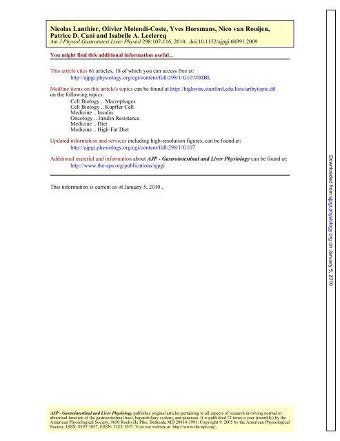
Patrice D. Cani and Isabelle A. Leclercq Nicolas Lanthier, Olivier ...
Patrice D. Cani and Isabelle A. Leclercq Nicolas Lanthier, Olivier ...
Patrice D. Cani and Isabelle A. Leclercq Nicolas Lanthier, Olivier ...

Create successful ePaper yourself
Turn your PDF publications into a flip-book with our unique Google optimized e-Paper software.
<strong>Nicolas</strong> <strong>Lanthier</strong>, <strong>Olivier</strong> Molendi-Coste, Yves Horsmans, Nico van Rooijen,<br />
<strong>Patrice</strong> D. <strong>Cani</strong> <strong>and</strong> <strong>Isabelle</strong> A. <strong>Leclercq</strong><br />
Am J Physiol Gastrointest Liver Physiol 298:107-116, 2010. doi:10.1152/ajpgi.00391.2009<br />
You might find this additional information useful...<br />
This article cites 61 articles, 18 of which you can access free at:<br />
http://ajpgi.physiology.org/cgi/content/full/298/1/G107#BIBL<br />
Medline items on this article's topics can be found at http://highwire.stanford.edu/lists/artbytopic.dtl<br />
on the following topics:<br />
Cell Biology .. Macrophages<br />
Cell Biology .. Kupffer Cell<br />
Medicine .. Insulin<br />
Oncology .. Insulin Resistance<br />
Medicine .. Diet<br />
Medicine .. High-Fat Diet<br />
Updated information <strong>and</strong> services including high-resolution figures, can be found at:<br />
http://ajpgi.physiology.org/cgi/content/full/298/1/G107<br />
Additional material <strong>and</strong> information about AJP - Gastrointestinal <strong>and</strong> Liver Physiology can be found at:<br />
http://www.the-aps.org/publications/ajpgi<br />
This information is current as of January 5, 2010 .<br />
AJP - Gastrointestinal <strong>and</strong> Liver Physiology publishes original articles pertaining to all aspects of research involving normal or<br />
abnormal function of the gastrointestinal tract, hepatobiliary system, <strong>and</strong> pancreas. It is published 12 times a year (monthly) by the<br />
American Physiological Society, 9650 Rockville Pike, Bethesda MD 20814-3991. Copyright © 2005 by the American Physiological<br />
Society. ISSN: 0193-1857, ESSN: 1522-1547. Visit our website at http://www.the-aps.org/.<br />
Downloaded from<br />
ajpgi.physiology.org<br />
on January 5, 2010
Kupffer cell activation is a causal factor for hepatic insulin resistance<br />
<strong>Nicolas</strong> <strong>Lanthier</strong>, 1 <strong>Olivier</strong> Molendi-Coste, 1 Yves Horsmans, 1 Nico van Rooijen, 2 <strong>Patrice</strong> D. <strong>Cani</strong>, 3<br />
<strong>and</strong> <strong>Isabelle</strong> A. <strong>Leclercq</strong> 1<br />
1 Laboratory of Gastroenterology, Université catholique de Louvain, Brussels, Belgium; 2 Department of Molecular Cell<br />
Biology, Vrije Universiteit Medical Center, Amsterdam, The Netherl<strong>and</strong>s; <strong>and</strong> 3 Louvain Drug Research Institute, Unit<br />
of Pharmacokinetics, Metabolism, Nutrition <strong>and</strong> Toxicology, Université catholique de Louvain, Brussels, Belgium<br />
Submitted 23 September 2009; accepted in final form 22 October 2009<br />
<strong>Lanthier</strong> N, Molendi-Coste O, Horsmans Y, van Rooijen N, <strong>Cani</strong> PD,<br />
<strong>Leclercq</strong> IA. Kupffer cell activation is a causal factor for hepatic insulin<br />
resistance. Am J Physiol Gastrointest Liver Physiol 298: G107–G116, 2010.<br />
First published October 29, 2009; doi:10.1152/ajpgi.00391.2009.—Recruited<br />
adipose tissue macrophages contribute to chronic <strong>and</strong> lowgrade<br />
inflammation causing insulin resistance in obesity. Similarly,<br />
we hypothesized here that Kupffer cells, the hepatic resident macrophages,<br />
play a pathogenic role in hepatic insulin resistance induced by<br />
a high-fat diet. Mice were fed a normal diet or high-fat diet for 3 days.<br />
Kupffer cell activation was evaluated by immunohistochemistry <strong>and</strong><br />
quantitative RT-PCR. Insulin sensitivity was assessed in vivo by<br />
hyperinsulinemic-euglycemic clamp <strong>and</strong> insulin-activated signaling<br />
was investigated by Western blot. Liposome-encapsulated clodronate<br />
was injected intravenously to deplete macrophages prior to a shortterm<br />
exposure to high-fat diet. Here, we characterized a short-term<br />
high-fat diet model in mice <strong>and</strong> demonstrated early hepatic insulin<br />
resistance <strong>and</strong> steatosis concurrent with Kupffer cell activation. We<br />
demonstrated that selective Kupffer cell depletion obtained by intravenous<br />
clodronate, without affecting adipose tissue macrophages, was<br />
sufficient to enhance insulin-dependent insulin signaling <strong>and</strong> significantly<br />
improve hepatic insulin sensitivity in vivo in this short-term<br />
high-fat diet model. Our study clearly shows that hepatic macrophage<br />
response participates to the onset of high-fat diet-induced hepatic<br />
insulin resistance <strong>and</strong> may therefore represent an attractive target for<br />
prevention <strong>and</strong> treatment of diet- <strong>and</strong> obesity-induced insulin resistance.<br />
macrophage; liver; steatosis; liposomes; high-fat diet<br />
NONALCOHOLIC FATTY LIVER DISEASE (NAFLD) becomes in our<br />
sedentary societies the most common cause of chronic liver<br />
disease (1). It is associated with obesity <strong>and</strong> insulin resistance<br />
<strong>and</strong> is now considered as the hepatic manifestation of the<br />
metabolic syndrome (50, 51). Fat accumulation in the liver is<br />
associated with higher cardiovascular mortality, in addition to<br />
liver-related cause of death, such as progression to nonalcoholic<br />
steatohepatitis <strong>and</strong> fibrosis or sensitization to other hepatic<br />
injuries (1, 49). Hepatic insulin resistance refers to<br />
inadequate insulin-dependent suppression of hepatic glucose<br />
production (HGP) <strong>and</strong> plays a pivotal role in the development<br />
of hyperglycemia <strong>and</strong> evolution toward type 2 diabetes (17,<br />
30). Hepatic insulin resistance is the hallmark of NAFLD (31,<br />
46). Hepatic lipid overload, lipid metabolites, <strong>and</strong> proinflammatory<br />
mediators have all been shown to alter hepatic insulin<br />
sensitivity in vivo <strong>and</strong> in vitro (38, 47). Knowing the role of<br />
diabetes in the progression of hepatic inflammation <strong>and</strong> fibrosis<br />
(7, 14, 18) <strong>and</strong> the potential benefits of improving insulin<br />
resistance in decreasing risk of cardiovascular related death,<br />
Address for reprint requests <strong>and</strong> other correspondence: I. <strong>Leclercq</strong>, Laboratoire<br />
de Gastro-entérologie, Université catholique de Louvain, GAEN 53/79, Ave.<br />
Mounier, 53, B-1200 Brussels, Belgium (e-mail: isabelle.leclercq@uclouvain.be).<br />
Am J Physiol Gastrointest Liver Physiol 298: G107–G116, 2010.<br />
First published October 29, 2009; doi:10.1152/ajpgi.00391.2009.<br />
targeting hepatic insulin resistance may represent a new therapeutic<br />
approach for NAFLD <strong>and</strong> related diseases.<br />
Several studies have linked insulin resistance to systemic<br />
inflammation (11, 45). Anti-inflammatory aspirin is long<br />
known to lower blood glucose (13). Mice lacking the proinflammatory<br />
cytokine TNF are protected from insulin resistance<br />
induced by chronic high-fat feeding (10). Conversely, activation<br />
of proinflammatory JNK or IB kinase- pathways have<br />
been found to promote obesity-induced insulin resistance (6,<br />
12). Fat-overloaded adipocyte, because it produces numerous<br />
chemokines <strong>and</strong> (adipo)cytokines affecting immunity <strong>and</strong> inflammation,<br />
was seen as an important player in this process.<br />
More recently, attention has been brought to bone marrowderived<br />
macrophages recruited to obese visceral adipose tissue<br />
(28, 56, 59) as a source for inflammatory factors altering<br />
metabolic functions of adipocytes <strong>and</strong> causing insulin resistance<br />
both locally <strong>and</strong> in distant organs. Accordingly, F4/<br />
80 CD11b CD11c M1 adipose tissue macrophages (ATM)<br />
increase in number in obese adipose tissue (28, 34) <strong>and</strong><br />
suppression of such inflammation improves insulin sensitivity<br />
<strong>and</strong> dissociates adiposity from insulin resistance (15, 37, 42,<br />
48, 55). Genetic manipulations of IB kinase- or JNK signaling<br />
in myeloid lineage affect systemic metabolic regulations<br />
(48, 58, 61). Collectively, data available to date show that,<br />
without a myeloid cell-driven inflammatory component, obesity<br />
itself does not cause insulin resistance.<br />
Recent data suggest that hepatic steatosis resulting from<br />
high-fat feeding is associated with activation of Kupffer cells<br />
(KC), the hepatic resident macrophages (2, 57). A causal link<br />
between KC activation <strong>and</strong> hepatic insulin resistance has been<br />
suggested (33, 44) but never convincingly experimentally addressed.<br />
The aim of our study is to characterize hepatic inflammation<br />
induced by high-fat feeding <strong>and</strong> determine whether such inflammation<br />
participate to the initiation of hepatic insulin resistance.<br />
We used a short-term high-fat diet (HFD) model in mice<br />
<strong>and</strong> demonstrated early hepatic macrophage activation associated<br />
with hepatic insulin resistance <strong>and</strong> steatosis. We addressed<br />
the role of activated resident macrophages in the liver (KC) on<br />
hepatic insulin sensitivity using experimental macrophage depletion<br />
obtained by intravenous injections of liposome-encapsulated<br />
clodronate (53, 60).<br />
MATERIALS AND METHODS<br />
Animals, diets, <strong>and</strong> treatment. Five-week-old male C57BL/6J (Janvier,<br />
Le Genest-Saint-Isle, France) mice were fed for 3 days a HFD in<br />
which 60% of calories are derived from fat (D12492 from Research<br />
Diets, New Brunswick) or a st<strong>and</strong>ard rodent chow (10% of calories<br />
from fat, Carfil Quality, Oud-Turnhout, Belgium) referred to as<br />
normal diet (ND). The animals were h<strong>and</strong>led according to the guide-<br />
http://www.ajpgi.org 0193-1857/10 $8.00 Copyright © 2010 the American Physiological Society<br />
G107<br />
Downloaded from<br />
ajpgi.physiology.org<br />
on January 5, 2010
G108 KUPFFER CELLS AND INSULIN RESISTANCE<br />
lines for humane care for laboratory animals established by the<br />
Université catholique de Louvain in accordance with European Union<br />
Regulation. The study protocol was approved by the local ethics<br />
committee. At the time of euthanasia, under anesthesia, mice received<br />
5 U insulin (Actrapid, Novo Nordisk, Bagsvaerd, Denmark) or an<br />
equal volume of PBS into the portal vein to analyze the intrahepatic<br />
response to insulin. The liver was removed 1 min after the injection,<br />
<strong>and</strong> epididymal <strong>and</strong> subcutaneous adipose tissues <strong>and</strong> right quadriceps<br />
were dissected <strong>and</strong> weighed. Portions of tissue were immersed in<br />
formalin 4% or frozen in optimal cutting temperature medium for<br />
histological studies; the remaining tissue was snap frozen in liquid<br />
nitrogen <strong>and</strong> kept at 80°C until analyses. To deplete macrophages,<br />
100 l/10 g body wt of 1 mg/ml liposome-encapsulated clodronate, or<br />
liposome-encapsulated PBS, or an equal volume of NaCl 0.9% were<br />
injected to mice intravenously (iv) via the retroorbital plexus, 24 h<br />
prior to the administration of the HFD. In a parallel experiment, mice<br />
received NaCl 0.9%, liposome-encapsulated PBS, or liposome-encapsulated<br />
clodronate by intraperitoneal injection. Clodronate (a gift from<br />
Roche Diagnostics, Mannheim, Germany) was encapsulated into<br />
liposomes as described previously (53). Clodronate <strong>and</strong> liposomes by<br />
themselves are not toxic. Liposomes, taken up by phagocytic cells<br />
(macrophages), are disrupted by lysosomal enzymes. This provokes<br />
the intracellular release of clodronate, causing apoptotic cell death of<br />
macrophage (54). Analyses were performed on eight animals per<br />
group.<br />
Hyperinsulinemic-euglycemic clamp study. Femoral iv catheter was<br />
implanted 7 days before clamp studies (4). HGP <strong>and</strong> whole body<br />
glucose turnover (TO) were evaluated by the hyperinsulinemic-euglycemic<br />
clamp technique (8, 16), conducted at 11:00 AM after a 5-h fast<br />
with a continuous infusion of 2.5 mUkg 1 min 1 human insulin<br />
(Actrapid) coupled with [3- 3 H]glucose (0.33 Ci, PerkinElmer, Boston,<br />
MA). Tail blood glycemia was obtained at time 0 <strong>and</strong> every 10<br />
min thereafter to adjust a variable 20% glucose infusion rate (GIR) to<br />
maintain euglycemia (90–110 mg/dl). When steady-state was obtained<br />
(90 min), blood samples (5 l) were collected every 10 min<br />
for 1 h for estimation of plasma glucose specific activity. At steady<br />
state, the rate of glucose appearance (Ra) measured as <br />
[3- 3 H]GIR/glucose specific activity equals the rate of peripheral<br />
glucose disposal (Rd) or TO (4, 5). In basal conditions, the rate of<br />
HGP (mg kg 1 min 1 ) equals Ra. In insulin stimulated conditions<br />
(2,5 mU kg 1 min 1 ), HGP was obtained by subtracting the GIR<br />
(mg kg 1 min 1 ) to the Rd previously calculated: HGP (Ra) <br />
Rd GIR. Analyses were performed on five to eight animals per<br />
group.<br />
Biochemical analyses. Blood glucose concentrations were determined<br />
with a glucose monitor (Accu-chek, Aviva, Roche) <strong>and</strong> plasma<br />
insulin levels by using an enzyme-linked immunoassay (Mercodia, Uppsala,<br />
Sweden). Total liver lipids were extracted with methanol <strong>and</strong><br />
chloroform <strong>and</strong> quantified by the vanillin-phosphoric acid reaction (22).<br />
Histology, immunohistochemistry, <strong>and</strong> immunofluorescence. Hematoxylin<br />
<strong>and</strong> eosin staining were used for routine histological<br />
evaluation. Immunohistochemical detection of F4/80 was performed<br />
on paraffin-embedded sections treated with proteinase K by using a<br />
primary rat anti-mouse F4/80 monoclonal Ab (1/200, Serotec, Oxford,<br />
UK), a rabbit anti-rat immunoglobulin (1/200, Dako, Glostrup, Denmark),<br />
<strong>and</strong> then a goat anti-rabbit streptavidin horseradish peroxidaseconjugated<br />
Ab (En Vision, Dako). The peroxidase activity was revealed<br />
with diaminobenzidine <strong>and</strong> slides were counterstained with<br />
hematoxylin. Morphometrical quantification of the surface stained<br />
was performed on microphotographs (20) taken from two r<strong>and</strong>om<br />
sections per liver (n 6 per group) by use of the Axiovision 4.6<br />
image analysis software (Carl Zeiss, Goentingen, Germany) <strong>and</strong><br />
expressed as percentage of total area as previously described (52).<br />
CD68, C11b, <strong>and</strong> CD11c-positive cells were detected on cryosections<br />
(5 m thick) by using a monoclonal rat anti-mouse CD68 (1/300,<br />
Serotec), a monoclonal rat anti-mouse CD11b (1/100, Developmental<br />
Studies Hybridoma Bank, Iowa City, IA) or a monoclonal hamster<br />
anti-mouse CD11c Ab (1/50, Serotec). For double immunofluorescence,<br />
the same primary Abs were used as well as a polyclonal F4/80<br />
rabbit anti-mouse Ab (1/500, Santa Cruz, CA) when indicated. Secondary<br />
Abs were anti-rat/AlexaFluor 594, anti-rabbit/AlexaFluor 488,<br />
or anti-hamster/AlexaFluor 488 (dilution 1/1,000, Invitrogen, Merelbeke,<br />
Belgium). Hoechst (dilution 1/10.000) was used to reveal the<br />
nuclei.<br />
Protein studies: Western blotting. Liver <strong>and</strong> muscle homogenates<br />
were prepared as described elsewhere (23). Proteins (100 g) were<br />
assayed by Western blotting using the Abs <strong>and</strong> conditions listed in<br />
Supplemental Table S1. The immunoreactivity was detected with a<br />
horseradish peroxidase-conjugated secondary Ab (1/10,000) <strong>and</strong><br />
chemiluminescence (PerkinElmer). One membrane was sequentially<br />
probed with the Ab against the total protein, the phosphoprotein, <strong>and</strong><br />
HSP90 to control for protein loading. The quantification of immunereactive<br />
proteins was obtained by densitometry using the Gel DocTM<br />
XR System 170–8,170 device <strong>and</strong> software (Bio-Rad). The levels of<br />
immunoreactivity relative to the invariant control are reported as<br />
arbitrary densitometry units.<br />
RNA extraction, reverse transcription, <strong>and</strong> quantitative PCR. Total<br />
RNA was extracted from frozen liver, subcutaneous or epididymal fat<br />
samples by using TRIpure Isolation Reagent (Roche Diagnostics<br />
Belgium, Vilvoorde, Belgium). cDNA synthesis <strong>and</strong> real-time PCR<br />
analysis were carried out as previously described (23). Primer pairs<br />
for transcripts of interest were designed using the Primer Express<br />
software (Applied Biosystems, Lennik, Belgium) <strong>and</strong> listed in Supplemental<br />
Table S2. RPL19 mRNA was chosen as an invariant<br />
st<strong>and</strong>ard. All experimental tissues <strong>and</strong> st<strong>and</strong>ard curve samples were<br />
run in duplicate in a 96-well reaction plate (MicroAmp Optical,<br />
Applied Biosystems). Results are expressed as fold expression relative<br />
to expression in the control group following the Ct method (26).<br />
Statistical analysis. All the data are presented as means SD.<br />
Statistical analysis was performed by ANOVA for multiple groups<br />
comparison <strong>and</strong> Student’s t-test for comparing two groups. When<br />
P was less than 0.05, differences were considered significant.<br />
RESULTS<br />
AJP-Gastrointest Liver Physiol • VOL 298 • JANUARY 2010 • www.ajpgi.org<br />
Short-term high-fat feeding induces obesity, adiposity, <strong>and</strong><br />
liver steatosis. Compared with ND, a 3-day course of HFD<br />
induced body weight gain, increased adiposity, <strong>and</strong> increased<br />
relative liver weight (Table 1). On histological examination,<br />
lipid vacuoles were seen predominantly in the intermediate <strong>and</strong><br />
periportal zones (Fig. 1B). Steatosis in HFD-fed animals was<br />
confirmed by a fourfold increase in hepatic lipid content (Table 1).<br />
Short-term high-fat fed mice are insulin resistant, with the<br />
evidence of a specific hepatic insulin resistance. HFD-fed<br />
animals had increased fasting glucose levels with normal insulinemia<br />
(Table 1). Insulin sensitivity, assessed by the gold st<strong>and</strong>ard<br />
hyperinsulinemic euglycemic clamp technique, was reduced<br />
Table 1. Morphological, hepatic, <strong>and</strong> metabolic changes<br />
induced by a 3-day HFD<br />
ND HFD<br />
Body weight gain, g<br />
Epididymal adipose tissue weight/ body<br />
0.30.5 3.80.9*<br />
weight, % 1.00.2 1.30.1*<br />
Liver weight/body weight, % 5.60.3 6.40.5†<br />
Hepatic lipid content, mg/100 mg liver 4.50.5 19.24.0*<br />
Blood glucose, mg/dl 136.612.3 164.18.4†<br />
Serum insulin, g/l 1.20.9 1.30.3<br />
Results are expressed as mean SD (n 8 per group). Mice were fed a<br />
normal diet (ND) or a high-fat diet for 3 days (HFD). *P 0.001 <strong>and</strong> †P <br />
0.01 in HFD vs. ND.<br />
Downloaded from<br />
ajpgi.physiology.org on January 5, 2010
in the HFD group as demonstrated by a lower GIR required to<br />
maintain euglycemia (Fig. 1C). This was owed to a marked<br />
impairment of insulin-dependent inhibition of HGP, a parameter<br />
completely inhibited in controls. In contrast, whole body glucose<br />
uptake (or TO) was not different in HFD <strong>and</strong> ND (Fig. 1C).<br />
In the liver, HFD decreased the expression of insulin receptor<br />
(IR) but did not induce significant changes in Akt protein<br />
levels (Fig. 1D). Insulin-stimulated phosphorylation of IR <strong>and</strong><br />
Akt (on Ser473 <strong>and</strong> Thr308) was significantly lower in HFD<br />
compared with ND livers (Fig. 1D). These changes were not<br />
KUPFFER CELLS AND INSULIN RESISTANCE<br />
AJP-Gastrointest Liver Physiol • VOL 298 • JANUARY 2010 • www.ajpgi.org<br />
G109<br />
Fig. 1. Short-term high-fat diet (HFD)-fed mice<br />
develop steatosis <strong>and</strong> hepatic insulin resistance.<br />
A <strong>and</strong> B: representative photomicrographs of hematoxylin<br />
<strong>and</strong> eosin-stained sections of livers from<br />
mice fed the normal diet (ND; A) <strong>and</strong> the HFD (B)<br />
for 3 days. Original magnification 20. PT, portal<br />
tract; CV, centrilobular vein. C: glucose infusion<br />
rate (GIR), glucose turnover (TO), <strong>and</strong> hepatic glucose<br />
production (HGP) during hyperinsulinemiceuglycemic<br />
clamps in mice fed the ND or the HFD<br />
for 3 days. Hepatic (D) <strong>and</strong> muscle protein expression<br />
(E) <strong>and</strong> phosphorylation of the insulin receptor<br />
(IR) <strong>and</strong> Akt evaluated by Western blot analysis in<br />
mice fed the ND or the HFD for 3 days, 1 min after<br />
the portal injection of insulin (ins) or PBS. Representative<br />
blots <strong>and</strong> graphs representing the ratio of the<br />
(insulin-stimulated phospho-) protein of interest on<br />
HSP90 as measured by densitometry analysis (n <br />
4/group). Data are expressed as means SD. *P <br />
0.05, **P 0.01, <strong>and</strong> ***P 0.001 in HFD vs. ND.<br />
observed in the muscle, where expression of IR, Akt <strong>and</strong><br />
insulin-stimulated p-IR <strong>and</strong> p-Akt were similar in HFD <strong>and</strong> ND<br />
groups (Fig. 1E).<br />
Collectively, those data demonstrate a marked hepatic insulin<br />
resistance but normal peripheral insulin sensitivity in mice<br />
fed the HFD for 3 days.<br />
HFD induces an early activation of hepatic <strong>and</strong> ATM.<br />
Compared with ND livers (Fig. 2A), enlarged F4/80-positive<br />
mature tissue resident macrophages were preferentially found<br />
in the vicinity of lipid-loaded hepatocytes in HFD livers (Fig.<br />
Downloaded from<br />
ajpgi.physiology.org<br />
on January 5, 2010
G110 KUPFFER CELLS AND INSULIN RESISTANCE<br />
Fig. 2. HFD activates Kupffer cells. Liver sections from mice fed the ND (A, D, G, I, L, O) <strong>and</strong> the HFD (B, E, H, J, M, P) immunoassayed with antibodies<br />
against F4/80 (A <strong>and</strong> B), CD68 (D <strong>and</strong> E), F4/80 <strong>and</strong> CD68 (G <strong>and</strong> H; arrows point to double-positive cells), CD11b (I <strong>and</strong> J), CD11c (L <strong>and</strong> M), F4/80 <strong>and</strong><br />
CD11c (O <strong>and</strong> P). CD11b staining revealed faintly stained, elongated cells (arrows) reminiscent of F4/80 cells <strong>and</strong> strongly positive round cells (arrowheads;<br />
I <strong>and</strong> J). CD11c cells were rare <strong>and</strong> predominantly located around the portal tract (L <strong>and</strong> M, arrows). Some F4/80 (red) <strong>and</strong> CD11c (green) double-positive<br />
cells (arrowheads) were found only in HFD livers (P). Original magnification 40. mRNA for macrophage markers assessed by real-time PCR (C, F, K, N) in<br />
ND <strong>and</strong> HFD livers (n 8 per group). Data are expressed as relative means SD to ND values arbitrarily set at 1. ***P 0.001 in HFD vs. ND.<br />
2B). This was confirmed by morphometrical analysis (F4/80stained<br />
surface of 2.31 0.85% in HFD livers vs. 0.85 <br />
0.21% in controls, P 0.005) <strong>and</strong> by the upregulation of F4/80<br />
mRNA (Fig. 2C). An increased number of cells expressing the<br />
AJP-Gastrointest Liver Physiol • VOL 298 • JANUARY 2010 • www.ajpgi.org<br />
CD68/macrosialin, a transmembrane protein expressed by activated<br />
tissue macrophages (39), was found in the HFD livers<br />
compared with controls (Fig. 2, D <strong>and</strong> E) <strong>and</strong> confirmed by<br />
upregulation of hepatic CD68 mRNA (Fig. 2F). Immunofluo-<br />
Downloaded from<br />
ajpgi.physiology.org<br />
on January 5, 2010
Table 2. Effect of clodronate on hepatic inflammationassociated<br />
markers in HFD-fed mice<br />
ND HFD CLO HFD<br />
TLR2 mRNA 10.10 1.720.48† 1.270.32<br />
TLR4 mRNA 10.17 1.470.21* 0.650.11‡<br />
CD14 mRNA 10.21 1.610.47† 0.430.26‡<br />
TNF mRNA 10.39 2.080.55* 0.760.51§<br />
mRNA expression of hepatic inflammatory genes assessed by RT-PCR, with<br />
expression in normal diet (ND) arbitrarily set at 1. Data are expressed as<br />
mean SD for n 8 per group. *P 0.001 <strong>and</strong> †P 0.01 in HFD group<br />
vs. ND. ‡P 0.001 <strong>and</strong> §P 0.01 in clodronate-treated high-fat fed mice<br />
(CLO HFD) vs. HFD.<br />
rescence double staining revealed that, in control livers, a small<br />
proportion of F4/80-positive macrophages also expressed<br />
CD68 (Fig. 2G). Under HFD, the proportion of F4/80 to CD68<br />
double-positive cells increased (Fig. 2H), supporting that hepatic<br />
macrophages are activated. CD11b/MAC-1 is classically<br />
expressed by granulocytes, monocytes, NK cells, <strong>and</strong> tissue<br />
macrophages (24). Two distinct liver populations of CD11b <br />
cells were detected: elongated <strong>and</strong> faintly stained cells morphologically<br />
<strong>and</strong> topologically reminiscent of tissue macrophages,<br />
<strong>and</strong> a population of round cells compatible with<br />
neutrophils or monocytes (Fig. 2I, arrows <strong>and</strong> arrowheads,<br />
respectively). Under HFD, the population of CD11b-positive<br />
cells appeared increased (Fig. 2J). The mRNA analysis showed<br />
an approximately fourfold upregulation of CD11b (Fig. 2K).<br />
CD11c is an integrin mainly expressed by dendritic cells <strong>and</strong><br />
KUPFFER CELLS AND INSULIN RESISTANCE<br />
by some macrophages (27). CD11c-positive cells were rare <strong>and</strong><br />
preferentially located around the portal tract (Fig. 2L) inthe<br />
normal liver. Upon HFD, slightly more numerous CD11cpositive<br />
cells, remaining located in the periportal zone (Fig.<br />
2M), were seen together with an approximately twofold increased<br />
in CD11c mRNA (Fig. 2N). Double immunostaining<br />
demonstrated that only a minority of F4/80-positive macrophages<br />
were CD11c positive in HFD livers (Fig. 2P). These<br />
were apparently absent in controls (Fig. 2O). In addition to<br />
phenotypic changes in hepatic macrophages, HFD also significantly<br />
induced TNF, TLR2, TLR4, <strong>and</strong> CD14 mRNA expression,<br />
considered as markers of inflammation (Table 2).<br />
In the adipose tissue, whether epididymal or subcutaneous,<br />
no apparent inflammation was detected by (immuno)histochemical<br />
techniques (Fig. 3, A <strong>and</strong> B), contrasting with adipose<br />
tissue inflammation classically described in chronic HFD <strong>and</strong><br />
obesity models (28, 56, 59). Nevertheless, a 3-day HFD increased<br />
F4/80, CD68, <strong>and</strong> CD11b mRNA levels by twofold<br />
(Fig. 3, C–E) in the epididymal adipose tissue, whereas CD11c<br />
mRNA remained unchanged (Fig. 3F). Those changes were not<br />
seen in the subcutaneous fat pads (not shown). Our data thus<br />
support the activation of ATM in intra-abdominal depots in our<br />
model despite the apparent absence of macrophage recruitment.<br />
Selective KC depletion ameliorates hepatic insulin sensitivity.<br />
The injection of PBS-loaded liposomes to HFD-fed mice had<br />
no effect on macrophage populations in the liver (Fig. 4, H–J). By<br />
contrast, the injection of liposome-encapsulated clodronate<br />
Fig. 3. Effects of a short-term HFD on adipose tissue expression of inflammatory markers. Immunohistochemical detection of the macrophage-specific antigen<br />
F4/80 in epididymal adipose tissue from mice fed the ND (A) or the HFD (B) for 3 days. Original magnification 40. C–F: epididymal adipose tissue mRNA<br />
expression of inflammatory markers. Results (n 8 per group) are expressed as means SD of fold expression compared with values in untreated ND animals<br />
arbitrarily set at 1. **P 0.01 in HFD vs. ND.<br />
AJP-Gastrointest Liver Physiol • VOL 298 • JANUARY 2010 • www.ajpgi.org<br />
G111<br />
Downloaded from<br />
ajpgi.physiology.org<br />
on January 5, 2010
G112 KUPFFER CELLS AND INSULIN RESISTANCE<br />
Fig. 4. Injection of liposome-encapsulated clodronate depletes Kupffer cells without affecting adipose tissue macrophages. F4/80 (A <strong>and</strong> D), CD68 (B <strong>and</strong> E),<br />
<strong>and</strong> CD11b (C <strong>and</strong> F) immunostaining on liver sections from mice fed the HFD with intravenous (iv) injection of PBS or clodronate loaded liposomes (PBS <br />
HFD or CLO HFD, respectively). Clodronate injection depleted F4/80, CD68, <strong>and</strong> CD11b (arrows)-positive cells with KC morphology (D–F); meanwhile a<br />
marginal population of round CD11b-positive cells (arrowheads) increased in the liver lobule (F). Immunohistochemistry on paraffin sections (F4/80) or frozen<br />
sections (CD68 <strong>and</strong> CD11b), brown staining. Original magnification 40. Effects of iv liposome-encapsulated PBS (PBS HFD) or clodronate injection<br />
(CLO HFD) on liver (H–J) <strong>and</strong> epididymal adipose tissue (K) mRNA expression of macrophage markers. Results (n 8 per group) are expressed as means <br />
SD of fold expression compared with untreated ND animal values arbitrarily set at 1. ***P 0.001.<br />
markedly <strong>and</strong> significantly depleted the liver of its F4/80,<br />
CD68, <strong>and</strong> CD11b-positive macrophages (Fig. 4, A–F). This<br />
was also assessed by a blunting in F4/80 <strong>and</strong> CD68 transcript<br />
expression (Fig. 4, H–I) whereas CD11b mRNA levels re-<br />
AJP-Gastrointest Liver Physiol • VOL 298 • JANUARY 2010 • www.ajpgi.org<br />
mained stable (Fig. 4J), reflecting the increased in nonmacrophage<br />
round CD11b-positive cells (Fig. 4F, arrowheads),<br />
consistent with the recruitment of blood monocyte precursors<br />
described by others (60). Hepatic CD11c mRNA remained<br />
Downloaded from<br />
ajpgi.physiology.org<br />
on January 5, 2010
stable in the clodronate-treated HFD group (not shown). Paralleling<br />
macrophage depletion, clodronate injection was associated<br />
with a significant decrease in hepatic TNF, TLR4, <strong>and</strong><br />
CD14 mRNA levels (Table 2).<br />
PCR analysis demonstrated that clodronate liposomes did<br />
not affect inflammatory cell activity in the adipose tissue (Fig.<br />
4K). Indeed, such liposomes, being unable to cross vascular<br />
barriers, are readily taken up by phagocytic cells in the reticuloendothelial<br />
system (53).<br />
Fasting blood glucose, serum insulin levels, hepatic lipid<br />
content <strong>and</strong> body weight gain were not different whether HFD<br />
animals received or not PBS or clodronate whereas epididymal<br />
fat weight was lower in the clodronate treated group compared<br />
with others HFD groups (Table 3).<br />
During clamp experiments, the GIR needed to maintain<br />
euglycemia significantly increased <strong>and</strong> insulin-dependent inhibition<br />
of HGP was significantly ameliorated in clodronatetreated<br />
animals compared with both PBS-treated <strong>and</strong> untreated<br />
HFD animals (Fig. 5A). At the level of insulin signaling, the<br />
specific depletion of hepatic macrophages did not improve IR<br />
phosphorylation whereas it significantly increased the phosphorylation<br />
of Akt in HFD livers (Fig. 5B).<br />
DISCUSSION<br />
In this study, we characterized a short-term HFD regimen in<br />
mice <strong>and</strong> used simultaneous ablation of KC through injection<br />
of liposome-encapsulated clodronate to demonstrate the role of<br />
hepatic macrophages in modulating hepatic insulin sensitivity.<br />
Consistent with previous reports in rats (17, 43), feeding<br />
mice a HFD for 3 days provoked a significant body weight<br />
gain, steatosis, <strong>and</strong> selective hepatic insulin resistance, proved<br />
in vivo by hyperinsulinemic-euglycemic clamp technique <strong>and</strong><br />
confirmed by defects in hepatic insulin signaling. Conversely,<br />
whole body glucose uptake as well as insulin signaling in<br />
skeletal muscle were unaltered, supporting that peripheral<br />
tissues remained insulin sensitive. In the liver, steatosis <strong>and</strong><br />
insulin resistance were associated with local inflammatory<br />
changes. In control livers, as previously reported by others<br />
(27), few F4/80-positive hepatic macrophages coexpressed<br />
CD68 but all were CD11c negative. After 3 days of HFD,<br />
F4/80-positive cells appeared enlarged <strong>and</strong> the proportion of<br />
F4/80 <strong>and</strong> CD68 double positive cells increased together with<br />
upregulation of mRNA expression of macrophage-specific<br />
Table 3. Effect of liposome-encapsulated clodronate on<br />
morphological, hepatic, <strong>and</strong> metabolic changes induced<br />
by a 3-day HFD<br />
HFD PBS HFD CLO HFD<br />
Body weight gain, g 2.40.9 3.40.7 2.11.2<br />
Epididymal adipose tissue<br />
weight/body weight, % 1.30.1 1.30.2 1.00.1*<br />
Liver weight/ body weight, % 5.70.4 5.80.2 6.10.5<br />
Hepatic lipid content, mg/100<br />
mg liver 9.32.8 11.74.7 11.64.0<br />
Blood glucose, mg/dl 133.821.1 134.915.9 138.117.6<br />
Serum insulin, g/l 1.41.5 1.21.2 1.50.8<br />
Results are expressed as mean SD (n 6–8 per group) for mice fed a<br />
HFD for 3 days preceded by an intravenous injection of liposome-encapsulated<br />
PBS (PBS HFD) or clodronate (CLO HFD). *P 0.001 in CLO HFD<br />
vs. HFD or PBS HFD.<br />
KUPFFER CELLS AND INSULIN RESISTANCE<br />
AJP-Gastrointest Liver Physiol • VOL 298 • JANUARY 2010 • www.ajpgi.org<br />
G113<br />
markers (TLR4, CD14, <strong>and</strong> TNF), whereas markers of alternative<br />
activation (35) remained unchanged (not shown). This<br />
supports the proinflammatory activation of resident hepatic<br />
macrophages (6, 57).<br />
In contrast with long-term exposure to high-fat diet in which<br />
hepatic steatosis is variable, if not absent (20, 21, 32), shortterm<br />
high-fat diet yields reproducible steatosis (19, 43). Moreover,<br />
whereas KC activation is obvious in our 3-day model as<br />
also reported in human nonalcoholic steatohepatitis (25, 29, 36,<br />
40), this is no longer found in the longer term regimen in<br />
rodents (9, 56, 59). By contrast, peripheral insulin resistance<br />
<strong>and</strong> recruitment of inflammatory cell in the adipose tissue, the<br />
hallmark of chronic obesity (28, 59), failed to be observed in<br />
our model. This implies that, in response to high-fat feeding,<br />
metabolic <strong>and</strong> inflammatory alterations occur first in the liver<br />
<strong>and</strong> secondarily in muscles <strong>and</strong> adipose tissues (peripheral<br />
tissues). We demonstrated here that selective ablation of KC<br />
significantly improved HFD-induced hepatic insulin resistance<br />
<strong>and</strong> alterations of hepatic insulin signaling, confirming a major<br />
role of KC in the initiating mechanism of HFD-induced hepatic<br />
insulin resistance, besides or irrespectively of inflammatory<br />
changes occurring in the adipose tissue.<br />
Our results contrast with a recent publication (9) showing<br />
that, in mice exposed to a HFD for 16 wk, a single intraperitoneal<br />
injection of liposome-encapsulated clodronate decreased<br />
hepatic insulin sensitivity. Besides depleting the liver<br />
of F4/80-positive cells, intraperitoneal clodronate also induced<br />
significantly F4/80 mRNA in the adipose tissue (9). Such<br />
adipose tissue inflammation might be a confounding factor. We<br />
also performed intraperitoneal injections of clodronate in our<br />
short-term model. Contrasting with the selective depletion of<br />
KC via intravenous injections (liposomes do not cross vascular<br />
barriers), intraperitoneal clodronate affected ATM, as also<br />
reported by others (3), <strong>and</strong> upregulated fat MCP-1 <strong>and</strong> TNF<br />
implicated in macrophage recruitment (Supplemental Figure<br />
S1). Therefore, the significant upregulation of F4/80 in an<br />
already inflamed adipose tissue under chronic high-fat feeding<br />
reported by Clementi et al. (9) is consistent with a recruitment<br />
of macrophages following the clodronate-induced depletion.<br />
The effect of selective KC depletion on hepatic insulin sensitivity<br />
in a long-term HFD model remains to be evaluated.<br />
Tissue fat accumulation is a cause of insulin resistance (31,<br />
47). A dissociation between adiposity induced by high-fat<br />
feeding <strong>and</strong> insulin sensitivity, however, occurs when inflammatory<br />
pathways are inhibited (42, 48). Similarly, hepatic<br />
macrophage depletion prevented hepatic insulin resistance <strong>and</strong><br />
alteration of hepatic insulin signaling without reducing hepatic<br />
steatosis elicited by high-fat feeding. Thus KC depletion also<br />
dissociates fat accumulation <strong>and</strong> hepatic insulin resistance.<br />
Therefore, hepatic accumulation of lipids or lipid metabolites<br />
known to alter insulin signaling in hepatocyte cultures (41)<br />
cannot be the sole cause of insulin resistance in vivo. Our<br />
results support that signals from local inflammatory cells acting<br />
downstream of IR concur to this effect, whether acting independently<br />
or amplifying those produced by the fat-loaded<br />
hepatocytes.<br />
Intriguingly, KC depletion decreased adipose tissue<br />
weight while having no effect on body weight gain, liver<br />
weight, or degree of steatosis. Decreased adipose tissue<br />
could be the consequence of amelioration of hepatic inflammation<br />
<strong>and</strong>/or insulin sensitivity. Alternatively, although<br />
Downloaded from<br />
ajpgi.physiology.org<br />
on January 5, 2010
G114 KUPFFER CELLS AND INSULIN RESISTANCE<br />
Fig. 5. Injection of liposome-encapsulated clodronate<br />
in HFD mice improves hepatic insulin sensitivity <strong>and</strong><br />
signaling. A: GIR <strong>and</strong> HGP during hyperinsulinemiceuglycemic<br />
clamps. B: insulin signaling in liver of mice<br />
fed a 3-day HFD preceded or not by the iv injection of<br />
liposome-encapsulated PBS or clodronate (PBS HFD<br />
or CLO HFD, respectively). Graphs represent the<br />
quantification by densitometry analysis of the insulin<br />
receptor (IR) <strong>and</strong> Akt (ser473) phosphorylation upon<br />
insulin. Results are expressed as means SD (n 4<br />
per group). *P 0.05, **P 0.01, <strong>and</strong> ***P 0.001.<br />
not associated with decreased inflammatory signals, decreased<br />
adiposity may possibly contribute to amelioration of<br />
hepatic insulin sensitivity. Such phenomenon might also<br />
participate in the amelioration of insulin resistance observed<br />
upon gadolinium chloride treatment, which prevented HFDinduced<br />
weight gain (33). Further experiments are needed to<br />
address those issues.<br />
Results presented here firmly establish that KC activation<br />
is a key mechanism in the induction of hepatic insulin<br />
resistance that develops upon high-fat diet feeding. Targeting<br />
KC to block this deleterious activation might therefore<br />
represent an effective strategy to prevent the development of<br />
hepatic insulin resistance. Further assessment of the role of<br />
KC in chronic obesity-induced metabolic alteration <strong>and</strong><br />
deciphering the priming for KC activation, which could be<br />
triggered either by dietary factors or by products, whether<br />
cytokines or fatty acids, released by the gut, the adipose<br />
tissue or the fat-loaded hepatocytes, will be of major relevance<br />
for the underst<strong>and</strong>ing of diet-induced insulin resistance<br />
<strong>and</strong> for the development of new therapeutic approaches<br />
for the metabolic syndrome.<br />
ACKNOWLEDGMENTS<br />
AJP-Gastrointest Liver Physiol • VOL 298 • JANUARY 2010 • www.ajpgi.org<br />
The authors are grateful to Martine Petit, Valérie Lebrun, <strong>and</strong> Jorge<br />
Abarca-Quinones for expert technical assistance <strong>and</strong> to Prof. Hubert Piessevaux<br />
for help with statistical analysis. The monoclonal CD11b Ab developed<br />
by Timothy A. Springer was obtained from the Developmental Studies Hybridoma<br />
Bank developed under the auspices of the National Institute of Child<br />
Health <strong>and</strong> Human Development <strong>and</strong> maintained by the University of Iowa,<br />
Department of Biological Sciences, Iowa City, IA 52242.<br />
GRANTS<br />
This work was supported by Fonds National de la Recherche Scientifique<br />
(FNRS) grants (no. 3.4578.07 <strong>and</strong> F.45.08.08), la Direction de la Recherche<br />
Scientifique de la Communauté Française de Belgique (Action de Recherche<br />
Concertée grant, 05/10-328), University FSR grants 2007 <strong>and</strong> 2008 to I. A.<br />
<strong>Leclercq</strong>, grant “liver, virus <strong>and</strong> diabetes” from the Fondation St Luc to I. A.<br />
<strong>Leclercq</strong> <strong>and</strong> Y. Horsmans. I. A. <strong>Leclercq</strong> <strong>and</strong> P. D. <strong>Cani</strong> are FNRS research<br />
associates.<br />
DISCLOSURES<br />
The authors have no conflict of interest in relation to this work to disclose.<br />
The foundation Clodronate Liposomes (N. van Rooijen) is a nonprofit foundation<br />
related to the Department of Molecular Cell Biology of the Free<br />
University in Amsterdam, The Netherl<strong>and</strong>s.<br />
Downloaded from<br />
ajpgi.physiology.org<br />
on January 5, 2010
REFERENCES<br />
1. Adams LA, Lymp JF, St Sauver J, S<strong>and</strong>erson SO, Lindor KD,<br />
Feldstein A, Angulo P. The natural history of nonalcoholic fatty liver<br />
disease: a population-based cohort study. Gastroenterology 129: 113–121,<br />
2005.<br />
2. Baffy G. Kupffer cells in non-alcoholic fatty liver disease: the emerging<br />
view. J Hepatol 51: 212–223, 2009.<br />
3. Biewenga J, van der Ende MB, Krist LFG, Borst A, Ghufron M, Van<br />
Rooijen N. Macrophage depletion in the rat after intraperitoneal administration<br />
of liposome-encapsulated clodronate: depletion kinetics <strong>and</strong> accelerated<br />
repopulation of peritoneal <strong>and</strong> omental macrophages by administration<br />
of Freund’s adjuvant. Cell Tissue Res 280: 189–196, 1995.<br />
4. Burcelin R, Dolci W, Thorens B. Portal glucose infusion in the mouse<br />
induces hypoglycemia: evidence that the hepatoportal glucose sensor<br />
stimulates glucose utilization. Diabetes 49: 1635–1642, 2000.<br />
5. Burcelin R, Uldry M, Foretz M, Perrin C, Dacosta A, Nenniger-<br />
Tosato M, Seydoux J, Cotecchia S, Thorens B. Impaired glucose<br />
homeostasis in mice lacking the alpha(1b)-adrenergic receptor subtype.<br />
J Biol Chem 279: 1108–1115, 2004.<br />
6. Cai DS, Yuan MS, Frantz DF, Melendez PA, Hansen L, Lee J,<br />
Shoelson SE. Local <strong>and</strong> systemic insulin resistance resulting from hepatic<br />
activation of IKK-beta <strong>and</strong> NF-kappa B. Nat Med 11: 183–190, 2005.<br />
7. Canbakan B, Senturk H, Tahan V, Hatemi I, Balci H, Toptas T,<br />
Sonsuz A, Velet M, Aydin S, Dirican A, Ozgulle S, Ozbay G. Clinical,<br />
biochemical <strong>and</strong> histological correlations in a group of non-drinker subjects<br />
with non-alcoholic fatty liver disease. Acta Gastroenterol Belg 70:<br />
277–284, 2007.<br />
8. <strong>Cani</strong> PD, Amar J, Iglesias MA, Poggi M, Knauf C, Bastelica D,<br />
Neyrinck AM, Fava F, Tuohy KM, Chabo C, Waget A, Delmee E,<br />
Cousin B, Sulpice T, Chamontin B, Ferrieres J, Tanti JF, Gibson GR,<br />
Casteilla L, Delzenne NM, Alessi MC, Burcelin R. Metabolic endotoxemia<br />
initiates obesity <strong>and</strong> insulin resistance. Diabetes 56: 1761–1772,<br />
2007.<br />
9. Clementi AH, Gaudy AM, Van Rooijen N, Pierce RH, Mooney RA.<br />
Loss of Kupffer cells in diet-induced obesity is associated with increased<br />
hepatic steatosis, STAT3 signaling, <strong>and</strong> further decreases in insulin signaling.<br />
Biochim Biophys Acta 1792: 1062–1072, 2009.<br />
10. De Taeye BM, Novitskaya T, McGuinness OP, Gleaves L, Medda M,<br />
Covington JW, Vaughan DE. Macrophage TNF-alpha contributes to<br />
insulin resistance <strong>and</strong> hepatic steatosis in diet-induced obesity. Am J<br />
Physiol Endocrinol Metab 293: E713–E725, 2007.<br />
11. Grimble RF. Inflammatory status <strong>and</strong> insulin resistance. Curr Opin Clin<br />
Nutr Metab Care 5: 551–559, 2002.<br />
12. Hirosumi J, Tuncman G, Chang LF, Gorgun CZ, Uysal KT, Maeda K,<br />
Karin M, Hotamisligil GS. A central role for JNK in obesity <strong>and</strong> insulin<br />
resistance. Nature 420: 333–336, 2002.<br />
13. Hundal RS, Petersen KF, Mayerson AB, R<strong>and</strong>hawa PS, Inzucchi S,<br />
Shoelson SE, Shulman GI. Mechanism by which high-dose aspirin<br />
improves glucose metabolism in type 2 diabetes. J Clin Invest 109:<br />
1321–1326, 2002.<br />
14. Jaskiewicz K, Rzepko R, Sledzinski Z. Fibrogenesis in fatty liver<br />
associated with obesity <strong>and</strong> diabetes mellitus type 2. Dig Dis Sci 53:<br />
785–788, 2008.<br />
15. K<strong>and</strong>a H, Tateya S, Tamori Y, Kotani K, Hiasa KI, Kitazawa R,<br />
Kitazawa S, Miyachi H, Maeda S, Egashira K, Kasuga M. MCP-1<br />
contributes to macrophage infiltration into adipose tissue, insulin resistance,<br />
<strong>and</strong> hepatic steatosis in obesity. J Clin Invest 116: 1494–1505,<br />
2006.<br />
16. Knauf C, <strong>Cani</strong> PD, Perrin C, Iglesias MA, Maury JF, Bernard E,<br />
Benhamed F, Gremeaux T, Drucker DJ, Kahn CR, Girard J, Tanti<br />
JF, Delzenne NM, Postic C, Burcelin R. Brain glucagon-like peptide-1<br />
increases insulin secretion <strong>and</strong> muscle insulin resistance to favor hepatic<br />
glycogen storage. J Clin Invest 115: 3554–3563, 2005.<br />
17. Kraegen EW, Clark PW, Jenkins AB, Daley EA, Chisholm DJ,<br />
Storlien LH. Development of muscle insulin resistance after liver insulin<br />
resistance in high-fat fed rats. Diabetes 40: 1397–1403, 1991.<br />
18. <strong>Lanthier</strong> N, Horsmans Y, <strong>Leclercq</strong> IA. The metabolic syndrome: how it<br />
may influence hepatic stellate cell activation <strong>and</strong> hepatic fibrosis. Curr<br />
Opin Clin Nutr Metab Care 12: 404–411, 2009.<br />
19. <strong>Lanthier</strong> N, Petit M, Lebrun V, Molendi O, <strong>Cani</strong> PD, Horsmans Y,<br />
<strong>Leclercq</strong> IA. Exogenous steatosis <strong>and</strong> hepatic insulin resistance are early<br />
consequences of high fat feeding. J Hepatol 50: S262, 2009.<br />
KUPFFER CELLS AND INSULIN RESISTANCE<br />
AJP-Gastrointest Liver Physiol • VOL 298 • JANUARY 2010 • www.ajpgi.org<br />
G115<br />
20. Larter CZ, Yeh MM. Animal models of NASH: getting both pathology<br />
<strong>and</strong> metabolic context right. J Gastroenterol Hepatol 23: 1635–1648,<br />
2008.<br />
21. Larter CZ, Yeh MM, Williams J, Bell-Anderson KS, Farrell GC.<br />
MCD-induced steatohepatitis is associated with hepatic adiponectin resistance<br />
<strong>and</strong> adipogenic transformation of hepatocytes. J Hepatol 49: 407–<br />
416, 2008.<br />
22. <strong>Leclercq</strong> IA, Farrell GC, Field J, Bell DR, Gonzalez FJ, Robertson<br />
GR. CYP2E1 <strong>and</strong> CYP4A as microsomal catalysts of lipid peroxides in<br />
murine nonalcoholic steatohepatitis. J Clin Invest 105: 1067–1075, 2000.<br />
23. <strong>Leclercq</strong> IA, Lebrun VA, Starkel P, Horsmans YJ. Intrahepatic insulin<br />
resistance in a murine model of steatohepatitis: effect of PPAR gamma<br />
agonist pioglitazone. Lab Invest 87: 56–65, 2007.<br />
24. Leenen PJM, Debruijn MFTR, Voerman JSA, Campbell PA,<br />
Vanewijk W. Markers of mouse macrophage development detected by<br />
monoclonal antibodies. J Immunol Methods 174: 5–19, 1994.<br />
25. Lefkowitch JH, Haythe JH, Regent N. Kupffer cell aggregation <strong>and</strong><br />
perivenular distribution in steatohepatitis. Mod Pathol 15: 699–704, 2002.<br />
26. Livak KJ, Schmittgen TD. Analysis of relative gene expression data<br />
using real-time quantitative PCR <strong>and</strong> the 2(T)(-Delta Delta C) method.<br />
Methods 25: 402–408, 2001.<br />
27. Lloyd CM, Phillips ARJ, Cooper GJS, Dunbar PR. Three-colour<br />
fluorescence immunohistochemistry reveals the diversity of cells staining<br />
for macrophage markers in murine spleen <strong>and</strong> liver. J Immunol Methods<br />
334: 70–81, 2008.<br />
28. Lumeng CN, DeYoung SM, Bodzin JL, Saltiel AR. Increased inflammatory<br />
properties of adipose tissue macrophages recruited during dietinduced<br />
obesity. Diabetes 56: 16–23, 2007.<br />
29. Malaguarnera L, Di Rosa M, Zambito AM, dell’Ombra N, Nicoletti F,<br />
Malaguarnera M. Chitotriosidase gene expression in Kupffer cells from<br />
patients with non-alcoholic fatty liver disease. Gut 55: 1313–1320, 2006.<br />
30. Marchesini G, Babini M. Nonalcoholic fatty liver disease <strong>and</strong> the<br />
metabolic syndrome. Minerva Cardioangiol 54: 229–239, 2006.<br />
31. Marchesini G, Brizi M, Morselli-Labate AM, Bianchi G, Bugianesi E,<br />
McCullough AJ, Forlani G, Melchionda N. Association of nonalcoholic<br />
fatty liver disease with insulin resistance. Am J Med 107: 450–455, 1999.<br />
32. Marsman HA, Al-Saady RL, Heger M, van Gulik TM. How reproducible<br />
are rat steatosis models using high-fat diets? J Hepatol 51: 822–823,<br />
2009.<br />
33. Neyrinck AM, <strong>Cani</strong> PD, Dewulf EM, De Backer F, Bindels LB,<br />
Delzenne NM. Critical role of Kupffer cells in the management of<br />
diet-induced diabetes <strong>and</strong> obesity. Biochem Biophys Res Commun 385:<br />
351–356, 2009.<br />
34. Nguyen MTA, Favelyukis S, Nguyen AK, Reichart D, Scott PA, Jenn<br />
A, Liu-Bryan R, Glass CK, Neels JG, Olefsky JM. A subpopulation of<br />
macrophages infiltrates hypertrophic adipose tissue <strong>and</strong> is activated by free<br />
fatty acids via toll-like receptors 2 <strong>and</strong> 4 <strong>and</strong> JNK-dependent pathways.<br />
J Biol Chem 282: 35279–35292, 2007.<br />
35. Odegaard JI, Ricardo-Gonzalez RR, Eagle AR, Vats D, Morel CR,<br />
Goforth MH, Subramanian V, Mukundan L, Ferrante AW, Chawla<br />
A. Alternative M2 activation of Kupffer cells by PPAR delta ameliorates<br />
obesity-induced insulin resistance. Cell Metab 7: 496–507, 2008.<br />
36. Park JW, Jeong G, Kim SJ, Kim MK, Park SM. Predictors reflecting<br />
the pathological severity of non-alcoholic fatty liver disease: comprehensive<br />
study of clinical <strong>and</strong> immunohistochemical findings in younger Asian<br />
patients. J Gastroenterol Hepatol 22: 491–497, 2007.<br />
37. Patsouris D, Li PP, Thapar D, Chapman J, Olefsky JM, Neels JG.<br />
Ablation of CD11c-positive cells normalizes insulin sensitivity in obese<br />
insulin resistant animals. Cell Metab 8: 301–309, 2008.<br />
38. Postic C, Girard J. Contribution of de novo fatty acid synthesis to hepatic<br />
steatosis <strong>and</strong> insulin resistance: lessons from genetically engineered mice.<br />
J Clin Invest 118: 829–838, 2008.<br />
39. Rabinowitz SS, Gordon S. Macrosialin, a macrophage-restricted membrane<br />
sialoprotein differentially glycosylated in response to inflammatory<br />
stimuli. J Exp Med 174: 827–836, 1991.<br />
40. Rensen SS, Slaats Y, Nijhuis J, Jans A, Bieghs V, Driessen A, Malle E,<br />
Greve JW, Buurman WA. Increased hepatic myeloperoxidase activity in<br />
obese subjects with nonalcoholic steatohepatitis. Am J Pathol 175: 1473–<br />
1482, 2009.<br />
41. Ruddock MW, Stein A, L<strong>and</strong>aker E, Park J, Cooksey RC, McClain D,<br />
Patti ME. Saturated fatty acids inhibit hepatic insulin action by modulating<br />
insulin receptor expression <strong>and</strong> post-receptor signalling. J Biochem<br />
144: 599–607, 2008.<br />
Downloaded from<br />
ajpgi.physiology.org<br />
on January 5, 2010
G116 KUPFFER CELLS AND INSULIN RESISTANCE<br />
42. Sabio G, Das M, Mora A, Zhang ZY, Jun JY, Ko HJ, Barrett T, Kim<br />
JK, Davis RJ. A stress signaling pathway in adipose tissue regulates<br />
hepatic insulin resistance. Science 322: 1539–1543, 2008.<br />
43. Samuel VT, Liu ZX, Qu XQ, Elder BD, Bilz S, Befroy D, Romanelli<br />
AJ, Shulman GI. Mechanism of hepatic insulin resistance in nonalcoholic<br />
fatty liver disease. J Biol Chem 279: 32345–32353, 2004.<br />
44. Schenk S, Saberi M, Olefsky JM. Insulin sensitivity: modulation by<br />
nutrients <strong>and</strong> inflammation. J Clin Invest 118: 2992–3002, 2008.<br />
45. Schmidt MI, Duncan BB, Sharrett AR, Lindberg G, Savage PJ,<br />
Offenbacher S, Azambuja MI, Tracy RP, Heiss G. Markers of inflammation<br />
<strong>and</strong> prediction of diabetes mellitus in adults (Atherosclerosis Risk<br />
in Communities study): a cohort study. Lancet 353: 1649–1652, 1999.<br />
46. Seppala-Lindroos A, Vehkavaara S, Hakkinen AM, Goto T, Westerbacka<br />
J, Sovijarvi A, Halavaara J, Yki-Jarvinen H. Fat accumulation<br />
in the liver is associated with defects in insulin suppression of glucose<br />
production <strong>and</strong> serum free fatty acids independent of obesity in normal<br />
men. J Clin Endocrinol Metab 87: 3023–3028, 2002.<br />
47. Shoelson SE, Herrero L, Naaz A. Obesity, inflammation, <strong>and</strong> insulin<br />
resistance. Gastroenterology 132: 2169–2180, 2007.<br />
48. Solinas G, Vilcu C, Neels JG, B<strong>and</strong>yopadhyay GK, Luo JL, Naugler<br />
W, Grivennikov S, Wynshaw-Boris A, Scadeng M, Olefsky JM, Karin<br />
M. JNK1 in hematopoietically derived cells contributes to diet-induced<br />
inflammation <strong>and</strong> insulin resistance without affecting obesity. Cell Metab<br />
6: 386–397, 2007.<br />
49. Targher G, Bertolini L, Poli F, Rodella S, Scala L, Tessari R, Zenari<br />
L, Falezzal G. Nonalcoholic fatty liver disease <strong>and</strong> risk of future cardiovascular<br />
events among type 2 diabetic patients. Diabetes 54: 3541–3546,<br />
2005.<br />
50. Torres DM, Harrison SA. Diagnosis <strong>and</strong> therapy of nonalcoholic steatohepatitis.<br />
Gastroenterology 134: 1682–1698, 2008.<br />
51. Van der Poorten D, Milner KL, Hui J, Hodge A, Trenell MI, Kench<br />
JG, London R, Peduto T, Chisholm DJ, George J. Visceral fat: a key<br />
mediator of steatohepatitis in metabolic liver disease. Hepatology 48:<br />
449–457, 2008.<br />
52. Van Hul NKM, Abarca-Quinones J, Sempoux C, Horsmans Y,<br />
<strong>Leclercq</strong> IA. Relation between liver progenitor cell expansion <strong>and</strong> extra-<br />
AJP-Gastrointest Liver Physiol • VOL 298 • JANUARY 2010 • www.ajpgi.org<br />
cellular matrix deposition in a CDE-induced murine model of chronic liver<br />
injury. Hepatology 49: 1625–1635, 2009.<br />
53. Van Rooijen N, S<strong>and</strong>ers A. Liposome-mediated depletion of macrophages:<br />
mechanism of action, preparation of liposomes <strong>and</strong> applications.<br />
J Immunol Methods 174: 83–93, 1994.<br />
54. Van Rooijen N, S<strong>and</strong>ers A, van den Berg TK. Apoptosis of macrophages<br />
induced by liposome-mediated intracellular delivery of clodronate<br />
<strong>and</strong> propamidine. J Immunol Methods 193: 93–99, 1996.<br />
55. Weisberg SP, Hunter D, Huber R, Lemieux J, Slaymaker S, Vaddi K,<br />
Charo I, Leibel RL, Ferrante AW. CCR2 modulates inflammatory <strong>and</strong><br />
metabolic effects of high-fat feeding. J Clin Invest 116: 115–124, 2006.<br />
56. Weisberg SP, McCann D, Desai M, Rosenbaum M, Leibel RL, Ferrante<br />
AW. Obesity is associated with macrophage accumulation in<br />
adipose tissue. J Clin Invest 112: 1796–1808, 2003.<br />
57. Wouters K, van Gorp PJ, Bieghs V, Gijbels MJ, Duimel H, Lutjohann<br />
D, Kerksiek A, van Kruchten R, Maeda N, Staels B, van Bilsen M,<br />
Shiri-Sverdlov R, Hofker MH. Dietary cholesterol, rather than liver<br />
steatosis, leads to hepatic inflammation in hyperlipidemic mouse models<br />
of nonalcoholic steatohepatitis. Hepatology 48: 474–486, 2008.<br />
58. Wunderlich FT, Luedde T, Singer S, Schmidt-Supprian M, Baumgartl<br />
J, Schirmacher P, Pasparakis M, Bruening JC. Hepatic NF-kappa<br />
B essential modulator deficiency prevents obesity-induced insulin resistance<br />
but synergizes with high-fat feeding in tumorigenesis. Proc Natl<br />
Acad Sci USA 105: 1297–1302, 2008.<br />
59. Xu HY, Barnes GT, Yang Q, Tan Q, Yang DS, Chou CJ, Sole J,<br />
Nichols A, Ross JS, Tartaglia LA, Chen H. Chronic inflammation in fat<br />
plays a crucial role in the development of obesity-related insulin resistance.<br />
J Clin Invest 112: 1821–1830, 2003.<br />
60. Yamamoto T, Naito M, Moriyama H, Umezu H, Matsuo H, Kiwada H,<br />
Arakawa M. Repopulation of murine Kupffer cells after intravenous<br />
administration of liposome-encapsulated dichloromethylene diphosphonate.<br />
Am J Pathol 149: 1271–1286, 1996.<br />
61. Yuan MS, Konstantopoulos N, Lee JS, Hansen L, Li ZW, Karin M,<br />
Shoelson SE. Reversal of obesity- <strong>and</strong> diet-induced insulin resistance with<br />
salicylates or targeted disruption of IKK beta. Science 293: 1673–1677,<br />
2001.<br />
Downloaded from<br />
ajpgi.physiology.org<br />
on January 5, 2010