Clinical Features Distinguish Eosinophilic and ... - NASPGHAN.org
Clinical Features Distinguish Eosinophilic and ... - NASPGHAN.org
Clinical Features Distinguish Eosinophilic and ... - NASPGHAN.org

You also want an ePaper? Increase the reach of your titles
YUMPU automatically turns print PDFs into web optimized ePapers that Google loves.
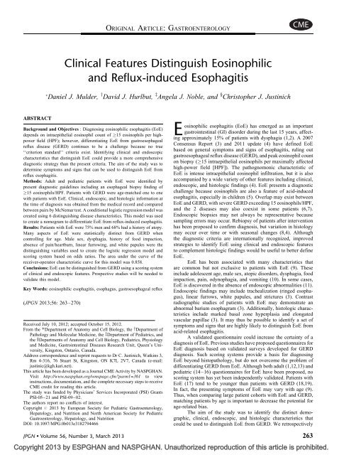
ABSTRACT<br />
<strong>Clinical</strong> <strong>Features</strong> <strong>Distinguish</strong> <strong>Eosinophilic</strong><br />
<strong>and</strong> Reflux-induced Esophagitis<br />
Daniel J. Mulder, y David J. Hurlbut, z Angela J. Noble, <strong>and</strong> § Christopher J. Justinich<br />
Background <strong>and</strong> Objectives : Diagnosing eosinophilic esophagitis (EoE)<br />
depends on intraepithelial eosinophil count of 15 eosinophils per highpower<br />
field (HPF); however, differentiating EoE from gastroesophageal<br />
reflux disease (GERD) continues to be a challenge because no true<br />
‘‘criterion st<strong>and</strong>ard’’ criteria exist. Identifying clinical <strong>and</strong> endoscopic<br />
characteristics that distinguish EoE could provide a more comprehensive<br />
diagnostic strategy than the present criteria. The aim of the study was to<br />
determine symptoms <strong>and</strong> signs that can be used to distinguish EoE from<br />
reflux esophagitis.<br />
Methods: Adult <strong>and</strong> pediatric patients with EoE were identified by<br />
present diagnostic guidelines including an esophageal biopsy finding of<br />
15 eosinophils/HPF. Patients with GERD were age-matched one to one<br />
with patients with EoE. <strong>Clinical</strong>, endoscopic, <strong>and</strong> histologic information at<br />
the time of diagnosis was obtained from the medical record <strong>and</strong> compared<br />
between pairs by McNemar test. A conditional logistic regression model was<br />
created using 6 distinguishing disease characteristics. This model was used<br />
to create a nomogram to differentiate EoE from reflux-induced esophagitis.<br />
Results: Patients with EoE were 75% men <strong>and</strong> 68% had a history of atopy.<br />
Many aspects of EoE were statistically distinct from GERD when<br />
controlling for age. Male sex, dysphagia, history of food impaction,<br />
absence of pain/heartburn, linear furrowing, <strong>and</strong> white papules were the<br />
distinguishing variables used to create the logistic regression model <strong>and</strong><br />
scoring system based on odds ratios. The area under the curve of the<br />
receiver-operator characteristic curve for this model was 0.858.<br />
Conclusions: EoE can be distinguished from GERD using a scoring system<br />
of clinical <strong>and</strong> endoscopic features. Prospective studies will be needed to<br />
validate this model.<br />
Key Words: eosinophilic esophagitis, esophagus, gastroesophageal reflux<br />
(JPGN 2013;56: 263–270)<br />
Received July 10, 2012; accepted October 15, 2012.<br />
From the Department of Anatomy <strong>and</strong> Cell Biology, the yDepartment of<br />
Pathology <strong>and</strong> Molecular Medicine, the zDepartment of Pediatrics, <strong>and</strong><br />
the §Departments of Anatomy <strong>and</strong> Cell Biology, Pediatrics, Physiology<br />
<strong>and</strong> Medicine, Gastrointestinal Diseases Research Unit, Queen’s University,<br />
Kingston, Ontario, Canada.<br />
Address correspondence <strong>and</strong> reprint requests to Dr C. Justinich, Watkins 3,<br />
Rm 4-316, 76 Stuart St, Kingston, ON K7L 2V7, Canada (e-mail:<br />
justinic@kgh.kari.net).<br />
This article has been developed as a Journal CME Activity by <strong>NASPGHAN</strong>.<br />
Visit http://www.naspghan.<strong>org</strong>/wmspage.cfm?parm1=361 to view<br />
instructions, documentation, <strong>and</strong> the complete necessary steps to receive<br />
CME credit for reading this article.<br />
The study was funded by Physicians’ Services Incorporated (PSI) Grants<br />
PSI-05–21 <strong>and</strong> PSI-09–02.<br />
The authors report no conflicts of interest.<br />
Copyright # 2013 by European Society for Pediatric Gastroenterology,<br />
Hepatology, <strong>and</strong> Nutrition <strong>and</strong> North American Society for Pediatric<br />
Gastroenterology, Hepatology, <strong>and</strong> Nutrition<br />
DOI: 10.1097/MPG.0b013e3182794466<br />
ORIGINAL ARTICLE: GASTROENTEROLOGY<br />
Copyright 2013 by ESPGHAN <strong>and</strong> <strong>NASPGHAN</strong>. Unauthorized reproduction of this article is prohibited.<br />
E osinophilic<br />
esophagitis (EoE) has emerged as an important<br />
gastrointestinal (GI) disorder during the last 15 years, affecting<br />
approximately 15% of patients with dysphagia (1,2). A 2007<br />
Consensus Report (3) <strong>and</strong> 2011 update (4) have defined EoE<br />
based on general symptoms <strong>and</strong> signs of esophagitis, ruling out<br />
gastroesophageal reflux disease (GERD), <strong>and</strong> peak eosinophil count<br />
on biopsy ( 15 intraepithelial eosinophils per maximally affected<br />
high-power field [HPF]). The pathognomonic characteristic of<br />
EoE is intense intraepithelial eosinophil infiltration, but it is also<br />
accompanied by a wide variety of other features including clinical,<br />
endoscopic, <strong>and</strong> histologic findings (4). EoE presents a diagnostic<br />
challenge because eosinophils are also a feature of acid-induced<br />
esophagitis, especially in children (5). Overlap may exist between<br />
EoE <strong>and</strong> GERD, with severe GERD exceeding 15 eosinophils/HPF,<br />
<strong>and</strong> the 2 diseases may also coexist in some patients (6,7).<br />
Endoscopic biopsies may not always be representative because<br />
sampling errors may occur. Rebiopsy of patients after intervention<br />
has been proposed to confirm diagnosis, but variation in histology<br />
may occur over time or with seasonal changes (8,4). Although<br />
the diagnostic criteria are internationally recognized, improved<br />
strategies to identify EoE using clinical <strong>and</strong> endoscopic features<br />
to complement histologic findings would be useful to better define<br />
EoE.<br />
EoE has been associated with many characteristics that<br />
are common but not exclusive to patients with EoE (9). These<br />
include adolescent age, male sex, atopic disorders, dysphagia, food<br />
impaction, pain, odynophagia, <strong>and</strong> vomiting (10). In some cases,<br />
EoE is discovered in the absence of endoscopic abnormalities (11).<br />
Endoscopic findings may include trachealization (ringed esophagus),<br />
linear furrows, white papules, <strong>and</strong> strictures (3). Contrast<br />
radiographic studies of patients with EoE may demonstrate an<br />
abnormal barium esophagram (3). Additionally, histologic characteristics<br />
include marked basal zone hyperplasia <strong>and</strong> elongated<br />
vascular papillae (3). It may thus be possible to identify a set of<br />
symptoms <strong>and</strong> signs that are highly likely to distinguish EoE from<br />
acid-related esophagitis.<br />
A validated questionnaire could increase the certainty of a<br />
diagnosis of EoE. Previous studies have proposed questionnaires for<br />
EoE diagnosis based on validated surveys developed for GERD<br />
diagnosis. Such scoring systems provide a basis for diagnosing<br />
EoE beyond histopathology, but do not overcome the problem of<br />
differentiating GERD from EoE. Although both adult (1,12,13) <strong>and</strong><br />
pediatric (14–16) questionnaires for EoE have been proposed, no<br />
scoring system has yet been independently validated. Patients with<br />
EoE (17) tend to be younger than patients with GERD (18,19).<br />
In fact, the presenting symptoms of EoE may vary with age (9).<br />
Thus, when comparing large patient cohorts with EoE <strong>and</strong> GERD,<br />
matching patients by age is important to decrease the potential for<br />
age-related bias.<br />
The aim of the study was to identify the distinct demographic,<br />
clinical, endoscopic, <strong>and</strong> histologic characteristics that<br />
could be used to distinguish EoE from GERD. We retrospectively<br />
JPGN Volume 56, Number 3, March 2013 263
Mulder et al JPGN Volume 56, Number 3, March 2013<br />
compared patients with EoE at the time of diagnosis with agematched<br />
patients with GERD. We hypothesize that there is a set of<br />
symptoms <strong>and</strong> signs that result from the unique immune processes<br />
of EoE <strong>and</strong> can be used to distinguish EoE from reflux esophagitis.<br />
In the present study, we have found a set of characteristics to be<br />
highly associated with EoE when compared with age-matched<br />
patients with GERD. Using independent odds ratios obtained<br />
from a conditional logistic regression model, we propose a<br />
scoring system based on these characteristics that is predictive of<br />
a diagnosis of EoE.<br />
METHODS<br />
Patient Identification<br />
An electronic search of the pathology records at the tertiary<br />
care center in Kingston, Canada, was performed for ‘‘esophagus<br />
AND (eosinophil OR eosinophils OR eosinophilia OR eosinophilic)’’<br />
in the final diagnosis section of all pathology reports from<br />
January 1, 1997 to December 31, 2009. EoE was defined, based<br />
on the 2007 consensus report (3) <strong>and</strong> 2011 update (4), as<br />
15 eosinophils per maximal HPF, symptoms of esophagitis,<br />
<strong>and</strong> absence of other causes of esophagitis. Four hundred eleven<br />
cases were identified by the initial search. A total of 232 cases did<br />
not meet the criteria for the diagnosis of EoE <strong>and</strong> were excluded.<br />
As a result of increasing awareness of EoE, our pathologists often<br />
include a statement such as ‘‘no esophageal eosinophils,’’ which<br />
was flagged in our electronic search of the keywords ’’esophagus’’<br />
<strong>and</strong> ‘‘eosinophil.’’ This accounts for the cases excluded after the<br />
initial search. Subsequently, any suggestion of having concurrent<br />
EoE <strong>and</strong> GERD resulted in exclusion from the study (n ¼ 3). For<br />
example, pH probe results suggesting GERD <strong>and</strong> 15 eosinophils/<br />
HPF, or patients diagnosed as having GERD who had previous or<br />
subsequent biopsies showing 15 eosinophils/HPF, were excluded<br />
from the study. Patients (n ¼ 9) were also excluded if they had<br />
evidence of other upper GI tract pathology, such as eosinophilic<br />
gastroenteritis, Barrett esophagus, or Crohn disease. From the<br />
search results, 167 (41 pediatric <strong>and</strong> 126 adult) cases of isolated<br />
EoE were identified. The medical record of each case was then<br />
reviewed to identify the initial clinical presentation <strong>and</strong> endoscopy<br />
findings for each case. Biopsy slides from the selected cases<br />
were then blinded <strong>and</strong> reevaluated to confirm the presence of<br />
15 intraepithelial eosinophils per maximal HPF. The Queen’s<br />
University Health Sciences research ethics board approved the<br />
present study.<br />
Age Matching<br />
A matching GERD cohort was identified by searching<br />
all pathology reports for ‘‘esophagus AND reflux’’ in the final<br />
diagnosis section during the same time period as the EoE<br />
cohort (n ¼ 564). Selection of GERD cases was based on symptoms<br />
of esophagitis, pathology demonstrating distal esophagitis with<br />
JPGN Volume 56, Number 3, March 2013 <strong>Eosinophilic</strong> Versus Reflux Esophagitis<br />
an allergist of allergic asthma, allergic rhinitis, or food allergy.<br />
Endoscopic features included were trachealization (defined as<br />
multiple rings seen at any level of the esophagus), linear<br />
furrows, white papules, stricture, <strong>and</strong> normal endoscopy. Archived<br />
esophageal tissue specimens from endoscopies in which EoE<br />
was first diagnosed were evaluated for histopathologic criteria;<br />
these included maximal intraepithelial eosinophil count, basal zone<br />
thickness, <strong>and</strong> elongation of vascular papillae. Basal zone<br />
hyperplasia <strong>and</strong> vascular papilla elongation were assessed in<br />
well-oriented sections, as we have previously reported (23).<br />
The basal zone was scored as: 0% to 25% (normal), 26% to<br />
50%, 51% to 75%, or 76% to 100% of the total thickness of<br />
the epithelium (23). For vascular papilla length, scores given were<br />
0% to 33% (normal), 34% to 67%, <strong>and</strong> 68% to 100% of the total<br />
thickness of the epithelium (23).<br />
Many patients in the present study had an upper GI<br />
radiographic study. A double-contrast barium swallow was done<br />
first. Patients were then asked to swallow three barium-coated<br />
marshmallows (approximately 1 1 1 cm) under fluoroscopy.<br />
An ordinal value was given to quantify the results based on<br />
descriptors used in the radiology report <strong>and</strong> studies of EoE with<br />
similar methodology (24,25). Scores were 0 ¼ normal, 1 ¼ mild,<br />
2 ¼ moderate, 3 ¼ severe.<br />
Statistical Analysis<br />
Graphpad Prism version 4.0 (GraphPad, La Jolla, CA) was<br />
used to perform statistics, except for the conditional logistic<br />
regression model, which was created using SPSS version 17.1<br />
(SPSS Inc, Chicago, IL), <strong>and</strong> the nomogram, which was originally<br />
created using the Orange Software Suite version 2.0. P < 0.05 was<br />
considered to be statistically significant. McNemar test was used to<br />
evaluate ‘‘yes/no’’ matched data from contingency tables. P values<br />
from this test were calculated using the continuity correction.<br />
For unmatched data analysis, a contingency table was created<br />
<strong>and</strong> analyzed by Fisher exact test. Linear correlation was evaluated<br />
using Spearman rank correlation coefficient. Confidence intervals<br />
(CI) were evaluated at 95%. For each disease characteristic investigated,<br />
data were only used for development of the regression<br />
model if available for both members of the pair. If one of the pairs<br />
was missing from the information being evaluated, that category<br />
data was excluded from both pairs automatically in the creation<br />
of the model. Interactions were not included in the model<br />
because the relations between the disease characteristics were<br />
not clinically relevant <strong>and</strong> did not significantly alter the model<br />
when using a backwards elimination strategy informed by the<br />
results of McNemar test. The odds ratios determined by conditional<br />
logistic regression were then used to establish a nomogram, with a<br />
score that would predict the relative odds of a patient having<br />
EoE versus GERD based on the selective set of clinical <strong>and</strong><br />
endoscopic characteristics.<br />
RESULTS<br />
<strong>Clinical</strong> Characteristics<br />
The clinical, endoscopic, <strong>and</strong> histologic data for the agematched<br />
EoE <strong>and</strong> GERD cohorts are summarized in Table 1.<br />
The matched odds ratios <strong>and</strong> P values in this table are based on<br />
the results of the McNemar test. The proportion of men in the EoE<br />
cohort (75%) was significantly greater than the proportion of men in<br />
the GERD cohort (52%, P < 0.0001).<br />
The most common age range to be diagnosed as having<br />
EoE in the present study patients was the second decade of life,<br />
although patients were diagnosed as having EoE at ages ranging<br />
from 1 to 79 years (Fig. 2A). Male sex was more common for<br />
patients with EoE in all age groups.<br />
Atopic <strong>Features</strong><br />
When stratified by age, the prevalence of atopic disease in<br />
patients with EoE was more than half of the patients in each<br />
decade of life (Fig. 2B). Atopic patients with EoE were found in<br />
every decade of life except the eighth, for which there was only<br />
1 patient. In the EoE cohort, 68% of patients had a history of<br />
atopy. Age-matched patients with GERD had significantly lower<br />
incidence of atopy (43%, P ¼ 0.03). Separating patients into adult<br />
<strong>and</strong> pediatric (younger than 18 years) groups did not alter these<br />
findings.<br />
Peripheral blood eosinophilia was not helpful in distinguishing<br />
EoE from GERD. Although blood eosinophil count was<br />
significantly greater in the EoE cohort, both EoE <strong>and</strong> GERD<br />
cohort average blood eosinophil levels were within the<br />
normal range. Seven of 82 matched patients with EoE <strong>and</strong> 2 of<br />
82 matched patients with GERD had a peripheral blood level<br />
>0.7 10 9 eosinophils/L. Using matched analysis, the amount of<br />
patients with EoE presenting with peripheral blood eosinophilia<br />
was not significantly different from the GERD cohort (Fig. 3A,<br />
P ¼ 0.18). Our tertiary care center considers an abnormal absolute<br />
eosinophil count to be >0.7 10 9 eosinophils/L. No consensus<br />
exists in the literature with regard to the definition of peripheral<br />
blood eosinophilia. Some studies have considered peripheral blood<br />
eosinophilia to be any value >0.5 10 9 eosinophils/L (26). There<br />
was no statistically significant difference in the incidence of<br />
eosinophilia between the EoE <strong>and</strong> GERD cohorts when using either<br />
cut-off value to define peripheral blood eosinophilia.<br />
Peripheral blood eosinophilia has been proposed as a<br />
possible surrogate marker of esophageal eosinophilia (27). Thus,<br />
the level of correlation between esophageal eosinophil count <strong>and</strong><br />
peripheral blood eosinophil level was investigated. These data were<br />
analyzed unpaired to investigate the possibility of blood eosinophil<br />
level alone being a predictor of the severity of esophageal eosinophilia<br />
in a particular disease state; neither EoE (Fig. 3B) nor GERD<br />
(Fig. 3C) peripheral blood eosinophil level correlated with maximal<br />
esophageal intraepithelial eosinophil count/HPF.<br />
Based on the month in which the diagnostic endoscopy was<br />
performed, patients were more likely to be diagnosed as having<br />
GERD during the winter months (December–February). During the<br />
remaining months of the year, patients were no more likely to be<br />
diagnosed as having EoE or GERD.<br />
Endoscopic <strong>and</strong> Radiologic Findings<br />
Four endoscopic features were significantly different<br />
between patients with EoE <strong>and</strong> age-matched patients with GERD.<br />
Trachealization, linear furrows, <strong>and</strong> white papules were significantly<br />
more likely to be found in the patients with EoE <strong>and</strong> a normal<br />
endoscopic appearance was more likely to be found in patients<br />
presenting with GERD. Barium-coated marshmallow esophagram<br />
was assessed for 105 EoE <strong>and</strong> 80 patients with GERD <strong>and</strong> was ranked<br />
as normal, mild, moderate, or severe. The proportion of patients with<br />
normal to severe dysmotility was similar in both disease states when<br />
analyzed age-matched <strong>and</strong> unmatched. The incidence of esophageal<br />
strictures was statistically similar between the cohorts.<br />
Histologic Characteristics<br />
In EoE, the mean st<strong>and</strong>ard deviation of eosinophils per<br />
maximal HPF was 76 56 <strong>and</strong> ranged from 15 to 308. The<br />
www.jpgn.<strong>org</strong> 265<br />
Copyright 2013 by ESPGHAN <strong>and</strong> <strong>NASPGHAN</strong>. Unauthorized reproduction of this article is prohibited.
Mulder et al JPGN Volume 56, Number 3, March 2013<br />
TABLE 1. Age-matched comparison of individual signs <strong>and</strong> symptoms of EoE <strong>and</strong> GERD<br />
proportion of epithelium occupied by the basal zone was significantly<br />
greater in patients with EoE, as was elongation of vascular<br />
papilla.<br />
Diagnosing EoE by Score<br />
EoE,<br />
n ¼ 163<br />
By evaluating the odds ratios <strong>and</strong> P values determined<br />
by comparison of individual disease features by McNemar test<br />
(evaluated in Table 1), as an approximation of that particular<br />
characteristic’s ability to predict EoE, a conditional logistic<br />
regression model was created using a backwards elimination<br />
strategy. The final model consisted of 6 characteristics (sex,<br />
dysphagia, pain/heartburn, history of food impaction, linear furrowing,<br />
<strong>and</strong> white papules; Table 2). In addition, the selection of<br />
characteristics for the model was informed by the knowledge that<br />
for 326 rows of data, having 6 to 11 covariates in the model, is<br />
optimal (28). Male sex, dysphagia, history of food impaction, linear<br />
furrowing, <strong>and</strong> white papules were associated with increased odds<br />
of EoE diagnosis. Pain/heartburn was associated with increased<br />
odds of GERD diagnosis. Missing data accounted 8.0% (n ¼ 13<br />
GERD,<br />
n ¼ 163<br />
Matched odds ratio<br />
(95% confidence interval) P<br />
<strong>Clinical</strong> characteristics<br />
Age at diagnosis, mean SD 31.8 17.7 31.8 17.7 0.83<br />
Male, n (%) 122 (75) 85 (52) 2.61 (1.59–4.42)
JPGN Volume 56, Number 3, March 2013 <strong>Eosinophilic</strong> Versus Reflux Esophagitis<br />
A B<br />
No. of cases<br />
40<br />
30<br />
20<br />
10<br />
0<br />
1<br />
Female<br />
Male<br />
2 3 4 5 6 7 8<br />
Decade of life<br />
The study discovered that multiple clinical <strong>and</strong> endoscopic<br />
findings are associated with EoE <strong>and</strong> the presence of these findings<br />
increases the statistical confidence with which the patient can be<br />
said to have EoE. Male sex, dysphagia, a history of food impaction,<br />
linear furrowing, <strong>and</strong> white papules were the clinical <strong>and</strong> endoscopic<br />
findings independently associated with EoE. Pain (including<br />
heartburn) was independently associated with a diagnosis of GERD.<br />
The finding of a normal appearing esophagus in 48% of the GERD<br />
group may not be representative of the GERD population as a whole<br />
because patients with a typical clinical presentation of erosive<br />
esophagitis are not always biopsied. Findings found to be significantly<br />
associated with EoE in previous studies including peripheral<br />
blood eosinophilia (27) <strong>and</strong> esophageal dysmotility (29) were not<br />
found to contribute to differentiating EoE <strong>and</strong> GERD in the present<br />
study population.<br />
The nomogram was based on the conditional logistic<br />
regression model determined by our data set. The purpose of the<br />
study was not to replace the histopathologic component of the EoE<br />
diagnosis, but to improve our underst<strong>and</strong>ing of the value of other<br />
clinical <strong>and</strong> endoscopic information that may aid in differentiating<br />
EoE <strong>and</strong> GERD. It is important to note that, based on the present<br />
model, a male patient with dysphagia <strong>and</strong> no pain or heartburn<br />
would have a score of 98 <strong>and</strong> a reasonably high probability of<br />
having EoE <strong>and</strong> not GERD. This result is noteworthy because these<br />
Total EoE cases<br />
Total GERD cases<br />
1 2 3 4 5 6 7 8 9<br />
Decade of life<br />
particular disease characteristics do not require endoscopy or<br />
biopsy. Future prospective studies can exp<strong>and</strong> on the present<br />
findings to create a more comprehensive diagnosis of EoE that<br />
may supplement present practices of evaluation for patients with<br />
suspected EoE.<br />
We aimed to build upon the findings of Dellon et al who<br />
performed a similar study, which was case-controlled, but not agematched<br />
(30). Similar to our study, this group also found<br />
that dysphagia <strong>and</strong> linear furrows supported a diagnosis of EoE<br />
compared with GERD. Six other studies have investigated questionnaires<br />
as a method to predict EoE (Table 3) (1,12–16,31–35).<br />
A symptom score based on a modified questionnaire for<br />
pediatric acid-peptic disease (31) has been used retrospectively<br />
to evaluate the safety <strong>and</strong> efficacy of budesonide treatment (15) <strong>and</strong><br />
prospectively to identify symptoms associated with EoE (14).<br />
The latter study found that dysphagia <strong>and</strong> early satiety were<br />
predictive of EoE in children. Two other scoring systems adapted<br />
from GERD questionnaires (32–34) have found that allergic<br />
asthma, male sex, trachealization (12), <strong>and</strong> dysphagia (1) were<br />
independent predictors of EoE in prospective study of adult subjects<br />
undergoing endoscopy. Many of these characteristics were significantly<br />
associated with EoE when considered individually in our<br />
patient cohort as well. Another study, using the same questionnaire<br />
as Veerappan et al (12), found no significant difference in symptom<br />
Copyright 2013 by ESPGHAN <strong>and</strong> <strong>NASPGHAN</strong>. Unauthorized reproduction of this article is prohibited.<br />
Cases with atopy<br />
30<br />
20<br />
10<br />
0<br />
n = 13<br />
n = 11<br />
32<br />
25<br />
20<br />
15<br />
20<br />
10<br />
16<br />
8<br />
8<br />
4<br />
6<br />
3<br />
EoE<br />
GERD<br />
FIGURE 2. A, Sex distribution of the eosinophilic esophagitis (EoE) cohort by decade of life, at the time of diagnosis. B, The distribution, by decade<br />
of life, of the number of cases with concurrent atopy in the EoE <strong>and</strong> gastroesophageal reflux disease (GERD) cohorts.<br />
A<br />
n = 82 n = 82<br />
B C<br />
1.6<br />
EoE<br />
Eosinophils*10 9 /L<br />
1.4<br />
1.2<br />
1.0<br />
0.8<br />
0.6<br />
0.4<br />
0.2<br />
0.0<br />
EoE<br />
GERD<br />
Blood eosinophils (*10ˆ9/L)<br />
2.0<br />
1.5<br />
1.0<br />
0.5<br />
r 2 = 0.001532<br />
0.0<br />
0.0<br />
0 50 100 150 200 250 300 350 0 5 10 15<br />
Esophageal eosinophils/Max HPF Esophageal eosinophils/Max HPF<br />
Blood eosinophils (*10ˆ9/L)<br />
2.5<br />
2.0<br />
1.5<br />
1.0<br />
0.5<br />
1<br />
1<br />
GERD<br />
0<br />
0<br />
r 2 = 0.009374<br />
FIGURE 3. A, Distribution of matched peripheral blood eosinophil level comparing eosinophilic esophagitis (EoE) <strong>and</strong> gastroesophageal reflux<br />
disease (GERD) cohorts (n ¼ 82 for each group). The dotted line represents the threshold value (0.7 10 9 eosinophils/L) for eosinophilia at our<br />
center. B, No significant correlation was found between unmatched intraepithelial esophageal eosinophils per maximal high-power field (HPF)<br />
<strong>and</strong> peripheral blood eosinophil level in EoE (n ¼ 119, P ¼ 0.673) or GERD (C; n ¼ 110, P ¼ 0.314) patients.<br />
www.jpgn.<strong>org</strong> 267
Mulder et al JPGN Volume 56, Number 3, March 2013<br />
TABLE 2. Results of the conditional logistic regression analysis for each<br />
parameter<br />
Parameter<br />
Odds<br />
ratio<br />
95% Confidence<br />
interval P<br />
Male sex 1.82 0.86–4.72 0.106<br />
Dysphagia 2.02 1.74–16.68 0.003<br />
Pain/heartburn 0.4 2.36–28.23 0.001<br />
History of food<br />
impaction<br />
5.39 0.19–0.86 0.018<br />
Linear furrowing 8.16 0.70–4.70 0.217<br />
White papules 9.61 1.33–69.28 0.025<br />
score when adult patients with EoE were stratified by the presence<br />
of nonspecific esophageal motility disorder (13). This finding is<br />
similar to our own finding that dysmotility, as subjectively<br />
assessed by barium swallow, does not aid in differentiating EoE<br />
from GERD. Pentiuk et al (16) developed a questionnaire that was<br />
based on symptom frequency <strong>and</strong> severity, <strong>and</strong> found that scores did<br />
not correlate with esophageal eosinophilia; thus, we did not attempt<br />
to evaluate symptom frequency or severity in our study.<br />
Logistic models can be useful for evaluating case-control<br />
studies, provided certain assumptions are met (36,37). Notably,<br />
case-controlled logistic regression models cannot be used to<br />
calculate positive or negative predictive values, which depend on<br />
prevalence. Because the cases <strong>and</strong> controls were matched in the<br />
present study in a one-to-one ratio, the disease prevalence in this<br />
population is artificial (50% of patients had EoE <strong>and</strong> 50% had<br />
GERD). The relation between EoE <strong>and</strong> the covariates determined<br />
in the present study is not necessarily causal. Other covariates<br />
that were not available for the present study may also influence<br />
identification of EoE over GERD. We cannot discount the<br />
possibility that the strength of the logistic regression model in<br />
the present study is based in part on the fact that little data were<br />
missing from the characteristics used. For example, even though<br />
some atopic characteristics were significantly different when<br />
analyzed individually in Table 1, these characteristics did not<br />
contribute when included in the conditional logistic regression<br />
analysis. The missing data in these categories may have decreased<br />
their influence on the model <strong>and</strong> subsequent odds ratios.<br />
A<br />
0 10 20 30 40 50 60 70 80 90 100<br />
Points<br />
0<br />
26<br />
Gender<br />
female<br />
0<br />
31<br />
Dysphagia<br />
no yes<br />
0<br />
41<br />
Pain/heartburn<br />
yes no<br />
History of<br />
0<br />
74<br />
food impaction<br />
no yes<br />
0<br />
93<br />
Furrowing<br />
no yes<br />
0<br />
100<br />
White papules<br />
yes<br />
B<br />
Total points<br />
0 100 200 300 365<br />
Relative odds<br />
of EoE 0.21 0.62 0.85 0.95<br />
male<br />
FIGURE 4. Nomogram based on the odds ratios from the conditional logistic regression model. A, The number of points for a given characteristic<br />
is proportional to the odds ratio for that characteristic. Note that the absence of pain/heartburn adds points to the total score. B, The sum of the<br />
points for each characteristic can be used to predict the relative odds that a patient has eosinophilic esophagitis (EoE) <strong>and</strong> not gastroesophageal<br />
reflux disease (GERD) (assuming an equal distribution of EoE <strong>and</strong> patients with GERD).<br />
Copyright 2013 by ESPGHAN <strong>and</strong> <strong>NASPGHAN</strong>. Unauthorized reproduction of this article is prohibited.<br />
Sensitivity<br />
1.00<br />
0.75<br />
0.50<br />
0.25<br />
0.00<br />
0.00<br />
0.25 0.50<br />
1 - specificity<br />
0.75 1.00<br />
FIGURE 5. Receiver-operator characteristic curve pertaining to the<br />
ability of the conditional logistic regression model to predict<br />
eosinophilic esophagitis versus gastroesophageal reflux disease in<br />
the population studied. The model was based on 6 clinical <strong>and</strong><br />
endoscopic characteristics. Area under the curve was 0.858 with a<br />
95% confidence interval of 0.816 to 0.900 (P < 0.0001).<br />
268 www.jpgn.<strong>org</strong>
JPGN Volume 56, Number 3, March 2013 <strong>Eosinophilic</strong> Versus Reflux Esophagitis<br />
TABLE 3. Details of the previous studies that make use of scoring systems to investigate EoE<br />
Histologic<br />
score Study type Adapted from<br />
Endoscopic<br />
score<br />
Symptom<br />
severity<br />
score<br />
Symptom<br />
frequency<br />
score<br />
No. symptoms<br />
questions<br />
Age<br />
group<br />
Patients who completed<br />
questionnaire<br />
Reference<br />
385 EGD (25 EoE) Adult 8 1–3 1–3 Not included Not included Prospective Vigneri et al (32)<br />
32 EoE Adult 6 1–3 1–3 Not included Not included Prospective Vigneri et al (32)<br />
Included Included Not included Not included Prospective Aanen et al (33)<br />
<strong>and</strong> DiSario<br />
et al (34)<br />
Not included Retrospective Dohil et al (31)<br />
Adult Dysphagia <strong>and</strong> GERD<br />
validated questionnaires<br />
261 Dysphagia/food<br />
impaction EGD (31 EoE)<br />
Veerappan<br />
et al (12)<br />
Bassett<br />
et al (13)<br />
MacKenzie<br />
et al (1)<br />
20 EoE Pediatric 7 sets Not included 1–2 4 categories,<br />
1–2<br />
100 (35 EoE, 27 GERD, Pediatric 7 sets Not included 1–2 4 categories, 8 features, Prospective Aceves et al (15),<br />
24 allergic nonEoE,<br />
1–2<br />
of 18<br />
added histology<br />
14 nonallergic nonEoE)<br />
scoring<br />
49 EoE Pediatric 10 for frequency,<br />
1–5 1–3 Not included Not included Prospective Konikoff et al (35)<br />
8 for severity<br />
Aceves<br />
et al (15)<br />
Aceves<br />
et al (14)<br />
Pentiuk<br />
et al (16)<br />
EGD ¼ esophagogastroduodenoscopy; EoE ¼ eosinophilic esophagitis; GERD ¼ gastroesophageal reflux disease.<br />
One of the strengths of the present study is that it<br />
controls for the high proportion of pediatric patients with<br />
EoE when compared with the generally older GERD population<br />
(22). EoE characteristics may change with age (9). The selection<br />
method used in the present study limited age-related bias,<br />
especially when determining the significance of atopic status<br />
to the diagnosis because pediatric patients in general are more<br />
likely to have atopic disease (38). We found that even when<br />
controlling for age, patients with EoE have an increased likelihood<br />
of having atopic disease.<br />
One of the limitations of the present study is that it is<br />
retrospective by design. A prospective study is being performed<br />
to evaluate the model, as a follow-up to the present study.<br />
An important aspect of future studies will be evaluating not only<br />
esophageal eosinophil count as an endpoint, but also the<br />
patient’s response to treatment. One recent change to the diagnostic<br />
criteria for EoE that has emerged is that the original diagnostic<br />
strategy could be supplemented with a trial of proton pump<br />
inhibitor (PPI) therapy to rule out severe GERD <strong>and</strong> possible<br />
‘‘PPI-responsive EoE’’ (4). Interventions, including PPI therapy,<br />
were not evaluated in the present study because the patients<br />
were evaluated at the time of initial presentation, not following<br />
a trial therapy.<br />
A potential confounding factor in the design of the present<br />
study is the possible overlap between the EoE <strong>and</strong> GERD patient<br />
groups. Unfortunately, no true criterion st<strong>and</strong>ard exists to define<br />
EoE at this time, so we are left with using esophageal eosinophil<br />
counts <strong>and</strong> the criteria set out in the published consensus statements<br />
(3,4). In this retrospective review, not all of the patients underwent<br />
repeat endoscopy, especially those who fulfilled criteria for<br />
GERD that responded to PPI therapy. This issue will be addressed<br />
by a prospective study to evaluate the scoring system.<br />
Future studies will be needed to assess the contribution of<br />
intraepithelial eosinophil number per HPF to the ability to distinguish<br />
EoE from GERD. Intraepithelial eosinophil count could<br />
not be included in our conditional regression model because it was<br />
used to define the cases <strong>and</strong> the controls. Based on previous<br />
epidemiologic studies (39), it is likely that both age <strong>and</strong> esophageal<br />
eosinophil count will contribute to the diagnosis in future prospective<br />
studies. The criteria for the scoring system will need to be<br />
updated when this information is available.<br />
In conclusion, our study has identified 6 symptoms <strong>and</strong> signs<br />
that can be used in a scoring system to aid in distinguishing EoE<br />
from GERD. The disease characteristics were determined by casecontrolled,<br />
age-matched comparison of EoE <strong>and</strong> patients with<br />
GERD. This scoring system will need to be validated by prospective<br />
studies before it can be used in clinical practice.<br />
Acknowledgments: The authors acknowledge support from<br />
Kingston General Hospital <strong>and</strong> the Queen’s University Gastrointestinal<br />
Diseases Research Unit.<br />
REFERENCES<br />
1. Mackenzie SH, Go M, Chadwick B, et al. <strong>Eosinophilic</strong> oesophagitis<br />
in patients presenting with dysphagia—a prospective analysis. Aliment<br />
Pharmacol Ther 2008;28:1140–6.<br />
2. Prasad GA, Talley NJ, Romero Y, et al. Prevalence <strong>and</strong> predictive factors<br />
of eosinophilic esophagitis in patients presenting with dysphagia:<br />
a prospective study. Am J Gastroenterol 2007;102:2627–32.<br />
3. Furuta GT, Liacouras CA, Collins MH, et al. <strong>Eosinophilic</strong> esophagitis in<br />
children <strong>and</strong> adults: a systematic review <strong>and</strong> consensus recommendations<br />
for diagnosis <strong>and</strong> treatment. Gastroenterology 2007;133:1342–63.<br />
4. Liacouras CA, Furuta GT, Hirano I, et al. <strong>Eosinophilic</strong> esophagitis:<br />
updated consensus recommendations for children <strong>and</strong> adults. J Allergy<br />
Clin Immunol 2011;128:3–20.<br />
www.jpgn.<strong>org</strong> 269<br />
Copyright 2013 by ESPGHAN <strong>and</strong> <strong>NASPGHAN</strong>. Unauthorized reproduction of this article is prohibited.
Mulder et al JPGN Volume 56, Number 3, March 2013<br />
5. Winter HS, Madara JL, Stafford RJ, et al. Intraepithelial eosinophils:<br />
a new diagnostic criterion for reflux esophagitis. Gastroenterology<br />
1982;83:818–23.<br />
6. Spechler SJ, Genta RM, Souza RF. Thoughts on the complex relationship<br />
between gastroesophageal reflux disease <strong>and</strong> eosinophilic esophagitis.<br />
Am J Gastroenterol 2007;102:1301–6.<br />
7. Ngo P, Furuta GT, Antonioli DA, et al. Eosinophils in the esophagus–<br />
peptic or allergic eosinophilic esophagitis? Case series of three patients<br />
with esophageal eosinophilia. Am J Gastroenterol 2006;101:1666–70.<br />
8. Wang FY, Gupta SK, Fitzgerald JF. Is there a seasonal variation in the<br />
incidence or intensity of allergic eosinophilic esophagitis in newly<br />
diagnosed children? J Clin Gastroenterol 2007;41:451–3.<br />
9. Noel RJ, Putnam PE, Rothenberg ME. <strong>Eosinophilic</strong> esophagitis. N Engl<br />
J Med 2004;351:940–1.<br />
10. Atkins D, Kramer R, Capocelli K, et al. <strong>Eosinophilic</strong> esophagitis:<br />
the newest esophageal inflammatory disease. Nat Rev Gastroenterol<br />
Hepatol 2009;6:267–78.<br />
11. Pasha SF, DiBaise JK, Kim HJ, et al. Patient characteristics, clinical,<br />
endoscopic, <strong>and</strong> histologic findings in adult eosinophilic esophagitis:<br />
a case series <strong>and</strong> systematic review of the medical literature. Dis<br />
Esophagus 2007;20:311–9.<br />
12. Veerappan GR, Perry JL, Duncan TJ, et al. Prevalence of eosinophilic<br />
esophagitis in an adult population undergoing upper endoscopy:<br />
a prospective study. Clin Gastroenterol Hepatol 2009;7:420–6.<br />
13. Bassett J, Maydonovitch C, Perry J, et al. Prevalence of esophageal<br />
dysmotility in a cohort of patients with esophageal biopsies consistent<br />
with eosinophilic esophagitis. Dis Esophagus 2009;22:543–8.<br />
14. Aceves SS, Newbury RO, Dohil MA, et al. A symptom scoring tool<br />
for identifying pediatric patients with eosinophilic esophagitis <strong>and</strong><br />
correlating symptoms with inflammation. Ann Allergy Asthma Immunol<br />
2009;103:401–6.<br />
15. Aceves SS, Bastian JF, Newbury RO, et al. Oral viscous budesonide:<br />
a potential new therapy for eosinophilic esophagitis in children. Am J<br />
Gastroenterol 2007;102:2271–9.<br />
16. Pentiuk S, Putnam PE, Collins MH, et al. Dissociation between<br />
symptoms <strong>and</strong> histological severity in pediatric eosinophilic esophagitis.<br />
J Pediatr Gastroenterol Nutr 2009;48:152–60.<br />
17. Sealock RJ, Rendon G, El-Serag HB. Systematic review: The epidemiology<br />
of eosinophilic oesophagitis in adults. Aliment Pharmacol Ther<br />
2010;32:712–9.<br />
18. Bonatti H, Achem SR, Hinder RA. Impact of changing epidemiology<br />
of gastroesophageal reflux disease on its diagnosis <strong>and</strong> treatment.<br />
J Gastrointest Surg 2008;12:373–81.<br />
19. El-Serag H, Hill C, Jones R. Systematic review: The epidemiology of<br />
gastro-oesophageal reflux disease in primary care, using the UK general<br />
practice research database. Aliment Pharmacol Ther 2009;29:470–80.<br />
20. Vakil N, van Zanten SV, Kahrilas P, et al. The montreal definition <strong>and</strong><br />
classification of gastroesophageal reflux disease: a global evidencebased<br />
consensus. Am J Gastroenterol 2006;101:1900–20.<br />
21. Sherman PM, Hassall E, Fagundes-Neto U, et al. A global, evidencebased<br />
consensus on the definition of gastroesophageal reflux disease in<br />
the pediatric population. Am J Gastroenterol 2009;104:1278–95.<br />
22. Fujiwara Y, Arakawa T. Epidemiology <strong>and</strong> clinical characteristics of<br />
GERD in the japanese population. J Gastroenterol 2009;44:518–34.<br />
23. Mulder DJ, Pacheco I, Hurlbut DJ, et al. FGF9-induced proliferative<br />
response to eosinophilic inflammation in oesophagitis. Gut 2009;<br />
58:166–73.<br />
24. Potter JW, Saeian K, Staff D, et al. <strong>Eosinophilic</strong> esophagitis in adults:<br />
an emerging problem with unique esophageal features. Gastrointest<br />
Endosc 2004;59:355–61.<br />
25. Vasilopoulos S, Murphy P, Auerbach A, et al. The small-caliber<br />
esophagus: an unappreciated cause of dysphagia for solids in patients<br />
with eosinophilic esophagitis. Gastrointest Endosc 2002;55:99–106.<br />
26. Morimoto S, Kubo N, Hiramitsu S, et al. Changes in the peripheral<br />
eosinophil count in patients with acute eosinophilic myocarditis. Heart<br />
Vessels 2003;18:193–6.<br />
27. Konikoff MR, Blanchard C, Kirby C, et al. Potential of blood eosinophils,<br />
eosinophil-derived neurotoxin, <strong>and</strong> eotaxin-3 as biomarkers of<br />
eosinophilic esophagitis. Clin Gastroenterol Hepatol 2006;4:1328–36.<br />
28. Peduzzi P, Concato J, Kemper E, et al. A simulation study of the number<br />
of events per variable in logistic regression analysis. J Clin Epidemiol<br />
1996;49:1373–9.<br />
29. Chehade M, Sampson HA, Morotti RA, et al. Esophageal subepithelial<br />
fibrosis in children with eosinophilic esophagitis. J Pediatr Gastroenterol<br />
Nutr 2007;45:319–28.<br />
30. Dellon ES, Gibbs WB, Fritchie KJ, et al. <strong>Clinical</strong>, endoscopic, <strong>and</strong><br />
histologic findings distinguish eosinophilic esophagitis from gastroesophageal<br />
reflux disease. Clin Gastroenterol Hepatol 2009;7:1305–13.<br />
31. Dohil R, Newbury RO, Sellers ZM, et al. The evaluation <strong>and</strong> treatment<br />
of gastrointestinal disease in children with cystinosis receiving<br />
cysteamine. J Pediatr 2003;143:224–30.<br />
32. Vigneri S, Termini R, Le<strong>and</strong>ro G, et al. A comparison of five maintenance<br />
therapies for reflux esophagitis. N Engl J Med 1995;333:<br />
1106–10.<br />
33. Aanen MC, Numans ME, Weusten BL, et al. Diagnostic value of<br />
the reflux disease questionnaire in general practice. Digestion 2006;<br />
74:162–8.<br />
34. DiSario JA, Baskin WN, Brown RD, et al. Endoscopic approaches to<br />
enteral nutritional support. Gastrointest Endosc 2002;55:901–8.<br />
35. Konikoff MR, Noel RJ, Blanchard C, et al. A r<strong>and</strong>omized, doubleblind,<br />
placebo-controlled trial of fluticasone propionate for pediatric<br />
eosinophilic esophagitis. Gastroenterology 2006;131:1381–91.<br />
36. Breslow NE, Day NE. Statistical methods in cancer research. volume<br />
I—the analysis of case-control studies. IARC Sci Publ 1980;32:5–338.<br />
37. Prentice RL, Kalbfleisch JD. Hazard rate models with covariates.<br />
Biometrics 1979;35:25–39.<br />
38. Chipps BE. Determinants of asthma <strong>and</strong> its clinical course. Ann Allergy<br />
Asthma Immunol 2004;93:309–15.<br />
39. Prasad GA, Alex<strong>and</strong>er JA, Schleck CD, et al. Epidemiology of<br />
eosinophilic esophagitis over three decades in olmsted county,<br />
minnesota. Clin Gastroenterol Hepatol 2009;7:1055–61.<br />
270 www.jpgn.<strong>org</strong><br />
Copyright 2013 by ESPGHAN <strong>and</strong> <strong>NASPGHAN</strong>. Unauthorized reproduction of this article is prohibited.