Rapport SBUF-projekt: Mykotoxiner i inomhusmiljöer. Förekomst ...
Rapport SBUF-projekt: Mykotoxiner i inomhusmiljöer. Förekomst ...
Rapport SBUF-projekt: Mykotoxiner i inomhusmiljöer. Förekomst ...

You also want an ePaper? Increase the reach of your titles
YUMPU automatically turns print PDFs into web optimized ePapers that Google loves.
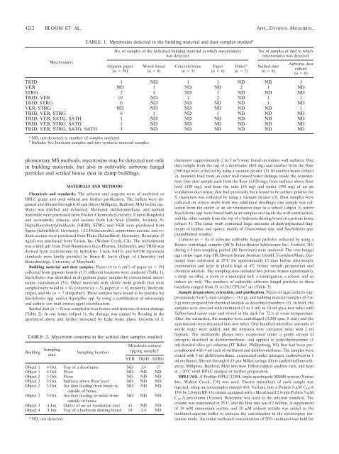
4212 BLOOM ET AL. APPL. ENVIRON. MICROBIOL.<br />
Mycotoxin(s)<br />
TABLE 1. Mycotoxins detected in the building material and dust samples studied a<br />
plementary MS methods, mycotoxins may be detected not only<br />
in building materials, but also in cultivable airborne fungal<br />
particles and settled house dust in damp buildings.<br />
MATERIALS AND METHODS<br />
Chemicals and standards. The solvents and reagents were of analytical or<br />
HPLC grade and used without any further purification. The buffers were degassed<br />
and filtered through 0.45-m filters (Millipore, Bedford, MA) before use.<br />
Water was distilled and deionized. Methanol, dichloromethane, and sodium<br />
hydroxide were purchased from Fischer Chemicals (Leicester, United Kingdom)<br />
and acetonitrile, toluene, and acetone from Lab Scan (Dublin, Ireland). N-<br />
Heptafluorobutyrylimidazole (HFBI), STRG, and VER were purchased from<br />
Sigma (Schnelldorf, Germany). 1,12-Dodecanediol, ammonium acetate, and sodium<br />
acetate were purchased from Fluka (Schnelldorf, Germany). Reserpine (5<br />
ng/l) was purchased from Varian, Inc. (Walnut Creek, CA). The trichodermin<br />
was a kind gift from Poul Rasmussen (Leo-Pharma, Denmark), and TRID was<br />
derived from trichodermin by hydrolysis. Crude SATG and SATH mycotoxin<br />
standards were kindly provided by Bruce B. Jarvis (Dept. of Chemistry and<br />
Biotechnology, University of Maryland).<br />
Building material and dust samples. Pieces (4 to 6 cm2 ) of paper (n 39)<br />
collected from gypsum boards at 31 different locations were analyzed (Table 1).<br />
Stachybotrys was identified in all gypsum paper samples by conventional microscopic<br />
examination (31). Other materials with visible mold growth that were<br />
sampled were wood (n 8), concrete (n 3), paper (n 6), masonite, linoleum,<br />
carpet, and tile (n 7 altogether). These samples were found to be positive for<br />
Stachybotrys spp. and/or Aspergillus spp. by using a combination of microscopy<br />
and culture (on malt extract agar) identification.<br />
Settled dust (n 8) was sampled in four homes with histories of water damage<br />
(Table 2). In one home (object 1), the damage was caused by flooding in the<br />
apartment above and further worsened by leaky waste pipes. Growths of S.<br />
No. of samples of the indicated building material in which mycotoxin(s)<br />
was detected<br />
Gypsum paper<br />
(n 39)<br />
Wood based<br />
(n 8)<br />
Concrete/stone<br />
(n 3)<br />
Paper<br />
(n 6)<br />
Other b<br />
(n 7)<br />
No. of samples of dust in which<br />
mycotoxin(s) was detected<br />
Settled dust<br />
(n 8)<br />
Airborne dust<br />
culture<br />
(n 8)<br />
TRID 1 ND 1 1 ND ND 3<br />
VER ND 1 ND ND 2 1 ND<br />
STRG 2 4 ND 1 ND ND ND<br />
TRID, VER 10 ND 1 2 ND 1 1<br />
TRID, STRG 6 ND ND ND ND 1 ND<br />
VER, STRG ND ND ND ND ND ND 1<br />
TRID, VER, STRG 6 1 ND 1 ND ND ND<br />
TRID, VER, SATG, SATH 1 ND ND ND ND ND ND<br />
TRID, VER, STRG, SATG 1 ND ND ND ND ND ND<br />
TRID, VER, STRG, SATG, SATH 3 ND ND ND ND ND ND<br />
a ND, not detected; n, number of samples analyzed.<br />
b Includes five linoleum samples and two synthetic material samples.<br />
TABLE 2. Mycotoxin contents in the settled dust samples studied<br />
Building Sampling<br />
date<br />
Sampling location<br />
Mycotoxin content<br />
(pg/mg sample) a<br />
VER TRID STRG<br />
Object 1 4 Oct. Top of a doorframe ND 3.4 17<br />
Object 1 4 Oct. Floor ND ND ND<br />
Object 2 5 Oct. Floor ND ND ND<br />
Object 2 5 Oct. Surfaces above floor level ND ND ND<br />
Object 2 5 Oct. Air duct leading from inside to<br />
outside of house<br />
ND ND ND<br />
Object 2 5 Oct. Air duct leading to inside from<br />
outside of house<br />
ND ND ND<br />
Object 3 4 Jan. Outlet of an air ventilation duct 43 ND ND<br />
Object 4 4 Jan. Top of a bedroom skirting board 19 2.4 ND<br />
a ND, not detected.<br />
chartarum (approximately 2 to 3 m 2 ) were found on indoor wall surfaces. One<br />
dust sample from the top of a doorframe (460 mg) and another from the floor<br />
(560 mg) were collected by using a vacuum cleaner (3). In another house (object<br />
2), moisture load from an outer wall caused water damage inside the construction.<br />
One dust sample each from the floor (1,020 mg), from surfaces above floor<br />
level (420 mg), and from the inlet (50 mg) and outlet (390 mg) of an air<br />
ventilation duct where dust had previously been found to be culture-positive for<br />
S. chartarum was collected by using a vacuum cleaner (3). Dust samples were<br />
collected on cotton swabs from two additional dwellings; one sample was collected<br />
from the outlet of an air ventilation duct in a school (object 3) where<br />
Stachybotrys spp. were found both in air samples and inside the wall construction,<br />
and the other sample from the top of a bedroom skirting board in a private home<br />
(object 4). The latter swab contained large amounts of dark-pigmented fragments<br />
of hyphae and spores, mainly of Chaetomium spp. and Stachybotrys spp.<br />
(unpublished results).<br />
Cultures (n 8) of airborne cultivable fungal particles collected by using a<br />
Reuter centrifugal sampler (RCS; Folex-Biotest-Schleussner Inc., Farfield, NJ)<br />
during a 4-min sampling period (40 liters/min) were analyzed. The rose bengal<br />
agar strips (agar strip HS; Biotest-Serum Institute GmbH, Frankfurt/Main, Germany)<br />
were cultivated at 25°C for approximately 12 days before microscopic<br />
examination and kept in plastic bags at 4°C before sample preparation and<br />
chemical analysis. The sampling sites included two private homes (apartments),<br />
a shop, an office, a room in a municipal hall, a kindergarten, a school, and an<br />
indoor ice rink. The numbers of cultivable airborne fungal particles in these<br />
locations ranged from 31 to 281 CFU/m 3 air (Table 3).<br />
Sample preparation, extraction, and purification. Pieces of agar cultures (approximately<br />
5 cm 2 ), dust samples (0.4 g), and building material samples (0.3 to<br />
3 g) were prepared for chemical analysis as described elsewhere (3). In brief, the<br />
samples were covered with methanol (3 to 5 ml) in 10-ml glass test tubes with<br />
Teflon-lined screw caps and stored in the dark for 72 h at room temperature.<br />
After the extraction, the samples were centrifuged (3,200 rpm, 5 min) and the<br />
supernatants were decanted into new tubes. One-hundred-microliter amounts of<br />
sterile water were added, and the mixtures were extracted twice with 2 ml<br />
heptane. The methanolic phases were evaporated under a gentle stream of<br />
nitrogen, dissolved in dichloromethane, and applied to polyethyleneimine (1<br />
ml)-bonded silica gel columns (JT Baker, Phillipsburg, NJ) that had been preconditioned<br />
with 4 ml each of methanol and dichloromethane. The samples were<br />
eluted with 5 ml dichloromethane, evaporated under nitrogen, redissolved in 1<br />
ml methanol, filtered through 0.45-m Millex syringe filters (polytetrafluoroethylene;<br />
Millipore, Bedford, MA) into new Teflon-capped analysis vials, and kept<br />
at 20°C until HPLC analysis or further preparation.<br />
HPLC-MS. A ProStar HPLC/1200L triple-quadrupole MSMS system (Varian<br />
Inc., Walnut Creek, CA) was used. Twenty microliters of each sample was<br />
injected, using an autosampler (model 410; Varian), into a Polaris 5-M C 18-A<br />
150- by 2.0-mm RP-18 column equipped with a MetaGuard 2.0-mm Polaris 5-M<br />
C 18-A precolumn (Varian). Reserpine was used as the internal standard. The<br />
column was maintained at 25°C, and the flow rate was 0.2 ml/min. A supplement<br />
of 10 mM ammonium acetate and 20 M sodium acetate was added to the<br />
methanol-aqueous buffer to increase the cationization in the electrospray ionization<br />
mode. An initial methanol concentration of 20% methanol was held for