

Year 2012 - Volume 1 - Issue 1 - IJMS
Year 2012 - Volume 1 - Issue 1 - IJMS
Year 2012 - Volume 1 - Issue 1 - IJMS

You also want an ePaper? Increase the reach of your titles
YUMPU automatically turns print PDFs into web optimized ePapers that Google loves.
Reviews<br />
The extraction process usually begins by lysing the cells of<br />
the target organism using enzymes such as proteinase K.<br />
Once lysed, the nucleic acid can then be bound to a membrane<br />
or to glass beads which are then washed several times<br />
to remove any possible inhibitors that may be present<br />
in the original sample and the remaining enzymes used for<br />
lysis. Once washing is complete, the nucleic acid can then<br />
be eluted off in to a clean collection tube. This extract is<br />
then ready for PCR. At this stage the eluate can be stored<br />
at -20°C for a short period of time if PCR is not planned<br />
immediately. Manual extraction processes also follow the<br />
same format of lysing, binding, washing and eluting [1-3].<br />
Principles of RT-PCR<br />
As with conventional PCR, in RT-PCR, double strand DNA (ds-<br />
DNA) is denatured at approximately 95°C, this high temperature<br />
breaks the hydrogen bonds that bind one side of the<br />
helix to the other allowing the two strands separate. The<br />
sample is cooled to between 50 to 60°C to allow annealing<br />
of primers that are complementary to a specific site on each<br />
strand. The temperature is raised to 72ºC and the addition<br />
of the heat-stable Taq polymerase extends the DNA from<br />
the primers generating four cDNA strands. A major improvement<br />
in this system is the addition of a dual labeled probe,<br />
which contains a flurophor on one end and a quencher<br />
molecule on the other. The shape of the un-bound probe<br />
allows the flurophor and quencher molecules to sit in close<br />
proximity to each other preventing the premature release<br />
of fluorescence. After each cycle, the level of fluorescence<br />
released is measured when bound to the ds-DNA i.e. the<br />
PCR product. The progress of PCR amplification can be continuously<br />
monitored in real time by acquiring fluorescence<br />
signals in each amplification reaction cycle. Thus, after 30<br />
Figure 1. The principle of RT-PCR with dual labeled probe containing a<br />
flurophor on one end and a quencher molecule on the other.<br />
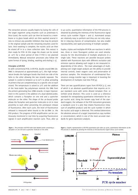
to 40 cycles, quantitative information of the PCR process is<br />
obtained by plotting the intensity of the fluorescence signal<br />
versus cycle number (Figure 1 and 2). Automated assays<br />
are relatively easy to perform and they are not only valuable<br />
in reducing chances of contamination, but also enable<br />
reproducibility and rapid processing of multiple samples<br />
Duplex, triplex and multiplex RT-PCRs are reactions in which<br />
two, three or more fluorogenic probes are used simultaneously<br />
for the discrimination of multiple amplicons in a<br />
single tube. These reactions are possible because probes<br />
labeled with fluorescent dyes with different excitation and<br />
emission spectra allowing each target to me measured independently<br />
of the others. The main advantages of multiplexing<br />
over single-target analysis are the ability to provide<br />
internal controls, lower reagent costs and preservation of<br />
precious samples. The introduction of combinatorial fluorescence<br />
energy transfer tags is important in boosting the<br />
development of multiplex real-time PCR [4].<br />
There are two quantifications types from RT-PCR [5-7], one<br />
of which is an absolute quantification that requires an input<br />
standard curve with series diluted template (five- or<br />
tenfold serial dilution). This curve is used as a reference<br />
standard for extrapolating quantitative information for target<br />
nucleic acid. Using the known copy level of the standard<br />
reagent, the software of the PCR instrument generates<br />
a standard curve in a plot that relates fluorescence (measure<br />
of amplified product) and the cycle number in which<br />
the nucleic acid target is detected. This method determines<br />
the absolute amount of a target (expressed as copy number<br />
or concentration), which is one of the most accurate standards<br />
for gene expression analysis.<br />
Figure 2. Graphic representation of RT-PCR quantitative plot after 40 cycles, quantitative information of the<br />
PCR process is obtained by plotting the intensity of the fluorescence signal versus cycle number.<br />
www.ijms.info • <strong>2012</strong> | Vol 1 | <strong>Issue</strong> 1 The International Journal of Medical Students 29