Selected MALDI Imaging Publications - Bruker Daltonics
Selected MALDI Imaging Publications - Bruker Daltonics
Selected MALDI Imaging Publications - Bruker Daltonics

Create successful ePaper yourself
Turn your PDF publications into a flip-book with our unique Google optimized e-Paper software.
Innovation with Integrity<br />
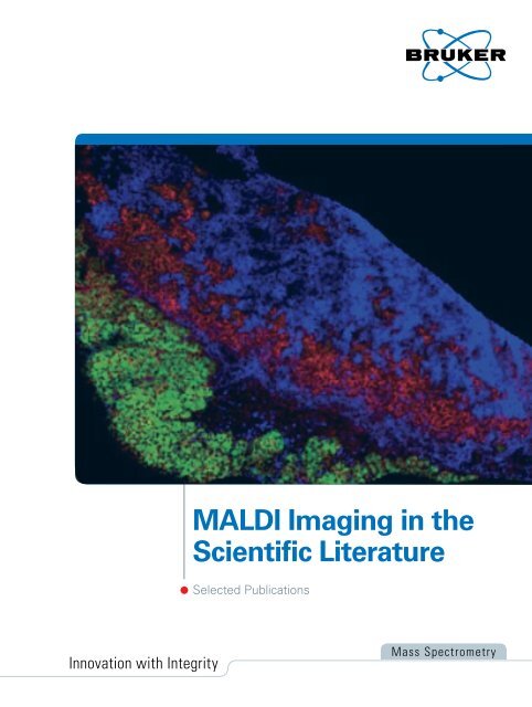
<strong>MALDI</strong> <strong>Imaging</strong> in the<br />
Scientific Literature<br />
<strong>Selected</strong> <strong>Publications</strong><br />
Mass Spectrometry
Figure 1: Drug <strong>Imaging</strong> / Histopharmacology:<br />
Correlation of<br />
histology and ion image in dog<br />
liver sections. The ion image (50<br />
µm spatial resolution) shows the<br />
lapatinib metabolite M10, localized<br />
only in the regions associated with<br />
the inflammation. Adapted from:<br />
Castellino et al. (2011) Bioanalysis<br />
3(21),2427-41 with kind permission<br />
of the authors.<br />
<strong>Selected</strong> <strong>MALDI</strong> <strong>Imaging</strong> <strong>Publications</strong><br />
Reviews<br />
<strong>Selected</strong> review articles on different <strong>MALDI</strong><br />
<strong>Imaging</strong> application areas<br />
Castellino S, Groseclose MR, Wagner D (2011(<br />
<strong>MALDI</strong> imaging mass spectrometry: bridging<br />
biology and chemistry in drug development.<br />
Bioanalysis. 3(21):2427-41<br />
This review explains how the combination<br />
of <strong>MALDI</strong> imaging with histology can solve<br />
problems in drug development. The review<br />
is illustrated with real life example from<br />
ClaxoSmithKline‘s DMPK unit, the data are<br />
acquired on a Solarix instrument. Highlights of<br />
the presented data include a Lapatinib metabolite<br />
that is only found in inflamed lesions in<br />
the liver, and another metabolite that is highly<br />
enriched in small bile ducts.<br />
Schwamborn K, Caprioli RM (2010)<br />
Molecular imaging by mass spectrometry -<br />
looking beyond classical histology.<br />
Nat Rev Cancer. 10(9):639-46<br />
A comprehensive review on imaging mass<br />
spectrometry in clinical cancer research.<br />
Walch A, Rauser S, Deininger SO, Hofler H<br />
(2008)<br />
<strong>MALDI</strong> <strong>Imaging</strong> mass spectrometry for direct<br />
tissue analysis: a new frontier for molecular<br />
histology.<br />
Histochem Cell Biol. 130(3):421-34<br />
A general review on imaging mass spectrometry<br />
with a detailed description on the importance<br />
of the combining histological information<br />
with <strong>MALDI</strong> <strong>Imaging</strong>.<br />
H&E stain<br />
Serial tissue sections<br />
Rauser S, Deininger SO, Suckau D, Hofler H,<br />
Walch A (2010)<br />
Approaching <strong>MALDI</strong> molecular imaging for<br />
clinical proteomic research: current state and<br />
fields of application.<br />
Expert Rev Proteomics. 7(6):927-41<br />
This review focuses on the application of<br />
<strong>MALDI</strong> <strong>Imaging</strong> for clinical proteomic research.<br />
It features an extensive discussion and explanation<br />
of relevant clinical questions where <strong>MALDI</strong><br />
<strong>Imaging</strong> can be applied.<br />
Bonnel D, Legouffe R, Willand N, Baulard A,<br />
Hamm G, Deprez B, Stauber J (2011)<br />
<strong>MALDI</strong> <strong>Imaging</strong> techniques dedicated to drugdistribution<br />
studies.<br />
Bioanalysis. 3(12):1399-406<br />
This review describes the use of <strong>MALDI</strong><br />
<strong>Imaging</strong> for drug-distribution studies. It also<br />
features an extensive discussion of <strong>Bruker</strong>s<br />
innovative FAST-SRM mode for drug imaging<br />
on <strong>MALDI</strong>-TOF instruments.<br />
Grassl J, Taylor NL, Millar AH (2011)<br />
Matrix-assisted laser desorption/ionisation<br />
mass spectrometry imaging and its development<br />
for plant protein imaging.<br />
Plant Methods. 7(1):21<br />
This review describes the use of <strong>MALDI</strong><br />
<strong>Imaging</strong> for plant research.<br />
Ion images
Clinical Research<br />
<strong>MALDI</strong> <strong>Imaging</strong> is perfectly suited for clinical<br />
biomarker studies. This sections contains<br />
selected papers that have used <strong>MALDI</strong><br />
<strong>Imaging</strong> to study human patient samples<br />
Meding S, Nitsche U, Balluff B, Elsner M,<br />
Rauser S, Schone C, Nipp M, Maak M, Feith M,<br />
Ebert MP, Friess H, Langer R, Hofler H, Zitzelsberger<br />
H, Rosenberg R, Walch A (2012)<br />
Tumor Classification of Six Common Cancer<br />
Types Based on Proteomic Profiling by <strong>MALDI</strong><br />
<strong>Imaging</strong>.<br />
J Proteome Res. 11(3):1996-2003<br />
The carcinoma of unknown primary (CUP) is a<br />
challenge in clinical oncology. <strong>MALDI</strong> imaging<br />
can classify tissue based on protein profiles. In<br />
this article, the authors describe how molecular<br />
classifiers allowed to differentiate different<br />
types of tumor. Interestingly, they were able to<br />
classify liver metastases from a known origin<br />
(colon) correctly. This shows that, in principle, a<br />
classification of metastases of unknown origin<br />
might be possible.<br />
Balluff B, Rauser S, Meding S, Elsner M,<br />
Schone C, Feuchtinger A, Schuhmacher C,<br />
Novotny A, Jutting U, Maccarrone G, Sarioglu<br />
H, Ueffing M, Braselmann H, Zitzelsberger<br />
H, Schmid RM, Hofler H, Ebert MP, Walch A<br />
(2011)<br />
<strong>MALDI</strong> <strong>Imaging</strong> Identifies Prognostic Seven-<br />
Protein Signature of Novel Tissue Markers in<br />
Intestinal-Type Gastric Cancer.<br />
Am J Pathol. 179(6):2720-9<br />
A comprehensive <strong>MALDI</strong> imaging study of<br />
gastric cancer: A seven protein signature was<br />
found to correlate with survival in a discovery<br />
cohort (n= 63). Three of the proteins were<br />
identified and the findings from the discovery<br />
cohort were subsequently validated on a larger<br />
independet cohort by immunohistochemistry.<br />
Hardesty WM, Kelley MC, Mi D, Low RL,<br />
Caprioli RM (2011)<br />
Protein signatures for survival and recurrence in<br />
metastatic melanoma.<br />
J Proteomics. 74(7):1002-14<br />
A study that utilised <strong>MALDI</strong> <strong>Imaging</strong> for survival<br />
and reccurence in metastatic melanoma.<br />
Twelve proteins could be associated with<br />
survival and three proteins were found to be<br />
associated with reccurence.<br />
Le Faouder J, Laouirem S, Chapelle M,<br />
Albuquerque M, Belghiti J, Degos F, Paradis V,<br />
Camadro JM, Bedossa P (2011)<br />
<strong>Imaging</strong> mass spectrometry provides fingerprints<br />
for distinguishing hepatocellular carcinoma<br />
from cirrhosis.<br />
J Proteome Res. 10(8):3755-65<br />
The differentiation of liver carcinoma and its<br />
predecessor, liver cirrhosis, was analyzed by<br />
<strong>MALDI</strong> <strong>Imaging</strong>. Among other mass signals,<br />
Ubiquitin was found to be up-regulated in the<br />
tumors, as was confirmed by immunohistochemistry.<br />
This paper also uses a supervised<br />
classification to differentiate cancerous from<br />
cirrhotic regions and shows how molecular<br />
images can be constructed from classification<br />
results („Classimaging“).<br />
Han EC, Lee YS, Liao WS, Liu YC, Liao HY,<br />
Jeng LB (2011)<br />
Direct tissue analysis by <strong>MALDI</strong>-TOF mass<br />
spectrometry in human hepatocellulat carcinoma.<br />
Clin Chim Acta. 412(3-4):230-9<br />
At present, prognosis of hepatocellular carcinoma<br />
(HCC) relies primarily on histopathological<br />
imaging applications after surgical removal.<br />
In this study, <strong>MALDI</strong> <strong>Imaging</strong> was utilised for<br />
classification of various tumour to supplement<br />
current histophatological analysis in tumour<br />
margins. A progressive change of fourteen<br />
peaks was observed in a distance-dependent<br />
manner from non-tumour to tumour regions of<br />
the junction section of HCC.<br />
Zaima N, Sasaki T, Tanaka H, Cheng XW,<br />
Onoue K, Hayasaka T, Goto-Inoue N, Enomoto<br />
H, Unno N, Kuzuya M, Setou M (2011)<br />
<strong>Imaging</strong> mass spectrometry-based histopathologic<br />
examination of atherosclerotic lesions.<br />
Atherosclerosis. 217(2):427-32<br />
This article describes the investigation of<br />
artherosclerotic lesions of ApoE deficient mice<br />
and humans with peripheral artery occlusive<br />
disease using IMS-based histopathologic<br />
examination (IbHE). Several lipids such as<br />
cholesterol linoleate (CE 18:2) and cholesterol<br />
oleate (CE 18:1) were specifically found in the<br />
lipid rich regions of both human and mouse,<br />
whilst triacylglycerol was localised in the<br />
lipid-rich regions of human samples. Other<br />
molecules were found to be localised in the<br />
calcified regions.
Schwamborn K, Krieg RC, Jirak P, Ott G,<br />
Knuchel R, Rosenwald A, Wellmann A (2010)<br />
Application of <strong>MALDI</strong> <strong>Imaging</strong> for the diagnosis<br />
of classical Hodgkin lymphoma.<br />
J Cancer Res Clin Oncol. 136(11):1651-5<br />
In this study, Hodgkin lymphoma was investigated<br />
by <strong>MALDI</strong> <strong>Imaging</strong>. the differentially<br />
expressed protein profiles allowed the differentiation<br />
between classical Hodgkin lymphoma<br />
and lymphadenitis with a sensitivity and specificity<br />
of 83.92 and 89.37%, respectively.<br />
Tanaka H, Zaima N, Yamamoto N, Sagara D,<br />
Suzuki M, Nishiyama M, Mano Y, Sano M,<br />
Hayasaka T, Goto-Inoue N, Sasaki T, Konno H,<br />
Unno N, Setou M (2010)<br />
<strong>Imaging</strong> mass spectrometry reveals unique<br />
lipid distribution in primary varicose veins.<br />
Eur J Vasc Endovasc Surg. 40(5):657-63<br />
Various lipid molecules have been measured<br />
in varicose and normal human veins. The<br />
accumulation of various specific lipids (identified<br />
using a combination of imaging mass<br />
spectrometry and MS/MS) in varicose veins<br />
suggested that inflammation with abnormal<br />
lipid metabolism is involved in the devlopment<br />
of varicose veins.<br />
Oppenheimer SR, Mi D, Sanders ME,<br />
Caprioli RM (2010)<br />
Molecular analysis of tumor margins by <strong>MALDI</strong><br />
mass spectrometry in renal carcinoma.<br />
J Proteome Res. 9(5):2182-90<br />
In surgery it is important to evaluate that the<br />
tumor margin has been completely resected.<br />
The tumor margin is usually evauated by histology.<br />
The <strong>MALDI</strong> <strong>Imaging</strong> analysis of tumor<br />
margins in renal carcinoma in this study revelaed<br />
that proteins that characteristically upregulated<br />
in the tumour were also up-regulated<br />
in an area adjacent to the tumor, indicating a<br />
molecular tumor margin that is different from<br />
the histological tumor margin.<br />
Balluff B, Elsner M, Kowarsch A, Rauser S,<br />
Meding S, Schuhmacher C, Feith M, Herrmann<br />
K, Rocken C, Schmid RM, Hofler H, Walch A,<br />
Ebert MP (2010)<br />
Classification of HER2/neu status in gastric<br />
cancer using a breast-cancer derived proteome<br />
classifier.<br />
J Proteome Res. 9(12):6317-22<br />
A paper that follows on an earlier breast cancer<br />
study (Rauser et al. J. Proteome Res. , 2010).<br />
It showed that a protein profile based classifier,<br />
trained to classify Her2 status in breast cancer,<br />
was also able to classify Her 2 status in an<br />
independent gastric cancer cohort, providing<br />
strong evidence of the robustness of <strong>MALDI</strong><br />
<strong>Imaging</strong> based tissue classifications.<br />
Willems SM, van Remoortere A, van Zeijl R,<br />
Deelder AM, McDonnell LA, Hogendoorn PC<br />
(2010)<br />
<strong>Imaging</strong> mass spectrometry of myxoid sarcomas<br />
identifies proteins and lipids specific to<br />
tumour type and grade, and reveals biochemical<br />
intratumour heterogeneity.<br />
J Pathol. 222(4):400-9<br />
An article that investigates the expression<br />
of proteins and lipids in myxoid sarcomas.<br />
Peptide, protein and lipid profiles have been<br />
correlated with tumor grades and intra-tumor<br />
heterogeneity has been investigated by hiearchical<br />
clustering of imaging data. In particular,<br />
lipid changes observed in myxoid liposarcomas<br />
could be related to pathways known to be<br />
affected during tumor development.<br />
Rauser S, Marquardt C, Balluff B, Deininger<br />
SO, Albers C, Belau E, Hartmer R, Suckau D,<br />
Specht K, Ebert MP, Schmitt M, Aubele M,<br />
Hofler H, Walch A (2010)<br />
Classification of HER2 receptor status in breast<br />
cancer tissues by <strong>MALDI</strong> <strong>Imaging</strong> mass spectrometry.<br />
J Proteome Res. 9(4):1854-63<br />
In this article, <strong>MALDI</strong> <strong>Imaging</strong> has been used<br />
to classify HER2 receptor status in human<br />
breast cancer specimens. Protein profiles<br />
correlated strongly with HER2 expression. A<br />
protein signal with a particularly high correlation<br />
with HER2 expression has been identified as<br />
cysteine rich intestinal protein 1, a protein that<br />
was previously not known to be associated<br />
with HER2 expression.<br />
Cazares LH, Troyer D, Mendrinos S, Lance RA,<br />
Nyalwidhe JO, Beydoun HA, Clements MA,<br />
Drake RR, Semmes OJ (2009)<br />
<strong>Imaging</strong> mass spectrometry of a specific<br />
fragment of mitogen-activated protein kinase/<br />
extracellular signal-regulated kinase kinase<br />
kinase 2 discriminates cancer from uninvolved<br />
prostate tissue.<br />
Clin Cancer Res. 15(17):5541-51<br />
Prostate cancer is a cancer type in which the<br />
differentiation of begning tissue and cancrous<br />
tissue may be difficult. In this study prostate<br />
cancer and normal tissue was differentiated by<br />
<strong>MALDI</strong> <strong>Imaging</strong>. The original findings in a small<br />
discovery set were verified by the analysis of<br />
a validation set with 54 tissue sections. One<br />
marker that correlated with the tumor was
identified as a fragment of mitogen-activated<br />
protein kinase/extracellular signal regulated<br />
kinase kinase kinase 2 (MEKK2).<br />
Groseclose MR, Massion PP, Chaurand P,<br />
Caprioli RM (2008)<br />
High-throughput proteomic analysis of formalinfixed<br />
paraffin-embedded tissue microarrays<br />
using <strong>MALDI</strong> <strong>Imaging</strong> mass spectrometry.<br />
Proteomics. 8(18):3715-24<br />
The analysis of tissue microarrays is of great<br />
interest, since it allows the analysis of a large<br />
number of samples in a relatively short time.<br />
This article describes how <strong>MALDI</strong> <strong>Imaging</strong><br />
can be used to analyze FFPE tissue by tryptic<br />
digestion and how tumor types can be classified<br />
based on their peptide patterns. The result<br />
of the classification of the individual pixels is<br />
visualized as image („Class imaging“).<br />
Lemaire R, Menguellet SA, Stauber J,<br />
Marchaudon V, Lucot JP, Collinet P,<br />
Farine MO, Vinatier D, Day R, Ducoroy P,<br />
Salzet M, Fournier I (2007)<br />
Specific <strong>MALDI</strong> <strong>Imaging</strong> and profiling for<br />
biomarker hunting and validation: fragment of<br />
the 11S proteasome activator complex, Reg<br />
alpha fragment, is a new potential ovary cancer<br />
biomarker.<br />
J Proteome Res. 6(11):4127-34<br />
Early paper that shows the power of <strong>MALDI</strong><br />
<strong>Imaging</strong> in clincial proteomic research for the<br />
analysis of ovary cancer. A specific 84 amino<br />
acid fragment from the protein Reg-alpha was<br />
identified as tumor marker and validated with<br />
other techniques, including immunohistochemsitry.<br />
Figure 2: Class <strong>Imaging</strong>: Visual representation of the statistical<br />
classification of four biopsies compared to the assignment based on<br />
histology. From: Groseclose et. al (2008),Proteomics 8(18) 3715<br />
© 2008 WILEY-VCH Verlag GmbH & Co. KGaA, Weinheim<br />
Drug imaging / Xenobiotics /<br />
Pharmaceutical research<br />
<strong>MALDI</strong> <strong>Imaging</strong> can be used to directly<br />
measure drugs, metabolites and xenobiotics in<br />
tissue<br />
Seppala U, Francese S, Turillazzi S, Moneti G,<br />
Clench M, Barber D (2011)<br />
In situ imaging of honeybee (Apis mellifera)<br />
venom components from aqueous and aluminum<br />
hydroxide-adsorbed venom immunotherapy<br />
preparations.<br />
J Allergy Clin Immunol. 129(5):1314-1320.e3<br />
An article that utilised <strong>MALDI</strong> <strong>Imaging</strong> to<br />
assess purified aqueous and aluminum hydroxide<br />
(Al[OH]3)adsorbed purified honeybee<br />
(Apis mellifera) venom (HBV) immunotherapy<br />
preparations in situ, which can reduce the incidence<br />
of side effects associated with venom<br />
immunotherapy. The study assessed the<br />
distribution of HBV components and showed<br />
that the majority of low-molecular-weight HBV<br />
components are rapidly removed from the site<br />
of venom immunotherapy administration. Furthermore,<br />
Al(OH)3-adsorbed HBV preparation<br />
demonstrated a depot effect, prolonging the<br />
availability of bee venom allergens at the site of<br />
administration.<br />
Goodwin RJ, Mackay CL, Nilsson A, Harrison<br />
D, Farde L, Andren PE, Iverson SL (2011)<br />
Qualitative and quantitative <strong>MALDI</strong> <strong>Imaging</strong><br />
of the positron emission tomography ligands<br />
raclopride (a D2 dopamine antagonist) and<br />
SCH 23390 (a D1 dopamine antagonist) in rat<br />
brain tissue sections using a solvent-free dry<br />
matrix application method.<br />
Anal Chem. 83(24):9694-701<br />
An article that describes a dry matrix preparation<br />
for the detection of two dopamine receptor<br />
antagonists. Rats were dosed at 5 mg/kg and<br />
the distribution of the drug in the brain could be<br />
detected.<br />
Enthaler B, Pruns JK, Wessel S, Rapp C,<br />
Fischer M, Wittern KP (2011)<br />
Improved sample preparation for <strong>MALDI</strong>-MSI<br />
of endogenous compounds in skin tissue sections<br />
and mapping of exogenous active compounds<br />
subsequent to ex-vivo skin penetration.<br />
Anal Bioanal Chem. 402(3):1159-67<br />
In this article, <strong>MALDI</strong> <strong>Imaging</strong> was utilised for<br />
the determination of the pathway and depth of<br />
penetration of an exogenous model compound,<br />
Nile Red, after ex-vivo penetration into<br />
porcine skin. Additionally, the localisation of
endogenous compounds, including cholesterol<br />
sulphate, was measured directly on tissue at a<br />
resolution of approximately 30 m.<br />
Miki A, Katagi M, Kamata T, Zaitsu K,<br />
Tatsuno M, Nakanishi T, Tsuchihashi H,<br />
Takubo T, Suzuki K (2011)<br />
<strong>MALDI</strong>-TOF and <strong>MALDI</strong>-FTICR <strong>Imaging</strong> mass<br />
spectrometry of methamphetamine incorporated<br />
into hair.<br />
J Mass Spectrom. 46(4):411-6<br />
This paper describes the potential use of<br />
<strong>MALDI</strong> <strong>Imaging</strong> in forensics. Cocaine was<br />
measured directly in human hair and the spatial<br />
profile of cocaine in the hair could be translated<br />
to a temporary consumption profile.<br />
Marshall P, Toteu-Djomte V, Bareille P, Perry H,<br />
Brown G, Baumert M, Biggadike K (2010)<br />
Correlation of skin blanching and percutaneous<br />
absorption for glucocorticoid receptor agonists<br />
by matrix-assisted laser desorption ionization<br />
mass spectrometry imaging and liquid extraction<br />
surface analysis with nanoelectrospray<br />
ionization mass spectrometry.<br />
Anal Chem. 82(18):7787-94<br />
In this study, the percutaneous absorption of<br />
three novel nonsteroid glucocorticoid receptor<br />
agonists has been investigated.<br />
Nilsson A, Fehniger TE, Gustavsson L,<br />
Andersson M, Kenne K, Marko-Varga G,<br />
Andren PE (2010)<br />
Fine mapping the spatial distribution and<br />
concentration of unlabeled drugs within tissue<br />
micro-compartments using imaging mass<br />
spectrometry.<br />
PLoS One. 5(7):e11411<br />
This article describes the use of <strong>MALDI</strong><br />
<strong>Imaging</strong> for monitoring in vivo transport of<br />
unlabelled drugs within specific organ tissue<br />
compartments. A reference compound (tiotropium)<br />
would be tracked and quantified within<br />
the lungs of dosed rats, using systematic pointby-point<br />
MS and MS/MS sampling across the<br />
tissue.<br />
Francese S, Lambardi D, Mastrobuoni G,<br />
la Marca G, Moneti G, Turillazzi S (2009)<br />
Detection of honeybee venom in envenomed<br />
tissues by direct <strong>MALDI</strong> MSI.<br />
J Am Soc Mass Spectrom. 20(1):112-23<br />
In this study, <strong>MALDI</strong> <strong>Imaging</strong> has been used<br />
to map the diffusion and distribution of venom<br />
allergen and toxins in skin.<br />
Acquadro E, Cabella C, Ghiani S, Miragoli L,<br />
Bucci EM, Corpillo D (2009)<br />
Matrix-assisted laser desorption ionization<br />
imaging mass spectrometry detection of a<br />
magnetic resonance imaging contrast agent in<br />
mouse liver.<br />
Anal Chem. 81(7):2779-84<br />
This article describes the use of <strong>MALDI</strong><br />
<strong>Imaging</strong> to detect a magnetic resonance<br />
imaging (MRI) contrast agent in both frozen and<br />
paraformaldehyde-fixed mouse livers after its in<br />
vivo administration.<br />
Dekker LJ, van Kampen JJ, Reedijk ML,<br />
Burgers PC, Gruters RA, Osterhaus AD,<br />
Luider TM (2009)<br />
A mass spectrometry based imaging method<br />
developed for the intracellular detection of HIV<br />
protease inhibitors.<br />
Rapid Commun Mass Spectrom. 23(8):1183-8<br />
HIV protease inhibitors have been measured in<br />
cultured cells using <strong>MALDI</strong>-TOF and <strong>MALDI</strong>-<br />
FTMS.<br />
Cornett DS, Frappier SL, Caprioli RM (2008)<br />
<strong>MALDI</strong>-FTICR <strong>Imaging</strong> mass spectrometry of<br />
drugs and metabolites in tissue.<br />
Anal Chem. 80(14):5648-53<br />
In this work, the authors show how the high<br />
precision of FTMS mass measurements<br />
increase the confidence in the results of drug<br />
imaging experiments.<br />
Meistermann H, Norris JL, Aerni HR,<br />
Cornett DS, Friedlein A, Erskine AR,<br />
Augustin A, De Vera Mudry MC, Ruepp S,<br />
Suter L, Langen H, Caprioli RM, Ducret A<br />
(2006)<br />
Biomarker discovery by imaging mass spectrometry:<br />
transthyretin is a biomarker for gentamicin-induced<br />
nephrotoxicity in rat.<br />
Mol Cell Proteomics. 5(10):1876-86<br />
In this article, <strong>MALDI</strong> <strong>Imaging</strong> has been used<br />
to find a surrogate biomarker in kidney that indicates<br />
the nephrotoxic effect of an administered<br />
drug, gentamicin.
Model Organisms<br />
A selection of studies that have used <strong>MALDI</strong><br />
<strong>Imaging</strong> for general biological or biomedical<br />
research on model organisms<br />
Qureshi A, Subathra M, Grey A, Schey K,<br />
Del Poeta M, Luberto C (2010)<br />
Role of sphingomyelin synthase in controlling<br />
the antimicrobial activity of neutrophils against<br />
Cryptococcus neoformans.<br />
PLoS One. 5(12):e15587<br />
This study performed <strong>MALDI</strong> <strong>Imaging</strong> on lungs<br />
infected with Cryptococcus neoformans to<br />
provide new insights into the key host cellular<br />
pathway(s) necessary to control infection<br />
caused from inhalation of Cryptococcus neoformans.<br />
<strong>MALDI</strong> <strong>Imaging</strong> revealed specific lipid<br />
(SM - sphingomyelin) species are associated<br />
with neutrophil infiltration at the site of the<br />
infection.<br />
Magalhaes BS, Melo JA, Leite JR, Silva LP,<br />
Prates MV, Vinecky F, Barbosa EA, Verly RM,<br />
Mehta A, Nicoli JR, Bemquerer MP,<br />
Andrade AC, Bloch C Jr (2008)<br />
Post-secretory events alter the peptide content<br />
of the skin secretion of Hypsiboas raniceps.<br />
Biochem Biophys Res Commun. 377(4):1057-<br />
61<br />
In this article, <strong>MALDI</strong> <strong>Imaging</strong> demonstrated<br />
that mature raniseptin peptides (a novel family<br />
of antimicrobial peptides characterised from<br />
skin secretion of the anuran Hypsibas raniceps)<br />
are in fact secreted as intact molecules within a<br />
defined glandular domain of the dorsal skin,<br />
Figure 3: Statistical Analysis: Spatial segmentation map based on<br />
hierarchical clustering of a rat whole body section and principal component<br />
analysis of selected organs. From: Speir. et al. Proceedings of<br />
the 60th ASMS Conference on MassSpectrometry and Allied Topics,<br />
May 20-24,2012<br />
challenging the physiological role of the observed<br />
raniseptin fragments, identified only as part<br />
of the crude secretion.<br />
Brand GD, Krause FC, Silva LP, Leite JR,<br />
Melo JA, Prates MV, Pesquero JB, Santos EL,<br />
Nakaie CR, Costa-Neto CM, Bloch C Jr (2006)<br />
Bradykinin-related peptides from Phyllomedusa<br />
hypochondrialis.<br />
Peptides. 27(9):2137-46 link<br />
Frog mucus contains biologically active peptides<br />
that are e.g. protective of fungal infections<br />
or predators. <strong>MALDI</strong> <strong>Imaging</strong> has been used to<br />
map such peptides directly in frog skin.<br />
Neuroscience<br />
<strong>Selected</strong> <strong>MALDI</strong> <strong>Imaging</strong> papers in neuroscience<br />
Hanrieder J, Karlsson A, Falth M, Eriksson-<br />
Mammo S, Bergquist J, Andersson M (2011)<br />
L-DOPA-induced dyskinesia is associated with<br />
regional increase of striatal dynorphin peptides<br />
as elucidated by imaging mass spectrometry.<br />
Mol Cell Proteomics. 10(10):M111.009308<br />
<strong>MALDI</strong> <strong>Imaging</strong> was used for characterization,<br />
localization, and relative quantification of striatal<br />
neuropeptides in a rat model of l-DOPA-induced<br />
dyskinesia (LID) in Parkinson‘s disease.<br />
Yuki D, Sugiura Y, Zaima N, Akatsu H,<br />
Hashizume Y, Yamamoto T, Fujiwara M,<br />
Sugiyama K, Setou M (2011)<br />
Hydroxylated and non-hydroxylated sulfatide<br />
are distinctly distributed in the human cerebral<br />
cortex.<br />
Neuroscience. 193:44-53<br />
This article studies the distribution of sulfatide<br />
lipids in human cerebral cortex. It was found<br />
that hydroxylated and non-hydroxylated sulfatides<br />
are differently distributed in white and<br />
grey matter. An alzheimer brain has also been<br />
investigated and showed a similar distribution<br />
of sulfatides compared to normal tissue.<br />
Yang H, Sugiura Y, Ikegami K, Konishi Y,<br />
Setou M (2011)<br />
Axonal gradient of arachidonic acid-containing<br />
phosphatidylcholine and its dependence on<br />
actin dynamics.<br />
J Biol Chem. 287(8):5290-300<br />
This article used <strong>MALDI</strong> <strong>Imaging</strong> to characterise<br />
the distribution of phosphatidylcholine (PC)<br />
species in cultured neurons of superior cervical<br />
ganglia.
Sugiura Y, Taguchi R, Setou M (2011)<br />
Visualization of spatiotemporal energy<br />
dynamics of hippocampal neurons by mass<br />
spectrometry during a kainate-induced seizure.<br />
PLoS One. 6(3):e17952<br />
In this paper, <strong>MALDI</strong> <strong>Imaging</strong> combined with<br />
capillary electrophoresis (CE) mass spectrometry<br />
were utilised to visualise energy metabolism<br />
by imaging energy-related metabolites<br />
in mouse brains to reveal the spatiotemporal<br />
energy metabolism of the mouse hippocampus.<br />
Sugiura Y, Shimma S, Konishi Y, Yamada MK,<br />
Setou M (2008)<br />
<strong>Imaging</strong> mass spectrometry technology and<br />
application on ganglioside study; visualization<br />
of age-dependent accumulation of C20-ganglioside<br />
molecular species in the mouse hippocampus.<br />
PLoS One. 3(9):e3232<br />
The distrubtion of ganglioside molecular<br />
species in the brain was revealed by <strong>MALDI</strong><br />
<strong>Imaging</strong>, enabling discrimination of species<br />
raised from both oligosaccharide and ceramide<br />
structure by the difference in mass-to-charge<br />
ratio and structural elucidation by MSMS.<br />
Plants<br />
<strong>MALDI</strong> <strong>Imaging</strong> in the investigation of plants<br />
Poth AG, Mylne JS, Grassl J, Lyons RE, Millar<br />
AH, Colgrave ML, Craik DJ (2012)<br />
Cyclotides associate with leaf vasculature and<br />
are the products of a novel precursor in Petunia<br />
(Solanaceae).<br />
J Biol Chem. doi:10.1074/jbc.M112.370841<br />
Cyclotides are defense related cylic peptides. In<br />
this study the spatial distribution of cyclotides<br />
was assessed by <strong>MALDI</strong> <strong>Imaging</strong>. The major<br />
cyclotide component showed a non-uniform<br />
distribution in petunia leaves.<br />
Holscher D, Shroff R, Knop K, Gottschaldt M,<br />
Crecelius A, Schneider B, Heckel DG,<br />
Schubert US, Svatos A (2009)<br />
Matrix-free UV-laser desorption/ionization (LDI)<br />
mass spectrometric imaging at the single-cell<br />
level: distribution of secondary metabolites of<br />
Arabidopsis thaliana and Hypericum species.<br />
Plant J. 60(5):907-18<br />
A paper that describes the direct analysis<br />
of secondary metabolites in Hypericum and<br />
Arabodopsis. The distribution of Naphtodianthrones<br />
were measured in Hypericum leavesm<br />
placenta, stamens and styli. The highest achieved<br />
spatial resolution was 10 micrometers.<br />
<strong>MALDI</strong> <strong>Imaging</strong> methodology<br />
<strong>MALDI</strong> <strong>Imaging</strong> is a field that is under research<br />
itself. This section shows interesting ongoing<br />
development in this field<br />
Thomas A, Charbonneau JL, Fournaise E,<br />
Chaurand P (2012)<br />
Sublimation of new matrix candidates for high<br />
spatial resolution imaging mass spectrometry<br />
of lipids: enhanced information in both positive<br />
and negative polarities after 1,5-diaminonapthalene<br />
deposition.<br />
Anal Chem. 84(4):2048-54<br />
As matrix sublimation has demonstrated to<br />
be a powerful approach for high-resolution<br />
<strong>MALDI</strong> <strong>Imaging</strong> of lipids, this study aimed to<br />
evaluate current and novel matrix candidates<br />
for high spatial resolution <strong>MALDI</strong> <strong>Imaging</strong> mass<br />
spectrometry of lipids from tissue section after<br />
deposition by sublimation.<br />
Angel PM, Spraggins JM, Baldwin HS,<br />
Caprioli R (2012)<br />
Enhanced sensitivity for high spatial resolution<br />
lipid analysis by negative ion mode matrix assisted<br />
laser desorption ionization imaging mass<br />
spectrometry.<br />
Anal Chem. 84(3):1557-64<br />
A paper that shows high spatial resolution of 10<br />
micrometer with an Ultraflextreme on tissue.<br />
Yang J, Caprioli RM (2011)<br />
Matrix sublimation/recrystallization for imaging<br />
proteins by mass spectrometry at high spatial<br />
resolution.<br />
Anal Chem. 83(14):5728-34<br />
A paper that investigates improved sample<br />
preparation methods based on matrix sublimation.<br />
It shows the technical possibilities of<br />
resolution obtainable with <strong>Bruker</strong>s advanced<br />
instrumentation.<br />
Khatib-Shahidi S, Andersson M, Herman JL,<br />
Gillespie TA, Caprioli RM (2006)<br />
Direct molecular analysis of whole-body animal<br />
tissue sections by imaging <strong>MALDI</strong> mass spectrometry.<br />
Anal Chem. 78(18):6448-56<br />
A paper that shows how different substance<br />
classes can be measured in whole body sections<br />
of rats.
Method - Computational<br />
Computational methods are an important part<br />
of <strong>MALDI</strong> <strong>Imaging</strong> analysis. This section lists<br />
interesting papers in this field<br />
Trede D, Schiffler S, Becker M, Wirtz S,<br />
Steinhorst K, Strehlow J, Aichler M, Kobarg<br />
JH, Oetjen J, Dyatlov A, Heldmann S, Walch A,<br />
Thiele H, Maass P, Alexandrov T (2012)<br />
Exploring Three-Dimensional Matrix-Assisted<br />
Laser Desorption/Ionization <strong>Imaging</strong> Mass<br />
Spectrometry Data: Three-Dimensional Spatial<br />
Segmentation of Mouse Kidney.<br />
Anal Chem. doi:10.1021/ac300673y<br />
This paper describes an efficient computational<br />
pipeline which allows analysis and interpretation<br />
of three dimensional <strong>MALDI</strong> <strong>Imaging</strong><br />
datasets.<br />
Andersson M, Groseclose MR, Deutch AY,<br />
Caprioli RM (2008)<br />
<strong>Imaging</strong> mass spectrometry of proteins and<br />
peptides: 3D volume reconstruction.<br />
Nat Methods. 5(1):101-8<br />
The article presents an optimised step-by-step<br />
procedure describing how to make 3D volume<br />
reconstructions of <strong>MALDI</strong> <strong>Imaging</strong> data, as<br />
applied to the rat brain.<br />
Sinha TK, Khatib-Shahidi S, Yankeelov TE,<br />
Mapara K, Ehtesham M, Cornett DS, Dawant<br />
BM, Caprioli RM, Gore JC (2008)<br />
Integrating spatially resolved three-dimensional<br />
<strong>MALDI</strong> IMS with in vivo magnetic resonance<br />
imaging.<br />
Nat Methods. 5(1):57-9<br />
The article describes a method for integration<br />
of 3D volume reconstructions of proteomic<br />
<strong>MALDI</strong> <strong>Imaging</strong> datasets of whole mouse<br />
heads with in vivo high-resolution images from<br />
other imaging modalities (magnetic resonance<br />
imaging).<br />
Hanselmann M, Kirchner M, Renard BY,<br />
Amstalden ER, Glunde K, Heeren RM,<br />
Hamprecht FA (2008)<br />
Concise representation of mass spectrometry<br />
images by probabilistic latent semantic analysis.<br />
Anal Chem. 80(24):9649-58<br />
This paper describes a very interesting statistical<br />
method, named probabibilistic latent<br />
sematics analysis (pLSA), for the concise representation<br />
of complex imaging mass spectrometry<br />
data. Although the data was not recorded<br />
on <strong>Bruker</strong> instrumentation,In the meantime,<br />
pLSA has been implemented in <strong>Bruker</strong>s <strong>MALDI</strong><br />
imaging software package.<br />
Figure 4: Drug <strong>Imaging</strong> / Histopharmacology: Sagittal rabbit brain section showing distribution of a cysteine<br />
conjugate metabolite. (A) Monochromatic ion image showing brain distribution of cysteine conjugate metabolite<br />
(170 µm spatial resolution). (B) Same ion image shown in rainbow intensity format. (C) Hematoxylin and eosin<br />
histology slide showing regions of the rabbit brain. From: Castellino et al. (2011) Bioanalysis 3(21),2427-41 with<br />
kind permission of the authors.
Development<br />
<strong>MALDI</strong> imaging papers in developmental<br />
biology<br />
Lagarrigue M, Becker M, Lavigne R,<br />
Deininger SO, Walch A, Aubry F, Suckau D,<br />
Pineau C (2011)<br />
Revisiting rat spermatogenesis with <strong>MALDI</strong><br />
<strong>Imaging</strong> at 20-microm resolution.<br />
Mol Cell Proteomics. 10(3):M110.005991<br />
This article investigates the protein expression<br />
in rat testis. It shows state-of-the-art protein<br />
images at a spatial resolution of 20 micrometer.<br />
The observed protein profiles were linked to<br />
the stages of spermatogenesis as determined<br />
by histology. The article therefore shows the<br />
importance of the correlation of molecular<br />
<strong>MALDI</strong> images and histology. Some of the<br />
observed proteins were identified and their<br />
expression further investigated by immunohistochemistry.<br />
Goto-Inoue N, Hayasaka T, Zaima N, Setou M<br />
(2009)<br />
The specific localization of seminolipid molecular<br />
species on mouse testis during testicular<br />
maturation revealed by imaging mass spectrometry.<br />
Glycobiology. 19(9):950-7<br />
<strong>MALDI</strong> <strong>Imaging</strong> was utilised to show cell-specific<br />
localisation of each molecular species of<br />
seminolipid (a unique sulphated glyceroglycolipid,<br />
which constitutes more than 90% of the<br />
glycolipid in mammalian testis) during testicular<br />
maturation.<br />
Burnum KE, Cornett DS, Puolitaival SM,<br />
Milne SB, Myers DS, Tranguch S, Brown HA,<br />
Dey SK, Caprioli RM (2009)<br />
Spatial and temporal alterations of phospholipids<br />
determined by mass spectrometry during<br />
mouse embryo implantation.<br />
J Lipid Res. 50(11):2290-8<br />
<strong>MALDI</strong> <strong>Imaging</strong> was used to characterise<br />
the spatial patial and temporal distribution of<br />
phospholipid species associated with mouse<br />
embryo implantation. LC-ESI-MSMS validated<br />
the phospholipid distribution patterns observed<br />
using <strong>MALDI</strong> <strong>Imaging</strong>.<br />
Burnum KE, Tranguch S, Mi D, Daikoku T, Dey<br />
SK, Caprioli RM (2008)<br />
<strong>Imaging</strong> mass spectrometry reveals unique<br />
protein profiles during embryo implantation.<br />
Endocrinology. 149(7):3274-8<br />
Chaurand P, Rahman MA, Hunt T, Mobley JA,<br />
Gu G, Latham JC, Caprioli RM, Kasper S (2008)<br />
Monitoring mouse prostate development by<br />
profiling and imaging mass spectrometry.<br />
Mol Cell Proteomics. 7(2):411-23<br />
This article describes the use of <strong>MALDI</strong><br />
<strong>Imaging</strong> to evaluate protein expression profiles<br />
in the normal mouse prostate during development,<br />
at sexual maturation and in adult stages.<br />
Microbiology<br />
Interesting papers of the use of <strong>MALDI</strong> <strong>Imaging</strong><br />
to study microbial interactions<br />
Liu WT, Kersten RD, Yang YL, Moore BS,<br />
Dorrestein PC (2011)<br />
<strong>Imaging</strong> mass spectrometry and genome<br />
mining via short sequence tagging identified<br />
the anti-infective agent arylomycin in Streptomyces<br />
roseosporus.<br />
J Am Chem Soc. 133(45):18010-3<br />
A paper that utilised <strong>MALDI</strong> <strong>Imaging</strong> along with<br />
a peptidogenomic approach (expanded to short<br />
sequence tagging SST) for discovery of antiinfective<br />
agent arylomycin and its biosynthetic<br />
gene cluster in an industrial daptomycin producing<br />
strain Streptomyces roseosporus.<br />
Liu WT, Yang YL, Xu Y, Lamsa A, Haste NM,<br />
Yang JY, Ng J, Gonzalez D, Ellermeier CD,<br />
Straight PD, Pevzner PA, Pogliano J, Nizet V,<br />
Pogliano K, Dorrestein PC (2010)<br />
<strong>Imaging</strong> mass spectrometry of intraspecies<br />
metabolic exchange revealed the cannibalistic<br />
factors of Bacillus subtilis.<br />
Proc Natl Acad Sci U S A. 107(37):16286-90<br />
This paper studies molecular interactions in<br />
bacterial cannibalism.<br />
Yang YL, Xu Y, Straight P, Dorrestein PC (2009)<br />
Translating metabolic exchange with imaging<br />
mass spectrometry.<br />
Nat Chem Biol. 5(12):885-7<br />
In this article the metabolic interaction of<br />
different bacterial strains on an agar plate are<br />
monitored by <strong>MALDI</strong> <strong>Imaging</strong>. This allowed a<br />
direct molecular characterization of a chemical<br />
fight between the bacterial strains and provided<br />
evidence that Bacillus subtilis silenced the<br />
defensive arsenal of Streptomyces coelicolor.
— 200 µm<br />
m/z726.5 PE 36:2<br />
m/z774.6 PE-p 40:6<br />
m/z885.6 PI 38:4<br />
Figure 5: Lipids in mouse cerebellum measured at 10 µm spatial resolution.<br />
Reprinted from: Thomas A, Charbonneau JL, Fournaise E, Chaurand P. Sublimation of new matrix<br />
candidates for high spatial resolution imaging mass spectrometry of lipids: enhanced information in both<br />
positive and negative polarities after 1,5-diaminonapthalene deposition. Anal Chem. 2012;84(4):2048-54<br />
Copyright (2012) American Chemical Society<br />
Sample Preparation<br />
m/z664.6 CerP 18:0<br />
m/z747.5 PG 34:1<br />
m/z786.5 PS 36:2<br />
m/z890.6 ST 24:0<br />
Data Acquisition<br />
Figure 6: <strong>Bruker</strong> offers the complete <strong>MALDI</strong> <strong>Imaging</strong> solution.<br />
m/z700.5 PE-p 34:1<br />
m/z762.6 PE 38:6<br />
m/z834.6 PS 40:6<br />
m/z700.5 + m/z 786.5<br />
m/z715.6 PA-o 38:1<br />
m/z766.6 PE 38:4<br />
m/z878.6 ST 22:0 (OH)<br />
m/z834.6 + m/z 890.6<br />
Data Evaluation<br />
Statistical Analysis
<strong>Bruker</strong> Author<br />
<strong>Bruker</strong> does actively contribute to research in<br />
the <strong>MALDI</strong> <strong>Imaging</strong> field, having introduced<br />
key innovations in both hard- and software.<br />
This section lists papers with key conttributions<br />
form <strong>Bruker</strong> authors<br />
Deininger SO, Cornett DS, Paape R, Becker M,<br />
Pineau C, Rauser S, Walch A, Wolski E (2011)<br />
Normalization in <strong>MALDI</strong>-TOF <strong>Imaging</strong> datasets<br />
of proteins: practical considerations.<br />
Anal Bioanal Chem. 401(1):167-81<br />
Normalization is an important step in the data<br />
preparation of <strong>MALDI</strong> <strong>Imaging</strong> data, mostly to<br />
account random real-world influences in the<br />
data. Commonly used normalization techniques<br />
such as the total ion count, can introduce<br />
artifacts in rare cases. This paper discusses the<br />
applicability of different normalization approaches<br />
and introduces robust methods, such<br />
as normalization to median intensity. These<br />
normalization options are included in <strong>Bruker</strong>s<br />
<strong>MALDI</strong> <strong>Imaging</strong> software.<br />
Alexandrov T, Becker M, Deininger SO,<br />
Ernst G, Wehder L, Grasmair M,<br />
von Eggeling F, Thiele H, Maass P (2010)<br />
Spatial segmentation of imaging mass spectrometry<br />
data with edge-preserving image<br />
denoising and clustering.<br />
J Proteome Res. 9(12):6535-46<br />
This paper describes how <strong>MALDI</strong> <strong>Imaging</strong> data<br />
can be improved by spatially aware de-noising.<br />
Compared to mere smoothing of the images,<br />
edge-preserving de-noising can improve<br />
<strong>MALDI</strong> images, while keeping highly resolved<br />
fine structures in the images.<br />
Agar NY, Kowalski JM, Kowalski PJ, Wong JH,<br />
Agar JN (2010)<br />
Tissue preparation for the in situ <strong>MALDI</strong> MS<br />
<strong>Imaging</strong> of proteins, lipids, and small molecules<br />
at cellular resolution.<br />
Methods Mol Biol. 656:415-31<br />
For research use only. Not for use in diagnostic procedures.<br />
<strong>Bruker</strong> Daltonik GmbH<br />
Bremen · Germany<br />
Phone +49 (0)421-2205-0<br />
Fax +49 (0)421-2205-103<br />
sales@bdal.de<br />
www.bruker.com/maldiimaging<br />
<strong>Bruker</strong> <strong>Daltonics</strong> Inc.<br />
This chapter presents three methods that<br />
minimise the delocalisation of analytes during<br />
matrix deposition, including a method where<br />
image resolution is not limited by the laser<br />
focal diameter.<br />
Deininger SO, Ebert MP, Futterer A,<br />
Gerhard M, Rocken C (2008)<br />
<strong>MALDI</strong> <strong>Imaging</strong> combined with hierarchical<br />
clustering as a new tool for the interpretation<br />
of complex human cancers.<br />
J Proteome Res. 7(12):5230-6<br />
This paper describes how hierarchical clustering<br />
can be used to do a highly interactive and<br />
consice detailed annotation of <strong>MALDI</strong> <strong>Imaging</strong><br />
datasets. The user interface for the clustering<br />
and the software to perform the analysis is<br />
part of <strong>Bruker</strong>s <strong>MALDI</strong> <strong>Imaging</strong> software.<br />
Taban IM, Altelaar AF, van der Burgt YE,<br />
McDonnell LA, Heeren RM, Fuchser J,<br />
Baykut G (2007)<br />
<strong>Imaging</strong> of peptides in the rat brain using<br />
<strong>MALDI</strong>-FTICR mass spectrometry.<br />
J Am Soc Mass Spectrom. 18(1):145-51<br />
The first paper that shows <strong>MALDI</strong> <strong>Imaging</strong><br />
with very high mass spectrometric resolution<br />
on FTMS instrumentation.<br />
Holle A, Haase A, Kayser M, Hohndorf J<br />
(2006)<br />
Optimizing UV laser focus profiles for improved<br />
<strong>MALDI</strong> performance.<br />
J Mass Spectrom. 41(6):705-16<br />
The laser is an important part of the <strong>MALDI</strong><br />
process; even being contained in the term<br />
„<strong>MALDI</strong>“. <strong>Bruker</strong>s proprietary smartbeam<br />
laser is widely recognized for its superior<br />
performance, softness of ionization and spatial<br />
resolution. This paper describes the principle<br />
and performance advantages of the smartbeam<br />
laser.<br />
Billerica, MA · USA Fremont, CA · USA<br />
Phone +1 (978) 663-3660 Phone +1 (510) 683-4300<br />
Fax +1 (978) 667-5993 Fax +1 (510) 490-6586<br />
ms-sales@bdal.com ms-sales@bdal.com<br />
<strong>Bruker</strong> <strong>Daltonics</strong> is continually improving its products and reserves the right<br />
to change specifications without notice. © BDAL 06-2012, #701576