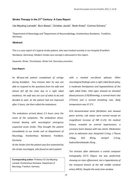
Stroke Therapy in the 21st Century: A Case Report Uta Meyding ...
Stroke Therapy in the 21st Century: A Case Report Uta Meyding ...
Stroke Therapy in the 21st Century: A Case Report Uta Meyding ...

You also want an ePaper? Increase the reach of your titles
YUMPU automatically turns print PDFs into web optimized ePapers that Google loves.
<strong>Stroke</strong> <strong>Therapy</strong> <strong>in</strong> <strong>the</strong> 21 st <strong>Century</strong>: A <strong>Case</strong> <strong>Report</strong><br />
13<br />
Brunei Darussalam Journal of Health, 2012, 5: 13-19<br />
<strong>Uta</strong> Meyd<strong>in</strong>g-Lamadé 1 , Burc Bassa 1 , Christian Jacobi 1 , Bodo Kress 2 , Cor<strong>in</strong>na Schranz 1<br />
1 Department of Neurology and 2 Department of Neuroradiology, Krankenhaus Nordwest, Frankfurt,<br />
Germany.<br />
Abstract:<br />
This is a case report of a typical stroke patient, who was treated recently <strong>in</strong> our hospital (Frankfurt<br />
Nordwest, Germany). Modern stroke care concept is discussed <strong>in</strong> this report.<br />
Keywords: <strong>Stroke</strong>, Thrombolysis, <strong>Stroke</strong> Unit, Secondary prevention<br />
<strong>Case</strong> <strong>Report</strong>:<br />
An 80-year-old patient compla<strong>in</strong>ed of vertigo<br />
dur<strong>in</strong>g breakfast. Few m<strong>in</strong>utes later he was not<br />
able to respond to <strong>the</strong> questions from his wife and<br />
almost fell off <strong>the</strong> chair due to a right sided<br />
weakness. His wife was not sure of what to do and<br />
decided to wait. As <strong>the</strong> patient had not improved<br />
after 2 hours, she <strong>the</strong>n called <strong>the</strong> ambulance.<br />
The ambulance arrived about 2.5 hours s<strong>in</strong>ce <strong>the</strong><br />
onset of <strong>the</strong> symptoms. The ambulance driver,<br />
tra<strong>in</strong>ed deal<strong>in</strong>g with neurological emergency<br />
suspected acute stroke. They brought <strong>the</strong> patient<br />
immediately to our stroke unit at Department of<br />
Neurology, Krankenhaus Nordwest, Frankfurt,<br />
Germany.<br />
At <strong>the</strong> <strong>Stroke</strong> Unit <strong>the</strong> patient was first exam<strong>in</strong>ed by<br />
<strong>the</strong> stroke neurologist, who found an alert patient<br />
Correspond<strong>in</strong>g author: Professor Dr.<strong>Uta</strong> Meyd<strong>in</strong>g-<br />
Lamadé, Krankenhaus Nordwest, Department of<br />
Neurology, Frankfurt, Germany<br />
with a marked non-fluent aphasia. O<strong>the</strong>r<br />
neurological f<strong>in</strong>d<strong>in</strong>gs were a right sided facial palsy,<br />
a moderate hemiparesis and hypoaes<strong>the</strong>sia of <strong>the</strong><br />
right sided limbs. Vital signs showed an elevated<br />
blood pressure (170/95mmHg), a normal heart rate<br />
(77/m<strong>in</strong>) and a normal breath<strong>in</strong>g rate. Body<br />
temperature was 37.3°C.<br />
ECG demonstrated atrial fibrillation and showed<br />
pacer activity. Lab values were normal except an<br />
<strong>in</strong>significant <strong>in</strong>crease of INR (1.23). His medical<br />
history revealed an arterial hypertension, a<br />
coronary heart disease with two stents. Medication<br />
prior to admission was: bisoprolol 2.5mg, L-Thyrox<br />
150µg, ASA 85mg, ramipril 2.5mg,<br />
hydrochlorothiazide 25mg .<br />
Ten m<strong>in</strong>utes after admission a cranial computer<br />
tomography (CCT) (Figure 1a) was performed,<br />
show<strong>in</strong>g no clear effacement, but a hyperdensity of<br />
<strong>the</strong> temporal branch of <strong>the</strong> left middle cerebral<br />
artery (MCA). Despite <strong>the</strong> early time w<strong>in</strong>dow
diffusion-MRI (Figure 1c) was performed to <strong>in</strong>crease<br />
treatment safety <strong>in</strong> this elderly patient.<br />
Figure 1a) Computertomography at admission<br />
show<strong>in</strong>g a thrombotic occlusion of a temporal branch<br />
of <strong>the</strong> distal MCA left side<br />
Figure 1b) MR-Angiography before thrombolysis<br />
show<strong>in</strong>g a proximal occlusion of <strong>the</strong> left MCA<br />
Figure 1c) Diffusion MRI before thrombolysis with a<br />
diffusion abnormality <strong>in</strong> <strong>the</strong> parietotemporal lobe less<br />
than 1/3 of <strong>the</strong> MCA territory<br />
14<br />
Brunei Darussalam Journal of Health, 2012, 5: 13-19<br />
As cl<strong>in</strong>ically suggested it showed a diffusion<br />
abnormality <strong>in</strong> <strong>the</strong> left parieto- temporal lobe less<br />
than a third of <strong>the</strong> entire media territory. MR-<br />
angiography (Figure 1b) demonstrated an occlusion<br />
of <strong>the</strong> proximal MCA on <strong>the</strong> left side. No <strong>in</strong>tracranial<br />
bleed<strong>in</strong>g was detectable <strong>in</strong> ei<strong>the</strong>r method.<br />
Because our patient was admitted with<strong>in</strong> 4.5 hours,<br />
CCT excluded <strong>in</strong>tracerebral haemorrhage and no<br />
o<strong>the</strong>r contra<strong>in</strong>dications could be detected. He was<br />
treated immediately with 60mg rt-PA (Alteplase)<br />
<strong>in</strong>travenously (0.9 mg/kg body weight, 10% as bolus,<br />
rema<strong>in</strong><strong>in</strong>g dose as <strong>in</strong>fusion over 60m<strong>in</strong>).<br />
A few hours later <strong>the</strong> neurological symptoms<br />
improved significantly. The patient was able to move<br />
all limbs and <strong>the</strong> aphasia was also <strong>in</strong> regression.<br />
Correspond<strong>in</strong>g to <strong>the</strong> cl<strong>in</strong>ical f<strong>in</strong>d<strong>in</strong>gs, <strong>the</strong> follow up<br />
CCT a day later showed only a small <strong>in</strong>farction of <strong>the</strong><br />
left sided lateral basal ganglia and a small<br />
parietocortical <strong>in</strong>farction.<br />
As carotid duplexsonography showed no stenosis or<br />
occlusion, <strong>the</strong> most probable reason for <strong>the</strong> embolic<br />
stroke was atrial fibrillation. For secondary<br />
prevention, <strong>the</strong> patient was <strong>in</strong>itially put on low<br />
molecular hepar<strong>in</strong> (Enoxapar<strong>in</strong> 40mg BID). Later,<br />
anticoagulation with Phenprocoumon was <strong>in</strong>itiated.
Public awareness<br />
Most people are not aware of stroke symptoms and<br />
important time is lost. The <strong>in</strong>terval from symptom<br />
onset to first call for medical help is <strong>the</strong> ma<strong>in</strong> cause<br />
of prehospital delay. Major reasons for delayed<br />
contact <strong>in</strong>clude not only lack of awareness of stroke<br />
symptoms and recognition of <strong>the</strong>ir severity, but also<br />
denial of <strong>the</strong> disease and <strong>the</strong> hope that symptoms<br />
would resolve. In Europe up to 50% of <strong>the</strong> patients<br />
do not realize stroke symptoms. This suggests that<br />
educat<strong>in</strong>g <strong>the</strong> population to recognize stroke<br />
symptoms, and chang<strong>in</strong>g people’s attitudes to acute<br />
stroke, may reduce <strong>the</strong> delay from stroke onset to<br />
emergency medical service (EMS) <strong>in</strong>volvement.<br />
While most people agree that stroke is an<br />
emergency, and that <strong>the</strong>y would seek medical help<br />
immediately, <strong>in</strong> reality only up to 50% call EMS. In<br />
many cases <strong>the</strong> first contact is with a family member<br />
or with a general practitioner; <strong>in</strong> some studies<br />
between 45% and 48% of patients were referred via<br />
a general practitioner 1 .<br />
Patient referral<br />
Once stroke symptoms are suspected, patients or<br />
<strong>the</strong>ir proxies should call Emergency Medical service<br />
(EMS). The EMS system should have an electronic<br />
validated algorithm of questions to diagnose stroke<br />
dur<strong>in</strong>g <strong>the</strong> phone <strong>in</strong>terview. The ambulance<br />
dispatchers and paramedics should be able to<br />
diagnose stroke us<strong>in</strong>g simple <strong>in</strong>struments such as <strong>the</strong><br />
Face-Arm-Speech-Test (Table 1) 2 . They should also<br />
be able to identify and provide appropriate help for<br />
patients with early complications or co-morbidities of<br />
stroke, such as impaired consciousness, seizures,<br />
15<br />
Brunei Darussalam Journal of Health, 2012, 5: 13-19<br />
vomit<strong>in</strong>g, or haemodynamic <strong>in</strong>stability. Suspected<br />
stroke victims should be transported without delay<br />
to <strong>the</strong> nearest stroke unit. Patients with onset of<br />
stroke symptoms with<strong>in</strong> 4.5 hours should be given<br />
priority <strong>in</strong> evaluation and transportation.<br />
Table 1) Face-arm-speech-test<br />
Item Yes No Uncerta<strong>in</strong><br />
Speech impairment<br />
Facial Palsy<br />
Arm weakness<br />
If at least one symptom is present, this patients is<br />
suspected of hav<strong>in</strong>g a stroke<br />
<strong>Stroke</strong> Units and telemedic<strong>in</strong>e<br />
A stroke unit consists of a discrete area of a hospital<br />
ward that exclusively takes care of stroke patients<br />
and is staffed by a specialist multidiscipl<strong>in</strong>ary team 1 .<br />
The core discipl<strong>in</strong>es of <strong>the</strong> team are medic<strong>in</strong>e,<br />
nurs<strong>in</strong>g, physio<strong>the</strong>rapy, occupational <strong>the</strong>rapy, speech<br />
and language <strong>the</strong>rapy and social workers. Treatment<br />
<strong>in</strong> a stroke unit reduces mortality and care-<br />
dependency by 25-30% compared to treatment <strong>in</strong><br />
general wards. All types of patients, irrespective of<br />
gender, age, stroke subtype and stroke severity,<br />
appear to benefit from treatment <strong>in</strong> stroke units 3 .<br />
Although stroke unit care is more costly, it reduces<br />
post-acute <strong>in</strong>patient care costs and is <strong>the</strong>refore cost-<br />
effective.<br />
In remote or rural areas without a close meshed<br />
stroke unit coverage telemedic<strong>in</strong>e can improve
access to treatment. Telemedic<strong>in</strong>e, as part of a<br />
regional stroke treatment concept, has been<br />
demonstrated to be a feasible, valid and reliable<br />
means of facilitat<strong>in</strong>g thrombolysis delivery to<br />
patients <strong>in</strong> distant or rural hospitals, where timely air<br />
or ground transportation is not feasible. The quality<br />
of treatment, complication rates, and short and long<br />
term outcomes are similar for acute stroke patients<br />
treated with rtPA via a telemedic<strong>in</strong>e consultation at<br />
local hospitals and those treated <strong>in</strong> academic centres<br />
4, 5 .<br />
Early/general stroke treatment<br />
First of all it is important for stroke patients to keep<br />
or to br<strong>in</strong>g <strong>the</strong>ir vital signs (blood pressure, body<br />
temperature, blood oxygen content, blood glucose<br />
level, fluid balance) <strong>in</strong> <strong>the</strong> normal range. Blood<br />
pressure monitor<strong>in</strong>g and treatment is a controversial<br />
area <strong>in</strong> stroke management. Patients outside <strong>the</strong><br />
highest and lowest levels of blood pressure<br />
recommended <strong>in</strong> <strong>the</strong> first 24 hours after stroke are<br />
more likely to have early neurological decl<strong>in</strong>e and<br />
poorer outcomes. It is still undeterm<strong>in</strong>ed whe<strong>the</strong>r<br />
blood pressure should be lowered after acute stroke,<br />
and whe<strong>the</strong>r antihypertensive <strong>the</strong>rapy should be<br />
cont<strong>in</strong>ued or stopped <strong>in</strong> <strong>the</strong> first few days after<br />
stroke. In <strong>the</strong> absence of reliable evidence from<br />
cl<strong>in</strong>ical trials, many cl<strong>in</strong>icians have developed<br />
protocols for <strong>the</strong> management of extremely high<br />
blood pressure. In some centres it is common<br />
practice to beg<strong>in</strong> cautious blood pressure reduction<br />
when levels exceed 220 mmHg systolic and<br />
120 mmHg diastolic. In patients undergo<strong>in</strong>g<br />
thrombolysis it is common practice to avoid systolic<br />
16<br />
Brunei Darussalam Journal of Health, 2012, 5: 13-19<br />
blood pressures above 185 mmHg. Most<br />
recommended drugs for treatment are i.v. labetalol<br />
and urapadil 1 .<br />
Hyperglycaemia occurs <strong>in</strong> up to 60% of stroke patients<br />
without known diabetes and is associated with larger<br />
<strong>in</strong>farct volumes and cortical <strong>in</strong>volvement, and with<br />
poor functional outcome. Despite <strong>the</strong> lack of data it is<br />
common practice <strong>in</strong> stroke units to reduce blood<br />
glucose levels exceed<strong>in</strong>g 180 mg/dl (10 mmol/l).<br />
Hypoglycaemia (37.5°C)<br />
with paracetamol is common practice <strong>in</strong> stroke<br />
patients 1 .<br />
Acute stroke imag<strong>in</strong>g<br />
Patients with suspected TIA or stroke should have<br />
clear priority over o<strong>the</strong>r patients for bra<strong>in</strong> imag<strong>in</strong>g,<br />
because time is crucial. Investigation of TIA is equally<br />
urgent, because up to 10% of <strong>the</strong>se patients will suffer<br />
stroke with<strong>in</strong> <strong>the</strong> next 48 hours 6 . Diagnostic imag<strong>in</strong>g<br />
must be sensitive and specific <strong>in</strong> detect<strong>in</strong>g stroke<br />
pathology, particularly <strong>in</strong> <strong>the</strong> early phase of stroke. CT<br />
is usually sufficient to guide rout<strong>in</strong>e thrombolysis and<br />
is <strong>the</strong> most cost-effective strategy for imag<strong>in</strong>g acute<br />
stroke patients. Overall, CT is less sensitive than<br />
multimodal MRI, but equally specific, for early<br />
ischaemic changes. Early CT changes <strong>in</strong> ischaemic<br />
stroke <strong>in</strong>clude decrease <strong>in</strong> tissue x-ray attenuation,
tissue swell<strong>in</strong>g with effacement of cerebrosp<strong>in</strong>al fluid<br />
spaces, and arterial hyperattenuation, which <strong>in</strong>dicates<br />
<strong>the</strong> presence of <strong>in</strong>tralum<strong>in</strong>al thrombus with high<br />
specificity 7 . The presence of early signs of ischaemia on<br />
CT should not exclude patients from thrombolysis with<strong>in</strong><br />
<strong>the</strong> first 3 hours, though patients with a hypoattenuat<strong>in</strong>g<br />
ischaemic lesion which exceeds one third of <strong>the</strong> middle<br />
cerebral artery (MCA) territory may benefit less from<br />
thrombolysis.<br />
Modern, multimodal MRI-techniques <strong>in</strong>clud<strong>in</strong>g diffusion,<br />
T2* and perfusion imag<strong>in</strong>g have <strong>the</strong> advantage of higher<br />
sensitivity for early ischaemic changes than CT 8 . This<br />
higher sensitivity is particularly useful <strong>in</strong> <strong>the</strong> diagnosis of<br />
posterior circulation strokes and lacunar or small cortical<br />
<strong>in</strong>farctions. MRI can also detect small and old<br />
haemorrhages for a prolonged period with T2* (gradient<br />
echo) sequences. However, DWI can be negative <strong>in</strong><br />
patients with def<strong>in</strong>ite stroke and <strong>the</strong> usefulness could<br />
not be demonstrated <strong>in</strong> randomized cl<strong>in</strong>ical trials 9 .<br />
Specific stroke treatment<br />
Thrombolytic <strong>the</strong>rapy with <strong>in</strong>travenous rtPA (Alteplase)<br />
is <strong>the</strong> only proven specific acute stroke <strong>the</strong>rapy, which<br />
was evaluated <strong>in</strong> several randomized, placebo controlled<br />
trials. The treatment was approved based on <strong>the</strong> results<br />
of <strong>the</strong> NINDS-trial with<strong>in</strong> a 3 hour time w<strong>in</strong>dow 10 . Three<br />
years ago <strong>the</strong> ECASS III trial showed that this treatment<br />
is effective and safe also <strong>in</strong> a 3 to 4.5 hour time w<strong>in</strong>dow<br />
11 . Recently <strong>the</strong> comb<strong>in</strong>ed analysis of NINDS and ECASS<br />
III aga<strong>in</strong> demonstrated that rtPA is effective and safe <strong>in</strong><br />
<strong>the</strong> time w<strong>in</strong>dow of 4.5 hours after symptom onset 12 .<br />
This analysis also confirms <strong>the</strong> clear correlation between<br />
17<br />
Brunei Darussalam Journal of Health, 2012, 5: 13-19<br />
<strong>the</strong> onset-to-treatment-time (OTT) and <strong>the</strong> treatment<br />
effect; <strong>the</strong> earlier <strong>the</strong> treatment <strong>the</strong> better <strong>the</strong><br />
outcome. The most feared risk of thrombolytic<br />
<strong>the</strong>rapy is <strong>in</strong>tracerebral haemorrhage, because it<br />
commonly leads to a cl<strong>in</strong>ical deterioration. The above<br />
mentioned analysis found a 5.2% risk for<br />
symptomatic <strong>in</strong>tracerebral haemorrhage, which is<br />
around 5 times higher than <strong>in</strong>-patients treated with<br />
placebo. It is important that <strong>the</strong>re was no significant<br />
<strong>in</strong>teraction between <strong>the</strong> bleed<strong>in</strong>g risk and <strong>the</strong> OTT.<br />
The NNT to achieve a favourable cl<strong>in</strong>ical outcome<br />
(patient with no or only mild symptoms) after 3<br />
months is 7. European regulatory agencies do not<br />
advocate rtPA treatment <strong>in</strong> patients with severe<br />
stroke (NIHSSS >25), extended early ischaemic<br />
changes on CT-scan, or age above 80 years (unlike<br />
<strong>the</strong> US labell<strong>in</strong>g). Never<strong>the</strong>less, observational studies<br />
suggest that rtPA given with<strong>in</strong> 3 hours of stroke onset<br />
is safe and effective <strong>in</strong> patients over 80 years of age 13 .<br />
In <strong>in</strong>dividual cases (time w<strong>in</strong>dow > 4.5h) or stroke<br />
patients with an uncerta<strong>in</strong> time w<strong>in</strong>dow <strong>the</strong> use of<br />
multimodal imag<strong>in</strong>g criteria may be helpful for<br />
patient selection 14 . Interventional techniques aim<strong>in</strong>g<br />
to remove occlud<strong>in</strong>g clots with mechanical retrievers<br />
are under development and are useful treatment<br />
options for patients with very severe strokes.<br />
Secondary prevention<br />
Secondary prevention depends on <strong>the</strong> etiological<br />
classification of <strong>the</strong> stroke and <strong>the</strong> <strong>in</strong>dividual risk<br />
factor profile. The etiological workup should be<br />
carried out dur<strong>in</strong>g <strong>the</strong> <strong>in</strong>itial 24 hours by ultrasound
and laboratory test<strong>in</strong>g, as <strong>the</strong> risk of recurrence is<br />
highest dur<strong>in</strong>g <strong>the</strong> first hours and days.<br />
In our case, <strong>the</strong> patient suffered his stroke from<br />
cardioembolic orig<strong>in</strong> due to atrial fibrillation. For<br />
patients with permanent or paroxysmal atrial<br />
fibrillation (AF) and concomitant vascular risk factors<br />
an oral anticoagulation with a target INR between 2.0<br />
and 3.0 is recommended 1 . Anticoagulation <strong>in</strong> elderly<br />
patients with Warfar<strong>in</strong> is important, because <strong>the</strong>y<br />
have a particular high risk of suffer<strong>in</strong>g from embolic<br />
stroke and <strong>the</strong> BAFTA-trial has confirmed that<br />
warfar<strong>in</strong> is also safe <strong>in</strong> those patients 15 . However,<br />
treatment with warfar<strong>in</strong> bears some problems, for<br />
example <strong>the</strong> need for regular blood tests, <strong>the</strong><br />
multiple <strong>in</strong>teractions with o<strong>the</strong>r drugs and a small<br />
<strong>the</strong>rapeutic range. In <strong>the</strong> near future new drugs like<br />
direct thromb<strong>in</strong>-<strong>in</strong>hibitors are expected as a<br />
promis<strong>in</strong>g alternative. The ReLy study exam<strong>in</strong>ed two<br />
dosages of dabigatran <strong>in</strong> <strong>the</strong> prevention of stroke<br />
recurrence <strong>in</strong> patients with atrial fibrillation and a<br />
prior stroke or TIA. The lower dosage had <strong>the</strong> same<br />
effectiveness as warfar<strong>in</strong> but was safer <strong>in</strong> terms of<br />
severe bleed<strong>in</strong>g complications, <strong>the</strong> higher dosage<br />
was more effective with comparable bleed<strong>in</strong>g<br />
complications 16 .<br />
Conclusion<br />
Optimization of <strong>the</strong> prehospital phase, public<br />
education for stroke recognition, immediate<br />
admission to a stroke unit may lead to a better<br />
strokecare. The only effective acute <strong>the</strong>rapy (rtPa,<br />
Alteplase) is now available for patients dur<strong>in</strong>g <strong>the</strong> 4.5<br />
hours w<strong>in</strong>dow with adherence to <strong>the</strong> <strong>in</strong>clusion- and<br />
exclusion criteria. Treatment at a specialized stroke<br />
18<br />
Brunei Darussalam Journal of Health, 2012, 5: 13-19<br />
centre has proven to save lives and reduces<br />
morbidity, that help enabl<strong>in</strong>g more patients to live an<br />
<strong>in</strong>dependent life after <strong>the</strong>ir stroke.<br />
References<br />
1 ESO Writ<strong>in</strong>g Committee, Guidel<strong>in</strong>es for<br />
Management of Ischaemic <strong>Stroke</strong> and Transient<br />
Ischaemic Attack 2008. Cerebrovasc Dis. 2008;<br />
25(5): 457-507.<br />
2 Harbison, J., et al., Diagnostic accuracy of stroke<br />
referrals from primary care, emergency room<br />
physicians, and ambulance staff us<strong>in</strong>g <strong>the</strong> face arm<br />
speech test. <strong>Stroke</strong>, 2003; 34(1): 71-6.<br />
3 Foley N, Salter K, Teasell R. Specialized stroke<br />
services: a meta-analysis compar<strong>in</strong>g three models<br />
of care. Cerebrovasc Dis 2007; 23:194-202<br />
4 LaMonte MP, Bahouth MN, Hu P, Pathan MY,<br />
Yarbrough KL, Gunawardane R, Crarey P, Page W:<br />
Telemedic<strong>in</strong>e for acute stroke: triumphs and pitfalls.<br />
<strong>Stroke</strong> 2003;34: 725-728.<br />
5 Audebert HJ, Schenkel J, Heuschmann PU, Bogdahn<br />
U, Haberl RL: Effects of <strong>the</strong> implementation of a<br />
telemedical stroke network: <strong>the</strong> Telemedic Pilot<br />
Project for Integrative <strong>Stroke</strong> Care (TEMPiS) <strong>in</strong><br />
Bavaria, Germany. Lancet Neurol 2006;5:742-748.<br />
6 Rothwell PM, Giles MF, Chandra<strong>the</strong>va A, Marquardt<br />
L, Geraghty O, Redgrave JN, Lovelock CE, B<strong>in</strong>ney LE,<br />
Bull LM, Cuthbertson FC, Welch SJ, Bosch S,<br />
Carasco-Alexander F, Silver LE, Gutnikov SA, Mehta<br />
Z. Effect of urgent treatment of transient ischaemic<br />
attack and m<strong>in</strong>or stroke on early recurrent stroke<br />
(EXPRESS study): a prospective population-based<br />
sequential comparison. Lancet 2007;370:1432-<br />
1442.<br />
7 von Kummer R, Allen KL, Holle R, Bozzao L,<br />
Bastianello S, Manelfe C, Bluhmki E, R<strong>in</strong>gleb P,<br />
Meier DH, Hacke W. Acute stroke: usefulness of<br />
early CT f<strong>in</strong>d<strong>in</strong>gs before thrombolytic <strong>the</strong>rapy.<br />
Radiology. 1997;205:327-333<br />
8 Chalela JA, Kidwell CS, Nentwich LM, Luby M,<br />
Butman JA, Demchuk AM, Hill MD, Patronas N,<br />
Latour L, Warach S: Magnetic resonance imag<strong>in</strong>g<br />
and computed tomography <strong>in</strong> emergency<br />
assessment of patients with suspected acute stroke:<br />
a prospective comparison. Lancet 2007;369:293-<br />
298.
9: Hacke W, Furlan AJ, Al-Rawi Y, Davalos A, Fiebach JB,<br />
Gruber F, Kaste M, Lipka LJ, Pedraza S, R<strong>in</strong>gleb PA,<br />
Rowley HA, Schneider D, Schwamm LH, Leal JS,<br />
Söhngen M, Teal PA, Wilhelm-Ogunbiyi K, W<strong>in</strong>termark<br />
M, Warach S. Intravenous desmoteplase <strong>in</strong> patients<br />
with acute ischaemic stroke selected by MRI<br />
perfusion-diffusion weighted imag<strong>in</strong>g or perfusion CT<br />
(DIAS-2): a prospective, randomised, double-bl<strong>in</strong>d,<br />
placebo-controlled study. Lancet Neurol. 2009;8:141-<br />
150<br />
10: The National Institute of Neurological Disorders and<br />
<strong>Stroke</strong> rt-PA <strong>Stroke</strong> Study Group. Tissue plasm<strong>in</strong>ogen<br />
activator for acute ischemic stroke.N Engl J Med.<br />
1995;333:1581-1587<br />
11: Hacke W, Kaste M, Bluhmki E, Brozman M, Davalos A,<br />
Guidetti D, Larrue V, Lees KR, Medeghri Z, Machnig T,<br />
Schneider D, von Kummer R, Wahlgren N, Toni D.<br />
Thrombolysis with alteplase 3 to 4.5 hours after acute<br />
ischemic stroke. N Engl J Med. 2008;359:1317-1329<br />
12: Lees, K.R., et al., Time to treatment with <strong>in</strong>travenous<br />
alteplase and outcome <strong>in</strong> stroke: an updated pooled<br />
analysis of ECASS, ATLANTIS, NINDS, and EPITHET<br />
trials. Lancet, 2010. 375(9727): p. 1695-703.<br />
13: R<strong>in</strong>gleb PA, Schwark C, Köhrmann M, Külkens S, Jüttler<br />
E, Hacke W, Schell<strong>in</strong>ger PD. Thrombolytic <strong>the</strong>rapy for<br />
acute ischaemic stroke <strong>in</strong> octogenarians: selection by<br />
magnetic resonance imag<strong>in</strong>g improves safety but does<br />
not improve outcome. J Neurol Neurosurg Psychiatry.<br />
2007 ;78:690-693<br />
14: Albers GW, Thijs VN, Wechsler L, Kemp S, Schlaug G,<br />
Skalabr<strong>in</strong> E, Bammer R, Kakuda W, Lansberg MG,<br />
Shuaib A, Copl<strong>in</strong> W, Hamilton S, Moseley M, Marks<br />
MP. Magnetic resonance imag<strong>in</strong>g profiles predict<br />
cl<strong>in</strong>ical response to early reperfusion: <strong>the</strong> diffusion<br />
and perfusion imag<strong>in</strong>g evaluation for understand<strong>in</strong>g<br />
stroke evolution (DEFUSE) study. Ann Neurol.<br />
2006;60:508-517<br />
15: Mant J, Hobbs FD, Fletcher K, Roalfe A, Fitzmaurice D,<br />
Lip GY, Murray E. Warfar<strong>in</strong> versus aspir<strong>in</strong> for stroke<br />
prevention <strong>in</strong> anelderly community population with<br />
atrial fibrillation (<strong>the</strong> Birm<strong>in</strong>gham Atrial Fibrillation<br />
Treatment of <strong>the</strong> Aged Study, BAFTA): a randomised<br />
controlled trial. Lancet. 2007;370:493-503<br />
19<br />
Brunei Darussalam Journal of Health, 2012, 5: 13-19<br />
16: Connolly SJ, Ezekowitz MD, Yusuf S, Eikelboom J,<br />
Oldgren J, Parekh A, Pogue J, Reilly PA, Themeles E,<br />
Varrone J, Wang S, Al<strong>in</strong>gs M, Xavier D, Zhu J, Diaz R,<br />
Lewis BS, Darius H, Diener H-C, Joyner CD, Wallent<strong>in</strong><br />
L, <strong>the</strong> RE-LY Steer<strong>in</strong>g Committee and Investigators.<br />
Dabigatran versus warfar<strong>in</strong> <strong>in</strong> patients with atrial<br />
fibrillation.N Engl J Med. 2009;361:1139-1151