International Plant Protection Convention Draft Annex to ISPM 27 ...
International Plant Protection Convention Draft Annex to ISPM 27 ...
International Plant Protection Convention Draft Annex to ISPM 27 ...

Create successful ePaper yourself
Turn your PDF publications into a flip-book with our unique Google optimized e-Paper software.
[G]<br />
[1]<br />
[2]<br />
[3]<br />
[4]<br />
[5]<br />
<strong>International</strong> <strong>Plant</strong> <strong>Protection</strong> <strong>Convention</strong><br />
<strong>Draft</strong> <strong>Annex</strong> <strong>to</strong> <strong>ISPM</strong> <strong>27</strong>:2006: Guignardia citricarpa Kiely on fruit (2004-<br />
023)<br />
DRAFT ANNEX <strong>to</strong> <strong>ISPM</strong> <strong>27</strong>:2006: Guignardia citricarpa Kiely on fruit (2004-023)<br />
Publication information<br />
Date of this document 2012-02-23<br />
Agenda Item:<br />
Document category <strong>Draft</strong> new annex <strong>to</strong> <strong>ISPM</strong> <strong>27</strong>:2006 (Diagnostic pro<strong>to</strong>cols for regulated pests)<br />
Current document<br />
stage<br />
<strong>Draft</strong> approved for member consultation by the SC – special process<br />
Origin Work programme <strong>to</strong>pic: Fungi and fungus-like organisms, CPM-1 (2006)<br />
Original subject: Guignardia citricarpa (2004-023)<br />
Major stages Approved for member consultation by the SC<br />
(2011_eSC_Nov_06_Guignardia citricarpa)<br />
Consultation on<br />
technical level<br />
Main discussion points<br />
during development of<br />
the diagnostic pro<strong>to</strong>col<br />
Notes<br />
CONTENTS (<strong>to</strong> be added at adoption)<br />
Adoption<br />
The first draft of this diagnostic pro<strong>to</strong>col was written by: Irene Vlou<strong>to</strong>glou<br />
(Benaki Phy<strong>to</strong>pathological Institute, Athens, Greece); Johan Meffert (<strong>Plant</strong><br />
<strong>Protection</strong> Service, Wageningen, The Netherlands); Luis Diaz Morales<br />
(Ministry of Agriculture and Fishery, Montevideo, Uruguay).<br />
This proposal has been commented upon by: Peter Bonants (<strong>Plant</strong> Research<br />
<strong>International</strong>, Wageningen, The Netherlands); Maria Fernandez (Servicio<br />
National de Sanidad y Calidad Agroalimentaria (SENASA), Buenos Aires,<br />
Argentina); Chirlei Glienke-Blanco (Universidade Federal do Paraná, Curitiba,<br />
Brazil); Marcel Spósi<strong>to</strong> (Universidade de São Paulo, São Paulo, Brazil);<br />
Lavern Timmer (University of Florida, Lake Alfred, USA); Mariette Truter<br />
(ARC-<strong>Plant</strong> <strong>Protection</strong> Research Institute, Queenswood, South Africa);<br />
Pinsahn Wu (Chinese Academy of Inspection and Quarantine, Beijing,<br />
China).<br />
The SC noted that there was no mention of an internal amplification control in<br />
the diagnostic pro<strong>to</strong>col, and wondered whether this should be added. The<br />
authors note that there is no internal amplification control (IAC) described for<br />
the conventional PCR. This has <strong>to</strong> be developed by the individual<br />
labora<strong>to</strong>ries.<br />
Page 1 of 22
Page 2 of 22<br />
<strong>Draft</strong> <strong>Annex</strong> <strong>to</strong> <strong>ISPM</strong> <strong>27</strong>:2006: Guignardia citricarpa Kiely on fruit (2004-023)<br />
[6] This diagnostic pro<strong>to</strong>col was adopted by the Xth Session of the Commission on Phy<strong>to</strong>sanitary Measures in<br />
20--.<br />
[7]<br />
1. Pest Information<br />
[8] Guignardia citricarpa Kiely, the causal agent of “citrus black spot” disease, is a leaf-spotting and fruitblemishing<br />
pest affecting Citrus, Poncirus, Fortunella and their hybrids. Except for Citrus aurantium L. and its<br />
hybrids and C. latifolia Tan., all commercially grown Citrus species are susceptible (Kotzé, 2000; Aguilar-<br />
Vildoso et al., 2002). Citrus limon L. is particularly susceptible and, thus, in an unaffected area, the disease<br />
usually appears first on C. limon (Kotzé, 2000).<br />
[9] Citrus black spot was first recorded in Australia in 1895 on C. sinensis (Linnaeus) Osbeck (Benson, 1895). It<br />
is now present in some citrus-producing areas in Asia, Africa, Australia and South America (CABI, 2011). In<br />
March 2010, G. citricarpa was detected for the first time in a few citrus groves in Florida (USA) (NAPPO,<br />
2010). Surveys on the distribution of the organism in the area are ongoing (USDA-APHIS, 2010). The<br />
organism has not been reported from Europe, Central America or the Caribbean region (EPPO/CABI, 1997;<br />
CABI/EPPO, 1998; CABI, 2011; NAPPO, 2010).<br />
[10] G. citricarpa has significant economic impact mainly because of the external blemishes it causes, which<br />
make citrus fruit unsuitable for the fresh market. Severe infections may cause premature fruit drop (Kotzé,<br />
2000). Some losses due <strong>to</strong> fruit drop occur in years favourable for pest development and when fruit is held on<br />
the trees past peak maturity (CABI, 2011). In addition, latently infected (asymp<strong>to</strong>matic) fruit at harvest may<br />
still develop symp<strong>to</strong>ms during transport or s<strong>to</strong>rage (Kotzé, 1996).<br />
[11] The epidemiology of citrus black spot is influenced by the availability of inoculum, the occurrence of<br />
environmental conditions favourable for infection (warm, wet and humid conditions), the growth cycle of the<br />
citrus tree and the age of the fruit in relation <strong>to</strong> its susceptibility <strong>to</strong> infection (Kotzé, 1981, 2000). In areas<br />
where rain is confined <strong>to</strong> a single season, pseudothecia with ascospores, produced exclusively on leaf litter,<br />
are the main source of inoculum. Where rain is not confined <strong>to</strong> a single season, where out-of-season fruit<br />
with lesions remain on the trees after flowering and fruit set, or where successive and irregular flowering<br />
occurs in the cultivated citrus species and varieties, pycnidia with conidia of the anamorph Phyllosticta<br />
citricarpa (McAlpine) Aa are also important as inoculum sources (Kotzé, 1981; Spósi<strong>to</strong> et al., 2008).<br />
[12] Pseudothecia develop 40–180 days after leaf drop, depending on the frequency of wetting and drying as well<br />
as on the prevailing temperatures (Kotzé, 1981). Citrus leaves drop all year round in some countries and<br />
seasonally in others, and this affects the availability of inoculum. The optimum temperature for pseudothecial<br />
formation is 21–28 °C and no pseudothecia are formed below 7 °C or above 35 °C (Lee and Huang, 1973).<br />
Ascospore release takes place during rainfall and occasionally during irrigation or heavy dew (Kiely, 1949a;<br />
Kotzé, 2000). Ascospores are forcibly released up <strong>to</strong> a height of 1.2 cm above pseudothecia and are carried<br />
by air currents throughout the canopy and over long distances (Kiely, 1949a). Ascospore discharges are<br />
closely influenced by the rainfall pattern (Kotzé, 1981). The critical period for infection starts at fruit set and<br />
lasts 4–6 months, whereas the first symp<strong>to</strong>ms on fruit do not appear until more than 6 months after fruit set<br />
(Baldassari et al., 2006). In Brazil, fruit of C. sinensis of “Valencia” and “Natal” varieties are susceptible until<br />
at least 24 weeks after the fall of 75% of the petals, when they are 5–6 cm in diameter (Baldassari et al.,<br />
2006).<br />
[13] After infection, the fungus remains in a quiescent state until the fruit becomes fully grown or mature, with<br />
symp<strong>to</strong>ms being produced many months after infection has taken place (Kotzé, 2000). Leaves remain<br />
susceptible from development up <strong>to</strong> 10 months of age (Truter et al., 2007). G. citricarpa has two asexual<br />
states: a macroconidial state in the genus Phyllosticta and a microconidial in the genus Lep<strong>to</strong>dothiorella<br />
(Kiely, 1949a). Pycnidia with conidia are produced on fruit, leaves, dead twigs, fruit pedicels and in<br />
abundance on leaf litter (Kotzé, 2000). They may be splash-dispersed on<strong>to</strong> the canopy or washed off from<br />
infected late-hanging fruit on<strong>to</strong> younger fruit and leaves that are still at the susceptible stage (Agostini et al.,<br />
2006; Spósi<strong>to</strong> et al., 2008). The microconidial state, Lep<strong>to</strong>dothiorella sp., also referred <strong>to</strong> as “spermogonial”<br />
state (Kiely, 1949a), usually appears on fallen leaves before pseudothecia develop. However, the role of
<strong>Draft</strong> <strong>Annex</strong> <strong>to</strong> <strong>ISPM</strong> <strong>27</strong>:2006: Guignardia citricarpa Kiely on fruit (2004-023)<br />
microconidia in the biology of G. citricarpa is still unclear.<br />
[14] Symp<strong>to</strong>m development on mature fruit is enhanced by rising temperatures, high light intensity, drought and<br />
poor tree vigour. Older trees usually have more citrus black spot than younger trees (Kotzé, 2000). Spread of<br />
G. citricarpa <strong>to</strong> new areas is assumed <strong>to</strong> have taken place through infected nursery s<strong>to</strong>ck or other planting<br />
material rather than through citrus fruit (Kotzé, 2000; Timmer, 2004).<br />
[15] It should be noted that in symp<strong>to</strong>mless citrus fruit or fruit with minute spots (
Page 4 of 22<br />
<strong>Draft</strong> <strong>Annex</strong> <strong>to</strong> <strong>ISPM</strong> <strong>27</strong>:2006: Guignardia citricarpa Kiely on fruit (2004-023)<br />
[21] Several types of symp<strong>to</strong>ms (e.g. hard spot, freckle spot, false melanose, virulent spot) appear on fruit,<br />
depending on the temperature and fruit maturity (Kotzé, 2000). The presence of G. citricarpa on fruit is<br />
unlikely <strong>to</strong> be accurately confirmed based only on visual examination, as symp<strong>to</strong>ms are variable in<br />
appearance and can easily be confused with those caused by other citrus pathogens, mechanical or insect<br />
damage (Snowdon, 1990; Kotzé, 2000). The following four main types of symp<strong>to</strong>ms, described by Kiely<br />
(1949a, 1949b, 1960), are widely recognized.<br />
[22] Hard spot: the most typical symp<strong>to</strong>m of citrus black spot, consisting of shallow lesions, 3–10 mm in diameter,<br />
with a grey <strong>to</strong> tan centre and a dark brown <strong>to</strong> black margin (Figure 1A). At advanced stages of symp<strong>to</strong>m<br />
development, the centre of the lesions becomes crater-like. Individual hard spot lesions may either remain<br />
small or coalesce <strong>to</strong> form larger lesions. A yellow halo, when the fruit is green, or a green halo, when the fruit<br />
is yellow or orange, may appear around these lesions. Quite often, pycnidia of the anamorph P. citricarpa are<br />
produced in the centre of these spots (Figure 1a) and can be detected by using a hand lens or a dissecting<br />
microscope. Hard spot usually appears when fruit starts maturing, even before colour change and on the side<br />
of the fruit most exposed <strong>to</strong> sunlight (Kotzé, 1981, 2000). Hard spot symp<strong>to</strong>ms with pycnidia would be the<br />
most easily identified as citrus black spot.<br />
[23] Freckle spot: grey, tan, reddish or colourless spots, 1–3 mm in diameter, slightly depressed at the centre and<br />
with no halo around them (Figure 1B). The spots turn brown with age and are almost always devoid of<br />
pycnidia (Figure 1b). Freckle spots mostly develop after the fruit has changed colour and may also appear as<br />
satellite spots around hard spot lesions (Bonants et al., 2003) (Figure 1C). Individual freckle spots may<br />
coalesce <strong>to</strong> form larger lesions that turn in<strong>to</strong> virulent spots (Figure 2C), especially during s<strong>to</strong>rage (Kotzé,<br />
1981, 2000).<br />
[24] False melanose or speckled blotch: usually appears on green fruit as small, raised, dark brown <strong>to</strong> black<br />
lesions often surrounded by dark specks (FUNDECITRUS, 2005) (Figure 2A, 2a, 2B). The lesions are devoid<br />
of pycnidia and may coalesce as the season progresses (CABI, 2011). This type of symp<strong>to</strong>m is observed in<br />
citrus-growing areas where G. citricarpa has been present for a long time (FUNDECITRUS, 2005).<br />
[25] Virulent spot or spreading spot or galloping spot: sunken, irregular, red <strong>to</strong> brown or colourless lesions, which<br />
appear on heavily infected mature fruit <strong>to</strong>wards the end of the season (Figure 2C). Numerous pycnidia<br />
eventually develop in these lesions under conditions of high humidity (Kotzé, 2000). Virulent spots grow<br />
rapidly covering two-thirds of the fruit surface within 4–5 days. It is the most damaging symp<strong>to</strong>m, because,<br />
unlike the other types of symp<strong>to</strong>ms, it extends deeply in<strong>to</strong> the mesocarp (albedo), occasionally involving the<br />
entire thickness of the rind, causing premature fruit drop and serious post-harvest losses (Kotzé, 1981).<br />
[26] Two additional types of symp<strong>to</strong>ms (below) have also been reported <strong>to</strong> occur on citrus fruit, though<br />
infrequently.<br />
[<strong>27</strong>] Lacy spot: superficial, yellow lesions with a dark yellow <strong>to</strong> brown centre, a smooth texture and no defined<br />
margins (Aguilar-Vildoso et al., 2002) (Figure 2D). This type of symp<strong>to</strong>m appears on green fruit and may<br />
cover a big part of its surface (Goes, 2001). The lesions are devoid of pycnidia and frequently appear as<br />
brown netting on a yellow background. Fruit showing lacy spot usually appear aggregated in the tree canopy<br />
(Dr M. Spósi<strong>to</strong>, personal communication).<br />
[28] Cracked spot: superficial, slightly raised, variable in size, dark brown <strong>to</strong> black lesions with a cracked surface<br />
and irregular margins (Goes et al., 2000) (Figure 2E). The lesions are devoid of pycnidia and appear on fruit<br />
older than six months. This type of symp<strong>to</strong>m has been associated with infestation by Phyllocoptruta oleivora<br />
Ashmead (FUNDECITRUS, 2005).<br />
[29] It should be noted that more than one of the above described types of symp<strong>to</strong>ms or intermediate stages<br />
between one type of symp<strong>to</strong>m and another may be observed on the same fruit (Figure 1C, 1c).
<strong>Draft</strong> <strong>Annex</strong> <strong>to</strong> <strong>ISPM</strong> <strong>27</strong>:2006: Guignardia citricarpa Kiely on fruit (2004-023)<br />
[30] In areas with high inoculum pressure, symp<strong>to</strong>ms may also appear on small fruit, calyxes and peduncles. The<br />
symp<strong>to</strong>ms on calyxes are red <strong>to</strong> dark brown lesions similar <strong>to</strong> freckle spots. On small fruit and peduncles,<br />
symp<strong>to</strong>ms appear as small black spots (Aguilar-Vildoso et al., 2002).<br />
[31]<br />
3.2 Symp<strong>to</strong>ms on leaves and twigs<br />
[32] Citrus black spot usually occurs on leaves as quiescent infections without any visible symp<strong>to</strong>ms (Sut<strong>to</strong>n and<br />
Waters<strong>to</strong>n, 1966). If symp<strong>to</strong>ms appear, they start as pinpoint spots visible on both leaf surfaces. The spots,<br />
which may increase in size up <strong>to</strong> 3 mm in diameter, are circular with their centres becoming gray or light<br />
brown in colour surrounded by a dark brown <strong>to</strong> black rim and a yellow halo (Kotzé, 2000) (Figure 3A).<br />
Pycnidia may occasionally be present in the centre of the lesions on the adaxial leaf surface.<br />
[33] Lesions similar <strong>to</strong> those on leaves may also occur on small twigs, more commonly on C. limon than on other<br />
citrus species (Dr M. Truter, personal communication). Symp<strong>to</strong>ms are small (0.5–2 mm in diameter), round,<br />
slightly sunken lesions with a brown <strong>to</strong> black margin and a grey <strong>to</strong> light brown centre (Figure 3B). Pycnidia<br />
may occasionally be present in the centre of the lesions.<br />
[34]<br />
3.3 Comparison of citrus black spot symp<strong>to</strong>ms with those caused by other organisms or abiotic<br />
fac<strong>to</strong>rs<br />
[35] Symp<strong>to</strong>ms on fruit are variable in appearance and often resemble those caused by other citrus pathogens<br />
(such as P. citriasiana, Diaporthe citri, Mycosphaerella citri, Alternaria alternata pv. citri, Sep<strong>to</strong>ria spp.,<br />
Colle<strong>to</strong>trichum spp.), insect or mechanical damage (Snowdon, 1990; Bonants et al., 2003) or cold damage,<br />
particularly in the case of freckle spot (Dr L. Diaz, personal communication).<br />
[36] As the symp<strong>to</strong>ms caused by G. citricarpa on citrus fruit are similar <strong>to</strong> those caused by other pathogens,<br />
reliable diagnosis can be made only by using the methods described below.<br />
[37]<br />
4. Identification<br />
[38] This pro<strong>to</strong>col describes the detection and identification of G. citricarpa on citrus fruit. Citrus fruit should be<br />
inspected for the detection of any symp<strong>to</strong>ms typical of citrus black spot (see section 3). If suspected<br />
symp<strong>to</strong>ms are present in the form of spots or lesions, they are examined with a magnifying lens or a<br />
dissecting microscope for the presence of pycnidia. However, as the pycnidia and spores of P. citricarpa<br />
(anamorph of G. citricarpa) are very similar <strong>to</strong> those of P. citriasiana, the recently described pathogen on C.<br />
maxima (Wulandari et al., 2009), the identity of G. citricarpa has <strong>to</strong> be confirmed by applying the diagnostic<br />
methods described below (Figure 4). The diagnostic Method A (isolation and culturing) is used for the<br />
identification of G. citricarpa on citrus fruit, but can also be used on leaves, twigs and pedicels, whereas<br />
Method B (molecular assays) is applied on citrus fruit only.<br />
[39] If after applying Method A the cultural characteristics of the colonies grown on cherry decoction agar (CHA)<br />
and oatmeal agar (OA) media are not consistent with those of G. citricarpa (see section 4.2, requirements (i),<br />
(ii) and (iii)), then the plant material is considered free of G. citricarpa. On G. citricarpa-like cultures that do<br />
not produce mature pycnidia within 14 days, application of real-time polymerase chain reaction (real-time<br />
PCR) (see section 4.3.2) or internal transcribed spacer (ITS) sequencing (see section 4.3.3) is<br />
recommended. However, isolation and culturing of the organism on appropriate media followed by a direct<br />
molecular test of the cultures is a time-consuming procedure and thus undesirable in time-critical diagnosis of<br />
consignments.<br />
[40] There are two PCR methods (conventional and real-time) available for the detection and identification of G.<br />
citricarpa on citrus fruit (see sections 4.3.1, 4.3.2). However, it has been recently observed during routine<br />
testing of C. maxima fruit showing typical symp<strong>to</strong>ms, that the real-time PCR method developed by Gent-<br />
Pelzer et al. (2007) gives no amplification (Dr J.P. Meffert, personal communication). The reason is that the<br />
citrus black spot-like symp<strong>to</strong>ms on C. maxima are caused by P. citriasiana, a newly described species,<br />
Page 5 of 22
Page 6 of 22<br />
<strong>Draft</strong> <strong>Annex</strong> <strong>to</strong> <strong>ISPM</strong> <strong>27</strong>:2006: Guignardia citricarpa Kiely on fruit (2004-023)<br />
closely related <strong>to</strong> G. citricarpa (Wulandari et al., 2009). As it is not clear whether G. citricarpa is able <strong>to</strong> cause<br />
typical symp<strong>to</strong>ms on C. maxima, fruit of this Citrus species showing such symp<strong>to</strong>ms should also be tested for<br />
the presence of G. citricarpa.<br />
[41] The real-time PCR method, developed by Gent-Pelzer et al. (2007) (see section 4.3.2), can be used for a<br />
positive diagnosis of G. citricarpa, as it will give a positive signal only when G. citricarpa is present, and not<br />
with any of the related species. The conventional PCR method (as described in section 4.3.1) will give<br />
amplification when either G. citricarpa or P. citriasiana is present. In this case, after a positive signal, isolation<br />
and culturing (see section 4.1) or a real-time PCR (see section 4.3.2) or ITS sequencing (see section 4.3.3)<br />
should be performed <strong>to</strong> discriminate between the two species.<br />
[42] It should be noted that occasionally acervuli of the common endophytic fungi Colle<strong>to</strong>trichum spp. may be<br />
present and may look similar <strong>to</strong> pycnidia of G. citricarpa. However, Colle<strong>to</strong>trichum spp. can be differentiated<br />
by the presence of setae in their acervuli, the production under humid conditions of pink- or salmon-coloured<br />
masses of conidia on the surface of the lesions, and the morphology of their conidia (Kotzé, 2000).<br />
[43] In the present pro<strong>to</strong>col, methods (including references <strong>to</strong> brand names) are described as published, as these<br />
define the original level of specificity achieved. Labora<strong>to</strong>ry procedures presented may be adjusted <strong>to</strong> the<br />
standard of individual labora<strong>to</strong>ries, provided that they are adequately validated.<br />
[44]<br />
4.1 Method A: Isolation and culturing of G. citricarpa<br />
[45] Fruit lesions are excised with a cork borer or scalpel, dipped in 70% ethanol for 30 s, surface disinfested with<br />
1% sodium hypochlorite (NaOCl) for 2 min, rinsed twice in sterilized distilled water, and blotted dry (Peres et<br />
al., 2007). For increasing the isolation frequency, lesions must be excised carefully with any asymp<strong>to</strong>matic<br />
tissue being removed prior <strong>to</strong> plating (Dr N.A. Peres, personal communication). Subsequently, the lesions are<br />
placed aseptically on Petri dishes (9 cm in diameter) with CHA or pota<strong>to</strong> dextrose agar (PDA) (see below,<br />
“Culture media”) or PDA amended with 50 μg ml -1 penicillin and 50 μg ml -1 strep<strong>to</strong>mycin (OEPP/EPPO,<br />
2003). If PDA is used and slow-growing, dark G. citricarpa-like cultures develop on it, they are subsequently<br />
transferred both <strong>to</strong> CHA dishes for testing the growth rate of the colonies and <strong>to</strong> OA (see below, “Culture<br />
media”) dishes for evaluating the yellow pigment production. At the same time, the cultures grown on PDA<br />
medium are placed under near-ultraviolet (NUV) light at 22 °C <strong>to</strong> induce pycnidium formation. Cultures that<br />
(i) grow slowly on CHA medium (see below, “Cultural characteristics”) and (ii) produce the characteristic<br />
pycnidia and conidia of the anamorph P. citricarpa (see below, “Cultural characteristics”) are identified as<br />
belonging <strong>to</strong> G. citricarpa.<br />
[46] The method has the following shortcomings: (a) G. citricarpa is a rather slow-growing fungus and often<br />
overgrown by other fungi in culture (e.g. C. gloeosporioides) (Peres et al., 2007), as none of the culture<br />
media used is selective for G. citricarpa, and (b) it is a time-consuming method, as it requires 7–14 days for<br />
the production of pycnidia.<br />
[47] Culture media<br />
[48] Cherry decoction agar (CHA). Cherry juice is made by boiling 1 kg of cherries, free of s<strong>to</strong>nes and petioles, in<br />
1 litre of tap water for approximately 2 h. The extract is filtered, poured in<strong>to</strong> bottles, sterilized for 30 min at<br />
110 °C and s<strong>to</strong>red until use. In a bottle containing 0.8 litres of distilled water, 20 g of technical agar no. 3 is<br />
added and sterilized for 15 min at 121 °C. Immediately after sterilization, 0.2 litres of the sterilized cherry<br />
extract (pH 4.5) is added, mixed well and sterilized for 5 min at 102 °C (Gams et al., 1998).<br />
[49] Oatmeal agar (OA). 30 g of oatmeal flakes is placed in<strong>to</strong> cheesecloth and suspended in a pan containing tap<br />
water. After simmering for approximately 2 h, the flakes are squeezed, filtered through cheesecloth and the<br />
extract is sterilized for 15 min at 121 °C. In a bottle containing 1 litre of the oatmeal extract, 20 g of technical<br />
agar no. 3 is added and sterilized for 15 min at 121 °C (Gams et al., 1998).
<strong>Draft</strong> <strong>Annex</strong> <strong>to</strong> <strong>ISPM</strong> <strong>27</strong>:2006: Guignardia citricarpa Kiely on fruit (2004-023)<br />
[50] Pota<strong>to</strong> dextrose agar (PDA). PDA is commercially available. Alternatively, it can be prepared according <strong>to</strong> the<br />
method described by Hawksworth et al. (1995).<br />
[51] Cultural characteristics<br />
[52] G. citricarpa colonies grow slowly on CHA medium with an average diameter 25–30 mm after 7 days at 22 °C<br />
in darkness (Baayen et al., 2002). On PDA, the colonies have irregular margins lined by a much wider<br />
translucent zone of colourless submerged mycelium (Figure 5A). The centre of the colony is dark with grey <strong>to</strong><br />
glaucous aerial mycelium, often with numerous small tufts. The reverse of the colony is very dark in the<br />
centre surrounded by areas of grey sepia and buff (Baayen et al., 2002). Stromata start <strong>to</strong> develop after 7–8<br />
days, whereas mature pycnidia with conidia are generally produced within 10–14 days (Figure 5B). On OA a<br />
distinct yellow pigment is often produced that diffuses in<strong>to</strong> the medium around the colony (Figure 6D, <strong>to</strong>p<br />
row). This pigment is produced weakly on the other culture media mentioned above. It is also worth<br />
mentioning that not all the G. citricarpa isolates produce a yellow pigment on OA medium (Dr C. Glienke,<br />
personal communication).<br />
[53] Morphology<br />
[54] Published data on morphology of G. citricarpa varies considerably, partly because of the confusion about the<br />
identity of pathogenic and “non-pathogenic” strains (Baayen et al., 2003). The following morphological and<br />
morphometric characteristics refer <strong>to</strong> fructifications and spores of G. citricarpa produced mainly in cultures;<br />
they are based on Sut<strong>to</strong>n and Waters<strong>to</strong>n (1966) and van der Aa (1973) data, as revised and amended by<br />
Baayen et al. (2002).<br />
[55] Ascocarps: pseudothecia are formed exclusively on leaf litter and not on any other plant material (e.g.<br />
attached leaves, fruit) or in culture. They are solitary or aggregated, globose <strong>to</strong> pyriform, immersed, dark<br />
brown <strong>to</strong> black, 125–360 μm, with a single papillate <strong>to</strong> rostrate ostiole and their surface often covered with<br />
irregular hyphal outgrowths. The outer wall layer is composed of angular cells with brown thickened walls,<br />
whereas the inner layer is composed of angular <strong>to</strong> globose cells with thinner colourless walls.<br />
[56] Asci: fasciculate, bitunicate, clavate, 8-spored with a rounded apex. Their dimensions are 40–65 μm × 12–15<br />
μm, before the rupture of the outer wall, becoming cylindrical-clavate and extending in length <strong>to</strong> 120–150 μm<br />
prior <strong>to</strong> dehiscence.<br />
[57] Ascospores: short, aseptate, hyaline, cylindrical, swollen in the middle, slightly curved, 12–16 μm × 4.5–6.5<br />
μm, heteropolar with unequal obtuse ends. The smaller upper end has a truncate, noncellular, mucoid caplike<br />
appendage, 1–2 μm long and the lower end has an acute or ruffled appendage 3–6 μm long.<br />
[58] Pycnidia: produced in vivo on fruit, attached leaves, dead twigs and leaf litter. They are solitary or<br />
occasionally aggregated, globose, immersed, mid-<strong>to</strong>-dark brown, 70–330 μm in diameter. The pycnidial wall<br />
is up <strong>to</strong> four cells thick, sclerotioid on the outside, pseudoparenchyma<strong>to</strong>us within, ostiole darker, slightly<br />
papillate, circular and 10–15 μm in diameter.<br />
[59] Conidia: obovate <strong>to</strong> elliptical, hyaline, aseptate, multiguttulate, 9.4–12.7 μm × 5–8.5 μm, with a colourless<br />
subulate appendage and a barely visible, colourless, gelatinous sheath,
Page 8 of 22<br />
<strong>Draft</strong> <strong>Annex</strong> <strong>to</strong> <strong>ISPM</strong> <strong>27</strong>:2006: Guignardia citricarpa Kiely on fruit (2004-023)<br />
[62] Cultures of G. citricarpa are very similar <strong>to</strong> those of the endophytic, non-pathogenic <strong>to</strong> citrus G. mangiferae<br />
(anamorph P. capitalensis) and P. citriasiana, a species recently described on C. maxima fruit (Wulandari et<br />
al., 2009).<br />
[63] Identification of G. citricarpa colonies is possible by combining (i) the colony growth on CHA medium (the<br />
average colony diameter after 7 days at 22 °C in darkness is 25–30 mm for G. citricarpa, >40 mm for G.<br />
mangiferae and 18–20 mm for P. citriasiana (average of two isolates), although the ranges may overlap), (ii)<br />
the thickness of the mucoid sheath surrounding the conidia (1.5<br />
μm for G. mangiferae) (Figures 5C, 5D, 6A, 6B, 6C), and (iii) the length of the conidial appendage (4–6 μm<br />
for G. citricarpa and G. mangiferae, 7–14 μm for P. citriasiana) (Dr J.P. Meffert, personal communication).<br />
[64]<br />
4.3 Method B: Molecular assays<br />
[65] Different molecular methods have been developed for the identification of G. citricarpa directly on pure<br />
cultures and fruit lesions (Bonants et al., 2003; Meyer et al., 2006; Gent-Pelzer et al., 2007; Peres et al.,<br />
2007; Stringari et al., 2009). However, none of these methods has been validated on plant material other<br />
than fruit (e.g. leaves, twigs). Two methods, a conventional PCR assay, developed by Peres et al. (2007),<br />
and a real-time PCR assay, developed by Gent-Pelzer et al. (2007), are recommended for the identification<br />
of G. citricarpa. It should be noted that a real-time PCR will pick up a signal from a single lesion, which is<br />
uncertain with the conventional PCR.<br />
[66]<br />
4.3.1 Identification of G. citricarpa by conventional PCR<br />
[67] Specificity (analytical specificity) was assessed in a study with 36 isolates of G. citricarpa, 13 isolates of G.<br />
mangiferae and isolates of common citrus pests, including A. alternata, C. acutatum, C. gloeosporioides, D.<br />
citri, M. citri and Penicillium digitatum. Only G. citricarpa gave a positive reaction. Sensitivity (analytical<br />
sensitivity; detection limit) is 1 pg DNA/μl (Peres et al., 2007). The method will give amplification in the<br />
presence of either G. citricarpa or P. citriasiana. There are three methods available <strong>to</strong> discriminate between<br />
the two species: isolation and culturing (see section 4.1), real-time PCR assay (see section 4.3.2), or<br />
sequencing (see section 4.3.3).<br />
[68]<br />
4.3.1.1 General information<br />
[69] The pro<strong>to</strong>col was developed by Peres et al. (2007). The nucleic acid source is mycelium or dissected fruit<br />
lesions. The assay is designed <strong>to</strong> amplify part of the internal transcribed spacer region producing an<br />
amplicon of 300 base pairs (bp). The oligonucleotide primers used are:<br />
[70] Forward primer: GCN (5'-CTG AAA GGT GAT GGA AGG GAG G -3')<br />
Reverse primer: GCMR (5'-CAT TAC TTA TCG CAT TTC GCT GC -3').<br />
[71] 2.5× Eppendorf ®1 MasterMix containing Taq DNA polymerase, reaction buffer containing Mg 2+ and<br />
nucleotides is used for PCR amplification. Molecular grade water (MGW) is used <strong>to</strong> make up reaction mixes;<br />
this should be purified (deionized or distilled), sterile (au<strong>to</strong>claved or 0.45 µm filtered) and nuclease-free. The<br />
amplification is performed in a Peltier-type thermocycler with heated lid.<br />
[72]<br />
4.3.1.2 Methods<br />
[73] Nucleic acid extraction and purification<br />
[74] DNA is extracted either from fungal cultures grown for 7 days in pota<strong>to</strong>-dextrose broth or from single fruit<br />
lesions. In the second case, the symp<strong>to</strong>matic tissue is dissected out, leaving behind as much pith and outer
<strong>Draft</strong> <strong>Annex</strong> <strong>to</strong> <strong>ISPM</strong> <strong>27</strong>:2006: Guignardia citricarpa Kiely on fruit (2004-023)<br />
rind as possible.<br />
[75] DNA extraction from mycelium is done using commercially available DNA extraction kits following the<br />
manufacturer’s instructions. For the extraction of DNA from single fruit lesions, the following alkaline lysis<br />
DNA extraction pro<strong>to</strong>col (Klimyuk et al., 1993) followed by purification using a dipstick method can be used<br />
as it proved <strong>to</strong> be the most effective (Peres et al., 2007).<br />
[76] Alkaline lysis pro<strong>to</strong>col. Symp<strong>to</strong>matic fruit tissue is placed in<strong>to</strong> sterile 2-ml eppendorf tubes containing 40 μl of<br />
0.25 M NaOH and incubated in a boiling (100 °C) water bath for 30 s (critical period). The content of the<br />
tubes is neutralized by addition of 40 μl 0.25 M HCl, 20 μl 0.5 M Tris-HCl, pH 8.0 and 0.25% (v/v) Nonidet P-<br />
40 and the tubes are placed again in the boiling water bath for 2 min. This material can be either used directly<br />
for purification by applying the dipstick method (see below) or s<strong>to</strong>red at 4 °C for several weeks. Prior <strong>to</strong><br />
purification after s<strong>to</strong>rage, the samples are incubated in a boiling water bath for 2 min.<br />
[77] Dipstick method. 150 μl of 100% ethanol and a small piece of cellulose thin-layer chroma<strong>to</strong>graphy plate<br />
(dipstick) are added <strong>to</strong> the 2-ml Eppendorf tube after alkaline lysis (see above). Tubes are placed on their<br />
sides on ice and shaken for 30 min. The liquid is aspirated off and 500 μl of wash buffer (10× [Tris, Na2EDTA<br />
and NaCl, pH 7.0] and 95% ethanol) diluted <strong>to</strong> 25% is added and the tubes are inverted <strong>to</strong> mix the contents.<br />
Washing is repeated twice. The dipsticks are placed in new tubes and dried under vacuum. Tubes are then<br />
placed on their sides and 50 μl of Tris-EDTA buffer is added <strong>to</strong> each tube. After incubation for 5 min, tubes<br />
are spun for 10 s, the dipsticks are removed and discarded, and the DNA is recovered. The purified DNA can<br />
be used immediately, s<strong>to</strong>red overnight at 4 °C or at −20 °C for longer periods.<br />
[78] Alternatively, DNA can be extracted from fruit lesions using commercially available DNA extraction kits,<br />
according <strong>to</strong> the manufacturer’s instructions.<br />
[79]<br />
Polymerase chain reaction<br />
[80] The master mix (concentration per 20 µl single reaction) is composed as follows: x µl of MGW, 8 µl of 2.5×<br />
Eppendorf ®1 MasterMix (Taq DNA polymerase at 0.06 U μl -1 ), 2.5× Taq reaction buffer (4 mM Mg 2+ , 500 μM<br />
of each dNTP), 0.8 μl of each primer at 10 μM (final concentration 0.4 μM) and 2 μl of template DNA. The<br />
PCR cycling parameters are 94 °C denaturation for 2 min, 39 cycles of 94 °C for 30 s, 64 °C for 30 s and<br />
72 °C for 1 min and 72 °C extension step for 10 min. A PCR product of 300 bp indicates the presence of G.<br />
citricarpa DNA.<br />
[81]<br />
4.3.1.3 Essential procedural information<br />
[82] After amplification, 10 μl of the reaction mixture is loaded along with a molecular weight marker (100 bp DNA<br />
Ladder) on<strong>to</strong> a 1% agarose gel, separated by electrophoresis, stained with ethidium bromide or alternative<br />
reagents, viewed and pho<strong>to</strong>graphed under UV light (Sambrook et al., 1989).<br />
[83] DNA from a reference strain of G. citricarpa (positive control) must also be included as an additional sample<br />
<strong>to</strong> ensure that amplification has been successful. PCR amplification must also be performed on a sample,<br />
where the G. citricarpa DNA extract has been replaced with the DNA extract of other related species or with a<br />
sample run using healthy exocarp (negative control). To moni<strong>to</strong>r possible reagent contamination and false<br />
positives, a sample must be substituted by water (reaction control). An internal amplification control (IAC) <strong>to</strong><br />
check for false negative reactions caused by inhibition of the amplification reaction is not described by Peres<br />
et al. (2007), and must therefore be developed and tested.<br />
[84]<br />
4.3.2 Identification of G. citricarpa by real-time PCR<br />
[85] Specificity (analytical specificity) was assessed with the G. citricarpa reference strain CBS 111.20<br />
(representative for 10 G. citricarpa isolates ITS sequence group I; Baayen et al., 2002), the G. mangiferae<br />
Page 9 of 22
[86]<br />
Page 10 of 22<br />
<strong>Draft</strong> <strong>Annex</strong> <strong>to</strong> <strong>ISPM</strong> <strong>27</strong>:2006: Guignardia citricarpa Kiely on fruit (2004-023)<br />
reference strain GC14 (representative for 22 G. mangiferae isolates ITS sequence group II; Baayen et al.,<br />
2002), 12 other citrus pests (Alternaria spp., Penicillium spp., Colle<strong>to</strong>trichum spp.), Phyllosticta ar<strong>to</strong>carpina<br />
and Guignardia bidwellii. Only G. citricarpa gave a positive reaction. The sensitivity (analytical sensitivity;<br />
detection limit) is 10 fg DNA per reaction and the diagnostic sensitivity is 100% (Gent-Pelzer et al., 2007).<br />
4.3.2.1 General information<br />
[87] The pro<strong>to</strong>col was developed by Gent-Pelzer et al. (2007). The nucleic acid source is mycelium or dissected<br />
fruit lesions. The assay is designed <strong>to</strong> amplify part of the ITS region producing an amplicon of 69 bp. The<br />
oligonucleotides used are:<br />
[88] Forward primer: GcF1 (5'-GGT GAT GGA AGG GAG GCC T-3')<br />
Reverse primer: GcR1 (5'-GCA ACA TGG TAG ATA CAC AAG GGT-3').<br />
[89] TaqMan probe GcP1 (5'-AAA AAG CCG CCC GAC CTA CCT TCA-3') labelled at the 5' end with the<br />
fluorescent reporter dye FAM (6-carboxy fluorescein) and modified at the 3' end with dye TAMRA (6carboxytetramethylrhod-amine)<br />
or Eclipse Dark Quencher.<br />
[90] 2× Premix Ex Taq (Takara Bio Europe S.A.S, France) 2 master mix containing Taq polymerase, reaction<br />
buffer containing MgCl2 and nucleotides is used for PCR amplification. ROX reference Dye (50×<br />
concentrated) is added <strong>to</strong> the Premix Ex Taq master mix. MGW is used <strong>to</strong> make up reaction mixes; this<br />
should be purified (deionized or distilled), sterile (au<strong>to</strong>claved or 0.45 µm filtered) and nuclease-free.<br />
Amplification is performed using a real-time PCR thermal cycler.<br />
[91]<br />
4.3.2.2 Methods<br />
[92] Nucleic acid extraction and purification<br />
[93] DNA is extracted either from plugs of mycelium (0.5 cm in diameter) taken from the edges of a colony grown<br />
on CHA (see section 4.1, “Culture media”) at 22 °C in darkness or from fruit lesions. Lesions are dissected<br />
from the peel, removing as much as possible of the surrounding albedo and peel tissue. Mycelium plugs or<br />
lesions are cut in<strong>to</strong> small pieces and placed in a 1.5-ml microcentrifuge tube with a secure-fitting flat-<strong>to</strong>p cap<br />
containing a stainless steel bead (3.2 mm in diameter) and 125 µl of extraction buffer (0.02M phosphatebuffered-saline<br />
(PBS), 0.5% Tween 20, 2% polyvinylpyrrolidone (PVP), 0.2% bovine serum albumin). The<br />
tube is shaken in a bead beater for 80 s at 5 000 bpm. The mixture is centrifuged for 5 s at maximum speed<br />
(16 100 × g) in a microcentrifuge and 75 µl of the resulting supernatant is used for DNA extraction. DNA can<br />
be extracted using commercially available DNA extraction kits, according <strong>to</strong> the manufacturer’s instructions.<br />
The final volume of the DNA solution is 50 µl. The DNA is further purified over spin columns filled with PVP.<br />
The columns are prepared by filling Axygen Multi-Spin columns with 0.5 cm PVP, placing it on an empty<br />
reaction tube and washing twice with 250 µl MGW by centrifuging the column for 5 min at 4 000 × g. The<br />
DNA suspension is applied <strong>to</strong> a PVP column and centrifuged for 5 min at 4 000 × g. The flow-through fraction<br />
is used as input for the PCR assay. Purified DNA can be used immediately; s<strong>to</strong>re overnight at 4 °C or at<br />
−20 °C for longer periods.<br />
[94]<br />
Polymerase chain reaction<br />
[95] The master mix (concentration per 30µl single reaction) is composed as follows: 12.5 µl of MGW, 15.0 µl of<br />
2× Premix Ex Taq (Takara) 2 , 0.15 µl of each primer (50 mM), final concentration 250 nM, 0.60 µl of TaqMan<br />
probe (5 mM), final concentration 100 nM, 0.60 µl of 50× ROX Reference Dye if applicable, 1.0 µl extracted<br />
DNA, obtained as described above.
<strong>Draft</strong> <strong>Annex</strong> <strong>to</strong> <strong>ISPM</strong> <strong>27</strong>:2006: Guignardia citricarpa Kiely on fruit (2004-023)<br />
[96] The PCR cycling parameters are 95 °C for 10 min, 40 cycles of 95 °C for 15 s and 60 °C for 1 min. A cycle<br />
threshold (Ct) value
[111] Sequencing of amplicons<br />
Page 12 of 22<br />
<strong>Draft</strong> <strong>Annex</strong> <strong>to</strong> <strong>ISPM</strong> <strong>27</strong>:2006: Guignardia citricarpa Kiely on fruit (2004-023)<br />
[112] Run 5 μl of the amplified mixture on a 1.5% agarose gel <strong>to</strong> check for positive test reactions. Purify the<br />
remaining 45 μl from positive test reactions using a suitable PCR purification kit, following the manufacturer’s<br />
instructions. Sequence with forward primer ITS1 and reverse primer ITS4.<br />
[113] 4.3.3.3 Essential procedural information<br />
[114] Amplification and analysis<br />
[115] Defrost extracted DNA, if necessary; prepare enough reaction mix for testing at least one sample of the<br />
unknown isolate, a positive control containing amplifiable DNA and negative control reactions of reaction mix<br />
loaded with water rather than DNA. Resolve samples on a 1.5% agarose gel. Compare consensus<br />
sequences for test samples (excluding primer sequences) with a confirmed strain for G. citricarpa, such as<br />
CBS 111.20 (GenBank ref FJ538314) on the NCBI database GenBank (http://www.ncbi.nlm.nih.gov). The<br />
level of identity should be between 99% and 100%.<br />
[116] 5. Records<br />
[117] The records and evidence detailed in section 2.5 of <strong>ISPM</strong> <strong>27</strong>:2006 (Diagnostic pro<strong>to</strong>cols for regulated pests)<br />
should be kept.<br />
[118] In cases where other contracting parties may be adversely affected by the results of the diagnosis, records<br />
and evidence of the results (in particular cultures, slides, pho<strong>to</strong>s of fungal structures, pho<strong>to</strong>s of symp<strong>to</strong>ms<br />
and signs, pho<strong>to</strong>s of gels and DNA extracts) should be retained for at least one year.<br />
[119] 6. Contact Points for Further Information<br />
[120] Further information on G. citricarpa and the methods for its detection and identification can be obtained from<br />
(in alphabetical order):<br />
[121] ARC-<strong>Plant</strong> <strong>Protection</strong> Research Institute, Biosystematics Division: Mycology, Private Bag x134, Queenswood<br />
0121, South Africa (Dr Mariette Truter; Tel: +<strong>27</strong> 12 808 8281; Fax: +<strong>27</strong> 12 808 8297; e-mail:<br />
truterm@arc.agric.za).<br />
<strong>Plant</strong> Research <strong>International</strong>, P.O. Box 26, 6700 AA Wageningen, The Netherlands (Dr Peter J.M. Bonants;<br />
e-mail: peter.bonants@wur.nl).<br />
Universidade de São Paulo, Escola Superior de Agricultura “Luiz de Queiroz –ESALQ/USP, Piracicaba, São<br />
Paulo, Brazil (Dr Marcel B. Spósi<strong>to</strong>; Tel.: +55 19 34294190 ext. 4190; email: mbsposi<strong>to</strong>@usp.br).<br />
University of Florida, Citrus Research and Education Center (CREC), 700 Experiment Station Rd., Lake<br />
Alfred, FL 33850, USA (Dr Lavern W. Timmer; Tel.: +1 863 9561151; Fax: +1 863 9564631; e-mail:<br />
lwtimmer@ufl.edu).<br />
[122] 7. Acknowledgements<br />
[123] The present pro<strong>to</strong>col was originally drafted by:
<strong>Draft</strong> <strong>Annex</strong> <strong>to</strong> <strong>ISPM</strong> <strong>27</strong>:2006: Guignardia citricarpa Kiely on fruit (2004-023)<br />
[124] Dr Irene Vlou<strong>to</strong>glou, Benaki Phy<strong>to</strong>pathological Institute, 8, St. Delta street, GR-145 61 Kifissia, Athens,<br />
Greece (Tel.: +30 210 8180201; Fax: +30 210 8077506; e-mail: i.vlou<strong>to</strong>glou@bpi.gr).<br />
[125]<br />
Dr Johan Meffert, <strong>Plant</strong> <strong>Protection</strong> Service, 15, Geertjesweg, 6706 EA, Wageningen, The Netherlands (Tel.:<br />
+31 417 496837; e-mail: j.p.meffert@minlnv.nl).<br />
Dr Luis E. Diaz, Ministry of Husbandry, Agriculture and Fisheries, General Direc<strong>to</strong>rate of Agricultural<br />
Services, Mycology Department, Av. Millán 4703, CP 12900, Montevideo, Uruguay (Tel.: +598 2 3043992;<br />
Fax: +598 2 3043992; e-mail: ldiaz@mgap.gub.uy).<br />
8. References<br />
[126] Aa, H.A. van der. 1973. Studies in Phyllosticta I. Studies in Mycology, 5: 1–110.<br />
Agostini, J.P., Peres, N.A., Mackenzie, S.J., Adaskaveg, J.E. & Timmer, L.W. 2006. Effect of fungicides<br />
and s<strong>to</strong>rage conditions on postharvest development of citrus black spot and survival of Guignardia<br />
citricarpa in fruit tissues. <strong>Plant</strong> Disease, 90: 1419–1424.<br />
Aguilar-Vildoso, C., Baldini, J., Feichtenberger, E., de Goes, A. & Spósi<strong>to</strong>, M. 2002. Manual técnico de<br />
procedimen<strong>to</strong>s da mancha preta dos Citros. Brasilia, Brazil, Ministério da Agricultura, Pecuária e<br />
Abastecimien<strong>to</strong>, Departamen<strong>to</strong> de Defesa e Inspeção Vegetal. Proje<strong>to</strong> CE-MERCOSUL ALA 93/143.<br />
59 pp.<br />
Baayen, R.P., Bonants, P.J.M., Verkley, G., Carroll, G.C., van der Aa, H.A., de Weerdt, M., van<br />
Brouwershaven, I.R., Schutte, G.C., Maccheroni Jr., W., Glienke de Blanco, C. & Azevedo, J.L.<br />
2002. Nonpathogenic isolates of the citrus black spot fungus, Guignardia citricarpa, identified as a<br />
cosmopolitan endophyte of woody plants, G. mangiferae (Phyllosticta capitalensis). Phy<strong>to</strong>pathology,<br />
92: 464–477.<br />
Baldassari, R.B., Reis, R.F. & de Goes, A. 2006. Susceptibility of fruits of the ‘Valência’ and ‘Natal’ sweet<br />
orange varieties <strong>to</strong> Guignardia citricarpa and the influence of the coexistence of healthy and<br />
symp<strong>to</strong>matic fruits. Fi<strong>to</strong>pa<strong>to</strong>logia Brasiliera, 31: 337–341.<br />
Benson, A.H. 1895. Some fruit pests: Black spot of the orange. Agricultural Gazette of New South Wales, 6:<br />
249–251.<br />
Bonants, P.J.M., Carroll, G.C., de Weerdt, M., van Brouwershaven, I.R. & Baayen, R.P. 2003.<br />
Development and validation of a fast PCR-based detection method for pathogenic isolates of the<br />
Citrus Black Spot fungus, Guignardia citricarpa. European Journal of <strong>Plant</strong> Pathology, 109: 503–513.<br />
CABI. 2011. Guignardia citricarpa. Crop <strong>Protection</strong> Compendium, 2011 Edition, Wallingford, UK, CAB<br />
<strong>International</strong>. www.cabi.org/isc/?compid=5&dsid=26154&loadmodule=datasheet&page=481&site=144<br />
CABI/EPPO. 1998. Guignardia citricarpa. Distribution maps of quarantine pests for Europe, no. 204.<br />
Wallingford, UK, CAB <strong>International</strong>.<br />
EPPO/CABI. 1997. Guignardia citricarpa. In I.M. Smith, D.G. McNamara, P.R. Scott & M. Holderness, eds.<br />
Quarantine Pests for Europe, 2nd edition, pp. 773–781. Wallingford, UK, CAB <strong>International</strong>. 1440 pp.<br />
FUNDECITRUS. 2005. Manual de Pinta Preta. Brazil, Fundo de Defesa da Citricultura.<br />
http://www.fundecitrus.com.br/manuais/fundec_manual_ppreta_0806.pdf<br />
Gams, W., Hoekstra, E.S. & Aptroot, A. 1998. CBS Course of Mycology, 4th edition. Baarn/Delft, The<br />
Netherlands, Centraal Bureau voor Schimmelcultures. 165 pp.<br />
Page 13 of 22
Page 14 of 22<br />
<strong>Draft</strong> <strong>Annex</strong> <strong>to</strong> <strong>ISPM</strong> <strong>27</strong>:2006: Guignardia citricarpa Kiely on fruit (2004-023)<br />
Gent-Pelzer, M.P.E. van, van Brouwershaven, I.R., Kox, L.F.F. & Bonants, P.J.M. 2007. A TaqMan PCR<br />
method for routine diagnosis of the quarantine fungus Guignardia citricarpa on citrus fruit. Journal of<br />
Phy<strong>to</strong>pathology, 155: 357–363.<br />
Goes, A. de, Baldassari, R.B., Feichtenberger, E., Aguilar-Vildoso, C.I. & Spósi<strong>to</strong>, M.B. 2000. Cracked<br />
spot, a new symp<strong>to</strong>m of citrus black spot in Brazil. In Abstracts of the 9th Congress of the <strong>International</strong><br />
Society of Citriculture. Orlando, USA, University of Florida. p. 145.<br />
Goes, A. de. 2001. Mancha preta dos Citros: Situação atual e perspectivas futuras. Ciência e Prática,<br />
Bebedouro, 20 December 2001. pp. 5–7.<br />
Hawksworth, D.L., Kirk, P.M., Sut<strong>to</strong>n, B.C. & Pegler, D.N. 1995. Ainsworth & Bisby’s Dictionary of the<br />
Fungi. 8th edition. Oxon, UK, CAB <strong>International</strong>. 650 pp.<br />
Hughes, K.J.D., Inman, A.J. & Cooke, D.E.L. 2000. Comparative testing of nested PCR-based methods<br />
with bait-plant tests for detecting Phy<strong>to</strong>phthora fragariae var. fragariae in infected strawberry roots<br />
from fruit crops in the UK. Bulletin OEPP/EPPO Bulletin, 30: 533–538.<br />
<strong>ISPM</strong> <strong>27</strong>. 2006. Diagnostic pro<strong>to</strong>cols for regulated pests. Rome, IPPC, FAO.<br />
Kiely, T.B. 1949a. Preliminary studies on Guignardia citricarpa n. sp., the ascigerous stage of Phoma<br />
citricarpa McAlp., and its relation <strong>to</strong> black spot of citrus. Proceedings of the Linnean Society of New<br />
South Wales, 73: 249–292.<br />
Kiely, T.B. 1949b. Black spot of citrus. The Agricultural Gazette of New South Wales, 60: 17–20.<br />
Kiely, T.B. 1960. Speckled blotch of citrus. The Agricultural Gazette of New South Wales, 71: 474–476.<br />
Klimyuk, V.I., Carroll, B.J., Thomas, C.M. & Jones, J.D. 1993. Alkali treatment for rapid preparation of<br />
plant material for reliable PCR analysis: technical advance. <strong>Plant</strong> Journal, 3: 493–494.<br />
Kotzé, J.M. 1981. Epidemiology and control of citrus black spot in South Africa. <strong>Plant</strong> Disease, 65: 945–950.<br />
Kotzé, J.M. 1996. His<strong>to</strong>ry and epidemiology of citrus black spot in South Africa. In: <strong>International</strong> Society of<br />
Citriculture, Proceedings of the 8th <strong>International</strong> Citrus Congress (Sun City, South Africa, 1966), pp.<br />
1296–1299. Orlando, USA, ISC.<br />
Kotzé, J.M. 2000. Black spot. In L.W. Timmer, S.M. Garnsey & J.H. Graham, eds. Compendium of Citrus<br />
Diseases, 2nd edition, pp. 23–25. St. Paul, Minnesota, USA, APS Press.<br />
Lee, Y.S. & Huang, C.S. 1973. Effect of climatic fac<strong>to</strong>rs on the development and discharge of ascospores of<br />
the citrus black spot fungus. Journal of Taiwan Agricultural Research, 22: 135–144.<br />
Meyer, L., Sanders, G.M., Jacobs, R. & Korsten, L. 2006. A one-day sensitive method <strong>to</strong> detect and<br />
distinguish between the citrus black spot pathogen Guignardia citricarpa and the endophyte<br />
Guignardia mangiferae. <strong>Plant</strong> Disease, 90: 97–101.<br />
NAPPO. 2010. Phy<strong>to</strong>sanitary Alert System: Confirmation of citrus black spot (Guignardia citricarpa) in Florida<br />
- United States. NAPPO. http://www.pestalert.org/oprDetail.cfm?oprID=421 (accessed on 26<br />
September 2011).<br />
OEPP/EPPO. 2003. Diagnostic pro<strong>to</strong>cols for regulated pests: Guignardia citricarpa. Bulletin OEPP/EPPO<br />
Bulletin, 33: <strong>27</strong>1–280.
<strong>Draft</strong> <strong>Annex</strong> <strong>to</strong> <strong>ISPM</strong> <strong>27</strong>:2006: Guignardia citricarpa Kiely on fruit (2004-023)<br />
[1<strong>27</strong>]<br />
Peres, N.A., Harakava, R., Caroll, G.C., Adaskaveg, J.E. & Timmer, L.W. 2007. Comparison of molecular<br />
procedures for detection and identification of Guignardia citricarpa and G. mangiferae. <strong>Plant</strong> Disease,<br />
91: 525–531.<br />
Sambrook, J., Fritsch, E.F. & Maniatis, T. 1989. Molecular cloning: a labora<strong>to</strong>ry manual. 2nd edn. Cold<br />
Spring Harbor, NY, USA, Cold Spring Harbor Labora<strong>to</strong>ry Press.<br />
Snowdon, A.L. 1990. Black spot. In A.L. Snowdon, ed. A colour atlas of post-harvest diseases and disorders<br />
of fruits and vegetables, Vol. I. General Introduction and fruits, pp. 62–63. London, UK, Wolfe<br />
Scientific Ltd.<br />
Spósi<strong>to</strong>, M.B., Amorim, L., Bassanezi, R.B., Bergamin Filho, A. & Hau, B. 2008. Spatial pattern of black<br />
spot incidence within citrus trees related <strong>to</strong> disease severity and pathogen dispersal. <strong>Plant</strong> Pathology,<br />
57: 103–108.<br />
Stringari, D., Glienke, C., Chris<strong>to</strong>, D., Maccheroni Jr., W. & Azevedo, J.L. 2009. High molecular diversity<br />
of the fungus Guignardia citricarpa and Guignardia mangiferae and new primers for the diagnosis of<br />
the citrus black spot. Brazilian Archives of Biology and Technology, 52: 1063–1073.<br />
Sut<strong>to</strong>n, B.C. & Waters<strong>to</strong>n, J.M. 1966. Guignardia citricarpa. CMI descriptions of pathogenic fungi and<br />
bacteria No. 85. Wallingford, UK, CAB <strong>International</strong>.<br />
Timmer, L.W. 2004. Evaluating the risks of introduction of citrus black spot in<strong>to</strong> the U.S. California Citrus<br />
Research Board 2004 Annual Report, pp. 36–38.<br />
Truter, M., Labuschagne, P.M., Kotzé, J.M., Meyer, L. & Korsten, L. 2007. Failure of Phyllosticta citricarpa<br />
pycnidiospores <strong>to</strong> infect Eureka lemon leaf litter. Australasian <strong>Plant</strong> Pathology, 36: 87–93.<br />
USDA-APHIS. 2010. Guignardia citricarpa (Citrus Black Spot, CBS). Technical Working Group Final Report.<br />
12 pp.<br />
White, T.J., Bruns, T.D., Lee S.B. & Taylor J.W. 1990. Amplification and direct sequencing of fungal<br />
ribosomal RNA genes for phylogenetics. In M.A. Innis, D.H. Gelfand, J.J. Sninsky & T.J. White, eds.<br />
PCR pro<strong>to</strong>cols: A guide <strong>to</strong> methods and applications, pp. 315–322. San Diego, CA, Academic Press.<br />
Wulandari, N.F., To-anun, C., Hyde, K.D., Duong, L.M., de Gruyter, J., Meffert, J.P., Groenewald, J.Z. &<br />
Crous, P.W. 2009. Phyllosticta citriasiana sp. nov., the cause of Citrus tan spot of Citrus maxima in<br />
Asia. Fungal Diversity, 34: 23–39 (http://www.fungaldiversity.org/fdp/sfdp/FD34-2.pdf).<br />
9. Figures<br />
Page 15 of 22
[128]<br />
Page 16 of 22<br />
<strong>Draft</strong> <strong>Annex</strong> <strong>to</strong> <strong>ISPM</strong> <strong>27</strong>:2006: Guignardia citricarpa Kiely on fruit (2004-023)<br />
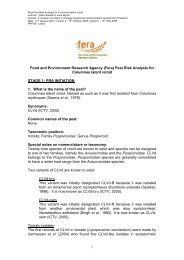
[129] Figure 1: Hard spot and freckle spot type of symp<strong>to</strong>ms caused by Guignardia citricarpa on sweet<br />
orange (Citrus sinensis) and lemon (C. limon) fruits: (A, a) hard spot lesions on sweet orange with the<br />
larger lesions containing pycnidia of the anamorph Phyllosticta citricarpa (arrows); (B) freckle spot lesions on<br />
lemon; (b) freckle spot lesions on sweet orange (the lesions are slightly depressed in the centre and devoid<br />
of pycnidia); (C) hard and freckle spot lesions on lemon; (c) freckle spot lesions (black arrows) and<br />
intermediate stage between freckle and hard spot lesions with pycnidia (white arrows) on sweet orange
<strong>Draft</strong> <strong>Annex</strong> <strong>to</strong> <strong>ISPM</strong> <strong>27</strong>:2006: Guignardia citricarpa Kiely on fruit (2004-023)<br />
[130]<br />
Courtesy: E. Feichtenberger, Institu<strong>to</strong> Biológico, Sorocaba, Brazil.<br />
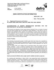
[131] Figure 2: False melanose, virulent spot, lacy spot and cracked spot symp<strong>to</strong>ms caused by Guignardia<br />
citricarpa on sweet orange (Citrus sinensis) and lemon (C. limon) fruits: (A) false melanose lesions on<br />
mature sweet orange; (a) mature sweet orange showing false melanose lesions surrounded by dark specks;<br />
(B) false melanose lesions on a green sweet orange; (C) virulent spot lesions on sweet orange (the lesions<br />
Page 17 of 22
[132]<br />
Page 18 of 22<br />
<strong>Draft</strong> <strong>Annex</strong> <strong>to</strong> <strong>ISPM</strong> <strong>27</strong>:2006: Guignardia citricarpa Kiely on fruit (2004-023)<br />
are depressed and extend deeply in<strong>to</strong> the albedo); (D) lacy spot symp<strong>to</strong>ms on a green sweet orange; (E)<br />
cracked spot type of lesions on sweet orange (the lesions are slightly raised, cracked with irregular margins<br />
and devoid of pycnidia)<br />
Courtesy: FUNDECITRUS (pho<strong>to</strong>s A, B, C, D, E) and E. Feichtenberger, Institu<strong>to</strong> Biológico, Sorocaba, Brazil<br />
(pho<strong>to</strong> a).<br />
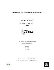
[133] Figure 3: Symp<strong>to</strong>ms of citrus black spot caused by Guignardia citricarpa on lemon (C. limon) leaves<br />
(A) and twigs (B)<br />
Courtesy: E. Feichtenberger, Institu<strong>to</strong> Biológico, Sorocaba, Brazil (pho<strong>to</strong> A) and M. Truter, <strong>Plant</strong> <strong>Protection</strong><br />
Research Institute, Agricultural Research Council, Pre<strong>to</strong>ria, South Africa (pho<strong>to</strong> B).
<strong>Draft</strong> <strong>Annex</strong> <strong>to</strong> <strong>ISPM</strong> <strong>27</strong>:2006: Guignardia citricarpa Kiely on fruit (2004-023)<br />
[134]<br />
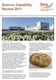
[135] Figure 4: Flow diagram for the diagnosis of Guignardia citricarpa on citrus fruit<br />
1 The molecular assays have been validated for the identification of the organism on pure cultures and fruit<br />
lesions and not on any other plant material (e.g. leaves, twigs).<br />
Page 19 of 22
[136]<br />
Page 20 of 22<br />
<strong>Draft</strong> <strong>Annex</strong> <strong>to</strong> <strong>ISPM</strong> <strong>27</strong>:2006: Guignardia citricarpa Kiely on fruit (2004-023)<br />
[137] Figure 5: Colony characteristics of Guignardia citricarpa and conidial morphology of the anamorph<br />
Phyllosticta citricarpa: (A) colony with irregular margin surrounded by a translucent zone of colourless<br />
submerged mycelium (arrow) after 30 days of growth on pota<strong>to</strong> dextrose agar (pH 5.5) at 25 °C and a 12-h<br />
pho<strong>to</strong>period; (B) conidial slime oozing from mature pycnidia; (C, D) conidia with a thin mucoid sheath (C,<br />
arrow) and a colourless subulate appendage (D, arrow, magnification 1,000× with immersion oil)<br />
Courtesy: L.E. Diaz, Ministry of Husbandry, Agriculture and Fisheries, Montevideo, Uruguay.
<strong>Draft</strong> <strong>Annex</strong> <strong>to</strong> <strong>ISPM</strong> <strong>27</strong>:2006: Guignardia citricarpa Kiely on fruit (2004-023)<br />
[138]<br />
[139] Figure 6: Conidia morphology and cultural characteristics of Guignardia citricarpa and G.<br />
mangiferae: (A) conidia of P. citricarpa, anamorph of G. citricarpa, with thin (1.5 μm) mucoid sheath (scale bar = 10<br />
μm) (pho<strong>to</strong> C was taken under a light microscope equipped with differential interference contrast); (D, E)<br />
colonies of G. citricarpa (D) and G. mangiferae (E), after 7 days of growth on (<strong>to</strong>p row) oatmeal agar, (middle<br />
row) malt extract agar, and (bot<strong>to</strong>m row) cherry decoction agar (Note production of a yellow pigment around<br />
the colony of G. citricarpa grown on oatmeal agar (D, arrows) and absence of this pigment in cultures of G.<br />
Page 21 of 22
Page 22 of 22<br />
mangiferae grown on the same medium (E).)<br />
<strong>Draft</strong> <strong>Annex</strong> <strong>to</strong> <strong>ISPM</strong> <strong>27</strong>:2006: Guignardia citricarpa Kiely on fruit (2004-023)<br />
Courtesy: G. Verkley, Centraalbureau voor Schimmelcultures, Utrecht, the Netherlands (pho<strong>to</strong>s A, B, C) and<br />
W. van Lienden, <strong>Plant</strong> <strong>Protection</strong> Service, Wageningen, The Netherlands (pho<strong>to</strong>s D, E).<br />
[140] Footnote 1 The use of the brand Eppendorf ® for PCR amplification in this diagnostic pro<strong>to</strong>col implies no<br />
approval of it <strong>to</strong> the exclusion of others that may also be suitable. This information is given for the<br />
convenience of users of this pro<strong>to</strong>col and does not constitute an endorsement by the CPM of the<br />
chemical, reagent and/or equipment named. Equivalent products may be used if they can be shown <strong>to</strong><br />
lead <strong>to</strong> the same results.<br />
[141] Footnote 2 The use of the brand Takara Bio Europe S.A.S for the 2× Premix Ex Taq master mix in this<br />
diagnostic pro<strong>to</strong>col implies no approval of it <strong>to</strong> the exclusion of others that may also be suitable. This<br />
information is given for the convenience of users of this pro<strong>to</strong>col and does not constitute an endorsement<br />
by the CPM of the chemical, reagent and/or equipment named. Equivalent products may be used if they<br />
can be shown <strong>to</strong> lead <strong>to</strong> the same results.<br />
[142] Footnote 3 The use of the brand Roche for the PCR reaction buffer and the DNA Taq-polymerase in this<br />
diagnostic pro<strong>to</strong>col implies no approval of them <strong>to</strong> the exclusion of others that may also be suitable. This<br />
information is given for the convenience of users of this pro<strong>to</strong>col and does not constitute an endorsement<br />
by the CPM of the chemical, reagent and/or equipment named. Equivalent products may be used if they<br />
can be shown <strong>to</strong> lead <strong>to</strong> the same results.