Laser refractive cataract surgery - FreeVis
Laser refractive cataract surgery - FreeVis
Laser refractive cataract surgery - FreeVis

You also want an ePaper? Increase the reach of your titles
YUMPU automatically turns print PDFs into web optimized ePapers that Google loves.
LenSx <strong>Laser</strong>s, Inc. 1/22/13<br />
Michael C. Knorz<br />
Medical Faculty Mannheim, University of Heidelberg<br />
Mannheim, Germany<br />
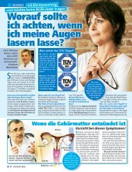
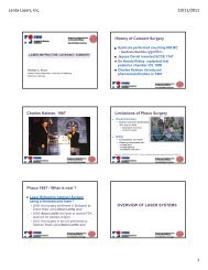
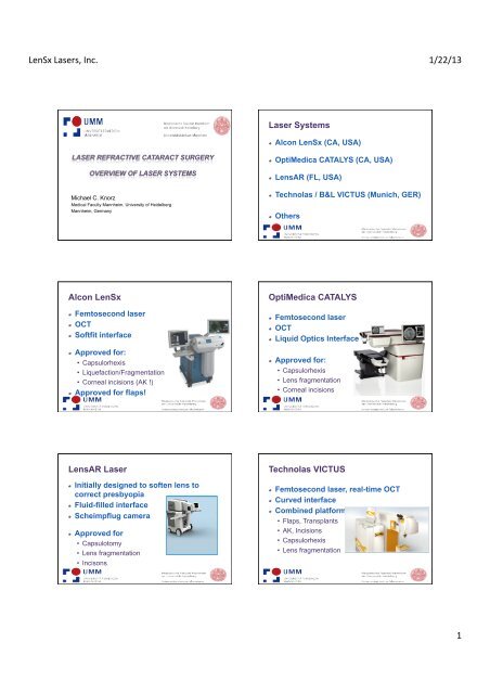
Alcon LenSx<br />
! Femtosecond laser<br />
! OCT<br />
! Softfit interface<br />
! Approved for:<br />
• Capsulorhexis<br />
• Liquefaction/Fragmentation<br />
• Corneal incisions (AK !)<br />
! Approved for flaps!<br />
LensAR <strong>Laser</strong><br />
1/22/13<br />
! Initially designed to soften lens to<br />
correct presbyopia<br />
! Fluid-filled interface<br />
! Scheimpflug camera<br />
! Approved for<br />
• Capsulotomy<br />
• Lens fragmentation<br />
• Incisons<br />
1/22/13<br />
<strong>Laser</strong> Systems<br />
! Alcon LenSx (CA, USA)<br />
! OptiMedica CATALYS (CA, USA)<br />
! LensAR (FL, USA)<br />
! Technolas / B&L VICTUS (Munich, GER)<br />
! Others<br />
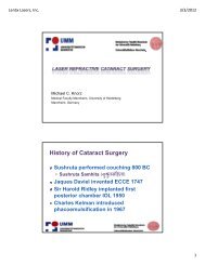
OptiMedica CATALYS<br />
! Femtosecond laser<br />
! OCT<br />
! Liquid Optics Interface<br />
! Approved for:<br />
• Capsulorhexis<br />
• Lens fragmentation<br />
• Corneal incisions<br />
Technolas VICTUS<br />
1/22/13<br />
1/22/13<br />
! Femtosecond laser, real-time OCT<br />
! Curved interface<br />
! Combined platform<br />
• Flaps, Transplants<br />
• AK, Incisions<br />
• Capsulorhexis<br />
• Lens fragmentation<br />
1/22/13<br />
1
LenSx <strong>Laser</strong>s, Inc. 1/22/13<br />
Personal Experience – Alcon LenSx<br />
! Surgery performed in Budapest,<br />
Hungary, 2010<br />
! Alcon LenSx laser in Mannheim since<br />
7-2011<br />
• > 250 laser <strong>refractive</strong> lens surgeries<br />
performed<br />
ALCON LENSX ® SOFTFIT PATIENT INTERFACE<br />
• Current PI Design with extended suction skirt<br />
• Proprietary soft contact lens insert<br />
• Lower IOP – 16mmHg increase<br />
• Simple docking process – better eye control, no fogging<br />
• Dramatically improved surgical performance<br />
ALCON LENSX ® SOFTFIT PI<br />
• • Eliminates corneal corneal compression compression<br />
• Lowers IOP – 16mmHg increase<br />
• Lowers IOP – 16mmHg increase<br />
• Better visibility w/ no fogging<br />
• Dramatically improves surgical performance<br />
• Improves docking and centration<br />
100% free-floating anterior capsulotomies!<br />
• Fixates cornea, no eye movement for precision incisions<br />
• Dramatically improves surgical performance<br />
• No liquid required………<br />
Alcon LenSx Company Update*<br />
• >200 <strong>Laser</strong>s<br />
• 34 Countries<br />
• More than 1,000<br />
MD's certified<br />
• More than 50,000<br />
procedures<br />
*Alcon Physician Training and Sales Data (PI procedures shipped)<br />
SOFTFIT PI: REDUCES CORNEAL<br />
COMPRESSION<br />
LenSx ® <strong>Laser</strong> Set Up<br />
Option 1: Placement in Pre-Op Room<br />
(non-sterile)<br />
• Maximizes procedure flow<br />
• Patient transported to OR on same bed<br />
• 1 laser easily feeds several ORs<br />
Dr Stephen G Slade, Houston, Texas<br />
1/22/13<br />
2
LenSx <strong>Laser</strong>s, Inc. 1/22/13<br />
LenSx ® <strong>Laser</strong> Set Up<br />
Option 2: Placement in Operating Room<br />
<strong>FreeVis</strong> <strong>Laser</strong> Center, Mannheim, Germany<br />
Patient positioned for LenSx ® <strong>Laser</strong><br />
treatment<br />
Capsulorhexis<br />
Patient bed swivels to operating<br />
microscope for I/A + IOL<br />
1/22/13<br />
! All lasers perform a reproducible<br />
capsulorhexis<br />
! No comparative studies published<br />
Femtosecond laser capsulotomy. [J Cataract Refract Surg. 2011] - PubMed - NCBI<br />
PubMed Search<br />
Display Settings: Abstract<br />
J Cataract Refract Surg. 2011 Jul;37(7):1189-98.<br />
Femtosecond laser capsulotomy.<br />
Friedman NJ, Palanker DV, Schuele G, Andersen D, Marcellino G, Seibel BS, Batlle J,<br />
Feliz R, Talamo JH, Blumenkranz MS, Culbertson WW.<br />
Department of Ophthalmology, Stanford University, the Mid-Peninsula Ophthalmology Medical<br />
Group, Palo Alto, USA. njfmd@pol.net<br />
Erratum in<br />
J Cataract Refract Surg. 2011 Sep;37(9):1742.<br />
Abstract<br />
PURPOSE: To evaluate a femtosecond laser system to create the<br />
capsulotomy.<br />
SETTING: Porcine and cadaver eye studies were performed at OptiMedica<br />
Corp., Santa Clara, California, USA; the human trial was performed at the<br />
Centro <strong>Laser</strong>, Santo Domingo, Dominican Republic.<br />
DESIGN: Experimental and clinical study.<br />
METHODS: Capsulotomies performed by an optical coherence tomographyguided<br />
femtosecond laser were evaluated in porcine and human cadaver eyes.<br />
Subsequently, the procedure was performed in 39 patients as part of a<br />
prospective randomized study of femtosecond laser-assisted <strong>cataract</strong> <strong>surgery</strong>.<br />
The accuracy of the capsulotomy size, shape, and centration were quantified<br />
and capsulotomy strength was assessed in the porcine eyes.<br />
RESULTS: <strong>Laser</strong>-created capsulotomies were significantly more precise in size<br />
and shape than manually created capsulorhexes. In the patient eyes, the<br />
deviation from the intended diameter of the resected capsule disk was 29 µm ±<br />
26 (SD) for the laser technique and 337 ± 258 µm for the manual technique.<br />
The mean deviation from circularity was 6% and 20%, respectively. The center<br />
of the laser capsulotomies was within 77 ± 47 µm of the intended position. All<br />
capsulotomies were complete, with no radial nicks or tears. The strength of<br />
laser capsulotomies (porcine subgroup) decreased with increasing pulse<br />
energy: 152 ± 21 mN for 3 µJ, 121 ± 16 mN for 6 µJ, and 113 ± 23 mN for 10<br />
µJ. The strength of the manual capsulorhexes was 65 ± 21 mN.<br />
CONCLUSION: The femtosecond laser produced capsulotomies that were<br />
more precise, accurate, reproducible, and stronger than those created with the<br />
conventional manual technique.<br />
Copyright © 2011 ASCRS and ESCRS. Published by Elsevier Inc. All rights<br />
reserved.<br />
PMID: 21700099 [PubMed - indexed for MEDLINE]<br />
Publication Types, MeSH Terms<br />
http://www.ncbi.nlm.nih.gov/pubmed/21700099<br />
1/22/13<br />
08.06.12 15:33<br />
1/22/13<br />
Seite 1 von 2<br />
17<br />
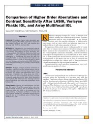
First Paper on LRCS<br />
published 2009 !<br />
Journal of Refractive Surgery Volume 25 December 2009<br />
ORIGINAL ARTICLES<br />
Initial Clinical Evaluation of an Intraocular<br />
Femtosecond <strong>Laser</strong> in Cataract Surgery<br />
Zoltan Nagy, MD; Agnes Takacs, MD; Tamas Filkorn, MD; Melvin Sarayba, MD<br />
ABSTRACT<br />
PURPOSE: To evaluate femtosecond laser lens fragmentation<br />
and anterior capsulotomy in <strong>cataract</strong> <strong>surgery</strong>.<br />
METHODS: Anterior capsulotomy and phacofragmentation<br />
procedures performed with an intraocular femtosecond<br />
laser (LenSx <strong>Laser</strong>s Inc) were initially evaluated<br />
in ex vivo porcine eyes. These procedures were then<br />
performed in an initial series of nine patients undergoing<br />
<strong>cataract</strong> <strong>surgery</strong>. In addition to standard intraoperative<br />
assessments (including capsulotomy diameter accuracy<br />
and reproducibility), optical coherence tomography was<br />
used to evaluate human procedures.<br />
RESULTS: For an intended 5-mm capsulorrhexis<br />
in porcine eyes, average achieved diameters were<br />
5.880.73 mm using a standard manual technique<br />
and 5.020.04 mm using the femtosecond laser.<br />
Scanning electron microscopy revealed equally smooth<br />
cut edges of the capsulotomy with the femtosecond<br />
laser and manual technique. Compared to control<br />
porcine eyes, femtosecond laser phacofragmentation<br />
resulted in a 43% reduction in phacoemulsifi cation<br />
power and a 51% decrease in phacoemulsifi cation<br />
time. In a small series of human clinical procedures,<br />
femtosecond laser capsulotomies and phacofragmentation<br />
demonstrated similarly high levels of accuracy<br />
and effectiveness, with no operative complications.<br />
CONCLUSIONS: Initial results with an intraocular femtosecond<br />
laser demonstrate higher precision of capsulorrhexis<br />
and reduced phacoemulsifi cation power in porcine<br />
and human eyes. [J Refract Surg. 2009;25:1053-1060.]<br />
doi:10.3928/1081597X-20091117-04<br />
<strong>Laser</strong> Capsulorhexis<br />
C<br />
ataract <strong>surgery</strong> with intraocular lens (IOL) implantation<br />
is the most common ophthalmic surgical procedure<br />
worldwide. It is also the most common <strong>surgery</strong><br />
that corrects <strong>refractive</strong> error, performed over fi ve times more<br />
frequently than corneal <strong>refractive</strong> <strong>surgery</strong>. 1 Phacoemulsifi -<br />
cation is the dominant form of <strong>cataract</strong> <strong>surgery</strong> in developed<br />
countries, accounting for 90% of procedures. 2,3 Although a<br />
number of recent developments have occurred in IOL technology,<br />
the basic phacoemulsifi cation procedure has remained<br />
largely unchanged over the past 20 years, involving a series<br />
of individual steps including corneal incision creation,<br />
capsulorrhexis, and phacofragmentation.<br />
Although highly successful, each of these manual steps<br />
presents an opportunity for improvement in both safety and<br />
effectiveness. For example, manual capsulorrhexis results in<br />
capsular tears in approximately 1% of cases and has limited<br />
diameter predictability, which can affect IOL centration,<br />
postoperative anterior chamber depth, and posterior capsular<br />
opacifi cation rates. 4-7 The surgical challenges posed by nuclear<br />
chopping techniques have hindered widespread adoption,<br />
despite evidence that they reduce ultrasound requirements<br />
relative to traditional phacoemulsifi cation. 2,8<br />
Femtosecond lasers represent an important technological<br />
advance in ophthalmic <strong>surgery</strong>. Combined with computercontrolled<br />
optical delivery systems, femtosecond lasers can<br />
produce precise surgical incisions without collateral damage<br />
to surrounding tissues. 9-13 Since 2001, several femtosecond<br />
laser systems have been introduced clinically and more than<br />
2 million ophthalmic procedures have been performed with<br />
femtosecond lasers, primarily for creation of a corneal fl ap in<br />
LASIK. The precision of femtosecond lasers exceeds that of<br />
From the 1st Department of Ophthalmology, Semmelweis University, Budapest,<br />
Hungary (Nagy, Takacs, Filkorn); and LenSx <strong>Laser</strong>s Inc, Aliso Viejo, Calif<br />
(Sarayba).<br />
Dr Nagy is a paid consultant to LenSx <strong>Laser</strong>s Inc. Dr Sarayba is an employee<br />
of LenSx <strong>Laser</strong>s Inc. Drs Takacs and Filkorn have no financial interest in the<br />
materials presented herein.<br />
Correspondence: Zoltan Nagy, MD, 1st Department of Ophthalmology, Semmelweis<br />
University, 39 St Maria St, Budapest, Hungary 1085. Tel: 36 20 815 8468;<br />
E-mail: zoltan.nagy100@gmail.com<br />
Received: November 2, 2009; Accepted: November 10, 2009<br />
1/22/13<br />
1/22/13<br />
1053<br />
18<br />
3
LenSx <strong>Laser</strong>s, Inc. 1/22/13<br />
Nucleus Fragmentation<br />
! All lasers perform nucleus fragmentation<br />
using different patterns<br />
! No comparative studies published<br />
1/22/13<br />
LenSx <strong>Laser</strong> Corneal Incision<br />
PostOp OCT image of LenSx 2-plane corneal incision<br />
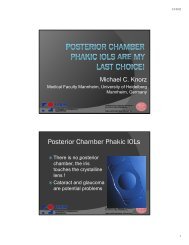
Astigmatism Results LenSx<br />
100%<br />
80%<br />
60%<br />
40%<br />
20%<br />
0%<br />
50%<br />
86%<br />
64%<br />
93%<br />
71%<br />
100%<br />
93%<br />
1/22/13<br />
100% 100%<br />
0% 0%<br />
1/22/13<br />
Pre Op (ΔKs)<br />
Post Op (MR Cyl.)<br />
≤0.50 ≤0.75 ≤1.00 ≤1.25 ≤1.50 ≤2.00 >2.00<br />
Astigmatism (D)<br />
19<br />
21<br />
Astigmatism Correction & Incisions<br />
! LenSx, Victus and Catalys<br />
• (AMO Intralase AK only)<br />
AK and incisions should be possible with<br />
all lasers, but published results are<br />
available with LenSx only<br />
1/22/13<br />
1/22/13<br />
20<br />
22<br />
4
LenSx <strong>Laser</strong>s, Inc. 1/22/13<br />
First Paper on LRCS<br />
published 2009 !<br />
Journal of Refractive Surgery Volume 25 December 2009<br />
ORIGINAL ARTICLES<br />
Initial Clinical Evaluation of an Intraocular<br />
Femtosecond <strong>Laser</strong> in Cataract Surgery<br />
Zoltan Nagy, MD; Agnes Takacs, MD; Tamas Filkorn, MD; Melvin Sarayba, MD<br />
ABSTRACT<br />
PURPOSE: To evaluate femtosecond laser lens fragmentation<br />
and anterior capsulotomy in <strong>cataract</strong> <strong>surgery</strong>.<br />
METHODS: Anterior capsulotomy and phacofragmentation<br />
procedures performed with an intraocular femtosecond<br />
laser (LenSx <strong>Laser</strong>s Inc) were initially evaluated<br />
in ex vivo porcine eyes. These procedures were then<br />
performed in an initial series of nine patients undergoing<br />
<strong>cataract</strong> <strong>surgery</strong>. In addition to standard intraoperative<br />
assessments (including capsulotomy diameter accuracy<br />
and reproducibility), optical coherence tomography was<br />
used to evaluate human procedures.<br />
RESULTS: For an intended 5-mm capsulorrhexis<br />
in porcine eyes, average achieved diameters were<br />
5.880.73 mm using a standard manual technique<br />
and 5.020.04 mm using the femtosecond laser.<br />
Scanning electron microscopy revealed equally smooth<br />
cut edges of the capsulotomy with the femtosecond<br />
laser and manual technique. Compared to control<br />
porcine eyes, femtosecond laser phacofragmentation<br />
resulted in a 43% reduction in phacoemulsifi cation<br />
power and a 51% decrease in phacoemulsifi cation<br />
time. In a small series of human clinical procedures,<br />
femtosecond laser capsulotomies and phacofragmentation<br />
demonstrated similarly high levels of accuracy<br />
and effectiveness, with no operative complications.<br />
CONCLUSIONS: Initial results with an intraocular femtosecond<br />
laser demonstrate higher precision of capsulorrhexis<br />
and reduced phacoemulsifi cation power in porcine<br />
and human eyes. [J Refract Surg. 2009;25:1053-1060.]<br />
doi:10.3928/1081597X-20091117-04<br />
C<br />
ataract <strong>surgery</strong> with intraocular lens (IOL) implantation<br />
is the most common ophthalmic surgical procedure<br />
worldwide. It is also the most common <strong>surgery</strong><br />
that corrects <strong>refractive</strong> error, performed over fi ve times more<br />
frequently than corneal <strong>refractive</strong> <strong>surgery</strong>. 1 Phacoemulsifi -<br />
cation is the dominant form of <strong>cataract</strong> <strong>surgery</strong> in developed<br />
countries, accounting for 90% of procedures. 2,3 Although a<br />
number of recent developments have occurred in IOL technology,<br />
the basic phacoemulsifi cation procedure has remained<br />
largely unchanged over the past 20 years, involving a series<br />
of individual steps including corneal incision creation,<br />
capsulorrhexis, and phacofragmentation.<br />
Although highly successful, each of these manual steps<br />
presents an opportunity for improvement in both safety and<br />
effectiveness. For example, manual capsulorrhexis results in<br />
capsular tears in approximately 1% of cases and has limited<br />
diameter predictability, which can affect IOL centration,<br />
postoperative anterior chamber depth, and posterior capsular<br />
opacifi cation rates. 4-7 The surgical challenges posed by nuclear<br />
chopping techniques have hindered widespread adoption,<br />
despite evidence that they reduce ultrasound requirements<br />
relative to traditional phacoemulsifi cation. 2,8<br />
Femtosecond lasers represent an important technological<br />
advance in ophthalmic <strong>surgery</strong>. Combined with computercontrolled<br />
optical delivery systems, femtosecond lasers can<br />
produce precise surgical incisions without collateral damage<br />
to surrounding tissues. 9-13 Since 2001, several femtosecond<br />
laser systems have been introduced clinically and more than<br />
2 million ophthalmic procedures have been performed with<br />
femtosecond lasers, primarily for creation of a corneal fl ap in<br />
LASIK. The precision of femtosecond lasers exceeds that of<br />
From the 1st Department of Ophthalmology, Semmelweis University, Budapest,<br />
Hungary (Nagy, Takacs, Filkorn); and LenSx <strong>Laser</strong>s Inc, Aliso Viejo, Calif<br />
(Sarayba).<br />
Dr Nagy is a paid consultant to LenSx <strong>Laser</strong>s Inc. Dr Sarayba is an employee<br />
of LenSx <strong>Laser</strong>s Inc. Drs Takacs and Filkorn have no financial interest in the<br />
materials presented herein.<br />
Correspondence: Zoltan Nagy, MD, 1st Department of Ophthalmology, Semmelweis<br />
University, 39 St Maria St, Budapest, Hungary 1085. Tel: 36 20 815 8468;<br />
E-mail: zoltan.nagy100@gmail.com<br />
Received: November 2, 2009; Accepted: November 10, 2009<br />
1/22/13<br />
Comparison of Intraocular Lens<br />
Decentration Parameters After<br />
Femtosecond and Manual Capsulotomies<br />
Zoltán Zsolt Nagy, MD, DSC; Kinga Kránitz, MD; Agnes I. Takacs, MD; Kata Miháltz, MD;<br />
Illés Kovács, MD, PhD; Michael C. Knorz, MD<br />
564<br />
ABSTRACT<br />
PURPOSE: To evaluate a laser technique and manual<br />
technique to perform capsulorrhexis in <strong>cataract</strong> eyes.<br />
METHODS: Anterior capsulotomy was performed with<br />
an intraocular femtosecond laser (LenSx <strong>Laser</strong>s Inc) in<br />
54 eyes (FS group) and manual continuous curvilinear<br />
capsulorrhexis was performed in 57 eyes (CCC group).<br />
Circularity and area of capsulotomy and IOL decentration<br />
were measured using Photoshop CS4 Extended<br />
(Adobe Systems Inc) 1 week after <strong>surgery</strong>. Average keratometry,<br />
axial length, and preoperative anterior chamber<br />
depth were examined with the Lenstar LS 900 (Haag-<br />
Streit AG).<br />
RESULTS: No statistically signifi cant differences were<br />
noted between groups in axial length, preoperative <strong>refractive</strong><br />
state, and in the area of capsulotomy. Circularity<br />
values were signifi cantly better in the FS group<br />
(P=.032). We found incomplete overlap of capsulotomies<br />
in 28% of eyes in the CCC group and 11% in the<br />
FS group (P=.033). Signifi cant correlations were noted<br />
between axial length and area of capsulotomy, and between<br />
average keratometry and area of the capsulotomy<br />
in the CCC group (R=0.278, P=.036; and R=0.29,<br />
P=.033, respectively), but both did not correlate in the<br />
FS group (P.05). In the CCC group, the pupillary area<br />
correlated signifi cantly with the area of the capsulotomy<br />
(R=0.27, P=.039). Signifi cant correlation was noted<br />
between IOL decentration and axial length in the CCC<br />
group (R=0.30, P=.026), but there was no correlation<br />
in the FS group (P.05).<br />
CONCLUSIONS: Femtosecond laser capsulorrhexis<br />
was more regularly shaped, showed better centration,<br />
and showed a better intraocular lens/capsule<br />
overlap than manual capsulorrhexis. [J Refract Surg.<br />
2011;27(8):564-569.]<br />
doi:10.3928/1081597X-20110607-01<br />
Conclusions<br />
M<br />
yopia and <strong>cataract</strong> are common disorders in the<br />
human population. Highly myopic eyes are more<br />
likely to develop <strong>cataract</strong>. 1 Cataract <strong>surgery</strong> has<br />
become a common, safe, and effective intervention performed<br />
worldwide. 2 However, <strong>surgery</strong> in eyes with long axial length<br />
is associated with increased risk of intra- and postoperative<br />
complications. 3<br />
Posterior capsular opacifi cation is the most common surgically<br />
related cause of reduced vision after <strong>cataract</strong> <strong>surgery</strong>.<br />
Capsulorrhexis size, centration, and completely overlapping<br />
anterior capsule on the optic edge of the intraocular lens (IOL)<br />
affect the severity of this disorder. Although new IOL designs<br />
have diminished the incidence of posterior capsular opacifi<br />
cation, a precise anterior capsulotomy is a crucial step in<br />
preventing the migration of lens epithelial cells. 4-6 Complete<br />
overlap helps prevent not only posterior capsular opacifi cation<br />
but also results in better IOL centration and less myopic<br />
shift by maintaining the IOL in the proper position. 7,8<br />
In recent years, the most commonly applied technique<br />
during phacoemulsifi cation is continuous curvilinear capsulorrhexis.<br />
Popularized by Gimbel and Neuhann, 9-11 it has<br />
several surgical and postoperative advantages, but special<br />
attention and surgical expertise are needed to complete it<br />
successfully. In highly myopic eyes, the larger size of the<br />
eye and pupillary diameter and optical distortion by the cornea<br />
may deceive surgeons to prepare a larger capsulorrhexis<br />
than intended. 12-14 This makes IOL malpositioning (eg, decentration,<br />
tilt, and luxation due to improper fi xation in a<br />
larger capsular bag) more likely and may cause myopization<br />
and an increase in higher order aberrations. 15-17<br />
From Semmelweis University Budapest, Faculty of Medicine, Department of<br />
Ophthalmology, Hungary (Nagy, Kránitz, Takacs, Miháltz, Kovács); and Medical<br />
Faculty Mannheim, University of Heidelberg, Mannheim, Germany (Knorz).<br />
Drs Nagy and Knorz are consultants to LenSx <strong>Laser</strong>s Inc. The remaining<br />
authors have no proprietary interest in the materials presented herein.<br />
Correspondence: Zoltán Zsolt Nagy, MD, DSC, 1085 Budapest, Mária u. 39,<br />
Hungary. Tel: 36 20 825 8468; Fax: 361 210 0309; E-mail: nz@szem1.sote.hu<br />
or zoltan.nagy100@gmail.com<br />
Received: December 1, 2010; Accepted: May 24, 2011<br />
Posted online: June 20, 2011<br />
Copyright © SLACK Incorporated<br />
1/22/13<br />
! LenSx, Victus, Catalys and LensAR can perform<br />
capsulorhexis and nucleus fragmentation<br />
• LenSx experience with more than 50,000 procedures<br />
! Incisions and AK should be possible with all lasers<br />
• Approved for LenSx, Catalys and Victus only (AMO AK only)<br />
! <strong>Laser</strong> Refractive Cataract Surgery improves results<br />
• Less ultrasound required<br />
• Better IOL centration and less IOL tilt<br />
• <strong>Laser</strong> incisions correct corneal astigmatism<br />
1/22/13<br />
1053<br />
558<br />
ORIGINAL ARTICLES<br />
Femtosecond <strong>Laser</strong> Capsulotomy<br />
and Manual Continuous Curvilinear<br />
Capsulorrhexis Parameters and Their<br />
Effects on Intraocular Lens Centration<br />
Kinga Kránitz, MD; Agnes Takacs, MD; Kata Miháltz, MD; Illés Kovács, MD, PhD;<br />
Michael C. Knorz, MD; Zoltán Z. Nagy, MD, DSC<br />
ABSTRACT<br />
PURPOSE: To measure and compare sizing and positioning<br />
parameters of femtosecond laser capsulotomy with<br />
manual continuous curvilinear capsulorrhexis (CCC).<br />
METHODS: Femtosecond capsulotomies (Alcon-LenSx<br />
<strong>Laser</strong>s Inc) and CCC were carried out in 20 eyes of 20<br />
patients, respectively. Intraocular lens (IOL) decentration,<br />
circularity, vertical and horizontal diameters of<br />
capsulotomies, and capsule overlap were measured<br />
with Adobe Photoshop (Adobe Systems Inc) 1 week, 1<br />
month, and 1 year after <strong>surgery</strong>. Between-group differences<br />
of parameters and predictors of IOL decentration<br />
were determined with repeated measures analysis of variance,<br />
chi-square test, and logistic regression analyses.<br />
RESULTS: Vertical diameter of CCC was statistically<br />
signifi cantly higher in the fi rst week and month. Signifi -<br />
cantly higher values of capsule overlap over 1 year and<br />
circularity in the fi rst week showed more regular femtosecond<br />
capsulotomies. Horizontal IOL decentration was<br />
statistically signifi cantly higher in the CCC group over 1<br />
year. A signifi cant difference was noted between the two<br />
groups in dichotomized horizontal decentration values<br />
at 0.4 mm with chi-square test after 1 week and 1 year<br />
(P=.035 and P=.016, respectively). In univariable general<br />
estimating equation models, type of capsulorrhexis<br />
(P.01) and capsule overlap (P=.002) were signifi cant<br />
predictors of horizontal decentration. Vertical diameter<br />
showed signifi cant correlation to the overlap in the CCC<br />
group (1 week: r=0.91; 1 month: r=0.76, P.01;<br />
1 year: r=0.62, P.01), whereas no signifi cant correlation<br />
was noted in the femtosecond group (P.05).<br />
CONCLUSIONS: More precise capsulotomy sizing and<br />
centering can be achieved with femtosecond laser. Properly<br />
sized, shaped, and centered femtosecond laser capsulotomies<br />
resulted in better overlap parameters that help<br />
maintain proper positioning of the IOL. [J Refract Surg.<br />
2011;27(8):558-563.]<br />
doi:10.3928/1081597X-20110623-03<br />
Journal of Refractive Surgery • Vol. 27, No. 10, 2011<br />
C<br />
reation of a precise anterior capsulorrhexis is one of<br />
the most important steps of <strong>cataract</strong> <strong>surgery</strong>. In recent<br />
years, the most commonly used technique during<br />
phacoemulsifi cation is continuous curvilinear capsulorrhexis<br />
(CCC). Popularized by Gimbel and Neuhann, 1-3 CCC has several<br />
surgical and postoperative advantages but its completion<br />
takes special attention and surgical expertise. Obtaining a precise<br />
capsulorrhexis is essential to reach demanding <strong>refractive</strong><br />
results because a properly sized and well-centered capsulorrhexis<br />
with a 360° overlapping capsular edge prevents optic<br />
decentration, tilt, myopic shift, posterior and anterior capsular<br />
opacifi cation due to symmetric contractile forces of the capsular<br />
bag, and shrink wrap effect. 4-10 However, an eccentric or<br />
irregularly shaped capsulorrhexis with a diameter extending<br />
beyond the optic edge may lose these advantages.<br />
Until now, capsulorrhexis has been a manual procedure.<br />
With the advent of femtosecond lasers in ophthalmic <strong>surgery</strong>,<br />
a predictably sized and centered anterior capsulotomy<br />
became possible through a laser–tissue interaction known as<br />
photodisruption. 11 Femtosecond lasers were initially developed<br />
for LASIK fl ap creation during corneal <strong>refractive</strong> <strong>surgery</strong>.<br />
Recently introduced laser technology enables surgeons<br />
to achieve effi cient lens fragmentation or liquefaction and<br />
precise and reproducible creation of capsulotomies and corneal<br />
incisions during <strong>refractive</strong> <strong>cataract</strong> <strong>surgery</strong>. 11-14<br />
The purpose of this study was to measure and compare sizing<br />
and positioning parameters of the femtosecond laser capsulotomy<br />
with manual CCC during 1-year follow-up. We also studied the<br />
effects of these differences on IOL centration. To our knowledge,<br />
no such comparisons have been performed previously.<br />
From Semmelweis University Budapest, Faculty of Medicine, Department of<br />
Ophthalmology, Hungary (Kránitz, Takacs, Miháltz, Kovács, Nagy); and Medical<br />
Faculty Mannheim, University of Heidelberg, Mannheim, Germany (Knorz).<br />
Drs Knorz and Nagy are consultants to Alcon-LenSx <strong>Laser</strong>s Inc. The remaining<br />
authors have no financial interest in the materials presented herein.<br />
Correspondence: Kinga Kránitz, MD, Semmelweiss University Budapest, Dept<br />
of Ophthalmology, Mária u. 39, 1085 Budapest, Hungary. Tel: 36 20 825 8503;<br />
Fax: 36 1 317 9061; E-mail: kranitzkinga@gmail.com<br />
Received: October 14, 2010; Accepted: June 3, 2011<br />
Posted online: June 30, 2011<br />
ORIGINAL ARTICLES<br />
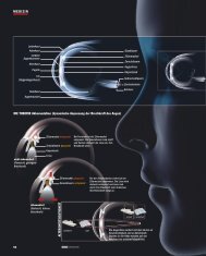
Internal Aberrations and Optical Quality<br />
After Femtosecond <strong>Laser</strong> Anterior<br />
Capsulotomy in Cataract Surgery<br />
Kata Miháltz, MD; Michael C. Knorz, MD; Jorge L. Alió, MD, PhD; Ágnes I. Takács, MD;<br />
Kinga Kránitz, MD; Illés Kovács, MD, PhD; Zoltán Z. Nagy, MD, DSc<br />
ABSTRACT<br />
PURPOSE: To compare ocular and internal aberrations<br />
after femtosecond laser anterior capsulotomy and continuous<br />
curvilinear capsulorrhexis in <strong>cataract</strong> <strong>surgery</strong>.<br />
METHODS: In this prospective study, anterior capsulotomy<br />
was performed during <strong>cataract</strong> <strong>surgery</strong> with an<br />
intraocular femtosecond (FS) laser (Alcon LenSx Inc) in<br />
48 eyes. As a control group, continuous curvilinear capsulorrhexis<br />
(CCC) was performed in 51 eyes. Wavefront<br />
aberrometry, corneal topography, and objective visual<br />
quality were measured using the OPD-Scan (NIDEK Co<br />
Ltd). Vertical and horizontal tilt, coma, and visual quality<br />
metrics were evaluated separately to determine whether<br />
the source of aberrations was ocular or internal. Main<br />
outcome measures included postoperative residual refraction,<br />
uncorrected and corrected visual acuities, ocular<br />
and internal aberrations, Strehl ratio, and modulation<br />
transfer function (MTF).<br />
RESULTS: No statistically signifi cant differences were<br />
noted between the FS and CCC groups, respectively, in<br />
postoperative sphere (0.601.50 vs 0.501.40<br />
diopters [D]), postoperative cylinder (1.301.01<br />
vs 1.101.10 D), uncorrected distance visual acuity<br />
(0.860.15 vs 0.880.08), or corrected distance<br />
visual acuity (0.970.08 vs 0.970.06). The FS<br />
group had signifi cantly lower values of intraocular<br />
vertical tilt (0.050.36 vs 0.270.57) and coma<br />
(0.0030.11 vs 0.10.15), and signifi cantly higher<br />
Strehl ratios (0.020.02 vs 0.010.01) and MTF values<br />
at all measured cycles per degree, compared to the<br />
CCC group.<br />
CONCLUSIONS: Capsulotomy performed with an intraocular<br />
FS laser induced signifi cantly less internal<br />
aberrations measured by the NIDEK OPD-Scan aberrometer<br />
compared to eyes that underwent CCC, which<br />
may result in better optical quality after the procedure.<br />
[J Refract Surg. 2011;27(10):711-716.]<br />
doi:10.3928/1081597X-20110913-01<br />
P<br />
Copyright © SLACK Incorporated<br />
1/22/13<br />
erforming a precise anterior capsulorrhexis is crucial<br />
in <strong>cataract</strong> <strong>surgery</strong>. A capsulorrhexis with a 360°<br />
overlapping capsular edge prevents optic decentra-<br />
tion, tilt, myopic shift, posterior and anterior capsular opacifi<br />
cation due to symmetric contractile forces of the capsular<br />
bag, and shrink wrap effect. 1-6 In earlier reports from our research<br />
group, we have demonstrated higher precision capsulorrhexis<br />
creation and reduced lens decentration with the<br />
intraocular femtosecond laser. 7,8 This technology also has<br />
the potential to reduce the risk of capsular tear and intraoperative<br />
complications during <strong>cataract</strong> <strong>surgery</strong> and reduced<br />
phacoemulsifi cation power. 9<br />
Optical quality is a subjective entity and can currently only<br />
be described indirectly by objective metrics, such as wavefront<br />
error measurements, and visual quality metrics or functional<br />
data, such as visual acuity and contrast sensitivity. 10-13<br />
Wavefront analysis isolates the effect of lower order aberrations<br />
(defocus, astigmatism) and higher order aberrations, as<br />
well as the contribution of individual aberrations on optical<br />
quality. Strehl ratio, point spread function (PSF), and modulation<br />
transfer function (MTF) are parameters of the quality of<br />
an optical system including a human eye. The PSF of an optical<br />
system is the intensity distribution of light from a point<br />
source projected onto the retina and indicates the extent of<br />
blurring of the retinal image. Modulation transfer function is<br />
defi ned as the amplitude of the image contrast divided by the<br />
amplitude of the object contrast and is a function of spatial<br />
frequency, which could describe the reduction in contrast of<br />
sine wave stimuli by the optical medium. Modulation transfer<br />
function can be measured by directly imaging the PSF on<br />
From Semmelweis University Budapest, Faculty of Medicine, Department<br />
of Ophthalmology, Budapest, Hungary (Miháltz, Takács, Kránitz, Kovács,<br />
Nagy); <strong>FreeVis</strong> LASIK Center, Medical Faculty Mannheim, University of<br />
Heidelberg, Mannheim, Germany (Knorz); and the Division of Ophthalmology,<br />
Instituto Oftalmológico de Alicante, Vissum Corporation, Universidad Miguel<br />
Hernández, Alicante, Spain (Alió).<br />
Drs Knorz, Alió, and Nagy are consultants for Alcon LenSx Inc. The remaining<br />
authors have no financial interest in the materials presented herein.<br />
Correspondence: Kata Miháltz, MD, 1085 Budapest, Mária u. 39, Hungary.<br />
Tel: 36 70 940 8296; Fax: 36 1 317 9061; E-mail: mihaltzkata@yahoo.com<br />
Received: July 13, 2011; Accepted: September 9, 2011<br />
711<br />
1/22/13<br />
5