Functional histology of the retina - Oftared
Functional histology of the retina - Oftared
Functional histology of the retina - Oftared

You also want an ePaper? Increase the reach of your titles
YUMPU automatically turns print PDFs into web optimized ePapers that Google loves.
Microscopy: Science, Technology, Applications and Education<br />
A. ______________________________________________<br />
Méndez-Vilas and J. Díaz (Eds.)<br />
<strong>Functional</strong> <strong>histology</strong> <strong>of</strong> <strong>the</strong> <strong>retina</strong><br />
F. Germain 1 , C. Pérez-Rico 2 , J. Vicente 1 and P. de la Villa 1<br />
1 Department <strong>of</strong> Physiology, University <strong>of</strong> Alcalá, Campus Universitario s/n Spain<br />
2 Hospital Universitario Príncipe de Asturias, Alcalá de Henares, Madrid, Spain<br />
Visual perception is a sensory process initiated at <strong>the</strong> <strong>retina</strong>, and completed in <strong>the</strong> cerebral cortex. Two main functions are<br />
currently performed by <strong>the</strong> <strong>retina</strong>: 1) <strong>the</strong> initial conversion <strong>of</strong> light energy into electric signals, phototransduction, which is<br />
carried out by photoreceptors; 2) a series <strong>of</strong> physiological processes performed by <strong>retina</strong>l interneurons (bipolar, horizontal<br />
and amacrine cells), in order to encode <strong>the</strong> different attributes <strong>of</strong> <strong>the</strong> visual stimuli (shape, movement and color) in<br />
electrical signals. The most direct pathway: photoreceptors - bipolar cells - ganglion cells is influence by two additional<br />
cell types: in <strong>the</strong> outer plexiform layer by horizontal cells that act on surrounding bipolar cells; and in <strong>the</strong> inner plexiform<br />
layer by amacrine cells, that modulate surrounding ganglion cells. Central nervous system, like <strong>retina</strong>, may be studied<br />
under several types <strong>of</strong> techniques. This chapter will deal with different microscopical techniques: iontophoretical<br />
intracellular injection, scanning and transmission electronic microscopy, and confocal immun<strong>of</strong>luorescence microscopy,<br />
that help us to understand <strong>the</strong> physiological mechanisms performed by <strong>retina</strong>l neurons.<br />
Keywords: scanning; transmission electronic microscopy; confocal immun<strong>of</strong>luorescence microscopy; isolated cells;<br />
immunohistochemistry; iontophoretical intracellular injection.<br />
1. Anatomical structure <strong>of</strong> <strong>the</strong> eye<br />
The eye is an organ specialized for <strong>the</strong> detection and analysis <strong>of</strong> light. The eye is a fluid chamber enclosed by three<br />
layers <strong>of</strong> tissue. The outermost <strong>of</strong> <strong>the</strong> three coasts <strong>of</strong> <strong>the</strong> eye consists <strong>of</strong> cornea, limbus and sclera. The transparent<br />
cornea makes up about 18% outer coat <strong>of</strong> <strong>the</strong> eye; <strong>the</strong> white opaque sclera accounts for most <strong>of</strong> <strong>the</strong> rest. Both tissues<br />
consist mainly <strong>of</strong> collagen fibers. The cornea is <strong>the</strong> principal refractive element <strong>of</strong> <strong>the</strong> eye. The sclera is rigid and<br />
resistant to penetration; it is able to protect <strong>the</strong> more delicate inner layers. The limbus is <strong>the</strong> region <strong>of</strong> transition from<br />
cornea to sclera.<br />
The middle layer, <strong>the</strong> uveal tract, includes <strong>the</strong> iris, ciliary body and choroid. The iris is a layer disposed in <strong>the</strong><br />
anterior pole <strong>of</strong> <strong>the</strong> eye, behind <strong>the</strong> cornea, that limits <strong>the</strong> quantity <strong>of</strong> light entering into <strong>the</strong> eye through <strong>the</strong> pupil. The<br />
ciliary body is adjacent to <strong>the</strong> iris and it consists in a ring <strong>of</strong> muscle cells encircling <strong>the</strong> anterior portion <strong>of</strong> <strong>the</strong> eye. The<br />
aqueous humor is formed at this site. The ciliary body musculature is part <strong>of</strong> <strong>the</strong> system for altering <strong>the</strong> refractive power<br />
<strong>of</strong> <strong>the</strong> lens, accommodation. The choroid is composed <strong>of</strong> blood vessels and pigmented epi<strong>the</strong>lium. This epi<strong>the</strong>lium is<br />
composed <strong>of</strong> dense melanin pigment to absorb and eliminate scattered light that might o<strong>the</strong>rwise degrade <strong>the</strong> image.<br />
The innermost eyeball layer is <strong>the</strong> <strong>retina</strong> that woks as computer that receives inputs from 100 million photodetectors,<br />
that are sampling <strong>the</strong> pattern <strong>of</strong> light and dark in <strong>the</strong> image formed by <strong>the</strong> optical system <strong>of</strong> <strong>the</strong> eye. The <strong>retina</strong> processes<br />
<strong>the</strong> light information that it receives from <strong>the</strong> surrounding world. This portion <strong>of</strong> <strong>the</strong> nervous system processes <strong>the</strong> light<br />
information and transmits it to <strong>the</strong> brain via <strong>the</strong> optic nerve which exits <strong>the</strong> eyeball from its posterior pole. Retina works<br />
detecting light in <strong>the</strong> <strong>retina</strong>l image and sending it to <strong>the</strong> brain, but not as a simple point-by-point representation <strong>of</strong> <strong>the</strong><br />
image. In <strong>the</strong> <strong>retina</strong>, complex processes are carried out before sending <strong>the</strong> information to superior centers.<br />
In general, good vision requires an in-focus <strong>retina</strong>l image, an appropriate pressure, integrity <strong>of</strong> vascular systems and<br />
anything that affects <strong>the</strong> milieu <strong>of</strong> <strong>the</strong> <strong>retina</strong>.<br />
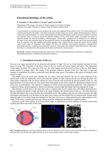
a) b) c)<br />
Fig 1. Structure <strong>of</strong> <strong>the</strong> eye. A shows a schematic draw <strong>of</strong> <strong>the</strong> eye. B shows a cross section <strong>of</strong> <strong>the</strong> whole <strong>retina</strong> stained with DAPI to<br />
stain <strong>the</strong> nuclei and viewed under epifluorescence. C shows a cross <strong>retina</strong>l section viewed under scanning.<br />
914 ©FORMATEX 2010
2. Retinal structure: layers and cells<br />
Vertebrate <strong>retina</strong> is organized in superimposed layers, formed by <strong>the</strong> different cells. The <strong>retina</strong> contains five mayor<br />
types <strong>of</strong> cells: photoreceptors (rods and cones), bipolar cells, horizontal cells, amacrine cells and ganglion cells (RGC).<br />
In general, cell somas are grouped in three distinct nuclear layers, separated by two connecting layers plexiform layers,<br />
where synapses between cells are formed. The innermost layer is <strong>the</strong> ganglion cell layer, which contains <strong>the</strong> cell bodies<br />
<strong>of</strong> <strong>the</strong> ganglion cells and displaced amacrine cells. The next cell layer is <strong>the</strong> inner nuclear layer, which contains <strong>the</strong> cell<br />
bodies <strong>of</strong> <strong>the</strong> amacrine cells, <strong>the</strong> bipolar cells, and <strong>the</strong> horizontal cells; it may also contain some displaced ganglion<br />
cells. The next cell layer is <strong>the</strong> outer nuclear layer, which contains <strong>the</strong> cell bodies <strong>of</strong> <strong>the</strong> photoreceptors. Outside <strong>of</strong><br />
<strong>the</strong>se layers, <strong>the</strong> layer <strong>of</strong> photoreceptor outer segments contains <strong>the</strong> light-sensitive elements <strong>of</strong> <strong>the</strong> <strong>retina</strong>. Light must<br />
pass through vitreous humor and <strong>the</strong> different layers <strong>of</strong> <strong>the</strong> <strong>retina</strong> before reaching <strong>the</strong> outer segments <strong>of</strong> <strong>the</strong><br />
photoreceptors. Interspersed between <strong>the</strong> ganglion cell layer and <strong>the</strong> inner nuclear layer is <strong>the</strong> inner plexiform layer,<br />
which contains <strong>the</strong> axons <strong>of</strong> bipolar cells, dendrites <strong>of</strong> ganglion cells and cell processes <strong>of</strong> amacrine cells (axons and/or<br />
dendrites). Between <strong>the</strong> outer and inner nuclear layers is <strong>the</strong> outer plexiform layer, which contains <strong>the</strong> axon terminals <strong>of</strong><br />
photoreceptors, <strong>the</strong> dendrites <strong>of</strong> bipolar and cell processes <strong>of</strong> horizontal cells (axons and/or dendrites).<br />
The basic system <strong>of</strong> <strong>retina</strong>l information processing consists on a direct pathway <strong>of</strong> visual information that flows from<br />
photoreceptors to bipolar cells to ganglion cells. The ganglion cells fire action potentials in response to light, and <strong>the</strong>se<br />
impulses propagate down <strong>the</strong> optic nerve to <strong>the</strong> projection nuclei in <strong>the</strong> brain. This direct pathway is influenced by two<br />
transverse fluxes <strong>of</strong> modulatory signals coming from horizontal in outer plexiform layer and amacrine cells in inner<br />
plexiform layer. Horizontal cells receive input from <strong>the</strong> photoreceptors and project <strong>the</strong>ir processes laterally to influence<br />
surrounding bipolar cells. Amacrine cells receive input from bipolar cells and project <strong>the</strong>ir processes laterally to<br />
influence surrounding bipolar and ganglion cells. Both, horizontal and amacrine cells usually make electrical and<br />
chemical synapses with neighbor cells <strong>of</strong> <strong>the</strong> same type.<br />
3. Design <strong>of</strong> histological techniques<br />
Microscopy: Science, Technology, Applications and Education<br />
______________________________________________<br />
A. Méndez-Vilas and J. Díaz (Eds.)<br />
Central nervous system may be studied under several types <strong>of</strong> techniques. Appropriate choosing <strong>of</strong> tissue is an<br />
important part <strong>of</strong> <strong>the</strong> study. Retina is a privileged part <strong>of</strong> <strong>the</strong> central nervous system for its accessibility and <strong>the</strong><br />
possibility <strong>of</strong> study isolated from <strong>the</strong> rest <strong>of</strong> <strong>the</strong> eye.<br />
For studying a tissue many histological techniques may be applied from a simple staining with hematoxiline-eosin to<br />
label nuclei and neuronal processes, respectively, to a Golgi staining that labels different cells depending on <strong>the</strong> cellular<br />
pH. Many works <strong>of</strong> Ramon y Cajal were made with this technique, since gives precious information <strong>of</strong> single cells,<br />
ra<strong>the</strong>r than <strong>the</strong> whole population <strong>of</strong> a tissue. The problem is we cannot decide which cell is stained.<br />
This problem may solved by o<strong>the</strong>r techniques. For example, a cell may be selectively stained by iontophoretical<br />
intracellular injection <strong>of</strong> fluorescent tracers like Lucifer Yellow, or substances that may be processed like neurobiotin,<br />
making stable and durable preparations. Previously to cell staining is necessary to be able <strong>of</strong> visualize and identify it. A<br />
way <strong>of</strong> labeling RGCs is injecting into <strong>the</strong> targets (superior colliculi) or into <strong>the</strong> axonal pathways (optic nerve) a<br />
neuronal tracer. In addition, <strong>the</strong>se techniques allow us to estimate neuronal populations in a selective way.<br />
With <strong>the</strong> development <strong>of</strong> immunohistochemistry <strong>the</strong> microscopy gained a great advance. The possibility <strong>of</strong><br />
selectively labeling a protein contained only in a type <strong>of</strong> cell allows studying specifically its structure and function.<br />
Initially immunohistochemistry were developed with H2O2 and diaminobencidine to obtain a brown stable labeling.<br />
These preparations could be observed along <strong>the</strong> time under light microscopy and without loss <strong>of</strong> staining. Later,<br />
immun<strong>of</strong>luorescence by adding a fluorocrome to <strong>the</strong> secondary antibody showed some advantages. It was takes less<br />
time and steps to complete <strong>the</strong> process. However, <strong>the</strong> fluorescence fades with time and exposition to <strong>the</strong> light. A new<br />
advance came with <strong>the</strong> application <strong>of</strong> different fluorocromes to <strong>the</strong> secondary antibody. It was possible labeling<br />
simultaneously, and differentiates, two or more proteins in <strong>the</strong> same preparation and to study <strong>the</strong>ir relationships. With<br />
<strong>the</strong> development <strong>of</strong> confocal immun<strong>of</strong>luorescence microscopy it was possible to improve focuses and images quality, to<br />
make co-localizations studies <strong>of</strong> two or more proteins in <strong>the</strong> same tissue, 3D reconstructions, and decreases fading <strong>of</strong><br />
fluorescence.<br />
Cellular ultrastructure could be studied in a more fine way by transmission electronic microscopy to analyze<br />
intracellular organelles. At this level, when <strong>the</strong> researcher is interested in <strong>the</strong> events on <strong>the</strong> surface (outer surface <strong>of</strong> <strong>the</strong><br />
membrane) may be preferable to use scanning electronic microscopy. Also it is possible to study isolated cells after<br />
dissociating <strong>of</strong> <strong>the</strong>ir tissues. By this process cells should maintain <strong>the</strong>ir vitality and many times <strong>the</strong>ir morphology. At <strong>the</strong><br />
present work are showed several samples <strong>of</strong> tissues and isolated cells studied by scanning.<br />
This chapter will deal with different microscopical techniques (scanning and transmission electronic microscopy, and<br />
light and confocal immun<strong>of</strong>luorescence microscopy) to study <strong>the</strong> physiological mechanisms performed by <strong>retina</strong>l<br />
neurons. Below are described some <strong>of</strong> <strong>the</strong> experimental procedures applied to this study.<br />
©FORMATEX 2010 915
Microscopy: Science, Technology, Applications and Education<br />
A. ______________________________________________<br />
Méndez-Vilas and J. Díaz (Eds.)<br />
4. Experimental procedures<br />
4.1 Animals and surgical procedures<br />
Adult New Zealand rabbits weighing 1000-1500 g, adult C57 BL/6j mice and adult Sprague-Dawley rats were used in<br />
this study. All experimental procedures were carried out in accordance with <strong>the</strong> European Communities Council<br />
Directive (86/609/ECC) for <strong>the</strong> use <strong>of</strong> laboratory animals. The animals were anes<strong>the</strong>tized by intramuscular injection <strong>of</strong><br />
5% ketamine chlorhydrate solution (Ketalar, Parke-Davis) 0.85 ml/kg. The animals had also previously received 2%<br />
tiazine chlorhydrate solution (Rompun, Bayer) 0.35 ml/kg. All surgical procedures were done under anes<strong>the</strong>sia. To prelabel<br />
RGCs, optic nerve was exposed by lateral approach and 10 µl <strong>of</strong> 0.1% fast blue (Sigma) was injected into <strong>the</strong><br />
optic nerve, 2 mm from <strong>the</strong> eyeball with a Hamilton microsyringe. Care was taken not to damage <strong>the</strong> blood vessels,<br />
especially <strong>the</strong> ophthalmic artery that enters <strong>the</strong> sclera from <strong>the</strong> ventral margin <strong>of</strong> <strong>the</strong> optic nerve. Preservation <strong>of</strong> <strong>the</strong><br />
structure <strong>of</strong> <strong>the</strong> central <strong>retina</strong>l artery was confirmed by ophtalmoscopy. Prelabeling <strong>of</strong> RGCs by injection <strong>of</strong> neuronal<br />
tracer (fast blue, fluorogold, diI, and o<strong>the</strong>rs) into <strong>the</strong> optic nerve allow us to count selectively RGCs to calculate<br />
populations in normal or abnormal situations.<br />
4.2 Retinal ganglion cell labeling by intracellular injection <strong>of</strong> Lucifer Yellow and neurobiotin<br />
Pre-labeled soma ganglion cells by injection <strong>of</strong> fast blue were easily identified under fluorescent microscopy, allowing<br />
<strong>the</strong> staining <strong>of</strong> RGCs by intracellular iontophoretic injection <strong>of</strong> Lucifer Yellow and neurobiotin. Labeling procedures<br />
have been described in detail previously [1]. Several days after labeling, <strong>the</strong> rabbits were sacrificed by an overdose <strong>of</strong><br />
sodium pentobarbital. The <strong>retina</strong> was removed from <strong>the</strong> eyecup and <strong>the</strong> vitreous humor teased away from its surface<br />
with a paintbrush in oxygenated (95% oxygen, 5% carbon dioxide) Ames medium [2]. The <strong>retina</strong> was flattened and<br />
fixed with <strong>the</strong> ganglion cell layer uppermost to a plate <strong>of</strong> Sylgard (Dow Corning Corporation Mich., USA) in a chamber<br />
<strong>of</strong> oxygenated Ames medium. Fast blue-labelled ganglion cells were filled intracellularly with Lucifer Yellow<br />
following a procedure similar to that described by Tauchi and Masland [3]. Micropipette electrodes filled with 1%<br />
Lucifer Yellow (Sigma) and 3% Neurobiotin (Vector) in 0.1 M Tris buffer (pH 7.4) were used. The tip resistance was<br />
20-100 MΩ. Lucifer Yellow was injected by negative pulse (1-3 nA, 1 Hertz, duration 500 msc) for about 1 min. Once<br />
<strong>the</strong> cell was impaled, Neurobiotin was ejected by passage <strong>of</strong> direct positive current (1-3 nA, 1 Hertz, duration 500 msc)<br />
for about 5 min. After this, <strong>the</strong> microelectrode was taken out <strong>the</strong> cell and <strong>the</strong> procedure repeated in o<strong>the</strong>r cells until <strong>the</strong><br />
electrode tip becomes clogged. After filling several cells, <strong>the</strong> <strong>retina</strong>s were equilibrated in Ames medium for 30 min [4,<br />
5], and fixed with 4% paraformaldehyde in 0.1 M phosphate buffer (PB) pH 7.4, at 22ºC for 2 hours. The <strong>retina</strong>s were<br />
<strong>the</strong>n washed in PB, cryoprotected in 30% sucrose in PB, and stored at -18ºC for fur<strong>the</strong>r processing.<br />
4.2.1 Processing <strong>of</strong> <strong>the</strong> <strong>retina</strong>l tissue<br />
After defrosting at room temperature and washing in phosphate buffer, <strong>the</strong> <strong>retina</strong>s were incubated in 0.2% albumin and<br />
0.1% Triton X-100 in 0.1 M PB for 10 min to avoid unspecific labeling and to make <strong>the</strong> <strong>retina</strong> permeable to antibodies,<br />
respectively. Then <strong>the</strong>y were incubated with 1% methanol and 0.15 % H2O2 in PB for 15 min. The next step was an<br />
incubation <strong>of</strong> <strong>the</strong> <strong>retina</strong>s in Avidin-Biotin complex solution (ABC-kit, Vectastatin, Vector) for 2 h. Following<br />
incubation, <strong>retina</strong>s were washed in phosphate buffer and incubated in <strong>the</strong> dark; with 0.05% diaminobenzidine for 20<br />
min. Neurobiotin was visualized after adding 0.03% H2O2 to <strong>the</strong> solution. The <strong>retina</strong>s were <strong>the</strong>n washed and dehydrated<br />
through an ascending ethanol series and mounted in DEPEX.<br />
4.2.2 Retinal shrinkage<br />
All RGCs were drawn using camera lucida attached to a microscope (Leitz Laborlux 12) under a 25x objective. The<br />
<strong>retina</strong>l area was measured and <strong>the</strong> shrinkage due to <strong>the</strong> histological procedures was estimated. The distance between <strong>the</strong><br />
optic disc and <strong>the</strong> cells were measured after injecting Lucifer Yellow (before any possible retraction), and compared<br />
with <strong>the</strong> distances after processing <strong>the</strong> <strong>retina</strong>s with diaminobenzidine and dehydrating. Some <strong>retina</strong>s had an area <strong>of</strong><br />
shrinkage ranging from 0% to 10%, o<strong>the</strong>rs had an area <strong>of</strong> shrinkage between 11% and 20%. Retinas with a larger<br />
shrinkage percentage were not used in <strong>the</strong> quantitative analysis.<br />
916 ©FORMATEX 2010
a) b) c)<br />
Fig 2. Morphometrical analysis <strong>of</strong> <strong>retina</strong>l ganglion cell. A, a RGC labeled by ionthophoretical injection <strong>of</strong> neurobiotin and<br />
developed with avidin-biotin and diaminobenzidine is shown. B, drawing <strong>of</strong> <strong>the</strong> same cell by camera lucida. C, computerized<br />
representation <strong>of</strong> <strong>the</strong> cell after morphometrical analysis<br />
4.2.3 Morphometrical analysis<br />
To study morphometrical features <strong>of</strong> RGCs (soma and dendritic field size, and number and length <strong>of</strong> dendritic branches,<br />
etc.) stained rabbit <strong>retina</strong>l ganglion cells were drawn using camera lucida projection as seen in whole-mount<br />
preparations and digitized using an HP-scanner with a resolution <strong>of</strong> 300 dpi. Gross image editing, such as removing<br />
large blocks <strong>of</strong> noise were performed with standard commercial s<strong>of</strong>tware (Adobe Photoshop 5.0) on a 6100 Power<br />
Macintosh. See figure 2C for morphometrical analysis.<br />
To obtain quantitative information about <strong>the</strong> true shape and morphology <strong>of</strong> neurons, <strong>the</strong> NIH Image program was<br />
used. This program has been developed at <strong>the</strong> US National Institutes <strong>of</strong> Health and is available on <strong>the</strong> Internet at<br />
http://rsb.info.nih.gov/nih-image [6]. A custom set <strong>of</strong> macros (modified version <strong>of</strong> <strong>the</strong> Neurite Macros v1.2, developed<br />
by Charles Thomas at <strong>the</strong> University <strong>of</strong> Wisconsin) allowed us to calculate different morphological parameters for each<br />
cell. Briefly, <strong>the</strong> first step was <strong>the</strong> acquisition <strong>of</strong> scale information followed by <strong>the</strong> acquisition <strong>of</strong> <strong>the</strong> soma and dendritic<br />
contours (convex hull around <strong>the</strong> dendritic area). The tracing <strong>of</strong> <strong>the</strong> dendritic tree were drawn by <strong>the</strong> operator via <strong>the</strong><br />
mouse or a digitizing table (Kurta IS/ONE) connected to a 6100 Apple Power Macintosh. Once tracing has begun, <strong>the</strong><br />
procedure permits <strong>the</strong> tracking <strong>of</strong> continuation points, branch points and end points to take place. From <strong>the</strong> traced tree,<br />
it is possible to automatically calculate non metric parameters such as <strong>the</strong> order <strong>of</strong> <strong>the</strong> neurite (e.g. primary, secondary,<br />
tertiary, etc.), number <strong>of</strong> branches, number <strong>of</strong> roots, number <strong>of</strong> bifurcations, number <strong>of</strong> terminal tips (topological<br />
degree) and sequences <strong>of</strong> branch order [7, 8, 9]. Using <strong>the</strong> scale information, metric variables such as <strong>the</strong> total dendritic<br />
length, dendritic length by branch order and so forth, were directly calculated. Fig. 2 summarizes <strong>the</strong> procedure for<br />
staining, drawing and morphometrical analyses performed on RGCs.<br />
4.2.4 Data analysis<br />
Morphometrical data <strong>of</strong> <strong>the</strong> cells was compared using a two-tailed Student t test. This was calculated using GraphPad<br />
InStat version 3.00 for Windows 95, GraphPad S<strong>of</strong>tware, San Diego California USA.<br />
4.3 Retinal cell dissociation<br />
Microscopy: Science, Technology, Applications and Education<br />
______________________________________________<br />
A. Méndez-Vilas and J. Díaz (Eds.)<br />
Dissociated <strong>retina</strong>l cells were prepared from adult rabbit <strong>retina</strong>s following a procedure described elsewhere [10].<br />
Animals were deeply anaes<strong>the</strong>tized with urethane (loading dose, 1.5 kg -1 , i.p.) prior to enucleation. Both globes were<br />
hemisected from each animal, <strong>the</strong> vitreous removed and <strong>the</strong> <strong>retina</strong>s isolated and kept in Ames medium [2]. Since <strong>retina</strong>l<br />
eccentricity influences on <strong>the</strong> RGC size, same areas <strong>of</strong> both <strong>retina</strong>s were used for dissociation; a piece <strong>of</strong> ca. 0.5 cm 2<br />
from <strong>the</strong> inferior-nasal quadrant, ca. 10 mm away from <strong>the</strong> optic nerve head was used for <strong>the</strong> dissociation. The <strong>retina</strong>l<br />
pieces were incubated separately for 30 min in standard Ringer solutions containing 40 U ml -1 papain (Worthington)<br />
and 0.1 mg ml-1 cysteine (Sigma, St. Louis, MO, U.S.A.) at 30ºC. Retinal pieces were rinsed with a standard Ringer<br />
solution containing 0.05% bovine serum albumin. The <strong>retina</strong>l fragments were mechanically dispersed on glass coverslip<br />
coated with concanavalin A (Sigma) (1 mg ml-1). This dissociation yielded a mixture <strong>of</strong> cells. Different <strong>retina</strong>l cells<br />
were identified by <strong>the</strong>ir characteristic morphology. The number <strong>of</strong> different neurons obtained per Petri dish was variable<br />
from day to day, from none to more than 10 cells. Cells were stored at 5ºC and <strong>the</strong> experiments were performed at room<br />
temperature within 1-4 hr after dissociation.<br />
©FORMATEX 2010 917
Microscopy: Science, Technology, Applications and Education<br />
A. ______________________________________________<br />
Méndez-Vilas and J. Díaz (Eds.)<br />
4.4 Scanning electron microscopy<br />
Flat-mounted <strong>retina</strong>s, <strong>retina</strong>l cross-sections preparations, and isolated <strong>retina</strong>l cells were used for scanning electron<br />
microscopy (SEM). In <strong>the</strong> flat <strong>retina</strong>, <strong>the</strong> inner limiting membrane (ILM), constituted by <strong>the</strong> feet <strong>of</strong> <strong>the</strong> Müller cells [11]<br />
need to be picked <strong>of</strong>f in order to clearly observe <strong>the</strong> RGCs and <strong>the</strong>ir membrane surface. To remove <strong>the</strong> ILM, we used a<br />
technique described in detail previously [12]. Briefly, a 2.5% poly-L-Lysine Hydrobromide solution was applied in a<br />
Petri dish. After an hour, <strong>the</strong> <strong>retina</strong> was extended with <strong>the</strong> ganglion cell layer down on <strong>the</strong> plate and glued to glass by<br />
poly-L-Lysine in dish perfused with Ames medium [2]. Five minutes later, <strong>the</strong> <strong>retina</strong> was carefully separated and taken<br />
to ano<strong>the</strong>r plate with a Sylgard floor and fixed (ganglion cells layer up) with micro-pins. Then, <strong>the</strong> <strong>retina</strong>s were<br />
incubated 10 minutes at 30ºC in a Ringer solution containing 15 U/ml Papain (Worthington, Lakewood NJ, USA) and 1<br />
mg/ml Cystein (Sigma, St.Louis, MO, USA). Retinas were washed with a continuous flow <strong>of</strong> Ringer to eliminate<br />
cellular debris (procedure modified from Edwards and cols.) [13]. Then, a <strong>retina</strong> patch <strong>of</strong> 1.7 x 2.24 mm was cropped<br />
and fixed in 1.5 % paraformaldehyde and 0.25% glutaraldehyde in phosphate buffered saline (PBS) for 2 hours at room<br />
temperature, rinsed and store at 4ºC.<br />
For <strong>retina</strong>l cross-section SEM, patches <strong>of</strong> <strong>retina</strong>s were isolated from <strong>the</strong> eyes and strongly adhered to a nitrocellulose<br />
paper (Membrane Filters; Advance; Tokyo) ganglion cell layer down. Then, <strong>the</strong> <strong>retina</strong>s were vertically cut in 200 to 500<br />
µm thick slices. The slices were lied down on a plate and stuck with poly-L-Lysine to expose <strong>the</strong> <strong>retina</strong>l layers. In <strong>the</strong><br />
next step, <strong>retina</strong>s were cleaned and fixed following <strong>the</strong> protocol previously described, and <strong>the</strong>n stored at 4ºC until<br />
fixation with osmium.<br />
Retinal cross-sections and flat mount <strong>retina</strong>s without ILM were <strong>the</strong>n immersed in 1% O4Os in Milloning (1.87%<br />
monosodium phosphate and 0.42% sodium hydroxide in distilled water) for 1.5 hour at room temperature in absolute<br />
darkness. Retinas were washed with distilled water and dehydrated in ethanol series and pure acetone. In next step,<br />
acetone was mixed with liquid CO2 (at room temperature and a pressure <strong>of</strong> 50 bar). When acetone was completely<br />
displaced by CO2, <strong>the</strong> chamber was heated to 37ºC (at 80 bar) and liquid CO2 became gas (g). In <strong>the</strong> critical point,<br />
chamber was slowly uncompressed to evacuate CO2 (g) (Polaron E 300 and Polaron E 5000/5100). These procedures<br />
ensured <strong>the</strong> plasma membrane preservation and observations by scanning (DSM-950, Zeiss, Germany). Retinal<br />
shrinkage due to SEM procedures was taken into account and preparations with a shrinkage percentage larger than 20%<br />
were not used for analysis.<br />
4.5 Immunohistochemistry, immun<strong>of</strong>luorescence, confocal mycroscopy<br />
Retinas were dissected and fixed by immersion for 2 hours at room temperature in a solution containing 4%<br />
paraformaldehyde in 0.1 M PBS (pH 7.4). Rinsed in 0.1 M PBS (pH 7.4) and incubated with 1% sodium borohydride<br />
for 30 min in 0.1 M phosphate buffer (pH 7.4). This step was carried out for decreasing to minimal <strong>the</strong> auto<br />
fluorescence <strong>of</strong> paraformaldehyde. The <strong>retina</strong>s were incubated in 0.2% albumin and 0.5% Triton X-100 in 0.1 M PB for<br />
1 hour at 4ºC in stirring to avoid unspecific labeling and to make <strong>the</strong> <strong>retina</strong> permeable to antibodies, respectively.<br />
Then, <strong>the</strong> tissues were incubated with <strong>the</strong> monoclonal antibody. For example, to study axons and somas <strong>of</strong> RGC<br />
Anti-β Tubulin Isotype III was used at a working dilution <strong>of</strong> 1:500 (Sigma, clone SDL.3D10, Product No. T8660) in 1<br />
% albumin 0.1 M PB (pH 7.4) for 4 days at 4ºC in stirring. After several rinsed, <strong>the</strong> <strong>retina</strong>s were immersed in<br />
fluorescent secondary antibodies (1:200) for 2 days at 4ºC in stirring. They were sheep anti-mouse IgG species-specific<br />
whole antibody (Amersham, Arlington Heights IL, USA). After rinsing, <strong>retina</strong>s were washed in 0.1 M PB (pH 7.4),<br />
mounted in fluoromount Vectashield (Vector Laboratories), and cover slipped for viewing by laser confocal microscopy<br />
(Leica TCS SP2 Leica Microsystems).<br />
Using o<strong>the</strong>r primary antibodies we can label different <strong>retina</strong>l cells, part <strong>of</strong> <strong>the</strong>m, or a combination. Some examples<br />
are:<br />
Rhodopsin labels rod photoreceptors; calbindin that labels preferentially horizontal cells, but also cone bipolar cells;<br />
α-PKC labels rod bipolar cells; Sodium-potasium-cloro cotransporter (NKCC1) labels horizontal cells; K-Cl<br />
cotransporter (KCC2) labels ON- cone bipolar cells; Bassoon antibody labels presynaptic cytomatrix protein Bassoon, a<br />
major component <strong>of</strong> <strong>the</strong> photoreceptor ribbon; melanopsin labels intrinsically photosensitive RGC (a RGC subtype<br />
discovered recently); Tiroxine hidroxilase labels dopaminergic amacrine cells; α1-GABA subunit antibody labels<br />
gabaergic amacrine cells; and many more. Also is possible a selective staining based on a selective protein affinity like<br />
Peanut agglutinin, a lectin with high binding affinity for galactose-galactosamine disaccharide. Peanut agglutinin binds<br />
preferentially to cones, not to rods.<br />
918 ©FORMATEX 2010
Fig. 3. Micropicture <strong>of</strong> a RGC. Notice <strong>the</strong> initio <strong>of</strong> a neural process, nucleus, mitochondrias, lisosomal apparatus and o<strong>the</strong>r cellular<br />
organelles.<br />
4.6 Immunohistochemistry, transmission electron microscopy<br />
Retinas were dissected and fixed by immersion for 2 hours at room temperature in a solution containing 4%<br />
paraformaldehyde and 0.1% glutaraldehyde in 0.1 M PBS (pH 7.4) with <strong>the</strong> addition <strong>of</strong> 0.15 mM CaCl2, 4% sucrose.<br />
Retinas were <strong>the</strong>n fixed in 4% paraformaldehyde, 0.15 mM CaCl2, 4% sucrose and bicarbonate phosphate buffer (pH<br />
10.4) at 4ºC overnight. To avoid tissue damage after freeze, <strong>retina</strong>s were exposed to a gradient <strong>of</strong> sucrose (15% thirty<br />
minutes, 20% one hour and 30% overnight) at 4ºC and quickly freeze and unfreeze. This step was carried out to make<br />
<strong>retina</strong>s permeable to antibodies; o<strong>the</strong>rwise <strong>the</strong> use <strong>of</strong> triton-X would destroy membranes. To avoid non specifically<br />
labeling, <strong>retina</strong>s were incubated in 10% normal serum in 0.1 M PBS (pH 7.4) at 4ºC in stirring. Then, <strong>the</strong> tissues were<br />
incubated with <strong>the</strong> monoclonal antibody Anti-β Tubulin Isotype III at a working dilution <strong>of</strong> 1:500 (Sigma, clone<br />
SDL.3D10, Product No. T8660) in 1 % albumin in phosphate buffer (pH 7.4) for 4 days at 4ºC in stirring. After several<br />
rinsed, <strong>the</strong> <strong>retina</strong>s were immersed in secondary antibodies (1:200) for 2 days at 4ºC in stirring. They were sheep antimouse<br />
IgG biotinylated species-specific whole antibody (Amersham, Arlington Heights IL, USA). After rinsing, <strong>retina</strong>s<br />
were incubated in Vectastatin, ABC-kit (Vector, Burlingame, California, USA) in a 1:250 dilution in 0.1 M PBS for one<br />
day at 4ºC in stirring. Finally, <strong>retina</strong>s were washed in phosphate buffer and incubated with 0.05% diaminobenzidine and<br />
0.03% H2O2 in 0.1 M PBS until <strong>the</strong> labeled cells were visualized by <strong>the</strong> light microscope.<br />
Retinas were placed overnight in cold 0.1M cacodylate-sucrose, pH 7.6 and post-fixed in cold 1% buffered OsO4, pH<br />
7.3 for one hour. Then, <strong>the</strong>y were placed in aqueous 2% uranyl acetate and dehydrated and embedded in Epon 812.<br />
Sectioning for electron microscopic examination followed standard procedures and was accomplished with an<br />
ultramicrotome (Vitracut E, Reichert-Jung, Austria). Segments <strong>of</strong> <strong>retina</strong>s were sectioned in <strong>the</strong> transverse plane<br />
(semithin, 1-2 µm) and stained with toluidine blue for light microscopy study. Blocks <strong>of</strong> <strong>retina</strong>s were <strong>the</strong>n cut again and<br />
thin sections (60-80 nm thick) were stained in lead citrate and viewed under a transmission electron microscope (M-10,<br />
Zeiss, Germany).<br />
5. Retinal cells<br />
Microscopy: Science, Technology, Applications and Education<br />
______________________________________________<br />
A. Méndez-Vilas and J. Díaz (Eds.)<br />
a) b) c)<br />
Fig. 4. Photoreceptors. A, Retinal cross-section, immunohistochemically labeled with NKCC1 (Na-K-Cl-Cotransporter) antibody<br />
(green) and calbindin (red). NKCC1 stains outer segments <strong>of</strong> photoreceptors (arrow in <strong>the</strong> upper) and horizontal cells. Calbindin<br />
stains horizontal cells. GCL: ganglion cell layer; INL: inner nuclear layer; ONL: outr nuclear layer. B, Outer and inner segments <strong>of</strong><br />
photoreceptors viewed under scanning microscopy. A cone is pointed by an arrow and a rod by a arrowhead. C, Somas and terminals<br />
<strong>of</strong> photoreceptors. Notice how a photoreceptor terminal (arrow) contacts with <strong>the</strong> outer plexiform layer.<br />
©FORMATEX 2010 919
Microscopy: Science, Technology, Applications and Education<br />
A. ______________________________________________<br />
Méndez-Vilas and J. Díaz (Eds.)<br />
5.1 Photoreceptors, rods and cones<br />
Almost all vertebrates have morphologically two types <strong>of</strong> photoreceptors, which can be classified as rod and cone cells<br />
[14]. Rods function in dim-light vision, whereas cones mediate bright-light and color vision. Rods and cones are<br />
differentiated on <strong>the</strong> basis <strong>of</strong> <strong>the</strong> shape <strong>of</strong> <strong>the</strong>ir outer segments (see Fig 4).<br />
Rod outer segments contain a set <strong>of</strong> membranous disc, derive from <strong>the</strong> surface membrane <strong>of</strong> <strong>the</strong> cell but no longer<br />
continuous with <strong>the</strong> surface membrane or with each o<strong>the</strong>r. They resemble a stack <strong>of</strong> coins. Cone outer segments are<br />
composed mainly <strong>of</strong> infoldings <strong>of</strong> <strong>the</strong> surface membrane <strong>of</strong> <strong>the</strong> cell. Outside <strong>the</strong> foveal region, <strong>the</strong>se outer segments<br />
taper slightly, and this gives <strong>the</strong> cones <strong>the</strong>ir name.<br />
O<strong>the</strong>r factors that may help in <strong>the</strong> identification are axonal terminal size and position, and soma location in <strong>the</strong> outer<br />
nuclear layer; it is smaller and closer to <strong>the</strong> outer plexiform layer in <strong>the</strong> case <strong>of</strong> rods. In most <strong>retina</strong>s <strong>the</strong>re is a single<br />
type <strong>of</strong> rods, but amphibians that have two types <strong>of</strong> rods, red and green, sensible to <strong>the</strong>se wave lengths [14]. Most<br />
species have multiple types <strong>of</strong> cones. Usually <strong>the</strong>y are three cone types, like in primates, and are termed L, M, and S,<br />
which absorb maximally in low, medium and supra frequency, respectively. These frequencies correspond to <strong>the</strong> three<br />
basic colors: red, green and blue, respectively. Spectral sensitivity <strong>of</strong> L and M are relatively close in <strong>the</strong> low frequency<br />
end <strong>of</strong> visible spectrum, while S cone lies at <strong>the</strong> high frequency end. These different absorptions are due to <strong>the</strong> presence<br />
<strong>of</strong> different pigments in <strong>the</strong> outer segments <strong>of</strong> <strong>the</strong> photoreceptors.<br />
Usually, <strong>the</strong> spatial distribution <strong>of</strong> S cones across <strong>the</strong> <strong>retina</strong> differs from that <strong>of</strong> L and M cones. They are only 10% <strong>of</strong><br />
cone population and are absent <strong>of</strong> <strong>the</strong> fovea.<br />
All photoreceptors have an outer segment (containing <strong>the</strong> visual pigments), an inner segment, a perikaryal region<br />
which contain <strong>the</strong> cell nucleus, and a terminal. Usually, rods have longer outer segments and smaller spherical<br />
terminals. Cones have a shorter outer segment, a fatter inner segment, and a larger terminal than rods. However, <strong>the</strong>re<br />
are exceptions for both types.<br />
Photoreceptor terminals are termed cone pedicles and rod spherules. Processes from cone pedicles contact with o<strong>the</strong>r<br />
cone pedicles as well as rod spherules. At each such contact <strong>the</strong>re is a low-resistance junction termed gap junction that<br />
provides a partial electrical coupling between <strong>the</strong> two cells. In each pedicle or spherule <strong>the</strong>re is an invagination and just<br />
above lies a specialized structure termed synaptic ribbon that plays an important role in <strong>the</strong> transmission <strong>of</strong> signals from<br />
a photoreceptor to <strong>the</strong> horizontal and bipolar cells.<br />
a) b) c)<br />
d) e)<br />
Fig. 5. Horizontal cells. A, isolated horizontal cell type HII (light microscopy). B, isolated horizontal cell type I (light microscopy).<br />
C, isolated horizontal cell type HII under scanning microscopy. D, Cross-<strong>retina</strong>l section immunohistochemically labeled with<br />
calbindin (labels horizontal cells and cone bipolar cells) and Propidium iodure to label <strong>retina</strong>l nucleus. E, horizontal cells in detail.<br />
Notice small dots in <strong>the</strong> upper that correspond to dendritic synapses.<br />
5.2 Horizontal cells<br />
Most vertebrate <strong>retina</strong>s contains only two types <strong>of</strong> horizontal cells: a cell with a short axon that typically runs 400 µm or<br />
fur<strong>the</strong>r, before ending in a prominent terminal expansion called telodendritic arbor, this cell is termed HI; and an<br />
920 ©FORMATEX 2010
axonless cell, termed HII. Axonless cells have not been observed in primate <strong>retina</strong>s; and it may be that o<strong>the</strong>r <strong>retina</strong>s are<br />
lacking <strong>of</strong> <strong>the</strong>se horizontal cell types [15].<br />
Electron microscopy <strong>of</strong> Golgi-stained horizontal cells from <strong>the</strong> cat has shown that <strong>the</strong> processes <strong>of</strong> <strong>the</strong> axonless cell<br />
and <strong>the</strong> dendritic processes <strong>of</strong> <strong>the</strong> axon cell connect exclusively with cone terminals, whereas <strong>the</strong> axon terminal<br />
processes <strong>of</strong> <strong>the</strong> axon cell end exclusively in <strong>the</strong> rod terminals [16]. However, in o<strong>the</strong>r <strong>retina</strong>s such segregation <strong>of</strong> cone<br />
and rod inputs has not been observed.<br />
Horizontal cells respond to illumination with sustained graded potentials. Usually, <strong>the</strong>y do not generate action<br />
potentials, and <strong>the</strong> axon <strong>of</strong> <strong>the</strong> horizontal cell is sufficiently long that <strong>the</strong> graded electrical activity taking place in <strong>the</strong><br />
telodendritic arbor is isolated from that <strong>of</strong> <strong>the</strong> dendritic arbor.<br />
Each rod synaptic terminal contacts with two to five rod bipolar cells. Each rod bipolar cell contacts with 30-50 rods.<br />
Horizontal cells work by lateral inhibitions modulating <strong>the</strong> magnitude <strong>of</strong> rod bipolar cell activation by rods. In <strong>the</strong> first<br />
form <strong>of</strong> lateral inhibition are involved HI horizontal cells telodendritic arbor, rods and rod bipolar cells. A greater<br />
stimulation <strong>of</strong> <strong>the</strong> telodendritic arbors <strong>of</strong> <strong>the</strong> horizontal cells by rods decreases <strong>the</strong> effect on <strong>the</strong> rod bipolar cells. Thus,<br />
<strong>the</strong> effect <strong>of</strong> light intensity depends more on differences in intensity within <strong>the</strong> image, such as at a border, than on <strong>the</strong><br />
overall level <strong>of</strong> light intensity.<br />
A second example <strong>of</strong> lateral inhibition involves dendrites <strong>of</strong> HI, cones, and cone bipolar cells. The dendrites <strong>of</strong><br />
adjacent HI are strongly coupled by gap junctions, which mediate spatial effects. The dendritic terminals <strong>of</strong> HI cells<br />
receive from both L and M cones, but not from S cones.<br />
The third example <strong>of</strong> lateral inhibition involves HII cells, S cones and <strong>the</strong> cone bipolar cells that contact with <strong>the</strong>m.<br />
HII horizontal cells do not have axon, but short dendritic branches and are also strongly coupled to one ano<strong>the</strong>r via gap<br />
junction.<br />
Although HII contact in equal measure with S cones and with a mixture <strong>of</strong> L and M cones, and since <strong>the</strong>re are ten<br />
times more L and M cones than S cones, L and M do not appear to be strongly influenced by HII. This neural circuit<br />
seems to have a role on color vision.<br />
a) b) c)<br />
Fig. 6. Bipolar cells. A, <strong>retina</strong>l cross-section labeled with α-PKC antibody to stain rod bipolar cells, (red) and DAPI to label nuclei<br />
(blue). B, rod bipolar cells in detail, bipolar cell somas are placed in <strong>the</strong> upper position, above are <strong>the</strong> dendritic processes. From <strong>the</strong><br />
inferior part <strong>of</strong> somas grow a unique axonal process that finish in <strong>the</strong> rod bipolar terminal. C, an isolated bipolar cell viewed under<br />
scanning microscopy. Notice dendritic processes (arrowhead) in <strong>the</strong> upper and axon terminal (arrow) in <strong>the</strong> bottom.<br />
5.3 Bipolar cells<br />
Microscopy: Science, Technology, Applications and Education<br />
______________________________________________<br />
A. Méndez-Vilas and J. Díaz (Eds.)<br />
Bipolar cells connect outer and inner <strong>retina</strong>. Their dendritic processes make synapses with photoreceptors and<br />
horizontal cells in <strong>the</strong> outer plexiform layer and <strong>the</strong>ir axon terminals contact with amacrine and ganglion cells in <strong>the</strong><br />
inner plexiform layer. Their somas are placed in <strong>the</strong> inner nuclear layer, between both synaptic layers.<br />
The first classification <strong>of</strong> bipolar cells depends on <strong>the</strong> type <strong>of</strong> photoreceptor <strong>the</strong>y receive input, rod bipolar cells and<br />
cone bipolar cells. These cone bipolar cells make two different kinds <strong>of</strong> synaptic connections with cones. One type <strong>of</strong><br />
<strong>the</strong> cone bipolar cells extends dendrites into <strong>the</strong> center <strong>of</strong> <strong>the</strong> invaginations <strong>of</strong> <strong>the</strong> cone receptor terminal (<strong>the</strong>y are called<br />
invaginating bipolar cells), and <strong>the</strong> axon terminals <strong>of</strong> <strong>the</strong>se cells end deep in <strong>the</strong> inner plexiform layer. Attending to its<br />
function, <strong>the</strong>se cells are termed ON-bipolar cells, because <strong>the</strong>y are activated by an increase in light intensity. The o<strong>the</strong>r<br />
cell type makes contacts along <strong>the</strong> flattened base <strong>of</strong> <strong>the</strong> cone receptor terminals (<strong>the</strong>y are called flat bipolar cells), and<br />
<strong>the</strong> axon terminals end in <strong>the</strong> distal part <strong>of</strong> <strong>the</strong> inner plexiform layer. This type responds to decreases <strong>of</strong> light intensity, it<br />
is also termed OFF-bipolar cells.<br />
Especially interesting is <strong>the</strong> synaptic contact made into <strong>the</strong> cone pedicle, in <strong>the</strong> triad. The most distinctive <strong>of</strong> <strong>the</strong>se are<br />
<strong>the</strong> invaginating, or central, processes directly below <strong>the</strong> synaptic ribbon. Usually, <strong>the</strong>re are three processes (from two<br />
horizontal cells and one bipolar cell) in an invagination, which are aligned along <strong>the</strong> length <strong>of</strong> <strong>the</strong> ribbon. Bipolar cells<br />
that receive from photoreceptors via invaginating processes all appear to have axonal arbors that stratify in <strong>the</strong> ONsublayer<br />
<strong>of</strong> <strong>the</strong> inner plexiform layer. O<strong>the</strong>r bipolar cells are triad associated, or semi-invaginating processes, branch on<br />
ei<strong>the</strong>r side <strong>of</strong> <strong>the</strong> invaginating processes, and constitute a heterogeneous group. Some are OFF bipolar cells and o<strong>the</strong>rs<br />
©FORMATEX 2010 921
Microscopy: Science, Technology, Applications and Education<br />
A. ______________________________________________<br />
Méndez-Vilas and J. Díaz (Eds.)<br />
are ON bipolar cells. The remaining bipolar cell processes are non-triad associated or flat processes, and are OFF<br />
bipolar cells.<br />
In <strong>the</strong> o<strong>the</strong>r extreme <strong>of</strong> <strong>the</strong> bipolar cell, <strong>the</strong>re are two postsynaptic processes apposed to <strong>the</strong> bipolar terminal synaptic<br />
ribbons, and this postsynaptic arrangement has been called a dyad [17]. The two postsynaptic processes <strong>of</strong> a dyad are<br />
most <strong>of</strong>ten a ganglion cell dendrite and an amacrine cell process or two amacrine cell processes [18]. It has been<br />
reported gap junctions between bipolar cells in some species [19, 20].<br />
Different types <strong>of</strong> bipolar cells may be distinguished: midget, diffuse, S cone, and giant. Midget bipolar cells are<br />
recognized by <strong>the</strong>ir small and compact dendritic arbors, which generally receive from only one cone, although in <strong>the</strong><br />
peripheral <strong>retina</strong> a few receive from two cones [21]. Midget bipolar cells can receive from cones via invaginating<br />
contacts (ON) or via triad-associated contacts (ON or OFF). Diffuse mean bipolar cell with dendritic processes that<br />
spread out in <strong>the</strong> outer plexiform layer to receive from a number <strong>of</strong> photoreceptors. S cone bipolar cells have<br />
invaginating dendritic processes that receive exclusively from S cones. Their axonal arbors ramify in <strong>the</strong> inner most<br />
portion <strong>of</strong> <strong>the</strong> inner plexiform layer, and are presynaptic to a ganglion cell type termed small bistratified.<br />
a) b) c)<br />
Fig. 7. Amacrine cells. A, <strong>retina</strong>l cross-section labeled with ChAT (Choline Acetyltransferase) that stains amacrine cells containing<br />
this enzyme. Somas <strong>of</strong> <strong>the</strong>se amacrine cells are distributed in inner nuclear layer and ganglion cell layer. B, <strong>retina</strong>l cross-section<br />
labeled with tiroxine hidroxilase antibody. This antibody stain cells placed only in <strong>the</strong> inner nuclear layer. C, Amacrine cells viewed<br />
under scanning in a very closer relationship. Scale is <strong>the</strong> same in A and B.<br />
5.4 Amacrine cells<br />
Ramón y Cajal discovered a group <strong>of</strong> neurons in <strong>the</strong> <strong>retina</strong> that had no axon, and he called a-macr-ine (a: lacking,<br />
macros: long, inos: fibers) [22]. There are many different types <strong>of</strong> amacrine cells, which vary greatly in size,<br />
morphology, and function. Cajal classified amacrine cells into two major types: diffuse that extends <strong>the</strong>ir processes<br />
throughout <strong>the</strong> thickness <strong>of</strong> <strong>the</strong> inner plexiform layer; and stratified that extend <strong>the</strong>ir processes on one or few strata <strong>of</strong><br />
<strong>the</strong> inner plexiform layer. Diffuse amacrine cells may be sub-classified in narrow and wide-field (on <strong>the</strong> basis <strong>of</strong> <strong>the</strong><br />
dendritic spread); and mono, bi, multi-stratified (on <strong>the</strong> basis <strong>of</strong> <strong>the</strong> strafication levels in <strong>the</strong> inner plexiform layer).<br />
Amacrine cells constitute <strong>the</strong> most diverse group <strong>of</strong> cell types within <strong>the</strong> <strong>retina</strong> with respect to morphology, size, and<br />
<strong>retina</strong>l coverage. There appear to be 30 to 40 types, but <strong>the</strong> functions <strong>of</strong> most are unknown. However, we know that<br />
many ganglion cell properties, such as directional selectivity or local edge detection is responsibility <strong>of</strong> amacrine cells.<br />
One <strong>of</strong> <strong>the</strong> most complete and recent classifications is given by Masland [23, 24].<br />
Cell bodies <strong>of</strong> amacrine cells are placed in <strong>the</strong> innermost layer <strong>of</strong> <strong>the</strong> inner nuclear layer, in <strong>the</strong> middle <strong>of</strong> <strong>the</strong> inner<br />
plexiform layer (A1 amacrine cell), and in <strong>the</strong> ganglion cell layer (displaced amacrine cells).<br />
Amacrine cells make numerous synapses onto ganglion cell dendrites, bipolar cell terminals, interplexiform<br />
processes and o<strong>the</strong>r amacrine cell processes [17]. Electron microscopy has shown that amacrine cell processes have<br />
characteristics <strong>of</strong> both axons and dendrites. Thus, amacrine cell processes can be both pre- and postynaptic over a very<br />
short portion <strong>of</strong> <strong>the</strong>ir length [18]. There are, at least, two types <strong>of</strong> synaptic arrangements. First, a postsynaptic amacrine<br />
cell process makes a synapse back onto <strong>the</strong> bipolar terminal a short distance away, <strong>the</strong>se are called reciprocal synapses.<br />
Second type <strong>of</strong> arrangement, one amacrine cell process makes a synapse onto an adjoining amacrine cell process, which<br />
makes a nearby synapse onto a third element (a ganglion cell dendrite, a bipolar cell terminal, or ano<strong>the</strong>r amacrine cell<br />
processes). These are called serial synapses. Ano<strong>the</strong>r kind <strong>of</strong> communication between amacrine cells processes are gap<br />
junctions that serve to extend <strong>the</strong> receptive field size <strong>of</strong> <strong>the</strong> cells.<br />
Attending to <strong>the</strong> diversity amacrine cell properties, we can show four main cell types: ON-starburst, OFF-starburst,<br />
dopaminergic, and A1.<br />
Starburst amacrine cell are present in every vertebrate class, and recognized by <strong>the</strong>ir distinctive morphology. Radiate<br />
processes extend from <strong>the</strong> cell body, branch, thicken, and branch again near <strong>the</strong>ir endings. Most <strong>of</strong> <strong>the</strong> synaptic contacts<br />
922 ©FORMATEX 2010
occur away from <strong>the</strong> cell body, at <strong>the</strong> thickenings. This gives place to two subtypes, OFF- starburst amacrine cells,<br />
stratified in <strong>the</strong> <strong>of</strong>f-sublayer <strong>of</strong> <strong>the</strong> inner plexiform layer; and ON- starburst amacrine cells stratified in <strong>the</strong> on-sublayer<br />
<strong>of</strong> <strong>the</strong> inner plexiform layer. They release acetylcholine (in excitatory synapses) and GABA (in inhibitory synapses).<br />
Dopaminergic amacrine cells are also common to all vertebrates. Their somas lie in <strong>the</strong> most inner portion <strong>of</strong> <strong>the</strong><br />
inner nuclear layer, and stratify in <strong>the</strong> outer portion <strong>of</strong> <strong>the</strong> inner synaptic layer to contact with AII amacrine cells. Its<br />
function is to control <strong>the</strong> degree <strong>of</strong> AII interconnection by gap junctions.<br />
A1 amacrine cells are characterized by two components: a dense dendritic field, and a more sparsely branched tree <strong>of</strong><br />
axon-like processes that extends for some millimeters beyond <strong>the</strong> dendritic arbor. A1 amacrine cells are coupled<br />
toge<strong>the</strong>r showing a regular array across <strong>the</strong> <strong>retina</strong>. They are stratified at <strong>the</strong> border between <strong>the</strong> ON- and OFF sublayers<br />
<strong>of</strong> <strong>the</strong> inner plexiform layer; and thus can respond in both ways. Whatever <strong>the</strong> different types <strong>of</strong> amacrine cells do, <strong>the</strong>ir<br />
ultimate role is to influence <strong>the</strong> response properties <strong>of</strong> ganglion cells, to which we now turn.<br />
a) b) c)<br />
Fig. 8. Retinal ganglion cells. A, ganglion cells in a flattened <strong>retina</strong>. These cells were labeled by retrograde injection <strong>of</strong> DTMR<br />
(dextrotetramethyl rodamine) into <strong>the</strong> optic nerve. B, Cross-<strong>retina</strong>l section labeled with melanopsin antibody. C, a RGC soma on <strong>the</strong><br />
inner plexiform layer viewed under scanning.<br />
5.5 Ganglion cells<br />
Microscopy: Science, Technology, Applications and Education<br />
______________________________________________<br />
A. Méndez-Vilas and J. Díaz (Eds.)<br />
Ganglion cells send a message to <strong>the</strong> brain in <strong>the</strong> form <strong>of</strong> action potentials, which are influenced by amacrine cells and<br />
bipolar cells that <strong>the</strong> ganglion cells receive from. In general, bipolar cells increase <strong>the</strong> ganglion cell activity and<br />
amacrine cells tend to decrease.<br />
Most ganglion cell types respond in specialized ways. For example, some cells show directional selectivity (respond<br />
in one direction, but not in <strong>the</strong> opposite one). This selectivity is a property <strong>of</strong> some amacrine cells <strong>the</strong>se ganglion cells<br />
receive from, ra<strong>the</strong>r than a property created by <strong>the</strong> ganglion cells from <strong>the</strong> signal received.<br />
Although <strong>the</strong> number <strong>of</strong> different types <strong>of</strong> ganglion cells is uncertain, based upon differences in morphology, might<br />
be close to 30 types. Each ganglion cell type is specialized for coding some particular aspect <strong>of</strong> <strong>the</strong> visual world<br />
(contrast, color, movement).<br />
On <strong>the</strong> basis <strong>of</strong> <strong>the</strong> morphology, Boycott and Wässle described in cat <strong>the</strong> basic RGCs types, [25]: α RGCs were<br />
characterized for a large soma and a monostratified dendritic arborization. This dendritic tree was composed <strong>of</strong> smooth<br />
and large processes that branched out at relatively acute angles to form a particular radial branching pattern, with<br />
virtually no spines or dendritic thickenings; β RGCs showed a small perykaria and dendritic tree; and more branches;<br />
and γ RGCs that form a more heterogeneous group that has been sub-classified in more groups. These researchers<br />
observed how <strong>the</strong> soma and dendritic size <strong>of</strong> <strong>the</strong> RGCs changed with <strong>the</strong> <strong>retina</strong> eccentricity, and established, in <strong>the</strong> cat<br />
<strong>retina</strong>, <strong>the</strong> morpho-physiological correlation <strong>of</strong> X- and Y- cells with β- and α-, respectively.<br />
Classically, from a physiological point <strong>of</strong> view, mammalian RGCs have been divided, in concentric and no<br />
concentric receptive field. Concentric receptive field may have a central part that respond with depolarization to<br />
lightness (ON), or to darkness (OFF), or to changes in illumination (ON-OFF). Non concentric receptive field RGCs are<br />
responsible for specific stimulus features, such as movement. Kuffer discovered <strong>the</strong> concentric, center-surround<br />
organization <strong>of</strong> ganglion cells in <strong>the</strong> mammalian <strong>retina</strong> [26].<br />
Different types <strong>of</strong> RGCs have different targets. In <strong>the</strong> primate <strong>retina</strong>, tonic ganglion cells with color-specific and<br />
small concentric receptive fields project to <strong>the</strong> parvocellular layers <strong>of</strong> <strong>the</strong> lateral geniculate nucleus (LGN), and phasic<br />
ganglion cells with non-color-specific large concentric receptive fields project to <strong>the</strong> magnocellular LGN layers.<br />
In <strong>the</strong> primate <strong>retina</strong>, we distinguish three RGCs groups: midget, parasol, and small bistratified. Midget RGCs<br />
constitute about 70% <strong>of</strong> <strong>the</strong> total ganglion cell population, with a highest percentage in central eccentricities. Midget<br />
RGCs are termed ON- and OFF-. The group <strong>of</strong> ON-midget bipolar cells is composed <strong>of</strong> two cell types, L cone ONmidget<br />
bipolar cells and M cone ON-midget bipolar cells. The population <strong>of</strong> OFF-midget RGCs shows territorial<br />
domains. However, <strong>the</strong>ir density is about 70% higher, and <strong>the</strong>ir dendritic fields are smaller than those <strong>of</strong> ON-midget.<br />
Parasol ganglion cells compose an 8-10% <strong>of</strong> <strong>the</strong> ganglion cell population. Parasol RGCs receive from diffuse bipolar<br />
cells, have larger dendritic fields, and collect from many more cones than midget RGCs. As with midget RGC, <strong>the</strong>y can<br />
be sub-classified in ON-parasol and OFF-parasol ganglion cells. Each type also forms tight territorial domains.<br />
©FORMATEX 2010 923
Microscopy: Science, Technology, Applications and Education<br />
A. ______________________________________________<br />
Méndez-Vilas and J. Díaz (Eds.)<br />
Small bistratified ganglion cells compare S cones with M and L cones. Their dendrites arborize in both <strong>the</strong> ON- and<br />
OFF- sub-layers <strong>of</strong> <strong>the</strong> inner synaptic layer.<br />
a) b) c)<br />
Fig. 9. Müller cells. A, Cross-<strong>retina</strong>l section immunohistochemically labeled with glutamine syn<strong>the</strong>tase. This antibody stains Müller<br />
cells from feet (lower position, inner limiting membrane) to apical microvilli (upper position, outer limiting membrane). B, isolated<br />
Müller cell viewed under light microscopy. C isolated Müller cell viewed under light microscopy.<br />
5.6 Muller cells<br />
The Müller (radial glial) cells are <strong>the</strong> predominant glia <strong>of</strong> <strong>the</strong> vertebrate <strong>retina</strong>. These cells extend vertically through <strong>the</strong><br />
<strong>retina</strong>, from <strong>the</strong> distal margin <strong>of</strong> <strong>the</strong> outer nuclear layer to <strong>the</strong> inner margin <strong>of</strong> <strong>the</strong> <strong>retina</strong>. The distal border <strong>of</strong> Müller<br />
cells is marked by <strong>the</strong> outer limiting membrane, which consists <strong>of</strong> junctional complexes made between <strong>the</strong> processes <strong>of</strong><br />
different Müller cells or between <strong>the</strong> processes <strong>of</strong> Müller cells and photoreceptors. The proximal border <strong>of</strong> <strong>the</strong> cells is<br />
marked by <strong>the</strong> inner limiting membrane, which is formed by <strong>the</strong> fusion <strong>of</strong> Müller cell feet [11]. The nuclei <strong>of</strong> Müller<br />
cells are usually in <strong>the</strong> middle <strong>of</strong> <strong>the</strong> inner nuclear layer.<br />
Müller cells arise, toge<strong>the</strong>r with rod photoreceptor cells, bipolar cells, and a subset <strong>of</strong> amacrine cells, from common<br />
precursor cells during a late proliferative phase. One Müller cell and a number <strong>of</strong> such neurons seem to form a columnar<br />
unit within <strong>the</strong> <strong>retina</strong>l tissue. Whereas ‘extracolumnar neurons’ (ganglion cells, cone photoreceptor cells, horizontal<br />
cells, and ano<strong>the</strong>r subset <strong>of</strong> amacrine cells) are born and start differentiation before most Müller cells are generated.<br />
Such neurons need to develop metabolic capacities sufficient to support <strong>the</strong>ir own survival, whereas late-born<br />
(‘columnar’) neurons seem to depend on a nursing function <strong>of</strong> Müller cell [27].<br />
The close relationship <strong>of</strong> Müller cells with neurons is reflected by a multitude <strong>of</strong> functional interactions between<br />
neurons and Müller cells, including a ‘metabolic symbiosis’ and <strong>the</strong> processing <strong>of</strong> visual information. Müller cells are<br />
also responsible for <strong>the</strong> maintenance <strong>of</strong> <strong>the</strong> homeostasis <strong>of</strong> <strong>the</strong> <strong>retina</strong>l extracellular milieu (ions, water, neurotransmitter<br />
molecules, and pH). Müller cells also seem to be involved in <strong>the</strong> control <strong>of</strong> angiogenesis, and <strong>the</strong> regulation <strong>of</strong> <strong>retina</strong>l<br />
blood flow. Every disease <strong>of</strong> <strong>the</strong> <strong>retina</strong> is associated with a reactive Müller cell gliosis which supports <strong>the</strong> survival <strong>of</strong><br />
<strong>retina</strong>l neurons via a release <strong>of</strong> neurotrophic factors, <strong>the</strong> uptake and degradation <strong>of</strong> <strong>the</strong> excitotoxin, glutamate, and <strong>the</strong><br />
secretion <strong>of</strong> <strong>the</strong> antioxidant, glutathione. However, <strong>the</strong>se reactive Müller cell gliosis also may accelerate <strong>the</strong> progress <strong>of</strong><br />
neuronal degeneration [28].<br />
References<br />
[1] Germain F, Fernández E, de la Villa P. Morphometrical analysis <strong>of</strong> dendritic arborization in axotomized <strong>retina</strong>l ganglion cells.<br />
Eur J Neurosci. 2003;18:1103-1109.<br />
[2] Ames A III, Nesbett FB. In vitro <strong>retina</strong> as an experimental model <strong>of</strong> <strong>the</strong> central nervous system. J.Neurochem. 1981;37: 867-<br />
877.<br />
[3] Tauchi M, Masland RH. The shape and arrangement <strong>of</strong> <strong>the</strong> cholinergic neurons in <strong>the</strong> rabbit <strong>retina</strong>, Proc. R. Soc. Lond. B. Biol.<br />
Sci.1984;223,(1230):101-119.<br />
[4] Vaney DI. Many diverse types <strong>of</strong> <strong>retina</strong>l neurons show tracer coupling when injected with biocytin or Neurobiotin. Neuroscience<br />
Letters. 1991;125:187-190.<br />
[5] Vaney DI. Photochromic intensification <strong>of</strong> diaminobenzidine reaction product in <strong>the</strong> presence <strong>of</strong> tetrazolium salts: applications<br />
for intracellular labeling and immunohistochemistry. J. <strong>of</strong> Neurosci. Methods. 1992;44:217-223.<br />
[6] Rasband WS, Bright DS. NIH Image: A public domain image processing program for <strong>the</strong> Macintosh. Microbeam Anal.<br />
1995;4:137-149.<br />
[7] Amthor FR, Oyster CW, Takahashi ES. Quantitative morphology <strong>of</strong> rabbit <strong>retina</strong>l ganglion cells. Proc. Roy. Soc. B.<br />
1983;217:341-355.<br />
[8] Montague PR, Friedlander MJ. Morphogenesis and territorial coverage by isolated mammalian <strong>retina</strong>l ganglion cells. J Neurosci.<br />
1991;11(5):1440-57.<br />
924 ©FORMATEX 2010
Microscopy: Science, Technology, Applications and Education<br />
______________________________________________<br />
A. Méndez-Vilas and J. Díaz (Eds.)<br />
[9] Villa MF, Amthor FR. Automating <strong>the</strong> quantitative analysis <strong>of</strong> 2-D neural dendritic trees. J. Neurosci. Meth. 1995; 56:77-88.<br />
[10] Blanco R, Vaquero CF, de la Villa P. The effects <strong>of</strong> GABA and glycine on horizontal cells <strong>of</strong> <strong>the</strong> rabbit <strong>retina</strong>. Vision Res.<br />
1996;36(24):3987-95.<br />
[11] Uga S, Smelser GK. Comparative study <strong>of</strong> <strong>the</strong> fine structure <strong>of</strong> <strong>retina</strong>l Müller cells in various vertebrates. Invest. Ophthalmol.<br />
Visual Sci. 1973;12:434-448.<br />
[12] Germain F, Fernández E, De la Villa P. Morphological signs <strong>of</strong> apoptosis in axotomized ganglion cells <strong>of</strong> <strong>the</strong> rabbit <strong>retina</strong>.<br />
Neuroscience. 2007;144,898-910.<br />
[13] Edwards FA, Konnerth A, Sakmann B, Takahashi T. A thin slice preparation for patch clamp recordings from neurones <strong>of</strong> <strong>the</strong><br />
mammalian central nervous system. Pfluegers Arch. 1989;414:600-612.<br />
[14] Walls GL. The vertebrate Eye and its Adaptative Radiation. New York: Hafner. 1942.<br />
[15] Gallego A. Horizontal and M. Laufer. <strong>of</strong> <strong>the</strong> tetrapoda <strong>retina</strong>. In The S-potential, ed. B.D.Drujan and M. Laufer. New York:<br />
Alan R. Liss, 1982:9-29.<br />
[16] Kolb H. The connections between horizontal cells and photoreceptors in <strong>the</strong> <strong>retina</strong> <strong>of</strong> <strong>the</strong> cat: electron microscopy <strong>of</strong> Golgi<br />
preparations. J. Comp. Neurol. 1974;155: 1-14.<br />
[17] Dowling JE, Boycott BB. Organization <strong>of</strong> <strong>the</strong> primate <strong>retina</strong>: electron microscopy. Proc. R. Soc. Lond. B 1966;166:80-111.<br />
[18] Dowling JE. Synaptic organization <strong>of</strong> <strong>the</strong> frog <strong>retina</strong>: an electron microscopy analysis comparing <strong>the</strong> <strong>retina</strong>s <strong>of</strong> frogs and<br />
primates. Proc. R. Soc. Lond. B. 1968;170:205-228.<br />
[19] Witkovsky P, Stell WK. Retinal structure in <strong>the</strong> smooth dogfish Mustelus canis: electron microscopy <strong>of</strong> serially sectioned<br />
bipolar cell synaptic terminals. J. Comp. Neurol. 1973;150:147-168.<br />
[20] Kujiraoka T, Saito T. Electrical coupling between bipolar cells in <strong>the</strong> carp <strong>retina</strong>. Proc. Natl Acad Sci. USA 1986;83:4063-4066.<br />
[21] Polyak S. The vertebrate visual system. Chicago, University <strong>of</strong> Chicago Press. 1941<br />
[22] Ramón y Cajal S. La rétine des vertébrés. La cellule 9:119-257. 1892.<br />
[23] Masland RH. The fundamental plan <strong>of</strong> <strong>the</strong> <strong>retina</strong>. Nat. Neurosci. 2001;4:877-886.<br />
[24] Masland RH. Neuronal diversity in <strong>the</strong> <strong>retina</strong>. Curr Opin Neurobiol. 2001 2001;11(4):431-6<br />
[25] Boycott BB, Wässle H. The morphological types <strong>of</strong> ganglion cells <strong>of</strong> <strong>the</strong> domestic cat´s <strong>retina</strong>. J. Physiol (Lond.) 1974;240,<br />
397-419.<br />
[26] Kuffer SW. Discharge patterns and functional organizations <strong>of</strong> mammalian <strong>retina</strong>. J.<strong>of</strong> Neurophysiol. 1953;16:37-68.<br />
[27] Reichenbach A, Stolzenburg J-U, Eberhardt W, Chao TI, Dettmer D, Hertz L. What do <strong>retina</strong>l Müller (glial) cells do for <strong>the</strong>ir<br />
neuronal ‘small siblings’? J. Chem Neuroanat. 1993;6:201-213.<br />
[28] Bringmann A, Pannicke T, Grosche J, Francke M, Wiedemann P, Skatchkov SN. Osborne NN, Reichenbach A. Müller cells in<br />
<strong>the</strong> healthy and diseased <strong>retina</strong>. Prog Retin Eye Res. 2006;25(4):397-424.<br />
©FORMATEX 2010 925