Temporomandibular joint and mandibular ramus ... - garpa
Temporomandibular joint and mandibular ramus ... - garpa
Temporomandibular joint and mandibular ramus ... - garpa

You also want an ePaper? Increase the reach of your titles
YUMPU automatically turns print PDFs into web optimized ePapers that Google loves.
<strong>Temporom<strong>and</strong>ibular</strong> <strong>joint</strong> <strong>and</strong> m<strong>and</strong>ibular <strong>ramus</strong> reconstruction in a patient with left<br />
hemifacial microsomia using stereolithographic(STL) techniques.<br />
Case presented by<br />
Dr. D. Trikeriotis, Dr. P. Diamantopoulos, Dr. I. Paravalou, Dr K. Gavakos, Dr N. Zotalis<br />
from Oral <strong>and</strong> Maxillofacial Surgery Unit of “Agia Olga” Hospital, Athens, Greece <strong>and</strong><br />
Bio-Medical Modelling Unit of the University of Sussex, UK<br />
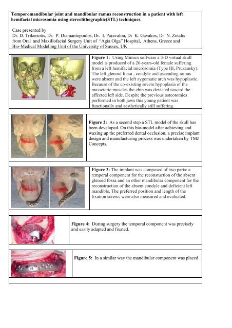
Figure 1: Using Mimics software a 3-D virtual skull<br />
model is produced of a 26-years-old female suffering<br />
from a left hemifacial microsomia (Type III, Pruzansky).<br />
The left glenoid fossa , condyle <strong>and</strong> ascending <strong>ramus</strong><br />
were absent <strong>and</strong> the left zygomatic arch was hypoplastic.<br />
Because of the co-existing severe hypoplasia of the<br />
masseteric muscles the chin was deviated toward the<br />
affected left side. Despite the previous osteotomies<br />
performed in both jaws this young patient was<br />
functionally <strong>and</strong> aesthetically still suffering.<br />
Figure 2: As a second step a STL model of the skull has<br />
been developed. On this bio-model after achieving <strong>and</strong><br />
waxing up the preferred dental occlusion, a precise implant<br />
design <strong>and</strong> manufacturing process was undertaken by TMJ<br />
Concepts.<br />
Figure 3: The implant was composed of two parts: a<br />
temporal component for the reconstuction of the absent<br />
glenoid fossa <strong>and</strong> an other m<strong>and</strong>ibular component for the<br />
reconstruction of the absent condyle <strong>and</strong> deficient left<br />
m<strong>and</strong>ible. The preferred position <strong>and</strong> length of the<br />
fixation screws were also measured <strong>and</strong> evaluated.<br />
Figure 4: During surgery the temporal component was precisely<br />
<strong>and</strong> easily adapted <strong>and</strong> fixated.<br />
Figure 5: In a similar way the m<strong>and</strong>ibular component was placed.
Figure 6: Patient's photos : 6a. Before reconstruction <strong>and</strong><br />
6b. After reconstruction without left chin deviation.<br />
Intermaxillary fixation was applied for two weeks after<br />
surgery.<br />
Figure 7: A month after surgery there was no deviation during the<br />
opening position. A more effective masticatory function <strong>and</strong> a more<br />
acceptable aesthetic result were achieved.<br />
CONCLUSION: Using the STL model in this patient with hemifacial microsomia the<br />
disturbed anatomy of the facial skeleton was thoroughly studied. A complex preoperarive<br />
reconstruction planning such as the implant-design <strong>and</strong> manufacturing process was done with<br />
high accuracy. During surgery both implant components were safely fixated on the anatomical<br />
sites without any need for further manual adaptation. This would never been possible without<br />
the making of a STL model. Also, less surgical time <strong>and</strong> tissue stress were observed.