Disposable electrochemical biosensors in microbiology - Formatex ...
Disposable electrochemical biosensors in microbiology - Formatex ...
Disposable electrochemical biosensors in microbiology - Formatex ...

You also want an ePaper? Increase the reach of your titles
YUMPU automatically turns print PDFs into web optimized ePapers that Google loves.
_______________________________________________________________________________________<br />
<strong>Disposable</strong> <strong>electrochemical</strong> <strong>biosensors</strong> <strong>in</strong> <strong>microbiology</strong><br />
O. Domínguez-Renedo, M.A. Alonso-Lomillo, M.J. Arcos-Martínez<br />
Department of Chemistry. Faculty of Sciences. University of Burgos. Plaza Misael Bañuelos s/n, 09001 Burgos, Spa<strong>in</strong>.<br />
Keywords Microorganism; screen-pr<strong>in</strong>ted electrodes; <strong>electrochemical</strong> <strong>biosensors</strong><br />
1.Introduction<br />
The analytical objective of develop<strong>in</strong>g sensitive, selective and rapid methods for the analysis of different substances <strong>in</strong><br />
complex matrices has led to the production of several chemical sensors. Among the analytical applications of chemical<br />
sensors, the one <strong>in</strong>volv<strong>in</strong>g the early detection of pathogen microorganisms can be considered essential <strong>in</strong> fields such as,<br />
• Food <strong>in</strong>dustry<br />
• Cl<strong>in</strong>ical and environmental analysis for avoidance of human health problems<br />
• Animals and plants epidemics [1-3].<br />
In this respect, chemical sensors can be considered as an alternative to the traditional analytical <strong>in</strong>strumentation <strong>in</strong><br />
rapid and sensitive analysis of these species.<br />
A chemical sensor is a device that responds to a particular analyte <strong>in</strong> a selective way through a chemical reaction.<br />
They can be used for the qualitative or quantitative determ<strong>in</strong>ation of the analyte [4]. There are two basic components <strong>in</strong><br />
a chemical sensor: a recognition system called receptor and, the transducer.<br />
The receptor is the sensitive part of the sensor and it also confers the selectivity needed for a suitable analyte<br />
determ<strong>in</strong>ation without other species <strong>in</strong>terference. Thus, the receptor recognizes the chemical <strong>in</strong>formation present <strong>in</strong> the<br />
sample. This <strong>in</strong>formation is then changed <strong>in</strong>to a signal <strong>in</strong> order to be recognized by the transducer. The latter f<strong>in</strong>ally<br />
carries out the conversion of the signal <strong>in</strong>to an observable response.<br />
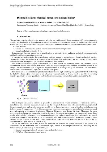
Biosensors can be considered as a type of chemical sensors with high sensitivity and applicability levels. Follow<strong>in</strong>g<br />
the IUPAC def<strong>in</strong>ition [5], a biosensor is an <strong>in</strong>tegrated receptor-transducer device, which is capable of provid<strong>in</strong>g<br />
selective quantitative or semi-quantitative analytical <strong>in</strong>formation us<strong>in</strong>g a biological recognition element (Fig. 1).<br />
Analyte<br />
Bioreceptor<br />
Fig. 1 Scheme of a biosensor<br />
Transducer<br />
Signal<br />
Signal<br />
Processor<br />
The biological recognition element is generally a macromolecule, which catalyzes a biochemical reaction,<br />
immobilized on a physical transducer. Enzymes are the biological elements most often used <strong>in</strong> the development of<br />
<strong>biosensors</strong> due to their high selectivity and easy use. However, they are expensive ow<strong>in</strong>g to their high cost of extract<strong>in</strong>g,<br />
isolat<strong>in</strong>g an purify<strong>in</strong>g processes [4]. In this way, on many occasions microorganisms are used as alternative sources of<br />
enzymatic activity <strong>in</strong> <strong>biosensors</strong> development. Microbial <strong>biosensors</strong> have longer life times. Moreover, the many<br />
enzymes and co-factors that co-exist <strong>in</strong> the cells give them the capacity to consume and consequently detect large<br />
number of chemicals. Furthermore, the advancement <strong>in</strong> molecular biology/recomb<strong>in</strong>ant DNA technologies has opened<br />
endless possibilities of modification of the microorganisms to improve the activity of an exist<strong>in</strong>g enzyme or even<br />
express foreign enzymes <strong>in</strong> host cell. These characteristics make microbial <strong>biosensors</strong> excellent devices <strong>in</strong> the<br />
determ<strong>in</strong>ation of a great number of substances [6].<br />
As for the transducer, this is that part of the biosensor that converts the change observed <strong>in</strong> the system as a<br />
consequence of the bioreaction <strong>in</strong>to a measurable signal related to the analyte concentration. Accord<strong>in</strong>g to the<br />
transducer type, <strong>biosensors</strong> may be classified as [7]:<br />
• Electrochemical: potentiometry, amperometry and conductimetry<br />
• FET (field effect transistor)-based sensors
• Optical<br />
• Piezoelectric devices<br />
• Surface acoustic waves<br />
• Thermal methods<br />
Among the various possible comb<strong>in</strong>ations of biocomponents and transducer techniques, the one <strong>in</strong>volv<strong>in</strong>g<br />
<strong>electrochemical</strong> detection has been used prom<strong>in</strong>ently <strong>in</strong> the analysis of different substances. This fact is due to the low<br />
detection limits that can be achieved on small samples volumes with modern <strong>electrochemical</strong> techniques. Moreover,<br />
electroanalytical techniques provide other important advantages <strong>in</strong>clud<strong>in</strong>g simplicity, low cost and <strong>in</strong> situ analysis<br />
options.<br />
Recently, the change of the conventional solid electrodes by screen-pr<strong>in</strong>ted electrodes (SPEs) has <strong>in</strong>creased the<br />
possibilities of electroanalytical techniques <strong>in</strong> the biosensor field. Various advantages <strong>in</strong>clud<strong>in</strong>g simple fabrication, low<br />
cost, small size, disposability, portability and easily mass-produced re<strong>in</strong>force the use of screen-pr<strong>in</strong>ted <strong>biosensors</strong> [8].<br />
Moreover, SPEs avoid some common problems related to traditional solid electrodes such as the needed clean<strong>in</strong>g<br />
processes. Thus, SPEs are extensively used <strong>in</strong> the fabrication of disposable <strong>biosensors</strong>.<br />
The fields of medic<strong>in</strong>e and public health require the rapid detection of bacterial pathogens, which cause severe<br />
<strong>in</strong>fectious diseases and poison<strong>in</strong>gs, <strong>in</strong> food and water. Traditional techniques of analysis, <strong>in</strong>clud<strong>in</strong>g cultur<strong>in</strong>g<br />
methodology, rema<strong>in</strong> as the preferred systems for the detection of these species. However, these techniques can be<br />
considered as time-consum<strong>in</strong>g and they often lead to false negatives results [9]. Biosensors have begun to play a<br />
significant role <strong>in</strong> the determ<strong>in</strong>ation of pathogens. Among them, <strong>electrochemical</strong> disposable devices have received<br />
considerable attention due to the comb<strong>in</strong>ation of the high sensitivity of <strong>electrochemical</strong> transducers with their low cost<br />
and their compatibility with modern microfabrication and m<strong>in</strong>iaturization technologies [10]. In this way, a great number<br />
of works describ<strong>in</strong>g the use of SPEs <strong>in</strong> the determ<strong>in</strong>ation of different microorganisms can be found <strong>in</strong> the bibliography.<br />
In the same way, microorganisms have been effectively used as the biological sens<strong>in</strong>g element <strong>in</strong> the construction of<br />
disposable <strong>biosensors</strong>. They present various advantages <strong>in</strong>clud<strong>in</strong>g ubiquitously and a great capacity to adapt to adverse<br />
conditions. Moreover, they are able to metabolize a wide range of chemical compounds [11]. Microorganisms can be<br />
also considered as excellent sources of enzymes be<strong>in</strong>g less expensive and more stable than purified enzymes usually<br />
employed <strong>in</strong> <strong>biosensors</strong> development. Therefore, a number of papers can be found <strong>in</strong> the literature describ<strong>in</strong>g the<br />
successfully determ<strong>in</strong>ation of several substances us<strong>in</strong>g disposable microbial <strong>biosensors</strong>.<br />
In this chapter, a description of the above-mentioned <strong>in</strong>terest<strong>in</strong>g applications of SPEs <strong>in</strong> <strong>microbiology</strong> has been<br />
reported. Several important aspects related to the type of biological element and immobilization procedure used have<br />
been <strong>in</strong>cluded. S<strong>in</strong>ce, the construction of the SPEs constitutes the first step <strong>in</strong> the development of disposable <strong>biosensors</strong>;<br />
a brief description of this fabrication process has been consequently <strong>in</strong>cluded.<br />
2. Screen-pr<strong>in</strong>ted electrodes<br />
Electroanalytical techniques are well considered <strong>in</strong> the Analytical Chemistry field. However, they have found several<br />
restrictions and practical difficulties for some applications. One of the most common problems of these techniques has<br />
been their lack of reproducibility, associated to the complexity of obta<strong>in</strong><strong>in</strong>g identical electrodes for all the<br />
measurements. Hang<strong>in</strong>g drop mercury electrodes have presented fewer problems <strong>in</strong> this respect but their toxicity has<br />
lead to the development of different alternatives. In this way, the possibilities of the <strong>electrochemical</strong> techniques can be<br />
improved by means of the replacement of the classical electrodes and cell systems by disposable screen-pr<strong>in</strong>ted devices.<br />
SPEs add many attractive advantages to the electroanalytical techniques, <strong>in</strong>clud<strong>in</strong>g the elim<strong>in</strong>ation of the surface<br />
regeneration needed <strong>in</strong> solid electrodes. Moreover, SPEs can be designed accord<strong>in</strong>g to the analytical problem<br />
characteristics by choos<strong>in</strong>g the adequate fabrication materials.<br />
Selective and disposable <strong>biosensors</strong> can be easily obta<strong>in</strong>ed by biomolecules immobilization on SPEs surfaces [12,<br />
13]. Procedures based on these disposable devices have been shown as practical systems for the fast, accessible and low<br />
cost analysis of many target species, <strong>in</strong>clud<strong>in</strong>g microorganisms. In the same way, the immobilization of microorganisms<br />
on the electrode has given rise to the development of selective and sensitive disposable <strong>biosensors</strong> for the analysis of<br />
different substances.<br />
2.1. SPEs fabrication<br />
_______________________________________________________________________________________<br />
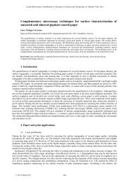
Screen-pr<strong>in</strong>t<strong>in</strong>g technology is based on the sequential layer deposition of different <strong>in</strong>ks on a ceramic or plastic substrate<br />
us<strong>in</strong>g the appropriate screen. A typical screen is made from a f<strong>in</strong>ely mesh of different materials <strong>in</strong>clud<strong>in</strong>g sta<strong>in</strong>less steel,<br />
polyester or nylon mounted under tension on a metal frame. The f<strong>in</strong>ished screen has open-mesh areas through which the<br />
desired pattern can be pr<strong>in</strong>ted (Fig. 2) [14].
_______________________________________________________________________________________<br />
Fig. 2 Scheme of a screen<br />
Sealed-mesh Open-mesh<br />
Metal frame<br />
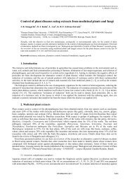
The screen-pr<strong>in</strong>t<strong>in</strong>g <strong>in</strong>k is then poured onto the top surface of the stencil. Then, a squeegee slowly moves from the<br />
rear to the front part of the screen, forc<strong>in</strong>g the <strong>in</strong>k through the open areas. The required pattern is thus deposited onto<br />
the substrate surface, as it can be seen <strong>in</strong> Fig. 3 [14].<br />
Screen<br />
(a) (b) (c)<br />
Substrate<br />
Squeege<br />
Ink<br />
Screen-pr<strong>in</strong>t<strong>in</strong>g<br />
step<br />
Pr<strong>in</strong>ted device<br />
Fig. 3 Screen-pr<strong>in</strong>ted device fabrication process scheme: (a) Ink poured; (b) Squeegee travers<strong>in</strong>g; (c) Ink deposited on<br />
the substrate surface<br />
The next phase of the process is to dry the pr<strong>in</strong>ted <strong>in</strong>k. Screen-pr<strong>in</strong>t<strong>in</strong>g <strong>in</strong>ks usually conta<strong>in</strong> various organic solvents,<br />
which are added with the aim of produc<strong>in</strong>g the accurate viscosity for screen pr<strong>in</strong>t<strong>in</strong>g. These solvents can be removed by<br />
dry<strong>in</strong>g the pr<strong>in</strong>ted <strong>in</strong>k <strong>in</strong> an oven at an adequate temperature. After dry<strong>in</strong>g, the substrate reta<strong>in</strong>s a rigid pattern that is<br />
relatively immune to smudg<strong>in</strong>g [14]. The comb<strong>in</strong>ation of different screens and <strong>in</strong>ks give rise to the def<strong>in</strong>ition of the<br />
different electrodes (work<strong>in</strong>g, reference and auxiliary) <strong>in</strong> the same configuration unit.<br />
F<strong>in</strong>ally, the disposable <strong>electrochemical</strong> biosensor is generated by the subsequent modification of the work<strong>in</strong>g<br />
electrode with the biosens<strong>in</strong>g material. This modification implies several steps <strong>in</strong> order to assure the robustness and<br />
durability of the developed biosensor.<br />
<strong>Disposable</strong> <strong>biosensors</strong> generated by modification of SPEs have been successfully applied <strong>in</strong> the <strong>microbiology</strong> field.<br />
The next section <strong>in</strong>volves the report of different disposable <strong>biosensors</strong> employed <strong>in</strong> such field, <strong>in</strong>clud<strong>in</strong>g the description<br />
of the different biocomponents and immobilization procedures used <strong>in</strong> their development.<br />
3. Screen-pr<strong>in</strong>ted electrodes <strong>biosensors</strong> <strong>in</strong> <strong>microbiology</strong><br />
As it has been mentioned, there is a great number of works describ<strong>in</strong>g the utilization of screen-pr<strong>in</strong>ted <strong>biosensors</strong> <strong>in</strong><br />
microbiological applications. These disposable <strong>biosensors</strong> have been developed by means of the immobilization of<br />
different nature biological elements. The immobilization procedure not only facilitates the required close proximity<br />
between the biomaterial and the transducer, but also helps <strong>in</strong> stabiliz<strong>in</strong>g it for reuse [11]. Thus, immobilization<br />
technology plays a very important role, be<strong>in</strong>g the critical step <strong>in</strong> <strong>biosensors</strong> manufactur<strong>in</strong>g. Many different procedures<br />
have been thus reported for the biological material immobilization conditions onto SPEs.<br />
A brief description of the different biological elements of the diverse immobilization procedures used <strong>in</strong> the<br />
development of disposable <strong>biosensors</strong> has been <strong>in</strong>cluded <strong>in</strong> the follow<strong>in</strong>g sections. This description has been focused <strong>in</strong><br />
the applications of this k<strong>in</strong>d of sensors with<strong>in</strong> <strong>microbiology</strong> field.<br />
3.1. Biological Element<br />
SPEs <strong>biosensors</strong> for detect<strong>in</strong>g microorganisms are mostly based on the <strong>in</strong>teraction between microorganisms and<br />
biological recognition elements such as antibodies giv<strong>in</strong>g rise to very sensitive and selective disposable immunosensors.<br />
The fabrication of DNA <strong>electrochemical</strong> sensors has also attracted a considerable recent attention <strong>in</strong> the development of
disposable genosensors for the analysis of pathogens agents. In the same way, different microorganisms have been used<br />
<strong>in</strong> the fabrication of disposable <strong>biosensors</strong> for many <strong>in</strong>terest<strong>in</strong>g analytical applications.<br />
3.1.1. Antibodies and antigens<br />
_______________________________________________________________________________________<br />
Antibodies are serum prote<strong>in</strong>s endowed with the capacity to recognize, by stereospecific association, a foreign<br />
substance <strong>in</strong> the organism it has <strong>in</strong>vaded. They are produced by two types of blood cells, B lymphocytes and plasma<br />
cells, <strong>in</strong> response to a foreign substance which is termed an immunogen, so-def<strong>in</strong>ed because it evokes an immune<br />
response. In most <strong>in</strong>stances, an <strong>in</strong>dividual antibody will recognize only one substance and this substance is termed the<br />
antigen. For antibody-based <strong>biosensors</strong>, the analyte is either the correspond<strong>in</strong>g antigen of the antibody or a part of it [7].<br />
The use of these biological elements has given rise to very high sensitive and selective sensors named immunosensors.<br />
Electrochemical immunosensors employs either antibodies or their complementary b<strong>in</strong>d<strong>in</strong>g partners (antigens and<br />
haptens) as biorecognition elements <strong>in</strong> comb<strong>in</strong>ation with <strong>electrochemical</strong> transducers. These sensors are based on the<br />
ability of antibodies to form complexes with the correspond<strong>in</strong>g antigens. This property of high specific molecular<br />
recognition leads to high selectivity of assays based on immune pr<strong>in</strong>ciples. Moreover, the extreme aff<strong>in</strong>ity of antigenantibody<br />
<strong>in</strong>teractions results <strong>in</strong> great sensitivity of immunoassay methods. This sensitivity is <strong>in</strong>creased when<br />
<strong>electrochemical</strong> transducers are used <strong>in</strong> immunosensors development [15, 16].<br />
A great number of immunoassay disposable <strong>electrochemical</strong> systems described <strong>in</strong> the literature are based on the<br />
pr<strong>in</strong>ciple of <strong>electrochemical</strong> detection of the labelled immunoagent. Most of these systems <strong>in</strong>volve the amperometric<br />
determ<strong>in</strong>ation of the product of a reaction of a substrate catalyzed by a labeled enzyme [16].<br />
Enzyme immunoassays comb<strong>in</strong>e the high sensitivity of the enzyme-catalyzed reactions with the extreme specificity<br />
of antigen-antibody <strong>in</strong>teractions. These advantages are <strong>in</strong>creased with the use of <strong>electrochemical</strong> transducers <strong>in</strong>clud<strong>in</strong>g<br />
disposable electrodes. Thus, disposable <strong>electrochemical</strong> enzyme-labelled immunosensors have been successfully<br />
applied <strong>in</strong> the sensitive and selective analysis of many species <strong>in</strong>clud<strong>in</strong>g microorganisms [17].<br />
A variety of different enzyme markers have been used for substrate transformation <strong>in</strong> <strong>electrochemical</strong> immunoassays<br />
systems. Alkal<strong>in</strong>e phosphatase (ALP) is one of the most popular enzymes used <strong>in</strong> imunoanalysis. This enzyme catalyzes<br />
a dephosphorylation reaction of different organic phosphates. Some of the products formed as a result of the reaction<br />
can be detected <strong>electrochemical</strong>ly <strong>in</strong> significantly low concentrations. Horseradish peroxidase (HRP), which catalyses<br />
the reduction of H2O2, is also a very frequently enzyme used <strong>in</strong> <strong>electrochemical</strong> immunosensors development [16].<br />
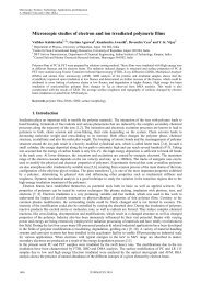
In respect to the immunosensors mechanism of analysis, a variety of methods have been developed <strong>in</strong>clud<strong>in</strong>g direct<br />
monitor<strong>in</strong>g, direct sandwich assays and <strong>in</strong>direct sandwich assays. The direct assay <strong>in</strong>volves the antigen immobilization<br />
of antigen on the electrode surface, followed by detection with an excess of enzyme-labelled conjugate (Fig. 4(a)). This<br />
method presents a great simplicity and a short assay time. For <strong>in</strong>stance, the analysis of Listeria monocytogenes (L.<br />
monocytogenes), an important food-borne pathogen with an extremely high mortality, has been analyzed us<strong>in</strong>g this<br />
simple method. The proposed immunosensor design was based on the immobilization of L. monocytogenes antigens on<br />
SPCEs. These modified SPCEs were then <strong>in</strong>cubated with goat anti-L. monocytogenes-ALP. F<strong>in</strong>ally, the amperometric<br />
signal was registered at + 300 mV follow<strong>in</strong>g the oxidation of p-am<strong>in</strong>ophenol (p-AP), the dephosphorylation product of<br />
the substrate p-am<strong>in</strong>ophenyl phosphate (p-APP) [18].<br />
Direct sandwich assay is one of the most frequently methods selected <strong>in</strong> the development of disposable<br />
immunosensors for the determ<strong>in</strong>ation of microorganisms. This method <strong>in</strong>volves the immobilization of a captur<strong>in</strong>g<br />
antibody on the electrode surface. Subsequently, an <strong>in</strong>cubation process with the specific antigen takes place and the<br />
antibody-antigen formed complex is f<strong>in</strong>ally <strong>in</strong>cubated with a labelled secondary antibody (Fig. 4(b)) [19]. This method<br />
has been successfully used <strong>in</strong> the determ<strong>in</strong>ation of Echerichia coli (E. coli). E. coli is an enterohemorrhagic bacterium<br />
which causes hemorrhagic colitis <strong>in</strong>clud<strong>in</strong>g symptoms such as bloody diarrhoea, hemolytic uremic syndrome, and<br />
thrombotic thrombocytopenic purpurea [20]. The analysis of this microorganism has been realized <strong>in</strong> samples of milk<br />
us<strong>in</strong>g a disposable immunosens<strong>in</strong>g strip based on a SPCE us<strong>in</strong>g HRP-conjugated polyclonal anti E. coli O157:H7 as the<br />
secondary antibody. Hydrogen peroxide and FeDC were used as the substrate for HRP and mediator, respectively, for<br />
the amperometric measurements at a potential of + 300 mV [20]. The electrode modification with mediators <strong>in</strong><br />
immunosensors development reduces the work<strong>in</strong>g potential, avoids <strong>electrochemical</strong> <strong>in</strong>terferences and <strong>in</strong>creases the<br />
reversibility of electrode reactions [21].<br />
Indirect sandwich assay is similar to the direct sandwich assay already described except that <strong>in</strong>stead of the secondary<br />
antibody, two detect<strong>in</strong>g antibodies are required. The first one is the specific antibody (captur<strong>in</strong>g antibody) to the<br />
antigen, while the second one is an enzyme-labelled anti-species conjugate (revell<strong>in</strong>g antibody) (Figure 4(c)). This<br />
system offers greater flexibility as it avoids the need to conjugate the antibody to an enzyme-label [18]. For example,<br />
Vibrio cholerae (V. cholerae) has been successfully determ<strong>in</strong>ed us<strong>in</strong>g this <strong>in</strong>direct method of analysis. V. cholerae<br />
causes cholera, a serious gastro<strong>in</strong>test<strong>in</strong>al disease present <strong>in</strong> many countries [21-23] . The detection of V. cholerae<br />
antigen has been performed us<strong>in</strong>g SPCEs coated with rabbit anti-Vibrio cholerae IgG captur<strong>in</strong>g antibody. The captur<strong>in</strong>g<br />
antibody was firstly adsorbed on the SPCE surface followed by block<strong>in</strong>g the free sites with seroalbum<strong>in</strong>e bov<strong>in</strong>e (BSA).<br />
The blocked SPCEs were <strong>in</strong>cubated with a solution conta<strong>in</strong><strong>in</strong>g V. cholerae cells. Afterward, these electrodes were<br />
further <strong>in</strong>cubated with a solution conta<strong>in</strong><strong>in</strong>g mice serum reveal<strong>in</strong>g antibody. F<strong>in</strong>ally, the electrodes were <strong>in</strong>cubated with<br />
a rabbit anti-mouse immunoglobul<strong>in</strong> ALP conjugated (rabbit anti-mouse ALP conjugate) solution. The analytical signal
_______________________________________________________________________________________<br />
measured was the amperometric response recorded for the enzyme substrate 1-napthyl phosphate by apply<strong>in</strong>g a<br />
potential of + 400 mV [22, 23].<br />
It can be found <strong>in</strong> the literature other assay methods similar to the <strong>in</strong>direct sandwich one described above, which can<br />
be called just <strong>in</strong>direct assay. In many cases, the antigen is first immobilized on the electrode surface followed by the<br />
<strong>in</strong>cubation with the specific antibody solution. F<strong>in</strong>ally, <strong>in</strong> a second <strong>in</strong>cubation step, the enzyme-labelled anti-species<br />
conjugate is fixed (Fig. 4 (d)). This method has been used <strong>in</strong> the analysis of numerous pathogen microorganisms. One<br />
of this analyzed microorganisms has been Salmonella typhimurium (S. typhimurium), one of the most common types of<br />
Salmonella, responsible of salmonellosis, a reported food-borne disease worldwide [24]. Therefore, the capacity to fast<br />
recognition of this pathogen is very important to protect public health safety and security. The immunosensor described<br />
by Rao et al. [25] for the analysis of S. typhimurium <strong>in</strong> serum samples was based on the immobilization of S.<br />
typhimurium flagell<strong>in</strong> antigen on SPCEs. After a wash<strong>in</strong>g step, the electrode was <strong>in</strong>cubated with anti-human ALP<br />
conjugated. The amperometric response of the modified SPCE was recorded at a potential of + 400 mV <strong>in</strong><br />
ethanoldiam<strong>in</strong>e. Next, the substrate 1-naphthyl phosphate was added and the change <strong>in</strong> the amperometric current was<br />
registered.<br />
Antigen-antibody complex formation can be also <strong>electrochemical</strong>ly directly detected us<strong>in</strong>g label-free immunosensors<br />
These <strong>biosensors</strong> display some important advantages <strong>in</strong> terms of speed and simplicity of operation [2]. The techniques<br />
are based on the detection of a change <strong>in</strong> the surface transducer physical properties as a result of the immunocomplex<br />
formation which causes a change <strong>in</strong> the transducer signal [16]. A redox probe is used <strong>in</strong> many cases <strong>in</strong> order to detect<br />
this change. The ma<strong>in</strong> advantage of label-free immunosensors is the s<strong>in</strong>gle stage analysis, which leads to a lower cost<br />
and easier use than labelled-immunosensors. As an example of this type of immunosensors, [Fe(CN)6 3- ]/[Fe(CN)6 4- ] has<br />
been used as a redox probe <strong>in</strong> label-free immunosensors for the successfully determ<strong>in</strong>ation of E. coli by different<br />
authors. This pathogen has been analyzed us<strong>in</strong>g SPAuEs [2] and SPCEs modified with self-assembled peptide<br />
nanotubes (PNTs) which have functional groups on their surface that allow an easier immobilization of antibodies [26].<br />
ELECTRODE<br />
(a)<br />
ENZYME<br />
ELECTRODE ELECTRODE<br />
ENZYME<br />
ELECTRODE<br />
(b) (c) (d)<br />
ENZYME<br />
Fig. 4 Schematic diagram of the different assay formats used <strong>in</strong> disposable <strong>electrochemical</strong> immunosensors: (a) Direct<br />
assay; (b) Direct sandwich assay; (c) Indirect sandwich assay; (d) Indirect assay<br />
antigen<br />
3.1.2. Nucleic Acids<br />
antibody<br />
ENZYME<br />
Enzyme-labelled<br />
antibody<br />
ENZYME<br />
Enzyme-labelled anti-species<br />
antibody<br />
Hybrid receptors such as DNA, which were first <strong>in</strong>troduced by Millan and Mikkelsen [27], have been shown to have<br />
promis<strong>in</strong>g applications <strong>in</strong> different fields of analysis. Nucleic acids operate <strong>in</strong> many ways like antibodies. The pr<strong>in</strong>ciple<br />
of selective detection is based on the highly specific hybridization of complementary strands of DNA. In this way,<br />
advances <strong>in</strong> molecular biology and biotechnology have led to a great number of possibilities for DNA <strong>electrochemical</strong><br />
<strong>biosensors</strong> development. In fact, the detection of specific DNA sequences provides the basis of many <strong>electrochemical</strong><br />
detect<strong>in</strong>g methods of microbial pathogens analysis. These methods are often based on hybridization <strong>biosensors</strong> for the<br />
detection of DNA sequences. The basics of nucleic acid hybridization devices is DNA base pair<strong>in</strong>g. Thus, the<br />
immobilization of a short synthetic oligomer on the electrode surface is first carried out. The sequence of this oligomer<br />
is complementary to the target element. Then, the exposure of this DNA modified electrode to the target sample results<br />
<strong>in</strong> the formation of the hybrid on the electrode surface. Electrochemical monitor<strong>in</strong>g of this duplex formation can result<br />
<strong>in</strong> a very useful transducer response. Thus, the formation of the hybrid is commonly detected by expos<strong>in</strong>g it to an
electroactive <strong>in</strong>dicator suitable to form strong bounds with the formed hybrid. An <strong>in</strong>crease <strong>in</strong> the <strong>electrochemical</strong><br />
response of this <strong>in</strong>dicator is then related to the duplex formation [28].<br />
Several genosensors based on the use of redox <strong>in</strong>dicators have been used for the determ<strong>in</strong>ation of pathogenic<br />
microbes <strong>in</strong>clud<strong>in</strong>g E. coli. The genosensor developed by Shiraishi et al. [29] was based on the immobilization of the<br />
probe DNA on a SPE fabricated by screen-pr<strong>in</strong>t<strong>in</strong>g a fullberene-impregnated carbon <strong>in</strong>k onto a<br />
poly(methylmethacrylate) substrate. In this case, Co(phen)3 3+ was used as the electroactive <strong>in</strong>dicator of the hybrid<br />
formation.<br />
In many occasions, the hybridization event is detected follow<strong>in</strong>g the enzymatic reaction of an enzyme, previously<br />
labelled on the electrode surface. Enzyme-labelled genosensors have been used, for <strong>in</strong>stance, <strong>in</strong> the determ<strong>in</strong>ation of<br />
Salmonella which its determ<strong>in</strong>ation with an ALP-labelled genosensor has been successfully carried out us<strong>in</strong>g SPCEs<br />
[30]. In this case, the hybridization reaction was detected though the sandwich format, based on the coupled of biot<strong>in</strong>streptavid<strong>in</strong><br />
<strong>in</strong>teraction us<strong>in</strong>g a streptavid<strong>in</strong> conjugated to ALP. α-naphtyl phosphate was used as substrate for the<br />
hybrid formation detection. An analogous genosensor is shown <strong>in</strong> [31] for the analysis of this pathogen us<strong>in</strong>g SPAuEs.<br />
3.1.3. Microorganisms<br />
As it has been po<strong>in</strong>ted up, microorganisms have been successfully used as biological elements <strong>in</strong> the development of<br />
disposable <strong>biosensors</strong> for a great number of analytical applications due to their already described great number of<br />
advantages. The microbial biosensor development <strong>in</strong>cludes several steps that are next summarized,<br />
• Cultivation step, <strong>in</strong> which the microorganism growth takes place. It <strong>in</strong>cludes the gradually <strong>in</strong>creas<strong>in</strong>g<br />
microorganism <strong>in</strong>oculation with the target analyte. This is the key step <strong>in</strong> order to make the biosensor specific to<br />
the analyte of <strong>in</strong>terest.<br />
• Microorganism immobilization on the SPE surface.<br />
• Determ<strong>in</strong>ation of the concentration of the target compound by chronoamperometric measurements, follow<strong>in</strong>g<br />
the oxygen consumption due to the metabolic activity of the immobilized microbe.<br />
Microbial disposable <strong>biosensors</strong> have been successfully applied <strong>in</strong> the determ<strong>in</strong>ation of many substances such as<br />
phenol [32], benzene [33] or the herbicide 2,4-dichloro phenoxy acetic acid (2,4-D) [13, 34, 35]. In most of the cases,<br />
the biosensor was constructed us<strong>in</strong>g Pseudomonas putida (P. putida) as the immobilized microorganism.<br />
3.2. Immobilization of Biological Elements<br />
The immobilization of the biological component must be carried out very carefully <strong>in</strong> order to assure the well<br />
performance of the biosensor. When select<strong>in</strong>g the most appropriate method various factors should be taken <strong>in</strong>to<br />
account, amongst which it is worth highlight<strong>in</strong>g the physical-chemical properties of the analyte, the nature of the<br />
biological element, the type of transducer used and the type of sample to be analyzed [7].<br />
A large number of immobilization methods can be found <strong>in</strong> the literature for the development of disposable<br />
<strong>biosensors</strong> with different applications <strong>in</strong> <strong>microbiology</strong>. The characteristic features of the most commonly used methods<br />
will be described <strong>in</strong> the follow<strong>in</strong>g sections.<br />
3.2.1. Adsorption<br />
Physical adsorption is the simplest method for biological element immobilization. This procedure has been successfully<br />
used <strong>in</strong> the development of disposable immunosensors for the determ<strong>in</strong>ation of pathogen microbes such as Helicobacter<br />
pylori (H. pylori) [17]. In this case, H. pylori antigens were immobilized by passive adsorption on the SPCE surface.<br />
In the case of microbial disposable <strong>biosensors</strong>, often a microbial suspension is <strong>in</strong>cubated with the electrode or an<br />
immobilization matrix, such as alum<strong>in</strong>a and glass bead, followed by r<strong>in</strong>s<strong>in</strong>g with buffer to remove unadsorbed microbe<br />
cells. The microbes are immobilized due to adsorptive <strong>in</strong>teractions such as ionic, polar or hydrogen bond<strong>in</strong>g and<br />
hydrophobic <strong>in</strong>teraction. However, immobilization us<strong>in</strong>g adsorption alone generally leads to poor long-term stability<br />
because of desorption of microbes [11].<br />
3.2.2. Entrapment<br />
_______________________________________________________________________________________<br />
Entrapment is a wide-rang<strong>in</strong>g term that <strong>in</strong>cludes different forms of work<strong>in</strong>g. One of the most commonly used methods<br />
consists of the entrapment of the biological element <strong>in</strong> an <strong>in</strong>ert polymer matrix on the electrode. In this case the<br />
biological component is placed <strong>in</strong> contact with a solution of a species susceptible to polymerization that forms a gel<br />
matrix <strong>in</strong> which the biocomponent is reta<strong>in</strong>ed [7].<br />
Alternatively, entrapment immobilization can be carried out by <strong>in</strong>clusion of the biological element by the either<br />
retention of it <strong>in</strong> close proximity of the transducer surface us<strong>in</strong>g dialysis or filter membrane or <strong>in</strong> chemical/biological<br />
polymers/gels such as (alg<strong>in</strong>ate, carrageenan, agarose, chitosan, collagen, polyacrylamide, polyv<strong>in</strong>ylachohol,<br />
poly(ethylene glycol), polyurethane, and so on. The major disadvantage of this immobilization method is the additional<br />
diffusion resistance offered by the entrapment material, which will result <strong>in</strong> lower sensitivity and detection limit [11].
_______________________________________________________________________________________<br />
Some reported examples of the application of this procedure <strong>in</strong> the development of microbial <strong>biosensors</strong> <strong>in</strong>clude the<br />
development of amperometric systems based on the immobilization of E. coli [36] and P. putida [37] us<strong>in</strong>g an Anopore<br />
membrane held to the SPCE surface us<strong>in</strong>g a micropore tape. Others reported E. coli based disposable <strong>biosensors</strong> use ĸ-<br />
Carrageenan, as immobilization matrix for entrapment of the bacterial cells onto SPAuEs [38].<br />
In the same way, disposable immunosensors have been developed for the analysis of the microbe pathogen Vibrio<br />
parahaemolyticus <strong>in</strong> food based on the immobilization of the biosens<strong>in</strong>g element us<strong>in</strong>g the natural polysaccharide<br />
agarose [8].<br />
3.2.3. Microencapsulation.<br />
Microencapsulation is an immobilization method which consists of bound<strong>in</strong>g the enzymes us<strong>in</strong>g a semi-permeable<br />
membrane which allows substrate molecules and products to pass through it, but which obstruct the biocomponent [21].<br />
There are different types of membranes (teflon, cellulose acetate (CA), polycarbonate, nafion, polyurethane) that are<br />
used <strong>in</strong> biosensor construction, the properties of which are decisive when apply<strong>in</strong>g the biosensor to the different types<br />
of analytes and matrices. So, as an example, CA membranes are impermeable to prote<strong>in</strong>s; teflon ones are permeable to<br />
some gases, etc. The most important advantage of this method is that membranes protect the biological components<br />
dim<strong>in</strong>ish<strong>in</strong>g its biodegradation and contam<strong>in</strong>ation. Moreover, the biosensor is also protected from temperature, pH and<br />
ionic force changes. However, the rate of the biochemical reaction is lower s<strong>in</strong>ce the analyte has to pass through the<br />
membrane to reach the biocomponent, which implies a less comprehensive analysis [7].<br />
A typical example of this immobilization procedure has been described for the bacteria P. Putida <strong>in</strong> the<br />
determ<strong>in</strong>ation of benzene [33].<br />
3.2.4. Cross-l<strong>in</strong>k<strong>in</strong>g<br />
Cross-l<strong>in</strong>k<strong>in</strong>g is a chemical immobilization method which <strong>in</strong>volves bridg<strong>in</strong>g between functional groups on the outer<br />
membrane of the microbe cells by multifunctional reagents such as GA and cyanuric chloride, to form a network.<br />
Because of the speed and simplicity, the method has found wide acceptance for immobilization of microorganisms. The<br />
cells can be bounded directly onto the electrode surface or on a removable support membrane, which can then be placed<br />
on the transducer surface [11].<br />
Several disposable <strong>biosensors</strong> have been developed for the analysis of phenol or the herbicide 2,4-dichloro phenoxy<br />
acetic acid (2,4-D) based on the immobilization of P. putida bacteria on SPEs by cross-l<strong>in</strong>k<strong>in</strong>g [13, 34, 35]. Likewise,<br />
E. coli selective immunosesnors have been developed based on the cross-l<strong>in</strong>k<strong>in</strong>g immobilization of bacteria antibodies<br />
on SPCEs [20].<br />
3.2.5. Covalent bound<strong>in</strong>g<br />
A highly stable b<strong>in</strong>d<strong>in</strong>g of biological material upon the <strong>electrochemical</strong> transducer can be achieved through the<br />
formation of covalent bonds between both materials. Covalent b<strong>in</strong>d<strong>in</strong>g methods rely on the formation of a stable<br />
covalent bond between functional groups of the biological components such as am<strong>in</strong>e, carboxylic or sulphydryl and the<br />
transducer such as am<strong>in</strong>e, carboxylic, epoxy or tosyl [6, 7].<br />
This immobilization has been frequently used <strong>in</strong> the development of disposable <strong>biosensors</strong> for different pathogens<br />
analysis. Thus, S. typhimurium can be determ<strong>in</strong>ed us<strong>in</strong>g a HRP-labelled immunosensor <strong>in</strong> which the biorecognition<br />
element was immobilized on a SPAuE surface covered with carboxymethyldextran <strong>in</strong> order to assure covalent bound<br />
formation [24].<br />
Covalent bound<strong>in</strong>g immobilization is the most frequently procedure used <strong>in</strong> the development of disposable<br />
genosensors for the determ<strong>in</strong>ation of pathogenic bacteria. For <strong>in</strong>stance, SPAuEs have been successfully used <strong>in</strong> the<br />
determ<strong>in</strong>ation of Salmonella bacteria us<strong>in</strong>g an ALP-labelled disposable genosensor. The immobilization of nucleic acid<br />
on the electrode surface was carried out via self-assembled monolayers (SAMs) formation. The procedure <strong>in</strong>volves a<br />
previous thiol-modification of the probe nucleotide [31].<br />
A common covalent immobilization procedure for disposable genosensors and immunosensors <strong>in</strong> <strong>microbiology</strong><br />
applications <strong>in</strong>volves the use of avid<strong>in</strong>-biot<strong>in</strong> <strong>in</strong>teraction to attach biot<strong>in</strong>ylated biocomponents on the electrode surface.<br />
For example, S. pneumoniae pathogen has been analyzed us<strong>in</strong>g both k<strong>in</strong>ds of sensors. Thus, a disposable genosensor<br />
has been developed for the determ<strong>in</strong>ation of this microbe based on the immobilization of biot<strong>in</strong>ylated oligonucleotides<br />
on the SPCE surface [10]. The analysis of this bacteria has been also performed us<strong>in</strong>g a disposable immunosensor<br />
fabricated by means of the immobilization of biot<strong>in</strong>ylated antibodies on SPCEs [39]. In both cases, the previous<br />
modification of the electrode surface with streptavid<strong>in</strong> was necessary <strong>in</strong> order to assure the necessary avid<strong>in</strong>-biot<strong>in</strong><br />
reaction.
Acknowledgements Authors would like to acknowledge fund<strong>in</strong>g via Junta de Castilla y León (GR177) and Spanish M<strong>in</strong>istry of<br />
Science and Innovation (TEC-2009/12029). M.A. Alonso-Lomillo is funded by a Ramón y Cajal fellowship from the Spanish<br />
M<strong>in</strong>istry of Science and Innovation and the European Social Found.<br />
References<br />
_______________________________________________________________________________________<br />
[1] Oczkowski T, Zwierkowska E, Bartkowiak S. Application of cell-based <strong>biosensors</strong> for the detection of bacterial elicitor<br />
flagell<strong>in</strong>. Bioelectrochemistry. 2007;70:192-197.<br />
[2] Escamilla-Gomez V, Campuzano S, Pedrero M, P<strong>in</strong>garron JM. Gold screen-pr<strong>in</strong>ted-based impedimetric immuno<strong>biosensors</strong> for<br />
direct and sensitive Escherichia coli quantisation. Biosensors & Bioelectronics. 2009; 24:3365-3371.<br />
[3] Gamella M, Campuzano S, Parrado C, Reviejo AJ, P<strong>in</strong>garron JM. Microorganisms recognition and quantification by lect<strong>in</strong><br />
adsorptive aff<strong>in</strong>ity impedance. Talanta. 2009; 78:1303-1309.<br />
[4] Egg<strong>in</strong>s BR, Chemical sensors and <strong>biosensors</strong>. Chchester: John Willey & sons; 2002.<br />
[5] Thévenot DR, Toth K, Durst RA, Wilson GS. Electrochemical <strong>biosensors</strong>: recommended def<strong>in</strong>itions and classification<br />
(Technical Report). Pure and Applied Chemistry. 1999;71:2333-2348.<br />
[6] Lei Y, Chen W, Mulchandani A. Microbial <strong>biosensors</strong>. Analytica Chimica Acta. 2006;568:200-210.<br />
[7] Domínguez O, Arcos MJ. Electrochemical Biosensors. Encyclopedia of Sensors. Vol. 3., USA: American Scientific Publishers;<br />
2006.<br />
[8] Zhao GY, X<strong>in</strong>g FF, Deng SP. A disposable amperometric enzyme immunosensor for rapid detection of Vibrio<br />
parahaemolyticus <strong>in</strong> food based on agarose/Nano-Au membrane and screen-pr<strong>in</strong>ted electrode. Electrochemistry<br />
Communications. 2007;9:1263-1268.<br />
[9] Yem<strong>in</strong>i M, Levi Y, Yagil E, Rishpon J. Specific <strong>electrochemical</strong> phage sens<strong>in</strong>g for Bacillus cereus and Mycobacterium<br />
smegmatis. Bioelectrochemistry. 2007;70:180-184.<br />
[10] Hernandez-Santos D, Diaz-Gonzalez M, Gonzalez-Garcia MB, Costa-Garcia A. Enzymatic genosensor on streptavid<strong>in</strong>modified<br />
screen-pr<strong>in</strong>ted carbon electrodes. Analytical Chemistry. 2004;76:6887-6893.<br />
[11] D'Souza SF. Microbial <strong>biosensors</strong>. Biosensors & Bioelectronics. 2001;16:337-353.<br />
[12] Domínguez-Renedo O, Alonso-Lomillo MA, Arcos-Mart<strong>in</strong>ez MJ. Recent developments <strong>in</strong> the field of screen-pr<strong>in</strong>ted electrodes<br />
and their related applications. Talanta. 2007;73:202-219.<br />
[13] Timur S, Della Seta L, Pazarlioglu N, Pilloton R, Telefoncu A. Screen pr<strong>in</strong>ted graphite <strong>biosensors</strong> based on bacterial cells.<br />
Process Biochemistry. 2004; 39:1325-1329.<br />
[14] White NM, Turner JD. Thick-film sensors: past, present and future. Meas. Sci. Technol. 1997;8:1-20.<br />
[15] Skladal P. Advances <strong>in</strong> <strong>electrochemical</strong> immunosensors. Electroanalysis. 1997;9:737-745.<br />
[16] Gh<strong>in</strong>dilis AL, Atanasov P, Wilk<strong>in</strong>s M, Wilk<strong>in</strong>s E. Immunosensors: Electrochemical sens<strong>in</strong>g and other eng<strong>in</strong>eer<strong>in</strong>g approaches.<br />
Biosensors & Bioelectronics. 1998;13:113-131.<br />
[17] Mess<strong>in</strong>a GA, De Vito IE, Raba J. Screen-pr<strong>in</strong>ted immunosensor for quantification of human serum IgG antibodies to<br />
Helicobacter pylori. Sensors and Actuators B-Chemical. 2007;128:23-30.<br />
[18] Crowley EL, O'Sullivan CK, Guilbault GG. Increas<strong>in</strong>g the sensitivity of Listeria monocytogenes assays: evaluation us<strong>in</strong>g<br />
ELISA and amperometric detection. Analyst. 1999;124:295-299.<br />
[19] Sadik OA, Van Emon JM. Applications of <strong>electrochemical</strong> immunosensors to environmental monitor<strong>in</strong>g. Biosensors and<br />
Bioelectronics. 1996;11:1-11.<br />
[20] L<strong>in</strong> YH, Chen SH, Chuang YC, Lu YC, Shen TY, Chang CA, L<strong>in</strong> CS. <strong>Disposable</strong> amperometric immunosens<strong>in</strong>g strips<br />
fabricated by Au nanoparticles-modified screen-pr<strong>in</strong>ted carbon electrodes for the detection of foodborne pathogen Escherichia<br />
coli O157 : H7. Biosensors & Bioelectronics. 2008;23:1832-1837.<br />
[21] Alonso-Lomillo MA, Domínguez-Renedo O, Arcos-Mart<strong>in</strong>ez, M.J. Enzyme modified screen pr<strong>in</strong>ted electrodes, <strong>in</strong> Biosensors:<br />
Properties, Materials and Applications. Hauppauge NY: Nova Publishers; 2009<br />
[22] Rao VK, Sharma MK, Goel AK, S<strong>in</strong>gh L, Sekhar K. Amperometric immunosensor for the detection of Vibrio cholerae O1<br />
us<strong>in</strong>g disposable screen-pr<strong>in</strong>ted electrodes. Analytical Sciences. 2006;22:1207-1211.<br />
[23] Sharma MK, Goel AK, S<strong>in</strong>gh L, Rao VK. Immunological biosensor for detection of Vibrio cholerae O1<strong>in</strong> environmental water<br />
samples. World Journal of Microbiology & Biotechnology. 2006;22:1155-1159.<br />
[24] Salam F, Tothill IE. Detection of Salmonella typhimurium us<strong>in</strong>g an <strong>electrochemical</strong> immunosensor. Biosensors &<br />
Bioelectronics. 2009;24:2630-2636.<br />
[25] Rao VK, Rai GP, Agarwal GS, Suresh S. Amperometric immunosensor for detection of antibodies of Salmonella typhi <strong>in</strong><br />
patient serum. Analytica Chimica Acta. 2005; 531:173-177.<br />
[26] Cho EC, Choi JW, Lee MY, Koo KK. Fabrication of an <strong>electrochemical</strong> immunosensor with self-assembled peptide nanotubes.<br />
Colloids and Surfaces a-Physicochemical and Eng<strong>in</strong>eer<strong>in</strong>g Aspects. 2008;313:95-99.<br />
[27] Millan KM, Mikkelsen SR. Sequence-selective biosensor for dna-based on electroactive hybridization <strong>in</strong>dicators. Analytical<br />
Chemistry. 1993;65:2317-2323.<br />
[28] Wang J, Rivas G, Cai X, Palecek E, Nielsen P, Shiraishi H, Dontha N, Luo D, Parrado C, Chicharro M, Farias PAM, Valera FS,<br />
Grant DH, Ozsoz M, Flair MN. DNA <strong>electrochemical</strong> <strong>biosensors</strong> for environmental monitor<strong>in</strong>g. A review. Analytica Chimica<br />
Acta. 1997;347:1-8.<br />
[29] Shiraishi H, Itoh T, Hayashi H, Takagi K, Sakane M, Mori T, Wang J. Electrochemical detection of E-coli 16S rDNA sequence<br />
us<strong>in</strong>g air-plasma-activated fullerene-impregnated screen pr<strong>in</strong>ted electrodes. Bioelectrochemistry. 2007;70:481-487.<br />
[30] Del Giallo ML, Ariksoysal D, Marrazza G, Masc<strong>in</strong>i M, Ozsoz M. <strong>Disposable</strong> <strong>electrochemical</strong> enzyme-amplified genosensor for<br />
Salmonella bacteria detection. Analytical Letters. 2005;38:2509-2523.<br />
[31] Farabull<strong>in</strong>i F, Lucarelli F, Palchetti I, Marrazza G, Masc<strong>in</strong>i M. <strong>Disposable</strong> <strong>electrochemical</strong> genosensor for the simultaneous<br />
analysis of different bacterial food contam<strong>in</strong>ants. Biosensors & Bioelectronics. 2007;22:1544-1549.
_______________________________________________________________________________________<br />
[32] Bachmann TT, Bilitewski U, Schmid RD. A microbial sensor based on Pseudomonas putida for phenol, benzoic acid and their<br />
monochlor<strong>in</strong>ated derivatives which can be used <strong>in</strong> water and n-hexane. Analytical Letters. 1998;31:2361-2373.<br />
[33] Lanyon YH, Tothill IE, Masc<strong>in</strong>i M. An amperometric bacterial biosensor based on gold screen-pr<strong>in</strong>ted electrodes for the<br />
detection of benzene. Analytical Letters. 2006;39:1669-1681.<br />
[34] Odaci D, Sezg<strong>in</strong>turk MK, Timur S, Pazarliolu N, Pilloton R, D<strong>in</strong>ckaya E, Telefoncu A. Pseudomonas putida Based<br />
Amperometric Biosensors for 2,4-D Detection. Preparative Biochemistry & Biotechnology. 2009;39:11-19.<br />
[35] Timur S, Pazarlioglu N, Pilloton R, Telefoncu A. Detection of phenolic compounds by thick film sensors based on<br />
Pseudomonas putida. Talanta. 2003;61:87-93.<br />
[36] Farré M, Pas<strong>in</strong>i O, Carmen Alonso M, Castillo M, Barceló D. Toxicity assessment of organic pollution <strong>in</strong> wastewaters us<strong>in</strong>g a<br />
bacterial biosensor. Analytica Chimica Acta. 2001;426:155-165.<br />
[37] Farre M, Barcelo D. Characterization of wastewater toxicity by means of a whole-cell bacterial biosensor, us<strong>in</strong>g Pseudomonas<br />
putida, <strong>in</strong> conjunction with chemical analysis. Fresenius Journal of Analytical Chemistry. 2001;371:467-473.<br />
[38] Held M, Schuhmann W, Jahreis K, Schmidt HL. Microbial biosensor array with transport mutants of Escherichia coli K12 for<br />
the simultaneous determ<strong>in</strong>ation of mono-and disaccharides. Biosensors & Bioelectronics. 2002;17:1089-1094.<br />
[39] Diaz-Gonzalez M, Gonzalez-Garcia MB, Costa-Garcia A. Detection of pneumolys<strong>in</strong> <strong>in</strong> human ur<strong>in</strong>e us<strong>in</strong>g an immunosensor on<br />
screen-pr<strong>in</strong>ted carbon electrodes. Sensors and Actuators B-Chemical, 2006;113:1005-1011.