Acid-Base Properties of Amino Acids
Acid-Base Properties of Amino Acids
Acid-Base Properties of Amino Acids

Create successful ePaper yourself
Turn your PDF publications into a flip-book with our unique Google optimized e-Paper software.
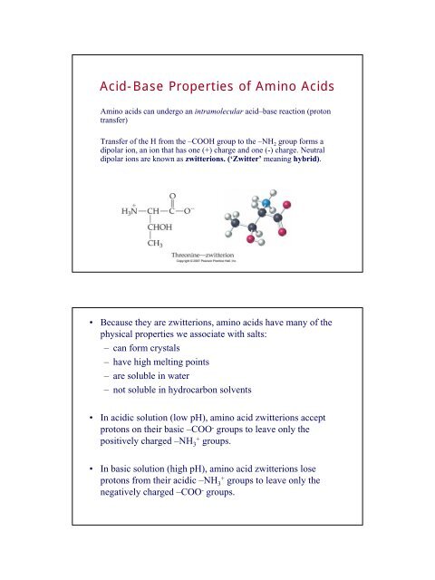
<strong>Acid</strong>-<strong>Base</strong> <strong>Properties</strong> <strong>of</strong> <strong>Amino</strong> <strong>Acid</strong>s<br />
<strong>Amino</strong> acids can undergo an intramolecular acid–base reaction (proton<br />
transfer)<br />
Transfer <strong>of</strong> the H from the –COOH group to the –NH 2 group forms a<br />
dipolar ion, an ion that has one (+) charge and one (-) charge. Neutral<br />
dipolar ions are known as zwitterions. (‘Zwitter’ meaning hybrid).<br />
• Because they are zwitterions, amino acids have many <strong>of</strong> the<br />
physical properties we associate with salts:<br />
– can form crystals<br />
– have high melting points<br />
– are soluble in water<br />
– not soluble in hydrocarbon solvents<br />
• In acidic solution (low pH), amino acid zwitterions accept<br />
protons on their basic –COO - groups to leave only the<br />
positively charged –NH 3 + groups.<br />
• In basic solution (high pH), amino acid zwitterions lose<br />
protons from their acidic –NH 3 + groups to leave only the<br />
negatively charged –COO - groups.
The net charge <strong>of</strong> an amino acid molecule at any given<br />
moment depends on the particular amino acid and the pH <strong>of</strong><br />
the medium.<br />
The pH at which the net positive and negative charges are<br />
equal is the amino acid’s isoelectric point (pI). At this point,<br />
the overall charge <strong>of</strong> all the amino acids in a sample is zero.<br />
Chirality in <strong>Amino</strong> <strong>Acid</strong>s<br />
• Chiral: Having right- or<br />
left-handedness with two<br />
different nonsuperimposable<br />
mirror<br />
image forms.<br />
• One hand does not match<br />
the other when<br />
superimposed.
• Achiral: The opposite <strong>of</strong><br />
chiral; having<br />
superimposable mirror<br />
images and thus no right- or<br />
left- handedness.<br />
• It is easy to visualize the<br />
chair on top <strong>of</strong> its mirror<br />
image.<br />
Molecular Handedness and <strong>Amino</strong><br />
<strong>Acid</strong>s<br />
• Chiral carbon atom<br />
(chirality center) A carbon<br />
atom bonded to four<br />
different groups.<br />
• If a molecule has an atom<br />
bonded to four different<br />
groups, it can be chiral.
• The mirror-image forms <strong>of</strong> a chiral molecule like alanine are<br />
called enantiomers or optical isomers.<br />
• Propane is an achiral molecule. The molecule and its mirror<br />
image are identical and it has no left- and right-handed<br />
isomers.<br />
• Enantiomers are one kind <strong>of</strong> stereoisomer, compounds that<br />
have the same formula and atomic connections but different<br />
spatial arrangements.<br />
• Pairs <strong>of</strong> enantiomers have the same physical properties<br />
except they always differ in their effect on polarized light<br />
and how they react with other chiral molecules.<br />
• Pairs <strong>of</strong> enantiomers <strong>of</strong>ten differ in their biological activity,<br />
odors, tastes, or activity as drugs.<br />
• 19 out <strong>of</strong> 20 natural amino acids are chiral – they have<br />
four different groups on the α-carbon. Only glycine is<br />
achiral.<br />
• Nature uses only one isomer out <strong>of</strong> a pair <strong>of</strong> enantiomers for<br />
each amino acid to build proteins. The naturally occurring<br />
amino acids are classified as left-handed or L-amino<br />
acids.
Tastes sweet!!<br />
The amino acids are in L<br />
Form. If you switch them<br />
With the D isomer, then<br />
It will not taste sweet.<br />
Shape Determining Interactions in Proteins<br />
(Tertiary 3D Structure)<br />
Covalent Bonds: The disulfide bonds <strong>of</strong> Cys is the most common<br />
Covalent bond. Found in structure <strong>of</strong> Insulin.<br />
Hydrogen bonds: Hydrogen bonding between backbone C=O<br />
And –N – H groups. H bonds also form between side chains and<br />
Between side chain – backbone.<br />
Salt Bridges: Electrostatic attractions between two A.A residues<br />
That have ionic side-chains. A Lysine side chain with an aspartic acid<br />
side chain.<br />
Hydrophobic Interactions: In aq. Solutions, proteins keep the polar<br />
groups for water solubility and the non-polar groups inward.<br />
The interaction within the non-polar groups interact and keeps water<br />
away. Weaker than the other interactions.<br />
Metal-Ion Coordination: Metal ions, which are positive cations,<br />
can bridge between negatively charged side chains. Hence the<br />
importance <strong>of</strong> trace minerals in our bodies.
Diagram <strong>of</strong> an Electrophoresis Apparatus<br />
Movement <strong>of</strong> charged molecules in electrophoresis. Movement<br />
varies with charge (depending on acid/base side chains)<br />
size and shape, strength <strong>of</strong> the electric field,<br />
And the nature <strong>of</strong> the medium (pH) in which the protein is moving.<br />
Electrophoresis diagram for normal and sickle cell Hemoglobin<br />
Hemoglobin in samples placed at the<br />
original position have moved left to right.<br />
The normal individual has only HbA.<br />
The individual with sickle-cell anemia has<br />
no HbA.<br />
The individual with sickle-cell trait<br />
has roughly equal amounts <strong>of</strong> HbA and HbS.<br />
HbA and HbS have negative charges <strong>of</strong><br />
different magnitudes because HbS has<br />
two fewer Glu residues than HbA.
A Blood test to determine the levels <strong>of</strong> the<br />
immune proteins (globulins, or antibodies),<br />
and albumin.<br />
www.wikipedia.org<br />
Proteins have been separated by<br />
size and stained blue for viewing.<br />
Diseases can cause changes in<br />
the protein patterns seen.<br />
Gel electrophoresis<br />
apparatus (DNA analysis)<br />
An agarose gel is placed in this<br />
buffer-filled box and electrical<br />
current is applied via the power<br />
supply to the rear.<br />
The negative terminal is at the<br />
far end (black wire), so DNA<br />
migrates toward the camera.