Bilateral Obstetric Palsy of Brachial Plexus - A Case Report
Bilateral Obstetric Palsy of Brachial Plexus - A Case Report
Bilateral Obstetric Palsy of Brachial Plexus - A Case Report

You also want an ePaper? Increase the reach of your titles
YUMPU automatically turns print PDFs into web optimized ePapers that Google loves.
126<br />
<strong>Bilateral</strong> <strong>Obstetric</strong> <strong>Palsy</strong> <strong>of</strong> <strong>Brachial</strong> <strong>Plexus</strong> - A <strong>Case</strong> <strong>Report</strong><br />
<strong>Bilateral</strong> Do¤umsal Brakiyal Pleksus Felci<br />
Summary<br />
<strong>Obstetric</strong> <strong>Brachial</strong> <strong>Plexus</strong> <strong>Palsy</strong> (OBPP) is one <strong>of</strong> the devastating<br />
complications <strong>of</strong> difficult or assisted deliveries. <strong>Brachial</strong> plexus palsy with<br />
upper root involvement most commonly affects the external rotators and<br />
abductors. Twenty percent <strong>of</strong> obstetrical brachial plexus palsies are bilateral<br />
and they represent a more severe condition. An eight-year-old girl patient<br />
with bilateral brachial plexus palsy was described and discussed in this<br />
report. Turk J Phys Med Rehab 2009;55:126-7.<br />
Key Words: <strong>Brachial</strong> plexus, rehabilitation<br />
Özet<br />
<strong>Case</strong> <strong>Report</strong> / Olgu Sunumu<br />
Özlem ALTINDA⁄, Savafl GÜRSOY, Ahmet METE*<br />
From Departments <strong>of</strong> Physical Medicine and Rehabilitation and *Radiology, Gaziantep University Medical Faculty, Gaziantep, Turkey<br />
Introduction<br />
<strong>Obstetric</strong> brachial plexus palsy (OBPP) is one <strong>of</strong> the<br />
devastating complications <strong>of</strong> difficult or assisted deliveries. The<br />
nature <strong>of</strong> this injury, with their severe loss <strong>of</strong> upper extremity<br />
function, leads to serious consequences for the personal and<br />
pr<strong>of</strong>essional life <strong>of</strong> the patient (1,2). The incidence <strong>of</strong> OBPP as<br />
reported in the literature varies from 0.9 to 2.4 per 1000 new<br />
live births (3,4). OBPP presents with either Erb’s paralysis<br />
(involving the C5, C6, 7), or total paralysis (involving C5, 6, 7, 8, and<br />
T1). Klumpke’s birth palsy (involving mainly the C7 root) is only a<br />
historical interest and is no longer seen in modern obstetric practice<br />
(5). Pure upper plexus lesions occur at 73%, followed by total<br />
plexus injury at 4%, and pure lower plexus injury at 2% (6).<br />
Risk factors for OBPP include macrosomia, assisted delivery or<br />
breech presentation, prolonged labor, excessive maternal weight gain,<br />
cephalopelvic disproportion, and subsequent shoulder dystocia.<br />
OBPP related injuries include clavicular fractures, physeal<br />
fractures <strong>of</strong> the humerus, fractures <strong>of</strong> the shoulder girdle,<br />
torticollis, facial and phrenic nerve palsy (7,8). Traction forces on<br />
nerves can cause various injuries, ranging from temporary<br />
conduction deficits to nerve root avulsion from the spinal cord.<br />
Do¤umsal brakiyal pleksus felci, zor do¤um s›ras›nda brakiyal pleksusun traksiyon<br />
yaralanmas› sonucu meydana gelen bir komplikasyondur. En yayg›n formu,<br />
eksternal rotator ve abduktorlar›n etkilendi¤i üst kök lezyonlar›d›r. Daha<br />
a¤›r bir klinik formda karfl›m›za ç›kan bilateral lezyonlar, do¤umsal brakiyal<br />
pleksus felçlerinin %20’sini teflkil eder. Bu yaz›da bilateral brakiyal pleksus<br />
felci olan 8 yafl›nda bir k›z anlat›ld› ve tart›fl›ld›. Türk Fiz T›p Rehab Derg<br />
2009;55:126-7.<br />
Anahtar Kelimeler: Brakiyal pleksus, rehabilitasyon<br />
<strong>Bilateral</strong> lesions are much less common and have been<br />
reported in 20% <strong>of</strong> the cases. We report a patient who sustained a<br />
bilateral brachial plexus palsy due to assisted delivery.<br />
Address for Correspondence/Yaz›flma Adresi: Özlem Alt›nda¤, MD, Department <strong>of</strong> Physical Medicine and Rehabilitation, Gaziantep University Medical Faculty, Gaziantep, Turkey<br />
Phone: +90 342 360 60 60/76220 E-mail: ozaltindag@yahoo.com Received/Gelifl Tarihi: January/Ocak 2008 Accepted/Kabul Tarihi: June/Haziran 2008<br />
© Turkish Journal <strong>of</strong> Physical Medicine and Rehabilitation, Published by Galenos Publishing. All rights reserved. / © Türkiye Fiziksel T›p ve Rehabilitasyon Dergisi, Galenos Yay›nc›l›k taraf›ndan bas›lm›flt›r. Her hakk› sakl›d›r.<br />
<strong>Case</strong><br />
An 8-year-old girl was referred to our clinic with decreased<br />
movements in her left and right arms since birth. The mother was a<br />
healthy 32-year-old woman. The patient was the fourth child <strong>of</strong> the<br />
parents and had been born at full term in breech presentation.<br />
Normal vaginal delivery had occurred after two hours.<br />
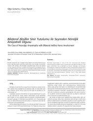
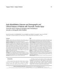
Physical examination revealed prominent muscle atrophy <strong>of</strong><br />
both arms (Figure 1). She had marked weakness in both <strong>of</strong> her<br />
shoulder muscles. The shoulder joints active range <strong>of</strong> motion was<br />
limited; however, its passive range <strong>of</strong> motion was normal. There was<br />
no sensory disturbance <strong>of</strong> her arms. Deep tendon reflexes were<br />
hypoactive in both limbs.<br />
Neurological examination revealed weakness <strong>of</strong> the upper limbs<br />
and the Medical Research Council (MRC) score was 3/5. Thoracic<br />
outlet maneuvers yielded negative results. The routine blood tests<br />
were normal.<br />
Electrophysiological findings <strong>of</strong> brachial plexopathy are<br />
demonstrated in Table 1.
Turk J Phys Med Rehab 2009;55:126-7<br />
Türk Fiz T›p Rehab Derg 2009;55:126-7<br />
Figure 1. Muscle atrophy in both arms. Figure 2. Magnetic resonance image <strong>of</strong> cervical spine.<br />
Table 1. The electrophysiological findings <strong>of</strong> the patient.<br />
Nerve Stimulation Record Amplitude Conduction Latancies<br />
(μV) (distal/proximal) Velocity (m/s) (ms) (distal/proximal)<br />
Motor<br />
Right medianus 14.6/13.5 48.2 3.7/6.5<br />
Left medianus 17.1/16.9 53.7 4.0/6.7<br />
Right ulnaris 15.8/12.5 50.0 2.3/5.2<br />
Left ulnaris 21.8/19.7 59.6 3.4/6.0<br />
Right musculocutaneus 3.0 4.2/4.2<br />
Left musculocutaneus 1.8/1.9 4.2/4.2<br />
The x-ray evaluation showed posterior shoulder subluxation.<br />
Cervical magnetic resonance imaging demonstrated a wide<br />
thecal sac from C2 to C4, spondylolisthesis at C3-C4 and a<br />
meningeal cystic lesion widening the neural foramina at right<br />
side (Figure 2).<br />
Based on our radiological, electrophysiological and clinical<br />
findings, we diagnosed our patient as having brachial plexopathy,<br />
shoulder subluxation, cervical dural ectasia and spondylolisthesis.<br />
Range <strong>of</strong> motion and scapular strengthening exercises were<br />
performed in the patient and orthopedic surgery consultation was<br />
obtained.<br />
Discussion<br />
Early diagnosed OBPP may recover completely with physical therapy<br />
only. A small percentage <strong>of</strong> cases require further<br />
physical therapy to achieve a better level <strong>of</strong> recovery. Significant<br />
improvement has occurred in 90% <strong>of</strong> these children as compared to a<br />
50-70% improvement rate in those whose treatment was delayed (9,10).<br />
Our patient had some differences from other infants born with<br />
OBPP. She had bilateral OBPP and no regular treatment<br />
since her birth till 8 years <strong>of</strong> age. In addition, there were<br />
traumatic lesions in her neck and posterior subluxation <strong>of</strong> both <strong>of</strong><br />
her shoulders.<br />
A multidisciplinary team approach is recommended for the management<br />
<strong>of</strong> OBPP. The initial goal <strong>of</strong> therapy is to maintain passive<br />
range <strong>of</strong> motion, supple joints and muscle strength. In our case,<br />
conservative treatment was not sufficient for recovery;<br />
deltoid and biceps muscles did not return to normal function.<br />
Surgical treatment was planned, including tendon transfer for<br />
internal rotation and shoulder joint fusion. A mobile arm support<br />
was recommended for the patient to facilitate her independent<br />
eating during the waiting period for surgery.<br />
Upper plexus injuries tend to be the least severe and have the<br />
best prognosis among brachial plexus injuries. Total plexus<br />
injuries require significantly higher traction forces and result in severe<br />
injuries with attendant root avulsions and they have a poorer<br />
prognosis. The upper and middle trunks <strong>of</strong> brachial plexus were<br />
involved in our patient. The present consensus for nerve<br />
reconstruction in OBPP is between 3 and 6 months after injury.<br />
Good results may not be achieved with a later reconstruction.<br />
<strong>Bilateral</strong> OBPP is a very rare condition. We suggest that OBPP<br />
should be kept in mind in cases with difficult and assisted<br />
delivery, and should be treated with conservative methods as<br />
soon as possible. Further, the possibility <strong>of</strong> presence <strong>of</strong> traumatic<br />
neck lesions and shoulder deformities must be considered in<br />
these patients.<br />
References<br />
Alt›nda¤ et al.<br />
<strong>Bilateral</strong> Plexopathy 127<br />
1. Millesi H. Trauma involving the brachial plexus. In Omer GE,<br />
Spinner M, Van Beek AL, editors. Management <strong>of</strong> Peripheral<br />
Nerve Problems. Philadelphia: W.B. Saunders; 1998. p 433-44.<br />
[Abstract] / [PDF]<br />
2. Sar›hasan B, Kelsaka E, Tomak Y, Karakaya D, Demirbilek Ö.<br />
Genel anestezi s›ras›nda gözlenen brakiyal pleksus zedelenmesi.<br />
Turkiye Klinikleri J Anest Reanim 2006;4:26-8. [Full Text] / [PDF]<br />
3. Gu YD, Chen L, Shen LY. Classification <strong>of</strong> impairment <strong>of</strong> shoulder<br />
abduction in obstetric brachial plexus palsy and its clinical<br />
significance. J Hand Surg [Br] 2000;25:46-8. [Abstract]<br />
4. DeMott RK. <strong>Brachial</strong> plexus deficits with and without shoulder<br />
dystocia. Am J Obstet Gynecol 2006;195:630. [Abstract]<br />
5. Al-Qattan MM. <strong>Obstetric</strong> brachial plexus palsy associated with<br />
breech delivery. Ann Plast Surg 2003;51:257-64. [Full Text] / [PDF]<br />
6. Dunham EA. <strong>Obstetric</strong>al brachial plexus palsy. Orthop Nurs<br />
2003;22:106-16. [Abstract] / [PDF]<br />
7. Wolfe GI, Young PK, Nations SP, Burkhead WZ, McVey AL, Barohn<br />
RJ. <strong>Brachial</strong> plexopathy following thoracoscapular fusion in<br />
facioscapulohumeral muscular dystrophy. Neurology<br />
2005;64:572-3. [Abstract]<br />
8. Geutjens G, Gilbert A, Helsen K. <strong>Obstetric</strong> brachial plexus palsy<br />
associated with breech delivery. A different pattern <strong>of</strong> injury.<br />
J Bone Joint Surg Br 1996;78:303-6. [Abstract] / [PDF]<br />
9. Gherman RB. A guest editorial: new insights to shoulder dystocia<br />
and brachial plexus palsy. Obstet Gynecol Surv 2003;58:1-2. [Full<br />
Text] / [PDF]<br />
10. Laurent JP, Lee R, Shenaq S, Parke JT, Solis IS, Kowalik L.<br />
Neurosurgical correction <strong>of</strong> upper brachial plexus birth injuries.<br />
J Neurosurg 1993;79:197-203. [Abstract] / [Full Text] / [PDF]