HEALTH EFFECTS OF AIR POLLUTION IN DELHI - Central ...
HEALTH EFFECTS OF AIR POLLUTION IN DELHI - Central ...
HEALTH EFFECTS OF AIR POLLUTION IN DELHI - Central ...

Create successful ePaper yourself
Turn your PDF publications into a flip-book with our unique Google optimized e-Paper software.
ENVIRONMENTAL <strong>HEALTH</strong> SERIES:<br />
EHS/1/2008<br />
EPIDEMIOLOGICAL STUDY<br />
ON EFFECT <strong>OF</strong> <strong>AIR</strong><br />
<strong>POLLUTION</strong> ON HUMAN<br />
<strong>HEALTH</strong> (ADULTS) <strong>IN</strong> <strong>DELHI</strong><br />
CENTRAL <strong>POLLUTION</strong> CONTROL BOARD<br />
M<strong>IN</strong>ISTRY <strong>OF</strong> ENVIRONMENT & FORESTS<br />
Website: http://www.cpcb.nic.in/ e-mail: cpcb@nic.in<br />
August 2008
ENVIRONMENTAL <strong>HEALTH</strong> SERIES:<br />
EHS/1/2008<br />
EPIDEMIOLOGICAL STUDY<br />
ON EFFECT <strong>OF</strong> <strong>AIR</strong><br />
<strong>POLLUTION</strong> ON HUMAN<br />
<strong>HEALTH</strong> (ADULTS) <strong>IN</strong> <strong>DELHI</strong><br />
CENTRAL <strong>POLLUTION</strong> CONTROL BOARD<br />
Ministry of Environment & Forests, Govt. of India<br />
Parivesh Bhavan, East Arjun Nagar<br />
Delhi – 110 032<br />
Website: http://www.cpcb.nic.in/ e-mail: cpcb@nic.in<br />
ii
J.M. Mauskar, I.A.S.<br />
Chairman<br />
FOREWORD<br />
<strong>Central</strong> Pollution Control Board has been entrusted with the responsibility of<br />
preparation of nation-wide plan for control of air pollution under the provision of<br />
Air Act, 1981. For rational planning of pollution control strategies, CPCB needs<br />
information on nature, magnitude and adverse health effects of air pollution.<br />
Over the years, rapid urbanization and economic growth has led to exponential<br />
growth of vehicles. Unfortunately, these changes pose a challenge to natural<br />
resources in general, and to the air quality in particular. Enormous increase in<br />
number of vehicles has resulted in increased emission of air pollutants and, as<br />
a result, levels of air pollutants such as respirable suspended particulate matter<br />
are found to exceed the prescribed standards in many cities. Respirable<br />
suspended particulate matter (RSPM) is a causative agent of mortality and<br />
morbidity. Fine particles on their own or with combination with other air<br />
pollutants are linked with a number of health problems.<br />
In order to determine health effects of air pollution, CPCB initiated a<br />
Epidemiological study in Delhi with the help of Chittaranjan National Cancer<br />
Institute, Kolkata. The study was carried out for over three years and several<br />
health camps were organized in different seasons covering different parts of the<br />
city. The study included questionnaire survey as well as clinical examination.<br />
The study carried out is a one-time study which requires further verification and<br />
detailed investigation. I am thankful to Chittaranjan National Cancer Institute,<br />
Kolkata for their relentless effort in carrying out the study. I am also thankful to<br />
my colleagues Dr. B. Sengupta, Member Secretary, Dr. R.C. Trivedi, Additional<br />
Director, Sh. Naresh Badhwar, Environmental Engineer and Dr. Sanghita<br />
Roychoudhury, SRF for assisting the project.<br />
I hope the findings of the report would be useful to all concerned.<br />
(J.M. Mauskar)<br />
Chairman, CPCB<br />
iii
CONTRIBUTIONS<br />
Guidance and Report<br />
Finalisation<br />
Coordination, Supervision<br />
& Report Review<br />
: Dr. B. Sengupta<br />
: Dr. R. C. Trivedi<br />
Sh. Naresh Badhwar<br />
CNCI personnel involved : Principal Investigator: Dr. Twisha Lahiri<br />
Co-investigator: Dr. Manas Ranjan Ray<br />
Research team: Dr. Twisha Lahiri<br />
Dr. Manas Ranjan Ray<br />
Prof. Pulak Lahiri<br />
Dr. G. Mukherjee<br />
Dr. N.B. Dey<br />
Dr. S. S. Mondal<br />
Ms. Saswati Choudhury<br />
Dr. Chandreyi Basu<br />
Dr. Senjuti Roy<br />
Dr. Sanghita Roychoudhury<br />
Ms. Madhuchanda Banerjee<br />
Ms. Shabana Siddique<br />
Dr. Ms. Sayali Mukherjee<br />
Ms. Purba Bhattacharjee<br />
Ms. Sreeparna Chakraborty<br />
Sh. Debanjan Bhattacharjee<br />
Sh. Pulin Behari Paul<br />
Sh. Manoj Kumar Sarkar<br />
CPCB personnel involved : Sh. Naresh Badhwar<br />
Dr. Pratima Akolkar<br />
Sh. Tarun Darbari<br />
Dr. Sanghita Roychaudhury<br />
Ms. Charu Sharma<br />
Ms. Abida Khatoon<br />
iv
CONTENTS<br />
Section<br />
no.<br />
EXECUTIVE SUMMARY<br />
Topic<br />
Page no.<br />
xxi<br />
1.0 BACKGROUND AND OBJECTIVE <strong>OF</strong> THE STUDY 1-41<br />
1.1 BACKGROUND 2<br />
1.2 URBANIZATION AND <strong>AIR</strong> <strong>POLLUTION</strong> <strong>IN</strong> <strong>IN</strong>DIA 3<br />
1.2.1 Particulate pollutants: the major toxic component of urban air 4<br />
1.2.2 Other pollutants 6<br />
1.2.3 Air toxics of biological origin 7<br />
1.3 <strong>AIR</strong> QUALITY STANDARDS 8<br />
1.4 <strong>DELHI</strong>, THE CAPITAL <strong>OF</strong> <strong>IN</strong>DIA 10<br />
1.4.1 General 10<br />
1.4.2 Sources of air pollution in Delhi 13<br />
1.5 ECONOMIC ASPECT <strong>OF</strong> <strong>AIR</strong> <strong>POLLUTION</strong> <strong>IN</strong> <strong>IN</strong>DIA 18<br />
1.6 <strong>HEALTH</strong> <strong>EFFECTS</strong> <strong>OF</strong> <strong>AIR</strong> <strong>POLLUTION</strong>: AN UPDATE 18<br />
1.6.1 Air pollution and respiratory system 20<br />
1.6.2 Systemic effects of air pollution 29<br />
1.6.3 Additive and synergistic effects of airborne pollutants 40<br />
1.6.4 Health effects of air pollution: modifying factors 40<br />
1.6.5 Mechanism of air pollution-related health injury 41<br />
1.7 OBJECTIVE <strong>OF</strong> THE STUDY 41<br />
2.0 MEASUREMENT <strong>OF</strong> AMBIENT <strong>AIR</strong> QUALITY <strong>OF</strong> <strong>DELHI</strong> 43-58<br />
2.1 AMBIENT <strong>AIR</strong> QUALITY MONITOR<strong>IN</strong>G <strong>IN</strong> <strong>DELHI</strong> 44<br />
2.2 RESULTS 45<br />
2.3 F<strong>IN</strong>D<strong>IN</strong>GS 58<br />
3.0 QUESTIONN<strong>AIR</strong>E SURVEY FOR PREVALENCE <strong>OF</strong><br />
60-97<br />
RESPIRATORY SYMPTOMS<br />
3.1 <strong>IN</strong>TRODUCTION 61<br />
3.2 MATERIALS AND METHODS 61<br />
3.3 RESULTS 67<br />
3.3.1 Demographic characteristic 67<br />
3.3.2 Prevalence of respiratory symptoms, in general 71<br />
3.4 F<strong>IN</strong>D<strong>IN</strong>GS 97<br />
4.0 ASSESSMENT <strong>OF</strong> LUNG FUNCTION BY SPIROMETRY 99-125<br />
4.1 <strong>IN</strong>TRODUCTION 100<br />
4.2 MATERIALS AND METHODS 100<br />
4.3 RESULTS 103<br />
4.3.1 Successful pulmonary function tests (PFT): 2816 in Delhi and 103<br />
780 in controls<br />
4.3.2 Prevalence of obesity: 9.8% in Delhi against 1.2% in controls 113<br />
4.3.3 Prevalence of COPD: 3.9% in Delhi against 0.8% in control 117<br />
4.3.4 Possible confounding factors for lung function decrement in 118<br />
non-smokers<br />
4.4 F<strong>IN</strong>D<strong>IN</strong>GS 125<br />
5.0 ASSESSMENT <strong>OF</strong> CELLULAR LUNG REACTION TO<br />
127-166<br />
<strong>DELHI</strong>’S <strong>AIR</strong> <strong>POLLUTION</strong><br />
5.1 <strong>IN</strong>TRODUCTION 128<br />
5.2 MATERIALS AND METHODS 128<br />
v
5.3 RESULTS 134<br />
5.3.1 Sputum cytology 134<br />
5.3.2 Functional alteration of sputum cells 159<br />
5.4 F<strong>IN</strong>D<strong>IN</strong>GS 166<br />
6.0 HEMATOLOGICAL, IMMUNOLOGICAL, METABOLIC AND 168-202<br />
CARDIOVASCULAR CHANGES ASSOCIATED WITH <strong>AIR</strong><br />
<strong>POLLUTION</strong><br />
6.1 <strong>IN</strong>TRODUCTION 169<br />
6.2 MATERIALS AND METHODS 170<br />
6.2.1 Clinical examination and blood pressure measurement 170<br />
6.2.2 Hematological studies 172<br />
6.2.3 Immunological studies 179<br />
6.2.4 Liver and kidney function 180<br />
6.2.5 Blood glucose measurement 181<br />
6.2.6 Assessment of antioxidant status 181<br />
6.2.7 Statistical evaluation 182<br />
6.3 RESULTS 182<br />
6.3.1 Prevalence of hypertension 182<br />
6.3.2 Hematological studies 186<br />
6.3.3 Immunological changes 191<br />
6.3.4 Assessment of liver and kidney function 199<br />
6.3.5 Prevalence of diabetes 199<br />
6.3.6 Assessment of antioxidant status 200<br />
6.4 F<strong>IN</strong>D<strong>IN</strong>GS 202<br />
7.0 BIOMONITOR<strong>IN</strong>G <strong>OF</strong> BENZENE EXPOSURE<br />
205-219<br />
AND GENOTOXICTY<br />
7.1 <strong>IN</strong>TRODUCTION 206<br />
7.2 MATERIALS AND METHODS 206<br />
7.3 RESULTS 211<br />
7.3.1 Concentration of t,t-MA in urine 211<br />
7.3.2 Elevated MN level in buccal epithelium of the residents of Delhi 211<br />
7.3.3 DNA damage in lymphocytes 217<br />
7.3.4 Association between air pollution exposure and MN formation 218<br />
7.4 F<strong>IN</strong>D<strong>IN</strong>GS 219<br />
8.0 NEUROBEHAVIORAL SYMPTOMS 220-227<br />
8.1 <strong>IN</strong>TROUCTION 221<br />
8.2 METHODOLOGY 221<br />
8.3 RESULTS 223<br />
8.4 F<strong>IN</strong>D<strong>IN</strong>GS 227<br />
9.0 IMPROVEMENT <strong>OF</strong> RESPIRATORY <strong>HEALTH</strong> FOLLOW<strong>IN</strong>G 229-235<br />
<strong>IN</strong>TERMITTENT EXPOSURES TO CLEANER <strong>AIR</strong><br />
9.1 <strong>IN</strong>TRODUCTION 230<br />
9.2 MATERIALS AND METHODS 231<br />
9.3 RESULTS 232<br />
9.4 <strong>IN</strong>FERENCE 235<br />
10.0 DISCUSSION 236<br />
11.0 SUMMARY AND RECOMMENDATIONS 262-273<br />
11.1 SUMMARY 263<br />
11.1.1 Measurement of ambient air quality of Delhi 263<br />
11.1.2 Questionnaire survey for prevalence of respiratory symptoms 263<br />
vi
11.1.3 Assessment of lung function by spirometry 264<br />
11.1.4 Assessment of cellular lung reaction to Delhi’s air pollution 264<br />
11.1.5 Hematological, immunological, metabolic and cardiovascular 265<br />
changes associated with air pollution<br />
11.1.6 Biomonitoring of benzene exposure and genotoxicty 266<br />
11.1.7 Neurobehavioral symptoms 267<br />
11.1.8 Improvement of respiratory health following intermittent<br />
267<br />
exposures to cleaner air<br />
11.2 SUMMARY <strong>OF</strong> THE SALIENT F<strong>IN</strong>D<strong>IN</strong>GS <strong>OF</strong> THE STUDY 267<br />
11.2.1 Air quality and benzene exposure 267<br />
11.2.2 Prevalence (%) of respiratory symptom complex (RSC), asthma 268<br />
and reduced lung function<br />
11.2.3 Changes in sputum cytology 268<br />
11.2.4 Changes in sputum cytology in percentage of subjects 268<br />
11.2.5 Alveolar macrophage activity (% +ve cells) 268<br />
11.2.6 Hematological and cardiovascular changes (% of individuals) 269<br />
11.2.7 Metabolic change 269<br />
11.2.8 Immunotoxicity 269<br />
11.2.9 Genotoxicity 269<br />
11.2.10 Neurotoxicity 269<br />
11.3 RECOMMENDATIONS 270<br />
11.3.1 Air quality monitoring 270<br />
11.3.2 Reduction of vehicular emissions 271<br />
11.3.3 Health impact 271<br />
11.3.4 Public education and awareness 272<br />
11.3.5 Better air quality management 273<br />
12.0 REFERENCES 274<br />
13.0 GLOSSARY 309<br />
vii
Table<br />
no.<br />
LIST <strong>OF</strong> TABLES<br />
Title<br />
Page no.<br />
1.1: Quality of gasoline in India 4<br />
1.2: Quality of diesel in India 4<br />
1.3: National Ambient Air Quality Standards (NAAQS) of India 9<br />
1.4: WHO Air Quality Guideline (AQG) values 9<br />
1.5: Districts of Delhi 11<br />
1.6: Relative contribution to air pollution in Delhi 13<br />
1.7: Emission comparison of diesel and CNG-powered buses 17<br />
1.8: Lymphocyte subtypes 32<br />
2.1: Air Quality Monitoring Stations in Delhi 44<br />
2.2: Suspended Particulate Matter concentrations (in µg/m 3 ) in 46<br />
Delhi’s air during 2002-2005<br />
2.3: Respirable suspended particulate matter (RSPM)<br />
47<br />
concentrations (µg/m 3 ) in Delhi’s air during 2002-2005<br />
2.4: Four-year (2002-05) average SPM and RSPM concentrations 50<br />
at different areas of Delhi<br />
2.5: Concentration of Sulfur dioxide (µg/m 3 ) in ambient air in<br />
50<br />
different areas of Delhi during 2002- 2005<br />
2.6: Concentrations of nitrogen dioxide (µg/m 3 ) in ambient air of 51<br />
different areas of Delhi during 2002- 2005<br />
2.7: Concentration of SPM-laden total polycyclic aromatic<br />
52<br />
hydrocarbons (ng/m 3 ) in Delhi’s air<br />
2.8: Concentrations of SPM-laden Benzo(a)pyrene (ng/m 3 ) in 53<br />
Delhi’s air<br />
2.9: Benzene (μg/m 3 ) level in Delhi during 2002-2004 55<br />
2.10: Volatile organic compound in ambient air in Delhi during<br />
57<br />
winter (Feb 3-10, 2005)<br />
2.11: Meteorological data of Delhi during 2004-2005. 58<br />
3.1: Number of participants in Delhi 64<br />
3.2: List of participants in the control areas 65<br />
3.3: Demographic characteristics of the participants 68<br />
3.4: Occupation-wise distribution of participants in Delhi 69<br />
3.5: Area-wise distribution of participants in Delhi 69<br />
3.6: Distribution (%) of participants according to season of<br />
69<br />
examination<br />
3.7: Distribution (%) of participants according to socio-economic 70<br />
status<br />
3.8: Prevalence (%) of respiratory symptoms in general in past 71<br />
three months<br />
3.9: Prevalence (%) of upper respiratory symptoms (URS) in past 73<br />
three months<br />
3.10: Prevalence (%) of URS in past three months with respect to<br />
smoking habit<br />
74<br />
viii
3.11: Prevalence (%) of sinusitis in past three months 76<br />
3.12: Prevalence of runny or stuffy nose in past three months 77<br />
3.13: Prevalence (%) of sneezing 77<br />
3.14: Prevalence of sore throat among individuals in past three<br />
78<br />
months in Delhi and rural (control) areas of West Bengal<br />
3.15: Prevalence of common cold and fever in past three months 79<br />
3.16: Prevalence (%) of upper respiratory symptoms in past three 80<br />
months<br />
3.17: Seasonal variation in the prevalence (%) of URS in Delhi 80<br />
3.18: Prevalence (%) of LRS in past three months 81<br />
3.19: Prevalence (%) of LRS in relation to smoking 82<br />
3.20: Prevalence (%) of LRS in different areas of Delhi 83<br />
3.21: Prevalence (%) of LRS in different socio-economic conditions 84<br />
3.22: Prevalence (%) of LRS among non-smokers of Delhi in relation 84<br />
to outdoor exposures<br />
3.23: Prevalence (%) of LRS in different seasons 85<br />
3.24: Prevalence (%) of dry cough in past three months 87<br />
3.25: Prevalence (%) of cough with phlegm in past three months 88<br />
3.26: Prevalence (%) of chest discomfort in past three months 89<br />
3.27: Prevalence (%) of breathless on exertion in past three months 89<br />
3.28: Prevalence (%) of wheezing breath in past three months 90<br />
3.29: Prevalence (%) of lower respiratory symptoms in past 3 months 91<br />
3.30: Prevalence (%) of LRS in different areas of Delhi 92<br />
3.31: Prevalence (%) of LRS in different socio economic status in 92<br />
Delhi<br />
3.32: Seasonal variation in the prevalence (%) of LRS in Delhi 92<br />
3.33: Prevalence of bronchial asthma in control subjects and citizens 93<br />
of Delhi<br />
3.34: Prevalence of doctor-diagnosed asthma in different areas of 94<br />
Delhi<br />
3.35: Prevalence (%) headache in past three months 95<br />
3.36: Prevalence (%) of eye irritation in past three months 96<br />
3.37: Prevalence (%) of skin irritation and dryness in past three<br />
months in Delhi and rural West Bengal (control)<br />
97<br />
4.1: GOLD diagnosis of COPD 102<br />
4.2: Significance of body mass index values 103<br />
4.3: Participants in pulmonary function test by spirometry 103<br />
4.4: Comparison of spirometric lung function measurements<br />
104<br />
between control subjects and participants in Delhi<br />
4.5: Prevalence (%) of reduced lung function 106<br />
4.6: Comparison of lung function between smokers and nonsmokers<br />
107<br />
of Delhi<br />
4.7: Percentage of never-smokers and ex-smokers with different 108<br />
grades of lung function decrement<br />
4.8: Reduction in FEF 25-75% in never-smokers and ex-smokers of 109<br />
Delhi<br />
4.9: Percentage of individuals with different grades of small airway<br />
obstruction<br />
ix
4.10: Percentage non-smoking individuals of Delhi (with control value 111<br />
in parentheses) with reduced lung function in different age groups<br />
4.11: Percentage of individuals with different grades of PEFR reduction 112<br />
4.12: Reduction in PEFR in different age groups 112<br />
4.13: Prevalence (%) of obesity in Delhi 113<br />
4.14: Change in BMI (kg/m 2 ) with advancing age in Delhi 114<br />
4.15: BMI of Delhi’s residents in relation to age 115<br />
4.16: Prevalence (%) of obesity in different age groups of Delhi 116<br />
4.17: Prevalence of overweight in different age groups 116<br />
4.18: Prevalence (%) of chronic obstructive pulmonary disease (COPD) 117<br />
4.19: Prevalence (%) of COPD among non-smokers of Delhi 118<br />
4.20: Relationship between BMI and lung function in Delhi 119<br />
4.21: Conditional logistic regression analysis of the association<br />
119<br />
between BMI and lung function deficits<br />
4.22: Lung function of ex-smokers and never-smokers of Delhi in<br />
120<br />
relation to socio-economic status (SES)<br />
4.23: Lung function of ex-smokers and never-smokers of Delhi in<br />
121<br />
relation to occupation<br />
4.24: Spearman’s rank correlation (r s value) between RSPM and<br />
122<br />
spirometric values<br />
4.25: Conditional logistic regression analysis of the association<br />
122<br />
between particulate pollution and lung function decrement<br />
4.26: Lung function of ex-and never-smoking residents of different 123<br />
areas of Delhi<br />
4.27: Prevalence (%) of lung function decrement in never-smokers and<br />
ex-smokers of different localities of Delhi<br />
123<br />
5.1: Sputum cytology of non-smoker adults of Delhi 134<br />
5.2: Diameter of alveolar macrophages in sputum 141<br />
5.3: Percentage of multinucleated alveolar macrophages in sputum 141<br />
5.4: Alveolar macrophage count in relation to air pollution level 144<br />
5.5: Percentage of individuals with abnormal sputum cytology 152<br />
5.6: Spearman’s rank correlation between PM 10 level and sputum cell 152<br />
count<br />
5.7: Cytological changes in Pap-stained sputum. Results are<br />
153<br />
expressed as percentage of individuals with changed cytology<br />
5.8: Percentage of alveolar macrophages with different grades of 161<br />
elastase activity<br />
5.9: Iron deposition in alveolar macrophages 163<br />
5.10: Spearman’s rank correlation test between sputum cell count and<br />
lung function<br />
166<br />
6.1: Classification of blood pressure according to JNC-7 172<br />
6.2: Prevalence (%) of hypertension in never-smokers 182<br />
6.3: Magnitude of hypertension in never-smokers 183<br />
6.4: Spearman’s rank correlation between RSPM level and blood 185<br />
pressure<br />
6.5: Conditional logistic regression analysis of hypertension 185<br />
x
6.6: Hematological values of the participants 186<br />
6.7: Absolute numbers of leukocytes in peripheral blood 187<br />
6.8: Prevalence (%) of abnormal cell types in peripheral blood 187<br />
6.9: Percentage of different lymphocyte subtypes in peripheral blood 192<br />
6.10: Absolute number of different lymphocyte subtypes in circulation 192<br />
of the residents of Delhi<br />
6.11: Absolute numbers of different lymphocyte subtypes in<br />
197<br />
peripheral blood of the residents of Delhi with in relation to<br />
occupation<br />
6.12: Biochemical assessment of liver function 199<br />
6.13: Biochemical assessment of kidney function 199<br />
6.14: Concentration of superoxide dismutase in erythrocytes 200<br />
6.15: Concentration of total antioxidant in plasma 201<br />
7.1: Micronucleated cell per 1000 exfoliated buccal epithelial cells 213<br />
7.2: Micronucleated cell per 1000 exfoliated airway epithelial cells 213<br />
7.3: Micronucleus (MN) count in buccal and airway epithelial cells of 214<br />
non-chewers and never-smokers of different areas of Delhi<br />
7.4: Micronucleus count (number/1000 cells) among the nonsmoking<br />
215<br />
and non-chewing residents of East Delhi<br />
7.5: Micronucleus count among the non-smoking and non-chewing 216<br />
residents of <strong>Central</strong> Delhi<br />
7.6: Micronucleus count among the non-smoking and non-chewing 216<br />
residents of North Delhi<br />
7.7: Micronucleus count among the non-smoking and non-chewing 217<br />
residents of West Delhi<br />
7.8: Micronucleus count among the non-smoking and non-chewing 217<br />
residents of South Delhi<br />
7.9: Assessment of DNA damage by COMET Assay in peripheral 218<br />
blood lymphocytes<br />
7.10: Spearman’s rank correlation between air pollution levels and 219<br />
micronucleus formation<br />
8.1: Prevalence (%) of depression among rural and urban subjects 224<br />
8.2: Prevalence of neurobehavioral symptoms 224<br />
8.3: Mean plasma catecholamine levels 226<br />
8.4: Plasma levels of acetyl cholinesterase 226<br />
9.1: Air quality of Paharpur 232<br />
9.2: Prevalence (%) of respiratory symptoms in past three months 233<br />
9.3: Prevalence (%) of lung function decrement 233<br />
9.4: Comparison of sputum cytology, genotoxity and hypertension<br />
data<br />
235<br />
xi
Figure<br />
no.<br />
LIST <strong>OF</strong> FIGURES<br />
Title<br />
Page<br />
no.<br />
1.1: Motor vehicle growth in India during 1971-2001 3<br />
1.2: District map of Delhi 11<br />
1.3: Population distribution per square kilometer in Delhi 12<br />
1.4: Growth in the number of vehicles (in lakhs) and the relative<br />
14<br />
contribution (%) of road traffic to Delhi’s air pollution in the past<br />
30 years (1971-2001)<br />
1.5: Growth in population and number of vehicles in Delhi over a 15<br />
period of 30 years (1970-2001)<br />
1.6: Percentage share of vehicles plying in Delhi 15<br />
1.7: Changing pattern in the number of different types of vehicles in 16<br />
Delhi over past 15 years (1985-2000)<br />
1.8: Estimated use of automotive fuel in India 17<br />
1.9: Changing pattern in the relative contribution (%) of different 18<br />
sources of air pollution in Delhi over a period of 30 years (1971-<br />
2001)<br />
1.10: Anatomy of the lungs 20<br />
1.11: Normal lung volumes and changes that occur in obstructive and<br />
restrictive types of lung function impairments (Adapted from<br />
Davidson, 1995)<br />
23<br />
2.1: Concentration of SPM (µg/m3) in residential areas of Delhi<br />
45<br />
during 1989-2005<br />
2.2: SPM concentration (µg/m3) in ambient air of residential and 45<br />
other areas of Delhi<br />
2.3: SPM levels ( 4 year average) in different areas of Delhi 46<br />
2.4: RSPM concentrations (µg/m3) at various Monitoring Stations in 47<br />
Delhi<br />
2.5: RSPM levels (µg/m3) in ambient air of different areas of Delhi 48<br />
2.6: Pattern of PM10 distribution in Delhi (2002-2005) 49<br />
2.7: Concentration of Sulfur dioxide in residential areas of Delhi 51<br />
2.8: Concentrations of Nitrogen dioxide in residential areas of Delhi 52<br />
2.9. Concentrations of Total PAHs and B(a)P in Delhi during winter 53<br />
(2004-05)<br />
2.10: Concentrations of SPM-laden Benzo(a) Pyrene (ng/m3) in Delhi 53<br />
2.11: Pattern of B(a)P distribution in Delhi 54<br />
2.12: Benzene levels in Delhi’s air during 2002-04 55<br />
2.13: Pattern of Benzene distribution in Delhi 56<br />
2.14: Benzene and Toluene in different areas of Delhi during winter 57<br />
3.1 Health check-up camp in progress at <strong>Central</strong> Pollution Control 62<br />
Board, East Arjun Nagar, Delhi during the first phase of the work<br />
3.2 Health check-up camp at offices of CSIR, Pusa Road, New Delhi 62<br />
3.3 Health check-up camp organized at Shahzada Bagh in west<br />
Delhi<br />
63<br />
xii
3.4: Collection of sputum samles of rural subjects at Taki, Indo-<br />
65<br />
Bangladesh border, North24 Parganas, West Bengal<br />
3.5: Prevalence of respiratory symptoms in general in past three<br />
71<br />
months among the citizens of Delhi and age and sex matched rural<br />
control<br />
3.6: Pattern of PM10 distribution and prevalence of respiratory<br />
72<br />
symptom complex among residents of Delhi<br />
3.7: Prevalence of upper respiratory symptoms in past three months in 73<br />
Delhi in relation to rural West Bengal where the level of particulate<br />
pollution in ambient air was much less<br />
3.8: Prevalence of upper respiratory symptoms in past three months 74<br />
with respect to smoking habit among the residents of Delhi and<br />
rural control<br />
3.9: Pattern of PM10 distribution and prevalence of upper respiratory 75<br />
symptoms among residents of Delhi<br />
3.10: Prevalence of sinusitis among the citizens of Delhi and their<br />
76<br />
matched controls from rural West Bengal<br />
3.11: Prevalence of runny or stuffy nose in past three months 77<br />
3.12: Prevalence of recurrent sneezing in past three months 78<br />
3.13: Prevalence of sore throat in past three months 79<br />
3.14: Prevalence of common cold and fever in past three months 79<br />
3.15: Prevalence of upper respiratory symptoms among the residents of 81<br />
Delhi in different seasons<br />
3.16: Prevalence of lower respiratory symptoms in past three months 82<br />
among 6005 citizens of Delhi and 1046 age- and sex- matched<br />
controls from rural West Bengal<br />
3.17: Prevalence of lower respiratory symptoms in past three months 83<br />
among current smokers and never/past smokers of Delhi and<br />
controls from rural West Bengal<br />
3.18: Prevalence (%) of LRS among individuals from different areas of 83<br />
Delhi<br />
3.19: Prevalence of lower respiratory symptoms in past three months 84<br />
among individuals from different socio-economic status of Delhi<br />
and controls from rural West Bengal<br />
3.20: Prevalence of lower respiratory symptoms in past three months 85<br />
among individuals with different outdoor exposure of Delhi<br />
3.21: Seasonal variation in the prevalence of lower respiratory symptoms 85<br />
among citizens of Delhi and rural control<br />
3.22: Pattern of PM10 distribution and prevalence of lower respiratory 86<br />
symptoms among residents of Delhi<br />
3.33: Prevalence of dry cough in past three months among the residents 87<br />
of Delhi and their matched controls from rural areas of West Bengal<br />
3.34: Prevalence of cough with phlegm in past three months among the 88<br />
residents of Delhi and matched controls<br />
3.35: Prevalence of chest discomfort in preceding three months among 89<br />
the residents of Delhi and rural control<br />
3.36: Prevalence of breathlessness in past three months among the<br />
citizens of Delhi and rural control<br />
90<br />
xiii
3.37: Prevalence of wheezing breath in past three months among the<br />
residents of Delhi (n=6005) and rural West Bengal (control,<br />
n=1046)<br />
3.38: Gender difference in the prevalence of current asthma (wheeze<br />
and dyspnea at any time in the past 12 months) in Delhi and rural<br />
West Bengal (control)<br />
3.39: Prevalence of physician-diagnosed asthma in males and females<br />
of Delhi and rural West Bengal (control)<br />
3.40: Prevalence (%) of physician diagnosed asthma among<br />
individuals from different areas of Delhi<br />
3.41: Prevalence of physician-diagnosed asthma in rural and urban<br />
individuals from different socio-economic status<br />
3.42: Prevalence of headache in past three months among the<br />
residents of Delhi and their age- and sex- matched rural controls<br />
3.43: Prevalence of eye irritation in past three months among 6005<br />
residents of Delhi compared with 1046 rural subjects of West<br />
Bengal (control)<br />
3.44: Prevalence of skin irritation and dryness in past three months in<br />
Delhi and rural<br />
4.1: Police offiers at a health check-up camp organized at a roadside<br />
camp at Sabzi Mandi, Janakpuri, New Delhi<br />
4.2: Degree of deviation in measured (i) FVC (L) and (ii) FEV1 (L/s)<br />
values from the predicted value in some residents of Delhi<br />
4.3: Comparison of spirometric lung function measurements between<br />
control subjects and participants of Delhi<br />
4.4: Comparison of prevalence of lung function impairment between<br />
residents of Delhi (b) and controls from rural West Bengal (a)<br />
4.5: Comparison of prevalence of lung function impairment in males<br />
(a) and females (b) of Delhi<br />
4.6: Prevalence of lung function impairment among smokers and past<br />
and never smokers of Delhi and rural subjects<br />
4.7: Comparison of severity of lung function decrement in nonsmokers<br />
of Delhi and rural control<br />
4.8: Percentage of individuals with reduced FEF 25-75% (%<br />
predicted) in non-smokers of Delhi<br />
4.9: Percentage of individuals with different grades of small airway<br />
obstruction<br />
4.10: Percentage of non-smoking individuals of Delhi with restrictive<br />
lung function in different age groups.<br />
4.11: Percentage of non-smoking individuals of Delhi with obstructive<br />
lung function in different age groups<br />
4.12: Percentage of individuals with different grades of PEFR (%<br />
predicted) reduction<br />
4.13: Reduction in PEFR among males and females of different age<br />
groups of Delhi<br />
4.14: Prevalence of underweight, overweight and obesity among<br />
residents of Delhi and controls from rural areas of West Bengal<br />
91<br />
93<br />
93<br />
94<br />
95<br />
95<br />
96<br />
97<br />
102<br />
104<br />
105<br />
106<br />
107<br />
108<br />
109<br />
110<br />
110<br />
111<br />
111<br />
112<br />
112<br />
113<br />
xiv
4.15: Prevalence of underweight, overweight and obesity among 114<br />
males (a) and females (b) of Delhi<br />
4.16: Change in BMI (kg/m2) with advancing age in Delhi 115<br />
4.17: Body mass index (BMI) of Delhi’s residents in relation to age 115<br />
4.18: Prevalence of obesity in different age groups of Delhi 116<br />
4.19: Prevalence of overweight in different age groups of Delhi 116<br />
4.20: Prevalence (%) of COPD among smokers and smokers of 117<br />
Delhi<br />
4.21: Prevalence of chronic obstructive pulmonary disease (COPD) 118<br />
among the residents of Delhi and rural control<br />
4.22: Prevalence of COPD among non-smoking residents of Delhi 118<br />
4.23: Relationship between BMI and lung function 119<br />
4.24: Lung function of ex-smokers and never-smokers of Delhi in 120<br />
relation to socio-economic status<br />
4.25: Lung function impairment among ex- and never-smokers of 121<br />
Delhi in relation to occupation<br />
4.26: Lung function impairment of non-smoking residents of different<br />
areas of Delhi<br />
123<br />
5.1: Sputum cytology by Papanicolaou (PAP) staining 129<br />
5.2: Alveolar Macrophage (Non-specific esterase) 131<br />
5.3: Detection of tissue degrading enzyme- elastase 132<br />
5.4: Diagnosis of hemorrhage in lung (Perl’s Prussian blue<br />
133<br />
reaction)<br />
5.5 a, b. Sputum cytology of non smokers of Delhi and controls from 135<br />
rural areas of West Bengal<br />
5.6: Sputum samples of a roadside hawker (a) and a trader (b) of 136<br />
Delhi showing sheets airway epithelial cells that may suggest<br />
airway injury. Papanicolaou-stained, x 400.<br />
5.7: Photomicrograph of non-specific esterase stained slide of 138<br />
sputum of a traffic policeman of Delhi showing heavy<br />
deposition of carbonaceous materials inside the alveolar<br />
macrophages that has lead to massive increase in size of<br />
some cells (a, b). x 1000.<br />
5.8: Sputum samples of Delhi’s residents showing heavy<br />
139<br />
deposition of carbonaceous (a) and fibrous materials (b) inside<br />
the lungs as a result of which cellular details are almost<br />
obliterated. Non-specific esterase-stained, x 1000.<br />
5.9: Photomicrographs of sputum showing marked increase in 140<br />
carbon-laden alveolar macrophages in an auto-rickshaw driver<br />
(a) and inflammatory cells in a student (b), both non smokers,<br />
of Delhi. Papanicolaou-stained, x 100 (a), x 200 (b)<br />
5.10: Photomicrographs showing aggregates of highly keratinized<br />
alveolar macrophages interspersed with inflammatory cells like<br />
neutrophils and eosinophils in sputum of citizens of Delhi who<br />
were lifelong non smokers. Note the presence of bi- and<br />
trinucleated alveolar macrophages which are generally absent<br />
in non smokers. Papanicolaou-stained, x 200 (a), x 400 (b)<br />
142<br />
xv
5.11: Photomicrographs of sputum of never-smoking citizens of Delhi 143<br />
showing nuclear heterogeneity in size and number of nucleus in<br />
the macrophages of a housewife (a), a roadside hawker at ITO (b),<br />
a taxi driver at Ajmeri Gate (c) and a traffic policeman at ITO (d).<br />
Papanicolaou-stained, x 1000.<br />
5.12: Alveolar macrophage count in relation to air pollution level 144<br />
5.13: Correlation between PM10 distribution in ambient air and<br />
prevalence of high alveolar macrophage in sputum among<br />
residents of Delhi<br />
5.14: Correlation between PM10 distribution in ambient air and alveolar<br />
macrophage count per high power field among residents of Delhi<br />
5.15: Photomicroraphs showing moderate to severe eosinophilia among<br />
a section of the residents of Delhi suggesting brobchial allergy, (a)<br />
a student, (b) housewife, (c) an office employee who was an<br />
asthmatic and (d) a taxi driver. Papanicolaou-stained, x 200 (a), x<br />
400 (b,c), x 1000 (d)<br />
5.16: Sputum sample of a housewife (a) and a college student (b0 of<br />
Delhi showing sputum neutrophilia (a) and lymphocytosis (b)<br />
suggesting bacterial and viral infections respectively.<br />
Papanicolaou-stained, x 400<br />
5.17: Sputum sample of residents of Delhi showing increased number of<br />
lymphocytes in sputum (a) along with thick mucus strands and<br />
clusters of goblet cells (b). Papanicolaou-stained, x 400<br />
5.18: Metaplasia of airway cell in sputum of two non-smoking residents<br />
of Delhi: (a) trader and (b) an office employee. Keratinization and<br />
nuclear features of cells suggest airway injury and consequent<br />
faulty repair. Papanicolaou-stained, x 400 (a), x 1000 (b)<br />
5.19: Prevalence of abnormal sputum cytology among residents of Delhi<br />
and rural subjects of West Bengal<br />
5.20: Sputum samples showing a loilocyte (a) indicating the possibility of<br />
Human Papilloma Virus (HPV) infection and ciliocytopthoria (b)<br />
which suggest cellular disintegration associated with viral<br />
pneumonia,Papanicolaou-stained, x 1000<br />
5.21: Cytological alterations in the residents of Delhi and controls from<br />
rural West Bengal<br />
5.22: Sputum cytology showing hyperplasia of goblet cells (a) and<br />
Curschmann’s spiral (b) in a non-smoking subject with chronic<br />
obstructive pulmonary disease. Papanicolaou-stained, x 1000<br />
5.23: Photographs of sputum sample showing presence of Charcot-<br />
Laden crystals (a) and conidium of the fungus Alternaria (b)<br />
indicative of allergic lung disease and exposure to funal bioaerosols<br />
respectively. Papanicolaou-stained, x 1000<br />
5.24: Photomicrograph showing cytochemical localization of elastase, a<br />
tissue degrading enzyme in neutrophils and alveolar macrophages<br />
(AM) in sputum. Elastase activity was moderate in neutroplils of<br />
control subjects (b) but high to very high in residents of Delhi (a, c).<br />
overproduced elastase is seen released in he surrounding that may<br />
cause tissue damage (d). upregulation of elastase production was<br />
recorded in AM of the residents of Delhi (a). Elastase-stained (Fast<br />
Blue B), x400 (b, c, d), x 1000 (a)<br />
145<br />
146<br />
147<br />
148<br />
149<br />
151<br />
152<br />
154<br />
155<br />
156<br />
158<br />
160<br />
xvi
5.25: Absolute number of elastase-positive AM/hpf in sputum 161<br />
5.26: Percentage of alveolar macrophages with different grades of<br />
161<br />
elastase activity<br />
5.27: Photomicrographs of Perl’s Prussian blue reaction in sputum. Note 162<br />
the abundance of blue-stained hemosiderin iron containing alveolar<br />
macrophages (siderophages) in a non-smoking office employee of<br />
Delhi (b) compared with negligible iron deposition in control subject<br />
(a) abundance of siderophages mey suggest covert pulmonary<br />
hemorrhage. Perl’s Prussian Blue-stained, x 400<br />
5.28: Iron-positive AM in sputum and Golde score in residents of Delhi 163<br />
and rural control<br />
5.29: Sputum samples showing different grades of iron deposition in 164<br />
alveolar macrophages. Note negligible iron deposition in alveolar<br />
macrophages in a control subject (a) compared with moderate to<br />
very high deposition in a housewife (b), office employee (c) and a<br />
road side hawker (d). Perl’s Prussian Blue-stained, x 1000<br />
5.30: Iron-positive AM in sputum among residents of Delhi in different<br />
seasons<br />
165<br />
6.1: Measurement of blood pressure at IP Police Station, Delhi 170<br />
6.2: Health check-up camp in progress at Banga Bhawan, Hailey Road, 171<br />
New Delhi (a) and sample collection at Clean Air Station, Paharpur<br />
(b) New Delhi<br />
6.3: Health check-up camp in progress at (a) National Physical<br />
173<br />
Laboratory, Pusa Road, New Delhi (b) and Mausam Bhawan, Lodhi<br />
Road, New Delhi<br />
6.4: Collection of blood samoles of a railway porter at Ajmeri Gate, New 174<br />
Delhi Railway Station<br />
6.5: Total counts of RBCs, WBCs and platelets 175<br />
6.6: Differential count of leukocytes 176<br />
6.7: Measurement of hemoglobin 176<br />
6.8: Checking of blood glucose (random) level at campsite of Delhi 176<br />
6.9: The research team at work at the Biolab of <strong>Central</strong> Pollution ontrol 178<br />
Board, Delhi<br />
6.10: Measurement of Platelet activation marker P-selectin 179<br />
6.11: Lymphocyte subtyping by flow cytometry 180<br />
6.12: Measurement of antioxidant enzyme superoxide dismutase (SOD) 181<br />
6.13: Prevalence of hypertension in never- smokers of Delhi and controls 183<br />
of rural areas of West Bengal<br />
6.14: Gender difference in the prevalence of hypertension in the<br />
184<br />
residents of Delhi and rural controls<br />
6.15: Prevalence of hypertension among different age groups in the 184<br />
residents of Delhi and rural controls<br />
6.16: Abnormalities in red cell morphology among the residents of Delhi 188<br />
and rural subjects<br />
6.17: Blood film of a resident of Delhi showing abundance of basophils<br />
(a), ‘target’ cells (b), poikilocytosis of red cells in association with<br />
small granular lymphocyte with natural killing activity (c) and toxic<br />
granulation in neutrophils (d). Leishman’s-stained, x 1000<br />
188<br />
xvii
6.18: Abnormalities in white cell morphology 189<br />
6.19: Prevalence of giant platelet in residents of Delhi and rural<br />
189<br />
controls<br />
6.20: Peripheral blood smears from an auto rickshaw drivwr (a) and a 190<br />
roadside hawker (b) showing abundance of giant platelets.<br />
Leishman’s-stained, x 1000<br />
6.21: Number of P-selectin -expressing circulating platelets in rural 191<br />
control and the residents of Delhi<br />
6.22: Percentage of CD4+ T-helper cell and CD8+T-cytotoxic cells in 192<br />
control subjects and residents of Delhi<br />
6.23: CD4+/CD8+ cells in peripheral blood of residents of Delhi 193<br />
6.24: CD19+ B cells in peripheral blood of residents of Delhi 194<br />
6.25: Percentage of CD19+ B cells and CD56+ NK cells in rural 195<br />
control and the residents of Delhi<br />
6.26: CD 56+ cells in peripheral blood of residents of Delhi 196<br />
6.27: Helper T cells in peripheral blood of individuals in different 197<br />
occupational groups<br />
6.28: Cytotoxic T cells in peripheral blood of individuals from different 197<br />
occupational groups of Delhi<br />
6.29: B cells in peripheral blood of individuals from different<br />
198<br />
occupational groups of Delhi<br />
6.30: Natural Killer cells in peripheral blood of individuals from<br />
198<br />
different occupational groups of Delhi<br />
6.31: Prevalence of diabetes in residents of Delhi and control<br />
200<br />
subjects<br />
6.32: Concentration of superoxide dismutase in Erythrocytes 200<br />
6.33: Gender difference in the concentration of superoxide dismutase 201<br />
in erythrocytes in the residents of Delhi and the respective rural<br />
control group<br />
6.34: Concentration of total antioxidant in plasma of citizens of Delhi 202<br />
and rural control of West Bengal<br />
6.35: Gender difference in the concentration of total antioxidant in<br />
plasma of citizens of Delhi and rural control of West Bengal<br />
202<br />
7.1: Micronucleus assay in buccal epithelial cell 208<br />
7.2: Comet assay 209<br />
7.3: t,t-MA concentration in urine 211<br />
7.4: Micronucleus in exfoliated buccal epithelial cellsof individuals 212<br />
from Delhi and controls of rural West Bengal<br />
7.5: Residents of Delhi showing micronucleus in airway (a) and 212<br />
buccal epithelial cells (b, c) and ‘comet’ in peripheral blood<br />
lymphocytes (d). Papanicolaou stained, x 1000 (a), Giemsastained<br />
x 1000 (b, c) and Single Cell Gel Electrophoresis<br />
(Comet) assay, x 400 (d)<br />
7.6: Micronucleus in exfoliated airway epithelial cells of residents of 214<br />
Delhi and control from rural West Bengal<br />
7.7: Micronucleus in buccal epithelial cells of non-smokers and nonchewers<br />
from different areas of Delhi<br />
214<br />
xviii
7.8: Micronucleus in airway epithelial cells in non-smokers and nonchewers<br />
from different areas of Delhi<br />
7.9: Percentage of cells with damaged DNA in peripheral blood<br />
lymphocytes in residents of Delhi and rural control<br />
7.10: Extent of DNA damage in peripheral blood lymphocytes in citizens<br />
of Delhi and controls from rural West Bengal<br />
215<br />
218<br />
218<br />
8.1: Acetylcholinesterase assay 223<br />
8.2: Prevalence of depression in the residents of Delhi and controls of 224<br />
rural West Bengal<br />
8.3: Prevalence of neurobehavioral symptoms in the<br />
225<br />
citizens of Delhi and controls from rural West Bengal<br />
8.4: Mean plasma catecholamine levels in the individuals of Delhi and 226<br />
rural control<br />
8.5: Plasma acetyl cholinesterase level among individuals of Delhi and<br />
rural control<br />
227<br />
9.1: Cleaning of air using plants at Clean Air Station Paharpur, Nehru<br />
Place, New Delhi<br />
9.2 Camp in progress at Paharpur Clean Air Station, Nehru Place, New<br />
Delhi<br />
9.3: Prevalence of respiratory symptoms in office workers of Clean Air<br />
Station and rest of Delhi in past three months<br />
9.4: Prevalence of headache and eye irritation in office workers of<br />
Clean Air Station and rest of Delhi in past three months<br />
9.5: Lung function pattern in office workers of Clean Air Station and rest<br />
of Delhi<br />
9.6: Sputum cytology in office workers of Clean Air Station and rest of<br />
Delhi<br />
9.7: Prevalence of squamous metaplasia in office workers of Clean Air<br />
Station and rest of Delhi<br />
9.8: Micronucleus in buccal epithelial cells of office workers from Clean<br />
Air station and rest of Delhi<br />
9.9: Prevalence of hypertension in office workers from Clean Air station<br />
and rest of Delhi<br />
230<br />
231<br />
232<br />
232<br />
233<br />
234<br />
234<br />
234<br />
235<br />
10.1 Interrelated pulmonary, systemic, and vascular chronic<br />
inflammatory responses to air pollution. Lung function and artery<br />
wall thickness are examples of markers of the continuous chronic<br />
process from health to disease. (Thin lines denote correlations<br />
established in epidemiological studies (adapted from Air pollution:<br />
from lung to heart by N. Künzli and I.B. Tager)<br />
252<br />
xix
ABBREVIATIONS USED<br />
AM : Alveolar macrophage<br />
ALT : Alanine aminotransferase<br />
AST : Aspartate aminotransferase<br />
ATS : American Thoracic Society<br />
BAL : Broncho-alveolar lavage<br />
B(a)P : Benzo(a)pyrene<br />
BMI : Body mass index<br />
CD : Cluster determinant<br />
CNG : Compressed Natural Gas<br />
COPD : Chronic obstructive pulmonary disease<br />
CO : Carbon monoxide<br />
CPCB : <strong>Central</strong> Pollution Control Board<br />
95%CI : 95% confidence interval<br />
CNCI : Chittaranjan National Cancer Institute<br />
CRP : C-reactive protein<br />
CSE : Centre for Science and Environment<br />
CVD : Cardiovascular disease<br />
DBP : Diastolic blood pressure<br />
DEP : Diesel Exhaust Particles<br />
FVC : Forced vital capacity<br />
FEV 1 : Forced expiratory volume in one second<br />
FEF 25-75% : Forced expiratory flow, 25-75%<br />
GOLD : Global Initiative for Chronic Obstructive Lung Diseases<br />
IAQ : Indoor air quality<br />
LDL : Low density lipoprotein<br />
LRS : Lower respiratory symptoms<br />
MMEF : Mid maximum expiratory force<br />
NAAQS : National ambient air quality standards<br />
NEERI : National environment engineering institute<br />
NHLI : National Heart, Lung institute<br />
NK : Natural killer<br />
NOx : Oxides of nitrogen<br />
O 3 : Ozone<br />
OR : Odds ratio<br />
PAH : Polycyclic aromatic hydrocarbon<br />
PEFR : Peak expiratory flow rate<br />
PFT : Pulmonary function test<br />
PM : Particulate matter<br />
PM 10 : Particulate matter with less than 10 µm diameter<br />
PM 2.5 : Particulate matter with less than 2.5 µm diameter<br />
Pap : Papanicolau<br />
RBC : Red blood corpuscles<br />
RSPM : Respiratory suspended particulate matter<br />
SBP : Systolic blood pressure<br />
SD : Standard deviation of mean<br />
SES : Socio-economic status<br />
SOD : Superoxide dismutase<br />
SOX : Oxides of sulfur<br />
TERI : Tata Energy Research Institute<br />
t,t-MA : Trans,trans muconic acid, a benzene metabolite<br />
UFP : Ultrafine particle with diameter of leas than 0.1µm<br />
UNEP : United Nations Environment Program<br />
URS : Upper respiratory symptom<br />
US EPA : United States environment protection agency<br />
VOC : Volatile organic compound<br />
WBC : White blood corpuscles<br />
WHO : World health organization<br />
xx
EXECUTIVE SUMMARY<br />
The Epidemiological study was carried out by Chittranjan National Cancer Institute<br />
(CNCI), Kolkata, an autonomous body under Ministry of Health and Family Welfare.<br />
The ambient air quality data was provided by <strong>Central</strong> Pollution Control Board. The<br />
study carried out is a one-time study which requires further verification and detailed<br />
investigation.<br />
1. OBJECTIVES<br />
The objective of the study are as follows:<br />
1. To assess air pollution related respiratory symptoms among the residents of Delhi.<br />
2. To assess the degree of lung function impairment in persons chronically exposed<br />
to city’s air.<br />
3. To explore the underlying mechanism of air pollution related pulmonary<br />
dysfunction at the cellular and subcellular level.<br />
2. STUDY DETAILS<br />
The study details are as follows<br />
‣ 6005 apparently healthy adults (4467 men and 1538 women) who were residents<br />
of Delhi for the past 10 years or more, aged between 21 – 66 years have been<br />
surveyed through questionnaires<br />
‣ 1438 individuals have been clinically examined in Health Camps.<br />
‣ Control group: 1046 apparently healthy subjects (775 men and 271 women), aged<br />
between 21 – 67 years from the rural areas of North and South 24-Parganas,<br />
Hooghly, Nadia, West and East Medinipur districts of West Bengal were enrolled<br />
‣ Study carried out during November 2002 and August 2005 in various parts of Delhi<br />
and in different seasons.<br />
3. STUDY PROTOCOL<br />
The study protocol is as follows:<br />
‣ Evaluation of respiratory symptoms through questionnaire survey and clinical<br />
examination<br />
‣ Assessment of lung function by spirometry<br />
‣ Assessment of cellular lung response to air pollution by sputum cytology and<br />
cytochemistry<br />
‣ Assessment of hematological, immunological, metabolic and cardiovascular<br />
changes associated with air pollution<br />
‣ Changes in liver and kidney function<br />
‣ Assessment of genotoxic effects<br />
‣ Assessment of neurobehavioral problems<br />
‣ Correlation between health effect and air quality<br />
4. F<strong>IN</strong>D<strong>IN</strong>GS<br />
The findings are as follows<br />
4.1 Respiratory Symptoms<br />
(a) 33.2% residents of Delhi had one or more respiratory symptoms compared to<br />
19.6% of control subjects indicating that respiratory symptoms were 1.7-times<br />
xxi
more prevalent in Delhi. Upper Respiratory Symptoms include sinusitis, runny or<br />
stuffy nose, sneezing, sore throat and common cold with fever.<br />
(b) Lower Respiratory Symptoms include Recurrent dry cough, cough with phlegm<br />
(wet cough), wheeze, breathlessness on exertion and chest discomfort. Residents<br />
of Delhi had 1.5-times greater prevalence of Upper Respiratory Symptoms. The<br />
prevalence of Lower Respiratory Symptoms was 1.8-times higher among the<br />
residents of Delhi. RSPM level was positively associated with LRS.<br />
4.2 Lung function<br />
(a) Lung function was reduced in 40.3% individuals of Delhi compared with 20.1% in<br />
control group. Residents of Delhi showed statistically significant (p
Background & Objective<br />
CHAPTER-1.0<br />
BACKGROUND AND<br />
OBJECTIVE <strong>OF</strong> THE STUDY<br />
1
Background & Objective<br />
1.1 BACKGROUND<br />
(a) Health effects of air pollution<br />
Air pollution is recognized as a major threat to human health. The United Nations<br />
Environment Programme has estimated that globally 1.1 billion people breathe unhealthy<br />
air (UNEP, 2002). Epidemiological studies have shown that concentrations of ambient air<br />
particles are associated with a wide range of effects on human health, especially on the<br />
cardio-respiratory system (Bates, 1992; Dockery and Pope, 1994). A growing body of<br />
evidence indicates that particulate pollution increases daily deaths and hospital<br />
admissions throughout the world (Pope et al., 1995; Zanobetti et al., 2001). Gaseous copollutants,<br />
seasonal patterns or weather did not confound the association between<br />
particulate pollution and cardiopulmonary mortality (Schwartz, 1994; Samet et al., 1998,<br />
2000). Similarly, it was not modified significantly by race, sex and socioeconomic status<br />
(Zanobetti and Schwartz, 2000c). Thus, the association between particulate air pollution<br />
exposures and cardio-pulmonary mortality appeared causal.<br />
(b) The historical perspective<br />
Our concern about air pollution and its effect on human health stemmed primarily from<br />
three major air pollution episodes- Meuse Valley of Belgium in 1930, Donora in<br />
Pennsylvania of USA in 1948, and London smog episode in 1952. These episodes<br />
prompted many countries in Europe and North America to initiate legislative and<br />
regulatory measures to control outdoor air pollution. From the 1960s through 1980s<br />
several population-based studies were taken up in the industrialized countries and these<br />
investigations confirmed the adverse effects of air pollution on human health (Pope,<br />
2000; Lave and Seskin, 1970). A series of epidemiological studies that followed in a<br />
short period of six years from 1989 to 1995 unveiled the role of particulate matter as the<br />
chief mediator of toxic effects of air pollution (Pope et al., 1995; Dockery et al., 1993;<br />
Schwartz and Dockery, 1992). This finding opened a floodgate of epidemiological and<br />
toxicological investigations on fine and ultrafine particulate air pollution. It became later<br />
obvious that besides respiratory diseases, air pollution adversely affect the<br />
cardiovascular system of the body (Pope et al., 2004; Brook et al., 2004). It was found<br />
that long-term, repeated exposures to air pollution increases the cumulative risk of<br />
chronic pulmonary and cardiovascular disease and even death (Pope et al., 2004; Brook<br />
et al., 2004; Pope et al., 2002; Clancy et al., 2002; Hoek et al., 2002). In fact, more<br />
deaths from air pollution occur due to cardiovascular causes rather than pulmonary<br />
diseases (Pope et al., 2004).<br />
The World Health Organization (WHO) has estimated that urban air pollution is<br />
responsible for approximately 800,000 deaths and 4.6 million lost life-years each year<br />
around the globe (WHO, 2002). The burden of ill-health is not equally distributed as<br />
approximately two-thirds of the deaths and lost life-years occur in developing countries of<br />
Asia. The estimated health impact of urban air pollution is based largely on the results of<br />
epidemiological studies conducted in industrialized countries of Europe and North<br />
America that had been extrapolated to other countries of the world. Admittedly, the<br />
constituents of air pollution in different parts of the world are largely similar, but the<br />
magnitude of exposure, general health status of the people, nutritional and other<br />
disparities and the level of health care facilities are different across the globe. These<br />
inherent differences make extrapolation of findings from developed to developing<br />
countries questionable.<br />
2
Background & Objective<br />
1.2 URBANIZATION AND <strong>AIR</strong> <strong>POLLUTION</strong> <strong>IN</strong> <strong>IN</strong>DIA<br />
Air pollution in Asian cities is closely tied to levels and trends in economic and social<br />
development. Besides, rapidly increasing industrialization, urbanization, population<br />
growth and demand for transportation along with meteorologic conditions influence air<br />
pollution in many Indian cities. In recent time, India is experiencing a rapid growth and<br />
economic development reflected by industrialization, urbanization, rise in income and<br />
motor vehicle use. Currently about two-third of Indians live in rural areas. But the pattern<br />
is changing rapidly as more people are moving to the cities in search of livelihood.<br />
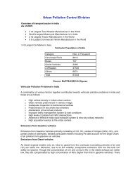
Vehicular sources of urban air pollution in India<br />
In general, combustion is the chief contributor to outdoor air pollution. In most cities, the<br />
major source of combustion is fuel use, which tends to increase along with population<br />
size and economic activity. In the last three decades, the number of motorized vehicles<br />
in India has increased 29-times, from 1.9 million in 1971 to 55.0 million in 2001 (Badami,<br />
2005; Figure 1.1). The increase was not uniform for all vehicle types: it was 7-fold for<br />
buses, 9-fold for trucks, 10-fold for car, Jeeps and taxis, but a remarkable 67-fold for twowheelers<br />
(Badami, 2005).<br />
Vehicle number (millions)<br />
60<br />
50<br />
40<br />
30<br />
20<br />
10<br />
0<br />
Figure 1.1. Motor vehicle growth in India during 1971-2001<br />
Million vehicle<br />
1971 1981 1991 2001<br />
Motor vehicles have internal combustion engine which burns a mixture of air and fuel to<br />
produce energy that propels the vehicle. The type and quantity of the pollutants released<br />
during this combustion is influenced by more than a dozen factors. The kind of fuelpetrol,<br />
diesel or compressed natural gas (CNG) is just one of them. However, fuel type<br />
is an useful indicator of potential emissions. Coal and biomass are high emitting solid<br />
fuels, petrol diesel and kerosene are mid-emitting liquid fuels and liquefied petroleum<br />
gas (LPG) and CNG are low-emitting gaseous fuels. Transport sector consumes half of<br />
the petroleum products in the world, and same is true for India. Petroleum product<br />
consumption by motorized vehicles has nearly doubled in India in the last decade.<br />
Fuel and lubricating oil quality have also contributed significantly to transport air pollution<br />
in India. Indian gasoline have a high volatility, and the vast majority of gasoline vehicles<br />
are carbureted, not fuel-injected (Table 1.1, 1.2). These facts coupled with India’s high<br />
ambient temperatures increases the potential for evaporating emissions rich in reactive<br />
hydrocarbons with the potential to generate ground-level ozone.<br />
3
Background & Objective<br />
Table 1.1: Quality of gasoline in India<br />
1993 1997 2002 Proposed<br />
Lead mg/L, max 560 150 13 5<br />
Sulfur % by mass, max 0.2 0.15 in unleaded 0.1; 0.02 in notified 0.015<br />
petrol<br />
areas<br />
Benzene % by volume, - - 1 in notified areas; 1<br />
max<br />
3 (metro’s); 5(rest of<br />
the country)<br />
Olefin % by vol, max - - - 21<br />
Aromatics % by vol, max - - - 42<br />
O 2 content, % by mass, - - - 2.7<br />
max<br />
Source: Badami (2005)<br />
Table 1.2: Quality of diesel in India<br />
Sulfur, total, % by mass,<br />
max<br />
Source: Badami (2005)<br />
1995 1999 2002 Proposed<br />
1 0.25 0.05 (in metros) 0.035<br />
Traffic congestion is yet another problem leading to high vehicular emissions. In Delhi,<br />
for example, the average speed for public transport vehicles ranged from 12 to 20 km/hr<br />
in the 1990’s (RITES, ORGO, 1994). Besides causing loss of time and productivity, traffic<br />
congestion increases fuel consumption and carbon monoxide and hydrocarbon<br />
emissions per vehicle-km by 200% or more (Faiz et al., 1992). Several studies have<br />
shown that maintenance is a significant factor in vehicular emissions.<br />
Epidemiological studies of outdoor air pollution in Western countries, in general, have not<br />
considered indoor sources and total exposures. Residents of slum households, who tend<br />
to have more health problems due to poverty, might also experience higher outdoor<br />
exposure because they live in road-side slums. In such cases, the effect of poverty on<br />
health can be confused with the effect of vehicular air pollution. Exposure to indoor air<br />
pollution or other factors associated with poverty may also increase the susceptibility of<br />
the poor to outdoor air pollution.<br />
Urban aerosols contain not only combustion products. Elevated PM 10 concentration in<br />
Delhi during summer can be attributed to windblown dust excursions from Rajasthan<br />
desert areas. These situations are dominated by fugitive dusts mostly associated with<br />
coarse particles (i.e. 2.5 μm to less than 10μm particles). Similar situation has been<br />
reported in Sun Joakin Valley in the USA (Tinkerton et al., 2000).<br />
1.2.1 Particulate pollutants: the major toxic component of urban air<br />
Particulate matter (PM) is a complex mixture of suspended solid and liquid particle in<br />
semi equilibrium with surrounding gases (Brook et al., 2003). The particle constituents<br />
vary greatly in size, composition, concentration, depending on origin and age. PM may<br />
be classified as primary (particles emitted directly by emission sources) and secondary<br />
(particles formed through the atmospheric reaction of gases). The size distributions of<br />
airborne particles are important for health impact. The particles larger than 10 µm in<br />
diameter are deposited almost exclusively in the nose and throat whereas those smaller<br />
than 1 µm reach the lower regions of the lung. The intermediate size range gets<br />
deposited between these two extremes of the respiratory tract. Outdoor (ambient) PM<br />
size ranges from approximately 0.001-100 µm in aerodynamic diameter. PM is<br />
4
Background & Objective<br />
considered as the single best indicator of potential harm. There are three main size<br />
categories for PM measured in urban air:<br />
(a) PM 10<br />
They consist of PM with a diameter upto 10 µm. However, for toxicity studies, the most<br />
important particles are those having a diameter of less than 10 µm (PM 10 ) because they<br />
are respirable whereas the larger particles are not. PM 10 deposit relatively quickly with a<br />
lifetime of less than 2 days, and exposure may lead to adverse responses in the lungs<br />
triggering an array of cardio-pulmonary problems (Brunekreef and Forsberg, 2005;<br />
Harrabi et al., 2006). PM 10 has also been associated with emergency hospital admission<br />
for asthma, bronchitis, and pneumonia in older people (Ye et al., 2001). For every 10-<br />
µg/m 3 increase of PM 10, mortality from all causes increases by 0.51% and from<br />
cardiopulmonary diseases by 0.68% (Samet et al., 2000). Moreover, the rise in daily<br />
mortality from increased concentrations of PM 10 persists for several days (Zeka et al.,<br />
2005).<br />
(b) Accumulation mode or fine particles (PM 2.5 )<br />
They consist of PM with a diameter upto 2.5 µm. Airborne particles smaller than 2.5 µm<br />
(PM 2.5 ) are usually called fine particles. These particles may penetrate deep inside the<br />
airways and are more strongly linked to adverse health effects (USEPA, 1996). Fine<br />
particles are composed mainly of carbonaceous materials (organic and elemental),<br />
inorganic compounds (sulfate, nitrate, and ammonium), and trace metal compounds<br />
(iron, aluminium, nickel, copper, zinc, and lead). There are potentially thousands of<br />
different compounds existing on fine particles that may exert harmful biological effects.<br />
On any day or location, the PM mass concentration may be similar, yet the composition<br />
may vary greatly enough to differentially impact human health (Brook et al., 2003). The<br />
relationship between PM 10 or PM 2.5 exposure and acute health effects is linear at<br />
concentrations below 100 μg/m 3 (Schwela, 2000). A modest rise in PM 10 or PM 2.5 level<br />
has been shown to be associated with small changes in cardiac function (Mar et al.,<br />
2005). Exposure to the fine particles induces oxidative stress (Furuyama, 2006).<br />
(c) Nuclei mode or ultra fine particles (UFP)<br />
The particles in this category are smaller than 0.1 µm. They are also known as ultrafine<br />
particle (UFP). UFP are present in great number in polluted urban air (Jaques and Kim,<br />
2000). They have a carbonaceous core with attached inorganic and organic materials<br />
that can cause adverse health effects (Oberdorster, 2000). The UFPs have less mass<br />
than course particle fraction but they are much greater in number and have a relatively<br />
large surface area to mass ratio, making them potential carriers of harmful gaseous<br />
compounds. Very tiny particles (UFP) escape alveolar macrophage surveillance, which is<br />
very efficient for larger particles (Hahn et al., 1977). Exposure to high doses of UFP can<br />
cause severe pulmonary inflammation and hemorrhage, high degree of alveolar and<br />
interstitial edema, disruption of epithelial and endothelial cell layers and even death<br />
(Oberdorster et al., 1992; Peters et al., 1997, Oberdorster, 2000). Biologic effects of<br />
ultrafine particles occur even at modest exposure, such as that occurring in traffic-related<br />
air pollution. UFPs cause health effects like cardiovascular problems, pulmonary<br />
disease, and development of cancer (Vinzents et al., 2005).<br />
Fate of the particles<br />
Following inhalation, the size of the particles determines where they are likely to deposit<br />
in the respiratory tract. Particles larger than 10 micrometer are mainly deposited in the<br />
nose and throat and are less likely to affect the health beyond the point of deposition.<br />
PM 2.5 and UFPs are able to penetrate into the airways all the way to the terminal alveoli.<br />
5
Background & Objective<br />
Smaller particles are present in larger numbers and have more total surface area and<br />
bioavailability, eliciting greater biological effect.<br />
1.2.2 Other pollutants<br />
(a) Sulfur dioxide (SO 2 )<br />
Sulfur dioxide (SO 2 ) is emitted in direct proportion to the amount of sulfur in fuel. Coal<br />
burning is a major source of SO 2 in air . It is an acidic gas, which combines with water<br />
vapor in the atmosphere to produce acid rain. SO 2 in ambient air can also affect human<br />
health ( Routledge et al., 2006), particularly in those suffering from asthma and chronic<br />
lung diseases and exacerbates respiratory symptoms and impaired breathing in sensitive<br />
individuals (Lipfert, 1994). It can also attach to particle surfaces and may form acidic<br />
coatings. It is considered more harmful when particulate and other pollution<br />
concentrations are high.<br />
(b) Oxides of nitrogen (NOx)<br />
Nitrogen oxides are formed during combustion processes at high temperatures from the<br />
oxidation of nitrogen in air. The major types of oxides of nitrogen are nitric oxide (NO)<br />
and nitrogen dioxide (NO 2). They are collectively known as NOx. The main source of NO<br />
is road traffic, which accounts for 49% of total NO emissions in Europe and 32% in the<br />
USA. It is emitted from both petrol- and diesel engine motor vehicles. Almost all NO x is<br />
emitted as NO, which is rapidly oxidized to more toxic NO 2 .<br />
NO x is a precursor of ozone formed in the troposphere. Oxides of nitrogen are<br />
immunotoxic and increase the susceptibility to respiratory tract infection such as<br />
influenza. Continued or frequent exposures to high concentrations of NOx in breathing<br />
air may cause irritation of the lungs and consequent acute respiratory illness (Hasselblad<br />
et al., 1992). In addition, NO x is a potent and selective vasodilator in pulmonary arterial<br />
hypertension (Perez-Penate et al., 2005).<br />
(c) Carbon monoxide (CO)<br />
Carbon monoxide (CO) is a toxic gas emitted into the atmosphere as a result of<br />
combustion processes. CO is also formed by the oxidation of hydrocarbons and other<br />
organic compounds. CO is produced almost entirely (90%) from road traffic in European<br />
cities. It remains in the atmosphere for approximately one month before being oxidized to<br />
CO 2 . The largest contributors of CO are petrol-fuelled vehicles. CO binds strongly to<br />
hemoglobin in red blood corpuscles resulting in the production of carboxyhemoglobin<br />
(COHb). This impairs the transport of oxygen within the blood and can result in adverse<br />
effect on tissues with high oxygen needs such as the cardiovascular and nervous<br />
systems. High concentration (>1000 ppm) for prolonged hours (>8 hr) can give rise to<br />
hypoxia. A recent study has shown that chronic exposures to CO may cause adverse<br />
birth outcomes such as reduced birth weight and intrauterine growth retardation (Salam<br />
et al., 2005).<br />
(d) Polycyclic aromatic hydrocarbons (PAHs)<br />
About 200 different kinds of hydrocarbons are emitted from combustion of petrol and<br />
diesel. Of these, the polycyclic aromatic hydrocarbons (PAHs) are of particular interest<br />
due to their carcinogenic potential. PAHs are usually adsorbed on the particulate<br />
pollutants. They enter the body through inhalation of these respirable particles. These<br />
compounds are semi-volatile in nature. Several PAHs like benzo(a)pyrene [B(a)P] are<br />
highly carcinogenic (Hrudkova et al., 2004). Incidence of lung cancer has been reported<br />
6
Background & Objective<br />
in persons directly exposed to B(a)P from automobile exhausts and biomass (wood,<br />
dung, agricultural wastes) fuel burning during household cooking (Cohen and Nikula,<br />
1999).<br />
(e) Volatile organic compounds (VOCs)<br />
VOCs consist of various classes of carbon-containing chemicals that are gases at room<br />
temperature. They are released into the environment from petrol and diesel, especially<br />
the former, by evaporation or as combustion products. Some VOCs (e.g. benzene) are<br />
human carcinogens while others are either respiratory tract irritants or neurotoxic (e.g.<br />
toluene, xylene).<br />
Benzene, a VOC, is a minor constituent of petrol. It is produced from combustion and<br />
evaporation of both petrol and diesel, especially the former. Combustion of petrol is the<br />
largest source (70% of total emissions) of benzene in air. Airborne benzene is primarily<br />
absorbed through the respiratory tract and then transported by blood to critical target<br />
organs. Therefore, it is possible that cumulative exposure to benzene could lead to<br />
systemic changes. True to this apprehension, benzene has been found very harmful for<br />
human health for its hematotoxic, neurotoxic, leukemogenic and carcinogenic effects<br />
(Wallace, 1984, 1989; Midzenski et al., 1992, Farris et al., 1993). Because of this, a<br />
sustained worldwide effort is on to reduce benzene exposure as far as possible.<br />
1.2.3 Air toxics of biological origin<br />
Biological agents present in polluted air may cause several diseases. The biological<br />
contaminants include bacteria, moulds, mildew, viruses, animal dander and cat saliva,<br />
house dust, mites, cockroaches, and pollen. There are many sources of these pollutants.<br />
Pollens originate from plants; people and animals transmit viruses; bacteria are carried<br />
by man, animal, soil, and plant debris; and household pets are sources of saliva and<br />
animal dander. The protein in urine from rats and mice is a potent allergen. When it<br />
dries, it can become airborne.<br />
(a) Bacteria<br />
Along with particulate pollution, numerous airborne bacteria enter the body during<br />
respiration. Several of these are pathogenic to humans. For example, Bordetella pertusis<br />
causes whooping cough, Corynaebacterium diphtheriae causes diphtheria,<br />
Mycobacterium tuberculosis causes tuberculosis and Mycobacterium pneumoniae cases<br />
bacterial pneumonia. Globally, pneumonia causes two million deaths of children (20% of<br />
all child deaths) every year and 70% of them occur in Africa and South-east Asia. The<br />
main causative organisms have identified as Streptococcus pneumoniae. It has been<br />
shown in animal studies that long-term exposures to diesel exhausts increase the risk of<br />
pulmonary tuberculosis (Hiramatsu et al., 2005). Tobacco smoke is a proven risk factor<br />
for bacterial infection. Smoking is associated with a significant increase in the relative<br />
risk of pneumonia (S. pneumoniae) and tuberculosis (Trosini-Desert et al., 2004).<br />
(b) Virus<br />
Like bacteria, viral infections have been linked to air pollution. Mumps virus (mumps),<br />
Myxovirus influenza (influenza), Poliovirus (poliomyelitis), Rhinovirus causing common<br />
cold, Rubella virus (German measles), Rubella (Measles), Varicella virus (Chicken pox)<br />
and Variola pox virus causing small pox, Haemophilus influenzae, Respiratory syncytial<br />
virus (RSV), influenza, parainfluenza and adenoviruses are some of the viruses which<br />
spread through polluted air. Measles infection increases pneumonia morbidity and<br />
mortality (Singh, 2005). Air pollution is known to enhance Human papilloma virus (HPV)-<br />
7
Background & Objective<br />
mediated cancer of the uterine cervix in women who are chronically exposed to biomass<br />
smoke (Velema et al., 2002).<br />
(c) Fungus<br />
Airborne fungi like Aspergillus fumigatus cause aspergillosis and Blastomyces dermitidis<br />
cause blastomycosis. Greater presence of fungi in indoor environment due to poorer<br />
house condition, older house age, relative lack of sun exposure and having no insulation<br />
enhances the risk of respiratory illness including oral toxicosis and airway allergy<br />
(Howden-Chapman et al., 2005). The problems may turn serious requiring hospitalization<br />
(Khalili et al., 2005).<br />
(d) Pollen and other allergens<br />
Airborne pollen and other allergens are major causative agents of bronchial<br />
hypersensitivity and asthma. Asthma exacerbation is the most common cause of hospital<br />
admission in children. Airborne bacteria and virus infections in allergic asthmatics further<br />
increase the risk of hospitalization (Murray et al., 2005). Pollen exposure is usually<br />
associated with respiratory tract allergy and eosinophil accumulation in the nasopharynx<br />
and the airways (Onbasi et al., 2005).<br />
1.3 <strong>AIR</strong> QUALITY STANDARDS<br />
National Standard<br />
National Ambient Air Quality Standards (NAAQS) of India has recommended that the<br />
annual average concentration of respirable suspended particulate matter (RSPM) with<br />
an aerodynamic diameter of less than 10 micrometer (PM 10 ) in ambient air of residential<br />
areas should be within 60µg/m 3 , while the 24-hr average should be within 120 µg/m 3<br />
(Table 1.3).<br />
8
Background & Objective<br />
Pollutant<br />
Sulfur dioxide<br />
Nitrogen<br />
dioxide<br />
Suspended<br />
particulate<br />
matter (SPM)<br />
Table 1.3: National Ambient Air Quality Standards (NAAQS) of India<br />
Time<br />
weighted<br />
average<br />
Annual<br />
24 hours<br />
Annual<br />
24 hours<br />
Annual<br />
24 hours<br />
Sensitive areas<br />
15<br />
30<br />
15<br />
30<br />
70<br />
100<br />
Concentration in ambient air (μg/ m 3 )<br />
Industrial Residential, Methods of<br />
areas rural and measurement<br />
80<br />
120<br />
80<br />
120<br />
360<br />
500<br />
other areas<br />
60<br />
80<br />
60<br />
80<br />
140<br />
200<br />
*Improved West and<br />
Gaeke method<br />
* Ultraviolet fluorescence<br />
* Jacob and hochheiser<br />
modified (Na-Arsenite)<br />
method<br />
* Gas-phase<br />
chemiluminescence<br />
High-volume sampling<br />
(average flow rate not<br />
less than 1.1m 3 /min)<br />
PM 10<br />
Annual<br />
24 hours<br />
50<br />
75<br />
120<br />
150<br />
60<br />
100<br />
Respirable particulate<br />
matter sampler<br />
Lead Annual 0.50 1.0 0.75 Atomic absorption<br />
spectrometry after<br />
sampling using EPM<br />
2000 or an equivalent<br />
filter paper<br />
Carbon<br />
monoxide<br />
24 hours<br />
8 hours<br />
1 hour<br />
0.75 (mg /m 3 )<br />
1.0 (mg /m 3 )<br />
2.0 (mg /m 3 )<br />
1.5 (mg /m 3 )<br />
5.0 (mg/m 3 )<br />
10.0 mg/m 3 )<br />
1.0 (mg /m 3 )<br />
2.0 (mg/m 3 )<br />
4.0 (mg/m 3 )<br />
Non-dispersive infrared<br />
spectroscopy<br />
Ammonia Annual<br />
400.00<br />
----<br />
24 hours<br />
100.00<br />
Source: National Ambient Air Quality Monitoring Series: NAAMQS/9/1996 – 97, <strong>Central</strong> Pollution<br />
Control Board, Government of India<br />
Compared with Indian standard, the air quality guidelines advocated by the World Health<br />
Organization (WHO, Table 1.4) and the United States Environmental Protection Agency<br />
(US EPA) are stricter. According to US EPA, the 24-hour average PM 10 concentration<br />
should be within 150 μg/ m 3 and should not be exceeded more than once per year, and<br />
the annual average concentration of PM 10 should not exceed 50 μg/ m 3 over the course<br />
of a calendar year. WHO guideline states that annual and 24-hr average PM 10 should be<br />
within 20 μg/ m 3 and 50 μg/ m 3 respectively.<br />
Table 1.4: WHO Air Quality Guideline (AQG) values<br />
Pollutant Averaging time AQG value<br />
Particulate matter<br />
PM 2.5<br />
1 year<br />
24 hour (99 th percentile)<br />
10 μg/m 3<br />
25 μg/m 3<br />
PM 10<br />
1 year<br />
24 hour (99 th percentile)<br />
20 μg/m 3<br />
50 μg/m 3<br />
Ozone, O 3 8 hour, daily maximum 100 μg/m 3<br />
Nitrogen dioxide, NO 2<br />
1 year<br />
1 hour<br />
40 μg/m 3<br />
200 μg/m 3<br />
Sulfur dioxide, SO 2<br />
24 hour<br />
10 minute<br />
20 μg/m 3<br />
500 μg/m 3<br />
Source: WHO air quality guidelines global update 2005. Report on a working meeting, Bonn,<br />
Germany, World health organization, 2005.<br />
9
Background & Objective<br />
1.4 <strong>DELHI</strong>, THE CAPITAL <strong>OF</strong> <strong>IN</strong>DIA<br />
1.4.1 General<br />
Delhi is the capital city of India. The name Delhi also refers to the National Capital<br />
Territory of Delhi (NCT), which is a special union territory jointly administered by the<br />
<strong>Central</strong> and local government. New Delhi, an urban area within the metropolis of Delhi, is<br />
the seat of the Government of India. The name Delhi has its origin either from the<br />
Persian word Dahleez (threshold, or frontier) or from the name of a Mauryan king, Raja<br />
Dhillu. Delhi was once Indraprastha, the capital city built around 5000 BC by the<br />
Pandavas of Mahabharata. Archaeological evidence suggests that Indraprastha once<br />
stood where the Old Fort is today. The earliest architectural relics of Delhi date back to<br />
the Mauryan Period (300 BC) and an inscription of the Mauryan emperor Ashoka (273-<br />
236 BC) was discovered near Srinivaspuri in 1966.<br />
The famous Iron pillar near the Qutub Minar was commissioned by the emperor Kumara<br />
Gupta I of the Gupta dynasty (320-540) and transplanted to Delhi during the 10 th century.<br />
Present-day Delhi contains the remnants of seven successive ancient cities including:<br />
Qila Rai Pithora built by Prithvi Raj Chauhan in Lal-Kot: Siri Fort built by Alauddin Khilji in<br />
1303; Tughluqabad built by Giyasuddin Tughluq (1321-25); Jahanpanah, built by<br />
Muhamad bin Tughluq (1325-51); Kotla Firoz Shah by Firoz Shah Tughluq (1351-88);<br />
Purana Qila, built by Sher Shah Suri and Dinpanah built by Humayun both near<br />
Indraprastha in 1538-45; and Shahjahanabad incluing Lal qila and Chandni Chowk built<br />
by Moghal Emperor Shah Jahan from 1638-1649.<br />
(a) Geography and meteorology<br />
Delhi is situated between the Himalayas and Aravali range in the heart of the Indian<br />
sub-continent, surrounded on three sides by the state Haryana and to the east, across<br />
the river Yamuna by the state Uttar Pradesh. Its location is between geocoordinates of<br />
76 0 50’ 24” to 77 0 20’ 37” E longitude and 28 0 24’ 17” to 28 0 53’ 00” N latitude. The major<br />
part of the territory lies on the western side of the river Yamuna. Its greatest length is<br />
around 33 miles and the greatest width is 30 miles. Delhi’s altitude ranges from 213 to<br />
305 meters above the sea level. Delhi has a semi-arid climate with high variation<br />
between summer and winter temperatures. Extreme temperature variations are<br />
characteristic of Delhi. Summers are long, from early March till October, with the rainy<br />
season in between. Hottest months are April to June when the temperature often rises to<br />
36 0 C to 47 0 C. Winter starts in November and peaks in January. Temperature falls to<br />
7.9 0 C or even lower during December-January. The average annual rainfall is<br />
approximately 670 mm (27 inches), most of which falls during the monsoons in July and<br />
August. Traditionally, the monsoons are supposed to touch Delhi by June 29 every year.<br />
Prevailing wind direction is in W-NW-N sector and E direction with 35% calm conditions.<br />
The predominant wind direction in winter season is from W-NW-N sector with 45% calm<br />
conditions (NEERI, 2003).<br />
(b) Area<br />
The national Capital Region of Delhi (NCT Delhi) has an area of 1483 sq.km. The city is<br />
divided into nine districts. Each district has three subdivisions. Below is the list of the<br />
districts and subdivisions of Delhi (Table 1.5, Fig. 1.2):<br />
10
Background & Objective<br />
Table 1.5: Districts of Delhi<br />
Districts<br />
Subdivisions<br />
1.<strong>Central</strong> Delhi Darya Ganj, Pahar Ganj, Karol Bagh<br />
2. North Delhi Sadar Bazar, Kotwali, Sabzi Mandi<br />
3. South Delhi Kalkaji, Defence Colony, Hauz Khas<br />
4. East Delhi Gandhi Nagar, Preet Vihar, Vivek Vihar, Vasundhara Enclave<br />
5. North East Delhi Seelampur, Shahdara,Seema Puri<br />
6. South West Delhi Vasant Vihar, Najafgarh,Delhi Cantonment<br />
7. New Delhi Connaught Place, Parliament Street, Chanakya Puri<br />
8. North West Delhi Saraswati Vihar, Narela, Model Town<br />
9. West Delhi Patel Nagar, Rajouri Garden, Punjabi Bagh<br />
Figure 1.2: District map of Delhi<br />
(c) Population, gender, literacy<br />
Delhi is one of the fastest-growing metropolis in Asia. Delhi’s population was 4.05 lakhs<br />
(0.4 million) at the beginning of 20 th century. According to 2001 census, Delhi’s<br />
population was 1,37,82,976 (approximately 13.8 million), making it the third largest<br />
city in India after Mumbai and Kolkata. District-wise distribution of population reveals<br />
that most thickly populated region is North-Western Delhi. It has 2.85 million inhabitants<br />
representing 20.66% of Delhi’s total population. Incidentally it is the largest district in<br />
Delhi with a total area of 444 sq.km, i.e. 29.66% of Delhi’s total area. The next populated<br />
regions are South (16.38%), West (15.38%), North-East (12.8%), South-West (12.69%),<br />
East (10.51%) and North (5.66%). <strong>Central</strong> district having only 25 sq.km area had 4.67%<br />
of total population, while New Delhi represents<br />
only 1.25% of Delhi’s total population (Fig. 1.3). According to 2001 Census, the sex ratio<br />
(number of females per 1000 males) of Delhi was 821. It was highest in North-East (851)<br />
and lowest in South-West district (783). Overall literacy rate in Delhi at the time of 2001<br />
11
Background & Objective<br />
Census was 81.82%, the 5 th highest position in the country. Highest literacy rate in Delhi<br />
was recorded in East district (85.1%), while North-East district had the lowest literacy<br />
rate (77.85%). Literacy rate among males was highest in South-West district (89.93%),<br />
and <strong>Central</strong> district had the lowest literacy (82.5%). In case of females, East district had<br />
the highest literacy (79.38%), and North-East district had the lowest female literacy<br />
(69.97%).<br />
12
Background & Objective<br />
(d) Administration<br />
The sixty-ninth amendment to the Constitution of India in 1991 granted Delhi the status<br />
of a special union territory and officially changed its name to the National Capital<br />
Territory of Delhi (NCT). Delhi has its own Legislative Assembly, Lieutenant Governor,<br />
Council of Ministers and Chief Minister. However, Delhi is jointly administered by the<br />
Union Government of India and the Territorial Government of Delhi. New Delhi, an urban<br />
area in Delhi, is the seat of both the State Government of Delhi and the Government of<br />
India. Each district is headed by a Deputy Commissioner and has three subdivisions.<br />
Each subdivision is headed by the Subdivision Magistrate.<br />
(e) Economy<br />
Delhi’s gross domestic product for 2004 was estimated at US $26 billion. Historically,<br />
Delhi has been the economic capital of northern India. In recent years, Delhi’s service<br />
sector has expanded exponentially. Key service industries include information<br />
technology (IT), telecommunications, banking, media and life sciences. Delhi and its<br />
suburbs account for over 30% of India’s IT and IT-enabled services (ITeS) exports - the<br />
second largest in the country after Bangalore’s 35% share. Delhi’s manufacturing<br />
industry has also grown considerably as many consumer goods industries have<br />
established manufacturing units and headquarters in and around Delhi.<br />
Delhi is one of India’s most affluent urban centers and is at the heart of India’s largest<br />
consumer belt. As an indicator, Delhi has more cars on its roads than India’s other<br />
metros Kolkata, Chennai and Mumbai combined. Delhi is one of India’s largest markets<br />
because per capita income is much higher than in other Indian cities. Gurgaon and<br />
Noida, two of Delhi’s largest satellite cities, have attracted more than $5 billion worth<br />
investments in the past three years. The city’s booming economy is also the main reason<br />
behind so many people migrated to Delhi in recent years in search of better living<br />
conditions and employment.<br />
1.4.2 Sources of air pollution in Delhi<br />
Vehicular emissions, industrial emissions, household activities and soil resuspension are<br />
the major source of air pollution in Delhi (Balachandran et al., 2000).<br />
(a) Vehicular pollution: motor vehicles in Delhi<br />
As in case of many other Asian cities, motor vehicles are responsible for a substantial<br />
part of Delhi’s air pollution. Delhi alone with only a little over 1% of India’s population<br />
account for about 8% of the national motor vehicles (Badami, 2005). The motor vehicle<br />
fleet presently stands at 4.2 million, of these 2.7 million are two wheelers (Badami,<br />
2005). Currently vehicular pollution contributes to 72% of the total air pollution load in<br />
Delhi (Goyal et al., 2006), which was only 23% in 1970-71 (Table 1.6). It has been<br />
estimated that vehicular source is responsible for generating more than 3000 metric tons<br />
of pollutants per day (MT /day) in Delhi.<br />
Table 1.6: Relative contribution to air pollution in Delhi<br />
Source<br />
Year<br />
1970 - 71 1980 - 81 1990 - 91 2000 – 2001<br />
Vehicle 23% 42% 63% 72%<br />
Industry 56% 40% 29% 20%<br />
Domestic 21% 18% 8% 8%<br />
Source: PHD Chamber of Commerce and Industry Report, November 1991<br />
13
Background & Objective<br />
Since the time of economic liberalization policy introduced by the Government of<br />
India, more and more households have graduated to higher income categories.<br />
Coupled with changing attitudes towards taking loans, people no longer found it<br />
difficult to own their personal car. Sales of passenger cars jumped more than<br />
three times from 209,203 units in 1994 to 638,815 units in 2000 - all in a matter of<br />
seven years. The National Capital Region of Delhi has the highest number of<br />
vehicles in the country - more than Mumbai, Calcutta and Chennai put together.<br />
There has been a steady increase in the number of vehicles in Delhi and their<br />
contribution to city’s air pollution in the last three decades (Figure 1.4). Till 1990, the rise<br />
in the number of motor vehicles in Delhi was proportional with the population growth.<br />
Thereafter, the growth of vehicles has been spectacular, surpassing population increase<br />
by several folds (Figure 1.5). The population of the city has increased from 3.53 million in<br />
1970 to 13.80 million in 2001, registering a rise of 3.9-fold over a period of 30 years. On<br />
the other hand, the number of registered motor vehicles in the city in 2001 was 34.2 lakh<br />
against 2 lakh during 1970-71. The growth in the vehicular population of the city during<br />
this period, however, has been 17-fold that far surpassed population growth. In 1975, the<br />
number of vehicles in Delhi and Mumbai was almost the same. Today Delhi has 3 times<br />
more vehicles than Mumbai, although Mumbai has 4 million more inhabitants than Delhi.<br />
In 1995 Delhi had 27 lakh vehicles compared with 7.2 lakh in Mumbai, 5.6 lakh in<br />
Kolkata and 8.1 lakh in Chennai. In essence, within a span of just 30 years (1971-2001)<br />
the city has 3.9-times more residents and 17-times more vehicles. It has been projected<br />
that the gap between the growth in human and vehicular population in Delhi will further<br />
widen in future.<br />
80<br />
60<br />
40<br />
Figure 1.4: Growth in the number of vehicles (in lakhs)<br />
and the relative contribution (%) of road traffic to Delhi’s<br />
air pollution in the past 30 years (1971-2001)<br />
Vehicle<br />
number<br />
Contribution<br />
20<br />
0<br />
1970-<br />
71<br />
1975 1980-<br />
81<br />
1985 1990-<br />
91<br />
1996 1998 2000-<br />
2001<br />
2004<br />
14
Background & Objective<br />
Figure 1.5: Growth in population and number of vehicles in<br />
Delhi over a period of 30 years (1970-2001)<br />
35<br />
30<br />
25<br />
20<br />
15<br />
10<br />
5<br />
0<br />
Vehicle (lakh)<br />
Population (million)<br />
1970 1975 1980 1985 1990 1996 2000 2001<br />
(i) Road transportation in Delhi<br />
Delhi’s road transport includes private vehicles such as 2-wheelers, cars, Jeeps etc.;<br />
public transport vehicles, such as bus, taxi, and autorickshaws; and goods transport<br />
vehicles such as trucks and tempos.<br />
Public vehicles<br />
Bus<br />
Delhi’s buses constitute only 1.2% of the total number of vehicles, but they cater to 60%<br />
of the total traffic load. Although personal vehicles such as cars and two wheelers<br />
represent nearly 94% of the total number of vehicles of the city (Figure 1.6, 1.7), they<br />
cater to only 30% of the travel demand (Dept. of Transport, Govt. of Delhi). Delhi<br />
Transport Corporation (DTC) operates the world’s largest fleet of compressed natural<br />
gas (CNG)-fueled buses. Besides, there are a large number of private-owned CNGfueled<br />
buses plying in Delhi. Delhi’s buses pollute much less than diesel-fueled buses of<br />
most other cities in India.<br />
Figure 1.6: Percentage share of vehicles plying in Delhi<br />
0.36<br />
2<br />
1<br />
3<br />
Private<br />
Auto Rickshaw<br />
Taxi<br />
Bus<br />
Goods vehicle<br />
94<br />
15
Background & Objective<br />
Taxi and Auto rickshaw<br />
Compared to Mumbai and Kolkata, taxis are fewer in Delhi as people rely more on their<br />
own cars. However, Delhi’s auto rickshaws provide a very important contribution in mass<br />
transport. Because they are CNG-powered, emissions are much less compared to pre-<br />
CNG era.<br />
Goods transport vehicles<br />
Lorries and trucks plying on Delhi roads including those coming with goods from<br />
adjoining states constitute 3% of city’s vehicular population. They are a significant<br />
source of air pollution, because the vehicles are run by diesel-powered engines.<br />
Private vehicles<br />
Private vehicles, such as cars, scooters and motor bikes, constitute 94% of Delhi’s<br />
vehicular population. The numbers of 2-wheelers and cars have increased exponentially<br />
in recent times in Delhi, although the number of public transport vehicles has increased<br />
only marginally (Figure 1.7). About two-third of the total vehicular population of Delhi is<br />
two wheelers. The total number of two wheelers in Delhi in 1993 was 14 lakh, which was<br />
1.5 times more than the combined number of two wheelers in three other metros of India<br />
– Mumbai 2.4 lakh, Kolkata 2.2 lakh and Chennai 4.6 lakh.<br />
lakhs<br />
18<br />
16<br />
14<br />
12<br />
10<br />
8<br />
6<br />
4<br />
2<br />
0<br />
Figure 1.7: Changing pattern in the number of different types of<br />
vehicles in Delhi over past 15 years (1985-2000)<br />
1985 1990 1995 2000<br />
Year<br />
Car, Jeep<br />
Two wheeler<br />
Three<br />
wheeler<br />
Taxi<br />
Bus<br />
Metro Railways<br />
A metro (mass rapid-transit system) built and managed by the Delhi Metro Rail<br />
Corporation is also operating. Three lines are currently operating on elevated as well as<br />
underground track totaling a length of 56 km. Line 1 runs east-west for 22 km on an<br />
elevated track between Rithala and Shahdara. Line 2 runs north-south for 11km in an<br />
underground tunnel between Vishwa Vidyalaya and the <strong>Central</strong> Secretariat. Line 3,<br />
opened on December 31, 2005, runs 22.8 km long on elevated tracks and an<br />
underground tunnel, between Indraprastha, Barakhamba Road and Dwarka.<br />
(ii) Automotive fuel<br />
Diesel and petrol (gasoline) were the principal automotive fuels till the introduction of<br />
compressed natural gas (CNG) for public transport vehicles in 2001. During 2001-02, the<br />
annual consumption of diesel in India was 38,500 tons against 6,640 tons of petrol<br />
(Figure 1.8). There has been a steady increase in vehicular pollution and automotive fuel<br />
16
Background & Objective<br />
consumption, especially diesel, in Delhi in the last three decades (Table 1.6, Figure 1.8).<br />
Diesel is considered more polluting fuel than petrol, because it emits more particulates,<br />
especially of fine and ultrafine categories, than petrol during combustion. Petrol, on the<br />
other hand, releases more volatile organic compounds, particularly benzene, during<br />
combustion and also due to evaporation.<br />
Thousand Tons/year<br />
40<br />
30<br />
20<br />
10<br />
Figure 1.8: Estimated use of automotive fuel in India<br />
Petrol<br />
Diesel<br />
0<br />
1987-88 1990-93 1995-96 2001-2002<br />
Introduction of CNG<br />
CNG vehicle program was launched in Delhi in 2001. In July 2001, Delhi had 34.2 lakh<br />
vehicles out of which CNG was used in 2450 buses, 1178 minibuses, 27,363 three<br />
wheelers and 1993 taxis (CPCB). During 2000-2001, over 1.5 million vehicles run on<br />
natural gas worldwide. Altogether, 14 countries had 10,000 or more natural gas-fueled<br />
vehicles (IANGV, 2001)<br />
Advantages of CNG<br />
Compared with diesel and petrol, CNG is a cleaner fuel as it emits substantially lower<br />
amount of particulates carbon monoxide and oxides of nitrogen (Table 1.7). Moreover,<br />
these vehicles have low vibrations and fewer odors than diesel engines.<br />
Table 1.7: Emission comparison of diesel and CNG-powered buses<br />
Diesel CNG % reduction<br />
CO 2.4 g/km 0.4 g/km 83<br />
NOx 21 g/km 8.9 g/km 58<br />
PM 380 mg/km 12 mg/km 97<br />
Source: Frailey et al., 2000<br />
(b) Industrial source of air pollution in Delhi<br />
The share of industries as source of air pollution is rapidly declining in Delhi for the past<br />
thirty years (Table 1.6). For example, during 1970-71, industrial pollution was the biggest<br />
contributor (56%) to city’s air pollution load. In contrast, only 20% of Delhi’s air pollution<br />
is now generated by the industries (Figure 1.9), mainly the three coal-based thermal<br />
power plants at Indraprastha, Badarpur and Rajghat.<br />
17
Background & Objective<br />
Sources of air pollution<br />
(%)<br />
75<br />
60<br />
45<br />
30<br />
15<br />
0<br />
Figure 1.9: Changing pattern in the relative contribution (%) of different<br />
sources of air pollution in Delhi over a period of 30 years (1971-2001)<br />
Vehicle<br />
Domesti<br />
c<br />
1970-71 1980-81 1990-91 2000-2001<br />
Year<br />
(c) Domestic source<br />
Like the industries, the contribution of household sector to city’s air pollution is steadily<br />
declining. Now a days only 8% of Delhi’s air pollution is contributed by household<br />
sources, compared with 21% in 1970-71 and 18% in 1980-81. Pollution<br />
from household sources is mainly due to the use of coal, kerosene and unprocessed<br />
solid biomass like firewood, cow dung and agricultural refuse like hay, husk, dried leaves<br />
etc.<br />
1.5 ECONOMIC ASPECT <strong>OF</strong> <strong>AIR</strong> <strong>POLLUTION</strong> <strong>IN</strong> <strong>IN</strong>DIA<br />
Improvement of air quality is associated with reduction in the number of premature<br />
deaths, episodes of acute illness such as asthma attacks and the number of chronic<br />
respiratory illness cases. The economists evaluate the value of avoiding an illness<br />
episode as:<br />
1. the value of work time lost due to the illness by the patient or the caregiver, or<br />
both<br />
2. the medical cost of treatment<br />
3. the amount paid to avoid the pain and suffering associated with the illness, and<br />
4. the value of leisure time lost due to the illness by the patient or caregiver.<br />
1.6 <strong>HEALTH</strong> <strong>EFFECTS</strong> <strong>OF</strong> <strong>AIR</strong> <strong>POLLUTION</strong>: AN UPDATE<br />
Harmful effects of air pollution on human health are recognized for centuries. It has been<br />
estimated that globally 8,000 people die every day from diseases related to air pollution<br />
exposure. Each year 60,000 deaths in the United States and 500,000 deaths in China<br />
occur due to air pollution. Several epidemiological studies have established a direct<br />
relationship between the pollutants and health hazards ranging from morbidity (illness) to<br />
mortality (death from illness).<br />
(a) Excess mortality<br />
The London fog incident in 1952 conclusively established an association between air<br />
pollution and increased mortality (Logan, 1952). Since then, several epidemiological<br />
studies in the USA and Europe have established a clear relationship between air<br />
18
Background & Objective<br />
pollution exposure and excess mortality (Samet et al., 1981; Dockery et al., 1982;<br />
Wichmann et al., 1989; Archer, 1990; Ostro et al., 1991; Ponka, 1991; Pope et al., 1992;<br />
Bobak and Leon., 1992; Schwartz, 1994; Lipfert, 1994; Thurston, 1996; Dockery, 1999).<br />
Air pollution is associated with increased risk of acute respiratory infections, the principal<br />
cause of infant and child mortality in the developing countries (Bendahmane, 1997).<br />
Each 10μg/m 3 increase in annual average PM 2.5 level may lead to 4%, 6% and 8% rise<br />
in the risk of all-cause, cardio-pulmonary, and lung cancer mortally respectively (Pope et<br />
al., 2002). An increase in PM 10 by 10 μg/m 3 has been reported to cause 0.76% excess<br />
deaths from cardiovascular causes and 0.58% excess mortality from respiratory<br />
diseases (Analitis et al., 2006).<br />
(b) Particulates and health impact<br />
Mortality and morbidity associated with air pollution are primarily due to the toxic effects<br />
of particulates (Schwartz, 1991, 1992, 1993; Morgan et al., 1998; Hong et al., 1999;<br />
Peters et al., 2000; Arena et al., 2006). Associations have also been reported with<br />
gaseous air pollutants viz. ozone (Anderson et al., 1996), nitrogen dioxide (Anderson et<br />
al., 1996), sulfur dioxide (Gouvea and Fletcher, 2000) and carbon monoxide (Gouvea<br />
and Fletcher, 2000). Compared with particulates, however, the relationship between<br />
gaseous pollutants and mortality is less consistent.<br />
(c) Direct relationship between death from heart and lung diseases and air pollution level<br />
Dockery and his co-workers (1993) showed an association between mortality rates and<br />
PM 10 levels not only from lung cancer but also from cardio-pulmonary diseases. They<br />
estimated 3.4% excess deaths from respiratory diseases and 1.4% from cardiovascular<br />
diseases for every 10 μg/m 3 increase of PM 10 . The overall increase in mortality was<br />
calculated as 1% for every 10 μg/m 3 rise in PM 10 (Dockery et al., 1999; Viegi et al., 1999,<br />
2000). Samet et al., (2000) has reviewed the subject and concluded that for every 10<br />
μg/m 3 rise of PM 10 , there was an increase in mortality from all causes by 0.51% and from<br />
cardiopulmonary diseases by 0.68%. This finding is similar to that of Katsouyanni et al.,<br />
(1997) who had suggested 0.4% increase in mortality with 10 μg/m 3 rise in PM 10 level.<br />
Further, Levy et al., (2000) showed a 0.7% increase in mortality per 10 μg/m 3 increase in<br />
PM 10 concentrations. In the same year Schwartz (2000), in a study in 10 cities in the<br />
USA, reported a 0.67% increase in mortality associated with every 10 μg/m 3 increase in<br />
PM 10 concentration.<br />
(d) Most deaths are from heart diseases<br />
The association between air pollution exposure and adverse health outcome has been<br />
summarized by Dockery (2001). He noted that although the relative risk for effects of<br />
particles are greater for respiratory than for cardiovascular deaths, the actual numbers of<br />
death are greater for cardiovascular than for respiratory causes.<br />
(e) Increased morbidity<br />
Besides mortality, air pollution could initiate and/or aggravate several diseases. Excess<br />
morbidity is often reflected in absenteeism from school and work, restricted activity spent<br />
at home, more attendance to outpatient medical services; emergency visits to clinics and<br />
hospitalization (Shy et al., 1978). Air pollution-related pulmonary diseases for which<br />
hospital admissions are usually required are acute bronchitis, pneumonia, emphysema,<br />
bronchiectasis, chronic airway obstruction and attacks of asthma. Besides lung diseases,<br />
air pollution is significantly associated with cardiac and vascular problems (von Klot,<br />
2005; Maheswaran et al., 2005; Mills, 2005).<br />
19
Background & Objective<br />
1.6.1 Air pollution and respiratory system<br />
Primary target of the pollutants: the lung and the airways<br />
Since airborne pollutant generally enters the body through inhalation, lung and the<br />
airways are the primary target organs. The airways of the lung are represented by the<br />
trachea (windpipe), and beyond it are bronchi (with cartilage cover) and bronchioles<br />
(without cartilage). The bronchioles lead to air spaces called alveoli which have an<br />
average diameter of 200 micrometer each (Figure 1.10). A recent study has<br />
demonstrated that there are approximately 480 million alveoli in both lobes of an adult<br />
human lung, and men have more alveoli and larger lung volume than women (Ochs et<br />
al., 2003). The mean size of a single alveolus is 4.2x10 6 µm 3 . Alveoli make up<br />
approximately 64% of the lung space (Ochs et al., 2004). Human lungs have a total<br />
surface area of 1,400 m 2 , and everyday we inhale approximately 15 m 3 of air (i.e.15,000<br />
litres). The weight of this inahled air is greater than the food we consume and the water<br />
we drink in a day. The lung volume and breathing frequencies of healthy adults at rest<br />
are 400-500 ml and 15-17 breaths per minute respectively (Tobin et al., 1983). Recent<br />
study has documented that a constant number of respiratory units is maintained from<br />
childhood to adulthood while both the smallest bronchioles and alveoli expand in size to<br />
produce the increased lung volume with increased age and height (Zeman and Bennett,<br />
2006).<br />
Figure 1.10: Anatomy of the lungs<br />
Respiratory illness following air pollution exposure<br />
Way back in 1981, Samet and co-workers reported statistically significant increase in<br />
respiratory emergency room visits in Ohio, USA in association with elevated levels of<br />
particulate pollution and SO 2 . In 1983, Lutz reported strong positive correlation between<br />
weekly particulate pollution levels and the percentage of patients with respiratory illness.<br />
Thurston and co-workers (1994) observed similar associations in Toronto and in New<br />
20
Background & Objective<br />
York. In 1989, a series of studies in Utah Valley, USA showed that particulate pollution<br />
was associated with a wide range of respiratory disorders and hospitalization (Pope,<br />
1989, 1991), lung function decrement and respiratory symptoms (Pope and Dockery,<br />
1992; Pope et al., 1991) and school absenteeism for children (Ransom and Pope, 1992).<br />
Dockery et al., (1989); Ostro (1990), Thurston et al., (1994), Abbey et al., (1995) and<br />
others have also shown association between air pollution and respiratory morbidity.<br />
Particulate air pollution was reported to be associated with emergency hospital visits for<br />
asthma in Seattle, USA (Schwartz et al., 1993), and emergency visits for chronic<br />
obstructive pulmonary disease (COPD) in Barcelona, Spain (Sunyer et al., 1993) and in<br />
Brazil (Martins et al., 2002).<br />
Schwartz (1999) reported that daily variations of PM 10 and CO were linked to daily<br />
hospital admissions among the elderly (more than 65 years of age). A study conducted<br />
amongst school children of China showed positive associations between morbidity<br />
prevalence and outdoor levels of particulate matter of all size fractions, but the<br />
association was strong for PM 10 and PM 2.5 (Zhang et al., 2002). The results also<br />
present some evidence that ambient levels of NOx and SO 2 are positively associated<br />
with respiratory symptoms in children, but the evidence for these two gaseous pollutants<br />
was weak than that for PM 10 and PM 2.5 (Zhang et al., 2002). Burnett et al., 2001)<br />
showed an association between ozone and hospitalization for acute respiratory diseases<br />
in children less than 2 years of age.<br />
(a) Air pollution and rise in the prevalence of respiratory symptoms<br />
Symptoms are a form of signals that act as an indicator of any underlying illness or<br />
disease. Epidemiological studies on respiratory and mental health are generally based<br />
on collection of data on the prevalence of respiratory and neurobehavioral symptoms to<br />
get an estimate of the disease (Bobak and Leon, 1992; Abbey et al., 1995; Dockery et<br />
al., 1996).<br />
Type of symptoms<br />
Respiratory symptoms are usually classified into two broad groups: a) Upper respiratory<br />
symptoms (URS) which include runny and stuffy nose, sinusitis, sore throat, wet cough,<br />
dry cough, cold head, fever, burning or red eyes, and b) Lower respiratory symptoms<br />
(LRS) which include wheezing, phlegm, shortness of breath, chest discomfort or pain.<br />
Most of the respiratory diseases underlying these symptoms are caused by bacterial,<br />
fungal or viral infections, or structural or functional damage to the respiratory system.<br />
Very often the symptoms of a multifactor respiratory disease like asthma or chronic<br />
obstructive pulmonary disease (COPD) are aggravated following exposure to air<br />
pollutant.<br />
Allergic rhinitis<br />
It is an inflammation of the membrane lining the nose and throat due to allergic reaction.<br />
The presence of rhinitis is indicated by itchy sensation in the nose, frequent sneezing,<br />
watery eyes, and running nose. The symptoms are commonly found following exposure<br />
to high level of particulate air pollution (Wongsurakiat et al., 1999) and benzene (Zuskin<br />
et al., 1997). Diesel exhaust particle have been identified as the major contributing factor<br />
to allergy (Sydbom et al., 2001; Riedl et al., 2005).<br />
Sore throat<br />
If the function of muco-ciliary escalator is impaired by high pollution exposure, the<br />
mucous is not cleared from the airways. The retained mucous traps inhaled pathogens<br />
resulting in damage to the airways, thickening and scarring of epithelium leading to<br />
21
Background & Objective<br />
fibrosis. This is associated by sore throat, an indicator of pharyngitis and tonsillitis, and<br />
respiratory or right heart failure.<br />
Chronic cough, bronchitis, sinusitis<br />
Cough is an important symptom frequently reported following exposures to air pollution.<br />
It is a reflex response to irritation from mucous or any foreign particle in the upper<br />
respiratory tract. When cough is accompanied by production of sputum it is termed<br />
productive or wet cough, whereas cough without sputum is known as dry cough. Cough<br />
may be caused by inflammation of the upper airways as a result of viral infections like<br />
common cold and influenza. Severe cough may indicate damage to the lungs caused by<br />
inflammation associated with pneumonia and chronic obstructive pulmonary disease<br />
(COPD). COPD is often indicated by morning cough producing sputum, frequent chest<br />
infection, specially during winter producing yellow or green sputum, wheezing particularly<br />
after cough, and shortness of breath even after mild exertion. COPD represents two<br />
separate lung conditions, chronic bronchitis and emphysema. Inflamed, congested and<br />
narrowed airway is associated with chronic bronchitis causing obstruction to airflow. In<br />
emphysema the alveoli are damaged or fused making them less efficient for transfer of<br />
oxygen. In acute bronchitis, cough is irritating, persistent and produces clear sputum.<br />
The associated symptoms are chest pain or tightness of the chest and wheezing.<br />
Morning cough and sputum production is associated with emphysema. High prevalence<br />
of chronic cough, phlegm and sinusitis has been reported among garage workers and<br />
taxi drivers exposed to vehicular exhaust in United Arab Emirates (Bener et al., 1998).<br />
Similarly, high prevalence of cough, sinusitis, bronchitis and asthma has been found in<br />
association with traffic-related air pollution in Kolkata (Basu et al., 2001).<br />
Chest tightness, dyspnea<br />
These symptoms along with rhinitis and nose and throat irritation along with lung function<br />
abnormality have been observed in workers from a shoe manufacturing plant in Croatia<br />
where benzene level was higher than allowable limit (Zuskin et al., 1997). High<br />
prevalence of cough with phlegm and rhinitis were recorded among traffic policemen in<br />
Bangkok (Wongsurakiat et al., 1999). Similarly high vehicle traffic resulted in asthma,<br />
cough and wheeze in children who were additionally exposed to environmental tobacco<br />
smoke in Germany (Nicolai et al., 2003).<br />
Bronchial asthma<br />
It results from intermittent narrowing of the airways and consequent shortness of breath.<br />
Its early symptoms are wheezing, tightening of chest, shortness of breath, and difficulty<br />
in exhaling and dry persistent cough. Although asthma is genetically controlled, exposure<br />
to air pollution exacerbate asthma attacks (Cakmak et al., 2004). A strong association<br />
between severe asthma symptoms and breath concentration of benzene has been<br />
demonstrated in children (Delefino et al., 2002). Asthma and bronchoconstriction have<br />
been linked to cumulative exposures to exhaust from diesel-fuelled engines (Riedl et al.,<br />
2005) and occupational exposures to VOCs (Cakmak et al., 2004).<br />
(b) Increased risk of lung function impairment<br />
Breathing and gas exchange is achieved by the lungs, chest wall, diaphragm, central<br />
nervous system and the pulmonary circulation acting in concert. Pollutants from<br />
vehicular exhaust could reach deep inside the lungs and result in the destruction of the<br />
alveoli or the proximal airways resulting in the malfunctioning of any of the components<br />
thereby hampering the balance. The measurement of mechanics of breathing is<br />
22
Background & Objective<br />
collectively known as Lung Function. An instrument called spirometer measures it. A<br />
spirometer is integrated with a microprocessor that measures flow rates (Mead et al.,<br />
1979).<br />
Types of functional lung impairment<br />
The two major patterns of abnormal lung function are restrictive and obstructive pattern.<br />
a) In restrictive lung function, there is a decrease in lung volume (FVC, Forced vital<br />
capacity). The subject inhales reduced volume of air due to reduction in total lung<br />
capacity. Restrictive lung may be caused by infection and inflammation such as in case<br />
of pneumonia or tuberculosis. These infections often leave scar on the lung tissue<br />
leading to the loss of elasticity of the lung. Obesity and neuromuscular problems also<br />
could lead to restrictive lung. b) Obstructive lung function is indicated by a decrease in<br />
the FEV 1 (Forced expiratory volume in one second)/FVC ratio. Decline in FEV 1 /FVC<br />
usually results from obstruction in large airways while fall in FEF 25-75% (Forced expiratory<br />
flow between 25-75% of FVC, mid expiratory force) signifies small airways obstruction<br />
(Dassen et al., 1986; Vedal et al., 1987). Obstructive lung function is common in<br />
diseases like asthma and COPD (Figure 1.11).<br />
Volume<br />
Total Lung<br />
capacity<br />
Functional residual<br />
capacity<br />
Residual volume<br />
Normal<br />
Obstructive<br />
Restrictive<br />
Figure 1.11: Normal lung volumes and changes that occur in<br />
Obstructive and restrictive types of lung function impairments<br />
(Adapted from Davidson, 1995)<br />
Reduction in lung function following air pollution exposure<br />
Pulmonary function can be affected by several factors including genetic predisposition,<br />
weather, season, time of the day, the basic respiratory health of the subject, smoking<br />
status, allergens and air pollution (Schoenberg et al., 1978). Lung function is a very<br />
important indicator of the adverse effects of air pollution on the lungs since impaired lung<br />
function has been identified as an independent risk factor for morbidity and mortality from<br />
heart diseases (Lipfert, 1988).<br />
In the 1980s, a large number of studies have shown a decline in forced vital capacity and<br />
forced expiratory volume with increasing concentration of air pollution (Johnson et al.,<br />
23
Background & Objective<br />
1982; Lebowitz et al., 1985; Asero et al., 2005). Burnekreef (1991) found a strong<br />
association between lung function decrement and particle concentration. Decline in FEV 1<br />
has been reported in school children following a particulate and SO 2 pollution episode in<br />
January 1985 in Netherlands (Dassen et al., 1986; Hoek and Brunekreef, 1994). Diesel<br />
exhaust exposure resulted in airway obstruction in railroad workers (Kilburn, 2000).<br />
Studies by Frye et al., (2003) have related decrement in lung function following urban air<br />
pollution episode. A study in Delhi has shown the adverse effect of air pollution on<br />
spirometric lung function, which was aggravated by smoking habit. The study also<br />
reported greater airway obstruction in bidi smokers (Chhabra et al., 2001). Decreased<br />
lung function in response to air pollution has also been reported by investigators from<br />
Germany (Ibald-Mulli et al., 2002) and Iran (Golshan et al., 2002). Workers from a shoe<br />
manufacturing plant in Croatia revealed several respiratory symptoms along with lung<br />
function abnormality where benzene level was found higher than permissible limit<br />
(Zuskin et al., 1997). Another study reported inverse relationship between peak<br />
expiratory flow rate and ambient VOC including benzene in children (Delefino et al.,<br />
2002). The additive effect of cigarette smoking and air pollution on reduction of FEV 1<br />
was shown in a survey of 7,685 individuals in Canada (Gan et al., 2005)<br />
(c) Cellular lung response to air pollution: Changes in pulmonary cytology<br />
In the course of development of diseases in the lung the cells of the airways usually<br />
undergo a number of changes that are accurately reflected in the sputum. Thus, sputum<br />
cytology is immensely helpful to study the initiation and progression of the underlying<br />
diseases in the airways and inner lung (Lahiri et al., 2000). Sputum contains mucus,<br />
inhaled substances including various microorganisms, epithelial cells, alveolar<br />
macrophages and immune cells like neutrophil, eosinophil and lymphocytes. Microscopic<br />
examination of these exfoliated cells and immunocytes provides useful information<br />
regarding the pathophysiological changes in the lung tissues.<br />
(i) Cell types in a normal lung<br />
More than 40 cell types have been identified in the normal lung and airway. The major<br />
cell types are: basal cell, Intermediate cell, ciliated and non-ciliated columnar epithelial<br />
cells, goblet cell, Type I and Type II pneumocytes and alveolar macrophages.<br />
Pneumocytes<br />
A rich capillary network surrounded by endothelial cells cover the alveolar space. The<br />
alveolar space is lined by a basement membrane on which lie the epithelial cells<br />
(pneumocytes).<br />
Type I pneumocytes<br />
These cells cover about 95% of alveolar surface and because of their extensive surface<br />
area they are thought to be at high risk of chemical and particulate attack despite being<br />
covered by a protective epithelial lining fluid and pulmonary surfactant. The cells permit<br />
efficient gaseous exchange by the red blood cells present in the alveolar capillary and<br />
contain enzymes, which are important in mediating toxicant biotransformation (Dinsdale,<br />
1998).<br />
Type II pneumocytes<br />
They cover only 3-5% area of alveolar surface and are located at the corners of the<br />
alveoli. These are secretory cells and synthesize pulmonary surfactant and also the<br />
components of the underlying basement membrane (Crouch et al., 1987). Type II cells<br />
24
Background & Objective<br />
also contain a spectrum of P-450 isoenzymes (Miller et al., 1986) and a high<br />
concentration of reduced glutathione (Horton et al., 1987). All these are important for the<br />
breakdown and biotransformation of inhaled or circulating chemicals.<br />
Surfactant<br />
Pulmonary surfactant or surface-active substance is a lipoprotein mixture rich in<br />
phospholipids. It has a ‘detergent’ property of lowering surface tension in the fluid layer<br />
that line the alveoli once air enters the lungs. Its ability to form a monomolecular layer at<br />
the interface between air and the alveolar lining fluid allows some air to be retained<br />
within the alveolus at all times. Surfactant is produced continually from Type-II epithelial<br />
cells because it has a half-life of only 14-24 hours. Deficiency of surfactant is associated<br />
with respiratory distress syndrome.<br />
Interstitium<br />
It represents the space between the alveoli. It is composed of fibroblasts and connective<br />
tissue components such as proteoglycans, collagen and elastin, which are responsible<br />
for maintaining the skeletal backbone of the innermost region of the lung.<br />
(ii) Cells normally found in sputum<br />
Squamous epithelial cells<br />
Squamous epithelial cells originate from normal lining of the mouth or pharynx. A mixture<br />
of intermediate and squamous epithelial cells is often found in sputum. The intermediate<br />
cells are characterized by round to oval nucleus and cyanophilic cytoplasm. Superficial<br />
cells have pyknotic nuclei and orangeophilic cytoplasm. Enucleated squamous and<br />
nucleated parabasal cells are occasionally present.<br />
Ciliated columnar cells<br />
These cells are shed singly, or in loose clusters or sheets from the lining of the<br />
respiratory tract, mainly as a result of traumatic exfoliation or inflammation. Following<br />
extensive damage to the respiratory epithelium these cells may be found in large<br />
numbers in sputum.<br />
Goblet cells<br />
Goblet cells are non-cilated epithelial cells. Together with the submucosal glands, they<br />
produce and secrete high molecular weight mucus glycoproteins, called mucins. Mucin<br />
helps entrapment of inhaled pollutants including particles and microorganisms, and their<br />
disposal by mucociliary clearance. These cells also produce lipids and small<br />
glycoproteins. Mucins are tightly packed in the intracellular granules of the goblet cells.<br />
Goblet cells discharge mucus in response to wide spectrum of stimuli- irritant gases,<br />
inflammatory mediators, reactive oxygen species, nerve activation, and changes in the<br />
biophysical environment (Rogers, 1994). Mucins are discharged rapidly (within<br />
milliseconds) and vast quantities of mucus are released.<br />
The ability of goblet cells to give rise to ciliated cells, to rapidly produce vast quantities of<br />
mucus in response to acute airway insult, and to change in number according to<br />
variations in chronic insult indicates that these cells are vitally important front-line<br />
defenders of the airways (Rogers, 1994).<br />
25
Background & Objective<br />
Alveolar macrophage (AM), the first line of cellular defense in lung<br />
Pulmonary macrophages include i) alveolar macrophages (AMs), ii) airway macrophages<br />
and iii) interstitial macrophages. These cells are present in sputum in varying numbers<br />
and their presence indicates the adequacy of sputum sample. AMs are the dominant<br />
phagocytic cells, which act as first line of cellular defense in the lungs. Their mobility,<br />
secretory behavior and bactericidal properties are essential for the maintenance of clean<br />
and sterile alveoli. Macrophages by their immense secretory potential may cause tissue<br />
injury while performing their defensive role.<br />
Heterogeneity<br />
Macrophages have an average diameter of 10-45 μm (mean diameter 20μm). Cline in<br />
1975 grouped it into three types: Type A – with a mean diameter of 20-25 µ, accounting<br />
for 94-98% of mononuclear cells, Type B – with a mean diameter of 30 µ constitutes 5%<br />
and Type C – with a mean diameter of 40 µ accounting for less than 10% of the<br />
mononuclear cells. The nucleus is oval or irregular and usually eccentric in position with<br />
nucleus: cytoplasmic ratio 1:3. Cytoplasm shows numerous granules and ingested<br />
particles.<br />
Number and origin of AM<br />
In adult human lung there are approximately 480 million alveoli and each alveolus is<br />
defended by about 73 macrophages. Therefore, it has been estimated that human lung<br />
contains 35.0 billion macrophages for its defense against inhaled pollutants.<br />
Macrophages originate from peripheral blood monocytes. Monocytes like other cells<br />
present in the blood originate from the bone marrow. The precursor stem cells in the<br />
bone marrow give rise to monoblasts that differentiate into promonocytes and then to<br />
monocytes. The monocytes circulate in blood for 36-104 hours and then leave the blood<br />
vessels, migrate through the endothelial barrier to interstitial space and then across the<br />
epithelium to the tissues where maturation and differentiation occur in response to<br />
environmental stimuli and they are called macrophage or histiocytes. The transformation<br />
is regulated by hematopoietic growth factors like colony stimulating factor (CSF) and<br />
interleukins (ILs) [Frampton, 2001; Zareba et al., 2001; Goldberg et al., 2001; Dockery,<br />
2001; Schwartz, 2001; Donaldson et al., 2001; von Klot et al., 2005, Ballester et al.,<br />
2006].<br />
The AM pool in lung is maintained by division of pre-existing interstitial macrophages or<br />
by influx of monocytes from blood to the lung (Lewis, 1995). Following exposure to<br />
inhaled pollutants, AM number in lung is increased either by cell division, or recruitment<br />
of monocytes from circulation or by enhancing the life span of existing cells by<br />
suppressing apoptosis (Foster, 1999). They have a normal life span ranging from months<br />
to years and are destroyed in the lymphatics.<br />
Function of AM<br />
AMs represent the first line of cellular defense in the lung. They play a key role in particle<br />
clearance from the inner airways by phagocytosis, generation of oxygen radicals, and<br />
endocytosis of insoluble particles and dust (Becker, 1995). They possess potent<br />
antimicrobial activities via local release of degradative enzymes and reactive oxygen<br />
metabolites. AM is perhaps the most secretory cell in the body. They actively participate<br />
in inflammation, wound healing and tissue repair through their vast array of secretory<br />
products (Laskin and Pendino, 1995). They release inflammatory cytokines like tumor<br />
necrosis factor-alpha (TNF-α), interleukin-1 (IL-1), chemotactic factors like leukotrine B-4<br />
(LTB 4 ), platelet-activating factor (PAF) and chemotactic cytokine such as interleukin-8<br />
(IL-8) that strengthens the pulmonary defense (Strieter et al., 1990). Macrophages also<br />
26
Background & Objective<br />
act as effector cells for killing of tumor cells in the lung (Shellito and Kaltreider, 1985),<br />
and known to regulate cellular and humoral immunity by processing and presenting<br />
antigen, interacting with helper/inducer T-lymphocytes (Thepen et al., 1994; Lewis,<br />
1995). They also perform various metabolic and homeostatic functions including lipid and<br />
iron metabolism (Madden et al., 1991).<br />
Enzymatic marker of AM<br />
The enzymatic marker is non-specific esterase (NSE) found in cytoplasmic granules of<br />
the cell. The enzyme comprises of three carboxylic ester hydrolases, namely<br />
carboxylesterase, arylesterase and acetylesterase. These three cytoplasmic enzymes<br />
are widely distributed in many tissues. The application of NSE method is helpful in<br />
defining cells of monocytic origin and thus detects histiocytes and macrophages. The<br />
type of NSE detected in AM is likely to be arylesterase as it acts on aromatic esters like<br />
α-naphthyl acetate.<br />
Inflammatory cells<br />
Neutrophils, eosinophils and lymphocytes are present in varying proportion depending<br />
upon the inflammatory or allergic reaction in the lung. The magnitude of inflammation<br />
can be assessed from the extent of rise in the number of these cells particularly the<br />
neutrophils.<br />
(iii) Change in airway cells following air pollution exposure<br />
Both chronic and acute exposures to air pollution have been shown to directly affect the<br />
structural integrity of the respiratory system. Continued chemical exposure can cause<br />
necrosis and subsequent sloughing off of ciliated epithelial cells. Microscopic<br />
examination of these exfoliated cells in spontaneously expectorated sputum provides<br />
important information regarding the pathophysiological changes in the lung tissue and<br />
development of lung disease including malignancy (Roby et al., 1990). Saccomanno and<br />
his co-workers (1970) demonstrated cytological changes in the respiratory epithelium<br />
following air pollution exposure. Air pollution exposure was associated with presence of<br />
abnormal columnar epithelial cells and squamous metaplasia in sputum of young adults<br />
and children (Plamenac et al., 1973, 1978). Exposure to diesel exhaust showed a<br />
significant increase in neutrophils, mast cells, CD4+ and CD8+ T-lymphocytes, along<br />
with upregulation of endothelial adhesion molecule, ICAM-1 and VCAM-1 in airway<br />
lavage (Salvi et al., 1999; Takizawa et al., 2000). Changes in airway epithelial cells were<br />
reported in sputum samples of smokers (Kulawik et al., 2003). Higher prevalence of<br />
sputum eosinophilia and neutrophilia along with marked rise in the prevalence of<br />
respiratory symptoms have been reported both in adults and children of Kolkata, and the<br />
changes have been attributed to the city’s high air pollution level (Lahiri et al., 2000a,<br />
2000b). Controlled exposure of healthy human volunteers to diesel exhaust particles<br />
(DEP) for 1 hour produced marked cellular inflammatory response in the airways<br />
involving neutrophils, mast cells and lymphocytes (Salvi et al., 1999). DEP exposure was<br />
accompanied by increased expression of endothelial adhesion molecules and their<br />
complementary ligands on inflammatory cells. It has been demonstrated that DEP elicits<br />
remarkable increase in neutrophils in the proximal airways, while the lymphocyte and<br />
histamine responses were found in the distal airways (Salvi et al., 1999).<br />
AM response to air pollution exposure<br />
A pioneering study by Mylius and Gullvag (1986) demonstrated several fold increase in<br />
the number of AM in sputum of persons exposed to industrial air pollution. The AM count<br />
was found to rise with increasing level of particulate pollution in the workplace. They<br />
advocated the number of AM as a reflection of the reaction of lung to air pollution.<br />
27
Background & Objective<br />
Following this study sputum AM has been used as a biomarker of pollution exposure and<br />
effect as it involves non-invasive procedure, the cells can be analyzed through relatively<br />
simple microscopic techniques, the cytological alterations in sputum accurately reflect<br />
the changes within the lung and the whole procedure is cost–effective. Hence the<br />
procedure seems well suited for large population based studies especially in the<br />
developing world. A comparative study by the current researchers among the residents<br />
of highly polluted city of Kolkata and the inhabitants of Sunderban islands in Bay of<br />
Bengal where air pollution level was significantly lower revealed several-fold rise in AM<br />
number in sputum of urban subjects (Lahiri et al., 2000a). Similar changes were<br />
observed in children exposed to high level of urban air pollution of Kolkata (Lahiri et al.,<br />
2000b). In another study, traffic- related air pollution was shown to be associated with<br />
6.5- fold rise in AM number in traffic policemen and street hawkers of Kolkata compared<br />
with relatively less exposed office workers (Basu et al., 2001). Thus, AM number in<br />
sputum appears to be a sensitive biomarker of cumulative exposure to air pollution. The<br />
number of alveolar macrophages with phagocytosed PM 10 has been correlated with<br />
atherosclerotic lesions in PM 10 exposed rabbits (Suwa et al., 2002).<br />
Functional impairment of AM following ultrafine particle exposures<br />
Ingestion of ultrafine particles by the AM may lead to impairment of phagocytic activity of<br />
these cells, particularly after infection that induce an increased production of Interferon<br />
gamma (Lundborg et al., 2001). Cytoskeletons of the macrophages are important for<br />
migration, phagocytosis of foreign materials and intra cellular transport and digestion.<br />
Ultrafine particles in urban dust and diesel exhaust have been shown to cause<br />
cytoskeletal toxicity leading to impaired function of the macrophages, compromising lung<br />
defects (Moller et al., 2002).<br />
Siderophages in sputum: indicator of pulmonary hemorrhage<br />
AM process hemoglobin from phagocytosed erythrocytes to form degradation products<br />
like iron (Perez-Arellano et al., 1992). These iron-containing macrophages are known as<br />
siderophages. Their presence in sputum in high numbers is indicative of either past<br />
intrathoracic bleeding or extravasations of red blood cells into the alveoli due to a<br />
sluggish blood flow (Grubb, 1994). Therefore, the presence of siderophages is of<br />
considerable value in detecting covert pulmonary hemorrhage. An earlier study by the<br />
present research team has demonstrated the presence of large number of siderophages<br />
in sputum of subjects chronically exposed to Kolkata’ air pollution (Roy et al., 2001). It<br />
was concluded that abundance of siderophages in sputum and associated microscopic<br />
hemorrhage in the inner airways is an indicator of adverse lung reaction to air pollution<br />
(Roy et al., 2001). Hemosiderin-laden AM were observed in sputa of traffic policemen in<br />
Italy (Giovagnoli, 1999) and automobile service station workers and hawkers in Kolkata<br />
(Roy et al., 2001); they were also found in steel workers in cases of pulmonary<br />
hemorrhage (Corhay, 1992). Becroft and co-workers (1997) reported abundance of intraalveolar<br />
siderophages as a cause of sudden death in infants. Animal studies have<br />
shown accumulation of iron in the rat lung following exposure to DEP. Deposition of iron<br />
causes oxidative stress, inflammation, and neutrophilic lung injury (Ghio et al., 2000).<br />
Elastase: a tissue-degrading enzyme<br />
Elastin is a fibrous protein that is widely distributed in the elastic tissues of lung.<br />
Elastase, a proteolytic enzyme found in the lysosomes of neutrophils and AM is capable<br />
of destroying elastin, collagen and fibronectin. Elastase is also involved in wound healing<br />
and disposal of damaged cells and debris. Uptake of non-degradable irritant materials<br />
from the atmosphere has been shown to enhance elastase production and secretion by<br />
the AM (Werb and Gordon, 1975). Release of excess elastase from neutrophils and<br />
28
Background & Objective<br />
activated AM promotes the development of emphysema as it leads to degradation of<br />
alveolar wall (Laskin and Pendido, 1995). Neutrophil and macrophage elastase plays a<br />
major role in emphysema in smokers (Churg et al., 2002; Shapiro et al., 2003; Valenca<br />
et al., 2004).<br />
1.6.2 Systemic effects of air pollution<br />
(a) Cardiovascular changes<br />
(i) Air pollution, increased plasma viscosity, hypertension<br />
There are reports that indicate air pollution may affect blood pressure. Indeed, high blood<br />
pressure (hypertension) is common among persons cumulatively exposed to high level<br />
of air pollution (Pope et al., 1999a). Hypertension is defined as systolic blood pressure at<br />
least 140 mm Hg, diastolic blood pressure at least 90 mm Hg, or both (Chalmers et al.,<br />
1999). Elevated plasma viscosity, increased heart rate (>80 beats/min), reduced heart<br />
rate variability, and increased risk of arterial hypertension have been reported in<br />
association with chronic air pollution exposure (Pope et al., 1999; Gold et al., 2000;<br />
Ibald-Mulli et al., 2001; Brook et al., 2003). Increase in heart rate in response to air<br />
pollution has been shown to be most marked in individuals who have high blood<br />
viscosity. Hypertension is one of the predictors of cardiovascular mortality with relative<br />
risk of up to 2.0 or more (Blazer et al., 2001).<br />
Air pollution exposure and cardiovascular diseases (CVD) are intimately related, and it is<br />
a growing concern worldwide (Zareba et al., 2001; Dockery, 2001; Goldberg et al., 2001;<br />
Donaldson et al., 2001). CVD associated with air pollution are angina, cardiac<br />
insufficiency, hypertension and myocardial infarction (MI) i.e. heart attack. Studies<br />
conducted from the late nineties have consistently shown that PM 10 is associated with<br />
overall hospital admissions for CVD (Schwartz, 1999; Burnett et al., 1999; Moolgavkar,<br />
2000; Linn et al., 2000). Air pollution has been associated with sudden death in patients<br />
with stable angina (Benchimol et al., 2000) and myocardial infarction (Lind et al., 2001,<br />
Peters et al., 2001, 2004). In combined analyses across six eastern US cities, each 10<br />
μg/m 3 increase in PM 2.5 was found to be associated with 2.1% increase in total mortality<br />
from ischaemic heart diseases (Schwartz et al., 1996). Among air pollution-related<br />
deaths in the US in 1997, only 8.5% were from respiratory diseases (COPD, pneumonia,<br />
influenza etc.) while cardiovascular deaths (heart, cerebrovascular and arterial diseases)<br />
accounted for 39.5% of all deaths (Greenle et al., 2000). Thus, CVD, rather than<br />
respiratory ailments, are the most important cause of death from air pollution exposure.<br />
A study in Delhi with high level of air pollution has reported higher risk of CVD and<br />
deaths at a younger age (Cropper et al., 1997).<br />
(ii) Underlying mechanism of cardiovascular effects of air pollution<br />
The mechanism by which air pollution affects the cardiovascular system is largely<br />
unknown. Seaton and co-workers (1995) first proposed that pollutant exposure induces a<br />
transient increase in blood coagulability, and this was subsequently corroborated by the<br />
study of Schwartz (2002) who demonstrated an association between PM 10 and all three<br />
markers of cardiovascular risk: higher level of fibrinogen in blood plasma, greater<br />
number of platelets in circulation, and elevated WBC count. Several other workers have<br />
linked air pollution, especially the PM 10 level, with markers of adverse cardiovascular<br />
events like increase in peripheral white cell counts (Salvi et al., 1999), elevation of<br />
interleukin-6 level (Ernst and Resch, 1993; Ridker et al., 2000), upregulation of C-<br />
reactive protein (CRP; Seaton et al., 1999; Peters et al., 2001), rise in plasma viscosity<br />
due to increase in fibrinogen level (Ghio et al., 2000; Peters et al., 2001; Schwartz,<br />
29
Background & Objective<br />
2001), and alteration in cardiovascular autonomic control (Ibald-Mulli et al., 2001).<br />
Raised fibrinogen and neutrophil levels is common in patients with coronary heart<br />
disease (Danesh et al., 1998). CRP, an index of inflammation, is an acute phase protein<br />
produced in the liver in response to injury and infection. A rise in CRP level increases the<br />
risk of coronary artery disease (Ridker and Haughie., 1998; Mendall et al., 1996). A<br />
survey in England has shown 1.5-fold increase in the incidence of coronary artery<br />
disease for each doubling of CRP level (Mendall et al., 1996).<br />
Inhaled particles elicit pulmonary inflammation, penetrate the blood stream, interact with<br />
platelets and trigger systemic increase in coagulability and procoagulant alteration in<br />
blood rheology that can promote the genesis of atherosclerosis leading to cardiac<br />
arrhythmias, arterial dysfunction, and acute MI (Seaton et al., 1995, Ruckerl et al., 2006).<br />
The important role of ultrafine particles (UFP), especially those containing transition<br />
metals, in mediating cardiovascular changes associated with urban air pollution has<br />
been supported by the work of Donaldson and co-workers (2001). They have shown that<br />
oxidative stress generated by UFP and subsequent oxidation of low-density lipoprotein<br />
(LDL) cholesterol destabilizes atheromatous plaque, which leads to an ischemic event.<br />
Alternatively, particles may induce activation of nuclear factor kappa beta (NF-kB)<br />
transcription factor facilitating generation and release of proinflammatory cytokines that<br />
stimulates the liver to generate more coagulation factors giving rise to an ischemic event<br />
(Donaldson et al., 2001). Besides PM 10 , exposure to SO 2 was found to be significantly<br />
associated with rise in WBC, NO 2 with raised platelet count and fibrinogen, while ozone<br />
had no effect on any of the outcomes (Schwartz, 2002).<br />
The proposed mechanism of air pollution- related cardiovascular events (Frampton,<br />
2001; Donaldson et al., 2001) is as follows.<br />
• Air pollution exposure causes deposition of particulates in airways<br />
• The particulates mediate generation of reactive oxygen species (ROS) by airway<br />
macrophages<br />
• ROS inflicts injury to airway and alveolar epithelial cells<br />
• Nuclear factor kappa beta (NFkB) is activated and proinflammatory cytokines are<br />
elaborated causing upregulation of adhesion molecules in vascular endothelium and<br />
circulating leucocytes<br />
• Immigration of neutrophil, eosinophils, lymphocytes and monocytes from blood to<br />
airways follows<br />
• Production of C-reactive protein (CRP), and fibrinogen in liver is increased, eliciting<br />
activation of platelets and coagulation cascade and oxidation of low-density<br />
lipoprotein (LDL) cholesterol<br />
• Generation of foam cells, plaque formation in arteries, atherosclerosis<br />
• Rupture of plaques leading to angina and myocardial infarction<br />
(iii) Susceptible groups<br />
People at highest risk tend to be those with pre-existing cardiovascular disease and<br />
elderly. In addition, persons with diabetes are also more susceptible to CVD mediated by<br />
airborne pollutants (Singh et al., 2000; Zanobetti and Schwartz, 2001).<br />
(b) Hematotoxicity of air pollution<br />
Air pollution exposure is associated with a multitude of hematological alterations.<br />
(i) Neutrophilia<br />
30
Background & Objective<br />
PM 10 exposure in laboratory animals increases circulating neutrophils<br />
(Polymorphonuclear cells, PMN), band cells, and an increase in the size of bone marrow<br />
mitotic pool of PMNs (Suwa et al., 2002). Repeated exposures to PM 10 in rabbits<br />
stimulates the bone marrow to increase the production of PMN in the marrow and<br />
accelerates the release the more mature PMN into the circulation, the magnitude of this<br />
changes was related to the amount of particles phagocytosed by AM (Mukae et al.,<br />
2001). Exposure to DEP for only 1 hour produced a marked increase in the neutrophils in<br />
the peripheral blood. Concomitantly a 4-fold increase in their numbers in the bronchial<br />
epithelium and a 3- fold increase in the submucosa was recorded (Salvi et al., 1999).<br />
This suggests that DEP is a potent stimulus for release of neutrophils from the bone<br />
marrow and the transit from blood to the airway tissues. Acute exposure to ambient<br />
particles accelerates the transit of PMN from marrow to the circulation, whereas chronic<br />
exposure expands the size of the bone marrow pool of PMN (van Eden and Hogg,<br />
2002). In vivo and in vitro studies have demonstrated that alveolar macrophages<br />
produce the mediators implicated in the bone marrow response to PM 10 exposures. The<br />
transit of neutrophils and other inflammatory cells from the blood into tissue occurs in a<br />
highly regulated manner involving sequential upregulation of several adhesion molecules<br />
on the endothelial cells and their respective ligands in the leucocytes, along with release<br />
of chemoattractant from the epithelial and inflammatory cells. Following acute exposure<br />
to DEP significant decrease in endothelial ICAM-1 and VCAM-1 expression was<br />
observed (Salvi et al., 1999). A combination of several pollutants present in diesel<br />
exhaust may be more potent in producing a biological response than each pollutant in<br />
isolation. Moreover, chronic low dose exposure to air pollution with intermittent acute<br />
high-dose exposures may elicit a response that is different from acute response seen in<br />
several experimental and epidemiological studies.<br />
(ii) Altered platelet count<br />
Exposure to DEP has been associated with markedly elevated platelet number in<br />
peripheral blood, i.e. thrombocytosis (Salvi et al., 1999). Thrombocytosis has been<br />
described as an acute phase reaction and has been linked to a range of inflammatory<br />
lung disorders (Sutor, 1995). Peters and colleagues (2000) have demonstrated elevated<br />
platelet count and increased plasma viscosity during rise in particulate pollution in<br />
ambient air. After rupture of an arterial sclerotic plaque in a coronary artery, platelets<br />
play a crucial role in the subsequent thrombus formation, leading to MI. Thrombocytosis,<br />
especially in elderly people compromised with cardiovascular function, may increase<br />
their risk of developing strokes and coronary vessel thrombosis, thereby increasing<br />
cardiovascular mortality and morbidity (Peters et al., 2000). Road repair workers<br />
exposed to asphalt fumes have higher platelet volume but lower platelet count (Chase et<br />
al., 1994). An increased mean platelet volume is an indicator of larger and more reactive<br />
platelets, and persons with these changes are at higher risk of myocardial infarction<br />
(Endler et al., 2002).<br />
(iii) Hematotoxicity of benzene<br />
Volatile organic compounds (benzene, toluene and xylene) are haematotoxic, and<br />
exposures to these pollutants are associated with higher prevalence of hematological<br />
abnormalities like alterations in WBC, RBC and platelet count in children (Lee et al.,<br />
2002). Rapidly proliferating stem cells in the bone marrow are most vulnerable to<br />
benzene toxicity (Marcus, 1990). Benzene cause bone marrow suppression, decreased<br />
erythrocyte, hemoglobin and hematocrit levels leading to anemia, suppression of WBC<br />
counts (leukopenia), and reduction in platelet number (thrombocytopenia). Suppression<br />
of all three elements (RBC, WBC and platelets) is called pancytopenia. Benzene<br />
exposure often leads to pancytopenia. Pancytopenia accompanied by necrosis of the<br />
bone marrow, is diagnostic of aplastic anemia caused by benzene. Bone marrow<br />
31
Background & Objective<br />
dysplasia including dyserythropoiesis, eosinophilic dysplasia and abnormal cytoplasmic<br />
granulation of neutrophilic precursors have been reported following occupational<br />
benzene exposure (Aksoy, 1991).<br />
Other hematological changes observed in humans exposed chronically to benzene<br />
include decreased leukocyte osmotic resistance, decreased phagocytic function of<br />
neutrophils, reduced glycogen content and decreased activity of peroxidase of<br />
neutrophils, and increased delta aminolevulinic acid activity in erythrocytes (Aksoy,<br />
1991). A report by Farris et al., (1996) showed significant reduction in the number of<br />
reticulocyte in blood, B-lymphocytes in the marrow and spleen and increased frequency<br />
of micronucleated reticulocyte in bone marrow following benzene exposure. Workers<br />
occupationally exposed to high level of benzene had higher incidence of anemia (Pyszel<br />
et al., 2005) and they had disturbances in the heme metabolism in lymphocytes and zinc<br />
level in plasma (Muzyka et al., 2002). Severe bone marrow dysplasia is frequently<br />
accompanied by clonal T cell expansion and alterations in T lymphocyte subsets which<br />
suggest that autoimmune-mediated bone marrow injury is an early or predisposing event<br />
in the pathogenesis of benzene-induced persistent hematopoietic disease (Irons et al.,<br />
2005).<br />
(c) Immunotoxicity of air pollution<br />
(i) Cells of the immune system<br />
Lymphocyte<br />
Lymphocytes are the key cells of the immune system responsible for acquired immunity<br />
and immunologic attributes of diversity, specificity, memory and self/non-self recognition.<br />
They are usually small, round or partially ovoid, slightly motile and non-phagocytic and<br />
non-granular leukocytes (except for a group of natural killer cells). There are<br />
approximately 10 10 lymphocytes in the human body. The size ranges from 6-15 µm.<br />
Lymphocytes can be broadly divided into T- (thymus-dependent), B- (Bursa of Fabricius<br />
in birds and bone marrow-dependent in mammals), and NK (Natural Killer) cells. These<br />
cells have specific surface marker glycoproteins (Cluster Determinants or CDs) by which<br />
they can be identified. The T-cells comprise of T-helper cell (CD4+) and T-cytotoxic cell<br />
(CD8+).<br />
Table 1.8: Lymphocyte subtypes<br />
CD marker Name Characteristic<br />
CD4+ T-helper 1 (Th-1) Production of interferon<br />
CD4+ T-helper 2 (Th-2) Production of Interleukin<br />
CD8+ T-cytotoxic (Tc) Cytotoxicity, production of perforin<br />
CD19+/ CD20+ B-cell Generation of immunoglobulin<br />
CD16+ CD56+ Natural killer (NK) cell Antibody-independent killing of virus and tumor<br />
cells<br />
CD4+ cells help in recognizing the antigen presented by antigen presenting cell (APC)<br />
like macrophages. It releases cytokines to activate the B-lymphocytes (CD19+ and<br />
CD20+ cells) to produce a clone of B-cells, mature to plasma cell and produce the<br />
specific type of antibody against the antigen. CD8+ T- cell mediate cellular cytotoxicity<br />
crucial for host defense against viral agents. NK cells (CD16+CD56+) are involved in<br />
natural killing of tumor cells and viruses (Table 1.8). A general increase in the number of<br />
lymphocytes is known as lymphocytosis whereas a decrease is lymphocytopenia.<br />
32
Background & Objective<br />
It has been shown that measurement of CD4+T lymphocytes provide a useful biological<br />
marker of past exposure to aromatic amines (Araki et al., 1993). Benzene, styrene and<br />
PAHs activate peripheral lymphocyte and caused changes in T-cells in workers exposed<br />
to aromatic hydrocarbons (Biro et al., 2002). The human immunodeficiency virus (HIV)<br />
destroys T cells (specifically, CD4+ lymphocytes). Without this key defense, the body is<br />
susceptible to opportunistic diseases. An increase in lymphocytes is usually a sign of a<br />
viral infection. In some rare cases, leukemias are found through an abnormally raised<br />
lymphocyte count in an otherwise normal person.<br />
Monocyte / macrophage<br />
A monocyte is the largest of all leukocytes measuring 12 to 20 μm in diameter. On a<br />
Wright's stained peripheral blood smear they show a bluish-gray cytoplasm with a large<br />
cytoplasm-to-nucleus ratio. The nucleus is ovoid or kidney shaped either centrally or<br />
eccentrically located. During hematopoiesis in the bone marrow, the granulocytemonocyte<br />
progenitor cells differentiate into promonocytes, enter the blood stream where<br />
they further differentiate into mature monocytes. Monocytes circulate in the blood stream<br />
for about 8 hours during which they enlarge migrate into tissues and differentiate into<br />
specific tissue macrophages at different anatomical locations. They serve different<br />
functions in different tissues and are named according to their tissue location: alveolar<br />
macrophages in the lung, histiocytes in connective tissue, Kupffer cells in the liver,<br />
mesangial cells in the kidney, microglial cells in the brain and osteoclasts in bone.<br />
Monocytes are responsible for phagocytosis (ingestion) of foreign substances in the<br />
body. Monocytes can perform phagocytosis using intermediary (opsonising) proteins<br />
such as antibodies or complement that coat the pathogen, as well as by binding to the<br />
microbe directly via pathogen-recognition receptors. Monocytes are also capable of<br />
killing infected host cells via antibody, termed antibody-mediated cellular cytotoxicity.<br />
Vacuolization may be present in a cell that has recently phagocytosed foreign matter.<br />
Monocytes, which migrate from the blood stream to other tissues, are called<br />
macrophages. Macrophages are responsible for protecting tissues from foreign<br />
substances but are also the predominant cells involved in atherosclerosis.<br />
Polymorphonuclear neutrophil (PMN)<br />
Polymorphonuclear neutrophils (PMN) are granulocytes with a diameter of 12-15 µm in<br />
peripheral blood smears. PMNs in turn account for 55-65% of all leukocytes in humans.<br />
PMN are active phagocytes, capable of only one phagocytic event, expending all of their<br />
glucose reserves in an extremely vigorous respiratory burst. The respiratory burst<br />
involves the activation of an NADPH oxidase enzyme, which produces large quantities of<br />
superoxide, a reactive oxygen species. Being highly motile, neutrophils quickly<br />
congregate at a focus of infection, attracted by cytokines expressed by activated<br />
endothelium, mast cells and macrophages.<br />
Low PMN counts are termed neutropenia. This can be congenital or due to acquired<br />
factors. It can also be a side effect of medication, including chemotherapy. Functional<br />
disorders are often hereditary. They are disorders of phagocytosis or deficiencies in the<br />
respiratory burst as in chronic granulomatous disease, a rare immune deficiency. In<br />
alpha 1-antitrypsin deficiency, the important neutrophil enzyme elastase is not<br />
adequately inhibited by alpha 1-antitrypsin, leading to excessive tissue damage in the<br />
presence of inflammation - most prominently pulmonary emphysema. Cytokines<br />
produced in the lung due to deposition of ambient particles also appear in the circulation<br />
and act on the bone marrow. PMN recently released from the bone marrow following the<br />
action of the cytokines are preferentially sequestered in pulmonary capillaries, are less<br />
33
Background & Objective<br />
chemotactic, contain more damaging granular enzymes, leading to damage of the<br />
alveolar tissue (van Eden and Hogg, 2002).<br />
Eosinophil<br />
Eosinophils are granulocytes responsible for combating allergen exposure and parasitic<br />
infestations in the body. Eosinophils make up about 2-4% of the all white blood cells, and<br />
are about 10-12 µm in size. The nucleus is bilobed with a narrow connection in between.<br />
The cytoplasm contains coarse granules, rounded in shape, staining reddish thereby<br />
indicating an acidophilic nature of the granules. These granules contain histamine and<br />
proteins such as eosinophil peroxidase, RNase, DNases, lipase, plasminogen, and major<br />
basic protein that are toxic to both parasites and the host's tissue.Eosinophils play a role<br />
in fighting viral infections, which is evident from the abundance of RNAses they contain<br />
within their granules. Eosinophils also play a role in the allergic response, and in fibrin<br />
removal in inflammation. Eosinophils are considered the main effector cells in asthma<br />
pathogenesis. An increase in eosinophils, i.e. the presence of more than 500<br />
eosinophils/µl of blood is called an eosinophilia, and is typically seen in people with a<br />
parasitic infection of the intestines, a collagen vascular disease (such as rheumatoid<br />
arthritis), malignant diseases such as Hodgkin's Disease, extensive skin diseases (such<br />
as exfoliative dermatitis), Addison's Disease and with the use of certain drugs such as<br />
penicillin. Eosinopenia is a decrease in eosinophil number, which occurs<br />
characteristically when glucocorticoids are administered.<br />
Basophil/mast cell<br />
Basophils are the least common of the granulocytes, representing about 1% of<br />
circulating leucocyte. They contain large cytoplasmic rounded granules, which stain deep<br />
blue or violet indicating its basophilic nature. The granules obscure the nucleus under<br />
the microscope. However, when unstained, the nucleus is visible and it usually has 2<br />
lobes. A cell in tissues, the mast cell, has many similar characteristics. For example, both<br />
cell types store histamine, a chemical that is secreted by the cells when stimulated in<br />
certain ways (histamine causes some of the symptoms of an allergic reaction). Like all<br />
circulating granulocytes, basophils can be recruited out of the blood into a tissue when<br />
needed.<br />
Basophils tend to appear in specific kinds of inflammatory reactions, particularly those<br />
that cause allergic symptoms. While the exact purpose of basophils has never been<br />
proven, they appear often in tissues where parasites are found. They can be found in<br />
unusually high numbers at sites of exoparasite infection, e.g., ticks. They also appear in<br />
tissues where allergic reactions are occurring and probably contribute to the severity of<br />
these reactions. Basophils have protein receptors on their cell surface that bind IgE<br />
antibody very tightly. It is the bound IgE antibody that confers a selective response of<br />
these cells to environmental substances, for example, pollen proteins.<br />
When activated, basophils secrete histamine, several proteoglycans, lipid mediators like<br />
leukotrienes, and several cytokines. Histamine and proteoglycans are pre-stored in the<br />
cell's granules while the other secreted substances are newly generated. Each of these<br />
substances contributes to inflammation. Recent evidence suggests that basophils are an<br />
important source of the cytokine, interleukin-4. Interleukin-4 is considered one of the<br />
critical cytokines in the development of allergies and the production of IgE antibody by<br />
the immune system. There are other substances that can activate basophils to secrete<br />
which suggests that these cells have other roles in inflammation.<br />
(ii) Immune response to air pollution<br />
34
Background & Objective<br />
Small number of T-lymphocyte resides in the bronchial tissue of normal subjects: CD4+<br />
cells predominate in the submucosa and CD8+ cells in the epithelial (Salvi et al., 1999).<br />
Following DEP exposure among human volunteers, T-lymphocytes, mostly CD4+ cells,<br />
infiltrate the submucosa and bronchial epithelium (Salvi et al., 1999). The number of B-<br />
lymphocytes in the bronchial tissue did not change, but their numbers increased in the<br />
bronchoalveolar lavage fluid with a corresponding decrease in the blood, suggesting<br />
trafficking of circulating B-cells to the bronchial lumen following DEP exposure (Salvi et<br />
al., 1999).<br />
In recent decades, increased prevalence of allergic conditions has been observed in<br />
developed countries. Although lifestyle, exposure to infection, and diet are important<br />
confounders, a strong link between industrialization and allergy has been established<br />
(Fernvik et al., 2002). The underlying mechanisms of hypersensitivity involved pollutantmediated<br />
stimulation of interleukin-5 production, immunoglobulin-E synthesis, eosinophil<br />
recruitment and bronchial hyperactivity (Fernvick et al., 2002).<br />
Chronic exposure to vehicular pollution is associated with airway inflammation, and<br />
diesel exhaust particles play an important role in this response (Salvi et al., 1999). The<br />
inflammatory response by DEP was not due to NO 2 , as NO 2 alone did not show any<br />
cellular inflammatory response in the airways (Blomberg et al., 1996). Instead, the<br />
particulate component of diesel exhaust is responsible for the action (Salvi et al., 1999).<br />
In vitro studies have shown that UFP elicit a greater inflammatory response in the<br />
alveolar space when compared with larger-sized particles, probably due to an interaction<br />
between a larger surface area of small particles within AM and interstitial cells (Albelda,<br />
1994). PM 10 collected by environmental sampling generate hydroxyl radicals in aqueous<br />
solution by an iron-dependent process (Gilmour, 1996). These free radicals cause<br />
activation of redox-sensitive transcription factors such as NF kB and AP-1 (Meyer et al.,<br />
1994) NFkB is important in transcription of many cytokines and chemokine genes,<br />
including inducible nitric oxide synthase (iNOS), cyclooxigenase-2 (COX2), and vascular<br />
and adhesion molecules (Lee et al., 1994). Oxidative stress induced by these free<br />
radicals increases the permeability of epithelial cells that would further facilitate the<br />
transfer of particles into the Interstitium (Lee et al., 1995). The proximity of the interstitial<br />
inflammatory cells to the endothelium and the blood spaces means that signals such as<br />
cytokines could be released into the blood causing systemic effects.<br />
(d) Reproductive toxicity<br />
Particulate matter can significantly increase the adverse reproductive outcomes in both<br />
males and females. Studies show relatively low level of air pollution (higher than 40 µg<br />
PM 10 /m 3 ) result in intrauterine growth retardation (IUGR) in the first gestational month in<br />
females and YY8 disomy in the sperms (Sram et al., 1999). Exposures to ambient air<br />
pollutants have also been associated with adverse birth outcomes. Investigation for the<br />
effects of air pollutants on birth weight mediated by reduced fetal growth among term<br />
infants who were born in California showed O 3 exposure during the second and third<br />
trimesters and CO exposure during the first trimester were associated with reduced birth<br />
weight and an increase of IUGR (Salam et al., 2005). Air pollutants viz. benzene exerts<br />
toxic effects on mammalian fetuses (Dobrzanska-Tatarczuch and Starek, 1991; Brown-<br />
Woodman et al., 1994). Disruption of embryonic development following exposure of<br />
pregnant women to aromatic hydrocarbons is well recognized. Benzene exposure<br />
induces a decrease in mean gestational age (Wang et al., 2000). Exposures to low level<br />
of benzene in work places interrupt the function of hypothalamic-pituitary-ovarian axis<br />
and affect normal levels of follicle stimulating hormone (FSH), pregnandiol-3-glucuronide<br />
(PgD) and estrone conjugate (EIC) with shortened luteal phase in female workers (Chen<br />
et al., 2001). Benzene has been detected above the maximum allowable concentration in<br />
semen of workers exposed to organic solvents, and the change has been attributed to<br />
35
Background & Objective<br />
abnormal pregnancy outcome among wives of the benzene-exposed workers (Xiao et<br />
al., 2001). Similarly, benzene exposure from the rubber industry resulted in alteration in<br />
viscocity, liquefaction capacity, sperm count, sperm motility and proportion of sperm with<br />
normal morphology in the workers (De Celis et al., 2000). Moreover, benzene induces<br />
numerical aberration in the chromosomes of sperm cells (Zhao et al., 2004). Diesel<br />
exhaust particles contain substances with estrogenic, antiestrogenic and antiandrogenic<br />
activities. An exposure to low level of diesel exhaust (0.1 mg DEP/m 3 ) have been shown<br />
to reduces the expression of several genes which play a key role in gonadal<br />
development and also a reduction in estrogen receptor mRNA expression (Takeda et al.,<br />
2004)<br />
(e) Neurotoxicity<br />
Besides physical health, air pollution exposure may lead to impairment of mental health,<br />
because toxic effects of particulate matters on central and peripheral nervous system<br />
has been reported. Difficulties with recall, response, concentration, and sleep disorders<br />
suggest central nervous system impairment due to vehicular emission (Kilburn, 2000).<br />
Benzene produced discrete changes in norepinephrine (NE) and dopamine (DA)<br />
turnover in certain areas of the hypothalamus (Andersson et al., 1983). Tyrosine<br />
hydroxylase is the key enzyme for biosynthesis of catecholamine and the hypothalamus<br />
is one of the major association centers in the central nervous system. Inhalation of<br />
benzene in high doses (>500 ppm) affects the functions of all these centers at an<br />
inhalation (Anderson et al., 1983). DEP selectively damages dopamine neurons through<br />
the phagocytic activation of microglial NADPH oxidase and consequent oxidative insult<br />
(Block et al., 2004). Several environmental toxicants promote or interfere with<br />
neurotransmitter function (Korpela et al., 1986; Sun et al., 1992) and evoke<br />
neurodevelopmental abnormalities by disrupting the timing or intensity of neurotrophic<br />
actions. This developmental neurotoxicity extends to late phases of brain maturation<br />
including adolescence (Slotkin, 2004). A recent study from this laboratory has<br />
demonstrated short-term memory loss in mice exposed to benzene (Banik and Lahiri,<br />
2005).<br />
(f) Change in antioxidant status<br />
Oxidative stress refers to the injury caused to cells resulting from increased formation of<br />
reactive oxygen species (ROS) and/or decreased antioxidant reserve. Reactive oxygen<br />
metabolites (O 2 - , OH . , H 2 O 2 ) are products of aerobic metabolism and are continuously<br />
produced in vivo which, if not neutralized with the endogenous antioxidants, may<br />
endanger the cellular integrity. These free radicals may cause oxidative DNA damage,<br />
lipid peroxidation and enzymatic oxidation leading to cellular damage. Air pollution<br />
containing a variety of toxic substances on their surface induce production of ROS by<br />
AM and neutrophils and induce an inflammatory reaction.<br />
Living organisms have a well-orchestrated machinery of antioxidant defense mechanism<br />
for their survival against oxidative stress. Superoxide dismutase (SOD) is a member of<br />
the family of metallo-enzymes and is the most important antioxidant present in the body<br />
to act against superoxide (O 2 - ) radicals. It accelerates the dismutation of superoxide<br />
anion into hydrogen peroxide (H 2 O 2 ) and molecular oxygen.<br />
SOD<br />
2 O 2 - + 2H + H 2 O 2 + O 2<br />
Other antioxidants enzymes are catalase and glutathione peroxidase. In addition there<br />
are several antioxidants present in food such as vitamin E, C and A, flavonoids.<br />
36
Background & Objective<br />
Several studies have shown PM induces cytotoxic, oxidative stress, and inflammatory<br />
responses in human epithelial lung cells. PM-exposed cells showed concentration- and<br />
time-dependent changes in lipid peroxidation, superoxide dismutase activity, 8-hydroxy-<br />
2'-deoxyguanosine formation, and poly(ADP-ribosylation, tumor necrosis factor-alpha<br />
secretion, inducible nitric oxide synthase activity, and nitric oxide release. These findings<br />
suggested that oxidative stress and inflammatory responses precede cytotoxicity in PMexposed<br />
L132 cells. PM 2.5 has been shown to significantly increase lipid peroxidation<br />
levels and decrease SOD, catalase and glutathione peroxidase activities in laboratory<br />
animals (Liu and Meng, 2005). These results led to a conclusion that airborne PM 2.5 was<br />
a systemic toxic agent, both to respiratory and cardiovascular systems. An exposure to<br />
DEP causes oxidative stress (Furuyama et al., 2006). A study conducted among petrol<br />
pump workers reported change in the antioxidant status (Georgieva et al., 2002). A fall<br />
in SOD level in liver has been documented following benzene poisoning (Pan et al.,<br />
2003). In a recent study, Holovska (2005) has shown decrease in glutathione peroxidase<br />
(GSHPx) and glutathione-S-transferase (GST) activities, but not in SOD and<br />
thiobarbituric acid reactive substances (TBARS), in the liver of rats after benzene<br />
exposure via inhalation.<br />
(g) Genotoxic effects of air pollution<br />
Besides affecting the respiratory system, exposure to vehicular emission may cause<br />
genetic changes as long-term adverse health effect. Urban atmospheres contain<br />
complex mixtures of air pollutants including mutagenic and carcinogenic substances<br />
such as benzene, diesel soot, heavy metals and PAHs (Klumpp et al., 2006). Different<br />
chemical agents or their metabolites may cause DNA strand breaks, impairment of DNA<br />
repair system, dysregulation of cell cycle and induction of programmed cell death i.e.<br />
apoptosis (Eastman and Barry, 1992; Tovalin et al., 2006). DNA strand break usually<br />
occurs when reactive oxygen species interact with DNA (Moller and Wallin, 1998). Diesel<br />
exhaust particles, urban particulate cause DNA damage (Don Porto et al., 2001).<br />
Chromosomal changes<br />
Chromosomal aberration and sister chromatid exchange have been reported following in<br />
vitro exposure of benzene metabolites to bovine lymphocytes (Sivikova, 2005). In human<br />
the loss or gain of a whole chromosome (aneuploidy) is common in the development of<br />
leukemia and other cancers. Chromosome 5 and 7 are highly sensitive to loss<br />
(monosomy) following hydroquinone and benzenetriol exposure in vitro whereas<br />
chromosomes 8 and 21 are highly sensitive to gain (trisomy) (Smith et al., 2000; Zhang<br />
et al., 2005).<br />
(i) Micronucleus formation and other nuclear anomalies<br />
Micronucleus (MN) formation is the accepted laboratory practice to detect chromosomal<br />
breaks in a cell. MN is defined as microscopically visible, round or oval chromatin<br />
masses in the cytoplasm next to the nucleus (Schmid, 1975). MN consists of a part of<br />
the chromosome or chromatid, or a whole chromosome that has not been incorporated<br />
in the spindle apparatus due to aberrant mitosis (Schmid, 1975). Assessment of the<br />
number of MN is widely used to identify the genotoxic damages and its formation is<br />
considered a simple biomarker of mutagenic effect of environmental pollutants (Stich et<br />
al., 1982; Belien et al., 1995). Since the air pollutants enter the body via nasaloropharyngeal<br />
route, the epithelial cells of the buccal mucosa are in constant touch with<br />
the foreign particles. Therefore, genotoxic effects of air pollutants can best be evaluated<br />
by MN test in buccal epithelial cells.<br />
37
Background & Objective<br />
Criteria for identifying micronucleus<br />
MN are round or oval in shape, consist of nuclear material that is fully separated from the<br />
parent nucleus and covering a total area
Background & Objective<br />
al., 2004, Sul et al., 2005). DNA strands break in liver cells has been recorded in<br />
benzene-exposed mice (Plappert et al., 1994; Vestergaard et al., 2002).<br />
(h) Air pollution and cancer incidence<br />
An association between air pollution exposure and cancer has been reported in<br />
laboratory animals and in human subjects by epidemiologic studies. Air pollution<br />
exposure has been linked to carcinoma of the lung (Kuper et al., 2002; Vineis et al.,<br />
2005; Chen, 2005), larynx (Zheng et al., 2001, Kuper et al., 2002), nasopharynx (Kuper<br />
et al., 2003; Sasco et al., 2004), esophagus (Kuper et al., 2002; Guo et al., 2004), oral<br />
cavity (Kuper et al., 2002), urinary bladder (Mastrangelo, 1996, Guo et al., 2004), uterine<br />
cervix (Sasco et al., 2004), breast (Johnson, 2005; Grant and Garland 2005; Bonner<br />
2005), and leukemia and lymphoma (Guo et al., 2004; Kasim et al., 2005; Fritschi et al.,<br />
2005). Diesel exhaust exposure has been linked to increased incidence of lung cancer<br />
(Garshick et al., 2002; Garshick et al., 2004; Laden et al., 2004).<br />
Carcinogens in air<br />
There are a large number of carcinogens present in automobile exhausts, industrial and<br />
household emissions. For example, cigarette smoke contains nearly 4,200 chemicals<br />
and 44 of these chemicals are carcinogenic. The most important environmental<br />
carcinogens are benzene and benzo(a)pyrene. Benzene is a Class 1 carcinogen<br />
(confirmed human carcinogen) while benzo(a) pyrene and diesel exhaust particles<br />
belong to Class 2A ( probably carcinogenic to humans) human carcinogens according to<br />
International Agency for Research on Cancer (IARC, 1982). The association between<br />
chronic benzene exposure and development of human leukemia has been established<br />
by epidemiological and case studies (IARC 1982), most of which have dealt with<br />
industrial exposures. Of the two major classes of leukemia (myeloid and lymphoid), the<br />
most consistent evidence for causal relationship in humans has been found between<br />
benzene exposure and myeloid leukemia (Goldstein, 1988).<br />
Carcinogenicity of benzene results from its cellular metabolism (Whysner, 2004). It is<br />
particularly carcinogenic to the hematopoietic system. Chronic exposure to benzene<br />
results in progressive decline of hematopoietic function inducing leukemia, aplastic<br />
anemia and myelodysplastic syndromes (Kang, 2005). Damage to macromolecules<br />
resulting from benzene metabolites and misrepair of DNA lesions may lead to changes in<br />
hematopoietic stem cells (HSCs) that give rise to leukemic clones (Faiola et al., 2004).<br />
Cumulative benzene exposures are strongly associated with acute myeloid leukemia<br />
(Descatha et al., 2005) and to a lesser extent with acute and chronic lymphocytic<br />
leukemia (Glass et al., 2003). It affects the bone marrow through the action of its highly<br />
reactive metabolites, especially p-benzoquinone (Xie et al., 2005).<br />
Wong (1987) reported a significant dose-response relationship between cumulative<br />
exposure to benzene and mortality from leukemia and all lymphopoietic cancers<br />
combined. In retrospective cohort study from China encompassing 28,460 workers<br />
exposed to benzene in 233 factories, 30 cases of leukemia (23 acute, 7 chronic) were<br />
found, as compared to four cases in the reference cohort of 28,257 workers in machine<br />
production, textile and cloth factories (Yin et al., 1987). A statistically significant excess<br />
risk for aplastic anemia and leukemia has been reported in studies conducted among<br />
shoe factory workers in Italy who were occupationally exposed to high level of benzene<br />
(Paci et al., 1989; Seniori et al., 2003). Similarly, benzene exposure may cause acute<br />
non-lymphocytic leukemia, aplastic anemia and myelodysplastic syndrome (Yin et al.,<br />
1996; Hayes et al., 2000; Steffen et al., 2004; Crosignani et al., 2004; Bernardini et al.,<br />
2005). In contrast, a study of 454 petroleum refinery workers in the USA employed<br />
between 1952 and 1978 showed no excess deaths from leukemia. However, the median<br />
39
Background & Objective<br />
exposure to benzene was relatively low 0.45 mg/m 3 (0.14 ppm) (Tsai et al., 1983).<br />
Besides leukemia, benzene exposure is believed to be responsible for cancers of the<br />
brain, lung, paranasal cavity and esophagus (Forni et al., 1996; Beach and Burstyn,<br />
2006). A study reported a high risk of traffic police developing cancer due to contact with<br />
traffic benzene vapor during daily work (Wiwanitkit et al., 2005).<br />
1.6.3 Additive and synergistic effects of airborne pollutants<br />
Following inhalation, air pollutants act on the target tissues in unison rather than<br />
individually. The pollutants may also react with each other and some of the compounds<br />
generated in the process may be more toxic than the primary pollutants (Hazucha,<br />
1999).<br />
The additive or cumulative response to a mixture is the sum of the effects induced by the<br />
individual components of the mixture. Conceptually, the additive effect occurs only when<br />
the action of each pollutant is independent. When a pollutant does not elicit a response<br />
when acting alone but increases the effect of another co-occurring pollutant, the effect is<br />
called potentiation. Synergism refers to any combination of action in which the result is<br />
more than which would be attained if the actions were entirely independent of each<br />
other. In other words, in a synergistic process the whole is greater than the sum of its<br />
parts. As for example smoking and exposure to vehicular emission or air pollution result<br />
in a greatly increased probability of lung cancer compared to the risk of either smoking or<br />
asbestos exposure alone.<br />
Human exposure to complex mixtures of air pollutants is a challenge to the toxicologists<br />
and epidemiologists because of the enormous range of variations and confounding<br />
factors making exposure assessment, study design and data interpretation difficult.<br />
Therefore, it is debatable whether the observed changes in human subjects could be<br />
attributed to benzene alone. To explore these points parallel experiments need to be<br />
conducted in experimental animals under controlled laboratory conditions where the<br />
animals are exposed to measured doses of benzene in drinking water and also<br />
inhalation. Comparing the health response following controlled benzene exposure to<br />
those obtained from vehicular emission exposed population, can give an insight into the<br />
possible health effects of benzene from vehicular emission.<br />
1.6.4 Health effects of air pollution: modifying factors<br />
Age and gender<br />
The effect of age and gender on response to particulate air pollution exposure is<br />
significant. Young people (mean age 24 year) exposed to 1 hour to 300 µg/m 3 PM 10 from<br />
disel engine showed rise in platelet and neutrophil number in peripheral blood (Salvi et<br />
al., 1999). PM 2.5 exposure in young girls (aged 24 year) resulted in incresased RBC and<br />
hemoglobin levels while no change was observed in men of similar age (Soresen et al.,<br />
2002). In contrast, decrease in platelet, RBC, and fibrinogen was reported in air pollution<br />
exposed aged (>60 years) individuals (Seaton et al., 1999). Personal exposure to fine<br />
particles (PM 2.5 ) in an ambient and/or indoor air can lead to changes in blood (Sorensen<br />
et al., 2003). However, except for elevated fibrinogen level no change was observed in<br />
hemoglobin, RBC, WBC, platelets in pollution exposed human volunteers (Ghio et al.,<br />
2000). In general, RBC and hemoglobin are higher in men while platelets, fibrinogen and<br />
oxidised protein in plasma are higher in women (Sorensen et al., 2003). It has been<br />
40
Background & Objective<br />
reported that for each 10 microgram per meter cube rise in PM 2.5 level RBC is elevated<br />
by 2.3% and hemoglobin by 2.6% in women but no such change was observed in men<br />
(Sorensen et al., 2003).<br />
The average lung size is 23% smaller in women than men. The functional residual<br />
capacity of lung varies between 3.0-3.9 litres with a range of 2.9-5.9 litres in men and<br />
2.1-3.9 litres in women (Jaques and Kim, 2000). The total lung deposition of particulate<br />
matter especially ultrafine particles is greater in women (Jaques and Kim, 2000). Lung<br />
function gradually detoriates with age even if we donot expose our lungs to tobacco<br />
smoke or other environmental pollutants that may provoke lung inflammation. Evidence<br />
of decline in lung function in healthy individuals becomes apparent in the fourth decade<br />
of life, thereafter the decline gradually progresses. Some individuals are much more<br />
susceptible to declining lung function as a function of age than others. Certain types of<br />
inflammatory disorders and bacterial lung infections tend to occur more frequently in<br />
older people (>60) and they have more CD4, neutrophils, IL-6 and IL8 in lungs than<br />
young people (Meyer, 1996).<br />
1.6.5 Mechanism of air pollution-related health injury<br />
Several recent studies suggest that general mechanistic pathways of air pollution-related<br />
diseases of the lungs and heart probably include pulmonary and systemic oxidative<br />
stress and inflammation, enhanced initiation and promotion of atherosclerosis, and<br />
altered cardiac autonomic functions (Pope 2004a, Brook et al., 2004, Tao, 2003). The<br />
consensus opinion is that the coarse and fine particles (PM 10 and PM 2.5 respectively) are<br />
the primary mediators of toxicity in the lungs and the airways, while fine and ultrafine<br />
particles (aerodynamic diameter less than 0.1μm) generally mediate the toxicity on the<br />
heart and blood vessels (Pope 2004a, Brook et al., 2004). It was also observed that<br />
exposures to fine particles from outdoor sources of combustion and from tobacco smoke<br />
might invoke similar pathophysiological processes (Pope 2004a, Brook et al., 2004).<br />
Indeed, airway inflammation, an important factor in mediating air pollution effects on<br />
lung, is a common finding among smokers as well as in persons who have lived for long<br />
in polluted environments (Gauderman et al., 2004).<br />
The most accepted mechanism of adverse health effect by particulate pollution is by<br />
oxidative stress. This is mediated partly by particle induced inflammation in the lungs<br />
causing macrophages to release reactive oxygen species, and partly by transitional<br />
metals such as iron, cobalt, nickel and manganese adsorbed on the particle surface<br />
which are capable of generating ROS through Fenton reaction (Donaldson et al., 2001;<br />
Li et al., 1997). Long-term exposure to particles increases the risk of developing<br />
atheroscelortic plaques through increased oxidation of low density lipoproteins. The<br />
involvement of oxidative stress is evidenced by the report that progressive<br />
atherosclerosis in heavy smokers by supplementing with combination of antioxidant<br />
vitamins C and D (Salonen et al., 2000). Other possible mechanisms of cardiovascular<br />
effects are increased blood viscosity due to lung inflammation (Peters et al., 1997) or<br />
direct effect of inhaled particles on platelets and red blood cells (Salvi et al., 1997;<br />
Seaton et al., 1999).<br />
1.7 OBJECTIVE <strong>OF</strong> THE STUDY<br />
Respirable particulates present in high concentrations in urban air are recognized as<br />
hematotoxic, neurotoxic, and carcinogenic. Several million Indians residing in the cities<br />
are exposed to these pollutants both occupationally and environmentally.<br />
41
Background & Objective<br />
With this background, the present study was undertaken in Delhi with following<br />
objectives:<br />
‣ To prepare a database on air pollution related respiratory symptoms among the<br />
residents of Delhi<br />
‣ To assess the degree of lung function impairment in persons chronically exposed to<br />
city’s air<br />
‣ To explore the underlying mechanism of air pollution related pulmonary dysfunction<br />
at the cellular and subcellular level<br />
42
Air Quality<br />
CHAPTER-2.0<br />
MEASUREMENT <strong>OF</strong> AMBIENT<br />
<strong>AIR</strong> QUALITY <strong>OF</strong> <strong>DELHI</strong><br />
43
Air Quality<br />
2.1 AMBIENT <strong>AIR</strong> QUALITY MONITOR<strong>IN</strong>G <strong>IN</strong> <strong>DELHI</strong><br />
Data on the concentration of ambient air pollutants with respect to RSPM (respirable<br />
particulate matter with an aerodynamic diameter of less than 10 µm, i.e. PM 10 ),<br />
carcinogenic organic compounds like polycyclic aromatic hydrocarbons (PAHs) and<br />
volatile organic compounds (VOCs), oxides of nitrogen (NOx), sulfur dioxide (SO 2 ),<br />
suspended particulate matter (SPM) and ozone in study areas during and preceding<br />
months of this study were obtained from different air quality monitoring stations<br />
operated under National Air Quality Monitoring Programme (NAMP). <strong>Central</strong><br />
Pollution Control Board and National Environmental Engineering Research Institute<br />
(NEERI) operate these stations under NAMP at the following locations as mentioned<br />
in Table 2.1.<br />
Table 2.1: Air Quality Monitoring Stations in Delhi<br />
Name Area Operated by<br />
1 Ashok Vihar North CPCB<br />
2 ITO <strong>Central</strong> CPCB<br />
3 Nizamuddin South-east CPCB<br />
4 Shahadara North-east CPCB<br />
5 Janak Puri West CPCB<br />
6 Shahzada Bagh North CPCB<br />
7 Siri Fort South CPCB<br />
8 Sarojini Nagar South NEERI<br />
9 Town Hall Library North NEERI<br />
10 Mayapuri Industrial Area West NEERI<br />
(a) Air quality measurements in control areas<br />
Air quality data of rural areas of West Bengal were obtained from monitoring stations<br />
of the West Bengal State Pollution Control Board. Additionally, real-time particulate<br />
pollutant concentration in air by portable, battery-operated laser photometer<br />
(DustTrak TM Aerosol monitor, model 8520, TSI Inc., MN, USA) were measured,<br />
particularly for those areas where monitoring stations were absent. The instrument<br />
contains 10-mm nylon Dor-Oliver cyclone, operates at a flow rate of 1.7 liter/min and<br />
measures particle load in the concentration range of 1µg-100 µg/m 3 . PM was<br />
measured with the aerodynamic diameter of less than 10 µm (PM 10 ), less than<br />
2.5µm (PM 2.5 ) and les than 1.0 µm (PM 1 ). The monitor was calibrated to the<br />
standard ISO 12103-1 A1 test dust. The monitor was placed in open space at least<br />
30 feet away from the roads, 3 feet above the ground level on a wooden stool. The<br />
monitoring was carried out 8 hours/day (07:30 –15:30 hours) for three consecutive<br />
days in a week and 3 alternate weeks in a season (summer, monsoon, and winter<br />
seasons). Monitor could not be used for longer periods due to the limitation of<br />
battery power.<br />
(b) Efficiency of real-time aerosol monitor and reliability of data<br />
Real-time Dust Trak monitors of TSI, USA was experimentally used in California,<br />
USA in late 90’s simultaneously with traditional monitors using filter-based<br />
gravimetric method. Researchers of Harvard School of Public Health have reported<br />
satisfactory performance (Kim et al., 2004). They measured PM 2.5 simultaneously by<br />
DustTrak direct-reading aerosol monitor Model 8520 (which was used in this study)<br />
and filter-based gravimetric method. Spearman correlation proved the two methods<br />
as highly correlated. A subsequent study in Canada by Zhu et al., (2005) confirmed<br />
the reliability of data generated by real-time monitor. They monitored concentrations<br />
44
Air Quality<br />
of fine particles in diesel exhausts with Dust Trak real-time monitors, and recorded<br />
the measured data every 5 seconds. Test variation of real-time monitoring in<br />
different test days was found similar to that measured by filter-based traditional<br />
gravimetric method, whereas the repeatability of the monitored data within the same<br />
day was better than that of gravimetric method (Zhu et al., 2005).<br />
(c) Statistical analysis<br />
The results are expressed as mean ± Standard Deviation (SD). The data were<br />
statistically analyzed by student ‘t’ test and p
Air Quality<br />
Table 2.2: Suspended Particulate Matter concentrations (in µg/m 3 ) in Delhi’s air during<br />
2002-2005<br />
Area 2002 2003 2004 2005 4-yr (Mean ±<br />
SD)<br />
Residential areas<br />
Ashok Vihar 425 356 315 318 354 ± 51<br />
Siri Fort 378 281 334 333 332 ± 40<br />
Sarojini Nagar 421 352 356 335 366 ± 38<br />
Janakpuri 442 291 328 320 345 ± 66<br />
Nizamuddin 329 315 345 268 314 ± 33<br />
Town Hall 534 478 508 516 509 ± 23<br />
Mean ± SD 422 ± 69 346 ± 72 364 ± 72 348 ± 86 370 ± 35<br />
Industrial area<br />
Mayapuri NA 425 484 523 477 ± 49<br />
Shahzada Bagh 468 354 338 308 367 ± 70<br />
Shahdara 415 343 357 300 354 ± 47<br />
Mean ± SD 442 ± 37 374 ± 45 393 ± 79 377 ± 127 396 ± 31<br />
Traffic intersection<br />
ITO 533 509 500 512 514 ± 14<br />
Overall 438 ± 67 370 ± 77 387 ± 78 373 ± 101 392 ± 32<br />
Source, CPCB, Delhi ; NA, data not available; *p
Air Quality<br />
In Industrial area, mean SPM concentrations (4 year average) was 396 µg/m 3 .<br />
However, highest SPM level of the city was found in traffic intersection point at ITO<br />
(514 ± 14 µg/m 3 , 4-year average).<br />
(b) Respirable Suspended Particulate Matter (RSPM/PM 10 )<br />
RSPM levels at various monitoring stations are given in Table 2.3. Out of the 10 air<br />
quality monitoring stations operative in Delhi, 6 were located in residential areas. The<br />
mean annual average respirable suspended particulate matter (RSPM) level during<br />
2002-2005 in these areas of Delhi was 142 µg/m 3 , which exceeded NAAQS. RSPM<br />
levels at various monitoring stations and areas are depicted in Figure 2.4 and 2.5<br />
respectively. Among the residential areas, highest RSPM concentration was reported<br />
at Town Hall (4-year mean 178 µg/m 3 ), followed by Sarojini Nagar (155 µg/m 3 ) and<br />
Janakpuri (141 µg/m 3 ).<br />
Table 2.3: Respirable suspended particulate matter (RSPM) concentrations (µg/m 3 ) in<br />
Delhi’s air during 2002-2005<br />
Area 2002 2003 2004 2005 4-yr (Mean ±<br />
SD)<br />
Residential and<br />
other areas<br />
Ashok Vihar 147 132 137 113 132 ± 14<br />
Siri Fort 138 110 127 111 122 ± 13<br />
Sarojini Nagar 167 166 151 137 155 ± 14<br />
Janakpuri 152 137 141 134 141 ± 8<br />
Nizamuddin 133 127 133 100 123 ± 16<br />
Town Hall 190 189 167 167 178 ± 13<br />
Mean ± SD 155 ± 21 144 ± 29 143 ± 14 127 ± 24 142 ± 11<br />
Industrial area<br />
Mayapuri NA 212 213 233 219 ± 12<br />
Shahzada Bagh 186 151 138 130 151 ± 25<br />
Shahdara 153 136 131 131 138 ± 10<br />
Mean ± SD 170 ± 23 166 ± 40 161 ± 45 165 ± 59 165 ± 4<br />
Traffic<br />
intersection<br />
ITO 270 244 228 259 250 ± 18<br />
Overall 171 ± 42 160 ± 42 157 ± 36 152 ± 53 160 ± 8<br />
Note:- NA- Data not available; *p
Air Quality<br />
RSPM (µg/m3)<br />
325<br />
300<br />
275<br />
250<br />
225<br />
200<br />
175<br />
150<br />
125<br />
100<br />
Figure 2.5: RSPM levels (µg/m3) in ambient air of<br />
different areas of Delhi<br />
Residential area<br />
Industrial area<br />
Traffic Intersection<br />
2002 2003 2004 2005<br />
There was a declining trend in RSPM level in residential areas in the preceding four<br />
years. Compared with 2002 average (155 µg/m 3 ), the RSPM level has been<br />
decreased by 18% in 2005 (127µg/m 3 ).Industrial areas had a 4-year mean of 165<br />
µg/m 3 of RSPM, which was higher than the NAAQS. At ITO crossing, the 4-year<br />
annual average RSPM concentration was 250 µg/m 3 , the highest in Delhi. Taking<br />
2000 data as the baseline, a sharp decline in RSPM level was recorded in 2001, the<br />
year in which CNG was introduced for public transport vehicles in Delhi. However, in<br />
the subsequent years the benefit was diluted perhaps due to the rise in the number<br />
of personal vehicles. The concentration was highest in traffic intersection point at<br />
ITO, followed by industrial areas and residential areas. RSPM level was lowest in<br />
residential areas. PM 10 distribution in Delhi is depicted in Figure 2.6.<br />
48
49<br />
Air Quality
Air Quality<br />
RSPM to SPM ratios are given in Table 2.4. RSPM:SPM ratio was appreciably higher<br />
at traffic intersection areas (0.49) than that of residential (0.39) and industrial areas<br />
(0.39), suggesting that the proportion of respirable particles in SPM was higher at<br />
traffic intersection.<br />
Table 2.4: Four-year (2002-05) average SPM and RSPM concentrations at different<br />
areas of Delhi<br />
Area SPM (µg/m 3 ) RSPM (µg/m 3 ) RSPM/SPM ratio<br />
Residential and other areas<br />
Ashok Vihar 354 132 0.37<br />
Siri Fort 332 122 0.37<br />
Sarojini Nagar 366 155 0.42<br />
Janakpuri 345 141 0.41<br />
Nizamuddin 314 123 0.39<br />
Town Hall 509 178 0.35<br />
Industrial area<br />
Mayapuri Industrial Area 477 219 0.46<br />
Shahzada Bagh 367 151 0.41<br />
Shahdara 354 138 0.39<br />
Traffic intersection<br />
ITO 514 250 0.49<br />
(c) Sulfur dioxide (SO 2)<br />
The concentrations of SO 2 in Delhi’s air were within the NAAQS during 2002-2005.<br />
The ambient SO 2 levels are given in Table 2.5. The 4-year (2002-05) average<br />
concentration of SO 2 in residential areas was 10µg/m 3 . SO 2 levels at ITO and<br />
industrial areas were 9 and 11 µg/m 3 respectively. Trend in ambient SO 2 (4 year<br />
average) levels in residential areas is depicted in Figure 2.7. SO 2 levels were within<br />
the prescribed NAAQS during all the years.<br />
Table 2.5: Concentration of Sulfur dioxide (µg/m 3 ) in ambient air in different areas of<br />
Delhi during 2002- 2005<br />
Area 2002 2003 2004 2005 4-yr mean±SD<br />
Residential and other areas<br />
Ashok Vihar 6 6 10 8 8 ± 2<br />
Siri Fort 12 9 8 9 10 ± 2<br />
Sarojini Nagar 7 7 7 5 7 ± 1<br />
Janakpuri 14 12 10 11 12 ± 2<br />
Nizamuddin 13 12 11 10 12 ± 1<br />
Town Hall 12 12 11 8 11 ± 2<br />
Mean ± SD 11 ± 3 10 ± 3 10 ± 2 9 ± 2 10 ± 1<br />
Industrial area<br />
Mayapuri NA 13 12 14 13 ± 1<br />
Shahzada Bagh 10 7 10 8 9 ± 2<br />
Shahdara 17 11 10 9 12 ± 4<br />
Mean±SD 14 ± 5 10 ± 3 11 ± 1 10 ± 3 11 ± 2<br />
Traffic intersection<br />
ITO 10 9 8 9 9 ± 1<br />
Overall 11 ± 3 10 ± 3 10 ± 2 9 ± 2 10 ± 1<br />
Note:- NA- data not available<br />
50
Air Quality<br />
SO 2 (µg/m 3 )<br />
70<br />
60<br />
50<br />
40<br />
30<br />
20<br />
10<br />
0<br />
Figure 2.7: Concentration of Sulfur dioxide in residential<br />
areas of Delhi<br />
NAAQS<br />
2002 2003 2004 2005<br />
(d) Nitrogen dioxide (NO ) 2<br />
The ambient NO 2 levels are given in Table 2.6. NO2 levels in Delhi’s air in<br />
residential areas varied between 32 and 59 µg/m 3 during 2002-2005. The 4-year<br />
average in residential areas was 44 µg/m 3 . NO2 levels at ITO was higher than other<br />
locations. NO 2 levels at ITO ranged from 75 µg/m 3 in 2002 and 83 µg/m 3 in 2005.<br />
Trend in ambient NO 2 (4 year average) levels inresidential areas is depicted in<br />
Figure 2.8.<br />
Table 2.6: Concentrations of nitrogen dioxide (µg/m 3 ) in ambient air of different areas<br />
of Delhi during 2002- 2005<br />
Area 2002 2003 2004 2005 4-yr<br />
(Mean±<br />
SD)<br />
Residential and other<br />
areas<br />
Ashok Vihar 26 32 39 49 37± 10<br />
Siri Fort 27 32 35 35 32 ± 4<br />
Sarojini Nagar 43 46 53 54 49 ± 5<br />
Janakpuri 40 44 41 48 43 ± 4<br />
Nizamuddin 39 43 45 45 43 ± 3<br />
Town Hall 53 59 60 64 59 ± 5<br />
Mean±SD 38 ± 10 43 ± 10 46 ± 9 49 ± 10 44 ± 5<br />
Industrial area<br />
Mayapuri NA 45 56 49 50 ± 6<br />
Shahzada Bagh 34 39 47 46 42 ± 6<br />
Shahdara 36 33 39 36 36 ± 2<br />
Mean±SD 35 ± 1 39 ± 6 47 ± 9 44 ± 7 41 ± 5<br />
Traffic intersection<br />
ITO 75 94 89 83 85 ± 8<br />
Overall 41 ± 15 47 ± 19 50 ± 16 51 ± 14 47 ± 4<br />
Source, CPCB and NEERI, Delhi; NA, data not available<br />
51
Air Quality<br />
NO2 (µg/m3 )<br />
70<br />
60<br />
50<br />
40<br />
30<br />
20<br />
10<br />
0<br />
Figure 2.8: Concentrations of Nitrogen dioxide in<br />
residential areas of Delhi<br />
NO2<br />
NAAQS<br />
2002 2003 2004 2005<br />
The annual average concentration of NO 2 in ambient air of Delhi’s was within the<br />
NAAQS at most of the locations.<br />
(e) Polycyclic aromatic hydrocarbons (PAHs)<br />
(i) Total PAHs<br />
SPM-laden PAH concentrations in air of Delhi in winter (December 2004 and<br />
January 2005) are presented in Table 2.7. The mean value of PAHs in residential<br />
areas of the city during this period was 23.8ng/m 3 . PAH concentration was highest in<br />
ITO traffic intersection area (54.4 ng/m 3 ), followed by Shahdara (44.4 ng/ m 3 ) and<br />
Nizamuddin (42.6 ng/ m 3 ). Lowest PAH level (10.8 ng/ m 3 ) was recorded in Siri Fort<br />
in South Delhi. Ashok vihar and Shahzada Bagh both in North Delhi had 18.0 and<br />
17.0 ng / m 3 of PAHs (2-month average) respectively.<br />
Table 2.7: Concentration of SPM-laden total polycyclic aromatic hydrocarbons (ng/m 3 )<br />
in Delhi’s air<br />
Location December 2004 January 2005 Mean<br />
Residential Areas<br />
Siri Fort 6.6 14.9 10.8<br />
Ashok Vihar 27.9 8.1 18.0<br />
Nizamuddin 65.2 20.0 42.6<br />
Residential areas, mean 33.2 14.3 23.8<br />
Industrial Areas<br />
Shahzada Bagh 22.7 11.3 17.0<br />
Shahdara 72.3 16.5 44.4<br />
Industrial areas , mean 47.5 13.9 30.7<br />
Traffic Intersection<br />
ITO 41.8 66.9 54.4<br />
Overall 39.4 23.0 31.2<br />
(ii) Benzo(a)pyrene (B(a)P)<br />
B(a)P is a potential carcinogenic PAH. B(a) P levels in Delhi are presented in Table<br />
2.8. Its concentration was highest in traffic intersection area of ITO (7.3 ng/m 3 ), and<br />
mean value in residential area was 2.77 ng/ m 3 in three residential areas monitored.<br />
Among the residential areas, Nizamuddin had highest level of this carcinogen in its<br />
52
Air Quality<br />
air (3.96 ng/m 3 ), followed by Siri Fort (2.30 ng/m 3 ) in southeast. The lowest B(a)P<br />
level (1.98 ng/m 3 ) was found in Shahzada Bagh in north Delhi.<br />
Concentration of Total PAHs and B(a)P is depicted in Figure 2.9. B(a)P levels in<br />
residential and industrial areas are depicted in Figure 2.10. Pattern of B(a)P<br />
distribution in Delhi is depicted in Figure 2.11.<br />
Table 2.8: Concentrations of SPM-laden Benzo(a)pyrene (ng/m 3 ) in Delhi’s air<br />
Location December 2004 January 2005 Mean<br />
Residential areas<br />
Siri Fort 2.74 1.87 2.30<br />
Ashok Vihar 3.34 0.78 2.06<br />
Nizamuddin 5.86 2.07 3.96<br />
Residential areas, mean 3.98 1.57 2.77<br />
Industrial areas<br />
Shahzada Bagh 2.74 1.22 1.98<br />
Shahdara 7.41 1.80 4.60<br />
Industrial areas, mean 5.08 1.51 3.29<br />
Traffic intersection<br />
ITO (SPM) 7.04 7.58 7.31<br />
Overall 4.86 2.55 3.70<br />
Benzo(a)pyrene (ng/m3)<br />
60<br />
50<br />
40<br />
30<br />
20<br />
10<br />
Figure 2.9. Concentrations of Total PAHs and B(a)P in<br />
Delhi during winter (2004-05)<br />
SPM- laden PAH<br />
SPM- laden B(a)P<br />
0<br />
Siri Fort Ashok Vihar Shahzada<br />
Bagh<br />
Shahdara Nizamuddin ITO<br />
Figure 2.10: Concentrations of SPM-laden Benzo(a) Pyrene<br />
(ng/m3) in Delhi<br />
Benz(a)pyrene in ng/m3<br />
6<br />
5<br />
4<br />
3<br />
2<br />
1<br />
0<br />
Dec-04<br />
Residential area<br />
Industrial<br />
Traffic Intersection<br />
Jan-05<br />
53
54<br />
Air Quality
Air Quality<br />
(f) Volatile organic compounds (VOCs): Benzene and toluene<br />
VOCs contain several organic chemicals like benzene, toluene, xylene (BTX),<br />
formaldehyde etc. Out of these, benzene and toluene levels have been measured in<br />
some selected areas of Delhi by CPCB. Benzene is a highly toxic compound for<br />
human health. Sustained exposure to benzene may cause damage to the bone<br />
marrow that may lead to life-threatening diseases like aplastic anemia and leukemia.<br />
(i) Benzene<br />
Benzene (μg/m 3 ) level in Delhi during 2002-2004 are presented in Table 2.9.<br />
Analysis of benzene data for the past four years (2002-05) shows a slow but steady<br />
decline in airborne benzene level till 2004 in residential localities and traffic<br />
intersection area of ITO. Benzene data of residential areas shows that north Delhi<br />
(Ashok Vihar and Moti Nagar) had higher benzene level in air than that of east (East<br />
Arjun Nagar) and JNU, south Delhi. JNU area had the lowest benzene level in the<br />
city. Benzene levels in residential areas and ITO are presented in Figure 2.12.<br />
Table 2.9: Benzene (μg/m 3 ) level in Delhi during 2002-2004<br />
Area 2002 2003 2004<br />
Traffic intersection area<br />
ITO crossing 20.7 14.0 12.3<br />
Residential and other areas<br />
Siri Fort 10.9 11.0 8.1<br />
Ashok Vihar NA 13.0 8.1<br />
Moti Nagar NA 13.0 9.5<br />
East Arjun Nagar NA 11.0 7.3<br />
JNU NA 6.0 4.7<br />
Town Hall 15.8 13.0 8.8<br />
Residential and other area,<br />
mean<br />
13.4 11.2 7.8<br />
Overall 15.8 11.6 8.4<br />
Note: NA- Data not available<br />
microg/m3<br />
35<br />
30<br />
25<br />
20<br />
15<br />
10<br />
5<br />
0<br />
Figure 2.12: Benzene levels in Delhi’s air during 2002-04<br />
Residential area<br />
ITO<br />
2002 2003 2004<br />
Compared with the residential areas, however, benzene concentration was higher in<br />
traffic intersection area of ITO. Pattern of Bezene distribution in Delhi is depicted in<br />
Figure 2.13.<br />
55
56<br />
Air Quality
Air Quality<br />
(ii) Toluene<br />
Benzene and Toluene levels in Delhi are presented in Table 2.10. Toluene level was<br />
relatively low in residential areas, and higher in traffic intersection area of ITO, and<br />
petrol/CNG refueling stations of the city. The petrol pump and CNG refueling stations<br />
had benzene: toluene ratio of 1.01-1.15. In contrast, roadside measurements at ITO<br />
showed a higher ratio of 1.44. Benzene and Toluene levels in different areas of Delhi<br />
are presented in Figure 2.14.<br />
Table 2.10: Volatile organic compound in ambient air in Delhi during winter (Feb 3-10,<br />
2005)<br />
Location<br />
Benzene<br />
(μg/m 3 )<br />
Toluene<br />
(μg/m 3 )<br />
Benzene:<br />
Toluene<br />
Residential areas<br />
Siri Fort (NAMP station) 3.39 3.13 1.08<br />
Ashok Vihar (NAMP station) 21.44 14.82 1.45<br />
Petrol Pump<br />
Preet Vihar Petrol Pump 34.38 29.81 1.15<br />
Mansa Petrol Pump 17.40 17.21 1.01<br />
CNG refueling station<br />
Sarai Kale Khan 17.15 15.15 1.13<br />
Traffic intersection area<br />
ITO crossing 34.57 24.02 1.44<br />
(µg/m3 )<br />
45<br />
36<br />
27<br />
18<br />
Figure 2.14: Benzene and Toluene in different areas of Delhi<br />
during winter<br />
Benzene<br />
Toluene<br />
9<br />
0<br />
ITO crossing<br />
Preet Vihar<br />
Petrol Pump<br />
Mansa Petrol<br />
Pump<br />
CNG station,<br />
Sarai Kale<br />
Khan<br />
Siri Fort<br />
(NAMP<br />
station)<br />
Ashok Vihar<br />
(NAMP<br />
station)<br />
(g) Meteorological Data<br />
Meteorological data of Delhi such as temperature, relative humidity, wind speed,<br />
wind direction and visibility during the study period was obtained from Indian<br />
Meteorological Department at Mausam Bhawan, Lodhi Road, New Delhi and the<br />
website wunderground.com and the meteorological data are presented in Table 2.11.<br />
For the rural areas of West Bengal data supplied by the regional Meteorological<br />
office at Alipore, Kolkata was used.<br />
57
Air Quality<br />
Table 2.11: Meteorological data of Delhi during 2004-2005.<br />
Summer<br />
(Mar-Jun)<br />
Monsoon<br />
(Jul-Oct)<br />
Winter<br />
(Nov-Feb)<br />
Temperature ( 0 C)<br />
Minimum 17.5 22.2 9.7<br />
Maximum 42.6 36.5 26.0<br />
Average 31.4 28.2 18.3<br />
Relative humidity (%)<br />
Minimum 14.2 47.6 29.0<br />
Maximum 72.9 94.8 97.0<br />
Average 38.7 63.6 64.2<br />
Wind speed (km/h)<br />
Maximum 29.3 31.5 38.3<br />
Average 5.9 4.6 3.4<br />
Wind direction WNW / W / WSW WSW /ENE/ ESE WSW / ENE<br />
Visibility (km) 3.1 3.3 2.1<br />
Source: wunderground.com<br />
(h) Air Quality of Control Areas<br />
The mean (SD) concentrations of SPM and PM 10 in air of the control areas of West<br />
Bengal were 179.8±24.7 and 82.5±14.2 µg/m 3 respectively. Mean PM 2.5<br />
3<br />
concentration of these areas was 45.8±5.9 µg/m . Concentrations of SO2 and NO2<br />
in these areas during the study period were 5.6± 2.2 and 30.3±5.2 µg/m 3<br />
respectively.<br />
Comparison of the air quality of residential areas of Delhi with that of control areas<br />
during 2002-2005 revealed significantly higher levels of all the pollutants in<br />
residential areas as compared to control areas. Concentrations of SO2 and NO2 were<br />
within standards in Delhi as well as in control areas, but Delhi had a substantially<br />
higher level as compared with control.<br />
2.3 SUMMARY<br />
1. Mean concentration of total suspended particular matter (SPM) in Delhi’s air<br />
during 2002-2005 was 370 µg/m 3 3<br />
in residential areas, 396 µg/m in industrial<br />
3<br />
areas, and 514 µg/m in traffic intersection point at ITO.<br />
2. Mean concentrations of the respirable suspended particulate matter (RSPM,<br />
particulate matter with less than 10 µm diameter, PM 10 ) during this period were<br />
3<br />
142, 165, and 250 µg/m in residential, industrial, and traffic intersection point<br />
respectively.<br />
3. Mean concentrations of sulfur dioxide (SO 2 ) and nitrogen dioxide (NO 2 ) in Delhi’s<br />
air during 2002-2005 were 10 and 47 µg/m 3 respectively. In the control areas the<br />
concentrations of SO 2 and NO 2 were 5.6 and 30.3 µg/m 3 respectively. The levels<br />
of these two pollutants were within the Standard in Delhi as well as in control<br />
areas.<br />
4. A small decline in the concentrations of SPM and RSPM in ambient air has been<br />
recorded in residential areas of Delhi during 2002-05.<br />
58
Air Quality<br />
5. The average concentration of benzo(a)pyrene in ambient air of Delhi was 3.70<br />
ng/m 3 during December 2004 and January 2005. The concentration was highest<br />
at ITO (7.31 ng/m 3 ).<br />
6. Benzene levels were 7.8 µg/m 3 in residential areas of Delhi in 2004. Highest<br />
concentration was found in traffic intersection point at ITO.<br />
59
Respiratory Symptoms<br />
l<br />
CHAPTER-3.0<br />
QUESTIONN<strong>AIR</strong>E SURVEY FOR<br />
PREVALENCE <strong>OF</strong> RESPIRA8TORY<br />
SYMPTOMS<br />
60
Respiratory Symptoms<br />
3.1 <strong>IN</strong>TRODUCTION<br />
Chronic exposures to outdoor and indoor air pollution have been implicated to<br />
several respiratory ailments. Diseases of the lungs and the airways are often<br />
manifested by one or more symptoms that can be easily recognized. Thus, the<br />
presence of a particular symptom or a group of symptoms helps in identifying the<br />
presence of an underlying disease in respiratory organs. This has been utilized by<br />
several epidemiological studies in which the prevalence of respiratory symptoms has<br />
been assessed in order to get an insight into the occurrence of a disease in the<br />
airways and the lungs. In this study the prevalence of respiratory symptoms were<br />
assessed in urban and rural subjects through questionnaire survey. Questionnaires<br />
were used because they are the most commonly used subjective instruments of<br />
measurement in respiratory epidemiology. Questionnaires represent a convenient<br />
tool for investigating large sample population due to low cost, easy to use by the<br />
investigator, and good compliance of the participants (Liard and Neukirch, 2000).<br />
3.2 MATERIALS AND METHODS<br />
(a) Organization of health camps<br />
The study was conducted in differents parts of Delhi and rural West Bengal<br />
simultaneously between November 2002 and August 2005. The sample areas were<br />
divided into different homogeneous strata based on air pollution level. Then sampling<br />
was done from each stratum randomly. Thus stratified random sampling procedure<br />
was followed under the general plan of Simple Random Sampling Without<br />
Replacement method. Altogether 7,051 people aged between 21 and 67 years<br />
participated in this study. To enrol the participants health camps were organized at<br />
different localities. For this different organizations such as local panchayat, cultural<br />
clubs, housing societies, trade unions, office and institutional authorities, and traders’<br />
associations and others were approached beforehand. Researchers discussed with<br />
them the objective and plan of work. Almost in all cases, these organizations<br />
informed and invited local people to attend a makeshift health camp held usually in a<br />
roadside corner, market place, basement of an office or in a club room from early<br />
morning till evening. The people’s response to this investigation was so<br />
overwhelming that in several places in Delhi the camps had to be continued till 10.00<br />
PM in the face of popular demand (Fig. 3.1, 3.2).<br />
61
Respiratory Symptoms<br />
Figure 3.1 Health check-up camp in progress at <strong>Central</strong> Pollution Control Board,<br />
East Arjun Nagar, Delhi during the first phase of the work<br />
Figure 3.2 Health check-up camp at offices of CSIR, Pusa Road, New Delhi<br />
62
Respiratory Symptoms<br />
(b) Inclusion and exclusion criteria<br />
Citizens of Delhi who were residing in the city for the last ten years or more without<br />
interruptions were included. Only apparently healthy individuals were included in this<br />
study. Subjects under treatment for tuberculosis, cancer, and serious heart, lung or<br />
kidney ailments were excluded. Also, pregnant and lactating women were not<br />
included as these conditions might modify some of the measured parameters.<br />
Participants in Delhi<br />
A total number of 6005 adults who were residents of Delhi for the past 10 years or<br />
more volunteered to participate in this study. Among the participants, 4467 (74.4%)<br />
were men and 1538 (25.6%) were women. The age of the participants was between<br />
21 and 66 years (Figure 3.3).<br />
Figure 3.3 Health check-up camp organized at Shahzada Bagh in west Delhi<br />
Health camps were organized at the following localities of Delhi and the number of<br />
subjects indicated in the Table 3.1 volunteered to participate.<br />
63
Respiratory Symptoms<br />
Table 3.1: Number of participants in Delhi<br />
Area Sampling month and year No. of participants<br />
EAST <strong>DELHI</strong><br />
East Arjun Nagar 11/ 02, 2/03, 4/03, 3/04, 12/04 749<br />
Shahdara 12/04 147<br />
Gandhi Nagar 12/04 169<br />
Vasundhara Enclave 2/03 35<br />
Total, East 1100<br />
CENTRAL <strong>DELHI</strong><br />
ITO 7/04, 9/04,3/04 346<br />
Ajmeri Gate 4/03, 9/03, 7/04 351<br />
Nizamuddin 3/04 173<br />
Hailey Road 9/03 121<br />
Old Rajinder Nagar 9/04 164<br />
New Rajinder Nagar 11/02 191<br />
Pusa Road 2/03, 8/05 106<br />
Karol Bagh 7/04, 8/05 332<br />
Hari Nagar 7/04 125<br />
Total, <strong>Central</strong> 1909<br />
NORTH <strong>DELHI</strong><br />
Chandni Chowk 12/04 120<br />
Civil Lines 7/04 67<br />
Darya Ganj 8/05 24<br />
Kamla Nagar 12/04 62<br />
Kalyan Vihar 12/04 15<br />
Ashok Vihar 3/04, 8/05 72<br />
Rohini 12/04 72<br />
Shalimar Bagh 8/05 58<br />
Sangam Park Extension 9/03 34<br />
Shahzada Bagh 7/04, 12/04 269<br />
Total, North 793<br />
WEST <strong>DELHI</strong><br />
Paschim Vihar 9/03 70<br />
Janak Puri 3/04, 9/04, 8/05 240<br />
Virendra Nagar 9/04 75<br />
Tilak Nagar 9/04 77<br />
Jaidev Park 9/04 49<br />
Kangan Heri 9/03 81<br />
Inder Puri 9/04 173<br />
Total, West 765<br />
SOUTH <strong>DELHI</strong><br />
R.K.Puram 3/04, 8/05 201<br />
Sarojini Nagar 9/03 38<br />
Vasant Place 7/04 175<br />
Safdarjung Enclave 7/04, 12/04 129<br />
Lajpat Nagar 3/04, 7/04 160<br />
Green Park 7/04 165<br />
Yusuf Sarai 3/04 113<br />
Tughlakabad Ind. Area 11/02 80<br />
Nehru Place 2/03, 9/03, 3/04, 8/05 207<br />
Lodhi Road 7/04 170<br />
Total, South 1438<br />
64
Respiratory Symptoms<br />
Control group<br />
As control, 1046 apparently healthy subjects from the rural areas of North and South<br />
24-Parganas, Hooghly, Nadia, West and East Medinipur districts of West Bengal<br />
were enrolled (Table 3.2). Within the control group, 775 (74.1%) were men and 271<br />
(25.9%) were women. The age of the participants varied between 21 and 67 years<br />
(Fig. 3.4). Health camps were organized in the following control areas:<br />
Table 3.2: List of participants in the control areas<br />
Area District Sampling<br />
month/ year<br />
No. of participants<br />
Taki 24-Parganas (N) 7/03, 12/04 143<br />
Ganga Sagar Island 24-Parganas (S) 4/04, 11/04 169<br />
Gayeshpur Nadia 2/03, 5/03 148<br />
Dhaniakhali Hoogly 5/04, 12/04 116<br />
Sabang East Medinipur 8/04, 6/05 107<br />
Kharaberia West Medinipur 2/05, 9/05 165<br />
Panskura East Medinipur 1/04, 7/05 198<br />
Total 1046<br />
Figure 3.4: Collection of sputum samles of rural subjects at Taki, Indo-Bangladesh<br />
border, North24 Parganas, West Bengal<br />
(c) Questionnaire survey protocol<br />
Respiratory health of the subjects was assessed through questionnaire survey. For<br />
this a modified, structured respiratory questionnaire was developed based on the<br />
respiratory questionnaire of British Medical Research Council (BMRC, Cotes, 1987),<br />
the American Thoracic Society (ATS) and National Heart and Lung Institute (NHLI)<br />
65
Respiratory Symptoms<br />
Division of Lung Diseases (DLD) questionnaire (ATS-DLD-78-C; Ferris, 1978), and<br />
Compendium of Respiratory Standard Questionnaires (CORSQ) for adults. The<br />
questions formulated to elicit information about respiratory symptoms and general<br />
health covering the last 12-month period of life of the subject. The 2-page<br />
questionnaires were prepared in English, Hindi, (for Delhi) and Bengali (for control<br />
group) were administered to the participants. The researchers described each<br />
question to the participants clearly so that they can comprehend and answer them<br />
properly. For illiterate and poorly educated participants, the researchers filled up the<br />
questionnaires on their behalf. The project personnel collected the filled up<br />
questionnaires from the participants and scrutinized whether every question has<br />
been attended to. Incompletely answered questionnaires were returned to the<br />
participants for completion. Thereafter the filled up questionnaires were collected<br />
and brought back to the laboratory for entry into computer programs for statistical<br />
analysis and interpretation.<br />
Information sought through questionnaire:<br />
• Prevalence of upper respiratory symptoms like sinusitis, rhinitis (runny or stuffy<br />
nose), common cold and fever and sore throat in past three months and in the<br />
preceding one year<br />
• Prevalence of lower respiratory symptoms like wet or dry cough, wheeze,<br />
heaviness in chest or chest pain, breathlessness in the preceding three months<br />
and one year<br />
• Prevalence of symptoms related to carbon monoxide exposure like headache,<br />
dizziness and eye irritation<br />
• Prevalence of current asthma through history of dyspnea attacks associated with<br />
wheezy breathing at any time in the last twelve months (Golshan et al., 2002),<br />
and prevalence of medically diagnosed asthma from subjects answer to the<br />
questions “Has a doctor ever told you that you had asthma?” And “ have you<br />
ever been treated by a medical practitioner for asthma?”<br />
• Information about subject’s residential proximity to main road, type of cooking<br />
fuel use at home, the amount of time spent outside, and overall activity and<br />
behavior<br />
• Other variables taken into account were age, sex, smoking habit, height and<br />
weight, calculated body mass index in kg/m 2<br />
• Passive smoking was defined by number of smokers at home<br />
Establishment of socio-economic status<br />
Socio-economic status (SES) was ascertained following the procedure of Srivastava<br />
(1978) and Tiwari et al., (2005) by scoring 0 to 10 of seven indicators: house,<br />
material possession (household gadgets, conveyance etc.), education (score 0 for<br />
illiterate, 10 for Ph.D., M.D, M.E. etc.), occupation (0 for no gainful employment, and<br />
10 for Class I or equivalent jobs) monthly income (per capita income of the family<br />
Rs.500/- and below got a score of 2, and >15,000 in urban and >10,000 in rural got a<br />
core of 10), land/house cost (0 for no land/house, 10 for costing >50 lakh), social<br />
participation and understanding. Scores of seven profiles were added and classified<br />
into 3 categories of SES-low, medium and high.<br />
66
Respiratory Symptoms<br />
(d) Statistical analysis<br />
The collected data were processed and analyzed in EPI info 6.0 and SPSS<br />
(Statistical Package for Social Sciences) software, and expressed as mean±<br />
standard deviation. Generalized estimating equations (GEE) more specifically<br />
generalized linear model (GLM) were used to examine the relationship between<br />
respiratory symptoms and possible confounders like RSPM levels.<br />
For these model-based methods, data were stratified according to age, sex, region,<br />
SES, RSPM levels etc, adjusting for continuous as well as discrete variables. After<br />
screening for outliers that might lead to misleading results, bivariate correlation for<br />
measuring the association between particulate exposure and outcome (e.g.<br />
respiratory symptoms, lung function impairment) was done following Spearman’s<br />
correlation (rho) test. For categorical variables Chi-square test was done.<br />
When the number of degrees of freedom was close to the total degrees of freedom<br />
available, conditional logistic regression model was followed. The step-up conditional<br />
logistic regression model was developed using variables that were significantly<br />
associated with the outcome variable (for example; presence of a respiratory<br />
symptom) in univariate analysis. Moreover, odds ratio and 95% confidence interval<br />
as calculated from logistic regression models were used to determine the degree of<br />
association between exposure and outcome. p
Respiratory Symptoms<br />
Table 3.3: Demographic characteristics of the participants<br />
Characteristics<br />
Control<br />
(n=1046)<br />
Delhi<br />
(n=6005)<br />
p-value<br />
Median age in year (range) 37(19-67) 35 (19-66) NS<br />
Gender (%): Male 74.1 74.4 NS<br />
Female 25.9 25.6 NS<br />
Median BMI in kg/m 2 20.9 23.4 NS<br />
Tobacco smoking/chewing habit (%)<br />
Current smoker 25.0 23.6 NS<br />
EX-smoker 0.8 0.7 NS<br />
Never smoker 74.2 75.7 NS<br />
Chewer 6.9 11.3
Respiratory Symptoms<br />
Table 3.4: Occupation-wise distribution of participants in Delhi<br />
Area Number of participants % of total<br />
Office employee 1873 31.2<br />
Teacher 294 4.9<br />
Student, unemployed 529 8.8<br />
Housewife 518 8.6<br />
Police personnel 172 2.9<br />
Autorickshaw driver 318 5.3<br />
Taxi, car driver 101 1.7<br />
Roadside hawker 265 4.4<br />
Trader, businessman 726 12.1<br />
Factory worker 48 0.8<br />
Laborer 705 11.7<br />
Retired person 167 2.8<br />
Other 289 4.8<br />
Total 6005 100.0<br />
In the control group, 25.9% (271/1046) were farmers and agricultural laborers, 19.1%<br />
were housewives and maids, 17% were teachers, 4.3% were office employees, 10%<br />
were students and unemployed, and remaining 23.6 % were artisans engaged in<br />
cottage industries like handloom, pottery etc.<br />
(d) Regional distribution of the participants in Delhi<br />
About 32% of the participants of Delhi were from central part of the city, 18.3% from<br />
eastern, 12.7% from western, 13.2% from northern, and 23.9% were from southern<br />
Delhi (Table 3.5).<br />
Table 3.5: Area-wise distribution of participants in Delhi<br />
Area Number of participants % of total<br />
East 1100 18.3<br />
West 765 12.7<br />
North 793 13.2<br />
South 1438 23.9<br />
<strong>Central</strong> 1909 31.8<br />
Total 6005 100.0<br />
(e) Seasonal distribution<br />
The study was conducted in all three seasons i.e. summer, monsoon and winter. Out<br />
of the total 6005 participants in Delhi, 30% (1798) were examined in winter, 25.2%<br />
(1515) in summer and 44.8% (2692) in monsoon. In control, 32.9%, 20.4%, and<br />
46.7% individuals were examined during winter, summer and monsoon respectively<br />
(Table 3.6).<br />
Table 3.6: Distribution (%) of participants according to season of examination<br />
Season<br />
Control<br />
(n=1046)<br />
Delhi<br />
(n=6005)<br />
Summer (March-June) 20.4 25.2<br />
Monsoon (July-October) 46.7 44.8<br />
Winter (November-February) 32.9 30.0<br />
69
Respiratory Symptoms<br />
(f) Socio-economic status (SES) of the participants<br />
SES of control and Delhi groups are presented in Table 3.7. About 44% of the<br />
participants of Delhi (2659) and 49% of control group (515) were from low SES.<br />
Persons belonging to medium and high SES in Delhi were 29.2% and 26.5%<br />
respectively in Delhi, and 32.8% and 18% in control.<br />
Table 3.7: Distribution (%) of participants according to socio-economic status<br />
Low Medium High<br />
Control (n= 1046) 49.2 32.8 18.0<br />
Delhi (n=6005) 44.3 29.2 26.5<br />
(g) Habits<br />
(i) Smoking<br />
Among male participants of Delhi, 31.3% (1398/4467) were current smokers, 0.8%<br />
(37) were ex-smokers, and 67.9% (3032) were never-smokers. On the other hand,<br />
1.4% of female participants of Delhi (22/1538) were current smokers, 0.2% (3) were<br />
ex-smokers, and 98.4% (1513) were never-smokers. Overall, 1420 participants<br />
(23.6%) were current smokers, 40 (0.7%) were ex-smokers and 4545 (75.7%) were<br />
never-smokers.<br />
In control, 33.8% (262/775) males were current smokers, 1% (8) were ex-smokers,<br />
and 65.2% were non-smokers. None of the control women was current or ex-smoker.<br />
Overall, 262 of1046 control subjects (25.0%) were current smokers, 0.8% were exsmokers,<br />
and remaining 776 (74.2%) were never-smokers. Both in Delhi and control<br />
group, smoking of bidi was common in persons belonging to low SES, while<br />
individuals belonging to medium and high SES preferred cigarettes. Overall, bidi<br />
smoking was more common in control group while cigarette smoking was more<br />
common in Delhi.<br />
(ii) Tobacco chewing<br />
In Delhi, 3.1% of females (48/1538) and 14.1% of males (630/4467) were habitual<br />
tobacco chewers. Overall, 11.3% of Delhiites who participated in this study were<br />
tobacco chewers. In contrast, 8.4% of the males and 6.3% of the females were<br />
tobacco chewers in the control group, with an overall prevalence of 6.9%. Thus,<br />
tobacco-chewing habit was more common in Delhi (p
Respiratory Symptoms<br />
3.3.2 Prevalence of respiratory symptoms, in general<br />
Symptoms more prevalent in Delhi<br />
The prevalence of respiratory symptoms in recent past (preceding 3 months) was<br />
recorded through questionnaire survey and personal interview by the researchers.<br />
One or more respiratory symptoms were present during this period in 33.2%<br />
participants of Delhi and 19.6% of control subjects (Table 3.8, Figure 3.5). Thus, the<br />
citizens of Delhi had 1.7- times more prevalence of respiratory symptoms compared<br />
with rural controls, and the difference between these two groups with respect to<br />
respiratory symptom was highly significant (p
72<br />
Respiratory Symptoms
Respiratory Symptoms<br />
Women had more symptoms than men<br />
The prevalence of respiratory symptoms was greater in women than in men. This is<br />
true both in rural and urban settings. For example, respiratory symptoms in general<br />
were present in 36.4% women of Delhi against 32.1% of city’s men. Similarly, 25.5%<br />
of women in control group had respiratory symptoms compared with 17.5% of control<br />
men (Table 3.8, Figure 3.5). The women: men ratio in symptom prevalence was 1.13<br />
in Delhi and 1.46 in control group. Thus, the gender difference in the prevalence of<br />
respiratory symptoms was wider in rural areas. In summary, respiratory symptoms<br />
were significantly more prevalent among the citizens of Delhi compared with the<br />
controls as one third of the citizens had respiratory symptoms in the previous three<br />
months, and women suffered more than the men in both rural and urban settings.<br />
(a) Prevalence of upper respiratory symptoms (URS)<br />
(i) URS more prevalent in Delhi, especially in women<br />
Sinusitis, rhinitis (runny or stuffy nose), sore throat and common cold with fever were<br />
the most prevalent upper respiratory symptoms (URS). In general, 21.5% participants<br />
from Delhi (1291/6005) had experienced one or more of these URS in the past 3<br />
months. In comparison, 14.7% of control subjects (154/1046) had URS (Table 3.9,<br />
Figure 26). The difference in the prevalence of URS between urban and control<br />
groups was significant (p< 0.05) in Chi-square test. Compared with control, the<br />
relative risk (RR) of URS in Delhi was 1.5 with an odds ratio (OR)=1.59, 95%<br />
confidence interval (95%CI) 1.32-1.91.<br />
Table 3.9: Prevalence (%) of upper respiratory symptoms (URS) in past three months<br />
Group<br />
Control<br />
(n=1046)<br />
Delhi<br />
(n=6005)<br />
Men 13.5 20.4*<br />
Women 18.1 24.6*<br />
Overall 14.7 21.5*<br />
*, p
Respiratory Symptoms<br />
URS was more prevalent in women than in men. In Delhi, 24.6% women had URS<br />
compared with 20.4% of men. Similarly 18.1% women in control group had URS<br />
against 13.5% of men, and the gender difference in this regard was significant<br />
(p
Respiratory Symptoms<br />
Considering OR=1 for 50-75 μg/m 3 of RSPM, Odds Ratio (OR) of 2.69 with 95% CI<br />
1.91-3.79 was observed for RSPM level above 150 μg/m 3 (Figure 3.9). Thus the risk<br />
factors for URS were female gender, smoking habit, low SES, high outdoor exposure<br />
and elevated RSPM level in ambient air.<br />
75
Respiratory Symptoms<br />
(iii) Prevalence of five individual symptoms under URS<br />
U1. Sinusitis<br />
Sinusitis is inflammation of one or more of the paranasal sinuses. The symptoms are<br />
purulent rhinorrhea, anosmia, nasal congestion, facial pain, headache, fever, cough<br />
and purulent discharge. Sinusitis can be subdivided into acute (symptoms present for<br />
less than 4 weeks), subacute (symptoms for 4-8 wk), chronic (symptoms for 8 wk or<br />
longer of varying severity) and recurrent (3 or more episodes of acute sinusitis per<br />
year). The diagnosis is based on symptoms, clinical history and physical examination<br />
of the subject. Sinusitis was present in 8.2% residents of Delhi compared with 6.9%<br />
in control, and the difference was not significant (p>0.05: Table 3.11; Figure 3.10). Its<br />
prevalence was slightly higher in women than in men both in control and urban<br />
groups.<br />
Table 3.11: Prevalence (%) of sinusitis in past three months<br />
Group Men Women Total<br />
Control 6.6 7.7 6.9<br />
Delhi 8.0 8.7 8.2<br />
% of individuals<br />
20<br />
15<br />
10<br />
5<br />
Figure 3.10: Prevalence of sinusitis among the citizens of<br />
Delhi and their matched controls from rural West Bengal<br />
Control<br />
Delhi<br />
0<br />
Men Women Total<br />
Sinusitis was most prevalent during monsoon (9.9% and 8.8% in Delhi and control<br />
group respectively) and least prevalent during winter both in Delhi and control group<br />
(6.7% and 4.4% respectively, Table 3.17). The relative risk of sinusitis among the<br />
residents of Delhi was 1.2, OR=1.21 (95%CI 0.93-1.57), suggesting that the rise in<br />
the prevalence of sinusitis in urban subjects was not significant when compared with<br />
that of rural controls (Table 3.16).<br />
U2. Running or stuffy nose<br />
Running or stuffy nose and sneezing are symptoms associated with rhinitis. In nearly<br />
90% cases, rhinitis is caused by hypersensitivity i.e. allergic reactions to a host of<br />
environmental allergens. Frequent presence of the symptom was recorded in 7.8%<br />
citizens of Delhi compared with 5.0% in rural control (p
Respiratory Symptoms<br />
Table 3.12: Prevalence of runny or stuffy nose in past three months<br />
Group Prevalence (%)<br />
Men Women Total<br />
Control 5.0 8.4 5.0<br />
Delhi 7.5* 8.6 7.8*<br />
*p
Respiratory Symptoms<br />
% of individuals<br />
20<br />
15<br />
10<br />
5<br />
Figure 3.12: Prevalence of recurrent sneezing in past three<br />
months<br />
Control<br />
Delhi<br />
0<br />
Men Women Total<br />
The greater prevalence of recurrent sneezing among the residents of Delhi, as<br />
compared with that of controls, was significant with a RR of 1.8, OR= 1.77 (95% CI<br />
1.22-2.34, Table 3.16).<br />
U4. Sore throat<br />
Sore throat was reported by 8.1% of Delhi’s residents (487/6005) against 4.5% in<br />
controls (p
Respiratory Symptoms<br />
20<br />
Figure 3.13: Prevalence of sore throat in past three months<br />
% of individuals<br />
15<br />
10<br />
5<br />
Control<br />
Delhi<br />
0<br />
Men Women Total<br />
The greater prevalence of sore throat among the residents of Delhi was significant<br />
with a RR of 1.8, OR=1.88 (95%CI 1.37-2.58, Table 3.16).<br />
U5. Common cold and fever<br />
Common cold and fever were present in 7.4% citizens of Delhi in past three months.<br />
In contrast, 4.1% of control subjects had this symptom during this period (Table<br />
3.15), and the difference in symptom prevalence was significant (p
Respiratory Symptoms<br />
The prevalence of the symptom was highest during the winter months both in Delhi<br />
(10.1%) and control (5.8%), intermediate in summer (7.8% and 4.2%), and lowest<br />
during monsoon (5.3% and 2.9% in Delhi and control respectively, Table 3.17, Figure<br />
3.15). Prevalence of common cold and fever was inversely associated with socioeconomic<br />
conditions.<br />
The greater prevalence of common cold with fever among the residents of Delhi was<br />
significant with a RR of 1.8, OR=1.87 (95%CI 1.34-2.61, Table 3.16).<br />
(iv) Summary of the prevalence of URS<br />
Prevalence<br />
Except for sinusitis, prevalence of remaining four symptoms under URS was<br />
significantly elevated among the residents of Delhi (Table 3.16). In general, the<br />
symptoms were most prevalent during winter when the air pollution level was highest,<br />
and lowest in monsoon when the air was least polluted (Figure 3.15). However,<br />
sinusitis was most prevalent during monsoon when the level of fungal aeroallergen is<br />
relatively high.<br />
Table 3.16: Prevalence (%) of upper respiratory symptoms in past three months<br />
Symptom Control Delhi Relative risk OR (95% CI)<br />
(RR)<br />
Sinusitis 6.9 8.2 1.2 1.21 (0.93-1.57)<br />
Runny or stuffy nose 5.0 7.8 1.6 1.62 (1.19-2.19)*<br />
Sneezing 3.8 6.7 1.8 1.77 (1.22-2.34)*<br />
Sore throat 4.5 8.1 1.8 1.88 (1.37-2.58)*<br />
Common cold & fever 4.1 7.4 1.8 1.87 (1.34-2.61)*<br />
URS, total 14.7 21.5 1.5 1.59 (1.32-1.91)*<br />
More than one symptom was present in many subjects; *, p
Respiratory Symptoms<br />
% of individuals<br />
14<br />
12<br />
10<br />
8<br />
6<br />
4<br />
2<br />
0<br />
Figure 3.15: Prevalence of upper respiratory symptoms<br />
among the residents of Delhi in different seasons<br />
Running nose<br />
Commom cold<br />
Sneeze<br />
Sore thraot<br />
PM10<br />
Winter Summer Monsoon<br />
200<br />
180<br />
160<br />
140<br />
120<br />
100<br />
80<br />
60<br />
40<br />
20<br />
0<br />
microgram/cubic metre<br />
(b) Prevalence of lower respiratory symptoms (LRS)<br />
(i) Greater prevalence in Delhi<br />
Recurrent dry cough, cough with phlegm (wet cough), wheeze, breathlessness on<br />
exertion and chest discomfort were the major lower respiratory symptoms (LRS)<br />
encountered. The prevalence of LRS was significantly higher (p
Respiratory Symptoms<br />
% of individuals<br />
40<br />
35<br />
30<br />
25<br />
20<br />
15<br />
10<br />
5<br />
0<br />
Figure 3.16: Prevalence of lower respiratory symptoms in past three<br />
months among 6005 citizens of Delhi and 1046 age- and sex- matched<br />
controls from rural West Bengal<br />
Control<br />
Delhi<br />
Men Women Total<br />
(ii) Women had more LRS than men<br />
Delhi’s women had higher prevalence of LRS than their male counterparts (25.5% vs.<br />
21.2%). In control group also, women had higher prevalence of LRS than men<br />
(17.1% vs. 11.1%, Table 3.18, Figure 3.16). However, the higher prevalence of LRS<br />
among Delhi’s women compared with their male counterparts was not significant in<br />
conditional logistic regression analysis (OR=1.02, 95% CI 0.90-1.15).<br />
(iii) Smoking and LRS<br />
Smoking was found to be associated with excess LRS, as the symptoms were more<br />
prevalent in current smokers than ex- and never-smokers. In Delhi, 402 out of 1420<br />
current smokers (27.6%) had LRS. In contrast, 937 out of 4585 never smokers and<br />
past smokers of the city (20.6%) had LRS (Table 3.19, Figure 3.17). The difference in<br />
LRS prevalence between these two groups was statistically significant in Chi-square<br />
test (p
Respiratory Symptoms<br />
% of individuals<br />
40<br />
35<br />
30<br />
25<br />
20<br />
15<br />
10<br />
5<br />
0<br />
Figure 3.17: Prevalence of lower respiratory symptoms in past<br />
three months among current smokers and never/past smokers of<br />
Delhi and controls from rural West Bengal<br />
Current smoker<br />
Control<br />
Delhi<br />
Never and past smoker<br />
(iv) More symptoms in North Delhi<br />
Air pollution level with respect to particulate pollution was higher in North, <strong>Central</strong><br />
and West Delhi than East and South Delhi. A similar pattern was found in the<br />
prevalence of LRS. The residents of North Delhi had greatest prevalence of LRS<br />
(26.7%), followed by residents of West and <strong>Central</strong> Delhi (24.3% and 23.0%) and<br />
least prevalent in inhabitants of South Delhi (18.3%, Table 3.20, Figure 3.18). The<br />
difference in LRS prevalence between South and North (OR= 2.04, 95% CI 1.65-<br />
2.45), West (OR= 1.46, 95% CI 1.21-1.76), and <strong>Central</strong> Delhi (OR= 1.43, 95% CI<br />
1.24-1.65) was significant in conditional logistic regression analysis.<br />
Table 3.20: Prevalence (%) of LRS in different areas of Delhi<br />
Symptoms<br />
East<br />
(n= 1100)<br />
West<br />
(n=765)<br />
North<br />
(n=793)<br />
South<br />
(n=1438)<br />
<strong>Central</strong><br />
(n=1909)<br />
LRS 21.6 24.3* 26.7* 18.3 23.0*<br />
* P
Respiratory Symptoms<br />
(v) People from low socio-economic status (SES) suffered most<br />
LRS was most prevalent among the weaker section of the society. Both in rural and<br />
urban settings, highest prevalence of LRS were found in people belonging to low<br />
SES. In Delhi, LRS prevalence in low, medium and high SES was 26.3%, 21.7% and<br />
16.3% respectively. In control group the prevalence was 14.6%, 11.6% and 9.6% in<br />
low, medium, and high SES respectively (Table 3.21, Figure 3.19). The impact of<br />
SES on LRS prevalence in Delhi was significant in conditional logistic regression<br />
analysis (OR= 1.75, 95% CI 1.55-1.98, high vs. low; and OR =1.64 95%CI 1.42-1.90<br />
high vs. medium SES).<br />
Table 3.21: Prevalence (%) of LRS in different socio-economic conditions<br />
SES Control Delhi<br />
Low 14.6 26.3*<br />
Medium 11.6 21.7*<br />
High 9.6 16.3*<br />
* P
Respiratory Symptoms<br />
% of individuals<br />
40<br />
35<br />
30<br />
25<br />
20<br />
15<br />
10<br />
5<br />
0<br />
Figure 3.20: Prevalence of lower respiratory symptoms in<br />
past three months among individuals with different<br />
outdoor exposure of Delhi<br />
Low Medium High<br />
The positive association between outdoor activity and LRS was significant in<br />
conditional logistic regression analysis (OR=1.35, 95%CI 1.15-1.68 for high vs. low<br />
exposure, and OR=1.24, 95% CI 1.02-1.40 for high vs. medium exposure, p
Respiratory Symptoms<br />
(viii) Positive association between ambient PM 10 level and LRS prevalence<br />
In multivariate logistic regression analysis, the level of RSPM (PM 10 ) in ambient air<br />
was found to be positively associated with the prevalence of LRS even after<br />
controlling potential confounders like smoking, SES, outdoor exposure and season<br />
(OR=1.42, 95% CI 1.22-1.63). In conditional logistic regression analysis taking LRS<br />
prevalence during RSPM level of 50-75 μg/m 3 as baseline (OR=1), odds ratio of 2.90<br />
with 95% CI 2.05-4.16 was found when RSPM level in ambient air crosses the level<br />
of 150 μg/m 3 (Figure 3.22).<br />
86
Respiratory Symptoms<br />
(ix) Prevalence of five individual symptoms under LRS<br />
L1. Dry cough<br />
Chronic dry cough was present in 7.3% adult individuals of Delhi (439/6005). In<br />
contrast, 4.2% of control subjects (44/1046; p0.05).<br />
Table 3.24: Prevalence (%) of dry cough in past three months<br />
Group Men Women Total<br />
Control 3.9 5.2 4.2<br />
Delhi 7.0* 8.3* 7.3*<br />
*, p
Respiratory Symptoms<br />
cough was more prevalent in men than in women both in Delhi (9.3 vs. 7.2%) and<br />
control group (4.9 vs. 3.7%).<br />
Table 3.25: Prevalence (%) of cough with phlegm in past three months<br />
Group Men Women Total<br />
Control 4.9 3.7 4.6<br />
Delhi 9.3* 7.2* 8.8*<br />
*, p
Respiratory Symptoms<br />
Table 3.26: Prevalence (%) of chest discomfort in past three months<br />
Group Men Women Total<br />
Control 2.3 5.2 3.1<br />
Delhi 6.0** 7.2* 6.3*<br />
*, p
Respiratory Symptoms<br />
20<br />
Figure 3.36: Prevalence of breathlessness in past three<br />
months among the citizens of Delhi and rural control<br />
% of individuals<br />
15<br />
10<br />
5<br />
Control<br />
Delhi<br />
0<br />
Men Women Total<br />
Current smokers had higher prevalence than never-smokers and ex-smokers in Delhi<br />
(12.5 vs. 8.7%) as well as in control (5.2% vs. 3.2%).<br />
The prevalence of breathlessness was more than city’s average in North (11.2%),<br />
West (11%), East (10.2%), and <strong>Central</strong> Delhi (10.1%). As in case of other LRS<br />
symptoms, residents of South Delhi had lowest incidents of breathlessness (8.2%,<br />
Table 3.30). Likewise, the symptom was more prevalent in low (11%) and medium<br />
(9.9%) SES compared with high SES (8.1%, Table 3.31). The relative risk of<br />
breathlessness among the residents of Delhi was 2.1, OR=2.19 (95% CI 1.61-2.98,<br />
Table 3.29).<br />
L5. Wheezing breath<br />
Wheeze or whistling sound during breathing was present in 396 (6.6%) participants<br />
from Delhi compared with 30 subjects in control group (2.9%), and the difference was<br />
significant (p
Respiratory Symptoms<br />
20<br />
Figure 3.37: Prevalence of wheezing breath in past three<br />
months among the residents of Delhi (n=6005) and rural<br />
West Bengal (control, n=1046)<br />
% of individuals<br />
15<br />
10<br />
5<br />
Control<br />
Delhi<br />
0<br />
Men Women Total<br />
The prevalence of wheeze was more or less uniform throughout the city, except for a<br />
slightly higher prevalence among the residents of North (7.2%), <strong>Central</strong> (6.8%) and<br />
West Delhi (6.7%, Table 3.30). Incidents of wheeze were more in high and low than<br />
in medium SES (6.9%, 6.5% and 5.9% respectively, Table 3.31).<br />
(x) Summary of the prevalence of LRS in Delhi<br />
Prevalence of LRS was significantly elevated among the residents of Delhi. The<br />
symptoms were most prevalent during winter when the air pollution level was highest,<br />
and lowest in monsoon when the air was least polluted. Excepting wheeze, all the<br />
symptoms were most prevalent in low SES and least prevalent in high SES.<br />
Most common LRS among the citizens of Delhi were breathlessness on exertion<br />
(9.95 vs. 4.8% in control), sputum-producing cough (8.8% vs. 4.6%) and dry cough<br />
(7.3 vs. 4.2%), while highest relative risk (2.3) was present for wheezing breath<br />
(Table 3.29).<br />
Table 3.29: Prevalence (%) of lower respiratory symptoms in past 3 months<br />
Symptom<br />
Control<br />
(n=1046)<br />
Delhi<br />
(n=6005)<br />
Relative<br />
Risk<br />
Odds Ratio<br />
(95% CI)<br />
Dry cough 4.2 7.3 1.7 1.80 (1.29-2.50)*<br />
Cough with phlegm 4.6 8.8 1.9 2.00 (1.47-2.75)*<br />
Wheeze 2.9 6.6 2.3 2.39 (1.62-3.55)*<br />
Breathlessness 4.8 9.9 2.1 2.19 (1.61-2.98)*<br />
Chest discomfort 3.1 6.3 2.0 2.13 (1.46-3.13)*<br />
LRS, total 12.7 22.3 1.7 1.97 (1.62-2.40)*<br />
More than one symptom were present in many subjects; *, p
Respiratory Symptoms<br />
Table 3.30: Prevalence (%) of LRS in different areas of Delhi<br />
Symptoms<br />
East<br />
(n= 1100)<br />
West<br />
(n=765)<br />
North<br />
(n=793)<br />
South<br />
(n=1438)<br />
<strong>Central</strong><br />
(n=1909)<br />
Dry cough 7.2 7.8 8.7 5.4 8.0<br />
Cough with phlegm 8.0 9.3 10.5 7.2 9.5<br />
Chest discomfort 6.4 6.8 6.3 5.3 6.8<br />
Breathlessness 10.2 11.0 11.2 8.2 10.1<br />
Wheezing breath 6.2 6.7 7.2 6.2 6.8<br />
LRS 21.6 24.3 26.7 18.3 23.0<br />
More than one symptom were present in many subjects<br />
Poor people belonging low socioeconomic status were most susceptible to LRS.<br />
They had highest prevalence of all the symptoms excepting wheeze, which was most<br />
prevalent in people belonging to high social and economic class (Table 3.31).<br />
Table 3.31: Prevalence (%) of LRS in different socio economic status in Delhi<br />
Symptoms<br />
Low<br />
(n=2660)<br />
Medium<br />
(n=1754)<br />
High<br />
(n=1591)<br />
Dry cough 8.2 7.6 5.3<br />
Cough with phlegm 11.4 8.3 5.0<br />
Chest discomfort 6.8 5.8 5.9<br />
Breathlessness 11.0 9.9 8.1<br />
Wheezing breath 6.5 5.9 6.9<br />
Total LRS 16.3 21.7 26.3<br />
More than one symptom were present in many subjects<br />
All the symptoms including wheeze were most prevalent during winter when the<br />
particulate pollution level in breathing air was highest. Conversely, lowest prevalence<br />
of the symptoms was found in monsoon when the air was least polluted (Table 3.32).<br />
Table 3.32: Seasonal variation in the prevalence (%) of LRS in Delhi<br />
Symptoms<br />
Winter<br />
(n=1802)<br />
Summer<br />
(n=1513)<br />
Monsoon<br />
(n=2690)<br />
Dry cough 8.7 8.4 5.6<br />
Cough with phlegm 10.6 9.6 7.1<br />
Chest discomfort 7.9 6.1 5.4<br />
Breathlessness 12.4 9.5 8.5<br />
Wheezing breath 8.9 6.1 5.3<br />
Total LRS 26.6 22.0 19.5<br />
More than one symptom were present in many subjects<br />
(c) Prevalence of bronchial asthma<br />
(i) More in Delhi than in rural areas<br />
The prevalence of current asthma (dyspnea and wheeze at any time in the last<br />
twelve months) and physician-diagnosed asthma among the participants of Delhi<br />
were 7.6% and 3.6 % respectively (Table 3.33, Figure 3.38) which were significantly<br />
higher than the corresponding prevalences in control group- 3.9% and 2.1%<br />
respectively (p
Respiratory Symptoms<br />
Table 3.33: Prevalence of bronchial asthma in control subjects and citizens of Delhi<br />
Type of asthma<br />
Control<br />
(n=1046)<br />
Delhi<br />
(n=6005)<br />
Current asthma<br />
Men 4.3 8.2
Respiratory Symptoms<br />
(iii) Smoking and asthma<br />
Out of the 1420 current smokers among the participants in Delhi, 53 (3.7%) had<br />
doctor-diagnosed asthma. In contrast, 163 of 4585 ex-smokers and never-smokers<br />
(3.6%) had asthma. The never-smokers and ex-smokers were clubbed together<br />
because there were only a few ex-smokers among the participants (n=40), and the<br />
difference in asthma prevalence between ex- and never-smokers was not<br />
statistically significant (5.0 vs. 3.5% p>0.05). Thus, it seems that smoking did not<br />
have significant influence on the prevalence of doctor-diagnosed asthma although it<br />
could have exacerbated the disease. Similarly, smoking marginally influenced the<br />
prevalence of current asthma. In Delhi, 8% of the smokers had current asthma<br />
against 7.5% of ex- and never smokers.<br />
(iv) Prevalence of asthma in different areas of Delhi<br />
The prevalence of doctor diagnosed asthma was more than the Delhi’s average<br />
among the residents of the East (4.0%) and North (3.9%), it was equal to average in<br />
South, and lower than average in West (3.0%) and <strong>Central</strong> Delhi (3.5%; Table 3.34,<br />
Figure 3.40).<br />
Table 3.34: Prevalence of doctor-diagnosed asthma in different areas of Delhi<br />
Area n Persons with doctor-diagnosed asthma % of total<br />
North 793 31 3.9<br />
South 1438 52 3.6<br />
East 1100 44 4.0<br />
West 765 23 3.0<br />
<strong>Central</strong> 1909 66 3.5<br />
Total 6005 216 3.6<br />
10<br />
Figure 3.40: Prevalence (%) of physician diagnosed<br />
asthma among individuals from different areas of Delhi<br />
% of individuals<br />
5<br />
0<br />
East North South <strong>Central</strong> West<br />
(v) Asthma and SES<br />
The prevalence of physician-diagnosed asthma in Delhi was more or less similar in<br />
people from all socioeconomic conditions, although a modest increase was found in<br />
high SES. The prevalence was 3.5%, 3.6% and 3.8% in low, medium and high SES<br />
respectively. In controls, subjects from medium SES has slightly higher prevalence<br />
(2.3%) than people belonging to low (1.9%) and high (2.1%) SES (Figure 3.41).<br />
94
Respiratory Symptoms<br />
% of individuals<br />
10<br />
5<br />
Figure 3.41: Prevalence of physician-diagnosed asthma in<br />
rural and urban individuals from different socio-economic<br />
status<br />
Co ntro l<br />
De lhi<br />
0<br />
Low Medium High<br />
(d) Prevalence of other symptoms<br />
(i) Headache<br />
Recurrent headache in past here months was present in 20.7% participants of Delhi.<br />
In contrast, only 7.3% of control subjects had chronic headache, and the difference<br />
was significant (p
Respiratory Symptoms<br />
(ii) Eye irritation<br />
Eye irritation including burning, dryness, itching, and eye watering in past three<br />
months was reported by 13.4% of the participants in Delhi. In contrast, only 2.5% of<br />
the control subjects had eye irritation, and the difference between urban and control<br />
group was highly significant (p
Respiratory Symptoms<br />
Table 3.37: Prevalence (%) of skin irritation and dryness in past three months in Delhi<br />
and rural West Bengal (control)<br />
Group Men Women Total<br />
Control 4.1 4.4 4.2<br />
Delhi 14.9* 12.2* 14.2*<br />
*, p
Respiratory Symptoms<br />
4. Barring sinusitis, the prevalence of all symptoms under URS was significantly<br />
higher (p
Lung Function Test<br />
`<br />
CHAPTER- 4.0<br />
ASSESSMENT <strong>OF</strong> LUNG<br />
FUNCTION BY SPIROMETRY<br />
99
Lung Function Test<br />
4.1 <strong>IN</strong>TRODUCTION<br />
Lungs are the primary target organ of all the inhaled pollutants. It is reasonable to<br />
assume therefore that the function of this organ could be adversely affected following<br />
interactions with air toxics. In view of this, cellular and functional lung activity is<br />
generally assessed to get an estimate of the health impact of air pollution.<br />
Lung function could be quantitatively measured using a broad array of tests that<br />
measure lung volume, airflow and gas diffusion. The most convenient test procedure<br />
is spirometry that measures how well the lung functions in exhaling air. Spirometric<br />
maneuvers are used to assess forced vital capacity (FVC), forced expiratory volume<br />
in one second (FEV 1 ), maximum mi-expiratory flow (MMEF), and peak expiratory<br />
flow rate (PEFR). The information gathered using spirometry is useful in assessing<br />
airway obstruction and functional lung capacity.<br />
Rapid lung growth begins in utero and continues until the late teens in girls and early<br />
20s in boys. Lung function reaches a maximum by 18-20 years of age in females and<br />
22-25 years in males (Tager et al., 1988). Some males may show a small increment<br />
in lung function even in their mid ‘20s. After that, lung function declines at a slow but<br />
steady rate. A non-smoker loses about 1% of lung function per year, whereas a<br />
smoker loses 1.5% per year (Dockery et al., 1988). Lung function varies widely<br />
among adults. The big difference in lung function in adults are due to attained lung<br />
function at maturity, which can differ by a factor of two for individuals of the same<br />
age, sex, height, weight and race (Dockery et al., 2005). Thus, factors that can affect<br />
growth of lung function in childhood are important in determining the level of lung<br />
function in adulthood. Chronic exposure to air pollution is one such factor that can<br />
reduce lung function growth. Against this background, lung function was measured in<br />
a representative group of urban and rural subjects to unveil the impact of Delhi’s air<br />
pollution on lung function of the residents.<br />
4.2 MATERIALS AND METHODS<br />
(a) Subjects<br />
A total number of 3616 subjects aged between 21 and 67 years participated in<br />
spirometric measurements of lung function. In Delhi, the number of participants was<br />
2833 of which 2165 (76.4%) were men and 668 (23.6%) were women. In the control<br />
group, 783 subjects participated in spirometry – 584 (74.6%) were men and<br />
199(25.4%) were women.<br />
(b) Pulmonary function tests by spirometry<br />
Lung function tests by spirometry were performed with informed consent of the<br />
participant. The tests were performed according to the methods suggested by the<br />
American Thoracic Society (1995) using a portable, electronic spirometer (Spirovit<br />
SP-1, Switzerland). The spirometers were calibrated daily in the morning using a 2.0<br />
liter syringe. Before performing the pulmonary function test, the height and weight of<br />
the subject was measured with shoes removed. Each subject performed at least<br />
three forced expiratory maneuvers while sitting with free mobility and nose closed<br />
with a nose clip to prevent passage of air through the nose to ensure reproducibility<br />
of results (Fig.4.1). Using a computer assisted quantitative assessment the best<br />
maneuver for acceptance was determined. The data were compared with predictive<br />
100
Lung Function Test<br />
values based on age, sex, height and ethnic group. Flow volume loops provided a<br />
graphic illustration of a subject’s spirometric efforts. Flow is plotted against volume to<br />
display a continuous loop from inspiration to expiration, the overall shape of the flow<br />
volume loop is important in interpreting spirometric results. The following spirometric<br />
parameters were recorded for analysis:<br />
• Forced vital capacity (FVC), i.e. the volume of air in liters that can be maximally<br />
forcefully exhaled<br />
• Forced expiratory volume at 1 second (FEV 1 ), i.e. volume of air (in liter) that is<br />
forcefully exhaled in one second.<br />
• Ratio of FEV 1 to FVC (FEV 1 /FVC), expressed as percentage<br />
• Forced expiratory flow at 25-75% (FEF 25-75% ) or maximal mid-expiratory flow<br />
rate (MMEF), which is the average expiration flow rate during the middle 50% of<br />
the FVC<br />
• Peak expiratory flow rate (PEFR) – the peak flow rate during expiration<br />
The abnormalities that could be detected by spirometry tests are obstruction,<br />
restriction and combined type of lung function deficits.<br />
In obstructive lung diseases such as emphysema or chronic bronchitis the FEV 1 is<br />
reduced disproportionately more than the FVC resulting in an FEV 1 /FVC ratio less<br />
than 70%. This reduced ratio is the primary criteria for diagnosing obstructive lung<br />
disease by spirometry. The appearance of the flow volume curve changes when<br />
there is an obstruction, as with a normal curve there is a rapid peak expiratory flow<br />
but the curve descends more quickly than normal and takes on a concave shape,<br />
reflected by a marked decrease in the FEF 25-75%. With more severe obstruction the<br />
peak becomes sharper and the expiratory flow rate drops precipitously. This results<br />
from dynamic airway collapse, which occurs as diseased conducting airways are<br />
more readily compressed during forced expiratory efforts. The following scale was<br />
used to grade the severity of obstruction:<br />
In restrictive lung disease, the FVC is reduced to less than 80% of predicted value<br />
based on height, weight, gender, age and ethnicity. The shape of the flow volume<br />
loop is relatively unaffected in restrictive disease, but the overall size of the curve<br />
appears smaller when compared to normal on the same scale. The following scale<br />
was used to grade the severity of restriction:<br />
In combined lung disease both FVC and FEV 1 /FVC ratio are decreased. FVC less<br />
than 80% and FEV 1 /FVC less than 70% are considered to be combined lung<br />
function.<br />
101
Lung Function Test<br />
Figure 4.1: Police offiers at a health check-up camp organized at a roadside camp<br />
at Sabzi Mandi, Janakpuri, New Delhi<br />
(c) Diagnosis of chronic obstructive pulmonary disease (COPD)<br />
Field surveys were conducted in different parts of Delhi with the help of a structured<br />
and validated questionnaire. COPD was initially diagnosed on the basis of symptoms<br />
of chronic bronchitis (presence of cough and expectorations on most of the days for<br />
at least three months in a year for two consecutive years or more). Confirmation of<br />
diagnosis and further classification of COPD were based on spirometric<br />
measurements following the criteria of Global Initiative for Chronic Obstructive Lung<br />
Diseases (GOLD) which are as follows (Pauwels et al., 2001), (Table 4.1):<br />
Table 4.1: GOLD diagnosis of COPD<br />
Stage of Severity Spirometric value Symptom<br />
COPD<br />
I Mild FEV 1 /FVC
Lung Function Test<br />
USA. In essence, BMI was calculated by dividing the body weight in kilogram by the<br />
square of standing height in meter. BMI is expressed as kg/m 2 . Relative body weight<br />
was calculated as follows (Bray and Gray, 1988, Table 4.2):<br />
Table 4.2: Significance of body mass index values<br />
BMI (kg/m 2 )<br />
Significance<br />
30.0 Obese<br />
The World Health Organization has categorized BMI values between 18.5-24.9 kg/m 2<br />
as normal; 25-29.9 kg/m 2 as grade I overweight; 30-39.9 kg/m 2 as grade II<br />
overweight; and 40 kg/m 2 or higher as grade III overweight (WHO, 1986).<br />
(e) Statistical analysis<br />
All data are expressed as mean± standard deviation. The collected data were<br />
processed and analyzed in EPI info 6.0 and SPSS (Statistical Package for Social<br />
Sciences, Chicago, IL, USA) software. Logistic regression analysis using generalized<br />
estimating equations (GEEs) was used to examine the relationship between lung<br />
function and possible confounders such as RSPM levels. Spearman’s rank<br />
correlation test for continuous variables and Chi-square test for categorical variables<br />
were done. P
Lung Function Test<br />
(a) Decrease in overall lung function in Delhi<br />
Compared with rural controls, the residents of Delhi had diminished levels of all<br />
spirometric measurements (Table 4.4, Figure 4.2, 4.3). Their mean FVC, FEV 1 ,<br />
FEF 25-75% and PEFR values were decreased by 9.4%, 13.3%, 10.4%, and 9.3%<br />
respectively. All these changes were statistically significant (p
Lung Function Test<br />
Figure 4.3: Comparison of spirometric lung function measurements<br />
between control subjects and participants of Delhi<br />
5<br />
4<br />
Control<br />
Delhi<br />
5<br />
4<br />
Litres<br />
3<br />
2<br />
1<br />
Litres/second<br />
3<br />
2<br />
1<br />
0<br />
FVC<br />
0<br />
FEV1 FEF 25-75% PEFR<br />
(b) Restrictive type of impairment more prevalent than obstructive type<br />
Reduction in lung function could be restrictive, obstructive, or combined (both<br />
obstructive and restrictive) type. The hallmark of restrictive type of lung function<br />
deficits is reduction of forced vital capacity (FVC) to less than 80% of predicted value<br />
which was present in 29.6% (834 out of 2816) participants of Delhi in contrast to<br />
13.5% (105/780) of control subjects. Thus, 29.6% citizens of Delhi and 13.5 % of<br />
control subjects had restrictive type of lung function decrement, and the difference<br />
between these two groups in this regard was highly significant (p
Lung Function Test<br />
Table 4.5: Prevalence (%) of reduced lung function<br />
Type of lung function deficits Men Women Total<br />
Restrictive<br />
Control 10.8 13.2 11.4<br />
Delhi 22.7* 21.9* 22.5*<br />
Obstructive<br />
Control 6.2 8.1 6.6<br />
Delhi 10.3* 12.1* 10.7*<br />
Combined<br />
Control 1.5 3.5 2.0<br />
Delhi 6.8* 8.0* 7.1<br />
Overall<br />
Control 18.5 24.9 20.1<br />
Delhi 39.8* 41.9* 40.3*<br />
*, p
Lung Function Test<br />
control women had greater prevalence of all three types of lung function deficits. For<br />
example, 12.1% and 8% of Delhi’s women had obstructive and combined lung<br />
deficits against 10.3% and 6.8% of city’s men. In the control group, 8.1% women had<br />
obstructive, 13.2% had restrictive and 3.5% had combined type of lung deficits<br />
against 6.2%, 10.8%, and 1.5% of men respectively (Table 4.5, Figure 4.5).<br />
Figure 4.5: Comparison of prevalence of lung function impairment in males (a)<br />
and females (b) of Delhi<br />
Normal<br />
Obstructive<br />
Restrictive<br />
Combined<br />
10.3<br />
6.8<br />
12.1<br />
8<br />
22.7<br />
60.2<br />
21.9<br />
58<br />
a<br />
b<br />
(e) Smokers had poorer lung function<br />
There were 568 current smokers (564/2153 male, 4/ female) among the participants<br />
in Delhi who successfully completed spirometry. In control group, 162 individuals (all<br />
male) were current smokers. Comparison of lung function between smokers and nonsmokers<br />
(never smokers and past smokers) revealed marked reduction in<br />
spirometric values in the latter group (Table 4.6, Figure 4.6). Greatest reduction was<br />
recorded in FEF 25-75% value (32%), and PEFR (30%).<br />
Lung function was decreased in 282 out of 568 current smokers (49.6%) of Delhi,<br />
compared with 853 out of 2248 non-smokers (37.9%; Figure 4.6). The difference was<br />
highly significant in Chi-square test (p
Lung Function Test<br />
% of individuals<br />
50<br />
40<br />
30<br />
20<br />
10<br />
Figure 4.6: Prevalence of lung function impairment among<br />
smokers and past and never smokers of Delhi and rural<br />
subjects<br />
Control<br />
Delhi<br />
0<br />
Smokers<br />
Non-smokers<br />
Smokers had increased prevalence of obstructive, restrictive as well as combined<br />
type of lung function deficits. For instance, 15% smokers in Delhi had obstructive<br />
lung compared with 9.6% of the non-smokers (p
Lung Function Test<br />
50<br />
Figure 4.7: Comparison of severity of lung function<br />
decrement in non-smokers of Delhi and rural control<br />
% of individuals<br />
40<br />
30<br />
20<br />
10<br />
Control<br />
Delhi<br />
0<br />
Mild Moderate Severe<br />
(g) Marked reduction in FEF 25-75% in non-smokers of Delhi, suggesting small airway<br />
obstruction<br />
FEF 25-75% was declined below 80% of predicted value in 867/2248 (38.6%)<br />
nonsmokers of Delhi. In the control group, 133/618 (21.5%) participants had FEF 25-<br />
75% value less than 80% predicted, suggesting obstruction in the small airways.<br />
Therefore, spirometric measurements indicate 1.8-times greater prevalence of small<br />
airway obstruction among the non-smokers of Delhi compared with age-matched<br />
non-smokers of control group (p
Lung Function Test<br />
% of individuals<br />
50<br />
40<br />
30<br />
20<br />
10<br />
0<br />
Figure 4.8: Percentage of individuals with reduced FEF 25-75%<br />
(% predicted) in non-smokers of Delhi<br />
60-79 40-59
Lung Function Test<br />
Table 4.10: Percentage non-smoking individuals of Delhi (with control value in<br />
parentheses) with reduced lung function in different age groups<br />
Age group n Restrictive lung<br />
(FVC60yrs<br />
% of individuals<br />
50<br />
40<br />
30<br />
20<br />
10<br />
0<br />
Figure 4.11: Percentage of non-smoking individuals of<br />
Delhi with obstructive lung function in different age<br />
groups<br />
Control<br />
Delhi<br />
21-30yrs 31-40yrs 41-50yrs 51-60yrs >60yrs<br />
(i) Reduction in PEFR: more in Delhi<br />
Decline in PEFR below 80% of predicted value based on height, weight, age, gender<br />
and ethnicity was found in 52.5% participants of Delhi. Men and women suffered<br />
equally: 52.6% of women had reduced PEFR against 52.5% of men (Table 4.11,<br />
4.12; Figure 4.12, 4.13). In the control group, 168 subjects (35.7%) had PEFR less<br />
than 80% predicted. Like urban subjects, men and women had similar prevalence.<br />
The difference in the prevalence of deficits in PEFR between control subjects and<br />
the participants of Delhi was significant (p
Lung Function Test<br />
Table 4.11: Percentage of individuals with different grades of PEFR reduction<br />
Severity of reduction in PEFR Control Delhi p<br />
Mild 60-79% predicted) 5.5 9.6
Lung Function Test<br />
The severity of PEFR reduction was mild (60-79% predicted) in 9.6%, moderate<br />
(PEFR 40-59% predicted) in 19.5% and severe (PEFR 30.0<br />
kg/m 2 ). In contrast, only 1.2% of rural controls were obese, and difference was highly<br />
significant (p
Lung Function Test<br />
Figure 4.15: Prevalence of underweight, overweight and obesity among<br />
smples (a) and females (b) of Delhi<br />
Underweight<br />
Overweight<br />
Normal<br />
Obese<br />
24.8<br />
7.9<br />
12.2<br />
15.4<br />
5.7<br />
55.1<br />
34.5 44.4<br />
a<br />
b<br />
(a) Prevalence of overweight: 27.3% in Delhi against 7.1% in controls<br />
Besides obesity, 27.3% residents of Delhi were overweight (BMI 25.0-30.0 kg/m 2 ) in<br />
contrast to only 7.1% overweight persons among controls (p
Lung Function Test<br />
40<br />
Figure 4.16: Change in BMI (kg/m2) with advancing age<br />
in Delhi<br />
30<br />
Kg/m2<br />
20<br />
10<br />
Male<br />
Female<br />
0<br />
21-30yrs 31-40yrs 41-50yrs 51-60yrs >60yrs<br />
The percentage of overweight and obese persons increased steadily with increasing<br />
age. There was a quantum jump when an individual reaches the 30+ age. Thereafter<br />
it increases progressively, reaching a peak at 51-60 years. A slight decline then<br />
follows. Overall, persons aged between 51 and 60 years had greatest prevalence of<br />
overweight and obesity (Table 4.15, Figure 4.17).<br />
Table 4.15: BMI of Delhi’s residents in relation to age<br />
BMI (kg/m 2 )<br />
Percentage of individuals<br />
21-30 yr 31-40 yr 41-50 yr 51-60 yr >60 yr<br />
30.0 2.8 10.3 15.3 18.8 15.9<br />
% of individuals<br />
70<br />
60<br />
50<br />
40<br />
30<br />
20<br />
Figure 4.17: Body mass index (BMI) of Delhi’s<br />
residents in relation to age<br />
Underweight<br />
Overweight<br />
Obese<br />
10<br />
0<br />
21-30yrs 31-40yrs 41-50yrs 51-60yrs >60yrs<br />
(d) Overweight and obesity among Delhi’s residents in relation to age<br />
The problem of overweight and obesity was more in females than in males in all the<br />
age groups. As many as 45.0% women aged 51-60 years were overweight and<br />
another 26.0% were obese leaving only 29.0% women having normal weight. In<br />
115
Lung Function Test<br />
contrast, 47.0% men of similar age had normal weight, 37.9% were overweight and<br />
15.1% were obese (Table 4.16, 4.17; Figure 4.18, 4.19).<br />
Table 4.16: Prevalence (%) of obesity in different age groups of Delhi<br />
Age (year) Male Female Total<br />
21-30 2.2 5.3 2.8<br />
31-40 8.5 15.3 10.3<br />
41-50 13.7 19.6 15.3<br />
51-60 15.1 26.0 18.8<br />
60+ 12.7 22.2 15.9<br />
Table 4.17: Prevalence of overweight in different age groups<br />
Age (year) Male Female Total<br />
21-30 13.1 18.0 14.1<br />
31-40 29.3 41.3 32.4<br />
41-50 35.7 39.7 36.6<br />
51-60 37.9 45.0 40.3<br />
60+ 27.0 39.7 31.2<br />
% of individuals<br />
70<br />
60<br />
50<br />
40<br />
30<br />
20<br />
Figure 4.18: Prevalence of obesity in different age<br />
groups of Delhi<br />
Male<br />
Female<br />
10<br />
0<br />
21-30yrs 31-40yrs 41-50yrs 51-60yrs >60yrs<br />
% of individuals<br />
70<br />
60<br />
50<br />
40<br />
30<br />
20<br />
Figure 4.19: Prevalence of overw eight in different age<br />
groups of Delhi<br />
Male<br />
Female<br />
10<br />
0<br />
21-30yrs 31-40yrs 41-50yrs 51-60yrs >60yrs<br />
116
Lung Function Test<br />
4.3.3 Prevalence of COPD: 3.9% in Delhi against 0.8% in control<br />
Chronic obstructive pulmonary disease (COPD) was present in 110 individuals out of<br />
2816 residents of Delhi who successfully completed spirometry procedures, with an<br />
overall prevalence of 3.9%. In contrast, only 6 individuals out of 780 control subjects<br />
(0.8%) had COPD (p
Lung Function Test<br />
Table 4.19: Prevalence (%) of COPD among non-smokers of Delhi<br />
Category<br />
Male<br />
(n=1589)<br />
Female<br />
(n=659)<br />
Total<br />
(2248)<br />
Stage-I 1.1 1.4 1.2<br />
Stage-IIa 0.7 1.1 0.8<br />
Stage –IIb 0.8 0.8 0.8<br />
Stage-III 0.3 0.4 0.4<br />
COPD, total 2.9 4.0 3.1<br />
10<br />
Figure 4.21: Prevalence of chronic obstructive pulmonary<br />
disease (COPD) among the residents of Delhi and rural control<br />
% of individuals<br />
8<br />
6<br />
4<br />
2<br />
Control<br />
Delhi<br />
0<br />
Male<br />
Female<br />
% of individuals<br />
5<br />
4<br />
3<br />
2<br />
1<br />
0<br />
Figure 4.22: Prevalence of COPD among non-smoking<br />
residents of Delhi<br />
Male<br />
Female<br />
Total COPD Stage-I Stage-IIa Stage- II b Stage-III<br />
4.3.4 Possible confounding factors for lung function decrement in nonsmokers<br />
(a) Body mass index (BMI)<br />
Reduced lung function in underweight and overweight/obese persons<br />
Highest prevalence of reduced lung function was recorded in obese subjects as<br />
46.5% of obese subjects of Delhi had decreased lung function compared with 36.6%<br />
118
Lung Function Test<br />
of individual with BMI in the normal range. Overweight and underweight persons also<br />
showed greater frequency of lung function decrement (43.5 and 44.6% respectively)<br />
than persons with normal body weight (Table 4.20, Figure 4.23). The result showed<br />
that obese, underweight and overweight persons had increased prevalence of lung<br />
function deficits than persons having normal body weight. Conditional logistic<br />
regression analysis showed that deviation in BMI from the normal range was<br />
associated with significant rise in the prevalence of reduced lung function (Table<br />
4.20).<br />
Table 4.20: Relationship between BMI and lung function in Delhi<br />
Lung function Underweight Normal Overweight Obese<br />
BMI<br />
Reduced (%) 44.6 36.6 43.5 46.5<br />
Restrictive (%) 29.5 24.8 18.3 14.1<br />
Obstructive (%) 6.7 5.3 17.0 26.1<br />
Combined (%) 8.4 6.5 8.1 6.2<br />
50<br />
Figure 4.23: Relationship between BMI and lung function<br />
Impairment Restrictive Obstructive Combined<br />
% of individuals<br />
40<br />
30<br />
20<br />
10<br />
0<br />
Normal Underweight Overweight Obese<br />
Although higher prevalence of lung function deficits was recorded in both<br />
underweight and overweight/obese subjects there was a marked difference in the<br />
type of impairment. Overweight and obese subjects had increased prevalence of<br />
obstructive type of lung, while underweight persons had predominantly restrictive<br />
type of lung function deficits (Table 4.21).<br />
Table 4.21: Conditional logistic regression analysis of the association between BMI<br />
and lung function deficits<br />
Body weight<br />
OR (95% CI) for reduction in lung function<br />
Restrictive type Obstructive type Overall<br />
Normal 1 1 1<br />
Underweight 1.71 (1.28-2.28)* 1.18 (0.82-1.66) 1.72 (1.30-2.30)*<br />
Overweight 0.83 (0.68-1.03) 1.37 (1.17-1.63)* 1.38 (1.12-1.55)*<br />
Obese 0.97 (0.71-1.32) 1.44(1.21-1.83)* 1.39(1.11-1.64)*<br />
*, p
Lung Function Test<br />
(b) Socio-economic status (SES)<br />
Highest prevalence of lung function deficits in low SES<br />
Persons from low SES had greatest prevalence of lung function decrement, as<br />
43.7% individuals of this group had reduced lung function compared with 35.2% and<br />
27.8% citizens from medium and high SES (Table 4.22, Figure 4.24). Persons<br />
belonging to low SES had greatest prevalence of all types of lung function deficitsrestrictive<br />
(24.2% vs. 17.3% in high SES, p
Lung Function Test<br />
Chronic exposure to vehicular exhausts could be a major contributor to higher<br />
prevalence of lung function deficits, because the roadside hawkers usually spend 8-<br />
10 hours per day, 6 days per week in the vicinity of high traffic load. Similarly, the<br />
drivers spend 10-14 hours per day, 6-7 days per week on city roads exposing<br />
themselves to automobile exhausts for hours every day.<br />
Table 4.23: Lung function of ex-smokers and never-smokers of Delhi in relation to<br />
occupation<br />
Area n Persons with decreased lung function %<br />
Office job 832 292 35.1<br />
Driver 82 41 50.0<br />
Roadside hawker 142 75 52.8<br />
Housewife 412 172 41.7<br />
Student 210 62 29.5<br />
Trader 206 81 39.3<br />
Others 364 130 35.7<br />
Delhi, total 2248 853 37.9<br />
Rural control 618 113 18.3<br />
Figure 4.25: Lung function impairment among ex- and<br />
never-smokers of Delhi in relation to occupation<br />
Student<br />
Office worker<br />
Others<br />
Trader<br />
Housewife<br />
Driver<br />
Hawker<br />
0 10 20 30 40 50 60<br />
% of individuals<br />
Housewives of the city also had greater-than-average prevalence of reduced lung<br />
function (41.7%). Since most of them were engaged in daily household cooking they<br />
were additionally exposed to cooking fuel and cooking oil emissions. That could be a<br />
reason for higher prevalence of lung function deficits in housewives. Lowest<br />
prevalence of reduced lung function (29.5%) was observed in non-smoking students,<br />
and the finding could be related to their younger age, since lung function in adults<br />
reduces progressively with age. In summary, among the non-smokers best lung<br />
activity was found in students while poorest lung function was found in roadside<br />
hawkers of the city.<br />
121
Lung Function Test<br />
(d) Particulate air pollution<br />
Of all the potential confounders, particulate air pollution was most intimately<br />
associated with lung function decrement. The correlation was strongest for FVC (rho<br />
value 0.74, p
Lung Function Test<br />
Table 4.26: Lung function of ex-and never-smoking residents of different areas of<br />
Delhi<br />
Area n Persons with decreased lung function %<br />
East 378 140 37.0*<br />
West 403 165 40.9*<br />
North 468 205 43.8*<br />
South 496 136 27.4*<br />
<strong>Central</strong> 503 207 41.1*<br />
Delhi, total 2248 853 37.9*<br />
Rural control 618 113 18.3<br />
*, p
Lung Function Test<br />
Table 4.27b: CENTRAL <strong>DELHI</strong><br />
Area n Persons with decreased lung function %<br />
ITO 44 22 50.0<br />
Ajmeri Gate 58 28 48.3<br />
Nizamuddin 29 13 44.8<br />
Hailey Road 42 14 33.3<br />
Old Rajinder Nagar 47 15 31.9<br />
New Rajinder Nagar 44 17 38.6<br />
Pusa Road 70 22 31.4<br />
Karol Bagh 152 68 44.7<br />
Hari Nagar 17 8 47.0<br />
Total 503 207 41.1<br />
Table 4.27c: NORTH <strong>DELHI</strong><br />
Area n Persons with decreased lung function %<br />
Chandni Chowk 55 28 50.9<br />
Civil Lines 27 8 29.6<br />
Darya Ganj 21 9 42.8<br />
Kamla Nagar 54 20 37.0<br />
Virendra Nagar 45 20 44.4<br />
Kalyan Vihar 13 6 46.1<br />
Ashok Vihar 37 17 45.9<br />
Rohini 52 19 36.5<br />
Shalimar Bagh 43 16 37.2<br />
Sangam Park Extension 20 8 40.0<br />
Shahzada Bagh 101 54 53.5<br />
Total 468 205 43.8<br />
Table 4.27d: WEST <strong>DELHI</strong><br />
Area n Persons with decreased lung function %<br />
Paschim Vihar 50 20 40.0<br />
Janak Puri 117 51 43.6<br />
Tilak Nagar 50 22 44.0<br />
Jaidev Park 18 8 44.4<br />
Kangan Heri 26 9 34.6<br />
Inder Puri 77 30 38.9<br />
Virendra Nagar 65 27 41.5<br />
Total 403 165 40.9<br />
Table 4.7e: SOUTH <strong>DELHI</strong><br />
Area n Persons with decreased lung function %<br />
R.K.Puram 16 5 31.2<br />
Sarojini Nagar 17 6 35.3<br />
Vasant Place 37 4 10.8<br />
Safdarjung Enclave 17 6 35.3<br />
Lajpat Nagar 23 9 39.1<br />
Green Park 48 16 33.3<br />
Yusuf Sarai 68 21 30.9<br />
Tughlakabad Industrial 58 11 18.9<br />
Area<br />
Nehru Place 162 44 27.2<br />
Lodhi Road 50 14 28.0<br />
Total 496 136 27.4<br />
124
Lung Function Test<br />
4.4 F<strong>IN</strong>D<strong>IN</strong>GS<br />
1. Lung function test was successfully carried out in 3596 subjects (2816 in<br />
Delhi and 780 in control) aged between 21 and 67 years by portable<br />
spirometer following the protocol of American Thoracic Society.<br />
2. Compared with rural controls, the residents of Delhi had diminished levels of<br />
all spirometric measurements. Their mean FVC, FEV 1 , FEF 25-75% and PEFR<br />
values were decreased by 9.4%, 13.3%, 10.4%, and 9.3% respectively. All<br />
these changes were statistically significant (p
Lung Function Test<br />
11. Greatest prevalence of reduced lung function was recorded in obese<br />
subjects, as 46.4% of obese and 43.4% of overweight citizens of Delhi had<br />
reduced lung function against 36.6% of citizens with normal body weight.<br />
Underweight individuals also had poor lung function as 44.6% of them had<br />
reduced lung function in Delhi. Overweight and obese subjects had increased<br />
prevalence of obstructive type of lung function deficits, while underweight<br />
persons had predominantly restrictive type of lung function reduction.<br />
12. Socio-economic status (SES) of the people had significant influence on their<br />
lung function. People from lower SES had greater prevalence of lung function<br />
deficits both in urban and areas. For instance, 43.7% non-smokers of Delhi<br />
belonging to low SES had lung function decrement compared with 35.2% and<br />
27.8% citizens from medium and high SES respectively. Persons belonging to<br />
low SES had greatest prevalence of all types of lung function deficitsrestrictive<br />
(24.2% vs. 17.3% in high SES, p
Sputum Cytology<br />
CHAPTER-5.0<br />
ASSESSMENT <strong>OF</strong> CELLULAR<br />
LUNG REACTION TO <strong>DELHI</strong>’S<br />
<strong>AIR</strong> <strong>POLLUTION</strong><br />
127
Sputum Cytology<br />
5.1 <strong>IN</strong>TRODUCTION<br />
In the previous chapter adverse effects of Delhi’s air pollution on lung function of the<br />
residents was demonstrated. A few studies have been conducted in Delhi (Chhabra<br />
et al., 2001, Natarajan et al., 2003, Ray et al., 2004, 2005) and elsewhere in India<br />
(Rao et al., 1992; Behera et al., 1998; Vijayan et al., 2000; Chattopadhyay et al.,<br />
2003; Singh et al., 2003; Sharma et al., 2004; Ingle et al., 2005) on impact of chronic<br />
urban air pollution on lung function. Like the present study, all these reports have<br />
demonstrated poor lung function in subject occupationally or environmentally<br />
exposed to high level of ambient air pollution.<br />
Functional impairment is the ultimate manifestations of a complex pattern of cellular<br />
changes that took place for a long time inside the lung. Therefore, chronic<br />
exposures to city’s air pollution are probably affecting the lung at the cellular level.<br />
To test this assumption, cellular lung response to Delhi’s air pollution has been<br />
evaluated via sputum cytology and cytochemistry.<br />
Objective<br />
The specific objective of this study was to examine whether sustained exposures to<br />
ambient air pollution of the city are causing:<br />
• Changes in cell growth and differentiation leading to the pathway of<br />
carcinogenesis<br />
• Downregulation of pulmonary defense with special reference to the activity of<br />
alveolar macrophages, the first line of cellular defense in the lung,<br />
• Inflammatory and allergic changes in the lung,<br />
• Damage to the alveolar wall via upregulaton of tissue-degrading enzyme<br />
elastase, and<br />
• Covert hemorrhages inside the lungs and airways that may impair lung activity<br />
Importance and significance<br />
Since most of the airway diseases including cancer take a long latent period to<br />
develop, the cellular changes are immensely helpful to identify the persons at risk so<br />
that the disease can be diagnosed at an early stage and medical intervention can be<br />
initiated for better therapeutic response. Secondly, the study is important from the<br />
point of biomonitoring of health effects of air pollution. In essence, it will help us to<br />
safeguard public health from the adverse effects of air pollution. For this reason this<br />
study has been undertaken. As a source of airway cells, spontaneously<br />
expectorated sputum was used, because it is a simple, sensitive and cost-effective<br />
technique ideally suited for such type of study in the developing countries.<br />
5.2 MATERIALS AND METHODS<br />
(a) Assessment of cellular lung reaction to air pollution: sputum cytology<br />
Sputum collection<br />
Sputum samples were collected from 550 control (male 357 and female 193) and<br />
1050 residents of Delhi (male 706, female 344). All of them were never-smokers.<br />
This was done in order to eliminate the known modulatory influence of smoking on<br />
sputum cytology. The participants were instructed to wash their mouth with saline<br />
water and to cough vigorously to expectorate sputum. The samples were collected in<br />
128
Sputum Cytology<br />
a sterile plastic container. Four smears were made on clean glass slides from the<br />
non-transparent high viscosity part of each sample.<br />
Fixation<br />
The slides were semi-dried in air, and fixed in appropriate fixatives immediately at the<br />
site of collection and brought to the laboratory at Kolkata for staining. The smears<br />
were fixed for 30 minute in ethyl alcohol for Papanicolaou staining, 20 min in buffered<br />
formalin (40% formaldehyde in 0.1M phosphate buffer, pH 7.4, 3:1, v/v) for nonspecific<br />
esterase, 10 min in 10% formalin for Perl’s Prussian blue reaction, and 10<br />
min in buffered formalin for elastase.<br />
(i) Papanicolaou (Pap) staining for cytology<br />
Papanicolaou is a multichromatic stain used principally on exfoliated cytologic<br />
specimens to get information about the cellular integrity, differentiation and functional<br />
state of different cell types. The staining was done following the procedure of Hughes<br />
and Dodds (1966; Figure 5.1).<br />
Preparation of Harris’ hematoxylin (stock solution)<br />
Hematoxylin dissolved in absolute alcohol and<br />
Potassium alum dissolved in distilled water<br />
↓<br />
The solutions are mixed and heated to boiling point<br />
↓<br />
Mercuric oxide added and stirred till color changes to purple<br />
↓<br />
Solution cooled in cold water bath and acetic acid added<br />
Staining procedure<br />
Fixed slides passed through graded alcohol and brought to water<br />
↓<br />
Stained with hematoxylin ( 1-2 min)<br />
↓<br />
Bluing under running tap water (10 min)<br />
↓<br />
Dehydrated in graded ethanol<br />
↓<br />
Stained with Orange-G ( 20 min)<br />
↓<br />
Washed in 95% ethanol ( 2- 3 dips)<br />
↓<br />
Stained with EA-50 ( 20 min)<br />
↓<br />
Washed in 95% ethanol ( 2- 3 dips)<br />
↓<br />
Dehydrated in absolute alcohol, cleared in xylene and mounted in DPX<br />
↓<br />
Observed under light microscope<br />
Figure 5.1: Sputum cytology by Papanicolaou (PAP) staining<br />
129
Sputum Cytology<br />
Reagents used<br />
Harris’ hematoxylin<br />
Orange G 6<br />
EA 50<br />
Dehydrated alcohol<br />
Distrene Plasticiser Xylene (DPX)<br />
Preparation of stock solutions<br />
Harris’ hematoxylin (Sigma Chemicals, USA)<br />
Hematoxylin (Sigma Chemicals, USA)<br />
Absolute alcohol (Bengal Chemical, India)<br />
Potassium alum<br />
Distilled water<br />
Mercuric oxide (SRL, India)<br />
Glacial acetic acid (Merck, India)<br />
Orange G 6<br />
Orange G crystals (Gurr, Germany)<br />
Distilled water<br />
Absolute alcohol<br />
Phosphotungstic acid (Sigma Chemical, USA)<br />
EA 50<br />
Light green (Gurr, Germany) SF solution (0.5% in 95% alcohol)<br />
Bismarck brown (Gurr, Germany) solution (0.5% in 95% alcohol)<br />
Eosin yellow (Sigma Chemical, USA) solution (0.5% in 95% alcohol)<br />
Phosphotungstic acid<br />
Lithium carbonate (Sigma Chem, USA) (saturated aqueous solution)<br />
1.0 g<br />
10.0 ml<br />
20.0 g<br />
200 ml<br />
0.5 g<br />
8.0 ml<br />
10.0 mg<br />
100 ml<br />
1 lit<br />
0.15 g<br />
45 ml<br />
10 ml<br />
45 ml<br />
0.2 g<br />
0.05 ml<br />
Fixation and staining procedure<br />
The semi dried smears were fixed in ethanol for 30 min. The fixed slides were then<br />
brought to 95% ethyl alcohol for 20 min, and ultimately to water through graded<br />
ethanol. The slides were stained with Harris’ hematoxylin for 30 sec, subsequently<br />
rinsed in distilled water and placed in Scott’s tap substitute for bluing. Thereafter, the<br />
slides were washed in running tap water, dehydrated in 70% and 90% ethanol,<br />
stained with Orange-G6 for 4 min, differentiated in 95% ethanol and subsequent<br />
staining with EA-50 solution for 4 min. Differentiation with absolute ethanol followed<br />
it. Finally, the slides were dehydrated in ethanol, cleared in xylene, mounted in DPX<br />
and observed under light microscope (Leitz, Germany).<br />
Observation and scoring<br />
The cell nucleus stained violet while cytoplasm and cytoplasmic granules stained<br />
green and/or orange depending on the differentiation stages of the cells. Under light<br />
microscope at least 20-50 high power fields at 400x magnification were observed and<br />
the total and differential sputum cell count were scored. Frequency (%) of neutrophil,<br />
eosinophil, lymphocyte and epithelial cells present in the sputum was recorded.<br />
Emphasis was given on the identification of epithelial cell metaplasia and dysplasia,<br />
Curschmann’s spiral, and indications of bacterial, viral and fungal infections.<br />
(ii) Cytochemical localization of non-specific esterase<br />
Staining for non-specific esterase, a marker enzyme for macrophages, was done by<br />
Fast Blue B method (Oliver et al., 1991; Figure 5.2).<br />
130
Sputum Cytology<br />
Reagents used<br />
Staining procedure<br />
Fixed slides are incubated in a mixture of α- naphthyl acetate,<br />
acetone, 0.1M phosphate buffer (pH 7.4) and Fast Blue B for 30 min<br />
at room temperature<br />
↓<br />
Washed in H 2 O, dehydrated in graded alcohol<br />
↓<br />
Cleared in xylene and mounted in DPX<br />
↓<br />
Observed under light microscope<br />
Figure 5.2: Alveolar Macrophage (Non-specific esterase)<br />
Buffered formalin (fixative) – 0.1M phosphate buffer (pH 7.4) and<br />
formalin (40% formaldehyde, Merck, India) mixed in ratio 3:1, v/v.<br />
α-naphthyl acetate (Sigma Chem, USA)<br />
Acetone (S.D. Fine Chemicals, India)<br />
Phosphate buffer (pH 7.4)<br />
Fast Blue B (BDH, England)<br />
Dehydrated alcohol (Bengal Chemical, India)<br />
Distrene Plasticiser Xylene (DPX, Qualigens, India)<br />
50 ml<br />
10 mg<br />
0.25ml<br />
20 ml<br />
100 mg<br />
Fixation and staining procedure<br />
The semi dried slides were fixed in buffered formalin for 20 min. The substrate<br />
solution was prepared by dissolving 10 mg of α-naphthyl acetate in 0.25 ml of<br />
acetone and then adding 20 ml of 0.1M phosphate buffer (pH 7.4), mixed well shaken<br />
well until most of the initial cloudiness disappeared. Thereafter 100 mg Fast Blue B<br />
was added, stirred and the mixture was filtered directly on to the smears. The slides<br />
were left undisturbed for 30 min at room temperature, then washed in running water,<br />
dehydrated in graded ethanol, cleared in xylene, mounted in DPX and observed<br />
under light microscope.<br />
Observation and scoring<br />
Non-specific esterase enzyme (NSE) activity was indicated by golden-brown staining<br />
in macrophages. At least 10 high power fields (hpf) at 400x magnification under<br />
microscope were observed and the average number of alveolar macrophages (AM)<br />
was recorded. The examined samples were graded on the basis of number of AM<br />
per hpf into four categories: Low, up to 5 AM/hpf; Moderate, 5.1 – 10 AM/hpf; High,<br />
10.1 – 40 AM/hpf; and alarming, >40 AM/hpf. Moreover, the slides were also<br />
classified into different subgroups, depending upon the morphology, particle load and<br />
number of nuclei in AM.<br />
(iii) Detection of tissue degrading enzyme, elastase<br />
The elastase activity in AM and neutrophils was studied by Simultaneous Azo-dye<br />
coupling method following the procedure of Lojda et al., (1991, Figure 5.3).<br />
131
Sputum Cytology<br />
Reagents used<br />
:<br />
Staining procedure<br />
Fixed slides are incubated in a mixture of naphthol AS-D chloroacetate,<br />
N,N dimethylformamide, phosphate buffer (pH 6.8) and Fast Blue B (60<br />
min, 37°C)<br />
↓<br />
Washed in distilled water<br />
↓<br />
Dehydrated in graded ethanol<br />
↓<br />
Cleared in xylene and mounted in DPX<br />
↓<br />
Observed under light microscope<br />
Figure 5.3: Detection of tissue degrading enzyme- elastase<br />
Naphthol AS-D chloroacetate (Sigma Chemical, USA) 3 mg<br />
N,N-dimethylformamide (Sisco, India)<br />
0.5 ml<br />
0.1 M Phosphate buffer (pH 6.8) 10 ml<br />
Fast Blue B (BDH, England)<br />
10 mg<br />
Fixation and staining<br />
The slides were fixed in buffered formalin for 10 min. The incubation mixture was<br />
prepared by dissolving 3 mg of naphthol AS-D chloroacetate in 0.5 ml of N,Ndimethylformamide<br />
and 10 ml of 0.1M phosphate buffer (pH 6.8). Thereafter, 10 mg<br />
of Fast Blue B was added, mixed well and filtered. The slides were incubated in this<br />
mixture for 60 min at room temperature, dehydrated in ethanol, cleared in xylene,<br />
mounted in DPX and observed under light microscope.<br />
Observation and scoring<br />
Elastase enzyme activity in neutrophils and macrophages was indicated by blue color<br />
of the cytoplasm. Background blue staining of the slides indicates extracellular<br />
elastase enzyme presumably liberated by the neutrophils and macrophages. The<br />
enzyme activity was graded subjectively as low, moderate and high on the basis of<br />
intensity of color reaction.<br />
(iv) Detection of iron deposition in lung: Perl’s Prussian blue staining for<br />
siderophages<br />
Perl’s Prussian blue reaction was done to identify deposition of ferric iron<br />
(hemosiderin) in airway and inflammatory cells, especially the AM, by the method of<br />
Pearse (1985, Figure 5.4).<br />
132
Sputum Cytology<br />
Reagents used<br />
Staining procedure<br />
Fixed slides are rinsed in distilled water<br />
↓<br />
Incubated in 2% potassium ferrocyanide & 2% HCl ( 60 min)<br />
↓<br />
Washed in distilled water<br />
↓<br />
Dehydrated in graded ethanol<br />
↓<br />
Cleared in xylene and mounted in DPX<br />
↓<br />
Observed under light microscope<br />
Figure 5.4: Diagnosis of hemorrhage in lung (Perl’s Prussian blue reaction)<br />
10% formalin (fixative; Merck, India)<br />
2% HCl (Merck, India) in iron-free distilled water 25 ml<br />
2% Potassium ferrocyanide (SD Fine Chem, India) in iron-free 25 ml<br />
distilled water<br />
Fixation and staining<br />
The staining was done in 205 sputum samples from control subjects and 411samples<br />
from the residents of Delhi. The slides were fixed in 10% formalin for 10 min at room<br />
temperature. Then the slides were brought to water and were exposed to a fresh<br />
mixture of equal parts of 2% potassium ferrocyanide and 2% HCl for 45 minutes. The<br />
slides were then washed in distilled water, dehydrated through graded ethanol,<br />
cleared in xylene and mounted in DPX.<br />
Observation and scoring<br />
Iron-containing AMs are known as siderophages. Presence of ferric iron in AM gives<br />
deep blue color reaction. Using a light microscope a minimum of 10 high power fields<br />
at 400x magnification were counted and the average frequency of siderophages was<br />
scored. The magnitude of iron deposition in macrophages was graded subjectively<br />
into 5 categories: 0, no stainable iron deposits, 1+, iron deposits visible in 1–25% of<br />
AM, 2+, iron deposits visible in 26–76% of AM; 3+, iron visible in 77–100% of AM and<br />
4+, ferric iron deposits are visible by naked eye inspection (Kolberg et al., 1975).<br />
Golde score<br />
The Golde score was used to assess alveolar hemorrhage (Golde et al., 1975). In<br />
slides stained for Perls Prussian blue reaction, an average of 100 AM were graded<br />
for hemosiderin on a scale of 0-4, 0 being the minimum and 400 the maximum score<br />
for 100 macrophages. The mean score of 100 AM was calculated. Hemosiderin<br />
resorption was considered normal if the Golde score ranged from 0-20, medium from<br />
20-70, and high when the score was more than 70 (Golde et al., 1975). The scoring<br />
was done in a blinded fashion in all cases.<br />
133
Sputum Cytology<br />
(b) Statistical analysis<br />
All data are expressed as mean± standard deviation. The collected data were<br />
processed and analyzed in EPI info 6.0 and SPSS (Statistical Package for Social<br />
Sciences) software. Logistic regression analysis using generalized estimating<br />
equations (GEEs) was used to examine the relationship between cellular lung and<br />
possible confounders such as RSPM levels. Spearman’s rank test for continuous<br />
variables and Chi-square test for categorical variables were done. P
Sputum Cytology<br />
cells/hpf<br />
cells/hpf<br />
90<br />
80<br />
70<br />
60<br />
50<br />
40<br />
30<br />
20<br />
10<br />
0<br />
20<br />
18<br />
16<br />
14<br />
12<br />
10<br />
8<br />
6<br />
4<br />
2<br />
0<br />
Figure 5.5 a, b. Sputum cytology of non smokers of Delhi<br />
and controls from rural areas of West Bengal<br />
Total cells Neutrophil AM<br />
Eosinophil Lymphocyte Epithelial cells<br />
Control<br />
Delhi<br />
a<br />
b<br />
135
Sputum Cytology<br />
Figure 5.6: Sputum samples of a roadside hawker (a) and a trader (b) of Delhi showing<br />
sheets airway epithelial cells that may suggest airway injury. Papanicolaou-stained, x<br />
400.<br />
136
Sputum Cytology<br />
(b) Alveolar macrophage (AM) count: remarkably increased in Delhi<br />
The residents of Delhi showed remarkable increase in AM number in sputum. NSEpositive<br />
AM per high power field (hpf) of light microscope (10x eye piece and 40x<br />
objective) was 12.9±2.6 in Delhi in contrast to 6.9±1.6 in controls (p
Sputum Cytology<br />
Figure 5.7: Photomicrograph of non-specific esterase stained slide of sputum of a<br />
traffic policeman of Delhi showing heavy deposition of carbonaceous materials inside<br />
the alveolar macrophages that has lead to massive increase in size of some cells (a, b).<br />
x 1000.<br />
138
Sputum Cytology<br />
Figure 5.8: Sputum samples of Delhi’s residents showing heavy deposition of<br />
carbonaceous (a) and fibrous materials (b) inside the lungs as a result of which cellular<br />
details are almost obliterated. Non-specific esterase-stained, x 1000.<br />
139
Sputum Cytology<br />
Figure 5.9: Photomicrographs of sputum showing marked increase in carbon-laden<br />
alveolar macrophages in an auto-rickshaw driver (a) and inflammatory cells in a<br />
student (b), both non smokers, of Delhi. Papanicolaou-stained, x 100 (a), x 200 (b)<br />
140
Sputum Cytology<br />
Table 5.2: Diameter of alveolar macrophages in sputum<br />
AM diameter (µm)<br />
Control<br />
(440)<br />
Delhi<br />
(980)<br />
Mean ±SD 16.2 ±1.4 27.8 ± 2.6*<br />
Range 13 - 22 16 - 45<br />
Results are mean ±SD; * p
Sputum Cytology<br />
Figure 5.10: Photomicrographs showing aggregates of highly keratinized alveolar<br />
macrophages interspersed with inflammatory cells like neutrophils and eosinophils in<br />
sputum of citizens of Delhi who were lifelong non smokers. Note the presence of biand<br />
trinucleated alveolar macrophages which are generally absent in non smokers.<br />
Papanicolaou-stained, x 200 (a), x 400 (b)<br />
142
Sputum Cytology<br />
Figure 5.11: Photomicrographs of sputum of never-smoking citizens of Delhi showing<br />
nuclear heterogeneity in size and number of nucleus in the macrophages of a<br />
housewife (a), a roadside hawker at ITO (b), a taxi driver at Ajmeri Gate (c) and a traffic<br />
policeman at ITO (d). Papanicolaou-stained, x 1000.<br />
143
Sputum Cytology<br />
Correlation with RSPM<br />
The rise in AM number in spontaneously expectorated sputum and the percentage of<br />
individuals with high AM count (>10 AM/hpf) in Delhi were significantly correlated with<br />
the city’s particulate pollution level (Table 5.4, Figure 5.12, 5.13, 5.14).<br />
Table 5.4: Alveolar macrophage count in relation to air pollution level<br />
Area Average PM 10<br />
(μg/m 3 )<br />
Mean AM/hpf % individual with high<br />
(>10/hpf) AM count<br />
North 140.1 11.8 17<br />
North East 160.5 13.2 24<br />
North West 158.2 12.8 22<br />
<strong>Central</strong> 186.9 16.7 32<br />
East 132.1 11.9 14<br />
West 181.4 16.8 30<br />
South 140.5 11.9 16<br />
South East 104.2 7.2 10<br />
South West 101.0 6.8 9<br />
AM/hpf<br />
20<br />
16<br />
12<br />
8<br />
4<br />
Figure 5.12: Alveolar macrophage count in relation to air<br />
pollution level<br />
AM/hpf<br />
PM10<br />
200<br />
150<br />
100<br />
50<br />
microgram/cubic metre<br />
0<br />
<strong>Central</strong> West<br />
North<br />
East<br />
North<br />
West<br />
South North East South<br />
East<br />
South<br />
West<br />
0<br />
144
145<br />
Sputum Cytology
146<br />
Sputum Cytology
Sputum Cytology<br />
(c) Rise in the number of inflammatory cells in sputum<br />
Besides AM, the number of neutrophils, eosinophils and lymphocytes in sputum was<br />
significantly increased (p
Sputum Cytology<br />
Rise in the prevalence of sputum neutrophilia<br />
The percentage of neutrophils in sputum of rural controls and citizens of Delhi was<br />
not significantly different (64.2% in control vs. 62.7% in Delhi). However, the absolute<br />
number of neutrophils per hpf was increased in Delhi by 61% (p
Sputum Cytology<br />
Change in sputum lymphocyte count<br />
The absolute number of lymphocytes in sputum was significantly increased among<br />
the residents of Delhi, although the relative percentage of these cells in sputum was<br />
exactly similar in control and urban subjects (Table 5.1, Figure 5.5b, 5.16, 5.17).<br />
Figure 5.17: Sputum sample of residents of Delhi showing increased number of<br />
lymphocytes in sputum (a) along with thick mucus strands and clusters of goblet cells<br />
(b). Papanicolaou-stained, x 400<br />
149
Sputum Cytology<br />
(d) Cytological changes in airway epithelial cells<br />
Two important cytopathological findings in sputum of urban subjects were the<br />
presence of metaplasia and dysplasia of bronchial epithelial cells (Figure 5.18).<br />
Metaplasia was present in 14 out of 440 (3.2%) control samples. In contrast, 156 out<br />
of 980 (15.9%) sputum samples of the residents of Delhi had metaplasia of airway<br />
cells (p
Sputum Cytology<br />
Figure 5.18: Metaplasia of airway cell in sputum of two non-smoking residents of Delhi:<br />
(a) trader and (b) an office employee. Keratinization and nuclear features of cells<br />
suggest airway injury and consequent faulty repair. Papanicolaou-stained, x 400 (a), x<br />
1000 (b)<br />
151
Sputum Cytology<br />
% of individuals<br />
18<br />
16<br />
14<br />
12<br />
10<br />
8<br />
6<br />
4<br />
2<br />
0<br />
Figure 5.19: Prevalence of abnormal sputum cytology among<br />
residents of Delhi and rural subjects of West Bengal<br />
Metaplasia<br />
Control<br />
Delhi<br />
Dysplasia<br />
Table 5.5: Prevalence of metaplasia and dysplasia of epithelial cells in expectorated<br />
sputum of the residents of different areas of Delhi in comparison with that of control<br />
Locations<br />
Number of<br />
sputum<br />
samples<br />
Metaplasia of epithelial cells,<br />
number (%)<br />
Dysplasia of epithelial<br />
cells, number (%)<br />
North Delhi 199 40 (20.1%)* 9 (4.5%)*<br />
South Delhi 182 20(11.0%)* 2 (1.1%)*<br />
East Delhi 208 26(12.5%)* 4 (1.9%)*<br />
West Delhi 202 37 (18.3%)* 8 (4.0%)*<br />
<strong>Central</strong> Delhi 189 33 (17.4%)* 6 (3.2%)*<br />
Delhi, overall 980 156 (15.9%)* 29 (3.0%)*<br />
Control areas 440 14 (3.2%) 3 (0.7%)<br />
*, p
Sputum Cytology<br />
(f) Other cytological changes in sputum<br />
Sputum samples of Delhi’s inhabitants displayed several other cytological changes.<br />
Ciliocytophthoria, characterized by the presence of disintegrated distal tufts of<br />
cytoplasm with intact cilia from columnar epithelial cells was present in 32 out of 980<br />
sputum samples (3.0%) from Delhi compared with 3 of 440(0.7%) of control samples<br />
(Table 5.7, Figure 5.5a, 5.20). Ciliocytophthoria is usually associated with respiratory<br />
virus infections.<br />
Table 5.7: Cytological changes in Pap-stained sputum. Results are expressed as<br />
percentage of individuals with changed cytology<br />
Finding<br />
Control<br />
(n=440)<br />
Delhi<br />
(n=980)<br />
Ciliocytophthoria 0.7 3.0*<br />
Aggregates of columnar epithelial cells 2.5 6.9*<br />
Goblet cell hyperplasia 2.0 6.4*<br />
Mucus strands 14.3 39.7*<br />
Curschmann spiral 0.7 1.6<br />
Koilocyte 1.1 2.7*<br />
Charcot –Leyden crystal 0 0.6*<br />
Alternaria 12.3 23.1*<br />
*, p
Sputum Cytology<br />
Figure 5.20: Sputum samples showing a loilocyte (a) indicating the possibility of<br />
Human Papilloma Virus (HPV) infection and ciliocytopthoria (b) which suggest cellular<br />
disintegration associated with viral pneumonia,Papanicolaou-stained, x 1000<br />
154
Sputum Cytology<br />
Aggregates or clumps of ciliated and non-ciliated columnar epithelial cells were<br />
present in 68 (6.9%) sputum samples in Delhi compared with 11 (2.5%) of control<br />
samples (Table 5.7, Figure 5.21a). The changes are usually associated with injury to<br />
the airway wall.<br />
Figure 5.21: Cytological alterations in the residents of Delhi<br />
and controls from rural West Bengal<br />
% of individuals<br />
10<br />
9<br />
8<br />
7<br />
6<br />
5<br />
4<br />
3<br />
2<br />
1<br />
0<br />
Ciliocytophthoria<br />
Clumps of<br />
columnar<br />
epithelial cells<br />
Goblet cell<br />
hyperplasia<br />
Control<br />
Delhi<br />
Charcot-Leyden<br />
cells<br />
a<br />
% of individuals<br />
40<br />
35<br />
30<br />
25<br />
20<br />
15<br />
10<br />
5<br />
0<br />
Curschmann<br />
spiral<br />
Control<br />
Delhi<br />
Koilocyte Alternaria Mucus strands<br />
b<br />
Goblet cell hyperplasia is usually followed by mucus hypersecretion and<br />
accumulation of these cells in the sputum (Figure 5.22). Such condition was present<br />
in 63 (6.4%) samples from Delhi and 9 (2.0%) of control (Table 5.7, Figure 5.21a).<br />
Goblet cell hyperplasia signifies mucus overproduction and hypersecretion<br />
presumably to contain higher load of inhaled pollutants. Indeed, mucus strands were<br />
a common feature of the sputum samples of Delhi’s inhabitants. In 39.7% of Delhi’s<br />
samples mucus strands were present compared with 14.3% of control samples<br />
(Figure 5.21b, 5.17). Curschmann spiral, representing inspissated mucus plug, was<br />
present in sputum of 1.6% of Delhi’s residents and 0.7% of controls (Table 5.7,<br />
Figure 5.21b, 5.22).<br />
155
Sputum Cytology<br />
Figure 5.22: Sputum cytology showing hyperplasia of goblet cells (a) and<br />
Curschmann’s spiral (b) in a non-smoking subject with chronic obstructive pulmonary<br />
disease. Papanicolaou-stained, x 1000<br />
156
Sputum Cytology<br />
Koilocytes are distinguished from other cell types for the presence of perinuclear<br />
halo. These cells were present in 26 urban (2.7%) and 5 (1.1%) control samples<br />
(Table 5.7, Figure 5.21b, 5.20). Koilocytes are usually associated with human<br />
papilloma virus (HPV) infection. Therefore, greater prevalence of HPV infection in<br />
airway epithelial cells among the citizens of Delhi is envisaged.<br />
The cell membrane of eosinophils contains several enzymes including<br />
lysolecithinase. Together with phospholipase A and D, lsolecithinase spontaneously<br />
forms rhomboid-shaped structures named as Charcot -Leyden crystals. These<br />
crystals were present in 6 (0.6%) sputum samples of Delhi but none in control<br />
samples (Table 5.7). The presence of these crystals indicates allergic condition<br />
(Figure 5.21a, 5.23).<br />
157
Sputum Cytology<br />
Figure 5.23: Photographs of sputum sample showing presence of Charcot-Laden<br />
crystals (a) and conidium of the fungus Alternaria (b) indicative of allergic lung disease<br />
and exposure to funal bio-aerosols respectively. Papanicolaou-stained, x 1000<br />
158
Sputum Cytology<br />
Spores and conidium of the fungus Alternaria were detected in 226 (23.1%) sputum<br />
samples of Delhi compared with 54 (12.3%) control samples, suggesting greater<br />
chances of fungal aeroallergen exposure in Delhi (Table 5.7, Figure 5.21b, 5.23).<br />
5.3.2 Functional alteration of sputum cells<br />
The results described so far documented marked eosinophilia, neutrophilia and<br />
elevated AM number in sputum of persons chronically exposed to Delhi’s air<br />
pollution. Investigations were also carried out to examine whether numerical changes<br />
in airway and lung defense cells were associated with functional alterations as well.<br />
Functional state of sputum AM and neutrophil was evaluated by cytochemical<br />
analysis of the generation and release of elastase.<br />
(a) High elastase activity in AM and neutrophils of citizens of Delhi<br />
Elastase is a tissue-degrading enzyme found abundantly in neutrophils and AM.<br />
Elastase helps in airway defense for its bacteria killing activity. But excess production<br />
and release of this enzyme may destroy the elastin protein of the alveolar wall,<br />
leading to the breakdown of alveolar wall causing emphysema. Localization of<br />
elastase was done in this study by enzyme cytochemistry in 93 non-smokers of Delhi<br />
and 55 age-and sex-matched non-smoking controls.<br />
Cytochemical analysis revealed increased production and release of elastase by AM<br />
and neutrophils in sputum of Delhi’s citizens (Figure 5.24). In control subjects, 42±<br />
12% of AM were stained positively for elastase. In contrast, 73±5% AM of the<br />
participants from Delhi had detectable elastase activity. The absolute number of<br />
elastase-positive AM in sputum was 2.9± 0.8/hpf in control and 9.4±1.9 /hpf in<br />
citizens of Delhi (p
Sputum Cytology<br />
Figure 5.24: Photomicrograph showing cytochemical localization of elastase, a tissue<br />
degrading enzyme in neutrophils and alveolar macrophages (AM) in sputum. Elastase<br />
activity was moderate in neutroplils of control subjects (b) but high to very high in<br />
residents of Delhi (a, c). overproduced elastase is seen released in he surrounding that<br />
may cause tissue damage (d). upregulation of elastase production was recorded in AM<br />
of the residents of Delhi (a). Elastase-stained (Fast Blue B), x400 (b, c, d), x 1000 (a)<br />
160
Sputum Cytology<br />
number of elastase +ve AM<br />
12<br />
10<br />
8<br />
6<br />
4<br />
2<br />
0<br />
Figure 5.25: Absolute number of elastase-positive<br />
AM/hpf in sputum<br />
Control<br />
Delhi<br />
Table 5.8: Percentage of alveolar macrophages with different grades of elastase activity<br />
Group Low Moderate High<br />
Control 70.3 ± 2.2 21.5 ± 1.1 8.1 ± 0.5<br />
Delhi 24.5 ± 2.4* 45.5 ± 5.0* 30.0 ± 7.2*<br />
% of elastase +ve AM<br />
80<br />
70<br />
60<br />
50<br />
40<br />
30<br />
20<br />
10<br />
0<br />
Figure 5.26: Percentage of alveolar macrophages w ith<br />
different grades of elastase activity<br />
Control<br />
Low<br />
Moderate<br />
High<br />
Delhi<br />
Neutrophil elastase<br />
All the neutrophils present in sputum of control and Delhi’s residents were positive for<br />
elastase. But the relative activity of this enzyme was greater in residents of Delhi.<br />
Subjective grading of enzyme activity in a scale of 1+ (mild) to 3+ (high) revealed that<br />
64 ± 12% of neutrophils of the residents of Delhi belonged to 3+ category, against<br />
41± 8 % neutrophils of control subjects in similar category (p
Sputum Cytology<br />
The residents of Delhi had a mean of 3.7 siderophage per hpf compared with 0.6<br />
siderophage/hpf in control, thereby demonstrating 7.4-fold increment over control<br />
(p
Sputum Cytology<br />
Table 5.9: Iron deposition in alveolar macrophages<br />
Siderophage in sputum<br />
Control<br />
(n=205)<br />
Delhi<br />
(n=411)<br />
Number/hpf 0.6 ± 0.2 3.7 ± 1.1*<br />
Golde score 12± 8 63± 32*<br />
% of subjects with Golde score ≥ 100 1.0 3.9*<br />
The results are mean ± SD; *, p
Sputum Cytology<br />
Figure 5.29: Sputum samples showing different grades of iron deposition in alveolar<br />
macrophages. Note negligible iron deposition in alveolar macrophages in a control<br />
subject (a) compared with moderate to very high deposition in a housewife (b), office<br />
employee (c) and a road side hawker (d). Perl’s Prussian Blue-stained, x 1000<br />
164
Sputum Cytology<br />
In essence, increased number of hemosiderin-laden macrophages i.e. siderophages<br />
in sputum of the never-smokers of Delhi was found compared with their control<br />
counterparts, and the degree of iron deposition was greater in urban subjects. Since<br />
the presence of siderophages generally indicates covert hemorrhage inside the<br />
lungs, microscopic pulmonary hemorrhage is not unlikely in a substantial number of<br />
residents of Delhi.<br />
Relationship between siderophage number with season, socio-economic conditions,<br />
and vehicular pollution<br />
Siderophage number among the residents of Delhi was higher in summer and winter<br />
when air pollution levels were high (4.2 and 3.9/hpf vs. 3.2/hpf in monsoon; Figure<br />
5.30), in persons belonging to low SES (4.5/hpf compared with 3.0 in high SES), and<br />
among auto rickshaw and taxi drivers (4.4 and 4.0 /hpf respectively compared with<br />
2.9/hpf in office employees) of the city who were occupationally exposed to high level<br />
of vehicular pollution.<br />
5<br />
Figure 5.30: Iron-positive AM in sputum among residents<br />
of Delhi in different seasons<br />
200<br />
Iron positive AM/hpf<br />
4<br />
3<br />
2<br />
1<br />
PM10<br />
150<br />
100<br />
50<br />
microgram/cubic metre<br />
0<br />
Winter Summer Monsoon<br />
0<br />
Relationship between siderophage number and lung function<br />
A strong, negative correlation exists between siderophage number in sputum and<br />
spirometric lung function measurements. The correlation was significant for FVC (r s =<br />
-0.587, p
Sputum Cytology<br />
Table 5.10: Spearman’s rank correlation test between sputum cell count and<br />
lung function<br />
Rho (r s ) value<br />
p value<br />
Total cells in sputum /hpf with FVC -0.228
Sputum Cytology<br />
columnar epithelial cells (6.9% vs. 2.5%), koilocytes (2.7% vs. 1.1%),<br />
Charcot-Leyden crystals (0.6% vs.0%), goblet cell hyperplasia (6.4% vs.<br />
2.0%) and mucus plugs (39.7% vs. 14.3%), spores and conidium of the<br />
fungus Alternaria (23.1% vs. 12.3%). The findings suggest greater chances of<br />
influenza and HPV infection, injury to the airway wall, greater exposure to<br />
fungal bioaerosols and hypersecretion of mucus that may obstruct the<br />
airways.<br />
7. The changes in sputum cytology were positively correlated with ambient PM 10<br />
level. Although the correlation was highly significant in Spearman’s rank<br />
correlation test for all the cell types (p
Changes in Blood<br />
CHAPTER-6.0<br />
HEMATOLOGICAL,<br />
IMMUNOLOGICAL, METABOLIC<br />
AND CARDIOVASCULAR<br />
CHANGES ASSOCIATED WITH<br />
<strong>AIR</strong> <strong>POLLUTION</strong><br />
168
Changes in Blood<br />
6.1 <strong>IN</strong>TRODUCTION<br />
Chronic exposures to high particulate air pollution, particularly in the ultrafine range,<br />
are known to provoke alveolar inflammation that release mediators capable of<br />
exacerbating lung disease and increased blood coagulability in eosinophils<br />
individuals (Seaton, 1995). The heart can be affected by air pollution exposure,<br />
because animal studies by Godleski and coworkers have shown that inflamed lung,<br />
as evidenced by neutrophil accumulation in bronchoalveolar lavage fluid, releases<br />
mediators that alter the autonomic nervous system control of cardiac rhythm<br />
(Godleski et al., 1997). Subsequent studies in human subjects have shown a rise in<br />
pulse rate in association with exposures to PM 10 (Pope et al., 1999a). Air pollution is<br />
associated with lower heart rate variability, implying poor autonomic control (Liao et<br />
al., 1999; Pope et al., 1999b). Poor autonomic control of the blood has also been<br />
reported in association with air pollution exposure (Gold et al., 2000). PM 2.5 has<br />
been implicated for these changes (Liao et al., 1999).<br />
Cropper et al., (1997) conducted a pioneering time-series study on the impact of<br />
Delhi’s particulate air pollution on daily mortality in early 1990’s.They found a<br />
significant relationship between particulate air pollution and daily deaths from<br />
respiratory and cardiovascular problems. In general, the impact of particulate matter<br />
on total non-trauma deaths in Delhi was smaller than the US.<br />
Pande and his co-workers (2002) conducted a 2-year (January 1997-December<br />
1998) time-series analysis in Delhi. Emergency room visits for acute asthma, acute<br />
exacerbation of chronic obstructive airway disease, and acute coronary events at All<br />
India Institute of Medical Sciences (AIIMS) increased by 21.3%, 24.9% and 24.3%<br />
respectively on account of higher than acceptable level of air pollutants (CO, NO x ,<br />
SO 2 ). It was concluded that there is a considerable burden of cardiopulmonary<br />
diseases in Delhi due to high level of ambient air pollution (Pande et al., 2002).<br />
Adverse effects of Delhi’s air pollution on the respiratory system have been<br />
documented in the preceding Chapter. The studies have documented abnormal red<br />
cell, neutrophil and platelet levels (Salvi et al., 1999), increase in blood viscosity<br />
(Schwartz, 2001), and changes in the number of T-lymphocytes, B-lymphocytes, and<br />
NK cells (Salvi et al., 1999) in response to air pollution exposure. In addition,<br />
neurotoxicity (Anderson et al., 1995; Kilburn, 2000), change in antioxidant defense of<br />
the body (Georgieva et al., 2002), and genotoxicity at the level of chromosomes<br />
(Zhang et al., 2005) and DNA (Eastman and Barry, 1992; Moller and Wallin, 1998;<br />
Don Porto et al., 2001) have been reported. In the line of these reports it seems<br />
important to examine the systemic changes, if any, in people chronically exposed to<br />
city’s air pollution. Accordingly, hematological and immunological toxicity have been<br />
examined in this study.<br />
It has been shown earlier that the residents of Delhi were exposed to higher levels of<br />
particulate pollution and benzene than that of rural controls. Recent studies have<br />
suggested that these pollutants elicit their toxic effects by oxidative stress through<br />
generation of free radicals. Antioxidant enzymes like SOD protect the body from the<br />
deleterious effects of free radicals. Considering this, SOD enzyme activity has been<br />
measured in this study to explore whether body’s antioxidant arsenal has been<br />
appropriately activated to combat Delhi’s vehicular pollution.<br />
169
Changes in Blood<br />
6.2 MATERIALS AND METHODS<br />
6.2.1 Clinical examination and blood pressure measurement<br />
Two physicians clinically examined the participants for general health problems at<br />
health check up camps organized in different parts of Delhi and in the village of West<br />
Bengal. Arterial blood pressure (BP) was measured by a sphygmomanometer. An<br />
inflatable cuff with a meter attached was placed around the subject’s arm over the<br />
artery, while he/she was seated. Systolic (the force that blood exerts on the artery<br />
walls as the heart contracts to pump out the blood) and diastolic (force as the heart<br />
relaxes to allow the blood to flow into the heart) blood pressures (SBP and DBP<br />
respectively) were expressed in millimeters of mercury (mm Hg). BP measurements<br />
were done in subject with empty bladder, in resting conditions sitting in a chair with<br />
back supported and arm supported at heart level. A minimum of two BP<br />
measurements was done at an interval of 5 minutes, and the readings were<br />
averaged (Figure 6.1, 6.2).<br />
Figure 6.1: Measurement of blood pressure at IP Police Station, Delhi<br />
170
Changes in Blood<br />
Figure 6.2: Health check-up camp in progress at Banga Bhawan, Hailey Road, New<br />
Delhi (a) and sample collection at Clean Air Station, Paharpur (b) New Delhi<br />
171
Changes in Blood<br />
(a) Diagnosis of hypertension<br />
Hypertension was diagnosed on the basis of Seventh Report of the Joint Committee<br />
on the Prevention, Detection, Evaluation and Treatment of High Blood Pressure<br />
(JNC-7, 2003). The condition was confirmed when SBP rosse to 140 mmHg or more,<br />
or DBP elevated to 90 mmHg or more, or both of these changes were prevalent in<br />
three or more consecutive measurement at 5 min intervals. Hypertension was<br />
subdivided into two based on severity of the problem: Stage 1(mild to moderate)<br />
when SBP was 140-159 mmHg or DBP was 90-99 mmHg; Stage 2 (severe<br />
hypertension) when SBP and DBP cross the 159 and 99 mmHg marks respectively<br />
(Table 6.1).<br />
Table 6.1: Classification of blood pressure according to JNC-7<br />
BP Classification SBP (mmHg) DBP (mmHg)<br />
Normal
Changes in Blood<br />
Figure 6.3: Health check-up camp in progress at (a) National Physical Laboratory, Pusa<br />
Road, New Delhi (b) and Mausam Bhawan, Lodhi Road, New Delhi<br />
173
Changes in Blood<br />
Figure 6.4: Collection of blood samoles of a railway porter at Ajmeri Gate, New Delhi<br />
Railway Station<br />
174
Changes in Blood<br />
Red Blood cell (RBC) count<br />
Blood is drawn in a RBC pipette and diluted 1:200 with RBC<br />
diluting fluid<br />
↓<br />
After thorough mixing, few drops of the resultant mixture are<br />
discarded and then the resultant mixture is discharged under<br />
the cover glass of Neubauer’s hemocytometer<br />
↓<br />
Corpuscles are allowed to settle for 3 minutes<br />
↓<br />
The number of erythrocytes in 80 small squares is counted<br />
under light microscope.<br />
White blood cell (WBC) count<br />
Blood is drawn in a WBC pipette and diluted 1:20 with WBC<br />
diluting fluid<br />
↓<br />
After thorough mixing, few drops of the resultant mixture are<br />
discarded and then the resultant mixture is discharged under<br />
the cover glass of Neubauer’s hemocytometer<br />
↓<br />
Corpuscles are allowed to settle for 3 minutes<br />
↓<br />
Total number of leukocytes is counted in 4 corner blocks of<br />
Neubauer hemocytometer under microscope<br />
Platelet count<br />
Blood is drawn in a RBC pipette and diluted 1:200 with Rees-<br />
Ecker diluting fluid<br />
↓<br />
After thorough mixing, few drops of the resultant mixture are<br />
discarded and then the resultant mixture is discharged under<br />
the cover glass of Neubauer’s hemocytometer<br />
↓<br />
Mixture is allowed to settle for 10 minutes<br />
↓<br />
The number of platelets in 80 small squares is counted<br />
under light microscope<br />
Figure 6.5: Total counts of RBCs, WBCs and platelets<br />
175
Changes in Blood<br />
Thin blood films are prepared on clean glass slides and air-dried<br />
↓<br />
Blood smears are fixed in methanol<br />
↓<br />
Smears are stained for 5 minutes with undiluted Leishman’s<br />
stain (Sigma Chem, USA) prepared in absolute methanol<br />
↓<br />
The stain is then diluted by adding distilled water drop-wise on<br />
the slides and kept for 10 minutes<br />
↓<br />
The slides are washed in running tap water, air-dried and<br />
observed under oil immersion lens of a light microscope (Leitz ,<br />
Germany) for differential distribution (%) of WBC<br />
Figure 6.6: Differential count of leukocytes<br />
20 µl of whole blood is diluted with 200µl of N/10 HCl<br />
↓<br />
After 10 min, the mixture is vortexed and transferred to the tube of the<br />
Hemoglobinometer<br />
↓<br />
The brown solution was diluted drop by drop with distilled water until the<br />
color matches that of the standard<br />
↓<br />
The final mark that the mixture reaches is the amount of Hb (g/dl) in<br />
peripheral blood<br />
Figure 6.7: Measurement of hemoglobin<br />
Figure 6.8: Checking of blood glucose (random) level at campsite of Delhi<br />
176
Changes in Blood<br />
(b) Total and differential counts<br />
Total counts of red blood cells, white blood cells (WBCs) and platelets were<br />
estimated by standard techniques (Dacie and Lewis, 1975; Figure 6.5). Blood smears<br />
were prepared immediately following blood drawing. Three slides were made for<br />
each person and stained with Leishman’s and blindly evaluated by a hematologist.<br />
Differential counts of WBC were done from Leishman’s-stained slides (Figure 6.6).<br />
Morphology of RBC, WBC and platelets was examined.<br />
(c) Detection of blood cell morphological changes<br />
Blood smears on clean glass slides were prepared immediately following blood<br />
drawing. Three slides were made for each person and allowed to air-dry. Smears<br />
were then stained with Leishman’s stain and examined under light microscope<br />
(Figure 6.9). Differential count of WBC was done after examining at least 200 cells<br />
per slide. The mean of three slides was accepted as individual value. Morphology of<br />
RBC, WBC and platelets was examined. Specific observations included percentage<br />
of schistocytes, ‘target’ cells, and toxic granulations in neutrophils. Schistocytes were<br />
evaluated as the number of fragmented red cells per 1,000 RBC expressed as a<br />
percentage, and was graded mild (2%)<br />
[Lesesve et al, 2001]. Reading was restricted to the thinner part of the smear.<br />
177
Changes in Blood<br />
Figure 6.9: The research team at work at the Biolab of <strong>Central</strong> Pollution ontrol Board,<br />
Delhi<br />
178
Changes in Blood<br />
(d) Platelet P-selectin expression<br />
P-selectin expression in non-stimulated, circulating platelets in whole blood was<br />
measured by flow cytometry following the procedure of Michelson et al., (2000) in<br />
45 control and 82 participants of Delhi (Figure 6.10). It was based on immediate<br />
sample preparation and minimal sample manipulation as RBC lysis and subsequent<br />
centrifugation may trigger platelet activation. Within 10 min of blood sampling, 10 µl<br />
of whole blood was transferred to polystyrene test tubes containing a saturating<br />
concentration of phycoerythrin (PE) –conjugated anti-human monoclonal antibody<br />
against CD62P and fluoroscein isothiocyanate (FITC) –conjugated anti-human<br />
CD41a monoclonal antibody (Becton Dickinson, USA) and incubated undisturbed for<br />
20 min at 20 0 C in the dark. Subsequently the samples were fixed for 1 hr with 1 ml<br />
cold 1% paraformaldehyde in Ca 2+ - Mg 2+ -free phosphate buffered saline (PBS)<br />
containing 0.1% sodium azide. The samples were analyzed for forward scatter<br />
(FSC), side scatter (SSC), FL-1 (FITC- CD41a) and FL-2 (PE-CD 62P) values of<br />
10,000 events in a flow cytometer (FACS Calibur with sorter, Becton Dickinson, USA)<br />
using Cell Quest software (Becton Dickinson, USA). Electronic compensation was<br />
used to remove spectral overlap and aligned daily with CaliBrite beads (Becton<br />
Dickinson, USA). Platelets were identified in whole blood by the characteristic FSC<br />
and SSC and FITC-conjugated CD41a positivity. P-selectin expression was recorded<br />
as percentage of PE-CD62P-positive platelets. Mean fluorescence intensity (MFI)<br />
was calculated after subtracting the MFI of isotype control IgG from.<br />
5 µl of whole blood sample is taken and 45 µl of PBS is added<br />
↓<br />
10 µl of PE conjugated anti-human CD 62P monoclonal antibody is added<br />
and incubated for 30 minutes in dark at room temperature<br />
↓<br />
The samples are fixed with 1% paraformaldehyde<br />
↓<br />
The samples are diluted with sheath fluid<br />
↓<br />
Analyzed in flow cytometer (FACS Calibur with sorter, BD, USA)<br />
↓<br />
Results are expressed as percentage of platelets with bound PE-CD62P in a<br />
total of 10,000 events observed by flow cytometry. Data are obtained from<br />
Cell Quest Software (B.D., USA)<br />
Figure 6.10: Measurement of Platelet activation marker P-selectin<br />
6.2.3 Immunological studies<br />
(a) Lymphocyte subtyping by flow cytometry<br />
The subpopulation of peripheral blood lymphocytes was determined in a flow<br />
cytometer (FACS Calibur with sorter, BD, USA) by using a panel of anti-human<br />
monoclonal antibodies for lymphocyte surface markers like CD4, CD8, CD20 and<br />
CD56 (BD-Pharmingen, USA). An average of 10,000 events was acquisitioned and<br />
the results were analyzed using Cell Quest software (BD, USA). Identification of<br />
lymphocyte subsets was done by the procedure of Fujimoto et al., (2000; Figure<br />
6.11).<br />
179
Changes in Blood<br />
25 µl of EDTA-anticoagulated whole blood sample is added to 75 µl of PBS<br />
↓<br />
10 µl of PE/FITC -conjugated anti-human CD4, CD8 ,CD19, or CD56<br />
monoclonal antibodies are added to each sample and incubated for 30 min<br />
in dark at room temperature<br />
↓<br />
2 ml of RBC lysis buffer is added and incubated for 5 min<br />
↓<br />
The samples are fixed with 1% paraformaldehyde<br />
↓<br />
Diluted 2x with sheath fluid<br />
↓<br />
Analyzed in flow cytometer<br />
↓<br />
Lymphocytes are gated from their characteristic forward and side scatter<br />
properties<br />
↓<br />
Lymphocyte subtypes are differentiated by fluorescence in channel- 1<br />
(FITC) and channel- 2 (PE)<br />
↓<br />
Data of individual lymphocyte subset is expressed as percentage of total<br />
lymphocytes in peripheral blood.<br />
Procedure<br />
Figure 6.11: Lymphocyte subtyping by flow cytometry<br />
Whole blood samples anticoagulated with K 3 EDTA were analyzed for lymphocyte<br />
subsets by flow cytometry within 8 hours of blood drawing. A 25μl aliquot of whole<br />
blood was diluted with 75 μl of phosphate buffered saline (PBS, pH 7.3) and the<br />
diluted blood samples were incubated with 10 μl each of fluorescence isothiocyanate<br />
(FITC) – and phycoerythrin (PE)-conjugated monoclonal antibodies (BD Pharmingen,<br />
USA) raised against human lymphocyte surface markers CD4 (T-helper), CD8 (Tcytotoxic/suppressive),<br />
CD19 (B cell), CD 16 and CD56 (natural killer cell) and<br />
isotype-matched negative controls for 30 min in the dark at room temperature. Then<br />
the erythrocytes were lysed by incubating the samples with 2 ml of RBC lysing<br />
solution (BD, USA) for 5 min at room temperature. Thereafter the cells were fixed<br />
with 0.5% paraformaldehyde (E. Merck, India), and 15,000 events were acquired and<br />
analyzed in a flow cytometer (FACS Caliber with sorter, BD, San Jose, CA, USA).<br />
Lymphocytes were identified from their characteristic forward and side scatter profile<br />
on dot plots and gated. Data acquisition and analysis of FL- 1 (FITC)) and FL- 2 (PE)<br />
were done using Cell Quest software (BD, USA). The relative proportion of each<br />
lymphocyte subset (such as CD4+ or CD8+) was calculated from statistical package<br />
of the Cell Quest software from quadrant gate setting for CD4, CD8, CD19, CD16<br />
and CD56 and isotype controls. Data of individual lymphocyte subset was expressed<br />
as percentage of total lymphocytes in peripheral blood.<br />
6.2.4 Liver and kidney function<br />
The concentrations of serum urea, creatinine, bilirubin, total protein, albumin, SGPT<br />
and SGOT were measured biochemically by established procedures.<br />
180
Changes in Blood<br />
6.2.5 Blood glucose measurement<br />
Blood glucose was measured at the health camps using a portable glucometer<br />
(Johnson and Johnson, USA). Diabetes was diagnosed if the random blood glucose<br />
level was 200 mg/dl (11.11mmol/L) or higher (Mayo Clinic Protocol-1998).<br />
6.2.6 Assessment of antioxidant status<br />
(a) Measurement of antioxidant enzyme superoxide dismutase (SOD)<br />
SOD is a member of the family of metallo-enzymes and is the most important<br />
enzyme in the front line of defense against oxidative stress. It accelerates the<br />
dismutation of superoxide anion (O 2 - ) into hydrogen peroxide (H 2 O 2 ) and molecular<br />
oxygen.<br />
SOD<br />
2 O 2 - + 2H H 2 O 2 + O 2 .<br />
The enzyme was assayed in human RBC spectrophotometrically following the<br />
procedure of Paoletti et al., (1986; Figure 6.12).<br />
1 ml of EDTA-anticoagulated whole blood centrifuged at 5000 rpm for 10 min<br />
↓<br />
Red cell pellet washed thrice in saline<br />
↓<br />
100µl of RBC pellet is diluted 9x with cold distilled water<br />
↓<br />
Tea-Dea buffer, EDTA/MnCl 2 , NADH and mercaptoethanol added to the RBC<br />
lysate<br />
↓<br />
Decrease in absorbance is measured at 340nm in UV-spectrophotometer at 1<br />
min interval<br />
Figure 6.12: Measurement of antioxidant enzyme superoxide dismutase (SOD)<br />
One eosinophil of EDTA-anticoagulated whole blood was centrifuged at 5000 rpm for<br />
10 min and the supernatant was discarded. RBC pellet was washed thrice in 0.9%<br />
saline. To 100µl of RBC pellet 900 µl of cold distilled water (1: 9 dilution) was added<br />
for lysis of the red cells. Following centrifugation at 500g for 5 min, the supernatant<br />
was collected for SOD assay.<br />
The absorption of the samples (RBC lysate) was measured at 340 nm in a UVspectrophotometer<br />
(Shimadzu, Japan). SOD in standard and sample will cause<br />
proportionate inhibition of the rate of NADH oxidation. This was calculated after<br />
measuring the absorbance at 340 nm at 1 min intervals up to 5 min. The absorbance<br />
values were graphically plotted against time after mercaptoethanol addition (0,<br />
1,2,3,4, and 5 min), and SOD activity (U/ml) in RBC was calculated from the standard<br />
curve following established procedure (Paoletti et al., 1986).<br />
181
Changes in Blood<br />
(b) Measurement of total antioxidant status (TAS)<br />
Total antioxidant refers to all the intracellular antioxidant enzymes involved in<br />
combating oxidative stress. This mainly includes superoxide dismutase, catalase and<br />
glutathione peroxidase.<br />
TAS was determined by commercially available kit (Randox, UK). Whole blood<br />
sample was centrifuged at 5000 rpm for 10 min at 4 o C and the supernatant plasma<br />
was taken. 20µl of plasma was then added to the chromogen containing<br />
Metmyoglobin (6.1µmol/l) and ABTS (2,2’-Azino-di-[3-ethylbenzthiazoline<br />
sulphonate]) 610 µmol/l and was incubated at 37ºC. Initial absorbance (A1) was<br />
measured at 600nm in a UV-vis Spectrophotometer (Shimadzu, Japan). The<br />
substrate was then added and the absorbance was recorded after 3 mins (A2)<br />
A2 – A1 = ∆ A of sample/standard/blank<br />
Factor = Concentration of standard<br />
(∆ A blank - ∆ A standard)<br />
Total Antioxidant Status (mmol/l) = Factor x (∆ A Blank - ∆ A Sample)<br />
6.2.7 Statistical evaluation<br />
All data are expressed as mean± standard deviation. The collected data were<br />
processed and analyzed in EPI info 6.0 and SPSS (Statistical Package for Social<br />
Sciences) software. Logistic regression analysis using generalized estimating<br />
equations (GEEs) was used to examine the relationship between hematological,<br />
immunological and metabolic changes and possible confounders such as RSPM<br />
levels. Spearman’s rank test for continuous variables and Chi-square test for<br />
categorical variables were done. P
Changes in Blood<br />
Hypertension<br />
Table 6.3: Magnitude of hypertension in never-smokers<br />
Control<br />
(n=642)<br />
Delhi<br />
(n=2218)<br />
Systolic hypertension<br />
Stage 1 6.1 15.4*<br />
Stage 2 0.9 4.0*<br />
Diastolic hypertension<br />
Stage 1 4.4 23.4*<br />
Stage 2 0.8 10.0*<br />
Results are expressed as percentage of individuals; *, p
Changes in Blood<br />
(d) Overall hypertension prevalence: 36.15 in Delhi against 9.5% in controls<br />
Overall, 800 (36.1%) residents of Delhi had hypertension compared with 9.5% (61<br />
out of 642) of control subjects (Table 6.2, Figure 6.13).<br />
(e) Gender difference in the prevalence of hypertension<br />
In Delhi, 34.5% of women (233/675) had hypertension compared with 36.7% of men<br />
(567/1543). Likewise, 8.3% of control women had hypertension against 10.1% of<br />
control men (Figure 6.14). Therefore, men marginally suffered more from<br />
hypertension than women both in urban and rural settings.<br />
Figure 6.14: Gender difference in the prevalence of<br />
hypertension in the residents of Delhi and rural controls<br />
% of individuals<br />
40<br />
35<br />
30<br />
25<br />
20<br />
15<br />
10<br />
5<br />
0<br />
Control<br />
Male<br />
Female<br />
Delhi<br />
(f) Hypertension in relation to age<br />
The prevalence of hypertension increased progressively with age. The prevalence in<br />
Delhi was 24.8% in 21-30 yr age group, 28.1% in 31-40 yr age group, 45.3% in 41-<br />
50yr age group, 52.5% in 51-60yr and 63.9% in 60+ yr age group (Figure 6.15).<br />
% of individuals<br />
70<br />
60<br />
50<br />
40<br />
30<br />
20<br />
10<br />
0<br />
Figure 6.15: Prevalence of hypertension among different<br />
age groups in the residents of Delhi and rural controls<br />
21-30yrs 31-40yrs 41-50yrs 51-60yrs >60yrs<br />
184
Changes in Blood<br />
(g) Magnitude of hypertension: more severe in Delhi<br />
Out of the 430 citizens of Delhi who had systolic blood pressure ≥ 140 mmHg, 342<br />
(15.4%) had relatively less severe Stage 1 hypertension (SBP 140-159 mmHg) and<br />
88 (4.0%) had more severe Stage 2 hypertension (SBP ≥160 mmHg). In contrast,<br />
39 (6.1%) and 6 (0.9%) of control subjects had Stage 1 and Stage 2 systolic<br />
hypertension respectively. Similarly, Stage 1 (DBP 90-99 mmHg) and Stage 2<br />
diastolic hypertension (DBP ≥100 mmHg) were present in 23.4% and 10.0% of the<br />
citizens of Delhi compared with 4.4% and 0.8% of control subjects respectively<br />
(Table 6.3).<br />
(f) Risk factors of hypertension<br />
(i) Poor air quality<br />
RSPM (PM 10 ) level in ambient air was found to be positively correlated with both<br />
systolic and diastolic blood pressure in Spearman’s rank correlation test (Table 6.4).<br />
The correlation was stronger for diastolic blood pressure (rho = 0.350, p
Changes in Blood<br />
(iii) Correlation between blood pressure and lung function<br />
Systolic blood pressure was negatively correlated with FVC (rho = -0.574, p
Changes in Blood<br />
Cell type<br />
Table 6.7: Absolute numbers of leukocytes in peripheral blood<br />
Control<br />
(n=417)<br />
Delhi<br />
(n=1312)<br />
Neutrophil / µl<br />
Male 4206 ± 423 4133 ± 542<br />
Female 4174 ± 576 4129 ± 456<br />
Lymphocyte / µl<br />
Male 2144 ± 375 2332±287*<br />
Female 2330± 269 2304± 226<br />
Monocyte / µl<br />
Male 170 ± 32 223 ± 52*<br />
Female 187 ± 27 262 ± 56*<br />
Eosinophil / µl<br />
Male 203 ± 45 236 ± 45*<br />
Female 221 ± 33 258± 57*<br />
Basophil /µl<br />
Male 14 ± 8 55 ± 22*<br />
Female 24 ±18 48 ± 27*<br />
*, p
Changes in Blood<br />
% of individuals<br />
35<br />
30<br />
25<br />
20<br />
15<br />
10<br />
5<br />
0<br />
Figure 6.16: Abnormalities in red cell morphology among the<br />
residents of Delhi and rural subjects<br />
Control<br />
Delhi<br />
Target cell Anisocytosis Poikilocytosis Hypochromia<br />
Figure 6.17: Blood film of a resident of Delhi showing abundance of basophils (a),<br />
‘target’ cells (b), poikilocytosis of red cells in association with small granular<br />
lymphocyte with natural killing activity (c) and toxic granulation in neutrophils (d).<br />
Leishman’s-stained, x 1000<br />
188
Changes in Blood<br />
Participants of Delhi and the controls subjects had similar frequencies of anisopoikilocytosis<br />
(changes in red cell shape and size), but the urban subjects had lesser<br />
prevalence of hypochromic (RBC with lowered hemoglobin content) cells (Table 6.8).<br />
Only a few immature neutrophils like metamyelocytes and band cells are found in<br />
peripheral blood under normal circumstances, but their numbers rise following<br />
greater demand such as in case of infection and inflammation. Likewise, toxic<br />
granulation appears in neutrophil cytoplasm during bacterial infection (Figure 6.17).<br />
Higher prevalence of circulating immature neutrophil (8.1 vs. 4.5%, p
Changes in Blood<br />
Figure 6.20: Peripheral blood smears from an auto rickshaw drivwr (a) and a roadside<br />
hawker (b) showing abundance of giant platelets. Leishman’s-stained, x 1000<br />
190
Changes in Blood<br />
(b) Upregulation of platelet number and activity<br />
Compared with control subjects, citizens of Delhi had elevated platelet count,<br />
especially in men (p
Changes in Blood<br />
Table 6.9: Percentage of different lymphocyte subtypes in peripheral blood<br />
Cell type and group<br />
Male<br />
(Control 30, Delhi<br />
55)<br />
Female<br />
(Control 15, Delhi<br />
20)<br />
Total<br />
CD4+cell, Control 43.6 ± 3.2 40.7± 3.9 42.6±3.0<br />
Delhi 33.4 ± 3.8* 36.5 ± 3.4 33.2±3.1*<br />
CD8+ cell, Control 24.4± 2.7 27.4±2.2 25.4± 2.1<br />
Delhi 32.4 ± 3.8* 26.8 ± 2.5 30.9± 2.2*<br />
CD19 cell, Control 21.2±2.6 19.8± 2.2 20.7±2.0<br />
Delhi 13.2 ± 2.5* 16.7 ± 2.0 14.1±2.0*<br />
CD16+CD56+ cell, Control 10.6± 2.1 11.8±2.2 11.0±2.1<br />
Delhi 20.8 ± 3.2* 19.4 ± 3.4* 20.4±2.8*<br />
*, p
Changes in Blood<br />
Figure 6.23: CD4+/CD8+ cells in peripheral blood of residents of Delhi<br />
193
Changes in Blood<br />
(b) Fall in CD19+ B-lymphocytes<br />
Like CD4+ cells, significant (p
Changes in Blood<br />
% of cells<br />
50<br />
45<br />
40<br />
35<br />
30<br />
25<br />
20<br />
15<br />
10<br />
5<br />
0<br />
Figure 6.25: Percentage of CD19+ B cells and CD56+<br />
NK cells in rural control and the residents of Delhi<br />
CD19+<br />
CD56+<br />
Control<br />
Delhi<br />
(c) Rise in natural killer (NK) cells<br />
The percentage and absolute number of CD 16+CD56+ natural killer (NK) cells was<br />
nearly doubled in Delhi. For instance, the percentage of NK cells was increased from<br />
11% in controls to 20.4% in Delhi (Figure 6.25, Figure 6.26). Similarly, the total<br />
number of NK cells in circulation was increased from 243/ µl in controls to 475/µl in<br />
residents of Delhi (+ 95%), suggesting remarkable increase (p
Changes in Blood<br />
Figure 6.26: CD 56+ cells in peripheral blood of residents of Delhi<br />
196
Changes in Blood<br />
(d) Vehicular pollution and lymphocyte changes<br />
In order to investigate whether occupational exposure to vehicular pollution had any<br />
impact on lymphocyte subsets, lymphocyte subtypes in persons of different<br />
occupation was compared. Peripheral blood of occupationally exposed persons to<br />
vehicular pollution such as auto rickshaw drivers and taxi drivers of Delhi exhibited<br />
more severe reduction of CD4+ and CD19+cells and rise of CD8+ and CD56+ cells<br />
than city’s housewives office employees (Table 6.11; Figure 6.27, 6.28, 6.29, 6.30).<br />
Table 6.11: Absolute numbers of different lymphocyte subtypes in peripheral<br />
blood of the residents of Delhi with in relation to occupation<br />
Group CD4+ cell CD8+ cell CD19+ cell CD16+56+ cell<br />
Office employees 801 ± 224* 646 ± 212* 317 ± 156* 336 ± 99*<br />
Housewife 826 ± 244* 621± 174* 372 ± 108* 456 ± 169*<br />
Auto rickshaw 746 ± 244* 753 ± 134* 232 ± 83* 367 ± 102*<br />
driver<br />
Taxi driver 759 ± 148* 748 ± 123* 221 ± 72* 353 ± 96*<br />
Rural control 939± 65 562±51 456± 45 243± 44<br />
*, p
Changes in Blood<br />
cells per microlitre<br />
960<br />
840<br />
720<br />
600<br />
480<br />
360<br />
240<br />
120<br />
0<br />
Figure 6.29: B cells in peripheral blood of individuals from<br />
different occupational groups of Delhi<br />
Office employees Housewife Auto rickshaw<br />
driver<br />
Taxi driver<br />
cells per microlitre<br />
960<br />
840<br />
720<br />
600<br />
480<br />
360<br />
240<br />
120<br />
0<br />
Figure 6.30: Natural Killer cells in peripheral blood of individuals<br />
from different occupational groups of Delhi<br />
Office employees Housewife Auto rickshaw<br />
driver<br />
Taxi driver<br />
(e) Association between immune alterations and air pollution<br />
The changes in the relative proportion and absolute number of different types of<br />
lymphocytes in peripheral blood of the citizens of Delhi were correlated with PM 10<br />
concentration in ambient air in the city. In Spearman’s test a negative correlation<br />
was found between PM 10 level and number of CD4+ and CD19+ cells (rho values –<br />
0.429 and –0.325 respectively, p
Changes in Blood<br />
6.3.4 Assessment of liver and kidney function<br />
(a) Liver function<br />
Liver function was assessed through biochemical measurements of four serum<br />
parameters- ALT, AST, albumin an total protein in 302 and 81 subjects of Delhi and<br />
control group respectively who were never-smoker and non-user of alcoholic drinks.<br />
Moreover, they did not have history of jaundice in past one year. Results showed<br />
that the concentrations of all four parameters were higher than the upper limit of the<br />
respective normal range in 14 out of 302 (4.6%) of subjects in Delhi. In control, the<br />
number of such persons was 1 out of 81 (1.2%). Thus, it seems that liver function<br />
was altered in 5.6% residents of Delhi compared with 2.4% of controls (Table 6.12).<br />
Serum enzymes<br />
and proteins<br />
Table 6.12: Biochemical assessment of liver function<br />
Normal range<br />
% individuals with<br />
higher value in<br />
Control (n=81)<br />
% individuals with<br />
higher value in<br />
Delhi (n=302)<br />
ALT 5-40 IU/l 1.2 4.6*<br />
AST 5-40 IU/l 1.2 4.6*<br />
Albumin 3-4.5 g/dl 1.2 4.6*<br />
Total protein 6-8 g/dl 1.2 4.6*<br />
*, p
Changes in Blood<br />
% of individuals<br />
8<br />
7<br />
6<br />
5<br />
4<br />
3<br />
2<br />
1<br />
0<br />
Figure 6.31: Prevalence of diabetes in residents of<br />
Delhi and control subjects<br />
Delhi<br />
Control<br />
Male<br />
Female<br />
6.3.6 Assessment of antioxidant status<br />
Air pollution exposure elicits oxidative stress that could be harmful for the cells. To<br />
circumvent such an eventuality, body has a strong efficient antioxidant mechanism.<br />
Several enzymes and non-enzymes constitute this antioxidant armory, most<br />
important of which is the enzyme superoxide dismutase (SOD).<br />
(a) Concentration of SOD<br />
Compared with the control group, a significant depletion (p
Changes in Blood<br />
Erythrocye SOD (U/mL)<br />
300<br />
250<br />
200<br />
150<br />
100<br />
50<br />
0<br />
Figure 6.33: Gender difference in the concentration of<br />
superoxide dismutase in erythrocytes in the residents of<br />
Delhi and the respective rural control group<br />
Control<br />
Delhi<br />
(b) Total antioxidant status<br />
Since SOD does not always reflect the antioxidant state of the body par se, total<br />
antioxidant concentration in plasma was measured by using commercially available<br />
kits. The normal reference value of total antioxidant level in blood is 1.3-1.77<br />
mmol/liter of plasma. The control male subjects of this study had higher than the<br />
normal limit, but the females had a lower value. Overall the control group had normal<br />
total antioxidant level. In contrast, the residents of Delhi had drastically reduced level<br />
of total antioxidant. The urban mean was one fourth of the control mean, and 65%<br />
lower than the lower limit of normal range (Table 6.15, Figure 6.34, 6.35).<br />
Particulate air pollution could be an important contributing factor to depleted SOD<br />
and total antioxidant level, because a negative correlation (rho values - 0.257, and –<br />
0.470 respectively, p
Changes in Blood<br />
3<br />
Figure 6.34: Concentration of total antioxidant in plasma of<br />
citizens of Delhi and rural control of West Bengal<br />
Total antioxidant in plasma<br />
(mmol/L)<br />
2.5<br />
2<br />
1.5<br />
1<br />
0.5<br />
0<br />
Control<br />
Delhi<br />
Figure 6.35: Gender difference in the concentration of<br />
total antioxidant in plasma of citizens of Delhi and<br />
rural control of West Bengal<br />
3<br />
Total antioxidant in plasma<br />
(mmol/L)<br />
2.5<br />
2<br />
1.5<br />
1<br />
0.5<br />
0<br />
Control<br />
Delhi<br />
Male<br />
Female<br />
6.4 F<strong>IN</strong>D<strong>IN</strong>GS<br />
1. Arterial blood pressure of 2218 residents of Delhi and 642 control subjects, all<br />
lifetime non-smokers, were measured and analysed for the diagnosis of<br />
hypertension according to the criteria of JNC-7 (2003).<br />
2. Overall, 36.1% residents of Delhi 9.5% of control subjects had hypertension.<br />
Thus the prevalence of hypertension was nearly 4-times higher in Delhi.<br />
3. Systolic hypertension (systolic blood pressure ≥ 140 mmHg) was present in<br />
19.4% subjects of Delhi compared with 7% of control subjects. The<br />
magnitude of systolic hypertension in Delhi was mild to moderate (Stage 1) in<br />
15.4% citizens and severe (Stage 2) in 4% citizens. In control group, 6.1%<br />
had Stage 1 and 0.9% had Stage 2 systolic hypertension.<br />
4. Diastolic hypertension (diastolic blood pressure ≥ 90 mmHg) was found in<br />
33.4% citizens of Delhi, compared with 5.1% of controls. Stage 1 and Stage<br />
2 diastolic hypertension were present in 23.4% and 10% residents of Delhi<br />
202
Changes in Blood<br />
respectively, while in control group 4.4% had Stage 1 and 0.8% had Stage 2<br />
diastolic hypertension.<br />
5. Both systolic and diastolic hypertension was present in 16.7% citizens of<br />
Delhi against 2.6% of controls. Thus, in Delhi, 2.7% had only systolic<br />
hypertension, 16.7% had only diastolic hypertension and another 16.7% had<br />
both systolic and diastolic hypertension. In control subjects, 4.4% had<br />
systolic, 2.5% had diastolic and 2.6% had both systolic and diastolic<br />
hypertension, making overall hypertension prevalence 36.1% in Delhi and<br />
9.5% in controls.<br />
6. Hypertension prevalence was marginally higher in men both in Delhi ( 36.5%<br />
vs. 34.5%) and in controls (10.1% vs. 8.3%).<br />
7. Like hypertension, the prevalence of pre-hypertension was much more in<br />
Delhi as 24.2% of urban subjects had systolic pre-hypertension compared<br />
with 8.1% of controls. Similarly, diastolic prehypertension was present in<br />
10.7% citizens of Delhi compared with 6.5% of controls.<br />
8. The prevalence of hypertension increased progressively with age. The<br />
prevalence in Delhi was 24.8% in 21-30 yr age group, 28.1% in 31-40 yr age<br />
group, 45.3% in 41-50yr age group, 52.5% in 51-60yr and 63.9% in 60+ yr<br />
age group.<br />
9. RSPM (PM 10 ) level in ambient air was positively correlated with both systolic<br />
and diastolic blood pressure in Spearman’s rank correlation test. The<br />
correlation was stronger for diastolic blood pressure (rho = 0.350, p
Changes in Blood<br />
14. A significant reduction (p
Genotoxicity<br />
CHAPTER-7.0<br />
BIOMONITOR<strong>IN</strong>G <strong>OF</strong><br />
BENZENE EXPOSURE<br />
AND GENOTOXICTY<br />
205
Genotoxicity<br />
7.1 <strong>IN</strong>TRODUCTION<br />
Urban air pollution is mixture of thousands of organic and inorganic compounds,<br />
some of which may cause damage to the DNA. These compounds are therefore<br />
genotoxic, and are considered extremely harmful for human health. The most notable<br />
examples are benzene and benzo(a)pyrene, which are genotoxic as well as<br />
carcinogenic. In addition, free radicals present in air pollution or generated in<br />
respiratory or defence cells such as neutrophils following pollution exposure may<br />
cause DNA damage. In view of these, genoxicity in exposed cells of person<br />
chronically exposed to Delhi’s air pollution was assessed.<br />
As a measure of genetic damage at the level of chromosomes, micronucleus (MN)<br />
test was employed. MN is defined as microscopically visible, round or oval<br />
cytoplasmic chromatin mass next to the nucleus (Schmid, 1975). They originate from<br />
aberrant mitoses and consist of acentric chromosomes, chromatid fragments or<br />
whole chromosomes that have failed to be incorporated into the daughter nuclei<br />
during mitosis. The MN test is the most frequent technique used to detect<br />
chromosome breakage or mitotic interference, events thought to be associated with<br />
increased risk for cancer (Stich et al., 1982, Tolbert et al., 1991). MN is useful<br />
because it can be studied directly in target cells of the buccal and airway epithelium.<br />
Considering these, MN frequency in exfoliated buccal epithelial cells of persons<br />
chronically exposed to high level of traffic-related emissions in Delhi was analysed. In<br />
addition, single cell gel electrophoresis or alkaline Comet assay in circulating<br />
lymphocytes was undertaken for detection of DNA damage.<br />
7.2 MATERIALS AND METHODS<br />
(a) Measurement of t,t-MA in urine<br />
(i) Subjects<br />
The concentration of t,t-MA, a benzene metabolite and a biomarker of benzene<br />
exposure, was measured in urine by HPLC-UV. Altogether, 54 samples from nonchewing<br />
and never-smoking males were measured: 24 samples from control<br />
subjects, 16 from Delhi’s office employees, and 14 samples from auto rickshaw and<br />
taxi drivers of Delhi who were occupationally exposed to city’s vehicular pollution.<br />
(ii) Measurement protocol<br />
Urine (25-50 ml) was collected into 100- ml plastic screw-cap vials for analysis of t,t-<br />
MA. Samples were analyzed following the procedure of Ducos et al., (1990). In<br />
brief, the samples were protected from light, brought to the laboratory at Kolkata in<br />
ice buckets and stored at –20 o C until analysis. The samples were thawed and a 1- ml<br />
aliquot of each was passed through a Bond elut extraction cartridge filled with 500<br />
mg of SAX sorbent preconditioned with 3 ml of methanol and 3 ml of distilled water.<br />
The cartridge was washed with 3 ml of 1% acetic acid solution. Then the t,t-MA was<br />
eluted with 3 ml of a 10% aqueous acetic acid solution, and measured in high<br />
performance liquid chromatography (HPLC, Waters, USA). Ten microliter fractions of<br />
the elute were used per injection in column filled with LiChrosorb C18, 5µM (Waters),<br />
and the detector was set at 259 nm. The eluent was a solution of 1% aqueous acetic<br />
acid/methanol (90/10). With a flow rate of 1.2 ml /min, the retention time of t,t-MA<br />
206
Genotoxicity<br />
was 10 min and the duration of an analytical run was 20 min. A stock solution of t,t-<br />
MA (100 mg/l, Sigma Chem, USA) was prepared in 10% acetic acid.<br />
(b) Micronucleus (MN) assay<br />
(i) Collection of buccal mucosal and airway epithelial cells<br />
The subject was asked to wash his/her mouth with 0.9% saline (NaCl) water. Then<br />
the inner side of the cheeks was scrapped using a sterile spatula to obtain the buccal<br />
mucosa, which was then smeared on clean glass slides. Airway epithelial cells were<br />
obtained from spontaneously expectorated sputum samples. For each subject, at<br />
least 3 slides each for buccal and airway cells were made: 2 slides were stained with<br />
Wright-Giemsa and one with Feulgen for confirmation of MN.<br />
(ii) Wright-Giemsa staining<br />
For Wright-Giemsa staining, the slides were fixed in methanol for 10 min, and stained<br />
for 10 min with a mixture of Giemsa stain and distilled water in the ratio 1:1.5, v/v.<br />
Thereafter, the slides were washed in running water, air dried and mounted in DPX.<br />
The nuclei of the cells stained violet.<br />
(iii) Feulgen staining for DNA<br />
For confirmation of nuclear changes, Feulgen staining for DNA was done following<br />
the procedure of Pearse (1991, Figure 7.1). Parallel slides were fixed in Carnoy’s<br />
fixative for 10 min (ethanol: chloroform: glacial acetic acid in the ratio 6 : 3 : 1, v/v ).<br />
207
Genotoxicity<br />
Preparation of Schiff’s Reagent<br />
Dissolve basic fuschin in boiling distilled water by constant shaking<br />
↓<br />
Cool to 50°C, filter solution and add 1N HCl to the filtrate<br />
↓<br />
Cool to 25°C and add Sodium metabisulphite<br />
↓<br />
Incubate at 4°C for 24 hours<br />
↓<br />
Add activated charcoal with constant shaking, filter solution and store at 4°C in dark<br />
↓<br />
Bring to room temperature prior to use<br />
Procedure<br />
Smeared samples are fixed in Carnoy’s fixative for 10 min<br />
↓<br />
Rinsed in distilled water (2 - 3 times)<br />
↓<br />
Incubated in 1N HCl at 37°C for 20 min and at 60°C for 12 min<br />
↓<br />
Stained in Schiff’s reagent for 2 hours, rinsed in bisulphate solution (3 times)<br />
↓<br />
Washed in distilled water and dehydrated in graded ethanol, cleared in xylene and<br />
mounted in DPX<br />
↓<br />
Observed under light microscope<br />
Figure 7.1: Micronucleus assay in buccal epithelial cell<br />
Reagents used<br />
1N HCl<br />
Bisulfite solution<br />
Schiff’s reagent<br />
100 ml<br />
50 ml<br />
50 ml<br />
Preparation of stock solutions<br />
Bisulfite solution<br />
10% H 2 SO 4 5 ml<br />
1N HCl<br />
5 ml<br />
Distilled water<br />
190 ml<br />
Schiff’s reagent<br />
Basic Fuchsin<br />
1g<br />
1N HCl<br />
20 ml<br />
Sodium metabisulfite 1 g<br />
Activated charcoal<br />
300 mg<br />
Distilled water<br />
200 ml<br />
Procedure<br />
The fixed slides were rinsed 2-3 times in distilled water, incubated in 1N HCl at 37°C<br />
for 20 min and at 60°C for 12 min. Then the slides are stained in Schiff’s reagent for<br />
208
Genotoxicity<br />
2 hours, rinsed 3 times in bisulfite solution, washed in distilled water, dehydrated in<br />
graded ethanol, cleared in xylene and mounted in DPX. DNA stained bright reddish<br />
purple or magenta. Using a light microscope, at least 1000 cells were counted at<br />
400x and 1000x magnification and the total number of MN per 1000 cells was<br />
recorded.<br />
(c) Single cell gel electrophoresis (Comet assay) in peripheral blood<br />
lymphocytes<br />
Analysis of DNA damage in peripheral blood lymphocytes was done following the<br />
procedure of Singh et al., (1988) with the modification of Zhu et al., (1999; Figure<br />
7.2).<br />
Procedure<br />
1% Normal melting agarose is coated on pre-frosted slide and<br />
immediately solidified at 4 o C<br />
↓<br />
0.6 % of low melting agarose is pipetted on the slide, covered with<br />
cover slip and allowed to solidify at room temperature<br />
↓<br />
Lymphocyte suspension (10 μl of isolated lymphocyte suspended in<br />
PBS mixed with 0.8% of low melting agarose) is transferred on the slide,<br />
covered with coverslip and solidified<br />
↓<br />
Coverslips are removed and slides are incubated in Lysis buffer for 1 hr<br />
at 4 o C<br />
↓<br />
The slides are placed in electrophoretic tank in alkaline electrophoretic<br />
buffer for 20 minutes at 25V, 300mA<br />
↓<br />
Slides are washed thoroughly with neutralizing buffer and stained in 20<br />
μl ethidium bromide<br />
↓<br />
Stained slides are immediately observed under green filter in<br />
fluorescence microscope<br />
Figure 7.2: Comet assay<br />
Reagents used<br />
Ficoll-Paque (Pharmacia Biotech, USA)<br />
High melting agarose (SRL, India)<br />
1% high melting agarose in distilled water 100 µl<br />
Low melting agarose (Sigma Chem, USA)<br />
0.6% low melting agarose in distilled water 70 µl<br />
0.8% low melting agarose in distilled water 10 µl<br />
Lysis buffer (pH 10.5)<br />
2.5M Sodium chloride 29.2 g<br />
0.3M NaOH 2.4 g<br />
0.1 M Na 2 EDTA 7.4 g<br />
10mM TRIS 0.242g<br />
Glass distilled water<br />
200 ml<br />
(10% DMSO (20 ml) and 1% Triton X (0.2ml) in were mixed<br />
with the lysis buffer just before use)<br />
209
Genotoxicity<br />
Electrophoretic buffer (pH 12.5)<br />
0.3 M NaOH 12 g<br />
1.0 mM Na 2 EDTA 0.372 g<br />
Distilled water<br />
1000 ml<br />
Neutralization buffer (pH 7.5)<br />
0.4M TRIS 4.84 g<br />
Distilled water<br />
100ml<br />
Ethidium bromide (25 µg/ml distilled water, Sigma Chem, USA) 20 µl/ slide<br />
Procedure<br />
Lymphocyte separation<br />
EDTA-anticoagulated whole blood was centrifuged for 10 min at 400 g at 20°C with<br />
Ficoll-Paque (Sigma Chem, USA) following the instruction of the manufacturer. This<br />
procedure separates a buffy coat layer of lymphocyte and platelet by the density<br />
gradient centrifugation technique. The buffy coat was pipetted out, collected in<br />
Eppendorf tubes and stored at –20 o C till further use.<br />
Comet Assay<br />
Frosted microscopic slides were covered with 100 μl of 0.1% high melting agarose,<br />
immediately covered with a cover glass and kept at 4 o C for 5 min to solidify. Next, the<br />
cover glass was removed and 70 μl of 0.6% low melting agarose was added to the<br />
slide, covered with a cover glass and kept at 4 o C for another 5 min to solidify.<br />
Thereafter, 10 μl of the thawed lymphocytes was mixed with 90 μl of 0.8% low<br />
melting agarose to form a cell suspension. After gently removing the cover glass, 50<br />
μl of this cell suspension was rapidly added onto the agarose layer, spreaded using a<br />
cover slip, and again kept cold for 10 min to solidify. Thereafter the cover glass was<br />
removed and 60 μl of 0.8% low melting agarose was added and kept at 4 o C for 10<br />
min to solidify. Then, the cover glass was removed and the slides were immersed in<br />
freshly prepared lysing solution at 4°C for 1 hr. After lysis, the slides were washed<br />
with electrophoretic buffer and then placed on a horizontal electrophoresis tank filled<br />
with fresh electrophoresis solution to a level approximately 0.25 cm above the slides<br />
for 20 min to allow the unwinding of the DNA and expression of alkali labile damage<br />
before electrophoresis. To electrophorese the DNA an electric current of 25 V and<br />
300 mA was applied for 30 min. All of these steps were conducted under dimmed<br />
light to prevent additional DNA damage. After electrophoresis, the slides were gently<br />
washed to neutralize excess alkali by placing the slides horizontally and flooding<br />
them with neutralization buffer. After 55 min the cells were stained with 25 μl of<br />
ethidium bromide (Sigma Chem, USA), covered with cover slip, kept in a humidified<br />
box and analyzed within 48 hr using a fluorescence microscope (Nikon, Japan).<br />
(d) Statistical analysis<br />
All data are expressed as mean± standard deviation. The collected data were<br />
processed and analyzed in EPI info 6.0 and SPSS (Statistical Package for Social<br />
Sciences) software. Logistic regression analysis using generalized estimating<br />
equations (GEEs) was used to examine the relationship between measured outcome<br />
and possible confounders such as RSPM levels. Spearman’s rank test for continuous<br />
variables and Chi-square test for categorical variables were done. P
Genotoxicity<br />
7.3 RESULTS<br />
7.3.1 Concentration of t,t-MA in urine<br />
The concentration of t,t-MA, a benzene metabolite and a biomarker of benzene<br />
exposure, was measured in urine by HPLC-UV. The control male subjects (n=24)<br />
had a mean of 102± 38 µg/g creatinine of t,t-MA . In contrast, the male residents of<br />
Delhi with office jobs (n=16) had 218±102 µg/g creatinine t,t-MA in urine, indicating<br />
2-times more benzene metabolites in their urine (p
Genotoxicity<br />
MN/1000 cells<br />
7<br />
6<br />
5<br />
4<br />
3<br />
2<br />
1<br />
0<br />
Figure 7.4: Micronucleus in exfoliated buccal epithelial cellsof<br />
individuals from Delhi and controls of rural West Bengal<br />
Control<br />
Delhi<br />
Smoker Chew er Smoker+Chew er Non-smoker+Nonchew<br />
er<br />
Figure 7.5: Residents of Delhi showing micronucleus in airway (a) and buccal epithelial<br />
cells (b, c) and ‘comet’ in peripheral blood lymphocytes (d). Papanicolaou stained, x<br />
1000 (a), Giemsa-stained x 1000 (b, c) and Single Cell Gel Electrophoresis (Comet)<br />
assay, x 400 (d)<br />
212
Genotoxicity<br />
Like smoking, tobacco chewing could enhance MN number, because genotoxic<br />
materials are present in chewing tobacco. To eliminate the effect of that confounder,<br />
the MN number of non-smoking, non-chewing individuals from control and urban<br />
group was compared. Non-smokers and non-chewers in control group had a mean<br />
MN count of 1.15 ± 0.43 per 1000 cells. In contrast, non-smokers and non-chewers<br />
residing in Delhi had 2.46 ±0.84 per 1000 cells, indicating more than 2-fold rise in MN<br />
number in the latter group. Smokers plus chewers of Delhi had 32% more MN than<br />
only smokers (Table 7.1, Figure 7.4) while only chewing increased the MN count by<br />
19% (from 2.46 to 2.93/1000 cells), suggesting synergistic effect of chewing on MN<br />
formation in smokers.<br />
Table 7.1: Micronucleated cell per 1000 exfoliated buccal epithelial cells<br />
Group Control Delhi % change over<br />
control<br />
Smoker 3.17 ± 1.74 4.17 ± 1.83* 32<br />
Chewer 1.76±0.69 2.93±1.68* 66<br />
Smoker plus chewer 3.82±1.54 5.49±2.32* 44<br />
Non-smoker plus non-chewer 1.15 ± 0.43 2.46 ±0.84* 114<br />
*, p
Genotoxicity<br />
MN/1000 cells<br />
7<br />
6<br />
5<br />
4<br />
3<br />
2<br />
1<br />
0<br />
Figure 7.6: Micronucleus in exfoliated airway epithelial cells of<br />
residents of Delhi and control from rural West Bengal<br />
Control<br />
Delhi<br />
Smoker Chewer Smoker+Chewer Nonsmoker+Nonchewer<br />
(b) MN frequency in never-smoking and non-chewing residents of different areas of<br />
Delhi<br />
Exfoliated buccal and airway epithelial cells collected for MN assay from 1396<br />
individuals throughout the city who never smoked or chewed tobacco or tobacco<br />
products. It was found that MN frequency was lowest in persons who reside in South<br />
Delhi, where the level of air pollution was lowest in the city. Conversely, highest MN<br />
frequency was found in residents of North Delhi, where the level of air pollution was<br />
highest in Delhi (Table 7.3, Figure 7.7, 7.8).<br />
Table 7.3: Micronucleus (MN) count in buccal and airway epithelial cells of<br />
non-chewers and never-smokers of different areas of Delhi<br />
Area n MN/1000<br />
buccal epithelial cells<br />
MN/1000<br />
airway cells<br />
East 255 2.23 ±1.32 2.73 ±1.32<br />
<strong>Central</strong> 326 2.89 ± 1.45 3.29 ± 1.53<br />
North 254 3.12 ±1.83 3.49 ±1.83<br />
West 236 2.48 ±1.42 3.10 ±1.42<br />
South 325 1.65 ±1.17 2.12 ±1.17<br />
Total Delhi 1396 2.46±0.84 2.92 ±1.24<br />
MN/1000 cells<br />
4<br />
3.5<br />
3<br />
2.5<br />
2<br />
1.5<br />
1<br />
0.5<br />
0<br />
Figure 7.7: Micronucleus in buccal epithelial cells of nonsmokers<br />
and non-chewers from<br />
different areas of Delhi<br />
MN/1000 cells<br />
PM10<br />
North <strong>Central</strong> West East South<br />
200<br />
180<br />
160<br />
140<br />
120<br />
100<br />
80<br />
60<br />
40<br />
20<br />
0<br />
microgram/cubic metre<br />
214
Genotoxicity<br />
Figure 7.8: Micronucleus in airway epithelial cells in<br />
non-smokers and non-chewers from different areas of Delhi<br />
MN/1000 cells<br />
4<br />
3.5<br />
3<br />
2.5<br />
2<br />
1.5<br />
1<br />
0.5<br />
0<br />
MN/1000 cells<br />
PM10<br />
North <strong>Central</strong> West East South<br />
200<br />
180<br />
160<br />
140<br />
120<br />
100<br />
80<br />
60<br />
40<br />
20<br />
0<br />
microgram/cubic metre<br />
(c) MN count among the residents of different localities of Delhi<br />
(i) East Delhi<br />
East Delhi had a mean of 2.23 and 2.73 MN per 1000 airway and buccal epithelial<br />
cells respectively, which were considerably lower than the Delhi’s average. It<br />
suggests a relatively better air quality in the eastern part of the city. However,<br />
Shahdara had a poor air quality, and this was reflected in MN count. Residents of<br />
this locality had highest MN count in all of eastern Delhi. In contrast, residents of<br />
Vasundhara Enclave displayed lowest MN count, indicating lower concentration of<br />
genotoxic pollutant in air of this region (Table 7.4).<br />
Table 7.4: Micronucleus count (number/1000 cells) among the non-smoking and<br />
non-chewing residents of East Delhi<br />
Area n Buccal epithelial cells Airway epithelial cells<br />
East Arjun Nagar 96 2.03±0.89 2.23±1.13<br />
Shahdara 62 2.61±0.73 3.15±1.27<br />
Gandhi Nagar 75 2.45±1.06 3.28±1.22<br />
Vasundhara Enclave 22 1.27±0.54 1.87±0.89<br />
Total 255 2.23±1.32 2.73±1.12<br />
Results are mean ± SD<br />
(ii) <strong>Central</strong> Delhi<br />
The mean MN count of the residents of <strong>Central</strong> Delhi was higher than the Delhi’s<br />
average. Highest count was observed among the street hawkers and shopkeepers of<br />
the ITO area, which, incidentally had highest PM 10 and benzene levels in the city.<br />
On the other hand, lowest MN count was recorded among the residents of Pusa<br />
Road, which had a relatively cleaner air (Table 7.5).<br />
215
Genotoxicity<br />
Table 7.5: Micronucleus count among the non-smoking and non-chewing residents of<br />
<strong>Central</strong> Delhi<br />
Area n MN/1000<br />
buccal epithelial cells<br />
MN/1000<br />
airway cells<br />
ITO 32 4.25±2.73 4.93±2.54<br />
Ajmeri Gate 45 3.64±2.12 4.33±2.27<br />
Nizamuddin 35 3.68±1.86 4.08±2.38<br />
Hailey Road 37 1.85±1.12 1.98±0.77<br />
Old Rajinder Nagar 45 2.15±1.32 2.42±1.42<br />
New Rajinder Nagar 34 2.15±1.20 2.92±1.25<br />
Pusa Road 33 2.32±1.27 1.82±1.02<br />
Karol Bagh 41 3.34±1.86 3.85±2.24<br />
Hari Nagar 24 2.53±1.24 3.15±1.69<br />
Mean 326 2.89±1.45 3.29±1.53<br />
(iii) North Delhi<br />
Residents of northern part of Delhi had the highest Mn count, suggesting poor air<br />
quality with respect to the concentrations of potential mutagens like benzene and<br />
benzo(a)pyrene. Highest MN count of the city was found among the residents and<br />
traders of Chandni Chowk-Daryaganj area. In contrast, a relatively lower level of MN<br />
was found in Civil lines and Rohini, both were quiter places than the rest of the areas<br />
examined in this study (Table 7.6).<br />
Table 7.6: Micronucleus count among the non-smoking and non-chewing residents of<br />
North Delhi<br />
Area n MN/1000<br />
buccal epithelial cells<br />
MN/1000<br />
airway cells<br />
Chandni Chowk 24 4.37±2.48 5.08±2.75<br />
Civil Lines 21 2.83±1.43 2.90±1.65<br />
Darya Ganj 14 3.92±2.39 4.35±2.84<br />
Kamla Nagar 17 3.29±2.43 3.02±2.21<br />
Virendra Nagar 16 2.33±2.12 3.05±2.22<br />
Kalyan Vihar 18 2.78±1.87 2.83±1.87<br />
Ashok Vihar 35 3.65±1.76 3.97±2.12<br />
Rohini 24 2.57±1.25 2.91±2.11<br />
Shalimar Bagh 29 2.89±1.79 3.01±2.24<br />
Sangam Park Extension 22 2.82±1.86 3.90±2.12<br />
Shahzada Bagh 34 2.77±1.82 3.55±2.32<br />
Mean 254 3.12 ±1.83 3.49 ±1.83<br />
(iv) West Delhi<br />
In West Delhi, highest MN count was recorded in Janak Puri, and lowest in Inder<br />
Puri.The average MN count of the people of western Delhi was slightly lower than<br />
Delhi’s average (Table 7.7).<br />
216
Genotoxicity<br />
Table 7.7: Micronucleus count among the non-smoking and non-chewing residents of<br />
West Delhi<br />
Area n MN/1000<br />
buccal epithelial cells<br />
MN/1000<br />
airway cells<br />
Paschim Vihar 53 2.69±1.22 3.18±1.33<br />
Janak Puri 52 2.82±0.87 3.42±1.45<br />
Tilak Nagar 43 2.41±1.12 3.27±1.61<br />
Jaidev Park 32 2.50±1.2 3.25±1.16<br />
Kangan Heri 24 1.44±0.88 2.76±1.24<br />
Inder Puri 32 2.37±1.08 2.31±1.32<br />
Mean 236 2.48 ±0.73 3.10 ±1.12<br />
(v) South Delhi<br />
Residents of South Delhi had the lowest MN count in Delhi, suggesting better air<br />
quality in this area. Vasant Place had the lowest MN count in entire Delhi, indicating<br />
best air quality in this area among all the areas covered under this study (Table 7.8).<br />
Table 7.8: Micronucleus count among the non-smoking and non-chewing residents of<br />
South Delhi<br />
Area n MN/1000<br />
buccal epithelial<br />
cells<br />
MN/1000<br />
airway cells<br />
R.K.Puram 22 1.65±0.63 2.36±1.04<br />
Sarojini Nagar 21 1.10±0.45 2.48±1.12<br />
Vasant Place 22 0.63±0.25 0.82±0.22<br />
Safdarjung Enclave 16 1.12±0.34 2.75±1.14<br />
Lajpat Nagar 34 2.52±1.27 2.73±1.19<br />
Green Park 25 2.00±0.89 2.52±1.03<br />
Yusuf Sarai 25 2.64±1.27 2.80±1.14<br />
Tughlakabad Industrial Area 42 1.23±0.69 1.35±0.66<br />
Srinivas Puri 20 1.95±0.78 2.22±0.82<br />
Nehru Place 53 1.34±0.58 1.87±0.49<br />
Lodhi Road 45 1.81±0.73 2.15±1.22<br />
Mean 325 1.65 ±0.63 2.12 ±0.82<br />
7.3.3 DNA damage in lymphocytes<br />
The frequency and extent of damage in DNA of peripheral blood lymphocytes was<br />
examined by Comet assay in 12 control and 15 Delhi’s residents who were nonchewer<br />
and never smokers. Compared to control subjects, significantly higher<br />
(p
Genotoxicity<br />
% of cells<br />
40<br />
35<br />
30<br />
25<br />
20<br />
15<br />
10<br />
5<br />
0<br />
Figure 7.9: Percentage of cells with damaged DNA in<br />
peripheral blood lymphocytes in residents of Delhi and<br />
rural control<br />
Control<br />
Delhi<br />
Tail length (micro meter)<br />
3.5<br />
3<br />
2.5<br />
2<br />
1.5<br />
1<br />
0.5<br />
0<br />
Figure 7.10: Extent of DNA damage in peripheral blood<br />
lymphocytes in citizens of Delhi and controls from rural<br />
West Bengal<br />
Control<br />
Delhi<br />
Table 7.9: Assessment of DNA damage by COMET Assay in peripheral blood<br />
lymphocytes<br />
n<br />
Percentage of cells with<br />
damaged DNA<br />
Extent of damage<br />
(Average tail length in<br />
µm)<br />
Delhi 15 33.8±7.3 2.8 ± 0.6<br />
Control 12 18.5±4.3* 1.7± 0.3*<br />
Results are mean± SD; *, p
Genotoxicity<br />
(b) Correlation of MN formation with ambient benzene and benzo(a)pyrene (B(a)P])<br />
levels<br />
A strong correlation (rho=0.77, p
Behavioral Changes<br />
Behavioral Changes<br />
CHAPTER-8.0<br />
NEUROBEHAVIORAL<br />
SYMPTOMS<br />
220
8.1 <strong>IN</strong>TROUCTION<br />
Behavioral Changes<br />
Vehicular pollution is a mixture of thousands of chemical agents, some of which are<br />
neurotoxic. Therefore it is possible that cumulative exposure may result in<br />
neurological problems affecting the behavior. To test this hypothesis, the prevalence<br />
of neurobehavioral problems among the residents of Delhi has been ascertained by<br />
questionnaire survey. In addition, plasma catecholamines (CA) and acetyl<br />
cholinesterase (AChE) enzyme activity have been measured in order to explain the<br />
underlying mechanism of neurobehavioral alterations, if any.<br />
8.2 METHODOLOGY<br />
(a) Questionnaire survey for neurobehavioral problems<br />
A total number of 919 subjects aged between 28 and 55 years were enrolled in<br />
neurobehavioral symptom survey. They include 272 control individuals (male 182,<br />
female 90), and 647 residents of Delhi (male 412 female 235). All the participants<br />
were never-smokers and non-chewers. The subjects were interviewed in Hindi,<br />
English (Delhi) and Bengali (control) by the research staff. A neurobehavioral<br />
symptom questionnaire, adopted from the subjective symptom questionnaire<br />
accompanying the World Health Organization Neurobehavioral Core Test Battery<br />
(WHO, 1986), Wechsler’s memory scale (Wechsler, 1945) and 21-item Beck<br />
depression inventory (Beck et al., 1961) was administered to them.<br />
The Beck’s depression inventory (BDI) includes 21 parameters- sadness, pessimism,<br />
sense of failure, dissatisfaction, guilt, expectation of punishment, dislike of self, selfaccusation,<br />
suicidal ideation, episodes of crying, irritability, social withdrawal,<br />
indecisiveness, change in body image, retardation, insomnia, fatigability, loss of<br />
appetite, loss of weight, somatic preoccupation, and low level of energy. Highest<br />
score on response to each question related to the above parameters was 3, and a<br />
total score of 5-9 was normal; 10-18: mild to moderate depression; 19-29: moderate<br />
to severe depression; and 30-63: severe depression (Beck et al., 1961).<br />
In addition, the questionnaire focused on other symptoms like burning sensation in<br />
extremities (feeling of burn in distal and terminal portions of the body such as hand<br />
and foot), tingling (repetitive moving pin prick-like sensation), numbness (temporary<br />
loss of sensation), vertigo (an illusionary sensation that the body or surrounding<br />
environment is revolving), and dizziness (sensation of unsteadiness with a feeling of<br />
movement within the head, giddiness). A five-point rating scale using simple and<br />
clear words like ‘never’, ‘rarely’, ‘sometimes’, ‘frequently’ and ‘very frequently’ was<br />
used in the questionnaire to elicit a better response for these symptoms. Afterwards,<br />
answers like ‘never’ and ‘rarely’ were considered as absence of that symptom, while<br />
responses like ‘sometimes’ frequently’ and very frequently’ were recognized as<br />
having such symptoms.<br />
(b) Measurement of plasma catecholamine (CA) by HPLC-ECD<br />
The concentrations of dopamine (DA), epinephrine (adrenalin, E), and<br />
norepinephrine (norepinephrine, NE) in blood plasma of 23 control subjects (male 15,<br />
female 8) and 45 (male 28 and female 17) residents of Delhi, aged 28-55 years who<br />
were all lifetime non-smokers and non-chewers, were measured by high performance<br />
221
Behavioral Changes<br />
Behavioral Changes<br />
liquid chromatography with electrochemical detection (HPLC-ECD) following the<br />
procedure of Davies and Molyneux (1982).<br />
Reagents required<br />
Epinephrine (Sigma Chem, USA)<br />
0.1 mg / ml in 0.1 M perchloric acid<br />
Norepinephrine (Sigma Chem, USA)<br />
0.1 mg / ml in 0.1 M perchloric acid<br />
Dopamine hydrochloride (Sigma Chem, USA) 0.1 mg / ml in 0.1 M perchloric acid<br />
Octane-1-sulfonate (Qualigens, India)<br />
Aluminium oxide (E Merck, Germany)<br />
Perchloric acid (Qualigens, India)<br />
Zinc Sulfate<br />
Mobile phase<br />
1000 ml<br />
Acetate citrate buffer, pH 5.2<br />
780 ml<br />
Citric acid monohydrate<br />
5.75 g<br />
Sodium acetate<br />
6.8 g<br />
Glacial acetic acid<br />
1.05 ml<br />
Sodium hydroxide<br />
2.4 g<br />
Methanol (HPLC grade, Qualigens, India) 220 ml<br />
Octane sulfonic acid<br />
1.081 g<br />
Washing buffer<br />
Distilled water<br />
100 ml<br />
1M Sodium metabisulfite 100 µl<br />
Tris HCl buffer, 0.5 M, pH 8.6<br />
1 ml<br />
Eluting buffer<br />
Sodium metabisulfite<br />
4 mg<br />
HClO 4 , 0.1M<br />
50 ml<br />
Tris HCl buffer, 0.5 m<br />
Tris (hydroxymethyl aminomethane, 2 M, 24.2 g in 100 ml distilled water<br />
(Sigma Chemicals, USA)<br />
HCl, 10 M<br />
30.93 ml in 100 ml distilled water<br />
(To 250 ml of tris solution 12.2 ml of HCl solution/l was added)<br />
Internal standard<br />
10 ml<br />
Sodium metabisulfite, 400 µM<br />
0.008 g<br />
HClO 4 , 0.1 M 600 µl<br />
3,4-dihydroxybenzylamine(DHBA, Sigma 1 mg/ml<br />
Chem, USA)<br />
Procedure<br />
Venous blood (5-ml) was collected after informed consent in vacutainer tubes (BD,<br />
USA) containing K 3 EDTA as anticoagulant. The subjects were requested to take rest<br />
in supine position in a relaxed environment before blood drawing to measure basal<br />
plasma CA level. Plasma was separated by centrifugation at 5000 rpm for 10 minutes<br />
at 4 o C of anticoagulated blood. Plasma samples were deproteinized by centrifuging<br />
with 10% zinc sulfate solution at 800g at 4°C. Two hundred microliter of 3, 4<br />
dihydroxybenzylamine prepared as 0.1 µM solution in 0.1 M perchloric acid<br />
(containing 400 µM sodium metabisulfite) was added as internal standard to 2 ml of<br />
plasma samples. Then 400 µl of 0.5 M tris HCl (pH 8.6) and 20 mg of activated<br />
alumina were added and the contents of the tube were shaken gently for 15 min.<br />
Following centrifugation at 600g for 2 min, the supernatant was removed and the<br />
catecholamines were eluted from alumina into 50 µl of 0.6 M perchloric acid<br />
containing 400 µM sodium metabisulfite. The mixture was centrifuged at 800g for 30<br />
min and 20 µl of the supernatant was injected into a reverse phase high performance<br />
liquid chromatographic column (Waters Novapak C 18 column, 3.9x150 nm, coupled<br />
222
Behavioral Changes<br />
into an electrochemical detector Waters 464 pulsed ECD; Waters, USA). The mobile<br />
phase consisted of 780 ml of acetate-citrate buffer pH 5.2; 220 ml of methanol and<br />
(c) Plasma acetylcholinesterase (AChE) assay<br />
Plasma cholinesterase was determined in 45 control subjects (male 34 female 11)<br />
and 66 Delhi’s residents (male 43 female 23), aged 28-55 years and all lifetime nonsmokers<br />
and non-chewers, following the procedure of Ellman et al., (1961; Figure<br />
8.1). In brief, plasma was isolated from venous blood by centrifugation at 5000 rpm<br />
for 10 minutes at 4 o C. Isolated plasma were serially diluted with 0.1M Na-phosphate<br />
buffer (150x) and 1000µl of the sample was then incubated with 10mM 5,5’-dithiobis-<br />
(2-nitrobenzoic acid) (DTNB) and 0.1M Na-phosphate buffer for 10 mins at room<br />
temperature (in dark); 50µl of acetylthiocholine iodide (Sigma, USA) was added to<br />
the solution and mixed thoroughly. Absorbance was measured in a UV-vis<br />
spectrophotometer (Shimadzu, Japan) at 412 nm for 180 secs starting 60secs after<br />
mixing of acetylthiocholine iodide. Each sample was tested in triplicates and plasma<br />
(butyl) cholinesterase activity was measured as micromole of substrate<br />
(acetylthiocholine iodide) hydrolyzed per minute per liter at 30 o C.<br />
Procedure<br />
3.0 ml of 5,5’-dithiobis-(2-nitrobenzoic acid) (DTNB) is taken in a 3.0 ml<br />
glass spectrophotometric cuvette and 20µl of plasma sample is added to it<br />
↓<br />
100 µl of acetylthiocholine iodide (Sigma Chem, USA) is added to the<br />
solution and mixed thoroughly<br />
↓<br />
Absorbance is measured at 406 nm, 60sec after mixing of acetylthiocholine<br />
iodide<br />
(d) Statistical analysis<br />
Figure 8.1: Acetylcholinesterase assay<br />
All data are expressed as mean ± standard deviation. The collected data were<br />
processed and analyzed in EPI info 6.0 and SPSS (Statistical Package for Social<br />
Sciences) software. Logistic regression analysis using generalized estimating<br />
equations (GEEs) was used to examine the relationship between measured outcome<br />
and possible confounders such as RSPM levels. Spearman’s rank test for continuous<br />
variables and Chi-square test for categorical variables were done. P
Behavioral Changes<br />
Behavioral Changes<br />
in 29% and severe in 15.9% citizens of Delhi, which were 3.4-6.6-times more than<br />
that of controls.<br />
Table 8.1: Prevalence (%) of depression among rural and urban subjects<br />
Depression BDI Score Control<br />
(n=272)<br />
Delhi<br />
(n=647)<br />
Absent 5 – 9 64.6 31.0*<br />
Mild to moderate 10 –18 24.4 24.1<br />
Moderate to severe 19 –29 8.5 29.0*<br />
Severe 30 – 63 2.4 15.9*<br />
Results are expressed as percentage of individuals; BDI, 21-question Beck’s<br />
depression inventory; *p
Behavioral Changes<br />
Figure 8.3: Prevalence of neurobehavioral symptoms in the<br />
citizens of Delhi and controls from rural West Bengal<br />
Disturbed sleep<br />
Blurred vision<br />
Delhi<br />
Control<br />
Feet numbness<br />
Reduced sense of smell<br />
Reduced sense of taste<br />
Transient memory loss<br />
Inability to concentrate<br />
Drunken feeling<br />
Anxiety<br />
Palpitation<br />
Dizziness<br />
Burning sensation<br />
0 5 10 15 20 25<br />
% of individuals<br />
Controlling for age and passive smoking as possible confounders, logistic regression<br />
analysis showed positive association between PM 10 level and the prevalence of<br />
transient loss of memory (OR = 1.43; 95% CI, 1.13-2.12), burning sensation in<br />
extremities (OR=1.87; 95% CI, 1.16-3.34), and depression (OR= 1.43; 95% CI, 1.04-<br />
2.35).<br />
Similarly, benzene exposure (t,t-MA level) was positively associated with transient<br />
loss of memory (OR= 1.55, 95%CI, 1.12-2.35), inability to concentrate (OR=1.44,<br />
(95% CI, 1.10-1.95) and anxiety (OR= 2.27, 95% CI, 1.56-3.21. Besides pollution<br />
exposure, however, there could be other factors that influence the onset of<br />
depression. Stress and strain of urban life and pressure at workplace could have<br />
important bearing on depression in urban subjects.<br />
Overall, the results demonstrated significantly higher prevalence of depression and<br />
other neurobehavioral problems, such as anxiety, transient memory loss, difficulty in<br />
concentrating, reduced sense of smell and sleep disturbance among the residents of<br />
Delhi compared with rural controls. High levels of RSPM and benzene in Delhi’s air<br />
were found to be significantly associated with these symptoms.<br />
(b) Changes in plasma CA levels<br />
Residents of Delhi had significantly elevated (p
Behavioral Changes<br />
Behavioral Changes<br />
NE. However, 22% depletion of dopamine level (p
Behavioral Changes<br />
12000<br />
Figure 8.5: Plasma acetyl cholinesterase level among<br />
individuals of Delhi and rural control<br />
Plasma Ache (U/L)<br />
10000<br />
8000<br />
6000<br />
4000<br />
2000<br />
Control<br />
Delhi<br />
0<br />
Male Female Total<br />
In essence, significant elevation of plasma epinephrine and norepinephrine levels<br />
was found along with marked decline in plasma cholinesterase level in citizens of<br />
Delhi compared with rural controls. The results underscore neurological changes in<br />
urban subjects that may, in part, explain their neurobehavioral problems.<br />
8.4 F<strong>IN</strong>D<strong>IN</strong>GS<br />
1. Depression and other neurobehavioral symptoms were screened by<br />
standardized questionnaire survey in 272 control subjects and 647 Delhi’s<br />
residents, aged 28-55 years, who were all lifetime non-smokers and nonchewers<br />
of tobacco. The percentage of depressed persons was 2-times more<br />
in Delhi (69% vs. 34.5%). Moreover, the magnitude of depression was much<br />
more in Delhi. About 16% citizens of Delhi showed severe depression<br />
compared with 2.4% control subjects.<br />
2. Besides depression, Delhi’s residents had increased prevalence of several<br />
other neurobehavioral symptoms like anxiety, burning sensation in<br />
extremities, inability to concentrate, transient loss of memory, and palpitation.<br />
3. PM 10 level was found to be positively associated with increased prevalence of<br />
transient loss of memory (OR = 2.23; 95%CI, 1.23-4.12), burning sensation in<br />
extremities (OR=3.87; 95% CI, 2.16-5.34), and depression (OR= 1.83; 95%<br />
CI, 1.24-3.05) although there may be other additional contributing factors like<br />
stress<br />
4. Benzene exposure, measured in terms of t,t-MA level in urine, was positively<br />
associated with transient loss of memory (OR= 1.55, 95%CI, 1.12-2.35),<br />
inability to concentrate (OR=1.44, (95% CI, 1.10-1.95) and anxiety (OR= 2.27,<br />
95% CI, 1.56-3.21).<br />
5. Subjects from Delhi had elevated epinephrine (E), norepinephrine (NE) levels<br />
in blood plasma than their rural counterparts. In contrast, plasma dopamine<br />
(DA) level was declined. Elevated t,t-MA excretion was found to be<br />
227
Behavioral Changes<br />
Behavioral Changes<br />
associated with rise in plasma E (OR = 2.15; 95% CI, 0.98-4.52), and NE<br />
(OR=2.24 95% CI, 1.22-3.61) levels.<br />
6. Compared to control subjects, plasma acetyl cholinesterase concentration<br />
was reduced by 43% in the residents of Delhi (p
Benefits of Cleaner Indoor Air<br />
CHAPTER-9.0<br />
IMPROVEMENT <strong>OF</strong><br />
RESPIRATORY <strong>HEALTH</strong><br />
FOLLOW<strong>IN</strong>G <strong>IN</strong>TERMITTENT<br />
EXPOSURES TO CLEANER <strong>AIR</strong><br />
229
9.1 <strong>IN</strong>TRODUCTION<br />
A frequently asked question is whether the adverse health effects of air pollution is<br />
reversible, and whether staying at a cleaner environment for at least some hours of a<br />
day has any beneficial effects on respiratory health. To address these questions,<br />
health data of persons employed at Paharpur Business Centre (PBC), Nehru Place,<br />
New Delhi was compared with that of Delhi’s office employees, in general. PBC is a<br />
provider of office facilities and air consultancy services to various corporate and<br />
multinational companies. The indoor air quality (IAQ) is maintained at PBC since<br />
1996 conforming to the standards for indoor air quality laid down by American<br />
Society for Housing, Refrigeration, and Air-conditioning Engineers (ASHRAE).<br />
For maintenance of a good IAQ, an eco-friendly approach is being followed at PBC.<br />
First, outside air is washed with water in scrubber before entering inside the building<br />
to reduce the water-soluble components like SO 2 , NO 2 and SPM. Then the washed<br />
air is passed through a green house of sleeted plants that effectively remove<br />
formaldehyde, benzene and carbon monoxide from air and enrich the air with fresh<br />
oxygen (Fig. 9.1). The processed air is then passed through micro-V filter to remove<br />
bacteria and pumped into the building as fresh air. The entire building is a no<br />
smoking zone, which is strictly maintained. There are several offices in the building<br />
and a number of people work there for 8-10 hours per day. In this study the answer<br />
was sought to the question whether staying in a much cleaner environment for a<br />
substantial duration had any beneficial effect on their respiratory health.<br />
Figure 9.1: Cleaning of air using plants at Clean Air Station<br />
Paharpur, Nehru Place, New Delhi<br />
230
Benefits of Cleaner Indoor Air<br />
9.2 MATERIALS AND METHODS<br />
A total number of 94 subjects working at Paharpur Business Center (PBC), male 61<br />
and female 33 who were life time non smokers were enrolled. The participants were<br />
aged between 24 and 56 years with a median of 39 years (Fig. 9.2). Prevalence of<br />
respiratory symptoms and lung function impairment was evaluated by questionnaire<br />
survey and spirometry respectively. The findings were compared with Delhi’s other<br />
office workers who were never smokers (n=1382, median age 42 year).<br />
Figure 9.2 Camp in progress at Paharpur Clean Air Station, Nehru Place, New Delhi<br />
231
9.3 RESULTS<br />
It is evident from Table.9.1 that indoor air of PBC had low pollutant levels during<br />
2002-2003.<br />
Table 9.1: Air quality of Paharpur<br />
Pollutant<br />
Daily average concentration at PBC<br />
In 2002 In 2003<br />
SPM 92 µg/m 3 91 µg/m 3<br />
RSPM 46 µg/m 3 45 µg/m 3<br />
NO 2 20 µg/m 3 20 µg/m 3<br />
SO 2 23 µg/m 3 21 µg/m 3<br />
Formaldehyde BDL BDL<br />
Benzene BDL BDL<br />
BDL, below detection level<br />
The prevalence of both URS and LRS in recent past (3 months) was significantly<br />
lower (p
Benefits of Cleaner Indoor Air<br />
Table 9.2: Prevalence (%) of respiratory symptoms in past three months<br />
Delhi, in general<br />
(n=1382)<br />
Paharpur<br />
(n=94)<br />
% change<br />
Respiratory symptoms<br />
URS 20.1 16.0* -20<br />
LRS 17.7 11.7* -34<br />
Medically-diagnosed asthma 3.5 3.2 -9<br />
Headache 16.8 12.8* -24<br />
Eye irritation 13.2 6.4* -52<br />
*, p
Sputum cytology of persons employed at PBC showed 19% lesser number of cells,<br />
and 30-33% lower number of alveolar macrophages, siderophages (Figure 9.6) and<br />
metaplastic squamous epithelial cells (Figure 9.7). The findings imply reduced<br />
exposure to particulates and a lower risk of microscopic hemorrhage in lung among<br />
PBC employees. Similarly, PBC employees had 36% lower micronucleus count<br />
(Figure 9.8) and 20% lower prevalence of hypertension (Figure 9.9), suggesting<br />
diminished risk of genotoxicity and cardiovascular diseases (Table. 9.4).<br />
cells/hpf<br />
90<br />
80<br />
70<br />
60<br />
50<br />
40<br />
30<br />
20<br />
10<br />
0<br />
Figure 9.6: Sputum cytology in office workers of<br />
Clean Air Station and rest of Delhi<br />
Clean air station<br />
Rest of Delhi<br />
Total cells AM Siderophage<br />
% of individuals<br />
18<br />
16<br />
14<br />
12<br />
10<br />
8<br />
6<br />
4<br />
2<br />
0<br />
Figure 9.7: Prevalence of squamous metaplasia in<br />
office workers of Clean Air Station and rest of<br />
Delhi<br />
Squamous metaplasia<br />
MN/1000 cells<br />
3.5<br />
3<br />
2.5<br />
2<br />
1.5<br />
1<br />
Figure 9.8: Micronucleus in buccal epithelial cells of<br />
office workers from Clean Air station and rest of<br />
Delhi<br />
Clean Air Station<br />
Delhi<br />
0.5<br />
0<br />
234
Benefits of Cleaner Indoor Air<br />
Figure 9.9: Prevalence of hypertension in office<br />
workers from Clean Air station and rest of Delhi<br />
% of individuals<br />
40<br />
35<br />
30<br />
25<br />
20<br />
15<br />
10<br />
5<br />
0<br />
Hypertension<br />
Table 9.4: Comparison of sputum cytology, genotoxity and hypertension data<br />
Parameters Rest of Delhi Paharpur % change<br />
Total cells /hpf in sputum 76.2 61.4* -19<br />
AM/hpf in sputum 12.9 8.7* -33<br />
Siderophage/hpf in sputum 3.7 2.6* -30<br />
Metaplasia (% individuals) 15.9 10.6* -33<br />
MN/1000 airway cells 2.92 1.86* -36<br />
Hypertension (% individuals) 36.1 28.7* -20<br />
*, p
Discussion<br />
CHAPTER-10.0<br />
DISCUSSION<br />
236
Discussion<br />
The study has demonstrated a multitude of health problems among the residents of<br />
Delhi who were chronically exposed to high level of air pollution primarily from road<br />
traffic. They had higher prevalence of respiratory symptoms, lung function<br />
decrement, pulmonary inflammation, platelet hyperactivity, immune alterations,<br />
neurobehavioral problems and genotoxic changes relative to rural controls.<br />
Air pollution and respiratory symptoms<br />
Respiratory symptoms, in general, reflect underlying problems in the airways and the<br />
alveoli. Therefore, greater prevalence of respiratory symptoms among the residents<br />
of Delhi may suggest elevated frequency of airway and alveolar damage. More than<br />
100 published studies have assessed the relationship between exposure to PM and<br />
excess mortality and morbidity in humans (Samet, 2002). Morbidity ranges from<br />
pulmonary function decrements to respiratory symptoms, and to hospital and<br />
emergency department admissions. Several of these epidemiological studies have<br />
described a close association between traffic flow and respiratory symptoms (Wjst et<br />
al., 1993) and decreased lung function (Brunekreef et al., 1997). Chhabra et al.,<br />
(2001) investigated the effects of urban air pollution on respiratory health in Delhi.<br />
They found that chronic respiratory symptoms excepting wheeze, and reduced lung<br />
function were significantly more common in high pollution zones (Chhabra et al.,<br />
2001). In Mumbai, Kamat and associates (1992) observed that respiratory symptoms<br />
and lung function were associated with NOx and PM, but not with SO 2 (Kamat et al.,<br />
1992). Like the present finding, air pollution in Bangkok was associated with higher<br />
prevalence of respiratory symptoms especially among the traffic policemen,<br />
emphasizing the health impact of vehicular pollution (Karita et al., 2001). Cumulative<br />
exposure to benzene through respiration could be an important contributor, because<br />
increase in both upper and lower respiratory symptoms has been reported in<br />
benzene- exposed subjects (Pappas et al., 2000). In many cases fungi, pollen,<br />
grains, insect and other materials of biological origin act as allergen (D’Amato et al.,<br />
2001; Mutius, 1998). The most important fungal aeroallergens in India belong to the<br />
genus Alternaria, Candida, Aspergillus, Neurospora and Helminthosporium (Singh<br />
and Kumar, 2002). Alternaria is ubiquitous in the environment and is easily isolated<br />
from soil, grains, human skin and the atmosphere (Conant et al., 1968). Conidia of<br />
Alternaria and hyphe of Aspergillus, Alternaria and Candida were frequently detected<br />
in sputum samples of citizens of Delhi of this study. Presence of these fungi is<br />
usually accompanied by acute inflammatory reaction in the airways. It seems that<br />
abundance of bioaerosols of fungal origin along with particulate pollution and<br />
benzene are major contributors to respiratory symptoms among the residents of<br />
Delhi.<br />
Greater prevalence of respiratory symptoms was observed in women both in Delhi<br />
and in control areas. Most of the women participants of this study were housewives<br />
who cooked regularly for 2-5 hours per day. Burning of cooking fuel emits high level<br />
of particulate pollutants, which these women were chronically exposed to. Moreover,<br />
in many of the Asian countries including India, indoor air pollution from cooking oil<br />
vapours is a major contributor to pollution in women folk (Lam 2005).<br />
Compared with rural controls, symptoms of rhinitis like runny or stuffy nose and<br />
sneezing were more prevalent in Delhi. Exposures to city’s air pollution could be<br />
implicated for this, because long-term exposure to air pollution increases prevalence<br />
of respiratory symptoms such as allergic rhinitis and atopy (Penard-Morand et al.,<br />
2005). Air pollutants may promote airway sensitization by acting as adjuvant. Rhinitis<br />
is significantly associated with SO 2 , CO and NOx while no relation was found<br />
between rhinitis and PM and ozone (Hwang et al., 2006). However, a recent study<br />
10<br />
237
Discussion<br />
in European cities has shown that ambient particles (PM 10 ) from combustion sources<br />
act as carrier of allergens such as pollen that could elicit allergic reactions in the<br />
airways following inhalation (Namork et al., 2006). Headache, skin and eye irritation<br />
are common problems among the residents of Delhi. Stress is an important factor<br />
which may contribute to headache. Saxena et al., (2003) found higher prevalence of<br />
redness of the eye, eye irritation and ocular surface disorder without any change in<br />
visual activity among the residents of Delhi. The authors suggested that the changes<br />
were due to high level of air pollution in Delhi (Saxena et al., 2003). Compared with<br />
Delhi, a lower prevalence of headache and eye irritation has been found among the<br />
residents of Mumbai (Kamat et al., 1992). The difference can be linked to the<br />
relatively lower level of ambient air pollution in Mumbai, where the current ambient<br />
PM 2.5 level is 43 μg/m 3 (Kumar and Joseph, 2006).<br />
Among the adult population of Delhi 5.6% current asthma and 3.6% physiciandiagnosed<br />
and treated asthma was found. Our findings are in general agreement<br />
with a recent multicentric population study of asthma prevalence in non-smokers of<br />
North India in which the overall prevalence of asthma was 2% (Gupta et al., 2006).<br />
In the present study, the prevalence of current asthma as well as medically<br />
diagnosed asthma was significantly higher than that of rural controls. The reason for<br />
this difference is only speculative. Obesity is positively associated with asthma<br />
prevalence. Since the problem is more common in Delhi it can be partially<br />
responsible for higher asthma prevalence in the city. In addition, increased level of<br />
vehicular pollution in Delhi may have some influence, since vehicular pollution from<br />
combustion of diesel increases the prevalence of wheeze (Hoppin et al., 2004).<br />
Air pollution and lung function<br />
Compared with rural controls, reduced lung function growth as well as lung function<br />
deficits was found at a given age both in men and women of Delhi. Decrement of<br />
lung function was observed in 47.4% residents of Delhi compared with 22.2% of rural<br />
controls. All the measured lung function parameters, such as FVC, FEV 1 , FEF 25-75% ,<br />
and PEFR, were decreased by about 10% among the citizens of Delhi. Lung<br />
development and lung function are influenced by several factors. Most important<br />
among these are birth weight, infections, nutrition and environmental factors such as<br />
air pollution. It is important to mention in this context that air pollution hampers lung<br />
development from childhood to adolescence and adulthood (Khan, 2004). Lung is<br />
affected by chronic exposures to high level of NO 2 , SO 2 and PM, of which the effect<br />
is strongest for PM (Ackerman-Liebrich et al., 1997). Recent study has identified<br />
PM 2.5 as the most dangerous particulate fraction in this regard (Bernstein and<br />
Abelson, 2005). Fine particulates (PM 2.5 ) are ubiquitous because they are largely<br />
derived from common combustion processes such as engines of motor vehicles,<br />
power generation, burning of biomass, and manufacturing, and they are transported<br />
over long distances and readily penetrate indoors (Pope 2004b). Exposure to fine<br />
particulate matter may be an important public health concern (Pope, 2004b). Such<br />
matters that can be breathed deeply into the lungs include sulfates, nitrates, acids,<br />
metals, and carbon particles with various chemicals adsorbed onto their surfaces<br />
(Pope, 2004b). About 31% and 34% of adult males in Delhi and control group<br />
respectively were current smokers. In addition, 1.4% women of Delhi against none in<br />
control group were current smokers. Bidi smoking was common in low SES, while<br />
cigarette smoking was more prevalent in higher SES. In addition to respiratory<br />
symptoms, a large number of citizens of Delhi had reduced lung function. Decline in<br />
lung function values could be associated with City’s vehicular pollution, because<br />
chronic exposure to high levels of PM 10 causes reduction in lung function (Churg et<br />
238
Discussion<br />
al., 2003). In agreement with this, reductions in FVC and FEV 1 have been found in<br />
highly polluted areas of Delhi (Chhabra et al., 2001), Kolkata (Lahiri et al., 2000a,b)<br />
and Kanpur (Sharma et al., 2004). A study among traffic policemen operating in a<br />
major highway crossing in Jalgaon, Maharastra showed significant reduction of<br />
FEV 1 , FVC and PEFR. Very high level of PM 10 (515µg/m 3 ) has been implicated for<br />
this as Sox and NOx levels were within standards (Ingle et al., 2005). Similar findings<br />
in traffic policemen have been reported from Thailand (Wongsurakiat et al., 1999).<br />
Inhalation of dust is an important cause of interstitial lung disease in India (Jindal et<br />
al., 2001). Inorganic metallic dusts cause progressive pulmonary fibrosis.<br />
Breathlessness, dry cough and general constitutional symptom usually accompany<br />
pulmonary fibrosis (Jindal et al., 2001). Inorganic metals (Pb, Cd, Zn, Ni) are<br />
abundant in particulate pollution of Delhi, especially in fine fraction of PM 10<br />
(Balachandran et al., 2000). Particulate pollution targets respiratory bronchioles, and<br />
chronic exposure decreases FEF 75 value, implying small airway problems (Hyde et<br />
al., 1978). Autopsy of lungs from women who were life-long residents of highly<br />
polluted Mexico City has shown that particulate matter penetrates and retained in the<br />
walls of the airways causing small airway remodeling and chronic air flow obstruction<br />
(Churg et al., 2003). It has been suggested that PM 10 is fibrogenic in individuals<br />
exposed to higher levels for longer periods (Churg et al., 2003). This may explain in<br />
part the higher prevalence of restrictive type of lung function defect among the<br />
residents of Delhi. Reduced FVC could be due to fibrosis, edema, hemorrhage,<br />
cellular hyperplasia and heavy particle loading. It is known that the respiratory<br />
system has a large functional reserve. Therefore, a relatively diffused and extensive<br />
lung lesion might have occurred in a substantial number of citizens of Delhi with<br />
detectable change in lung function.<br />
It has been shown that for 34 μg/m 3 rise in total suspended particulate matter from<br />
the standard, FVC is decreased by 2.25%. (Chestnut et al., 1991). A study in Kanpur<br />
with 91 adult subjects has documented decreased spirometric lung function in<br />
polluted commercial and residential areas while normal lung function was found in<br />
relatively clean area of IIT campus (Sharma et al., 2004). The peak expiratory flow<br />
rate (PEFR) was also markedly reduced in urban subjects of this study. Marked<br />
decline in morning PEFR has been shown to be associated with exposure to PM 10<br />
(van der Zee et al., 2000). It was estimated that 100 µg/m 3 increase of PM 10 could<br />
reduce PEFR by 3.2 l/min (Sharma et al., 2004). Particulate pollution from<br />
combustion of organic matters has also been shown to reduce mean PEFR (Ige and<br />
Awoyemi, 2002). The main source of particles in accumulation mode (most intimately<br />
related to lung function impairment in urban air) is the coagulation of ultrafine<br />
particles (0.01–0.1 μm) present in automobile exhausts. On the other hand,<br />
increased concentration of transitional metal like iron, a component of the fine and<br />
ultrafine particle range, tended to be associated with decline in lung function in adult<br />
asthmatics (Dusseldorp et al., 1995). Likewise, heavy iron deposition in AM, as<br />
observed in urban individuals of this study, may contribute to impairment in lung<br />
function. Studies in young adults have shown that lung function decrement is<br />
associated with long-term elevated levels of particulates, and lung function<br />
decrement can be further worsened by concomitant exposure of PM 10 with ozone<br />
(Abbey et al., 1998). Indeed, the 24-cities study in North America has shown a strong<br />
association of annual mean PM 10 , ozone and acid aerosols with lung function<br />
(Raizenne et al., 1996). The authors reported 2.4% decrement in FVC and 2.1%<br />
decrement in FEV 1 for a rise of 17.3 µg/m 3 in annual mean PM 10 level.<br />
239
Discussion<br />
COPD is a disease state characterized by airflow limitation that is not fully reversible.<br />
The airflow limitation is usually progressive, with an abnormal inflammatory response<br />
of the lungs elicited by noxious particles or gases generally from tobacco smoking.<br />
COPD is a leading cause of morbidity and mortality worldwide. It is the fourth leading<br />
cause of death after heart diseases, cancer and stroke in USA and Europe. The<br />
annual mortality due to COPD in USA alone is more than one lakh per year, and<br />
COPD mortality in females has more than doubled over the last 20 years. Prevalence<br />
and mortality data greatly underestimate the total burden of COPD because the<br />
disease is usually not diagnosed until it is clinically apparent and moderately<br />
advanced.<br />
In the present study, 3.9% of adult individuals of Delhi had chronic obstructive<br />
pulmonary disease (COPD), compared with only 0.8% of control subjects, indicating<br />
nearly 5-times more relative risk in the city. A recent multicentric epidemiologic study<br />
of COPD diagnosis based on chronic bronchitis criteria in questionnaire survey<br />
among the residents of four cities – Bangalore, Chandigarh, Delhi and Kanpur who<br />
were above 35 years of age revealed COPD in 4.1% participants with a male: female<br />
ratio of 2.65:1. Prevalence was greater in smokers, men, elderly, lower socioeconomic<br />
status and urban or mixed residence (Jindal et al., 2006). Thus the<br />
prevalence of COPD in the present study was largely similar with the findings of<br />
multicentric study. SPM and RSPM levels of Delhi were found to have significant<br />
positive correlation with COPD (rho=0.476 and 0.353 respectively; Agarwal et al.,<br />
2006).<br />
Confounding factors of lung function:<br />
Obesity<br />
Besides air pollution and smoking, BMI is considered as a modifying factor for lung<br />
activity. It is important to mention in this context that the proportion of overweight and<br />
obesity in adults is increasing very rapidly in India among urban residents and highincome<br />
rural residents (Popkin et al., 1995). The percentage of obese women has<br />
been reported to be 0.1% in South Asia, 2.5% in Sub-Saharan Africa, 9.6% in Latin<br />
America and the Carribbean, 15.4% in Europe, 17.2% in the Middle East and North<br />
Africa and 20.7% in the USA (Martorell et al., 2000). Levels of obesity in countries<br />
increased sharply until a per capita gross national product is reached US dollars<br />
1,500 (1992 value) and thereafter it changed very little. In the poor countries obesity<br />
levels were greatly concentrated among urban and higher educated women whereas<br />
in more developed countries obesity levels are equally distributed in the general<br />
population (Martorell et al., 2000). Rising national income in developing countries<br />
along with increased “westernization” have been partially attributed to rising trend in<br />
obesity (Martorell et al., 2000). The increase in fat reserves that characterizes obesity<br />
results from an energy imbalance (Bocquier et al., 2006). In India, the diet is shifting<br />
towards higher intakes of high-energy dairy products and added sugar, and India’s<br />
cost of under-nutrition is declining continually and the cost for over-nutrition is<br />
increasing rapidly (Popkin et al., 2001). The higher level of obesity found in Delhi<br />
may also be due to the higher level of nutrition as well as the higher availability of<br />
junk food in Delhi compared to the control areas.<br />
Emphysema, a clinical entity under COPD, is significantly associated with<br />
underweight conditions (BMI
Discussion<br />
(Ulger et al., 2006). Besides poor lung function, obesity is associated with metabolic<br />
disturbances, cardiovascular and pulmonary complications, predisposition to some<br />
cancers and psychosocial repercussions (Bocquier et al., 2006). Elevated BMI is<br />
associated with thromboembolic stroke in non-smoking men 55-68 years of age who<br />
were free of commonly observed conditions related to cardiovascular diseases<br />
(Abbott et al., 1994). Of the 7 million deaths from cancer worldwide in 2001,<br />
approximately 30% deaths were attributable to nine potentially modifiable risk factors<br />
one of which is BMI (Danaei et al., 2005). In high-income countries, smoking, alcohol<br />
use and overweight/obesity were the most important causes of cancer (Danaei et al.,<br />
2005). Moreover, obesity increases the risk of adult leukemias (Kasim et al., 2005).<br />
SES<br />
Study in England among people aged 16-79 years has shown that low social class<br />
and poor air quality are independently associated with decreased lung function<br />
(FEV 1), but not with asthma prevalence after controlling potential confounders.<br />
Importantly, social class effect was not attenuated by adjustment for air quality. The<br />
effect of low SES on lung function was more in men than in women (Wheeler and<br />
Ben-Shlomo, 2005).<br />
Implication of lung function decrement<br />
Extensive studies in different parts of the world have established that lung function is<br />
not only an indicator of existing pulmonary disease but also an important predictor of<br />
mortality (Bates, 1989; Lipfert, 1994). It is also considered an index of general<br />
physical robustness (Schwartz, 1989). Since apparently healthy, mostly non-smoking<br />
subjects participated in this study, impairment of lung function in 47% citizens of<br />
Delhi is a matter of great concern.<br />
Changes in lung defense<br />
The lung defence against inhaled particles and gaseous pollutants include innate<br />
mechanism such as aerodynamic filtration, mucociliary clearance, particle transport<br />
and detoxification by alveolar macrophages, as well as local and systemic innate and<br />
acquired antiviral immunity. In particular, alveolar macrophages (AMs) provide an<br />
innate defence mechanism against bacteria and viruses. Virus particles are ingested<br />
by phagocytosis and macrophages, like epithelial and other virus-infected cells,<br />
produce interferons that potentially inhibit viral replication. AMs also contribute to the<br />
neutralization of viral infection by removing the debris of the destroyed virus-<br />
containing cells and by presenting viral antigen to T lymphocytes. In addition, cell<br />
mediated immunity play an important role in the control of many viral infections of the<br />
respiratory tract. Many of these functions can be modulated by exposure to PM 10 ,<br />
NO 2 and other air pollutants (Chauhan and Johnston, 2003; Chauhan et al., 2005).<br />
PM 10 from highly polluted Mexico City was able to induce the production of<br />
inflammatory mediators TNF-alpha and IL-6 in monocytic cell lines in vitro (Alfaro-<br />
Monaro et al., 2002). Polycyclic aromatic hydrocarbon (PAH) fraction of diesel<br />
exhausts particles (Nel et al., 2001) and NO 2 (Chauhan et al., 1998) elicited<br />
proinflammatory effects such as persistent neutrophilic infiltration in human airways<br />
(Blomberg et al., 1999) and increased histamine release in nasal eosinophil<br />
(Schierhorn et al., 1999). Association between aeroallergens and respiratory<br />
symptoms are strong for pollen and fungi (Delfino et al., 2002). Pollutant mix act<br />
independently of aeroallergens, while pollen and fungal allergens cause allergic<br />
respiratory disease via inflammatory mechanism (Metzger et al., 1987).<br />
241
Discussion<br />
Acute exposures to air pollutants result in ciliostatis in both upper and lower airways,<br />
which may prevent the nasal and bronchial mucosal cells from filtering inhaled<br />
particles such as aeroallergens, bacteria and viruses (Devalia et al., 1993). In vitro<br />
study has shown that very high concentration of NO 2 reduces ciliary beat frequency<br />
of human bronchial epithelial cells and causes ciliary damage. The results have been<br />
confirmed in vivo in human volunteers (Helleday et al., 1995). Air pollution also<br />
increases the number of mast cells, lymphocytes and natural killer cells in<br />
bronchoalveolar lavage fluid (BALF) (Sandstrom et al., 1990). Repeated exposures<br />
to NO 2 impair bronchial immunity by reducing total lymphocytes in peripheral blood<br />
and CD4+ and CD8+ cell ratio in BALF (Sandstrom et al., 1990). Particulate air<br />
pollutants are able to penetrate the lower airways, and the percentage of particle<br />
containing AM was found increased in subjects who lived on a main road compared<br />
with those living on a quiet residential road (Bunn et al., 2001).<br />
Activated AM provide protection against bacterial and viral infections by a variety of<br />
mechanism including superoxide radicle-anion generation and cytokine production.<br />
Therefore it is likely that alterations in AM function are important risk factors for<br />
infections. Indeed, exposure of human AMs to pollutants for short durations resulted<br />
in functional impairment of these cells (Kienast et al., 1996). Particulate pollutants<br />
contain materials that may promote antigen presentation by monocytes, while the<br />
ability to specifically recruit T helper lymphocyte is contained in AM stimulated with<br />
PM 10 (Baker and Soukup, 2003). Exposure of AM to PM 10 significantly reduced<br />
inflammatory responses to viral infection (Baker and Soukup, 1999). Prior exposure<br />
of laboratory animals to diesel exhaust particles (DEP) has been shown to increase<br />
viral replication, lung inflammation and impairment in host defence mechanism<br />
(Harrod et al., 2003). DEP synergistically enhance neutrophilic lung injury related to<br />
endotoxin from gram-negative bacteria. The combustion of fuels produces a variety<br />
of outdoor pollutants such as NO 2 , ozone, SO 2 and PM 10 . There is increasing<br />
evidence to suggest that exposure to these pollutants elicits adverse health effects<br />
often at levels well below the current WHO guidelines. Viral infections can cause<br />
severe pathological abnormalities in both the upper and lower respiratory tract, the<br />
extent of epithelial damage varied with the virus type. The lower airway epithelium<br />
between the bronchial and alveolar regions is particularly susceptible. It is possible<br />
that the penetration of allergens into the epithelium could be facilitated both by<br />
epithelial shedding and by reduced ciliary clearance, resulting in easier access of<br />
allergen to antigen presenting cells leading to increased inflammation.<br />
Cellular lung reaction to air pollution: importance of sputum cytology<br />
Pulmonary cytology is an accurate diagnostic modality for the evaluation of<br />
pulmonary neoplasm and non-neoplastic pulmonary diseases. Sputum, bronchial<br />
washings, and broncho-alveolar lavage (BAL) fluid provide adequate samples from<br />
the proximal and distal airways and alveoli, the portions of the lung most frequently<br />
encountered by inhaled particles and adsorbed organic and inorganic pollutants.<br />
Sputum is derived mostly from central airways of the alveolar compartment whereas<br />
BAL is obtained from more peripheral airways (Fireman, 2001). Although<br />
bronchoscopy and BAL collection provide the gold standard assessment of<br />
pulmonary cellularity, the procedure is expensive and is associated with some risk<br />
and therefore less suitable for epidemiological studies in the third world countries<br />
(Fireman, 2001). In comparison, spontaneously expectorated sputum is a safe, noninvasive<br />
and cost-effective technique for the collection of samples from the airways<br />
(Erkilic et al., 2003). The technique can identify any changes in the airways and lung<br />
that may be attributed to air pollution. Quantification of cellular morphologic integrity<br />
242
Discussion<br />
of ciliated tracheobroncho epithelial cells provides the most sensitive index of<br />
pulmonary damage. More importantly, sputum cytology is used for early detection of<br />
the disease process. For example, sputum cytology has been used for screening<br />
early changes in the process of lung carcinogenesis (Roby et al., 1990). Although<br />
examination of sputum cells and differential counts of each cell type from smears are<br />
time consumable, they are informative and reproducible and aid to diagnosis of<br />
several disease conditions (Lewis et al., 1998). In comparison, sputum induction is<br />
costly and its superiority over routine expectorated sputum in diagnosis of<br />
tuberculosis is debatable (Merrick et al., 1997). Considering these, cytology of<br />
spontaneously expectorated sputum has been used in the present study to analyze<br />
the pulmonary effects of vehicular pollution of Delhi.<br />
Cytological alteration in the bronchial epithelium is a valid indicator of the<br />
carcinogenic potentiality of environmental toxins (Frost et al., 1973; Fullmer, 1968).<br />
Changes in macrophage number, morphology and function, presence of mucus and<br />
mucus spirals, and abundance of columnar epithelial cells are useful for monitoring<br />
cancer of the airways. Chronic presence of neutrophil in sputum is associated with<br />
development of emphysema (Janoff, 1985). Cytomorphological changes of the<br />
airway epithelial cells by air pollution exposure such as smoking precede alteration in<br />
lung function. Similarly, presence of reactive bronchial cells and squamous<br />
metaplasia are predictive of an abnormal FEV 1/FVC ratio indicating obstructive lung<br />
disease (Madison et al., 1984).<br />
Alveolar macrophage response to air pollution<br />
An important finding of this study is the marked increase in AM number in sputum of<br />
citizens of Delhi. Exposure to particulate pollution increases in number of AM in<br />
sputum (Mylius and Gullvag, 1986, Nobutomo, 1978; Brain, 1986; Talbot et al., 1987;<br />
Hornby and Kellington, 1990; Mauderly, 1994; Kyi et al., 2000; Lahiri et al., 2000 a,<br />
b). The rise in AM count could be due to increased proliferation of resident<br />
inflammatory cells, release and migration of existing cells from reservoir within the<br />
lungs, increased production of macrophage precursors in the lung, or increased influx<br />
of circulating monocytes into the lungs (Johnston 1988; Kyi et al., 2000). Since AM<br />
are recognized as the first line of cellular defense in the lung, marked rise in these<br />
cells may indicate upregulation of lung defense in the face of greater challenge from<br />
airborne pollutants. In general there are three mechanisms by which AM play a<br />
critical role in protecting the lung from bacterial and viral infections: production of<br />
inflammatory cytokines that recruit and activate lung phagocytes, production of<br />
antimicrobial reactive oxygen species (ROS), and production of interferon, an<br />
antiviral agent. Exposure to diesel exhaust decreases the ability of AM to produce<br />
ROS and interferes with the ability of the lung to clear bacteria, depresses the ability<br />
of the lung to produce interferon facilitating viral multiplication and decreases TNF-<br />
and IL-1 production (Castranova et al., 2001). Therefore DEP increases the<br />
alpha<br />
susceptibility of the lung to infection by depressing the antimicrobial potential of AM.<br />
This inhibitory effect was shown to be due to adsorbed organic chemicals rather than<br />
the carbonaceous core of the particle (Castranova et al., 2001).<br />
The diameter of sputum AM in residents of Delhi was appreciably increased<br />
presumably to accommodate greater particle load. This hypothesis is supported by<br />
the observation that murine AMs substantially increase their size following exposure<br />
to cigarette smoke (Hornby and Kellington, 1990). A sharp rise was observed in the<br />
frequency of multinucleated and micronucleated AM in urban subjects.<br />
Multinucleated AM could result from DNA synthesis without cell division<br />
(endomitosis) while micronucleus is formed following chromosomal breakage or<br />
243
Discussion<br />
mitotic apparatus disruption. Therefore, the findings can be interpreted as evidence<br />
of dysregulated AM production from their precursors following chronic exposure to<br />
higher level of ambient air pollution in Delhi.<br />
On an average, 50-100 ingested particles were detected per AM under microscope in<br />
urban subjects compared with ≤10 particles per AM of rural individuals. Assuming<br />
that the AM of urban subjects had a mean diameter of 20 μm, the volume of an AM<br />
was calculated as 4190 μm 3 whereas human AM normally has a volume of 1000 μm 3<br />
(Oberdorster et al., 1992). The nucleus and the cytoplasmic organelles like<br />
mitochondria, Golgi bodies, ribosomes and lysosomes occupy approximately 40% of<br />
the cell volume. Therefore, at most 60% of AM volume is available to accommodate<br />
the inhaled particles. Accordingly, the urban subjects of this study had a maximum of<br />
2514 μm 3 of AM cell volume available for the ingested particles. More than 50<br />
particles was found in that space. Thus each particle occupies a volume of 50 μm 3 . It<br />
suggests that each particle had a diameter of less than 2.5 μm i.e. PM 2.5 .<br />
Considering the limitation of the resolution of the microscopic lens and the possibility<br />
of aggregation of fine particles such as from diesel exhaust, it is possible that the<br />
number of particles per AM is actually much higher than counted. In that case, the<br />
ingested particles may contain a fair proportion of ultrafine particles.<br />
The enormous increase in particle load in the lungs is likely to cause ‘overloading’. It<br />
has been estimated that macrophage overloading begins when the total volume of<br />
particles ingested by a cell reaches 60 μm 3 i.e. only 6% of the estimated cell volume<br />
(Morrow, 1994). Overload of a macrophage becomes complete when the total<br />
volume of the ingested particles covers 60% of macrophage volume. The volume of<br />
ingested particle correlates with macrophage activity, greater the load lesser the<br />
activity. In case of complete overload by particles, the cells’ activity with respect to<br />
mobility is reduced to zero (Morrow, 1994). Based on these reports, it is reasonable<br />
to conclude that a majority of AM of subjects occupationally exposed to vehicular<br />
pollution of Delhi had lost their mobility to a large extent for their greater ingestion of<br />
airborne particles. Particle overloading of macrophages and their subsequent inability<br />
to move triggers the release of proinflammatory cytokines from particle-laden AM<br />
leading to acute and chronic inflammation of the lung (Morrow, 1988).<br />
Activated AMs produce and release a myriad of enzymes and regulatory cytokines. It<br />
has been reported that the initiation of acute lung injury is associated with the<br />
activation of alveolar macrophages (Wang et al., 1999).<br />
Sputum neutrophilia and eosinophilia<br />
A large number of urban subjects had airway inflammation, as evident from sputum<br />
neutrophilia and eosinophilia. Influx of neutrophils has been reported following<br />
exposure to PM 10 (Salvi et al., 1999; Ghio et al., 2000). Therefore, the changes can<br />
be attributed to Delhi’s high PM 10 level. Particulate pollution such as cigarette smoke<br />
increases the number of inflammatory cells in BAL fluid (Johnston 1988; Kyi et al.,<br />
2000). Breathing of polluted air with high levels of PM 10 and SO 2 has been shown to<br />
be associated with increased release of WBC and their precursors from the bone<br />
marrow and elevated number of band cells in peripheral blood (Athens, 1993; Tan et<br />
al., 2000). A band cell is an immature neutrophil recognized by an incomplete<br />
separation of the lobes of the nucleus that is characteristic of mature granulocytes.<br />
These cells are commonly found in the marrow and form 2-6% of the normal<br />
circulating WBC. An increase in their number indicated that the marrow has been<br />
stimulated to increase the release of PMN cells. Elevated percentages of myelocytes,<br />
244
Discussion<br />
metamyelocytes and band cells have been recorded in peripheral blood of urban<br />
subjects, suggesting granulopoietic stimulation of bone marrow. Like the present<br />
study, stimulated production of neutrophilic cells from the bone marrow has been<br />
shown during air pollution exposures (Athens, 1993; Tan et al., 2000).<br />
Sputum neutrophils are more phagocytic than peripheral blood neutrophils (Alexis et<br />
al., 2000). Accumulation of immature neutrophils in the lungs could be helpful for<br />
better antimicrobial defense because granules of immature neutrophils contain<br />
greater defensin levels than the mature ones (Klut et al., 2000). Defensin plays a<br />
major role in cytotoxicity against bacteria, fungi and some enveloped viruses (Lehrer<br />
et al., 1993; Ganz and Lehrer, 1995). The driving force behind neutrophil recruitment<br />
is the stimulation of cytokines like interleukin-1 and TNF-α and the chemokines viz.<br />
interleukin-8 (IL-8) produced by an array of cells including AM and airway epithelial<br />
cells (Fahy et al., 1995). The observed neutrophilia and eosinophilia could be linked<br />
to lung function decrement because an inverse relationship exists between FEV 1 ,<br />
FEV 1 /FVC ratio and sputum neutrophil and eosinophils counts (Sagel et al., 2002;<br />
Birring et al., 2002). In fact, neutrophil accumulation in the lung is a prominent feature<br />
of COPD (Williams and Jose, 2001; Birring et al., 2002). Highest percentage of<br />
neutrophils is usually found in sputum of patients with worst airflow obstruction,<br />
suggesting that neutrophilic inflammation in small airways is the key factor in the<br />
pathogenesis of COPD (Merrick et al., 1997; Green et al., 2002). Sputum<br />
eosinophilia, on the other hand, could partially explain cough because sputum<br />
eosinophils count > 3% is usually associated with eosinophilic bronchitis, wheeze<br />
and chronic cough (Ayik et al., 2003). Sputum eosinophilia is a common finding in<br />
smokers with bronchial allergy (Maestrelli et al., 1996; Komori et al., 2001) and in<br />
patients with COPD (Brightling et al., 2002).<br />
Increased number of lymphocytes<br />
Lymphocytes are present in increased numbers in sputum of urban subjects. In<br />
contrast to bacterial infection, host responses to viruses, fungi and mycobacteria are<br />
more complex and require the phagocytic cells as well as antibody-mediated and<br />
cell-mediated immunity. While neutrophils play a role in host defense against fungal<br />
and mycobacterial pathogens, the recruitment and activation of macrophages and T<br />
lymphocytes are essential for effective clearance of these organisms from the lung.<br />
Macrophages engulf, process and present antigen to reactivate T-helper cells, which,<br />
in turn, release cytokines resulting in enhanced phagocytic and cytotoxic activities.<br />
The T-helper cells also stimulate B cells to produce antibody, which further facilitate<br />
microbial phagocytosis. Cytotoxic (CD8+) T cells mediate cellular cytotoxicity crucial<br />
for host defense against viral agents. The presence of lymphocytes in increased<br />
numbers, therefore, probably indicates adverse lung reaction to a greater load of<br />
inhaled pathogens.<br />
Air pollution and airway epithelial cells<br />
Several cytological changes were found in sputum of the residents of Delhi. Sputum<br />
cytology was employed because it is a cost-effective and useful technique to assess<br />
possible adverse effects of chronic expoure to vehicular pollution on respiratory<br />
mucous membrane (Gluck et al., 2003). Customs officers who were chronically<br />
exposed to diesel engine emissions from trucks (diesel exhaust particles 31-60<br />
µg/m 3 , and B(a)P 10-15 ng/m 3 ) had rhinitis and chronic inflammatory changes in<br />
nasal epithelia (Gluck et al., 2003). As in the present study, the investigators found<br />
increased prevalence of cellular changes like metaplasia and dysplasia in exposed<br />
245
Discussion<br />
subjects, and explained these changes as due to genotoxic insults of mutagens<br />
present in diesel exhausts (Gluck et al., 2003). Exposure of airways to environmental<br />
toxins (ozone, endotoxin, cigarette smoke) or allergens induces proliferation of<br />
epithelial cells. Depending on the type of exposure, existing and newly formed cells<br />
can differentiate into mucus-producing cells resulting in mucous cell metaplasia.<br />
During recovery, the epithelium reduces the number of epithelial cells by inducing<br />
apoptosis to return to original state (Tesfaigzi, 2002).<br />
Sheets of ciliated and non-ciliated columnar epithelial cells were often observed in<br />
sputum of the residents of Delhi. These cells line the respiratory surface of the upper<br />
airways. They are usually absent in sputum or present as isolated cells. The<br />
appearance of these cells in large sheets, therefore, suggests damage to the<br />
respiratory epithelium following chronic pollution exposure. The change could be<br />
detrimental to respiratory physiology because it will affect the efficacy of the<br />
mucociliary escalator for pollutant disposal. Moreover it will facilitate entry of the<br />
microorganisms into the lung tissues thereby enhancing pulmonary infection.<br />
Curschmann’s spiral, found in sputum of Delhi’s non-smokers, is a matter of concern<br />
because it is usually associated with COPD. The finding is possibly associated with<br />
city’s vehicular pollution, because traffic policeman of Italy who never smoked had<br />
increased number of Curschmann’s spiral in sputum (Cenci et al., 1998; Alderisio et<br />
al., 2006), and the authors have causally associated its presence with urban air<br />
pollution exposure.<br />
Multinucleated ciliated epithelial cells, metaplasia and dysplasia of epithelial cells that<br />
has been seen in some residents of Delhi are present more often in subjects with<br />
high risk of cancer development (Chalone et al., 1973). In epithelial carcinogenesis,<br />
progressive accumulation of molecular lesions, such as activation of oncogenes,<br />
inactivation of tumor suppressor genes and methylation of promoter is paralleled by a<br />
morphological journey of the cells from normality via progressively more severe<br />
abnormalities (metaplasia-dysplasia-carcinoma-in-situ) to malignancy. Metaplasia is<br />
thus the initial morphological change in this journey towards neoplasia (Grubb, 1994).<br />
Squamous metaplasia usually develops as an adaptive response to toxic insults and<br />
the cells behave differently from that of normal airway epithelium. Metaplasia often<br />
leads to dysplasia when cell turnover becomes more rapid (Snead et al., 2003).<br />
Although it is rarely seen in sputum samples of non-smokers, metaplasia with atypia,<br />
a hallmark of cytological change in the lower respiratory tract after carcinogen<br />
exposure (Kamei et al., 1993), and a risk factor for lung cancer (Vine et al., 1990) has<br />
been frequently recorded in occupationally exposed subjects of this study.<br />
Metaplasia of squamous epithelial cells is an indicator of predisposition to chronic<br />
obstructive pulmonary disease (Madison et al., 1984) and lung cancer (Djuricic and<br />
Plamenac, 1999). Increased prevalence of squamous metaplasia in urban subjects of<br />
this study therefore signifies a greater risk of lung diseases including cancer.<br />
Enhanced release of elastase<br />
Human AM produces several protein-degrading enzymes like elastase, collagenase–<br />
I, gelatinase–B, stromelysin–I and matrilysin (Shapiro, 1999). Elastase is capable of<br />
destroying elastin, a fibrous protein widely distributed in elastic tissues of the lung.<br />
The enzyme is important for antimicrobial activity, but its excess production may<br />
cause lung damage. AM and neutrophils are the major sources of elastase in the<br />
lung. Besides tissue degradation, AM elastase helps recruit macrophages within the<br />
alveolus causing an increase in AM number. Elastase disintegrates the elastin<br />
molecules into smaller fragments, and these fragments attract monocytes to leave<br />
the bloodstream and migrate to the alveolus as AM (Huninghake et al., 1981). The<br />
246
Discussion<br />
macrophage-recruiting role of elastase has been demonstrated in laboratory animals<br />
in which elastase-lacking mice failed to recruit macrophage in their lungs in response<br />
to cigarette smoke (Bedard et al., 1993). Elevated elastase production and release<br />
coupled with massive increase in AM number indicate a profound increase in<br />
elastase activity in urban subjects of this study. Prolonged and elevated secretion of<br />
this enzyme by activated AM may lead to breakdown of the elastic tissue of the lung<br />
and injury to pulmonary blood vessels. The consequence would be reduction in the<br />
number of alveolus and depletion of surface area for gas exchange as in case of<br />
emphysema. An association between AM with the chain of events linking smoking<br />
and emphysema is well recognized (Janoff, 1985, Clark et al., 1998). Mice lacking<br />
AM elastase did not develop emphysema in response to long-term cigarette smoke<br />
exposure (Hautamaki et al., 1997). Neutrophil elastase, on the other hand, is capable<br />
of causing eosinophils degranulation in atopic asthmatic subjects (Liu et al., 1999).<br />
Thus neutrophil elastase may be one of the several agents responsible for asthmatic<br />
attack.<br />
Mucus hypersecretion<br />
Mucus hypersecretion is a prominent manifestation of chronic airway diseases and<br />
contributes to their morbidity and mortality by plugging airway and causing recurrent<br />
infections (Shao and Nadel, 2005). Airway mucus hypersecretion is a serious and<br />
presently untreatable symptom of COPD (Kim and Nadel, 2004). A special type of<br />
epithelial cell known as goblet cell produces mucus. Goblet cells are non–ciliated<br />
mucus-producing epithelial cells usually shed singly or in loose clusters in sputum.<br />
They are seldom seen in abundance in routine smears except in certain diseases like<br />
asthma and chronic bronchitis. However, a large number of these cells along with<br />
sheets of mucus was found in sputum of apparently healthy, never smokers of Delhi.<br />
The reason could be chronic exposure to high level of air pollution and related airway<br />
stress that increase goblet cell number and the quantum of mucus production as a<br />
part of pulmonary defense to trap and dispose off inhaled particles and<br />
microorganisms (Rogers, 1994).<br />
Goblet cells are situated in the epithelium of the conducting airways, often with their<br />
apical surfaces protruding into the lumen, a location which fits them for a rapid<br />
response to inhaled airway insults (Rogers, 1994). In animal studies, Wistar rats<br />
exposed to diesel emissions containing variable concentrations of NO 2 and particles<br />
exhibited increase in the number of mucus-producing goblet cells, infiltration of the<br />
airways and alveoli with AM, PMN, mast cells and plasma cells, and development of<br />
alveolar holes, an early marker of alveolar destruction (Kato et al., 2000). The<br />
observed cellular changes in the lung appeared to be due to the activity of particles,<br />
rather than NO 2 , because elimination of particles from diesel emissions led to<br />
reduction of morphological changes (Kato et al., 2000). In a subsequent study,<br />
proliferation of goblet cells, increased production of mucus in the airway, and<br />
infiltration of submucosa by inflammatory cells such as AM, mast cells and plasma<br />
cells were found following exposure of rats to polluted roadside air of Kawasaki city<br />
for 60 consecutive weeks (Kato and Kagawa, 2003).<br />
Role of neutrophils in mucus production<br />
Human neutrophil elastase (HNE) exists in high concentrations in airway secretions<br />
of subjects with COPD and induces overproduction of MUC5AC mucin, a major<br />
component of airway mucus. HNE induces MUC5AC induction through generation of<br />
ROS, induction of TNF-alpha-converting enzyme, and activation of PKC and<br />
epidermal growth factor receptor (EGFR) in human airway cells (Shao and Nadel,<br />
247
Discussion<br />
2005). EGFR expression and activation have been implicated in mucin production by<br />
goblet cells. Activated PMN recruited to the airways and their secreted product play<br />
several key roles in EFGR-dependent mucin hypersecretion. The secretion of active<br />
products by PMN is carefully regulated. The local release of PMN elastase requires<br />
close contact between the PMN and another cell, mediated by surface adhesion<br />
molecule, thus limiting proteolysis to the immediate pericellular environment. In the<br />
airway lumen, PMN undergoes apoptosis and are cleared by macrophage without<br />
releasing their intracellular content. In contrast, PMN that dies by necrosis disgorge<br />
proteases and ROS into the lumen. In COPD, PMN necrosis occurs within lumen,<br />
causing release of high concentration of eosinophil and ROS in lumen. Thus,<br />
inflammatory cells (PMN), molecules (elastase and ROS), signaling pathway (EGFR)<br />
and cellular processes (PMN necrois) contribute to mucus hypersecretion in COPD<br />
(Kim and Nadel, 2004).<br />
Iron disposition in AM<br />
Association between chronic exposure to Delhi’s air pollution and microscopic<br />
hemorrhage inside the lung was investigated by enumerating siderophages.<br />
Generally smokers contain in their sputum AM with iron deposits (Smith and Raso,<br />
1999; Mateos et al., 1998). However, non-smokers of Delhi also recorded abundance<br />
of siderophages in sputum. Hence, reason other than smoking appears to be<br />
responsible for the change. The possible sources of high level of iron in AM are<br />
chronic inhalation of iron-rich dust as observed in iron-foundry workers (Plamenac et<br />
al., 1974), phagocytosis of moribund cells including erythrocytes (Richter, 1978), and<br />
uptake of iron from iron-binding proteins eosinophils and lactoferrin via the<br />
eosinophils receptors (Mateos et al., 1998). Inhalation of metallic iron may not be a<br />
significant contributor in Delhi (Balachandran et al., 2000). A likely possibility is that<br />
the iron in AM is derived from hemoglobin of phagocytosed erythrocytes as in case of<br />
patients with Goodpasture’s disease (Mateos et al., 1998). Hemosiderin-laden<br />
macrophages are a common finding in bronchoalveolar lavage (BAL) of patients with<br />
alveolar bleeding (Schnabel et al., 1999). Likewise, abundance of siderophages in<br />
sputum of pollution-exposed individuals of Delhi may suggest covert pulmonary<br />
hemorrhage. However, there is no direct proof to support that inhaled particles<br />
damage the lung vasculature or red cells leading to microscopic hemorrhage and<br />
subsequent phagocytosis of erythrocytes by AM. But it has been suggested that<br />
some components of PM 10 may affect the lung endothelial cells or erythrocytes<br />
causing sequestration of red cells in the circulation (Seaton et al., 1999). Moreover,<br />
premature destruction of red cells has been indicated in persons chronically exposed<br />
to traffic-related air pollution (Giovagnoli et al., 1999).<br />
The AMs have only a limited ability to metabolize hemoglobin (Custer et al., 1982)<br />
and the cells lack heme oxygenase, the enzyme required for liberation of iron from<br />
hemoglobin (McGowan et al., 1986). However, heme can induce the synthesis of this<br />
enzyme (Mateos et al., 1998). It is possible, therefore, that heme oxygenase is<br />
induced in AM of pollution-exposed individuals actively engaged in phagocytosis of<br />
the moribund cells like that in patients with Goodpasture’s disease (Mateos et al.,<br />
1998). Besides erythrocytes, the AM may acquire iron and ferritin from alveolar cells<br />
destroyed during infection and inflammation of the lung (Wesselius et al., 1996). It is<br />
important to mention in this context that eosinophils are synthesized within the<br />
human alveolus by T lymphocytes, especially the CD4+ cells (Thompson et al.,<br />
1991).<br />
A higher Golde’s score was found among the residents of Delhi suggesting occult<br />
alveolar hemorrhage. Alveolar hemorrhage is associated with thrombocytopenia<br />
248
Discussion<br />
(platelet count less than 50,000 per microliter) abnormal coagulation, renal failure<br />
and history of heavy smoking, but not with pneumonia (De Lassence et al., 1995).<br />
The changes could be related to hemorrhage because intranasal instillation of blood<br />
in mice resulted in marked rise of siderophage in BAL within 3 days with a peak at<br />
day 7 and it persisted for 2 months (Epstein et al., 2001). Alveolar hemorrhage is a<br />
characteristic feature of a rare, pulmonary veno-occlusive disease (POVD; Rabiller et<br />
al., 2006). But the participants of this study were apparently healthy, without<br />
evidence of gross pulmonary impairment. Thus, the possibility of POVD may be<br />
excluded. Thrombocytopenia and invasive fungal infections are associated with<br />
severe hemorrhage in the lung that leads to an increased number of siderophages in<br />
the BAL (Kahn et al., 1987). Several fungi were detected in sputum of a large number<br />
of Delhi’s individuals that can be instrumental in part for high Golde score, suggesting<br />
pulmonary hemorrhage.<br />
The consequence of iron overload in AM<br />
The accumulated iron is likely to be retained for the entire life of the cell because<br />
AMs lack the mechanism of iron release for recycling (Lee and Herbert, 1999). This<br />
could be detrimental to the lung because excessive accumulation of iron, especially<br />
in unbound, ‘free’ form, facilitates the generation of potentially toxic hydroxyl radicals<br />
from less reactive superoxide and hydrogen peroxide via Fenton and Haber-Weiss<br />
reaction (Mateos et al., 1998). The consequence would be tissue injury. Fibrosis of<br />
the lung is another possibility because iron has the ability to stimulate fibrogenesis<br />
(Britton et al., 1994). Moreover, iron, at least in certain forms, promotes the<br />
development of cancer (Hueper and Payne, 1962).<br />
Mechanism of air pollution-related lung injury: ultrafine particle and oxidative stress<br />
Ultrafine particles (UFP) in vehicular exhausts are extremely important for pulmonary<br />
toxicity of vehicular emissions, as they induce a greater inflammatory response per<br />
given mass than larger particles (Oberdorster, 2000). Exposure of rats to a<br />
concentration of 5x10 5 UFP per cm 3 of air for 10-20 minutes resulted in the<br />
development of severe pulmonary inflammation and hemorrhage within 4 hours and<br />
high mortality afterwards (Oberdorster, 1995). Lung lavage fluid of these animals<br />
illustrated neutrophilia (up to 80% of total cells) and increased lysosomal and<br />
cytoplasmic enzymes. UFP-exposed animals showed a high degree of alveolar and<br />
interstitial edema following disruption of the epithelial and endothelial cell layers, and<br />
upregulation of pro-inflammatory cytokines such as IL-6, IL-1A, Il-1B, TNF-α and<br />
antioxidants such as Mn-SOD suggesting that the lung injuries were due to severe<br />
oxidative stress (Oberdorster, 1995, 2000). Therefore, UFP could be a significant<br />
contributor to pulmonary inflammation observed in a large number of residents of<br />
Delhi. Deposition of UFP in the respiratory tract occurs by diffusion process, and<br />
human nose is highly equipped to collect inhaled UFP by this process in the nasal<br />
compartment (Cheng et al., 1991, Swift et al., 1992). The translocation of UFP<br />
deposited in the lung epithelial, interstitial and endothelial sites appear to be rapid<br />
because these tiny particles escape AM surveillance, which is otherwise very efficient<br />
for larger particles (Hahn et al., 1977).<br />
The changes in airway epithelial cells of the residents of Delhi can be attributed to<br />
pollution related oxidative stress. This hypothesis is supported by animal studies<br />
where smoke exposure caused loss of epithelium and exposure of the basement<br />
membrane of trachea (Dubick et al., 2002). Thiobarbituric acid reactive substances<br />
(TBARS), a measure of oxidative stress, was 2-3 times higher in lung and liver of<br />
smoke-exposed rats, suggesting that the cellular changes in the lungs of smoke-<br />
249
Discussion<br />
exposed rats were due to oxidative stress (Dubick et al., 2002). There is extensive<br />
evidence that air pollutants cause lung disease by generating oxidative stress. PM 10 ,<br />
PM 2.5 and transition metals including iron are known to mediate the oxidative stress<br />
(Kadiiska et al., 1997; Jimenez et al., 2000). There is a link between oxidative stress<br />
and inflammation in the lung via activation of oxidative stress-responsive transcription<br />
factor nuclear factor kappa beta, which control pro-inflammatory genes in the cells<br />
(Rahman and MacNee 1998).<br />
Protection by antioxidants<br />
Several lung diseases are associated with oxidative stress evoked by environmental<br />
pollutants. It is important to mention in this context that dietary factors and nutrients<br />
including fruits and vegetables, antioxidant vitamins such as vitamin C, vitamin E,<br />
beta carotene and other carotenoids, vitamin A, fatty acids and some minerals such<br />
as sodium, magnesium and selenium confer protection against oxidative process and<br />
inflammatory response responsible for the genesis of these diseases (Romieu,<br />
2005).<br />
Hematological effects of air pollution<br />
Recent evidence suggests that effect of inhaled particles is not merely restricted to<br />
the lungs. Ambient particles are known to exert significant effect on several<br />
hematologic parameters after inhalation (Gordon et al., 1998). Inhaled ultrafine<br />
particles may directly enter the blood circulation resulting in translocation of air<br />
pollutant to extra-pulmonary sites (Oberdorster, 2001). Therefore the hematologic<br />
profile of the citizens of Delhi were examined and compared to the findings with that<br />
of the rural control. A small, but consistent rise was found in hemoglobin and RBC<br />
level among the residents of Delhi. This could be associated with chronic air pollution<br />
exposures, because exposure to particles (PM 2.5 ) can induce oxidative stress and<br />
increase RBC and hemoglobin levels by enhancing hypoxia-inducible factor-1 –<br />
mediated erythropoietin production particularly in women (Sorensen et al., 2003). An<br />
increase in RBC would cause an increase in blood viscosity, which is a risk factor for<br />
cardiovascular events (Donaldson et al., 2001). ‘Target’ cells are erythrocytes with<br />
increased surface-to-area ratio. They are rare in normal, healthy individuals.<br />
Presence of these cells in excess in peripheral blood is associated with, among<br />
others, thalassemia and liver toxicity related to cholesterol metabolism (Dacie and<br />
Lewis, 1975). The overall frequency of thalassemia carriers in Indian population is 4-<br />
6%. Since a much higher percentage of Delhiites had target cells, it is unlikely that<br />
the finding is attributable to thalassemia trait. Instead, it may indicate a change in<br />
liver function in association with cumulative pollution exposure.<br />
The residents of Delhi had increased number of total leukocytes in their circulation<br />
than the rural controls. They had significantly increased number eosinophils and<br />
monocytes besides elevated neutrophil numbers while lymphocyte count remained<br />
relatively unaltered. The urban subjects showed marked increase in the number of<br />
immature neutrophils in circulation in the form of band cells, myelocytes and<br />
metamyelocytes. Band cells, recognized by an incomplete separation of nuclear<br />
lobes, are found in the bone marrow and form 2 to 6% of normal circulating<br />
neutrophilic leukocytes. An increase in their number in blood indicates that the<br />
marrow is been stimulated to increase the release of polymorphonuclear (PMN) cells<br />
(Athens et al., 1993). Rise in the number of these immature neutrophils in peripheral<br />
blood could result from increased rate of granulocyte production in the bone marrow<br />
and their premature release into the bloodstream. Current studies in laboratory<br />
250
Discussion<br />
animals and human volunteers have conclusively established that particulate air<br />
pollution enhances the rate of granulocyte production in the bone marrow and their<br />
release into the circulating blood. Immature cells released from the marrow following<br />
air pollution exposure marginate more readily in the lung and have greater tissue<br />
damaging capabilities than do mature PMN (Terrisema et al., 1997). Therefore it is<br />
possible that these immature cells released from the marrow have an important role<br />
on the pathogenesis of cardiopulmonary mortality and morbidity associated with air<br />
pollution. Indeed, Weiss (1995) and colleagues have shown that an increase in WBC<br />
count is the predictor of total mortality.<br />
Following exposure of rats to ambient particulate matter the percentage of<br />
neutrophils in peripheral blood was significantly elevated while the percentage of<br />
lymphocytes was significantly decreased (Gordon et al., 1998). Rise in PM 10 level<br />
results in granulocytosis in bone marrow therefore releasing band cells into the<br />
circulation (Tan et al., 2000). Exposure of human volunteers to diesel exhaust for<br />
only one hour produced a marked increase in the number of neutrophils in the<br />
peripheral blood (Salvi et al., 1999). Levels of exercise can also influence peripheral<br />
WBC counts, stress and health related events such as acute infections. The control<br />
and exposed subjects of this study were controlled for this variation in lifestyle<br />
stresses. The subjects were of the same sex, similar age, working outdoors and<br />
living in the same environment. The absence of these confounding influences on<br />
WBC kinetics and the clear temporal relationship between the band cell count and<br />
the indices of air pollution strongly suggest the elevation in band cell count can be<br />
attributable to high pollution load.<br />
Peripheral blood of urban subjects had more platelets. Moreover, a significantly<br />
increased percentage of these platelets expressed P-selectin, an activation marker.<br />
P-selectin plays an important pro-inflammatory role in mediating interactions among<br />
neutrophils, platelets, and the vascular endothelium (Zimmerman et al., 1992). P-<br />
selectin exposed on the activated platelet membrane binds its counter receptor, the<br />
P-selectin glycoprotein ligand 1 (PSGL1) present on leukocytes and endothelial cells,<br />
resulting in platelet-leukocyte and platelet-endothelial cell interactions (Palabrica et<br />
al., 1992) , leading to fibrin formation via induction of tissue factor on monocytes,<br />
release of superoxide anion and proinflammatory cytokines from monocytes and<br />
neutrophils, and modulation of the coagulant properties of endothelium (Nagata et<br />
al., 2000; Celi et al., 1994). The net result could be a sharp rise in the risk of<br />
cardiovascular diseases (Rider et al., 2001; Blann et al., 2004). Indeed, in patients<br />
with peripheral artery disease and deep vein thrombosis the platelets are hyperactive<br />
and circulate in an activated (P-selectin-expressing) state (Robless et al., 2003;<br />
Kappelmayer et al., 2004). Therefore, rise in the number of activated platelets in<br />
citizens of Delhi appeared to be a potential cardiovascular risk.<br />
Air pollution and the cardiovascular risk<br />
Although the adverse health impact of air pollution on cardiovascular system is well<br />
recognized, the pathophysiology is not clearly understood. Heart rate and the entire<br />
cardiovascular system are under the constant influence of both sympathetic and<br />
parasympathetic innervations from the autonomic nervous system. Parasympathetic<br />
effect is conveyed through vagus nerves originating from the CNS while sympathetic<br />
nerve originates from spinal chord. Increase in parasympathetic activity slows the<br />
heart rate whereas sympathetic activity increases the heart rate by directly affecting<br />
the sinus node. Normal resting heart beats 55-70 times per minute. In case of stress<br />
there is an increase in sympathetic discharge leading to an increased heart rate.<br />
Various conditions such as hypertension, diabetes and COPD are associated with<br />
251
Discussion<br />
altered autonomic function due to increased activation or withdrawal of<br />
parasympathetic or both (Zareba et al., 2001). Air pollution is associated with<br />
decreased heart rate variability, implying impaired autonomic nervous activity and<br />
increased risk of cardiac events associated with imbalance of sympathetic and<br />
parasympathetic system (Zareba et al., 2001). Biologic or psychologic stress induces<br />
a cascade of events by increasing sympathetic activation, which may increase<br />
myocardial vulnerability leading to ventricular tachyarrythymias, aggravation of<br />
ischemia or heart failure and death (Zareba et al., 2001). The stress from pollution<br />
exposure and consequent high levels of circulating catecholamines may cause<br />
myocardial damage (Wood et al., 1991). Air pollution as a stressor can trigger this<br />
chain of events, especially in vulnerable subjects such as the elderly and patience<br />
with heart problem or COPD (Zareba et al., 2001). Stable atherosclerotic plaque may<br />
narrow the arterial lumen and cause stable angina pectoris but are relatively<br />
harmless.<br />
LUNG<br />
Oxidative<br />
stress<br />
inflammation<br />
Lung<br />
function<br />
Chronic<br />
lung<br />
disease<br />
Respiratory<br />
death<br />
LIVER<br />
Systemic<br />
inflammation<br />
BONE<br />
MARROW<br />
Endothelial<br />
dysfunction<br />
inflammation<br />
Artery<br />
wall<br />
thicknes<br />
Cardiovascular<br />
disease<br />
Cardiovascular<br />
death<br />
ARTERIES<br />
Figure 10.1 Interrelated pulmonary, systemic, and vascular chronic<br />
inflammatory responses to air pollution. Lung function and artery wall<br />
thickness are examples of markers of the continuous chronic process from<br />
health to disease. (Thin lines denote correlations established in<br />
epidemiological studies (adapted from Air pollution: from lung to heart by N.<br />
Künzli and I.B. Tager)<br />
252
Discussion<br />
Due to compensatory enlargement of artery with atherosclerosis lumenar diameter of<br />
the vessel remains normal until approximately 40% is occupied by plaque. It is the<br />
large unstable plaques that rupture and form occlusive thrombosis, resulting in MI<br />
and unstable angina. A thin fibrous scrap infiltrated by macrophages characterizes<br />
vulnerable plaques prone to rupture. Inflammatory process are important in the<br />
pathogenesis of acute MI and accumulation of mast cells develop at the site of<br />
atheromatous lesions or rupture. Smoke exposure increases the risk of both MI and<br />
ischemic stroke due to occlusion of the carotid arteries (Smith et al., 2000).<br />
Systolic and diastolic hypertension was observed in 33.9% and 21.3% individuals of<br />
Delhi. Moreover, systolic and diastolic pre-hypertension was present in 23.9% and<br />
10.7% respectively. Our findings are in general agreement with the recent report from<br />
urban West Bengal showing systolic and diastolic hypertension in 40.9% (21.3% in<br />
this study) and 29.3% (33.9% in Delhi) subjects respectively (Das et al., 2005). Like<br />
the present study, they also found greater prevalence of hypertension in women<br />
(Das et al., 2005). Our findings also agree with the WHO report in which 65%<br />
overall prevalence of hypertension among elderly (age>60 year) in India and<br />
Bangladesh has been reported (WHO, 2002). Hypertension is emerging as a major<br />
health problem in India, and the problem is more severe in urban than in rural<br />
subjects (Gupta, 1997). Hypertension is a multifactorial disease with stress, obesity<br />
and socio-economic status having a srong contribution. In the early 50’s,<br />
hypertension prevalence ranged from 1-4% in Indian cities like Kolkata, Kanpur and<br />
Mumbai. The prevalence increased to 11-and14% in mid 80’s to 90’s in Jaipur and<br />
Ludhiana respectively. There has been a significant increase in systolic than in<br />
diastolic BP (Gupta, 1997).<br />
Hypertension was more prevalent among women, and in persons with high BMI and<br />
in high SES in Delhi. Arterial hypertension is influenced by some non-modifiable and<br />
modifiable factors. Non-modifiable factors such as age, female gender, and family<br />
history of hypertension increases the risk of increased SBP and DBP. Among the<br />
modifiable factors, lower physical activity, body weight gain, excess sodium chloride<br />
intake (> 5-7 g/day), and alcohol consumption increase BP. Higher prevalence of<br />
hypertension has been noted in upper socio-economic groups in countries at a<br />
transitional stage of economics. In contrast, in developed countries with affluent<br />
economics, greater prevalence is found in lower socio-economic groups. In women,<br />
hormonal (estrogen-progesterone) contraceptives cause a distinct increase in blood<br />
pressure (Antezana et al., 1996).<br />
Air pollutants, especially the particulates from road traffic, are known risk factors for<br />
serious cardiac problems including myocardial infarction. Evidence in support of this<br />
statement comes from studies conducted in Europe in which increased risk of<br />
myocardial infarction has been reported in professional drivers, such as bus, truck<br />
and taxi drivers (Bigert et al., 2003). Pande and his co-workers (2002) conducted a 2-<br />
year (January 1997-December 1998) time-series analysis in Delhi. They found<br />
ambient level of pollutant exceeded the national air quality standard on most of the<br />
days over the two-year period of study. Emergency room visits for acute coronary<br />
events at All India Institute of Medical Sciences increased by 24.3% on account of<br />
higher than acceptable level of air pollutants. It was concluded that there is a<br />
considerable burden of cardiopulmonary diseases in Delhi due to high level of<br />
ambient air pollution (Pande et al., 2002). In addition to air pollution, unfavorable life<br />
style, social factors, and duration of work were found to be potential risk factors.<br />
Likewise, exposure to fine particulate pollution (PM 2.5 ) is associated with increased<br />
cardiovascular mortality (Samet et al., 2000; Pope et al., 2002).<br />
253
Discussion<br />
Exposure to air pollution has been shown to cause arterial vasoconstriction and alter<br />
autonomic balance (Gold et al., 2000; Devlin et al., 2003). Inflammation of the<br />
airways and alveoli is considered as a risk factor for cardiovascular problems.<br />
Increase in the number of neutrophils in BAL fluid in dogs has been reported to be<br />
associated with disturbances in cardiac rhythm (Godleski et al., 1997a).<br />
Remarkable increase in the number of P-selectin- expressing activated platelets in<br />
the circulation of the residents of Delhi may indicate increased risk of thrombosis.<br />
This is supported by reports showing association between raised P-selectin<br />
expression in platelets with deep vein thrombosis (Yang et al., 2002), and acute<br />
myocardial infarction (Matsumoto et al., 2002). In addition, over-expression of P-<br />
selectin in platelets may promote systemic alterations because up-regulation of<br />
platelet P-selectin may accompany liver injury (Massaguer et al., 2002), eosinophil<br />
syndromes (Sirolli et al., 2002), and depression (Walsh et al., 2002). In fact, a<br />
significantly higher prevalence of depression was found among the residents of<br />
Delhi. Incidentally, a close relationship exists between cardiovascular disease and<br />
depression (Walsh et al., 2002).<br />
Air pollution and diabetes<br />
Diabetes is a highly prevalence chronic illness: 5.1% of US population older than 20<br />
years of age has diagnosed diabetes and an additional 2.7% has undiagnosed<br />
diabetes (Harris et al., 1998). A 7.9% prevalence of diabetes was found in Delhi<br />
compared with 1.9% in rural controls. A recent multicentric study has concluded that<br />
the prevalence of diabetes mellitus in Indian population is 3.3% (Sadikot et al., 2004).<br />
In agreement with our observation, the study reported a significantly higher<br />
prevalence in urban subjects (4.6% vs. 1.9% in rural). Till date, there is no evidence<br />
to suggest that air pollution causes diabetes. But, a number of studies have<br />
demonstrated that diabetics are more susceptible to adverse health effects of air<br />
pollution than persons without diabetes. For example, a 10mg/m 3 increase in PM 10<br />
was associated with 2.01% (95% CI 1.4-2.62%) increase in hospital admissions for<br />
heart diseases in persons with diabetes, but only 0.94% (95% CI 0.61-1.28%)<br />
increase in persons without diabetes (Zanobetti et al., 2000a, 2002).<br />
Airborne particles are associated with reduced heart rate variability, increased<br />
plasma C-reactive protein, increased fibrinogen and white cell counts (Peters et al.,<br />
2001; Schwartz 2001; Salvi et al., 1999). These indicators of cardiovascular<br />
vulnerability are affected by diabetes as well. It raises the question whether other<br />
cardiovascular risk factors generally associated with diabetes are also affected by<br />
airborne particles. For example, diabetics have increased risk of vascular injury.<br />
Whether particulate exposure has similar effect is an area of intense current<br />
research. Some preliminary reports have strengthened these assumptions, as urban<br />
particles increased endothelin I and III concentrations in plasma (Vincent et al., 2001)<br />
and atherosclerotic lesions in coronary arteries (Suwa et al., 2001).<br />
Besides heart problems, diabetes increases PM 10 -induced hospital admissions for<br />
COPD in older people and the risk of pneumonia in younger people (Zanobetti et al.,<br />
2001). These findings indicate that the interaction of particulate pollution with<br />
diabetes is a matter of serious concern and deserves more attention due to its large<br />
public health implication in India, termed as the diabetic capital of the world.<br />
Immunology, lymphocyte subtypes and lymphocyte activation<br />
254
Discussion<br />
A depletion of CD4+ T-helper cells and B cells, but increase in the number of natural<br />
killer (NK) cells in peripheral blood of the residents of Delhi was found, suggesting<br />
change in body’s defense against infections. In agreement with the present finding,<br />
significant increase in the percentage of NK cells has been reported among residents<br />
of highly polluted Teplice district of Czech Republic compared to relatively less<br />
polluted district of Prachatice (Dostal et al., 2000; Hertz-Picciotto et al., 2002). In a<br />
separate study in Czech Republic, women from highly polluted city had lower<br />
percentages of CD4+ and lower ratios of CD4+: CD8+ than women from less<br />
polluted cities. Higher percentages of NK cells and depleted proportions of T cells<br />
were also found in cord blood of these women from polluted areas (Hertz-Picciotto et<br />
al., 2002). Thus, higher levels of air pollution affect the immune system of pregnant<br />
women and their fetuses.<br />
The change in immune status can be attributed, at least in part, to Delhi’s vehicular<br />
pollution. Human volunteers after exposure to diesel exhausts showed fall in CD4+ T<br />
cells and B cells in peripheral blood due to migration of these cells from circulation<br />
to the airways (Salvi et al., 1999). Decrement of CD4+ cells but increment of CD8+<br />
cells has been reported in non-smoking urban policemen in Italy (Boscolo et al.,<br />
2000). Chronic inhalation of diesel exhaust particles has been shown to increase the<br />
susceptibility of lung towards infection by depressing the antimicrobial potential of<br />
alveolar macrophages (Kastranova et al., 2001). On the other hand, substantial<br />
decline in circulating CD4+ T cells has been reported in workers exposed to aromatic<br />
amines in Japan (Araki et al., 1993). Similarly, Chinese shoe workers exposed to low<br />
level of benzene (
Discussion<br />
elicited by benzene, because chronic exposure to benzene caused alteration of T<br />
lymphocyte subsets (Irons et al., 2005). In any case, the present study showed<br />
changes in immune defense among the citizens of Kolkata that could make them<br />
more vulnerable to bacterial and viral infections. In agreement with this,<br />
downregulation of pulmonary immunity to infection by Listeria monocytogenes has<br />
been observed in rats following exposure to diesel exhaust particles (Yang et al.,<br />
2001, Yin et al., 2005).<br />
Air pollution and metabolic changes, Liver function<br />
Cumulative exposures to traffic-related air pollution in laboratory animals cause<br />
biochemical changes in the liver. Exposure of rats to diesel exhausts particles (0.3<br />
mg/m 3 ) 12h per day for 4 weeks significantly increased P4501A1 and P450 1B1<br />
metabolizing enzymes in lung and liver. The induced P4501B1 and P4501B1 may<br />
catalyze the genotoxic activation of diesel exhausts particles (Hatanaka et al., 2001).<br />
In a subsequent comparative study among 111 traffic policemen and 118 office<br />
workers, in Rome, higher AST, ALT values were found in the traffic policemen,<br />
suggesting impairment of liver function (Tomao et al., 2002). It has been suggested<br />
that urban air pollution, even within admissible level, contains chemical that cause<br />
liver damage (Tomao et al., 2002), and the change can be attributed, at least in part,<br />
to city’s vehicular pollution.<br />
Genotoxic effect of air pollution<br />
The citizens of Delhi illustrated greater prevalence of micronucleus formation in<br />
buccal and airway epithelial cells and DNA damage in peripheral blood lymphocytes,<br />
suggesting genetic damage. Micronuclei (MN) are formed in cells as a result of<br />
breakage in chromosome. The chromosomal fragments that cannot be attached to<br />
spindle apparatus due to the absence of centromere in the break away portion during<br />
cell division are excluded from the nucleus. These chromosomal fragments stay in<br />
the cytoplasm as MN. Thus, presence of MN is an indication of chromosomal<br />
damage.<br />
The assessment of MN in exfoliated cells is a useful tool to study the degree of<br />
cytogenetic damage in target tissues by human carcinogens (Stich and Rosin, 1984,<br />
Stich et al., 1982; Belien et al., 1995). The population in general is exposed to a<br />
variety of carcinogens and co-carcinogens present in ambient air that can act in an<br />
additive, synergistic or antagonistic manner. A study in China has shown marked rise<br />
in MN frequency in lymphocytes of traffic policemen who worked outdoors than their<br />
colleagues who worked in offices. It was also reported that the enhanced MN count<br />
was attributed to their smoking habit (Zhao et al., 1998). Smoking and chewing<br />
appeared to have additive effect on MN formation. But nonsmokers and nonchewers<br />
of the present study also showed elevated MN count. The results may suggest<br />
chronic inhalation of Delhi’s air elicits genotoxic changes in the exposed cells. Like<br />
the present study, significant increase in MN frequency has been demonstrated in<br />
buccal epithelial cells of traffic police (Karahalil et al., 1999; Maffei, 2005), and taxi<br />
drivers and engine repair workers of Turkey (Karahalil et al., 1999). It is conceivable<br />
that carcinogens present in air induces chromosomal damage to the dividing basal<br />
cells of the buccal epithelium resulting in the production of micronuclei in the<br />
daughter cells which migrate up through the epithelium and are exfoliated. But an<br />
increase in the frequency of MN does not necessarily indicate the formation of a<br />
precancerous lesion or a carcinoma because the aberrant cell may be destroyed<br />
through apoptosis. But elevated frequency of MN does indicate increased probability<br />
256
Discussion<br />
of carcinogenesis. In agreement with this, increased risk for oral cancer among urban<br />
individuals has been reported (Stich et al., 1982).<br />
Compared with rural controls, we found significantly higher frequency of DNA<br />
damage in lymphocytes in citizens of Delhi. The change can be linked to higher level<br />
of air pollution in the city, because air pollution is a conglomerate of many organic<br />
chemicals, some of which are potentially genotoxic. In agreement with the present<br />
finding, particulate pollutant (PM 10 ) from Mexico City has been shown to cause DNA<br />
damage in Comet assay (Alfaro-Monaro et al., 2002). DNA damage may lead to<br />
several diseases including cancer. The most notable examples of genotoxic and<br />
cancer-causing compounds in urban air are benzene and benzo(a)pyrene that come<br />
primarily from road traffic. Burgaz and co-workers (2002) showed that occupational<br />
exposure to urban air pollutants leads to induction of cytogenetic damage in<br />
lymphocytes of traffic policemen and taxi drivers. Excess DNA damage in<br />
lymphocytes has been recorded in bus manufacturing workers (Zhu et al., 2001),<br />
elevator manufacturing workers (Lam et al., 2002), rubber factory workers<br />
(Somorovska et al., 1999) and gasoline service attendants (Navasumrit, 2005).<br />
Mechanism of pollution-mediated cellular injury: Oxidative stress<br />
Oxidative stress imposed by pollutants affects certain pathways involved in DNA<br />
damage and the antioxidative defense system (Nia et al., 2001). Thus, the damage<br />
gets multiplied whenever there is an oxidative stress. Our results also show<br />
significant depletion of superoxide dismutase and total antioxidant level in blood of<br />
the citizens of Delhi, suggesting decreased antioxidant activity. It implies that<br />
although the Delhiites are more exposed to genotoxic air pollutants, their antioxidant<br />
machinery has been down regulated, rather than enhanced, to combat the oxidative<br />
stress. In conformity with our findings, depletion of antioxidant proteins, such as<br />
catalase, SOD, and glutathione peroxidase, has been observed in traffic policemen<br />
of Hyderabad (Suresh et al., 2000). Thus, chronic expoure to vehicular pollution<br />
causes decrease in the antioxidant level and increase in oxidative stress. Reactive<br />
oxygen species, which are generated during reduction of O 2 , are able to break DNA<br />
strands, to destroy proteins, and to induce the process of lipid peroxidation (Doelman<br />
et al., 1990). The radicals can attack DNA bases or deoxyribose residues to produce<br />
damaged bases or strand breaks (Ward et al., 1987). The presence of a methylene<br />
group between two double bonds renders the fatty acid sensitive to oxidation (Porter,<br />
1986). The oxidized products are either reduced by glutathione peroxidases to<br />
unreactive fatty acid alcohols or they react with metals to produce a variety of<br />
products, such as epoxides and aldehydes which themselves are reactive and<br />
mutagenic (Schauenstein and Esterbauer, 1978). Thus, oxygen radicals can oxidize<br />
lipid or protein molecules to generate intermediates that react with DNA to form<br />
adduct (Esterbauer et al., 1990). This could be a reason for excess genetic damage<br />
in these subjects.<br />
Air pollution and cancer<br />
Urban air pollution consists of a complex mixture of organic compounds, many of<br />
which are potentially cancer causing for humans. An association has been found<br />
between high level of urban air pollution and lung cancer. Diesel exhausts particles<br />
(DEP) are classified as probably carcinogenic for humans by the International<br />
Agency for Research on Cancer (IARC, 1988). Some occupational groups, such as<br />
professional drivers working in urban areas, are exposed to a high level of ambient<br />
pollution (Guillemin et al., 1992). An increased risk of several types of cancer,<br />
including cancer of the lungs, urinary bladder and gastrointestinal tract, was<br />
257
Discussion<br />
observed among men working as drivers of passenger car, bus, and truck in urban<br />
areas (Hansen et al., 1998; Balarajan and McDowal, 1988; Risch et al., 1988;<br />
Guberman et al., 1992; Steenland et al., 1992; Barbone et al., 1995; Jacobsson et<br />
al., 1997; Soll-Johanning et al., 1998;; Gustavsson et al., 2000). Greater prevalence<br />
of lung cancer has also been reported among traffic policemen in China (Zhu et al.,<br />
2003). The yearly excess of lung cancer due to air pollution in Europe and the USA<br />
has been estimated as 30-150 cases per million people (Hemminki and Pershagen,<br />
1994). Squamous cell carcinoma is still commonest, but adenocarcinoma is rapidly<br />
increasing. In addition to smoking, occupational and environmental exposures to<br />
carcinogens, indoor air pollution and dietary factor are important (Behera and<br />
Balamugesh, 2004).<br />
Benzo(a)pyrene (B(a)P), benzene in breathing air<br />
Delhi had 31.2 ng/m 3 total PAHs in its air during winter, which is comparable with that<br />
of three cities of the USA: 4.2-64 ng/m 3 in Los Angeles, 10-160 ng/m 3 in Houston,<br />
and 12-110 ng/m 3 in Elizabeth (Naumova et al., 2002). However, a more recent<br />
study in Roxbury near Boston has reported a remarkably lower level of 8ng/m 3<br />
3<br />
median concentration (range 4-57 ng/m ) of particle-bound PAHs at the side of the<br />
road (Levy et al., 2003). The concentrations of PAHs in vehicular exhausts in Delhi<br />
varied with the vehicle type. It was 50.76 mg/g for bus, 57.72 mg/g for truck, 27.27 for<br />
car, 28.61 for autorickshaw and 29.81 for 2-wheelers (Khillare et al., 2005a,b). The<br />
authors found higher concentration of PAHs in scooter exhaust than in car and auto<br />
rickshaw exhausts.<br />
The concentration of B(a)P, a carcinogenic PAH, in five residential areas of Delhi<br />
during winter of 2004-2005 was 3.13 ng/m 3 with a lowest concentration of 1.98<br />
ng/m 3 in Shahzada Bagh and highest of 4.60 ng/m 3 in Shahdara. Traffic intersection<br />
area at ITO had even more- 7.31 ng/m 3 . The overall concentration of B(a)P in Delhi’s<br />
air was 3.82 ng/m 3 . In contrast, cities of the USA and Europe had a mean B(a)P level<br />
of 0.1-1.0 ng/m 3 . For example, 9-year mean of B(a)P in San Francisco’s Bay area<br />
was 0.4 ng/m 3 (Flessel et al., 1991), 1.0 ng/m 3 of B(a)P was found in ambient air of<br />
3 3<br />
Berlin (Fromme et al., 2004), and 0.37 ng/m and 0.12 ng/m of B(a)P were found<br />
Italy during winter and summer respectively (Minoia et al., 1997). Higher values (4-61<br />
ng/m 3 ) are usually found in occupational settings such as in rubber factory (Barnski et<br />
al., 1992). A study among ng/m 3 which was significantly greater than controls (0.16<br />
ng/m 3 ) and the values were not significantly different from ambient B(a)P level<br />
(Piccardo et al., 2004).<br />
Delhi had a mean of 21.4 μg/m 3 of benzene in air in 2005: 12.4 μg/m 3 in residential<br />
locations and 34.6 μg/m 3 in roadside areas. Compared to other Asian cities, benzene<br />
level in Delhi’s air was significantly higher. For example, Fuji in Japan had 0.78-3.17<br />
μg/m 3 of benzene in summer and 1.35-6.04 μg/m 3 in winter (Amagai et al., 2002).<br />
The concentration of benzene in urban air of Korea was 6.8 μg/m 3 (Jo et al., 2003).<br />
However, a higher mean level of 20.4 μg/m 3 (range15.4-27.9 μg/m 3 ) has been<br />
reported in Athens, Greece (Chatzis et al., 2005). Similarly, higher levels of ambient<br />
benzene in residential/commercial areas (average 70 μg/m 3 ) and in petrol pumps<br />
(1100μg/m 3 ) have been reported in Bangalore (Raghavan and Basavaiah, 2005). In<br />
contrast, toluene level in Delhi was comparable with that of Korea (29.1 μg/m 3 ; Jo et<br />
al., 2003) and lower than the current level in Greece (47-84 μg/m 3 ; Alexopulos et al.,<br />
2006). Therefore, the levels of B(a)P and benzene are substantially higher in Delhi’s<br />
air compared with Europe, US and Asian cities. Both of these chemicals are<br />
potentially cancer causing, and there is no threshold for their cancer-causing<br />
258
Discussion<br />
potential. Thus, there is no safe limit for B(a)P or benzene, and efforts should be<br />
made by all concerned to lower the levels of these carcinogens in breathing air as far<br />
as possible.<br />
Meteorological influence on air pollution<br />
Meteorological factors have considerable influence on air pollution. A study in Cairo<br />
has emphasized the importance of relative humidity on the concentration of urban air<br />
pollutants. Low humidity (less than or equal to 40%) increases the levels of NO2 and<br />
ozone while high (>80%) humidity increases the concentration of PM 10 , CO, and<br />
SO2. Moreover, low wind speed was found to be associated with higher levels of<br />
vehicular pollution while high wind speed is associated with elevated crustal pollution<br />
(Elminir, 2005). Incidentally, PM 10 in urban areas are mainly anthropogenic in origin<br />
whereas in rural areas its source is mainly natural such as resuspended soil dust<br />
associated with traffic on unpaved roads and soil dusts from agricultural activities<br />
(Celis et al., 2006). In a study in Delhi during a 4-year period of 2000-2003, Agarwal<br />
et al., (2006) have concluded that winter months had greater expoure risk as<br />
pollutant often trapped in the lower layers of atmosphere resulting in high<br />
concentrations. They also found a significant negative correlation between<br />
meteorological factor such as temperature with COPD (r = -0.318). A study in Chile<br />
has shown that mean PM 10 level during the cold season was twice that of warm<br />
season. Moreover, carbonaceous substances are the most abundant components of<br />
PM 10 during winter; whereas while crustal material is the most abundant component<br />
of PM 10 during summer (Celis et al., 2006).<br />
Biomonitoring and health impact of benzene<br />
Benzene is an important constituent of vehicular pollution. Benzene is well known for<br />
its neurotoxicity besides hematologic toxicity including leukemia and lymphoma<br />
(Wallace 1984, 1989; Midzenski et al., 1992; Farris et al., 1993; Ritchie, 2001;<br />
Burbacher, 1993). Long-term occupational exposure to high concentrations of<br />
benzene is reported to be associated with leukemogenesis (Snyder and Kalf, 1994).<br />
From the late 1980s on, proposals were made and adopted in many countries to<br />
lower the occupational exposure limit value for benzene from 10 to 1ppm (8-hr timeweighted<br />
average [TWA]). In some countries, even lower values have been adopted<br />
(e.g., 0.5 ppm in Sweden) or proposed (0.3 ppm in USA). Benzene exposure,<br />
calculated from urinary t,t-MA levels, in urban subjects varied between 1.4 ppm and<br />
5. 7 ppm respectively. The t,t-MA values closely resembled actual measured benzene<br />
concentration of 5.5 ppm (17.6 μg/m 3 ) in ambient air. Since measurement of t,t-MA is<br />
easier and cost-effective than benzene measurement; it can be used for all future<br />
biomonitoring study for benzene in the country.<br />
Medical treatments are usually very expensive and often ineffective for benzeneinduced<br />
blood diseases like aplastic anemia and leukemias. Therefore, emphasis<br />
should be given on early detection of reversible health effects of benzene through<br />
medical surveillance system. Medical surveillance, a secondary preventive measure<br />
in occupational health control strategies, is based on detection of preclinical<br />
abnormalities before a worker generally seek medical attention. Since assessment<br />
of benzene toxicity by sensitive procedures like bone marrow biopsy is difficult to<br />
perform routinely for technical and economic reasons especially in the developing<br />
countries, peripheral blood indices, though not sensitive enough, serve as the ideal<br />
parameters for surveillance program on benzene exposure (Bogadi-Sare et al.,<br />
2003). In this context, present findings like rise in band neutrophils in circulation,<br />
259
Discussion<br />
increase in MCV, depletion of B lymphocytes and overexpression of P-selectin on<br />
platelet surface can be used as valuable monitoring endpoints for health surveillance<br />
of cumulative benzene exposures in the country.<br />
Air pollution and the brain<br />
Increased prevalence of neurobehavioral symptoms among the residents of Delhi<br />
could be the fall-out of possible neurological changes, because behavior is the<br />
outcome of multiple mechanisms within the central nervous system (CNS) and<br />
changes are sensitive indicators of nervous system dysfunction. Besides, a sharp<br />
increase has been demonstrated in plasma epinephrine and norepinephrine and<br />
depletion of dopamine and cholinesterase among the residents of Delhi, suggesting<br />
neurological alterations in these subjects. Stress is an important factor causing such<br />
changes. The changes could be partially attributed to city’s air pollution, because<br />
exposure of rats to ambient particles increases the concentration of norepinehrine in<br />
brain, and corticosterone in serum (Sirivelu et al., 2006). Several other studies have<br />
shown brain damage following sustained exposures to high level of air pollution. In a<br />
pioneering study, Calderon-Garciduenas et al., (2004) have demonstrated<br />
inflammation (elevated level of COX-2) and Alzheimer’s-like pathology (accumulation<br />
of beta-amyloid) in autopsy of brain of persons who lived in cities with severe air<br />
pollution such as in Mexico City. It has been argued that particulate pollutants can act<br />
on the brain directly to stimulate the stress axis that predisposes individuals to<br />
respiratory diseases (Sirivelu et al., 2006).<br />
It is now well recognized that air pollution can adversely affect psychological health of<br />
the people. Exposures to increased levels of some air pollutants are associated with<br />
psychiatric symptoms, including anxiety and changes in mood, cognition and<br />
behavior (Lundberg, 1996). Toxic air pollutants including carbon monoxide interfere<br />
with the development and adult functioning of the CNS causing impairment of<br />
memory, learning ability, attention and concentration (Amitai et al., 1998). It is now<br />
well established that ultrafine particles present in combustion products cross the<br />
alveolar-capillary barrier, reach the blood stream, and influence the activities of all<br />
vital organs including the CNS. Therefore, it is an interesting proposition to explore<br />
whether cumulative exposures to high background air pollution level mediate<br />
changes in CNS function that, in turn, result in neurobehavioral symptoms. It has<br />
been proposed that inflammation of the lung by UFP could lead to systemic<br />
responses (Oberdorster, 2000). Animal studies have established that pulmonary<br />
inflammation affects behavior and work performance up to 40%, and the effect starts<br />
immediately after exposure and lasts for several days (Oberdorster, 2000).<br />
The lower level of blood cholinesterase in citizens of Delhi raises the possibility that<br />
they could be exposed to higher level of cholinesterase-inhibiting pesticides viz.<br />
organophosphates and carbamates. It is not unlikely because vegetables available in<br />
markets in North India are highly contaminated with pesticides. Besides, urban<br />
people consume lots of soft drinks that are alleged to contain high level of pesticides.<br />
Since plasma NE is used as a measure of sympathetic activity (Landsberg and<br />
Young, 1994), stimulation of the sympathetic activity in citizens of Delhi is envisaged.<br />
But the mechanism by which both E and NE levels are increased in these subjects is<br />
currently unknown. Carbon monoxide can be excluded, as it has no significant effect<br />
on plasma CA levels (Zevin et al., 2001). Alternatively, it can be attributed to more<br />
strenuous and stressful urban life because strenuous exercise and mental stress<br />
cause significant increase in both plasma E and NE levels (Kilburn, 1994). Emotional<br />
or physical stress can increase plasma E up to 8-fold, whereas physical stress<br />
260
Discussion<br />
elevates plasma NE level more acutely than emotional stress (Klein, 2001). The net<br />
impact of elevated plasma CA level could be alteration of the activity of all major<br />
organ systems. CA stimulates vasoconstriction and increases heart rate and cardiac<br />
output. Cardiac stimulation increases myocardial oxygen consumption, a major factor<br />
in the pathogenesis of myocardial ischemia. Indeed, rise in plasma NE is considered<br />
as a potential cardiovascular risk (Landsberg and Young, 1994). Rise in plasma E<br />
and NE could also alter immune function by increasing the number of natural killer<br />
cells, T-suppressor/cytotoxic cells and total WBC, and by depleting T-helper cells and<br />
B cells in peripheral blood (Imrich et al., 2004). In essence, the study has<br />
demonstrated stimulation of sympathetic nervous system and increased prevalence<br />
of several neurobehavioral symptoms in residents of Delhi. Stress of urban living<br />
could have played a role in eliciting neurobehavioral problems as observed among<br />
the participants from Delhi. The consequence of these findings could be far reaching,<br />
because the changes may interfere with mental health of the people (Fiedler et al.,<br />
1996).<br />
Adaptation to air pollution<br />
One of the oft-repeated questions is whether people can adapt themselves with air<br />
pollution. Almost 80 years ago, Drinker (1927) observed that repeated exposures to<br />
metal such as zinc and cadmium in workplace induce a state of tolerance for<br />
subsequent exposures. Subsequent studies have confirmed the initial finding by<br />
documenting adaptation after exposure to ozone (van Bree et al., 1993; Dodge et al.,<br />
1994) and ultrafine polymer fumes. Such adaptation was age dependent, because<br />
the risk of oxidative lung injury from ultrafine carbon particles in combination with<br />
ozone was greater in aged individuals than young ones (Oberdorster, 2003). As a<br />
possible mechanism of adaptation, increase in antioxidant proteins including<br />
metallothionein, which protect the lungs from toxic effects of subsequent exposures<br />
to high concentrations of these pollutants, has been proposed (Hart et al., 1989).<br />
In summary, high level of air pollution in Delhi was associated with higher incidence<br />
of upper and lower respiratory symptoms; greater prevalence of lung function<br />
decrement; very high alveolar macrophage count in sputum suggesting higher<br />
particle load; changes in sputum cytology indicating adverse cellular reaction of the<br />
airways; presence of iron laden AM in sputum that may indicate covert pulmonary<br />
hemorrhage; increased secretions of elastase by AM and neutrophils suggesting<br />
inflammation and damage to the bronchial and alveolar wall; alterations in<br />
lymphocyte subtype implying changes in immunity; greater prevalence of altered<br />
blood cell morphology indicating hematological changes; increased prevalence of<br />
micronucleus in buccal and airway epithelial cells and DNA damage in lymphocyte<br />
illustrating genotoxicity, and alterations in plasma cholinesterase and catecholamine<br />
levels and higher prevalence of neurobehavioral symptoms indicating<br />
neurobehavioral complications mediated by<br />
air pollution. Fortunately, most of these<br />
health impairments are reversible. Therefore, concerted efforts should be made by all<br />
concerned for reduction of particulate air pollution in Delhi in order to safeguard the<br />
physical and mental health of the citizens.<br />
261
Summary and Recommendations<br />
CHAPTER-11.0<br />
SUMMARY AND<br />
RECOMMENDATIONS<br />
262
Summary and Recommendations<br />
11.1 SUMMARY<br />
Air pollution from road transport has become an almost inescapable part of urban life<br />
throughout the world. Studies have revealed that exposures to high level of air<br />
pollution has adverse effects on human health like premature deaths due to diseases<br />
of the heart and lungs, aggravates preexisting cardio-pulmonary diseases, mediates<br />
development of respiratory and cardiac problems, exacerbates asthma attacks,<br />
causes suppression of body’s immune defense to fight against infections, inflicts<br />
damage to the DNA that may lead to several diseases including cancer, affects<br />
child’s growth and development which frequently cause reduced birth weight and<br />
congenital defects, and influences the activity of the brain which often manifests itself<br />
by neurobehavioral changes such as depression, anxiety and impaired memory. In<br />
essence, living in an environment of high air pollution for long can affect a person’s<br />
physical and mental health. One of the main objectives of Air Act is to protect human<br />
health from air pollution. Keeping this in mind <strong>Central</strong> Pollution Control Board initiated<br />
a detailed study on health impact of air pollution on human health with Chittaranjan<br />
National Cancer Institute. The study was carried out through questionnaire survey<br />
and study of detailed physiological parameters. The major findings of the study are<br />
summarized as below:<br />
11.1.1 Measurement of ambient air quality of Delhi<br />
a. Mean concentration of total suspended particular matter (SPM) in Delhi’s air<br />
during 2002-2005 was 370 µg/m3 in residential areas, 396 µg/m3 in industrial<br />
areas, and 514 µg/m3 in traffic intersection point at ITO.<br />
b. Mean concentrations of the respirable suspended particulate matter (RSPM,<br />
particulate matter with less than 10 µm diameter, PM10) during this period were<br />
142, 165, and 250 µg/m3 in residential, industrial, and traffic intersection point<br />
respectively.<br />
c. Mean concentrations of sulfur dioxide (SO2) and nitrogen dioxide (NO2) in Delhi’s<br />
air during 2002-2005 were 10 and 47 µg/m3 respectively. In the control areas the<br />
concentrations of SO2 and NO2 were 5.6 and 30.3 µg/m3 respectively. The<br />
levels of these two pollutants were within the Standard in Delhi as well as in<br />
control areas.<br />
d. A small decline in the concentrations of SPM and RSPM in ambient air has been<br />
recorded in residential areas of Delhi during 2002-05.<br />
e. The average concentration of benzo(a)pyrene in ambient air of Delhi was 3.70<br />
ng/m3 during December 2004 and January 2005. The concentration was highest<br />
at ITO (7.31 ng/m3).<br />
f. Benzene levels were 7.8 µg/m3 in residential areas of Delhi in 2004. Highest<br />
concentration was found in traffic intersection point at ITO.<br />
11.1.2 Questionnaire survey for prevalence of respiratory symptoms<br />
a. One or more respiratory symptoms were present in 33.2% residents of Delhi and<br />
19.6% of control subjects. Respiratory symptoms were 1.7-times more prevalent in<br />
Delhi indicating breathing problem<br />
263
Summary and Recommendations<br />
b. Respiratory symptoms were grossly divided into two: Upper respiratory symptoms<br />
(URS), such as sinusitis, runny or stuffy nose, sneezing, sore throat and common<br />
cold with fever, and lower respiratory symptoms (LRS), such as chronic dry cough,<br />
recurrent sputum-producing cough, wheezing breath, breathlessness on exertion,<br />
and chest pain or tightness. URS was present in 21.5% residents of Delhi<br />
compared with 14.7% control subjects. Therefore, Delhiites had 1.5-times greater<br />
prevalence of URS indicating increased viral infection.<br />
11.1.3 Assessment of lung function by spirometry<br />
a. Lung function was reduced in 40.3% individuals of Delhi compared with 20.1% in<br />
control group. Residents of Delhi showed increased prevalence of restrictive<br />
(22.5% vs. 11.4% in control), obstructive (10.7% vs. 6.6%), as well as combined<br />
(both obstructive and restrictive) type of lung functions deficits (7.1% vs. 2.0%)<br />
indication labored air intake, labored expiration and both respectively.<br />
b. Lung function reduction was more prevalent in women than in men both in rural<br />
and urban settings. In Delhi, 41.9% women had lung function deficits against<br />
39.8% of men. In control group, 24.9% women had lung function deficit than<br />
18.5% of men.<br />
c. Chronic obstructive pulmonary disease (COPD) was detected in 3.9% residents of<br />
Delhi against 0.8% of controls indicating lung obstruction. Current smokers had<br />
significantly higher prevalence of COPD than ex-smokers and never-smokers.<br />
3.1% of non-smokers of Delhi who participated in this study had COPD<br />
compared with only 0.2% of non-smokers with COPD in control group.<br />
d. Nearly 10% adult individuals of Delhi were obese, against 1.2% of controls.<br />
Another 27.3% Delhiites were overweight against 7.1% of controls. Obesity in<br />
Delhi was more prevalent in women than in men (15.4% vs. 7.9% p
Summary and Recommendations<br />
alveolar macrophage (AM) number by 1.8-fold and epithelial cells by 1.8-fold.<br />
Sputum cytology indicates allergy and inflammatory changes in the lung among<br />
the residents of Delhi.<br />
c. Sputum of Delhi’s citizens contained 12.9±2.6 alveolar macrophage per hpf in<br />
contrast to 6.9±1.6 AM/hpf in controls, which were heavily loaded with particles<br />
indicating high particulate exposure.<br />
d. Metaplasia (15.9% of non-smokers) and dysplasia (3.0% individuals) of airway<br />
epithelial cells were more frequent in Delhi’s residents which are risk factors for<br />
cancer in the exposed tissues.<br />
e. The citizens of Delhi also had greater prevalence of several cytological changes<br />
in sputum like presence of ciliocytophthoria (3.0% vs. 0.7% in control),<br />
aggregates of columnar epithelial cells (6.9% vs. 2.5%), koilocytes (2.7% vs.<br />
1.1%), Charcot-Leyden crystals (0.6% vs.0%), goblet cell hyperplasia (6.4% vs.<br />
2.0%) and mucus plugs (39.7% vs. 14.3%), spores and conidium of the fungus<br />
Alternaria (23.1% vs. 12.3%). The findings suggest greater chances of influenza<br />
and HPV infection, injury to the airway wall, greater exposure to fungal<br />
bioaerosols and hypersecretion of mucus that may obstruct the airways.<br />
f. The rise in AM count and percentage of individual with high AM count (≥10<br />
AM/hpf) in Delhi significantly correlated (p
Summary and Recommendations<br />
e. Greater prevalence of several hematological abnormalities like target cells, toxic<br />
granulation, anisocytosis, poikilocytosis, hypochromic RBC, immature neutrophils,<br />
metamyelocytes and giant platelets were found in the individuals from Delhi in<br />
comparison to the control population indicate altered liver function, increased<br />
bacterial infection and cardiovascular risk.<br />
f. Platelet P-selectin was remarkably unregulated (2.8-times more activated platelets<br />
in circulation than controls) in residents of Delhi, suggesting hyper activation of<br />
circulating platelets and cardiovascular risk.<br />
g. A significant reduction in the percentage of CD4+ T-helper cells and CD19+ B cells<br />
and concomitant increase in the percentage of CD8+ T-cytotoxic cells and CD56+<br />
natural killer (NK) cells was found among the residents of Delhi indicating altered<br />
immunity and increased risk of infection.<br />
h. Liver function was altered in 5.6% residents of Delhi compared with 2.4% of<br />
controls. On the other hand, kidney function was impaired in 2.6% citizens of Delhi<br />
compared with 1.2% of controls.<br />
i. Diabetes (random blood glucose >200mg/dl) was recorded in 7.4% residents of<br />
Delhi compared with 1.9% of controls. Prevalence of diabetes in Delhi was more in<br />
men than in women (7.9% vs. 5.4%).<br />
j. 30% of erythrocyte superoxide dismutase level, and 76% reduction in total<br />
antioxidant status were observed among the citizens of Delhi, compared with rural<br />
controls. The findings imply down-regulation of body’s antioxidant defense and<br />
consequent rise in the risk of oxidative stress-mediated cellular injury among the<br />
citizens of Delhi. The change could be attributed to city’s high level of particulate<br />
air pollution, because a significant negative correlation was found between PM 10<br />
level and superoxide dismutase and total antioxidant status (rho values –0.257,<br />
and –0.470 respectively, p
Summary and Recommendations<br />
11.1.7 Neurobehavioral symptoms<br />
a. About 16% citizens of Delhi showed severe depression compared with 2.4%<br />
control subjects.<br />
b. Delhi’s residents had increased prevalence of other neurobehavioral symptoms<br />
like anxiety, burning sensation in extremities, inability to concentrate, transient<br />
loss of memory, and palpitation.<br />
c. PM 10 level was found to be positively associated with increased prevalence of<br />
transient loss of memory, burning sensation in extremities, and depression.<br />
Benzene exposure was positively associated with transient loss of memory,<br />
inability to concentrate and anxiety<br />
d. Delhi residents had elevated epinephrine (E), norepinephrine (NE) and a decline in<br />
plasma dopamine (DA) levels in blood plasma than their rural counterparts<br />
indicating a stress effect. Elevated t,t-MA excretion was found to be associated<br />
with rise in plasma E and NE<br />
e. The residents of Delhi had depleted level of AChE in plasma, suggesting alteration<br />
in cholinergic neurotransmission.<br />
11.1.8 Improvement of respiratory health following intermittent<br />
exposures to cleaner air<br />
Stay in a cleaner indoor environment for 8-10 hours a day reduces the prevalence<br />
and magnitude of health impairments associated with chronic exposures to air<br />
pollution.<br />
11.2 SUMMARY <strong>OF</strong> THE SALIENT F<strong>IN</strong>D<strong>IN</strong>GS <strong>OF</strong> THE STUDY<br />
11.2.1 Air quality and benzene exposure<br />
Parameter Control Delhi Magnitude of change<br />
PM 10 (µg/m 3 ) 82.5±14.2 136.8±16.5 1.6-fold rise<br />
SO 2 (µg/m 3 ) 5.6±2.2 9.4±1.3 1.6- fold rise<br />
NO 2 (µg/m 3 ) 30.3±5.2 43.4±5.9 1.4- fold rise<br />
Benzene (µg/m 3 ) 2.2±0.6 12.4±12.7 5.6-fold rise<br />
t,t-MA in urine (µg/g 102 218 2.1-fold rise<br />
creatinine )<br />
267
Summary and Recommendations<br />
11.2.2 Prevalence (%) of respiratory symptom complex (RSC), asthma and reduced<br />
lung function<br />
Parameter Control Delhi Magnitude of<br />
change<br />
Significance<br />
Total RSC 19.6 33.2 1.7- fold rise Breathing problem<br />
URS 14.7 21.5 1.4- fold rise Increased viral infection<br />
LRS 12.7 22.3 1.7- fold rise Increased bacterial infection<br />
Current asthma 3.9 7.6 1.9- fold rise Hypersensitivity<br />
Doctor-diagnosed asthma 2.1 3.6 1.7-fold rise Hypersensitivity<br />
Lung function reduction 20.1 40.3 2- fold rise Short of breath<br />
Restrictive type 11.4 22.5 1.9- fold rise Laboured air intake<br />
Obstructive type 6.6 1.7 1.6- fold rise Laboured expiration<br />
Combined type 2.0 7.1 3.5-fold rise Laboured inspiration &<br />
expiration<br />
COPD 0.8 3.9 4.9-fold rise Serious lung obstruction<br />
11.2.3 Changes in sputum cytology<br />
Parameter Control Delhi Magnitude of<br />
change<br />
Significance<br />
AM /hpf 6.9 12.9 1.8- fold rise High particulate exposure<br />
Eosinophil /hpf 0.6 3.3 5.5- fold rise Bronchial allergy<br />
Lymphocyte /hpf 2.9 4.7 1.6- fold rise Viral infection<br />
Neutrophil /hpf 29.8 48.1 1.6- fold rise Inflammation<br />
Metaplasia (%) 3.2 15.9 4.9- fold rise Cancer risk<br />
Dysplasia (%) 0.7 3.0 4.3-fold rise Cancer risk<br />
AM diameter (µm) 16.2 27.8 1.7- fold rise High particulate exposure<br />
11.2.4 Changes in sputum cytology in percentage of subjects<br />
Parameter Control Delhi Magnitude of<br />
change<br />
Significance<br />
Ciliocytophthoria 0.7 3.0* 4.3-fold rise Viral infection<br />
Epithelial cell aggregates 2.5 6.9* 2.7-fold rise Injury to airway wall<br />
Goblet cell hyperplasia 2.0 6.4* 3.2-fold rise Mucus hypersecretion,<br />
obstructive lung<br />
Mucus strands 14.3 39.7* 2.8-fold rise Obstructive lung<br />
Curschmann spiral 0.7 1.6 2.3-fold rise Obstructive lung<br />
Koilocyte 1.1 2.7* 2.4-fold rise HPV infection<br />
Charcot -Leyden crystal 0 0.6* Marked rise Respiratory allergy<br />
Presence of Alternaria 12.3 23.1* 1.9-fold rise Respiratory allergy<br />
11.2.5 Alveolar macrophage activity (% +ve cells)<br />
Parameter Control Delhi Magnitude of<br />
change<br />
Significance<br />
Elastase-positive AM/hpf 2.9 9.4 3.2- fold rise Emphysema<br />
Neutrophil with high elastase activity (%) 41 64 1.6-fold rise Emphysema<br />
Siderophage/hpf 0.6 3.7 6.2- fold rise Hemorrhage in lung<br />
Golde score 12 63 5.2- fold rise Hemorrhage in lung<br />
268
Summary and Recommendations<br />
11.2.6 Hematological and cardiovascular changes (% of individuals)<br />
Parameter Control Delhi Magnitude of change Significance<br />
‘Target’ RBC 3.3 11.7 3.5- fold rise Altered liver function<br />
Toxic granulation in PMN 15.8 34.5 2.2- fold rise Increased bacterial infection<br />
Giant platelet 2.6 4.8 1.8- fold rise Cardiovascular risk<br />
P-selectin +ve platelet (%) 1.6 3.4 2.1- fold rise Cardiovascular risk<br />
Hypertension 9.5 36.1 3.8-fold rise Cardiovascular risk<br />
Obesity 1.2 9.8 8.2-fold rise Cardiac and pulmonary risk<br />
11.2.7 Metabolic change<br />
Parameter Control Delhi Magnitude of<br />
change<br />
Significance<br />
Hyperglycemia (%) 1.9 7.4 3.9- fold rise Diabetes<br />
Raised serum ALT, AST (%) 1.2 4.6 3.8- fold rise Change in liver function<br />
Raised serum urea,<br />
1.2 2.6 2.1- fold rise Change in kidney function<br />
creatinine (%)<br />
Superoxide dismutase (U/ml) 255.3 179.4 1.4- fold decline Depleted antioxidant status,<br />
cell injury<br />
Total antioxidant (mmol/L) 1.93 0.46 4.2- fold decline Depleted antioxidant status,<br />
cell injury<br />
11.2.8 Immunotoxicity<br />
Parameter Control Delhi Magnitude of<br />
change<br />
Significance<br />
CD4+ T cell /μl of blood 939 795 1.2- fold decline Altered immunity and<br />
increased risk of infection<br />
CD8+ T cell /μl 562 719 1.3- fold rise do<br />
CD19+ B cell /μl 456 329 1.4- fold decline do<br />
CD56+ NK cell /μl 243 475 1.9- fold rise do<br />
11.2.9 Genotoxicity<br />
Parameter Control Delhi Magnitude of<br />
change<br />
Significance<br />
MN / 1000 buccal epithelial cells 1.15 2.46 2.1- fold rise Chromosome breakage<br />
MN / 1000 airway epithelial cells 1.35 2.92 2.1- fold rise Chromosome breakage<br />
Comet-positive lymphocyte (%) 18.5 33.8 1.8- fold rise DNA damage<br />
Tail length of Comet µm) 1.7 2.8 1.6- fold rise Greater magnitude of DNA damage<br />
11.2.10 Neurotoxicity<br />
Parameter Control Delhi Magnitude of change Significance<br />
Norepinephrine (nmol/l) 2.34 3.71 1.6- fold rise Stress effect<br />
Epinephrine (nmol/l) 0.31 0.51 1.6- fold rise -do-<br />
Dopamine (nmol/l) 2.73 2.13 1.3- fold decline -do-<br />
Cholinesterase (U/l) 6325.1 3603 1.7- fold decline Alteration in cholinergic<br />
neurotransmission<br />
Depression (% of<br />
individuals)<br />
35.4 69.0 1.9- fold rise Depression<br />
269
Summary and Recommendations<br />
11.3 RECOMMENDATIONS<br />
11.3.1 Air quality monitoring<br />
a. There has been a rapid expansion of Delhi in recent times. Therefore the number<br />
of air quality monitoring stations should be increased from the present 10 to keep<br />
pace with city’s growth. More stations are needed particularly in hot spots where<br />
the vehicular and industrial emissions are high. Similarly, monitoring stations at<br />
areas with low background air pollution level such as in the extreme south of the<br />
city may be considered.<br />
b. Besides increasing the number of monitoring stations, the performance of the<br />
existing stations should be overhauled and streamlined ensuring strict quality<br />
control. It should be ensured that all the existing stations of CPCB and NEERI are<br />
operational for sampling for a mandatory minimum period of 104 days/ year. The<br />
collected air quality data should be comprehensively and statistically analyzed to<br />
get an insight into temporal and spatial trends.<br />
c. Urban air pollution causes five times as many deaths and illnesses as malaria<br />
and is almost the largest contributor of regional burden of diseases in South Asia.<br />
Information collected till date suggests that the main air pollutant of concern in<br />
relation to public health in India is the particulate matter. More recent<br />
epidemiological studies have identified that PM 10 and PM 2.5 are the principal<br />
mediators of health effects of air pollution. There is a general consensus today of<br />
mitigation measures targeted on fine particles (PM 2.5 ), the most serious pollutant<br />
in relation to health in South Asia. Therefore, a review is needed as to which<br />
pollutant should be monitored at a regular basis. Monitoring of PM 2.5 at regular<br />
basis is recommended. To save cost, time and energy, monitoring of TSP may be<br />
discontinued instead.<br />
d. Source apportionment of airborne pollutants is weak in India. For example, resuspension<br />
of dust may contribute significantly but it is rarely quantified. Current<br />
results show that there are a number of sources, rather than a single dominant<br />
source, for PM 2.5. Contribution of different sources in relation to season may vary<br />
across the cities. Emphasis should be given on these aspects. Similarly there is<br />
no reliable emission inventory for PM 10 , and when available, they often differed<br />
considerably for the same city. Efforts should be made to bridge the gaps in our<br />
understanding in these respects.<br />
e. Till date, attention has been focused almost entirely on controlling emissions from<br />
road traffic. Obviously, this is important. But the contributions of other significant<br />
sources of pollution, for example, small stationary sources like refuse burning,<br />
emissions from small-scale industries, and household use of biomass are not<br />
known with any degree of certainty. Efforts should be made at all levels to control<br />
these sources through an effective urban air quality management strategy.<br />
f. Ozone is one of the most harmful pollutants. It is not directly emitted but it is<br />
formed in complex photochemistry of oxides of nitrogen and hydrocarbons in the<br />
atmosphere. Only a limited amount of information is available for photochemical<br />
air pollution in Indian cities. Regular monitoring of ozone is recommended in other<br />
cities. Meteorological variables like wind speed should be taken into account<br />
during monitoring days. Besides, ozone levels have diurnal variation; therefore it<br />
should be measured at different times of the day to get an overall picture.<br />
270
Summary and Recommendations<br />
g. Volatile organic compounds such as benzene, and polycyclic aromatic<br />
hydrocarbons such as benz(a)pyrene are potential cancer-causing agents<br />
present in ambient air and their main source is vehicular exhaust, which is the<br />
principle contributor of urban air pollution in India. Considering their health impact<br />
coupled with absence of a threshold level for their carcinogenicity, these two<br />
compounds should be monitored regularly in order to minimize their emissions to<br />
protect public health.<br />
h. Recent published works have identified transition metals adsorbed on<br />
particulates as the main source of oxidative stress that mediate most of the health<br />
effects of air pollution. Therefore, regular monitoring of iron, copper, nickel,<br />
chromium and manganese is recommended in urban air. In addition, special<br />
emphasis should be given on the concentration of heavy metals particularly lead<br />
for its neurotoxicity.<br />
i. Estimation of various aeroallergens including pollen and fungal spores and their<br />
relationship with asthma and other forms of bronchial allergy should be<br />
undertaken in the city at regular intervals.<br />
j. Since people on average spend two third of their daily time indoor, indoor air<br />
quality has profound effect on human health. In fact ‘sick building syndrome’ is a<br />
growing concern worldwide. Besides smoking, indoor air quality in Indian<br />
households is affected by emissions from burning biomass and kerosene during<br />
cooking, emissions from mosquito-repellants, from molds, and from cooking oil<br />
vapors during cooking at high temperatures. Reports are scanty on air pollution<br />
level in indoor air and contribution of each potential source in Indian households.<br />
Therefore indoor air quality needs to be monitored periodically.<br />
11.3.2 Reduction of vehicular emissions<br />
a. The use of CNG and LPG in all classes of vehicles, both private and public, should<br />
be encouraged in Delhi and in other cities with high level of vehicular pollution.<br />
b. The authority should explore the avenues of improvement in quality of automotive<br />
fuels, lubricating oils, and vehicle types.<br />
c. The use of cetane enhancer and detergent additive in diesel fuel may be<br />
encouraged to combat the build-up of detrimental deposits of fuel injectors that is<br />
expected to reduce emissions and increase fuel economy and extended<br />
component life.<br />
11.3.3 Health impact<br />
a. An important lacuna is the absence of facilities for regular monitoring of public<br />
health in relation to air pollution exposures despite the fact that protection of<br />
public health is the ultimate goal of air quality monitoring. Regular monitoring of<br />
public health may be carried out.<br />
b. The concentrations of air-borne pollutants in different parts of India have been<br />
measured by various organizations but in most cases these studies were not<br />
linked to health of the exposed subjects. How air pollution affects public health<br />
and which fraction or component need to be controlled is important for reduction<br />
271
Summary and Recommendations<br />
of air pollution in order to safeguard health of the citizens. Unfortunately, data on<br />
health effects of air pollution from Indian cities is scanty, and investigation on the<br />
health effects of chronic, long-term exposures to ambient air pollution with special<br />
reference to particulate matter is almost lacking. More epidemiological studies on<br />
health impact of air pollution in urban as well as rural India with special reference<br />
to chronic exposures may be undertaken. In this context, it is recommended that<br />
the epidemiological study may be conducted at regular intervals to detect and<br />
analyze the health effects of air pollution.<br />
c. Recent studies in Europe and the United States have emphasized the need to<br />
explore the effect of ambient air pollution on the cardiovascular system because<br />
most people die due to air pollution exposures from diseases of the heart.<br />
Unfortunately, very little is known about this problem in India. Therefore, efforts<br />
need to be made to investigate the effects of chronic air pollution exposures on<br />
the cardiovascular system of the Indian population.<br />
d. Biological markers of pollution exposure and effect are now readily available.<br />
Notable examples are alveolar macrophage response and sputum cytology for<br />
respiratory toxicity, platelet activity, fibrinogen and C-reactive protein for<br />
cardiovascular effects, urinary excretion of trans,trans-muconic acid for benzene<br />
exposure, and micronucleus assay for genotoxicity. It is recommended that these<br />
biomarkers in air pollution-related epidemiological studies in India.<br />
11.3.4 Public education and awareness<br />
a. Mass awareness campaigns involving local bodies, voluntary organizations,<br />
students, trade unions and others may be initiated educating people about the<br />
health impact of air pollution. Moreover, air quality management and air pollution<br />
mitigation measures taken up by the government at the state or central level may<br />
be widely promoted through educational and information programmmes.<br />
b. Environmental Health subject may be introduced in medical education syllabus in<br />
India.<br />
c. Mass transportation should be strengthened in Delhi. Introduction of metro railway<br />
service is a right step in the right direction. On the other hand, people should be<br />
advised to maintain their vehicles properly. At the same time they should be<br />
encouraged to walk or use bicycles for traveling short distances, and to share<br />
vehicles for long distances.<br />
d. In many cases pollution victims are not beneficiary of the polluting facility, such as<br />
a pedestrian is affected by the exhaust fumes of a moving car. ‘The polluter<br />
should pay’ philosophy should be introduced in India as well.<br />
e. Consumer awareness should be promoted about air quality benefits by opting to<br />
eco-friendly products such as use of water-based acrylic paints that contains less<br />
VOCs.<br />
d. From the economic point of view, the total cost of control measures should be<br />
measured in terms of total benefits to the society. The monetary benefits of<br />
reducing illness and premature mortality associated with a small change in air<br />
pollution exposure is important to estimate the value of unit reduction in each<br />
272
Summary and Recommendations<br />
pollutant that can serve as an input for a cost-benefit analysis of air pollution<br />
mitigation programs. Besides, it helps in calculating the relative benefits from<br />
controlling one pollutant versus another.<br />
11.3.5 Better air quality management<br />
a. The state and central pollution control boards may continue to collaborate in the<br />
ongoing development and implementation of monitoring, data analysis, modeling,<br />
prediction and reporting, R&D, and health impact studies. Whenever necessary,<br />
they may continue to invite expertise from the universities, basic and biomedical<br />
research institutes, non-government organizations and private consultants. For<br />
data interpretation, modeling and prediction, GIS based methodologies may be<br />
adopted.<br />
b. Personal exposure estimates, which are most important for evaluation of health<br />
impact of air pollution, are almost non-existent in India. The monitoring agencies<br />
should consider this aspect. Admittedly, it is a tough proposition to implement.<br />
But someone should start it somewhere, because it is necessary.<br />
c. In many cities hot spots are highly influenced by surrounding buildings.<br />
Furthermore, due to lack of space, many new developments of infrastructure and<br />
housing need to be realized in very small areas, with high risk of creating new hot<br />
spots of air pollution. An improvement in our understanding regarding the effect<br />
of buildings on hot spot concentrations is needed.<br />
d. For integrating local policies regarding air, water, soil and waste, cities require<br />
information on how pollutants travel between environmental media. More<br />
knowledge about this would be beneficial directly at the local level and indirectly<br />
by making it more feasible to develop consistent regional legislation among the<br />
South Asian countries.<br />
273
References<br />
CHAPTER-12.0<br />
REFERENCES<br />
274
References<br />
Abbey DE, Hwang BL, Burchette RJ, Vancuren T, Mills PK. Estimated long-term ambient<br />
concentrations of PM 10 and development of respiratory symptoms in a nonsmoking<br />
population. Arch Environ Health, 50, 139-152, 1995.<br />
Abbey DE, Burchette RJ, Knutsen SF, MacDonnel WF, Lebowitz MD, Enright TL. Long-term<br />
particulate and other air pollutants and lung function in non-smokers. Am J Respir Crit Care<br />
Med, 158, 289-298, 1998.<br />
Abbott RD, Behrens GR, Sharp DS, Rodriguea BL, Burchfiel CM, Ross GW, Yano K, Curb<br />
JD. Body mass index and thromboembolic stroke in non-smoking men in older middle age:<br />
The Honolulu Heart Program. Stroke, 25, 2370-2376, 1994.<br />
Agarwal R, Jayaraman G, Anand S, marimuthu P. Assessing respiratory morbidity through<br />
pollution status and meteorological conditions for Delhi. Environ Monit Assess 114, 489-504,<br />
2006.<br />
Aksoy M. Hematotoxicity, leukemogenicity and carcinogenicity of chronic exposure to<br />
benzene. In: Arinc, E.; Schenkman, J.B.; Hodgson, E., Eds. Molecular Aspects of<br />
Monooxygenases and Bioactivation of Toxic Compounds. New York: Plenum Press, pp. 415-<br />
434, 1991.<br />
Albelda SM, Smith CW, Ward PA. Adhesion molecule and inflammatory injury. F.A.S.E.B.J, 8,<br />
504-512, 1994.<br />
Alderisio M, Cenci M, Mudu P, Vecchione A, Giovagnoli MR. Cytological value of sputum in<br />
workers daily exposed to air pollution. Anticancer Res, 26, 395-403, 2006.<br />
Alexis N, Soukup J, Ghio A, Becker S. Sputum phagocytes from healthy individuals are<br />
functional and activated: a flow cytometric comparison with cells in bronchoalveolar lavage<br />
and peripheral blood. Clin Immunol, 97, 21-32, 2000.<br />
Alfaro-Monaro E, Martinez L, Garcia-Cullar C, Bonner JC, Murray JC, Rosales SPDL,<br />
Osornio-Vargus AR. Biologic effects induced in vitro by PM 10 from 3 different zones of Mexico<br />
city. Environ Health Perspect, 110, 715-720, 2002.<br />
Amagai T, Ohura T, Sugiyama T, Fusaya M, Matsushita H. Gas chromatographic/ mass<br />
spectrometric determination of benzene and its alkyl derivatives in indoor and outdoor air in<br />
Fuji, Japan. J AOAC Int., 85, 203-211, 2002.<br />
Amitai Y, Zlotogorski Z, Golan-Katzav V, Wexler A, Gross D. Neuropsychological impairment<br />
from acute low-level exposure to carbon monoxide. Arch Neurol, 55, 845-848,1998.<br />
Analitis A, Katsouyanni K, Dimakopoulou K, Samoli E, Nikoloulopoulos AK, Petasakis Y,<br />
Touloumi G, Schwartz J, Anderson HR, Cambra K, Forastiere F, Zmirou D, Vonk JM, Clancy<br />
L, Kriz B, Bobvos J, Pekkanen J. Short-term effects of ambient particles on cardiovascular<br />
and respiratory mortality. Epidemiol, 17, 230-233, 2006.<br />
Anderson D, T-W Yu and P Schmezer. An investigation of the DNA damaging ability of<br />
benzene and its metabolites in human lymphocytes using the COMET assay. Environ Mol<br />
Mutagen, 26, 305-314, 1995.<br />
Anderson HR, de Leon AP, Bland JM, Bower JS, Strachan DP. Air pollution and daily<br />
mortality in London: 1987-92. BMJ, 312, 665-669, 1996.<br />
Andersson K, Fuxe K, Eneroth P, Isaksson O, Nyberg F, Roos P. Rat growth hormone and<br />
hypothalamic catecholamine nerve terminal systems evidence for rapid and discrete<br />
reductions in dopamine and noradrenaline levels and turnover in the median eminence of the<br />
hypophysectomized male rat. Eur J Pharmacol, 95, 271-275, 1983.<br />
275
References<br />
Aneja VP, Agarwal A, Roelle PA, Phillip SB, Tong Q, Watkins N, Yablonsky R. Measurements<br />
and analysis of criteria pollutants in New Delhi, India. Environ Int, 27, 35-42, 2001.<br />
Antezana FS. Epidemiologic aspects of hypertension in the world. WHO, 1996.<br />
http://www.gfmer.ch/TMCAM/hypertension/Epidemiologic aspects.<br />
Araki S, Tanigawa T, Ishizu S, Minato N. Decrease of CD4-positive T lymphocytes in workers<br />
exposed to benzidine and beta-naphthylamine. Arch. Environ. Health, 48, 205-208, 1993.<br />
Archer VE. Air pollution and fatal lung disease in three Utah counties. Arch Environ Health,<br />
45, 325-334, 1990.<br />
Arena VC, Mazumdar S, Zborowski JV, Talbott EO, He S, Chuang YH, Schwerha JJ. A<br />
retrospective investigation of PM 10 in ambient air and cardiopulmonary hospital admissions in<br />
Allegheny County, Pennsylvania: 1995-2000. J Occup Environ Med., 48, 38-47, 2006.<br />
Asero V. Mistretta A. Arcidiacono G. Polosa R. Sin DD, Gan WQ, Man SFP. The Puzzling<br />
Relationship Between Cigarette Smoking, Reduced Respiratory Function, and Systemic<br />
Inflammation, Chest, 128, 3772-3773, 2005.<br />
Athens JW, Granulocytes. In: G.R. Lee, T.C. Bithel, J. Foerster, J.W. Athens, J.N. Lukens,<br />
eds. Wintrobe’s Clinical Hematology, 9 th ed. Lea and Febiger, Malvern, PA. pp. 224t, 1993.<br />
ATS (American Thoracic Society). Standardization of Spirometry. Am J Respir Crit Care<br />
Med,152, 1107-1136, 1995.<br />
Badami MG. Transport and uban air pollution in India. Environ Menage, 36, 195-204, 2005.<br />
Balachandran S, Meena BR, Khillare PS. Particle size distribution and its elemental<br />
composition in the ambient air of Delhi. Environ Int, 26, 49-54, 2000.<br />
Balarajan R, McDowall ME. Professional drivers in London: a mortality study. Br J Ind Med,<br />
45, 483-486, 1988.<br />
Ballester F, Rodriguez P, Iniguez C, Saez M, Daponte A, Galan I, Taracido M, Arribas F,<br />
Bellido J, Cirarda FB, Canada A, Guillen JJ, Guillen-Grima F, Lopez E, Perez-Hoyos S,<br />
Lertxundi A, Toro S. Air pollution and cardiovascular admissions association in Spain: results<br />
within the EMECAS project, J Epidemiol Community Health, 60, 328-336, 2006.<br />
Banik S, Lahiri T. Decrease in brain serotonin level and short term memory loss in mice: a<br />
preliminary study. Environ Toxicol Pharmacol, 19, 367-370, 2005.<br />
Baranaski B, Palus J, Rogaczewska T, Sjymczak W, Spiechowicz E. Correlation between<br />
polycyclic aromatic hydrocarbons concentration and air borne particle mutagenecity in the<br />
rubber factory. Pol J Occup Med Environ Health, 5, 357-362,1992.<br />
Barbone F, Bovenzi M, cavallieri F, Stanta G. Air pollution and lung cancer in Trieste, Italy.<br />
Am J Ind Med, 141, 1161-1169, 1995.<br />
Basu C, Ray MR, Lahiri T. Traffic-related air pollution in Calcutta associated with increased<br />
respiratory symptoms and impaired alveolar macrophage activity. J Environ Pollut, 8,187-195,<br />
2001.<br />
Bates DV. Respiratory Function in Disease. 3 rd edn. WB Saunders. Philaladelphia PA. 1989.<br />
Beach J, Burstyn I. Cancer risk in benzene exposed workers. Occup Environ Med, 63, 71-72,<br />
2006.<br />
276
References<br />
Beck AT, Ward CH, Mendelson M, Mock J, Erbaugh J. An inventory for measuring<br />
depression. Arch Gen Psychiatry, 4, 561-571, 1961.<br />
Becker S. Fc-mediated macrophage phagocytosis. Meth Immunol Toxicl, 2, 27-38, 1995.<br />
Becroft DM, Lockett BK. Intra-alveolar pulmonary siderophages in sudden infant death: a<br />
marker for previous imposed suffocation. Pathology, 29, 60-63, 1997.<br />
Bedard M, McClure CD, Schiller L, Francoeur C, Cantin A, Denis M. Release of interleukin-8,<br />
interleukin-6 and colony stimulating factors by upper airway epithelial cells: implication for<br />
cystic fibrosis, Am J Respir Cell Mol Biol, 9, 455-462, 1993.<br />
Behera D, Sood P, Singh S. Passive smoking, domestic fuels and lung function in north<br />
Indian children. Indian J Chest Dis Allied Science, 40, 89-98,1998.<br />
Behera D, Balamugesh T. Lung cancer in India. Ind J Chest Dis Allied Sci, 46, 269-281, 2004.<br />
Belien JAM, Copper MP, Braakhuis BJM, Snow GB, Baak JPA. Carcinogenesis, 16, 2395-<br />
2400, 1995.<br />
Benchimol D, Dubroca B, Bernard V, Lavie J, Paviot B, Benchimol H, Couffinhal T, Pillois X,<br />
Dartigues J, Bonnet J. Short and long term risk factors for sudden deaths in patients with<br />
stable angina. Int J Cardiol, 76, 147-156, 2000.<br />
Bener A, Galadari I, al-Mutawa JK, al-Maskari F, Das M, Abuzeid MS. Respiratory symptoms<br />
and lung function in garage workers and taxi drivers. J R Soc Health, 118, 346-353, 1998.<br />
Bendahmane DB. Air pollution and child health: Priorities for action. Environmental Health<br />
Project, Activity Report no. 38. Washington DC: US AID. 1997.<br />
Benschop RJ, Geenen R, Mills PJ, Naliboff BD, Kiecolt-Glaser JK, Herbert TB, van der<br />
Pompe G, Miller GE, Matthews KA, Godaert GL, Gilmore SL, Glaser R, Heijnen CJ, Dopp JM,<br />
Bijlsma JW, Solomon GF, Cacioppo JT.Cardiovascular and immune responses to acute<br />
psychological stress in young and old women: a meta-analysis. Psychosom Med, 60, 290-<br />
296, 1998.<br />
Bernardini P, Giannandrea F, Voso MT, Sica S. Myeloproliferative disorders due to the use of<br />
gasoline as a solvent: report of three cases. Med Lav, 96, 119-125, 2005.<br />
Bernstein AS, Abelson HT. PM 2.5 - A killer in our midst. Arch Pediatr Adolesc Med, 159, 786-<br />
786, 2005.<br />
Bigert C, Gustavsson P, Hallqvist J, Hogstedt C, Lewne C, Plato N, Reuterwall C, Scheele P.<br />
Myocardial infarction among professional drivers. Epidemiology, 14, 333-339, 2003.<br />
Biro A, Pallinger E, Major J, Jakab MG, Klupp T, Falus A, Tompa A. Lymphocyte phenotype<br />
analysis and chromosome aberration frequency of workers occupationally exposed to<br />
styrene, benzene, polycyclic aromatic hydrocarbons or mixed solvents. Immunol Lett, 81,133-<br />
140, 2002.<br />
Blakey DH, Duncan AM, Wargovich MJ, Goldberg MT, Bruce WR, Heddle JA. Detection of<br />
nuclear anomalies in the colonic epithelium of the mouse. Cancer Res, 45, 242-249, 1985.<br />
Blann A, Caine G, Bareford D. Abnormal vascular, platelet and coagulation markers in<br />
primary thrombocythaemia are not reversed by treatments that reduce the platelet count.<br />
Platelets, 15, 447-449, 2004.<br />
277
References<br />
Blazer DG, Landerman LR, Hays JC, Grady TA, Havlik R, Corti MC. Blood pressure and<br />
mortality risk in older people: Comparison between African Americans and Whites. J Am<br />
Geriatr Soc, 49, 375-381, 2001.<br />
Block ML, Wu X, Pei Z, Li G, Wang T, Qin L, Wilson B, Yang J, Hong JS, Veronesi B.<br />
Nanometer size diesel exhaust particles are selectively toxic to dopaminergic neurons: the<br />
role of microglia, phagocytosis, and NADPH oxidase. FASEB J, 18,1618-1620. 2004.<br />
Blomberg A, MT Krishna, FJ Kelly, AJ Frew, ST Holgate, T Sandstrom. Effects of 2ppm NO 2<br />
on cytokines, airway lavage fluid and the bronchial mucosa of healthy nonsmokers. Eur<br />
Respir J, 9,447s, 1996.<br />
Blomberg A, Krishna MT, Halleday R, Soderberger M, Ledin MC, Kelly FJ, Frew AJ, Holgate<br />
ST, Sandstrom T. Persistence air way inflammation but accommodated antioxidant and lung<br />
function responses after repeated daily exposure to nitrogen dioxide. Am J Respir Crit Care<br />
Med, 159, 536-543, 1999.<br />
Bobak M, Leon D. Air pollution and infant mortality in the Czech Republic, 1986-1988. Lancet,<br />
340, 1010-1014, 1992.<br />
Bocquier A, Boullu-Ciocca S, Verger P, Oliver C. Obesity: where are we now? Press Med, 35,<br />
270-276, 2006.<br />
Bogadi-Sare A, Zavalic M, Turk R. Utility of a routine medical surveillance program with<br />
benzene exposed workers. Am J Ind Med, 44,467-473, 2003.<br />
Bonner MR, Han D, Nie J, Rogerson P, Vena JE, Muti P, Trevisan M, Edge SB, Freudenheim<br />
JL. Breast cancer risk and exposure in early life to polycyclic aromatic hydrocarbons using<br />
total suspended particulates as a proxy measure. Cancer Epidemiol Biomarkers Prev, 14, 53-<br />
60, 2005.<br />
Boscolo P, Di Gioacchino M, Sabbioni E, Di Giacomo F, Reale M, Volpe AR, Di Sciascio MB,<br />
Conti P, Giuliano G. Lymphocyte subpopulations, cytokines and trace elements in<br />
asymptomatic atopic women exposed to an urban environment. Life Sci, 67, 119-126, 2000.<br />
Brain JD. Toxicological aspects of alteration of pulmonary macrophage function. Ann Rev<br />
Pharmacol Toxicol, 26, 547-565, 1986.<br />
Brightling CE, Symon FA, Birring SS, Wardlaw AJ, Robinson R, Pavord ID. A case of cough,<br />
lymphocytic bronchoalveolitis and coeliac disease with improvement following a gluten free<br />
diet. Thorax, 57,91-92, 2002.<br />
Britton RS, Tavill AS, Bacon BR. Mechanisms of iron toxicity. In: Iron metabolism in health<br />
and disease. Brock, J.H., Halliday, J.W., Pippard, M.J., Powell, L.W. (eds.). WB Saunders,<br />
London, pp. 311-351, 1994.<br />
Brook RD, Brook JR, Rajagopala S. Air pollution: the ‘Heart’ of the problem. Curr Hypertens<br />
Rep, 5, 32-39, 2003.<br />
Brook RD, Franklin B, Cascio W, Hong Y, Howard G, Lipsett M, Luepker R, Mittleman M,<br />
Samet J, Smith SC Jr, Tager I. Expert Panel on Population and Pevention Science of the<br />
American Heart Association. Air pollution and cardiovascular disease: a statement for<br />
healthcare professionals from the Expert Panel on Population and Prevention Science of the<br />
American Heart Association. Circulation, 109, 2655-2671, 2004.<br />
Brown-Woodman PDC, Webster WS, Picker K, Huq F. In vitro assessment of individual and<br />
interactive effects of aromatic hydrocarbons on embryonic development of the rat. Reprod<br />
Toxicol, 8,121-135, 1994.<br />
278
References<br />
Brunekreef B, Janssen NA, de Hartog J, Harssema H, Knape M, van Vliet P. Air pollution<br />
from truck traffic and lung function in children living near motorways. Epidemiology, 8, 298-<br />
303, 1997.<br />
Brunekreef B, Forsberg B. Epidemiological evidence of effects of coarse airborne particles on<br />
health. Eur Respir J, 26,309-18, 2005.<br />
Bunn HJ, Dinsdale D, Smith T, Grigg J. Ultrafine particles in alveolar macrophages from<br />
normal children. Thorax, 56, 932-934, 2001.<br />
Burbacher TM. Neurotoxic effects of gasoline and gasoline constituents. Environ Health<br />
Perspect, 101, 133-141, 1993.<br />
Burnett RT, Smith-Doiron M, Stieb D, Raizenne ME, Brook JR, Dales RE, Leech JA, Cakmak<br />
S, Krewski D. Association between ozone and hospitalization for acute respiratory diseases in<br />
children less than 2 years of age. Am J Epidemiol, 153,444-452. 2001.<br />
Cakmak A, Ekici A, Ekici M, Arslan M, Iteginli A, Kurtipek E, Kara T. Respiratory findings in<br />
gun factory workers exposed to solvents. Respir Med, 98, 52-56, 2004.<br />
Calderon-Garciduenas L, Reed W, Maronpot RR, Henriquez-Roldan C, Delgado-Chavez R,<br />
Calderon-Garciduenas A, Dragustinovis I, Franco-Lira M, Aragon-Flores M, Solt AC,<br />
Altenburg M, Torres-Jardon R, Swenberg JA. Brain inflammation and Alzheimer’s-like<br />
pathology in individuals exposed to severe air pollution. Toxicol Pathol, 32, 650-658, 2004.<br />
Castranova V, Ma JYC, Yang HM, Antonini JM, Butterworth L, Barger MW, Roberts J, Ma<br />
JKH. Effect of exposure to diesel exhaust particles on the susceptibility of the lung to<br />
infection. Environ Health Perspect, 109, 609-612, 2001.<br />
Celi A, Cianchetti S, Petruzzelli S, Carnevali S, Baliva F, Giuntini C. ICAM-1-independent<br />
adhesion of neutrophils to phorbol ester-stimulated human airway epithelial cells. Am J<br />
Physiol., 277, L465-L471, 1999.<br />
Celis JE, Flocchini RG, Carvacho <strong>OF</strong>, Morales JR, Zarror CA, Inzunza JC, Tineda MJ.<br />
Analysis of aerosol particles and coarse particulate matter concentrations in Chillan, Chile<br />
2001-2003. J Air West Manag Assoc, 56, 152- 158, 2006.<br />
Chalmers J, MacMahon S, Mancia G, Whitworth J, Beilin L, Hansson L, Neal B, Rodgers A,<br />
Ni Mhurchu C, Clark T. World Health Organization-International Society of Hypertension<br />
Guidelines for the management of hypertension. Guidelines sub-committee of the World<br />
Health Organization. Clin Exp Hypertens, 21, 1009-1060, 1999.<br />
Chase RM, Liss GM, Cole DC, Heath B. Toxic health effects including reversible<br />
macrothrombocytosis in workers exposed to asphalt fumes. Am J Ind Med, 25, 279-289,<br />
1994.<br />
Chattopadhyay BP, Alam J, Roychowdhury A. Pulmonary function abnormalities associated<br />
with exposure to automobile exhaust in a diesel bus garage and road. Lung, 18, 291-302,<br />
2003.<br />
Chatzis C, Alexopoulos EC, Linos A. Indoor and outdoor personal exposure to benzene in<br />
Greece. Sci Total Environ, 349, 72-80, 2005.<br />
Chauhan AJ, Krishna MT, Frew AJ, Holgate ST. Exposure to nitrogen dioxide (NO2) and<br />
respiratory disease risk. Rev Environ Health, 13, 73-90, 1998.<br />
Chauhan A, Johnston SL. Air pollution and infection in respiratory illness. Brit Med Bull 68,<br />
95-112, 2003.<br />
279
References<br />
Chauhan A, Chatterjee A, Johnston SL. Acute Respiratory Infections. In; Effects of Air<br />
Pollution on Children’s Health and Development, A review of the evidence. World Health<br />
Organization; Special Programme on Health and Environment, European Center for<br />
Environment and Health, Bonn pp 44-69, 2005.<br />
Chhabra SK, Chhabra P, Rajpal S, Gupta RK. Ambient air pollution and chronic respiratory<br />
morbidity in Delhi. Arch Environ Health , 56, 58-64, 2001.<br />
Chhabra SK, Rajpal S, Gupta R. Pattern of smoking in Delhi and comparison of chronic<br />
respiratory morbidity among beedi and cigarette smokers. Indian J Chest Di Allied Sci, 43, 19-<br />
26, 2001.<br />
Chen H, Wang X, Xu L. Effects of exposure to low-level benzene and its analogues on<br />
reproductive hormone secretion in female workers. Zhonghua Yu Fang Yi Xue Za Zhi, 35, 83-<br />
86, 2001.<br />
Chen Y, Yang Q, Krewski D, Burnett RT, Shi Y, McGrail KM. The effect of coarse ambient<br />
particulate matter on first, second, and overall hospital admissions for respiratory disease<br />
among the elderly. Inhal Toxicol, 17, 649-55, 2005.<br />
Chestnut LG, Schwartz J, Savitz DA, Burchfiel CM. Pulmonary function and ambient<br />
particulate matter: epidemiological evidence from NHANES I. Arch Environ Health, 46;pp.<br />
135-144, 1991.<br />
Chung A, Chang DP, Kleeman MJ, Perry KD, Cahill TA, Dutcher D, McDougall EM, Stroud K.<br />
Comparison of real-time instruments use to monitor airborne particulate matter. J Air Waste<br />
Manag Assoc, 51, 109-120, 2001.<br />
Churg A, Zay K, Shay S, Xie C, Shapiro SD, Hendricks R, Wright JL. Acute cigarette smokeinduced<br />
connective tissue breakdown requires both neutrophils and macrophage<br />
metalloelastase in mice. Am J Respir Cell Mol Biol, 27, 368-374, 2002.<br />
Churg A, Brauer M, Avila-Casado MC, Fortoul TI, Wright JL. Chronic Exposure to High Levels<br />
of Particulate Air Pollution and Small Airway Remodeling. Environ Health Perpect. 111, 714-<br />
718. 2003.<br />
Clancy L, Goodman P, Sinclair H, Dockery DW. Effect of air pollution control on death rates in<br />
Dublin, Ireland: an intervention study. Lancet, 360, 1210-1214, 2002.<br />
Clark KD, Wardrobe-Wong N, Elliott JJ, Gill PT, Tait NP, Snashall PD. Cigarette smoke<br />
inhalation and lung damage in smoking volunteers. Europ Resp J, 12,395-399, 1998.<br />
Cline MJ. The White Cell. Harvard University Press. Cambridge. Masachusetts. USA, 1975.<br />
Cohen AR, Nikula K. The health effects of diesel exhaust: Laboratory and epidemiologic<br />
studies. In: Air pollution and health. Holgate ST, Samet JM, Koren HS, Maynard RL (eds).<br />
Academic Press, San Diego. pp. 707-735, 1999.<br />
Conant NF, Smith DT, Baker RD, Callaway JL: Manual of Clinical Mycology. Third edition.<br />
Philadelphia, WB Saunders, pp 690, 1968.<br />
Corhay JL, Weber G, Bury T, Mariz S, Roelandts I, Radermecker MF. Iron content in human<br />
alveolar macrophages. Eur Respir J, 5, 804-809, 1992.<br />
Cotes JE. Medical Research Council Questionnaire on Respiratory Symptoms. Lancet, 31,<br />
1028, 1987.<br />
280
References<br />
Cropper ML, Simon NB, Alberini A, Sharma PK. The health effects of air pollution in Delhi,<br />
India.Policy Research Working Paper 1860. The World Bank Development Research Group,<br />
December 1997.<br />
Crosignani P, Tittarelli A, Borgini A, Codazzi T, Rovelli A, Porro E, Contiero P, Bianchi N,<br />
Tagaliabue G, Fissi R, Rossitto F, Berrino F. Childhood leukemia and road traffic: a<br />
population based case control study. Int J Cancer, 108,596-599, 2004.<br />
Crouch EC, Moxley MA, Longmore L. Synthesis of collagenous proteins by pulmonary type II<br />
epithelial cells. Am Rev Respir Dis, 135, 1118-1123, 1987.<br />
D'Amato G, Liccardi G, D'Amato M, Cazzola M. The role of outdoor air pollution and climatic<br />
changes on the rising trends in respiratory allergy. Respir Med, 95, 606-611, 2001.<br />
Dacie, J.V., Lewis, S.M. 1975. In: Practical Haematology. 5 th edition. Churchill Livingstone,<br />
London.<br />
Danaei G, Vander-Hoorn S, Lopez AD, Murray CJ, Ezzati M, Comparative Risk Assessment<br />
collaborating group (Cancers). Causes of cancer in the world: comparative risk assessment of<br />
nine behavioral and environmental risk factors. Lancet, 366, 1784-1793, 2005.<br />
Danesh J, Appleby P. Persistent infection and vascular disease: a systematic review. Expert<br />
Opin Investig Drugs, 7, 691-713, 1998.<br />
Daniel V, Huber W, Bauer K, Opelz G. Impaired in-vitro lymphocyte responses in patients with<br />
elevated pentachlorophenol (PCP) blood levels. Arch Environ Health, 50, 287-292,1995.<br />
Das SK, Sanyal K, Basu A. Study of urban community survey in India: growing trend of high<br />
prevalence of hypertension in a developing country. Int J Med Sci, 2, 70-78, 2005.<br />
Dassen W, Brunekreef B, Hoek G. Decline in children’s pulmonary function during an air<br />
pollution episode. J Air Pollut Control Assoc, 36, 1223-1227, 1986.<br />
Davidson TM, Murphy C, Mitchell M. Management of chronic sinusitis in cystic fibrosis.<br />
Laryngoscope, 105, 354-358, 1995.<br />
De Celis R, Feria-Velasco A, Gonzalez-Unzaga M, Torres-Calleja J, Pedron-Nuevo N. Semen<br />
quality of workers occupationally exposed to hydrocarbons. Fertil Steril, 73, 221-228, 2000.<br />
Delfino RJ, Zeiger RS, Seltzer JM, Street DH, McLaren CE. Association of asthma symptoms<br />
with peak particulate air pollution and effect modification by anti inflammatory medication use.<br />
Environ Health Perspect, 110A, 607-617, 2002.<br />
Descatha A, Jenabian A, Conso F, Ameille J. Occupational exposures and haematological<br />
malignancies: overview on human recent data. Cancer Causes Control, 16, 939-953, 2005.<br />
Devalia JL, Davies RJ. Airway epithelial cells and mediators of inflammation. Respir Med, 87,<br />
405-408, 1993.<br />
Devlin RB, Ghio AJ, Kehrl H, Sanders G, Cascio W. Elderly humans exposed to concentrated<br />
air pollution particles have decreased heart rate variability. Eur Respir J, 76S-80S, 2003.<br />
Dinsdale D. Biotransformation in the pulmonary epithelium: the role of different cell types.<br />
Resp Med, 92, A10, 1998.<br />
Djuricic S, Plamenac P. The effect of sex factors on cytologic changes in the sputum of young<br />
adults exposed to urban air pollution. Srp Arh Celok Lek, 127, 16-20, 1999.<br />
281
References<br />
Dockery DW, Ware JH, Ferris BG Jr. Change in pulmonary function in children associated<br />
with air pollution episodes. J Air Pollut Control Assoc, 32, 937-942, 1982.<br />
Dockery DW, Speizer FE, Ferris BG Jr, Ware JH, Louis TA, Spiro A 3rd. Cumulative and<br />
reversible effects of lifetime smoking on simple tests of lung function in adults. Am Rev Respir<br />
Dis, 137,286-292,1988.<br />
Dockery DW, Speizer FE, Stram DO, Ware JH, Spengler JD, Ferris BG (Jr). Effects of<br />
inhalable particles on respiratory health of children. Am Rev Respir Dis, 139, 587-594, 1989.<br />
Dockery DW, Arden Pope III C, Xu X, Spengler JD, Ware JH, Fay ME, Ferris (Jr) BG, Speizer<br />
FE. An association between air pollution and mortality in six U.S. cities. N Eng J Med,<br />
329,1753-1759, 1993.<br />
Dockery DW, Cunningham J, Damokosh AI, Neas LM, Spengler JD, Koutrakis P, Ware JH,<br />
Raizenne M, Speizer FE. Health effects of acid aerosols on North American children:<br />
respiratory symptoms. Environ Health Perspect, 104, 500-505, 1996.<br />
Dockery DW, Pope CA III, Kanner RE, Martin Villegas G, Schwartz J. Daily changes in<br />
oxygen saturation and pulse rate associated with particulate air pollution and barometric<br />
pressure. Health Effects Inst Rep, 83, 1–19, 1999.<br />
Dockery DW. Epidemiologic evidence of cardiovascular effects of particulate air pollution.<br />
Environ Health Perspect, 109, 483-486, 2001.<br />
Dockery DW, Skerrett PJ, Walters D, Gilliland F. Development of lung function. In: Effects of<br />
air pollution on children’s health and development : A review of the evidence. World Health<br />
Organization Special Programme on Health and Environment. European Centre for<br />
Environment and Health. Bonn, pp. 108-133, 2005.<br />
Dobrzanska-Tatarczuch L, Starek A. Evaluation of combined toxic action of benzene and<br />
ethanol in the rat fetus. Folia Medica Cracov, 32257-32273, 1991.<br />
Dodge DE, Rucker RB, Pinkerton KE, Haselton CJ, Plopper CG. Dose dependent tolerance<br />
to ozone. III. Elevation of intracellular clara cell 10-k protein in central acini of rats exposed for<br />
20 months. Toxicol Appl Pharmacol, 127,109-123, 1994.<br />
Doelman CJ, Leurs R, Oosterom WC, Bast A. Mineral dust exposure and free radicalmediated<br />
lung damage. Exp Lung Res, 16, 41-55, 1990.<br />
Don Porto, Carero A, Hoet PH, Verschaeve L, Schoeters G, Nemery B. Genotoxic effects of<br />
carbon black particles, diesel exhaust particles and urban air particulates and their extracts on<br />
a human alveolar epithelial cell line (A549) and a human monocytic cell line (THP-1). Environ<br />
Mol Mutagen, 37,155-163, 2001.<br />
Donaldson K, Stone V, Seaton A, MacNee W. Ambient particle inhalation and the<br />
cardiovascular system: potential mechanism. Environ Health Perspect 109, 523-527, 2001.<br />
Drinker P, Thomson RM, Finn JL. Metal fume fever. II. Resistance acquired by inhalation of<br />
zinc oxide on two successive days. J Ind Hygiene Toxicol, 9, 98-105, 1927.<br />
Dubick MA, Carden SC, Jordan BS, Langlinais PC, Mozingo DW. Indices of antioxidant status<br />
in rats subjected to wood smoke inhalation and/or thermal injury. Toxicology, 176. 145-157,<br />
2002.<br />
Ducos P, Gaudin R, Robert A, Francin, J.M., Marie, C. Improvement in HPLC analysis of<br />
urinary trans, trans-muconic acid, a promising substitute for phenol in the assessment of<br />
benzene exposure. Int Arch Occup Environ Health, 62, 529-534, 1990.<br />
282
References<br />
Duncan AM, Heddle JA, Blakey DH. Mechanism of induction of nuclear anomalies by gammaradiation<br />
in the colonic epithelium of the mouse. Cancer Res, 45, 250-252, 1985.<br />
Dusseldorp A, Kruize H, Brunekreef B, Hofschreuder P, de Meer G, Oudvorst AB.<br />
Associations of PM 10 and air borne iron with respiratory health of adults living near a steel<br />
factory. Am J Respir Crit Care Med, 152, 1932-1939, 1995.<br />
Eastman A, Barry MA. The origin of DNA breaks: a consequence of DNA damage, DNA<br />
repair, or apoptosis? Cancer Invest, 10, 229-240, 1992.<br />
Elminir HK. Dependence of urban air pollutants on meteorology. Sci Total Environ, 350, 225-<br />
237, 2005.<br />
Endler G, Klimesch A, Sunder-Plassman H, Schillinger M, Exner M, Mannhalter C, Jordanova<br />
N, Christ G, Thalhammer R, Huber K, Sunder-Plassman R. Mean platelet volume is an<br />
independent risk factor for myocardial infarction but not for coronary artery disease. Br J<br />
Haematol, 117, 399-404, 2002.<br />
Erkilic S, Coskun Ozsarac MD, Kullu S. Sputum cytology for the diagnosis of lung cancer.<br />
Acta Cytol, 47, 1023-1027, 2003.<br />
Ernst E, Resch KL. Fibrinogen as a cardiovascular risk factor: a meta-analysis and review of<br />
the literature. Ann Intern Med, 118, 956-963, 1993.<br />
Esterbauer H, Dieber-Rotheneder M, Waeg G, Puhl H, Tatzber F. Endogenous antioxidants<br />
and lipoprotein oxidation. Biochem Soc Trans, 18, 1059-1061,1990.<br />
ESMAP. Energy Sector Management Assistance Programme of UNDP/World Bank.<br />
International experience with CNG vehicles. In: South Asia Urban Air Quality Management<br />
Briefing Note No. 2, October 2001.<br />
ESMAP. Energy Sector Management Assistance Programme of UNDP/World Bank.<br />
Economic valuation of the health benefits of reduction in air pollution. In: South Asia Urban<br />
Air Quality Management Briefing Note No. 12, February 2003.<br />
Fahy JV, Kim KW, Liu J, Boushey HA. Prominent neutrophilic inflammation in sputum from<br />
subjects with asthma exacerbation. J Allergy Clin Immunol, 95, 843-852, 1995.<br />
Faiola B, Fuller ES, Wong VA, Pluta L, Abernethy DJ, Rose J, Recio L. Exposure of<br />
hematopoietic stem cells to benzene or 1,4-benzoquinone induces gender-specific gene<br />
expression. Stem cells, 22, 750-758, 2004.<br />
Farris GM, Everitt JI, Irons RD, Popp JA. Carcinogenicity of inhaled benzene in CPA mice.<br />
Fundam Appl Toxicol, 20, 503-507, 1993.<br />
Farris GM, Robinson SN, Gaido KW, Wong BA, Wong VA, Leonard L, Shah R. Effects of<br />
Low Concentrations of Benzene on Mouse Hematopoietic Cells in Vivo: A Preliminary Report.<br />
Environ Health Perspect, 104, 1275-1276, 1996.<br />
Ferris BG. Epidemiology Standardization Project (American Thoracic Society). Am Rev<br />
Respir Dis, 118, 1-120, 1978.<br />
Fernvik E, Peltre G, Senechal H, Vargaftig BB. Effects of birch pollen and traffic particulate<br />
matter on Th2 cytokines, immunoglobulin E levels and bronchial hyper-responsiveness in<br />
mice. Clin Exp Allergy, 32, 602-611, 2002.<br />
Fiedler N, Feldman RG, Jacobson J, Rahill A, Wetherell A. The assessment of<br />
neurobehavioral toxicity: SGOMSEC joint report. Environ Health Perspect, 104, 179-191,<br />
1996.<br />
283
References<br />
Fireman E. Induced sputum: opening a new window to the lung. Sarcoidosis Vasc Diffuse<br />
Lung Dis, 18, 263-271, 2001.<br />
Flessel P, Wang YY, Chang KI, Wesolwski JJ, Guirguis GN, Kim IS, Levaggi D, Siu W.<br />
Seasonal variations and trends in concentrations of filter-collected polycyclic aromatic<br />
hydrocarbons (PAH) and mutagenic activity in the San Francisco Bay area. J Air Waste<br />
Manage Assoc, 41, 276-281,1991.<br />
Forni A. Benzene-Induced Chromosome Aberrations: A Follow-up Study. Environ Health<br />
Perspect, 104, 1309-1312,1996.<br />
Foster WM. Deposition and clearance of inhaled particles. In: Air pollution and health.<br />
Holgate ST, Samet JM, Koren HS, Maynard RL (eds). Academic Press, San Diego. pp. 295-<br />
324, 1999.<br />
Frailey M, Norton P, Clark NN, Lyons DW. An evaluation of natural gas versus diesel in<br />
medium duty buses. SAE Technical Paper Series 2000-01-2822, Warrendale, Pennsylvania.<br />
Frampton MW. Systemic and cardiovascular effects of airway injury and inflammation:<br />
Ultrafine particle exposure in humans. Environ Health Perspect, 109, 529-532, 2001.<br />
Frampton MW, Stewart JC, Oberdorster G, Morrow PE, Chalupa D, Pietropaoli AP, Frasier<br />
LM, Speers DM, Cox C, Huang LS, Utell MJ. Inhalation of ultrafine particles alters blood<br />
leukocyte expressions of adhesion molecules in humans. Environ Health Perspect, 114, 51-<br />
58, 2006.<br />
Fritschi L, Benke G, Hughes AM, Kricker A, Vajdic CM, Grulich A, Turner J, Milliken S, Kaldor<br />
J, Armstrong BK. Risk of non-Hodgkin lymphoma associated with occupational exposure to<br />
solvents, metals, organic dusts and PCBs (Australia). Cancer Causes Control, 16, 599-607,<br />
2005.<br />
Fromme H, Lahrz T, Piloty M, Gebhart H, Oddoy A, Ruden H. Occurrence of phthalates and<br />
musk fragrances in indoor air and dust from apartments and kindergartens in Berlin<br />
(Germany). Indoor Air, 14, 188-195, 2004.<br />
Frost JK, Gupta PK, Erozan YS, Carter D, Hollander DH, Levin ML, Ball WC Jr. Pulmonary<br />
cytologic alterations in toxic environmental inhalation. Hum Pathol, 4, 521-536, 1973.<br />
Frye C, Hoelscher B, Cyrys J, Wjst M, Wichmann HE, Heinrich J. Association of lung function<br />
with declining ambient air pollution. Environ Health Perspect, 111, 383-388, 2003.<br />
Fujimoto H, Sakata T, Hamaguchi Y, Shiga S, Tohyama K, Ichiyama S, Wang F, Houwen B.<br />
Flow cytometric Method for Enumeration and Classification of Reactive Immature Granulocyte<br />
Populations. Cytometry (Communications in Clinical Cytometry), 42, 371-378, 2000.<br />
Fullmer CD. Cytological aspects of long-term cigarette smoking. 1968 Winley-Morris Lecture<br />
presented at the 32nd annual convention, C.S.L.T., Edmonton. Can J Med Technol, 30, 249-<br />
253, 1968.<br />
Furuyama A, Hirano S, Koike E, Kobayashi T. Induction of oxidative stress and inhibition of<br />
plasminogen activator inhibitor-1 production in endothelial cells following exposure to organic<br />
extracts of diesel exhaust particles and urban fine particles. Arch Toxicol, 80,154-162. 2006.<br />
Ganz T, Lehrer RI. Defensins. Pharmacol Ther, 66, 191-205, 1995.<br />
Garshick E, Smith TJ, Laden F. Quantitative assessment of lung cancer risk from diesel<br />
exhaust exposure in the US trucking industry: A feasibility study. In Diesel Epidemiology<br />
Working Group. Research Directions to Improve Estimates of Human Exposure and Risk from<br />
Diesel Exhaust. Health Effects Institute, Boston MA. 2002.<br />
284
References<br />
Garshick E, Laden F, Hart JE, Rosner B, Smith TJ, Dockery DW, Speizer FE. Lung cancer in<br />
railroad workers exposed to diesel exhaust. Environ Health Perspect, 112,1539-1543, 2004.<br />
Gaskell M, McLuckie KI, Farmer PB. Genotoxicity of the benzene metabolites parabenzoquinone<br />
and hydroquinone. Chem Biol Interact, 153-154, 267-270, 2005.<br />
Gauderman, WJ. The Effect of Air Pollution on Lung Development from 10 to 18 Years of<br />
Age. N Engl J Med, 351, 1057-1067, 2004.<br />
Georgieva T, Michailova A, Panev T, Popov T. Possibilities to control the health risk of<br />
petrochemical workers. Int Arch Occup Environ Health, 75, 1-6, 2002.<br />
Ghio AJ, Kim C, Devlin RB. Concentrated ambient air particles induce mild pulmonary<br />
inflammation in healthy human volunteers. Am J Respir Crit Care Med, 162, 981-988, 2000.<br />
Gilmour PS, Brown DM, Lindey TG, Beswich PH, McNee W, Donaldson K. Adverse health<br />
effects of PM 10 particles: involvement of iron in generation of hydroxyl radical. Occup Environ<br />
Med, 53, 817-822. 1996.<br />
Giovagnoli MR, Alderisio M, Cenci M, Nofroni I, Vecchione A. Carbon and hemosiderin-laden<br />
macrophages in sputum of traffic policeman exposed to air pollution. Arch Environ Health, 54,<br />
284-290, 1999.<br />
Glass DC, Gray CN, Jolley DJ, Gibbons C, Sim MR, Fritschi L, Adams GG, Bisby JA, Manuell<br />
R. Leukemia risk associated with low-level benzene exposure. Epidemiology, 14, 569-577,<br />
2003.<br />
Gluck U, Schutz R, Gebbers JO. Cytopathology of the nasal mucosa in chronic exposure to<br />
diesel engine emissions: a five-year survey of Swiss cutoms officers. Environ Health<br />
Perspect, 111, 925-929, 2003.<br />
Godleski LS, Sernyak MJ.Agranulocytosis after addition of risperidone to clozapine treatment.<br />
Am J Psychiatry, 153, 735-736,1996.<br />
Godleski JJ, Sioutas C, Kalter M, Koutrakis P. Death from inhalation of concentrated ambient<br />
particles in animal models of pulmonary disease (Abtract). Am J Respir Crit Care Med, 155,<br />
A246, 1997a.<br />
Godleski JJ, Sioutas C, Verrier RL et al. Inhalation exposure of canines to concentrated<br />
ambient air particles (Abtract). Am J Respir Crit Care Med, 153, A15, 1997b.<br />
Gold DR, Litonjua A, Schwartz J, Lovett E, Larson A, Nearing B et al. Ambient pollution and<br />
heart rate variability. Circulation, 101, 1267-1273, 2000.<br />
Goldberg MS, Burnett RT, Bailar III JC, Tamblyn R, Ernst P, Flegel K, Brook J, Bonvalot Y,<br />
Singh R, Valois MF, Vincent R. Identification of persons with cardiorespiratory conditions who<br />
are at risk of dying from the acute effects of ambient air particles. Environ Health Perspect,<br />
109, 487-494, 2001.<br />
Golde DW, Drew WL, Klein HZ, Finley TN, Cline MJ. Occult pulmonary hemorrhage in<br />
leukaemia. Br Med J, 2, 166-168, 1975.<br />
Goldstein BD. Benzene toxicity. Occupational medicine: State of the Art Reviews. 3, 541-554,<br />
1988.<br />
285
References<br />
Golshan M, Faghihi M, Roushan-Zamir T, Masood Marandi M, Esteki B, Dadvand P.,<br />
Farahmand-Far H, Rahmati S, Islami F. Early effects of burning rice farm residues on<br />
respiratory symptoms of villagers in suburbs of Isfahan, Iran. Int J Environ Health Res,<br />
12,125-1 31, 2002.<br />
Gordon T, Nadziejko C, Schlesinger R, Chen LC. Pulmonary and cardiovascular effects of<br />
acute exposure to concentrated ambient particulate matter in rats. Toxicol Lett, 97, 285-288,<br />
1998.<br />
Gouvea N, Fletcher T. Time series analysis of air pollution and mortality: effects by cause,<br />
age and socioeconomic status. J Epidemiol Community Health, 54, 750–755, 2000.<br />
Goyal SK, Ghatge SV, Nema P, Tamhane S. Understanding urban vehiclar pollution problem<br />
via-a vis ambient air quality- a case study of a megacity (Delhi, India). Environ Monit Assess,<br />
2006 (Epub ahead of print).<br />
Green RH, Brightling CE, Woltmann G, Parker D, Wardlaw AL, Pavord ID. Analysis of<br />
induced sputum in adult with asthma: Identification of a subgroup with isolated sputum<br />
neutrophilia and poor response to inhale corticosteroids. Thorax, 57, 875-879, 2002.<br />
Greenle RT, Murray T, Bolden S, Wingo PA. Cancer Statistics. CA Cancer J Clin, 50, 7-33,<br />
2000.<br />
Grubb C. Diagnostic Cytopathology, A text and colour atlas. Churchill Livingstone, London.<br />
pp. 65-112, 1994.<br />
Guerra S, Sherrill DL, Bobadilla A, Martinez FD, Barbee RA. The relation of body mass index<br />
to asthma, chronic bronchitis, and emphysema. Chest, 122, 1256-1263, 2002.<br />
Guillemin MP, Herrera H, Huynh CK, Droz PO, Vu Duc T. Occupational exposure of truck<br />
drivers to dust and polynuclear aromatic hydrocarbons: a pilot study in Geneva, Switzerland.<br />
Int Arch Occup Environ Health, 63, 439-447, 1992.<br />
Guo J, Kauppinen T, Kyyronen P, Heikkila P, Lindbohm ML, Pukkala E. Risk of esophageal,<br />
ovarian, testicular, kidney and bladder cancers and leukemia among Finnish workers exposed<br />
to diesel or gasoline engine exhaust. Int J Cancer, 111, 286-292, 2004.<br />
Gupta D, Aggarwal AN, Chaudhry K, Chhabra SK, D’Souza GA, Jindal SK, Katiyar SK,<br />
Kumar R, Shah B, Vijayan VK. Household environmental tobacco smoke exposure,<br />
respiratory symptoms and asthma in non-smokers adults: a multicentric population study from<br />
India. Indian J Chest Dis Allied Sci, 48, 31-36, 2006.<br />
Gupta R. Meta-analysis of prevalence of hypertension in India. Indian heart J, 49, 43-48,<br />
1997.<br />
Gustavsson P, Jakobsson R, Nyberg F. Occupational exposure and lung cancer risk: a<br />
population based case-referent study in Sweden. Am J Epidemiol, 152, 32-40, 2000.<br />
Hagberg IA, Lyberg T. Evaluation of circulating platelet-leukocyte conjugates: a sensitive flow<br />
cytometric assay well suited for clinical studies. Platelets, 11, 151-160, 2000.<br />
Hahn FF, Newton GJ, Bryant PL. In vitro phagocytosis of respirable – sized monodisperse<br />
particles by alveolar macrophages. In : Pulmonary macrophages and epithelial cells (eds.) CL<br />
Sanders, RP Schnider, GE Dagle and HA Ragen, Energy research and development<br />
administration symposium series, pp. 424-435, 1977.<br />
Hansen J, Raaschou-Nielsen O, Olsen JH. Increased risk of lung cancer among different<br />
types of professional drivers in Denmark. Occup Environ Med, 55, 115-118, 1998.<br />
286
References<br />
Harrabi I, Rondeau V, Dartigues JF, Tessier JF, Filleul L. Effects of particulate air pollution on<br />
systolic blood pressure: A population-based approach. Environ Res, 21, 2006. (in press).<br />
Harrod KS, Jaramilo RJ, Rosenberger CL, Wang SZ, Berger JA, McDonald JD, Reed MD.<br />
Increased susceptibility RSV infection by exposure to inhaled diesel engine emissions. Am J<br />
Respir Cell Mol Biol, 28, 451-463, 2003.<br />
Hart BA, Voss GW, Willean CL. Pulmonary tolerance to Cadmium following cadmium aerosol<br />
pretreatment. Toxicol Appl Pharmacol, 101, 447-460, 1989.<br />
Hasselblad V, Kotchmar DJ, Eddy DM. Synthesis of Environmental Evidence: Nitrogen<br />
Dioxide Epidemiology Studies, EPA/600/8-91/049A, U.S. Environmental Protection Agency,<br />
Research Triangle Park, NC. 1992.<br />
Hatanaka N, Yamazaki H, Kizu R, Hayakawa K, Aoki Y, Iwanari M, Nakajima M, Yokoi T.<br />
Induction of cytochrome P450 1B1 in lung, liver and kidneys of rats exposed to diesel<br />
exhaust. Carcinogenesis, 22, 2033-2038, 2001.<br />
Hautamaki RD, Kobayashi DK, Senior RM, Shapiro SD. Requirement for macrophage<br />
elastase for cigarette smoke-induced emphysema in mice. Science, 277, 2002-2004, 1997.<br />
Hayes RB, Yin S, Rothman N, Dosemeci M, Li G, Travis LT, Smith MT, Linet MS. Benzene<br />
and lymphohematopoietic malignancies in China. J Toxicol Environ Health A, 61, 419-432,<br />
2000.<br />
Hazucha MJ. Controlled exposure to ozone, Nitrogen oxides and acids. Air pollution and<br />
Health, 23,511-526, 1999.<br />
Helleday R, Huberman D, Blomberg A, Stjernberg N, Sandstrom T. Nitrogen dioxide exposure<br />
impairs the frequency of the mucociliary activity in healthy subjects. Eur Respir J, 8, 1664-<br />
1668, 1995.<br />
Hemminki K, Pershagen G. Cancer risk of air pollution: epidemiological evidence. Environ<br />
Health Perspect, 102, 187-192, 1994.<br />
Hertz-picciotto I, Dostla M, Dejmek J, Selevan SG, Wegienk G, Gomez-Caminero A, Sram<br />
RJ. Air pollution and distribution of lymphocyte immunophenotypes in cord and maternal<br />
blood at delivery. Epidemiology, 13, 172-183, 2002.<br />
Hiramatsu K, Saito Y, Sakakibara K, Azuma A, Takizawa H, Sugawara I. The effects of<br />
inhalation of diesel exhaust on murine mycobacterial infection. Exp Lung Res, 31, 405-415,<br />
2005.<br />
Hockertz S, Emmendorffer A, Scherer G, Ruppert T, Daube H, Tricker AR, Adlkofer F. Acute<br />
effects of smoking and high experimental exposure to environmental tobacco smoke (ETS) on<br />
the immune system. Cell Biol Toxicol, 10,177-190, 1994.<br />
Hoek G, Brunekreef B. Effects of low level winter air pollution concentration on respiratory<br />
health of Dutch children. Environ Res, 64,136-150, 1994.<br />
Hoek G, Brunekreef B, Goldbohm S, Fischer P, van den Brandt PA. Association between<br />
mortality and indicators of traffic-related air pollution in the Netherlands: a cohort study.<br />
Lancet, 360, 1203-1209, 2002.<br />
Hong YC, Leem JH, Ha EH, Christiani DC. PM 10 exposure, gaseous pollutants, and daily<br />
mortality in Inchon, South Korea. Environ Health Perspect, 107, 873-878, 1999.<br />
287
References<br />
Hoppin JA, Umbach DM, London SJ, Alvanja MC, Sandler DP. Diesel exhaust, solvents, and<br />
other occupational exposures as risk factors for wheeze among farmers. Am J Resir Crit Care<br />
Med, 169, 1308-1313, 2004.<br />
Hornby SB, Kellington JP. DNA synthesis in alveolar macrophages and other changes in<br />
lavaged cells following exposure of CBA/H mice to cigarette smoke. Environ Health Perspect,<br />
85, 107-112, 1990.<br />
Horton JK, Meredith MJ, Bend JR. Glutathione biosynthesis form sulphur-containing amino<br />
acids in enriched populations of clara and type II cells macrophages freshly isolated from<br />
rabbit lung. J Pharmacol Exp Ther, 240, 376-380, 1987.<br />
Howden-Chapman P, Saville-Smith K, Crane J, Wilson N.Risk factors for mold in housing: a<br />
national survey. Indoor Air, 15, 469-476, 2005.<br />
Hrudkova M, Fiala Z, Borska L, Novosad J, Smolej L. The effect of polycyclic aromatic<br />
hydrocarbons to bone marrow. Acta Medica (Hradec Kralove), 47, 75-81, 2004.<br />
Hueper WC, Payne WW. Experimental studies in metal carcinogenesis. Arch Environ Health<br />
(Chicago), 5, 445-462, 1962.<br />
Huninghake GW, Davidson JM, Rennard S, Szapiel S, Gadek JE, Crystal RG. Elastin<br />
fragments attract macrophage precursors to diseased sites in pulmonary emphysema.<br />
Science, 212, 925-927, 1981.<br />
Hwang BF, Jaakkola JJ, Lee YL, Lin YC, Guo YL. Relation between air pollution and allergic<br />
rhinitis in Taiwanese school children. Respir Res, 7, 23, 2006.<br />
Hyde D. Morphometric and morphologic evaluation of pulmonary lesions in beagle dogs<br />
chronically exposed to high ambient level of air pollutants. Lab invest, 38, 455-469, 1978.<br />
IANGV. International Natural gas Vehicle Statistics. August 2001. Available at<br />
http://www.iangv.org/html/ngv/stats.html<br />
IARC.“Some industrial chemicals and dyestuffs” in Monographs on the Evaluation of<br />
Carcinogenic Risk of Chemicals to Humans, vol 29, International Agency for Research on<br />
Cancer, Lyon, pp345-389, 1982.<br />
IARC (International Agency for Research on Cancer) IARC Monographs on the Evaluation of<br />
Carcinogenic Risks to Humans, Suppl 7: Overall Evaluations of Carcinogencity, an Updating<br />
of IARC Monographs. Lyons, France: International Agency for Research on Cancer, pp- 1-42.<br />
1987.<br />
IARC. International Agency for Research on Cancer. Monographs on the Evaluation of<br />
Carcinogenic Risks to Humans. Vol 46. Diesel and Gasoline Engine Exhausts and Some<br />
Nitroarenes. Lyon, 1988.<br />
Ibald-Mulli A, Stieber J, Wichmann HE, Koenig W, Peters A. Effects of air pollution on blood<br />
pressure: A population-based approach. Am J Public Health, 91, 571-577, 2001.<br />
Ibald-Mulli, A., Wichmann, H.E., Kreyling, W., Peters, A. Epidemiological evidence on health<br />
effects of ultrafine particles. J Aerosol Med, 15, 189-201, 2002.<br />
Ige OM, Awoyemi OB. Respiratory symptoms and ventilatory function of the bakery workers<br />
in Ibadan, Nigeria.West Afr J Med, 21, 316-318, 2002.<br />
Imrich R, Tibenska E, Koska J, Ksinantova L, Kvetnansky R, Bergendiova-Sedlackova K,<br />
Blazicek P, Vigas M. Repeated stress-induced stimulation of catecholamine response is not<br />
followed by altered immune cell redistribution. Ann N Y Acad Sci, 1018, 266-272, 2004.<br />
288
References<br />
Ingle ST, Pachpande BG, Wagh ND, Patel VS, Attarde SB. Ind Health, 43, 656-662, 2005.<br />
Irons RD, Lv L, Gross SA, Ye X, Bao L, Wang XQ, Ryder J, Armstrong TW, Zhou Y, Miao L,<br />
Le AT, Kerzic PJ, Ni W, Fu H. Chronic exposure to benzene results in a unique form of<br />
dysplasia. Leuk Res, 29, 1371-1380, 2005.<br />
Jacobsson R, Gustavsson P, Lundberg I. Increased risk of lung cancer among male<br />
professional drivers in urban but not in rural areas of Sweden. Occup Environ Med, 54, 189-<br />
193, 1997.<br />
Janoff A. Elastase and emphysema. Am Rev Respir Dis, 132, 417-433, 1985.<br />
Jaques PA, Kim CS. Measurement of total lung deposition of inhaled ultrafine particles in<br />
healthy men and women. Inhale Toxicol, 12, 715-731, 2000.<br />
Jindal SK, Aggarwal AN, Gupta D. Dut-induced intertitial lung diease in the tropics. Curr Opin<br />
Pulm Med, 7, 272-277, 2001.<br />
Jindal SK, Aggarwal AN, Chaudhry K, Chhabra SK, D’Souza GA, Gupta D, Katiyar SK,<br />
Kumar R, Shah B, Vijayan VK. A multicentric study on epidemiology of chronic obstructive<br />
pulmonary disease and its relationship with tobacco smoking and environmental tobacco<br />
smoke. Indian J Chest Dis Allied Sci, 48, 23-29, 2006.<br />
JNC-7. Seventh Report of the Joint Committee on the Prevention, Detection, Evaluation and<br />
Treatment of High Blood Pressure. Bethesda, MD, National Heart, Lung, Blood Institute,<br />
National High Blood Pressure Education Program, May 2003. Accessed at<br />
www.nhbli.nih.gov/guidelines/hypertension<br />
Jo WK, Kim KY, Park KH, Kim YK, Lee HW, Park JK. Comparison of outdoor and indoor<br />
mobile source-related volatile organic compounds between low-and high-floor apartments.<br />
Environ Res, 92, 166-171, 2003.<br />
Johnson KG, Loftsgarden DO, Gideon RA. The effects of Mount St. Helens Volcanic Ash on<br />
the Pulmonary Function of 120 Elementary School-children. Am Rev Respir Dis, 126, 1066-<br />
1069, 1982.<br />
Johnston RB Jr. Current concepts: immunology. Monocytes and macrophages. N Engl J Med,<br />
318, 747–752, 1988.<br />
Johnson KC. Accumulating evidence on passive and active smoking and breast cancer risk.<br />
Int J Cancer, 117, 619-628, 2005.<br />
Joo WA, Sul D, Lee DY, Lee E, Kim CW. Proteomic analyis of plasma proteins of workers to<br />
benzene. Mutat Res, 588, 35-44, 2004.<br />
Kahn FW, Jones JM, England DM. Diagnosis of pulmonary hemorrhage in the<br />
immunocompromised host. Am Rev Respir Dis, 136, 155-160, 1987.<br />
Kappelmayer J, Nagy B Jr, Miszti-Blasius K, Hevessy Z, Setiadi H. The emerging value of P-<br />
selectin as a disease marker. Clin Chem Lab Med, 42, 475-86, 2004.<br />
Kamat SR, Patil JD, Gregart J, Dalal N, Deshpande JM, Hardikar P. Air pollution related<br />
respiratory morbidity in central and north-eastern Bombay. J Assoc Physicians India. 40, 588-<br />
593, 1992.<br />
Kamei T, Kohno T, Ohwada H, Takeuchi Y, Hayashi Y, Fukuma S. Experimental study of the<br />
therapeutic effects of folate, vitamin A and vitamin B 12 on squamous metaplasia of the<br />
bronchial epithelium. Cancer, 71, 2477-2483,1993.<br />
289
References<br />
Kang SK, Lee MY, Kim TK, Lee JO, Ahn YS. Occupational exposure to benzene in South<br />
Korea. Chem Biol Interact, 153-154, 65-74, 2005.<br />
Karahalil B, Karakaya AE, Burgaz S. The micronucleus assay in exfoliated buccal cells:<br />
application to occupational exposure to polycyclic aromatic hydrocarbons. Mutat Res, 442,<br />
29-35, 1999.<br />
Karita K, Yano E, Jinsart W. Respiratory symptoms and pulmonary function among traffic<br />
policemen in Bangkok. Arch Environ Health, 56, 467-470, 2001.<br />
Kasim K, Levallois P, Abdous B, Auqer P, Johnson KC. Lifestyle factors and the risk of adult<br />
leukemia in cancer. Cancer Causes Control, 16, 489-500, 2005.<br />
Kato A, Nagai A, Kagawa J. Morphological changes in rat lung after long-term exposure to<br />
diesel emissions. Inhal Toxicol, 12, 469-490, 2000.<br />
Kato A, Kagawa J. Morphological effects in rat exposed to urban roadside air. Inhal Toxicol,<br />
15, 799-818, 2003.<br />
Katsouyanni K, Touloumi G, Spix C, Schwartz J, Balducci F, Medina S, Rossi G, Wojtyniak B,<br />
Sunyer J, Bacharova L. Short-term effects of ambient sulphur dioxide and particulate matter<br />
on mortality in 12 European cities: results from time-series data from the APHEA project. Br<br />
Med J, 314, 1658-1663, 1997.<br />
Khalili B, Montanaro MT, Bardana EJ Jr. Inhalational mold toxicity: fact or fiction? A clinical<br />
review of 50 cases. Ann Allergy Asthma Immunol, 95, 239-246, 2005.<br />
Khan A. Air pollution hampers teenagers’ lung development. Thorax, 59, 1045-1045, 2004.<br />
Khillare PS, Balachandran S, Hoque RR. Profile of PAH in the diesel exhaust in Delhi.<br />
Environ Monit Assess, 115, 411-417, 2005a.<br />
Khillare PS, Balachandran S, Hoque RR. Profile of PAH in the exhaust of gasoline driven<br />
vehicle in Delhi. Environ Monit Assess, 110, 217-225, 2005b.<br />
Kienast K, Knorst M, Muller-Quernheim J, Ferlinz R. Modulation of IL-1 beta, IL-6, IL-8, TNFalpha,<br />
and TGF- beta secretions by alveolar macrophages under NO 2 exposure. Lung, 174,<br />
57-67, 1996.<br />
Kilburn KH, Warshaw RH. Airways obstruction from asbestos exposure. Effects of asbestosis<br />
and smoking. Chest, 106, 1061-1070, 1994.<br />
Kilburn KH. Effects of diesel exhaust on neurobehavioral and pulmonary function. Arch<br />
Environ Health, 55, 11-17, 2000.<br />
Kim JY, Magari SR, Herrick RF, Smith TJ, Christiani DC. Comparison of fine particle<br />
measurements from a direct-reading instrument and a gravimetric sampling method. J Occup<br />
Environ Hyg, 1, 707-715, 2004.<br />
Kim S, Nadel JA. Role of neutrophils in mucus hypersecretion in COPD and implications for<br />
therapy. Treat respir Med, 3, 147-159, 2004.<br />
Klein HH. Stress and myocardial infarction. Herz, 26, 360-364, 2001.<br />
Klumpp A, Ansel W, Klumpp G, Calatayud V, Garrec JP, He S, Penuelas J, Ribas A, Ro-<br />
Poulsen H, Rasmussen S, Sanz MJ, Vergne P. Tradescantia micronucleus test indicates<br />
genotoxic potential of traffic emissions in European cities. Environ Pollut, 139, 515-522, 2006.<br />
290
References<br />
Klut ME, Whalen BA, Hogg JC. Flow cytrometric analysis of defensins in blood and marrow<br />
neutrophils. Eur J Haematol, 64, 114-120, 2000.<br />
Komori M, Inoue H, Matsumoto K, Koto H, Fukuyama S, Aizawa H, Hara N. PAF mediates<br />
cigarette smoke-induced goblet cell metaplasia in guinea pig airways. Lung Cellular Mol<br />
Physiol, 280, L436-L441, 2001.<br />
Korpela M, Tahti H. Effect of organic solvents on human erythrocyte membrane<br />
acetylcholinesterase activity in vitro. Arch Toxicol Suppl, 9, 320-323, 1986.<br />
Kozlowska, E., Krzystyniak, K., Drela, N., Grabarczyk, P., Izdebska-Szymona, K. Thymusdirected<br />
immunotoxicity of airborne dust particles from Upper Silesia (Poland) under acute<br />
extrapulmonary studies in mice. J Toxicol Environ Health. 49, 563-579, 1996.<br />
Kumar A, Phadke KM, Tajne DS, Hassan MZ. Increase in inhalable particulates’<br />
concentration by commercial and industrial activities in the ambient air of a select Indian<br />
metropolis. Environ Sci Technol, 35, 487-492, 2001.<br />
Kumar R, Joseph AE. Air pollutions concentrations of PM 2.5 , PM 10 and NO 2 at ambient and<br />
kerbsite and their correlation in metro city-Mumbai. Environ Monit Assess, 119, 191-199,<br />
2006.<br />
Kunzli N, Tager IB. Air pollution: from lung to heart. Swiss Med Wkly, 135, 697-702, 2005.<br />
Kuper H, Boffetta P, and Adami HO. Tobacco use and cancer causation: association by<br />
tumour type. J Intern Med, 252, 206-224, 2002.<br />
Kyi M, Miyazaki Y, Inoue T, Miyake S, Matsukawa A, Yoshizawa Y. Acute effects of smoke<br />
exposure on the cellular and cytokine profile in isolated perfused lungs. Resp Physiol, 123,<br />
143-151, 2000.<br />
Laden F, Stampfer MJ, Walker AM. Lung Cancer and Mesothelioma among male automobile<br />
mechanics: a review. Reviews on Env Health, 19, 39-61, 2004.<br />
Lahiri T, Ray MR, Mukherjee S, Basu C, Lahiri P. Marked increase in sputum alveolar<br />
macrophages in residents of Calcutta: Possible exposure effect of severe air pollution.<br />
Current Science, 78, 399-404, 2000a.<br />
Lahiri T, Roy S, Basu C, Ganguly S, Ray MR, Lahiri P. Air pollution in Calcutta elicits adverse<br />
pulmonary reaction in children. Ind J Med Res, 112, 21-26, 2000b.<br />
Lam CW, James JT, Latch JN, Hamilton RF Jr, Holian A. Pulmonary toxicity of simulated<br />
lunar and Martian dusts in mice: II. Biomarkers of acute responses after intratracheal<br />
instillation. Inhal Toxicol, 14, 917-928, 2002.<br />
Lan Q, Zhang L, Li G, Vermeulen R, Weinberg RS, Dosemeci M, Rappaport SM, Shen M,<br />
Alter BP, Wu Y, Kopp W, Waidyanatha S, Rabkin C, Guo W, Chanock S, Hayes RB, Linet M,<br />
Kim S, Yin S, Rothman N, Smith MT. Hematoxicity in workers exposed to low levels of<br />
benzene. Science, 306, 1774-1776, 2004.<br />
Laskin DL, Pendino KJ. Macrophages and inflammatory mediators in tissue injury. Annu Rev<br />
Pharmacol Toxicol, 35, 655-677, 1995.<br />
Lave LB, Seskin EP. Air pollution and human health. Science, 169, 723-733, 1970.<br />
Lebowitz MD, Holberg CJ, Boyer B, Hayes C. Respiratory symptoms and peak flow<br />
associated with indoor and outdoor air pollutants in the southwest. J Air Pollut Control Assoc,<br />
35,1154-1158, 1985.<br />
291
References<br />
Lee JC, Transcription factor NF-kB: an emerging of inflammation. Ann Reports Medicinal<br />
Chem, 29, 235-244, 1994.<br />
Lee KS, Buck M, Houglum K, Chojkier M. Activation of hepatic stellate cells by TGF alpha and<br />
collagen type I is mediated by oxidative stress through c-myb expression. J Clin Invest, 96,<br />
2461-2468, 1995.<br />
Lee GR, Herbert V. Nutritional factors in the production and function of erythrocytes. In:<br />
Wintrobe’s Clinical Hematology, 10 th ed. Lee GR, Foerster J, Lukens F, Paraskevas JP,<br />
Greer GM (eds.), Rodgers. Baltimore, Williams and Wilkins. vol I, pp. 228-266, 1999.<br />
Lee SC, Chiu MY, Ho KF, Zou SC, Wang X. Volatile organic compounds (VOCs) in urban<br />
atmosphere of Hong Kong. Chemosphere, 48, 375-382, 2002.<br />
Lehrer RI, Lichtenstein AK, Ganz T. Defensins: antimicrobial and cytotoxic reptiles of<br />
mammalian cells. Annu Rev Immunol, 11, 105-128, 1993.<br />
Leonardi GS, Houthuijs D, Steernberg PA, Fletcher T, Armstrong B, Antova T, Lochman I,<br />
Lochmanova A, Rudnai P, Erdei E, Musial J, Jazwiec-Kanyion B, Niciu EM, Durbaca S,<br />
Fabianova E, Koppova K, Lebret E, Brunekreef B, van-Loveren H. Immune biomarkers in<br />
relation to exposure to particulate matters: A cross-sectional survey in 17 cities of <strong>Central</strong><br />
Europe. Inhal Toxicol, 12, 1-14, 2000.<br />
Lesesve JF, Salignac S, Lecompte T. Contribution of the complete blood cell count to the<br />
efficiency of fragmentation haemolytic anaemia diagnosis. Ann Biol Clin, 59, 551-558, 2001.<br />
Levy J, Hammit JK, Spengler J. Estimating the mortality impacts of particulate matter: what<br />
can be learned from between-study variability? Environ Health Perspect, 108, 109-117, 2000.<br />
Levy JI, Bennett DH, Melly SJ, Spengler JD. Influence of traffic patters on particulate matter<br />
and polycyclic aromatioc hydrocarbon concentration in Roxbury, Massachusetts. J Expo Anal<br />
Environ Epidemiol, 13, 364-371, 2003.<br />
Lewis JG. Introduction to the mononuclear phagocyte system. Methods in Immunotoxicol, 2,<br />
3-13, 1995.<br />
Lewis PR, Hensley MJ, Wlodarczyk J, Toneguzzi RC, Westley-Wise VJ, Dunn T, Calvert D.<br />
Outdoor air pollution and children’s respiratory symptoms in the steel cities of New South<br />
Wales. Med J Aust, 169, 459-463, 1998.<br />
Li XY, Gilmour TS, Donaldson K, MacNee W. In vivo and in vitro proinflammatory effects of<br />
particulate air pollution (PM 10 ). Environ Health Perspect, 105, 1279-1283, 1997.<br />
Liao D, Creason J, Shy C, Williams R, Watts R, Zweidinger R. Daily variation of particulate air<br />
pollution and poor cardiac autonomic control in the elderly. Environ Health Perpect, 107, 521-<br />
525,1999.<br />
Liard R, Neukirch F. Questionnaires: a major instrument for respiratory epidemiology. Eur<br />
Respir Mon, 15, 154-166, 2000.<br />
Lind P, Hedblad B, Stavenow L, Janzon L, Eriksson KF, Lindgarde F. Influence of plasma<br />
fibrinogen levels on the incidence of myocardial infarction and death is modified by other<br />
inflammation-sensitive proteins: a long-term cohort study. Arterioscler Thromb Vasc Biol, 21,<br />
452–458, 2001.<br />
Lipfert FW, Malone RG, Daum ML, Mendell NR, Yang CC. A statistical study of the<br />
macroepidemiology of air pollution and total mortality. Rep no BNL 52112. Upton,<br />
NY:Brookhaven National Laboratory, 1988.<br />
292
References<br />
Lipfert FW. Air Pollution and Community Health: A critical Review and Data Sourcebook, Van<br />
Nostrand Reinhold, New York, USA. pp 437, 1994.<br />
Liu H, Lazarus SC, Caughey GH, Fahy JV. Neutrophil elastase and elastase-rich cystic<br />
fibrosis sputum degranulate human eosinophils in vitro. Am J Physiol, 276, L28-L34, 1999.<br />
Logan, W.P.D. Mortality in the London fog incident. Lancet, 264, 336-338, 1952.<br />
Lojda Z, Gossrau R, Stoward PJ. Histochemical methods for proteases. In: Histochemistry,<br />
Theoretical and Applied. Stoward PJ and Pearse AGE (eds). Vol 3, Churchill Livingstone,<br />
London, pp. 639-652, 1991.<br />
Lundborg M, Johard U, Lastbom L, Gerde P, Camner P. Human alveolar macrophage<br />
phagocytic function is impaired by aggregates of ultrafine carbon particles. Environ Res, 86,<br />
244-253, 2001.<br />
Lutz LJ. Health effects of air pollution measured by outpatient visits. J Fam Pract, 16, 301-<br />
313, 1983.<br />
Madden MC, Eling TE, Dailey LA, Friedman M. The effect of ozone exposure on rat alveolar<br />
macrophage arachidonic acid metabolism. Exp Lung Res, 17, 47-63, 1991.<br />
Madison R, Afifi AA, Mittman C. Respiratory impairment in coke oven workers: Relationship to<br />
work exposure and bronchial inflammation detected by sputum cytology. J Chron Dis, 37,167-<br />
176, 1984.<br />
Maestrelli P, Calcagni PG, Saetta M, Bertin T, Mapp CE, Sanna A, Veriter C, Fabbri LM,<br />
Stanescu D. Integrin upregulation on sputum neutrophils in smokers with chronic airway<br />
obstruction. Am J Respir Crit Care Med, 154, 1296-1300, 1996.<br />
Maffei F, Buschini A, Rossi C, Poli P, Forti GC, Hrelia P. Use of the Comet test and<br />
micronucleus assay on human white blood cells for in vitro assessment of genotoxicity<br />
induced by different drinking water disinfection protocols. Environ Mol Mutagen, 46, 116-125,<br />
2005.<br />
Maheswaran R, Haining RP, Brindley P, Law J, Pearson T, Fryers PR, Wise S, Campbell MJ.<br />
Outdoor air pollution, mortality, and hospital admissions from coronary heart disease in<br />
Sheffield, UK: a small-area level ecological study. Eur Heart J, 26, 2543-2549, 2005.<br />
Marcus WL. Chemical of current interest: benzene. Cancer risk from benzene. Adv Mod<br />
Environ Toxicol, 17,127-188, 1990.<br />
Martins LC, Latorre Mdo, R, Saldiva PH, Braga AL. Air pollution and emergency room visits<br />
due to chronic lower respiratory diseases in the elderly: an ecological time-series study in Sao<br />
Paulo, Brazil. J Occup Environ Med, 44, 622-627, 2002.<br />
Martorell R, Khan lK, Hughes ML, Grummer-Strawn LM. Obesity in women from developing<br />
countries. Eur J Clin Nutr, 54, 247-252, 2000.<br />
Massaguer A, Perez-Del-Pulgar S, Engel P, Serratosa J, Bosch J, Pizcueta P. Concanavalin-<br />
A-induced liver injury is severely impaired in mice deficient in P-selectin. J Leukoc Biol, 72,<br />
262-270, 2002.<br />
Mastrangelo G, Fadda E, Marzia V. Polycyclic aromatic hydrocarbons and cancer in man.<br />
Environ Health Perspect, 104, 1166-1170, 1996.<br />
Mateos F, Brock JH, Perez-Arellano JL. Iron metabolism in the lower respiratory tract. Thorax,<br />
53, 594-600, 1998.<br />
293
References<br />
Matsumoto N, Nomura S, Kamihata H, Kimura Y. Association of platelet-derived<br />
microparticles with C-C chemokines on vascular complication in patients with acute<br />
myocardial infarction. Clin Appl Thromb Hemost, 8, 279-286, 2002.<br />
Mauderly Jl. Toxicological and epidemiological evidence for health risks from inhaled engine<br />
emissions. Environ Health Perspect, 102, 165-171, 1994.<br />
McGowan SE, Murray JJ, Parrish MG. Iron binding, internalisation, and fate in human alveolar<br />
macrophages. J Lab Clin Invest, 108, 587-595, 1986.<br />
Mead J. Problems in interpreting common tests of pulmonary mechanical function. In: The<br />
lung in the trasition between health and disease. Macklem PT and Permutt S (eds). Marcel<br />
Dekker, New York, pp 43-51, 1979.<br />
Mendall M, Patel P, Ballam L, Strachan D, Northfield T. C-reactive protein and its relation to<br />
cardiovascular risk factors: A population based cross sectional study. British Medical Journal,<br />
312, 1049-1050, 1996.<br />
Merlo F, Andreassen A, Weston A, Pan CF, Haugen A, Valerio F, Reggiardo G, Fontana V,<br />
Garte S, Puntoni R, Abbondandolo A.Urinary excretion of 1-hydroxypyrene as a marker for<br />
exposure to urban air levels of polycyclic aromatic hydrocarbons. Cancer Epidemiol<br />
Biomarkers Prev., 7, 147-155, 1998.<br />
Merrick ST, Sepkowitz KA, Walsh J, Damson L, McKinley P, Jacobs JL. Comparison of<br />
induced versus expectorated sputum for diagnosis of pulmonary tuberculosis by acid-fast<br />
smear. Am J Infect Control, 25, 463-466, 1997.<br />
Metzger WJ, Zavala D, Richerson HB, Moseley P, Iwamota P, Monick M, Sjoerdsma K,<br />
Hunnighake GW. Local allergen challenge and broncholalveolar lavage of allergic asthmatic<br />
lungs. Description of the model and local airway inflammation. Am Rev Respir Dis, 135, 433-<br />
440, 1987.<br />
Meyer M, Pahl L, Baeuerle PA. Regulation of the transcription factors NF- B and AP-1 by<br />
redox changes. Chem-Biol Interact, 91, 91-100, 1994.<br />
Meyer KC, Ershler W, Rosenthal N, Lu X, Peterson K. Immune disregulation in the aging<br />
human lung. Am J Respir Crit Care Med, 153, 107 2-1079, 1996.<br />
Michelson AD, Barnard MR, Krueger LA, Frelinger AL III, Furman MI. Evaluation of platelet<br />
function by flow cytometry. Methods, 21, 259-270, 2000.<br />
Midzenski MA, McDiarmid MA, Rothman N, Kolodner K. Acute high dose exposure to<br />
benzene in shipyard workers. Am J Ind Med, 22, 553-565, 1992.<br />
Mills NL, Amin N, Robinson SD, Anand A, Davies J, Patel D, de la Fuente JM, Cassee FR,<br />
Boon NA, Macnee W, Millar AM, Donaldson K, Newby DE. Do inhaled carbon nanoparticles<br />
translocate directly into the circulation in humans? Am J Respir Crit Care Med, 173, 426-<br />
431,2005.<br />
Minoia C, Magnahi S, Micoli G, Firoentino ML, Turci R, Angeleri S, Berri A. Determination of<br />
environmental reference concentration of six PAHs in urban areas (Pavia, Italy). Sci Total<br />
Environ, 198, 33-41, 1997.<br />
Mittal A, Arora A, Mandal A. Vehicular pollution by introduction of CNG in Delhi- a case<br />
study. (Abstract). Clean Air Initiative: BAQ 2004.<br />
Moller P, Wallin H. Adduct formation, mutagenesis and nucleotide excision repair of DNA<br />
damgage produced by reactive oxygen species and lipid peroxidation products. Mutat Res,<br />
410, 271-290, 1998.<br />
294
References<br />
Moller W, Hofer T, Ziesenis A, Karg E, Heyder J. Ultrafine particles cause cytoskeletal<br />
dysfunctions in macrophages. Toxicol Appl Pharmacol, 182, 197-207, 2002.<br />
Moller P. Genotoxicity of environmental agents assessed by the alkaline comet assay. Basic<br />
Clin Pharmacol Toxicol, 96, 1-42, 2005.<br />
Morgan G, Corbett S, Wlodarczyk J, Lewis P. Air pollution and daily mortality in Sydney,<br />
Australia, 1989 through 1993, Am J Public Health, 88, 759-764,1998.<br />
Moolgavkar S. Air pollution and daily mortality in three US counties. Environ Health Perspect,<br />
108, 777-784, 2000.<br />
Morrow PE. Possible mechanism to explain dust overloading of the lungs. Fundam App<br />
Toxicol, 10, 369-384, 1988.<br />
Morrow PE. Mechanisms and significance of ‘particle overload’. In: Toxic and carcinogenic<br />
effects of solid particles in the respiratory tract. Mohr, U. (ed). Hemisphere Publishing,<br />
Washington DC. pp. 17-25, 1994.<br />
Mukae H, Hogg JC, English D, Vincent R, van Eeden SF.Phagocytosis of particulate air<br />
pollutants by human alveolar macrophages stimulates the bone marrow. Lung Cell Mol<br />
Physiol, 279, L924-L931, 2000.<br />
Murray CS, Poletti G, Kebadze T, Morris J, Woodcock A, Johnston S, Custovic A. A study of<br />
modifiable risk factors for asthma exacerbations: virus infection and allergen exposure<br />
increase the risk of asthma hospitalization in children. Thorax, 29, 2005 (in print).<br />
Mutius E. The rising trends in asthma and allergic disease. Clin Exp Allergy, 28, 45-49, 1998.<br />
Muzyka V, Bogovski S, Viitak A, Veidebaum T. Alterations of heme metabolism in<br />
lymphocytes and metal content in blood plasma as markers of diesel fuels effects on human<br />
organism. The Science of the Total Environment, 286, 73-81, 2002.<br />
Mylius EA, Gullvag B. Alveolar macrophage count as an indicator of lung reaction to industrial<br />
air pollution. Acta Cytol, 30, 157-162, 1986.<br />
Nagata K, Tsuji T, Matsushima K, Hanai N, Irimura T. Redistribution of selectin counterligands<br />
induced by cytokines. Int Immunol, 12, 487-492, 2000.<br />
Namork E, Johansen PV, Lovik M. Detection of allergens adsorbed to ambient air particles<br />
collected in four European cities. Toxicol Lett, 165, 71-78, 2006.<br />
Natarajan S, Gupta P, Singh S, Gambhir JK. Lung function tests in electroplaters of East<br />
Delhi. Indian J Physiol. Pharmacol, 47, 337-342, 2003.<br />
Naumova YY, Isenrich SJ, Turpin BJ, Weisel CP, Morandi MT, Colome SD, Totten LA, Stock<br />
TH, Winter AM, Alimokhtari S, Kwon J, Shendell D, Jones J, Maberti S, Wall SJ. Polycyclic<br />
aromatic hydrocarbons in the indoor and outdoor of three cities in the U.S. Environ Sci<br />
Technol, 36, 2552-2559, 2002.<br />
Navasumrit P, Chanvaivit S, Intarasunanont P, Arayasiri M, Lauhareungpanya N, Parnlob V,<br />
Settachan D, Ruchirawat M. Environmental and occupational exposure to benzene in<br />
Thailand. Chem Biol Interact, 153-154, 75-83, 2005.<br />
NEERI. National Environmental Engineering Research Institute. Ambient Air Quality Status<br />
for Ten Cities of India. No.12. September, 2003.<br />
295
References<br />
Nel AE, Diaz-Sanchez D, Li N. The role of particulate pollutants in pulmonary inflammation<br />
and asthma; Evidence for the involvement of organic chemicals and oxidative stress. Curr<br />
Opin Pulm Med, 7, 20-26, 2001.<br />
Nia AB, Van Schooten FJ, Schilderman PA, De Kok TM, Haenen GR, Van Herwijnen MH, van<br />
Agen E, Pachen D, Kleinjans JC. A multi-biomarker approach to study the effects of smoking<br />
on oxidative DNA damage and repair and antioxidative defense mechanisms.<br />
Carcinogenesis, 22, 395-401, 2001.<br />
Nicolai T, Carr D, Weiland SK, Duhma H, Ehrestein OV, Wagner C, Mutlus EV. Urban traffic<br />
and pollutant exposure related to respiratory outcomes and atopy in a large sample of<br />
children. Eur Respir J, 21, 956-963, 2003.<br />
Nobutomo K. Air Pollution and cytological changes in sputum. Lancet, 11, 523-526, 1978.<br />
Oberdorster G, Ferin J, Morrow PE. Volumetric loading of alveolar macrophages (AM): a<br />
possible basis for diminished AM-mediated particle clearance. Exp Lung Res, 18, 87-104,<br />
1992.<br />
Oberdorster G, Gelein RM, Ferin J, Weiss B. Association of particulate air pollution and acute<br />
mortality: involvement of ultrafine particles? Inhal Toxicol, 7,111-124, 1995.<br />
Oberdorster G. Toxicology of ultrafine particles; in vivo studies. Phil Trans R Soc Lond A, 358,<br />
2719-2740, 2000.<br />
Oberdorster G. Pulmonary effects of inhaled ultrafine particles. Int Arch Occup Environ<br />
Health, 74,1-8, 2001.<br />
Oliver C, Lewis PR, Stoward PJ. Histochemical methods for esterases. In Histochemistry,<br />
Theoretical and Applied. Stoward PJ and Pearse AGE (eds). Vol.3, Churchill Livingstone,<br />
London, pp. 607-618, 1991.<br />
Onbasi K, Sin AZ, Doganavsargil B, Onder GF, Bor S, Sebik F. Eosinophil infiltration of the<br />
oesophageal mucosa in patients with pollen allergy during the season. Clin Exp Allergy,<br />
35,1423-1431, 2005.<br />
Ostro, B.D. Associations between morbidity and alternative measures of particulate matter.<br />
Risk Anal, 10, 421-427, 1990.<br />
Ostro BD, Lipsett MJ, Wiener MB, Selner JC. Asthmatic responses to airborne acid aerosols.<br />
Am J Public Health, 81,694-702. 1991.<br />
Paci E, Buiatti E, Costantini AS, Miligi L, Pucci N, Scarpelli A, Petrioli G, Simonato L,<br />
Winkelmann R, Kaldor JM. Aplastic anemia, leukemia, and other cancer mortality in a cohort<br />
of workers exposed to benzene. Scand J Work Environ Health, 15, 313-318, 1989.<br />
Palabrica T, Lobb R, Furie BC, Aronovitz M, Benjamin C, Hsu YM, Sajer SA, Furie B.<br />
Leukocyte accumulation promoting fibrin deposition is mediated in vivo by P-selectin on<br />
adherent platelets. Nature, 29, 848-851,1992.<br />
Pan HZ, Na LX, Tao L. DNA damage and changes of antioxidative enzymes in chronic<br />
benzene poisoning mice. Zhonghua Lao Dong Wei Sheng Zhi Ye Bing Za Zhi, 21, 423-425,<br />
2003.<br />
Pande JN, Bhatta N, Biswas D, Pandey RM, Ahluwalia G, Siddaramaiah NH, Khilnani GC.<br />
Outdoor air pollution and emergency room visit at a hopital in Delhi. Indian J Chest Dis Allied<br />
Sci, 44, 13-19, 2002.<br />
296
References<br />
Paoletti F, Aldinucci D, Mocali A, Caparrini A. A sensitive spectrophotometrc method for the<br />
determination of superoxide dismutase activity in tissue extracts. Anal Biochem, 154, 536–<br />
541, 1986.<br />
Pappas GP, Herbert RJ, Henderson W, Koenig J, Stover B, Barnhart S. The respiratory<br />
effects of volatile organic compounds. Int J Occup Environ Health, 6, 1-8, 2000.<br />
Pauwels RA, Buist AS, Calverely PMA, Jenkins CR, Hurd SS. Global strategy for the<br />
diagnosis, management, and prevention of Chronic Obstructive Pulmonary Disease. Am J<br />
Respir Crit Care Med, 163, 1256-1276, 2001.<br />
Pearse AGE. Pigments and pigment precursor. In: Histochemistry, Theoretical and Applied.<br />
Stoward PJ and Pearse AGE (eds). 4 th edition, Vol 2, Churchill Livingstone, Edinburgh, pp.<br />
874-928, 1985.<br />
Pearse A.G.E. Histochemistry (eds Peter J. Stoward and Pearse AGE), Churchill Livinstone,<br />
Edinburgh, 4 th ed. Vol 3, pp 607,1991.<br />
Penard-Morand C, Charpin D, Raherison C, Kopferschmitt C, Caillaud D, Lavaud F, Annesi-<br />
Maesano I. Clin Exp Allergy, 35, 1279-1287, 2005.<br />
Percy AJ, Moore N, Chipman JK. Formation of nuclear anomalies in rat intestine by benzidine<br />
and its biliary metabolites. Toxicology, 57, 217-23, 1989.<br />
Perez-Arellano JL, Losa Garcia JE, Garcia Macias MC, Gomez Gomez F, Jimenez Lopez A,<br />
de Castro S. Hemosiderin-laden macrophages in bronchoalveolar lavage fluid. Acta Cytol,<br />
36, 26-30, 1992.<br />
Perez-Penate G, Cabrera Navarro P, Ponce Gonzalez M, Gorriz Gomez E, Pulido Duque J,<br />
Garcia Quintana A, Julia Serda G. Long-term inhaled nitric oxide plus dipyridamole for<br />
pulmonary arterial hypertension. Respiration, 72, 419-422, 2005.<br />
Peters A, Doring A, Whichmann HE, Koenig W. Increased plasma viscosity during an air<br />
pollution episode; a link to mortality? Lancet, 349, 1582-1587, 1997.<br />
Peters A, Liu E, Verrier RL, Schwartz J, Gold DR, Mittleman M, Baliff J, Oh JA, Allen G,<br />
Monahan K. Air pollution and incidence of cardiac arrhythmia. Epidemiol, 11, 11-17. 2000.<br />
Peters A, Dockery DW, Muller JE, Mittleman MA. Increased particulate air pollution and the<br />
triggering of myocardial infarction. Circulation, 103, 2810-2815, 2001.<br />
Peters A, von Klot S, Heier M, Trentinaglia I, Hormann A, Wichmann HE, Löwel H. Exposure<br />
to traffic and the onset of myocardial infarction. N Engl J Med, 351, 1721-1730, 2004.<br />
Plamenac P, Nikulin A, Pikula B. Cytologic changes of the respiratory tract in young adults as<br />
a consequence of high levels of air pollution exposure. Acta Cytol, 17, 241-244, 1973.<br />
Plamenac P, Nikulin A, Pikula B. Cytologic changes of the respiratory epithelium in iron<br />
foundry workers. Acta Cytol, 18, 34-40, 1974.<br />
Plamenac P, Nikulin A, Pikula B, Markovic Z. Cytologic changes in the respiratory tract in<br />
children smokers. Acta Cytol, 23, 389-391, 1978.<br />
Plappert U, Barthel E, Raddatz K, Seidel HJ. Early effects of benzene exposure in mice.<br />
Hematologicl versus genotoxic effects. Arch Toxicol, 68, 284-290, 1994.<br />
Ponka A. Asthma and low level air pollution in Helsinki. Arch. Environ. Health, 46, 262-270,<br />
1991.<br />
297
References<br />
Pope CA III. Respiratory disease associated with community air pollution and a steel mill,<br />
Utah Valley. Am J Public Health, 79, 623-628. 1989.<br />
Pope CA III. Respiratory hospital admissions associated with PM 10 pollution in Utah, Salt<br />
Lake, and Cache Valleys. Arch Environ Health, 46, 90-97,1991.<br />
Pope CA III, Dockery DW. Acute health effects of PM 10 pollution on symptomatic and<br />
asymptomatic children. Am Rev Respir Dis, 145, 1123-1128, 1992.<br />
Pope CA, Dockery DW, Schwartz J. Review of epidemiologic evidence of health effects of<br />
particulate air pollution. Inhal Toxicol, 7, 1-18, 1995.<br />
Pope CA III, Dockery DW. Epidemiology of particle effects. In: Air pollution and health.<br />
Holgate, S.T., Samet, J.M., Koren, S.H., Maynard, R.L. (Eds), Air Pollution and Health.<br />
Academic Press, San Diego. pp. 673-705, 1999a.<br />
Pope CA, Verrier RL Lovett EG, Larson AC, Raizenne ME, Kaner RE, Schartz J, Villegas M,<br />
Gold DR, Dockery DW. Heart rate variability associated with particulate pollution. Amer Heart<br />
J, 138, 890-899, 1999b.<br />
Pope CA III. Epidemiology of fine particulate air pollution and human health: biological<br />
mechanisms and who’s at risk? Environ Health Perspect, 108, 713-72, 2000.<br />
Pope CA III, Barnett RT, Thun MJ, Calle EE, Krewski D, Ito K, Thurston GD. Lung cancer,<br />
cardiopulmonary mortality, and long term exposure to fine particulate air pollution. JAMA, 287,<br />
1132-1141, 2002.<br />
Pope CA III, Burnett RT, Thurston GD, Thun MJ, Calle EE, Krewski D, Godleski JJ.<br />
Cardiovascular mortality and long-term exposure to particulate air pollution: epidemiological<br />
evidence of general pathophysiological pathways of disease. Circulation, 109, 71-77, 2004a.<br />
Pope CA III. Air pollution and health- good news and bad news. (Editorial). N Eng J Med, 351,<br />
1132-1134, 2004 b.<br />
Popkin BM, Paeratakul S, Zhai F, Ge K. A review of dietary and environmental correlates of<br />
obesity with emphasis on developing countries. Obes Res, 3, 145s-153s, 1995.<br />
Popkin BM, Horton S, Kim S, Mahal A, Shuigao J. Trends in diet, nutritional status and dietrelated<br />
noncommunicable diseases in China and India: The economic costs of the nutrition<br />
transition. Nutr Rev, 59, 379-390, 2001.<br />
Porter NA. Mechanism for the auto-oxidation of polyunsaturated lipids. Acc Chem Res, 19,<br />
262-268, 1986.<br />
Pyszel A, Wrobel T, Szuba A, Andzejak R. Effect of metals, benzene, pesticides and ethylene<br />
oxide on the haematopoietic system. Med Pr, 56, 249-255, 2005.<br />
Rabiller A, Jais X, Hamid A, Resten A, Parent F, Haque R, Capron F, Sitborn O, Simonneau<br />
G, Humbert M. Occult alveolar haemorrhage in pulmonary veno-occlusive disease. Eur<br />
Respir J, 27, 108-113, 2006.<br />
Raghavan S, Basavaiah K. Biological monitoring among benzene-exposed workers in<br />
Bangalore city, India. Biomarkers, 10, 336-341,2005.<br />
Ransom MR, Pope CA. III. Elementary school absences and PM 10 pollution in Utah Valley.<br />
Environ Res, 58, 204-219, 1992.<br />
Rao NM, Patel TS, Raiyani CV, Aggarwal AL, Kulkarni PK, Chatterjee SK, Kashyap SK.<br />
Pulmonary function status of shopkeepers of Ahmedabad exposed to auto exhaust pollutants.<br />
Indian J Physiol Pharmacol, 36, 60-64, 1992.<br />
298
References<br />
Ray MR, Roychoudhury S, Mukherjee G, Roy S, Lahiri T. Respiratory and general health<br />
impairments of ragpickers in India. A study in Delhi. Int Arch Occup Environ Health, 77, 595-<br />
598, 2004.<br />
Ray MR, Mukherjee G, Roychoudhury S, Lahiri T. Respiratory and general health<br />
impairments of workers employed in a municipal solid waste disposal at an open landfill site in<br />
Delhi. Int J Hyg Environ Health, 208, 255-62, 2005.<br />
Richter GW. The iron laden cell: The cytopathology of iron storage. Am J Pathol, 91, 361-406,<br />
1978.<br />
Rider V, Jones S, Evans M, Bassiri H, Afsar Z, Abdou NI. Estrogen increases CD40 ligand<br />
expression in T cells from women with systemic lupus erythematosus. J Rheumatol, 28, 2644-<br />
2649, 2001.<br />
Ridker PM, Hennekens CH, Buring JE, Rifai N. C-reactive protein and other markers of<br />
inflammation in the prediction of cardiovascular disease in Women. N Engl J Med, 342, 836-<br />
843, 2000.<br />
Riedl M, Diaz-Sanchez D. Biology of disel exhaust effects on respiratory function. J Allergy<br />
Clin Immunol, 115, 221-228, 2005.<br />
Risch HA, Burch JD, Miller AB, Hill GB, Steele R, Howe GR. Occupational factors and the<br />
incidence of cancer of the bladder in Canada. Br J Ind Med., 45, 361-367, 1988.<br />
Ritchie GD, Still KR, Alexander WK, Nordholm AF, Wilson CL, Rossi J 3rd, Mattie DR. A<br />
review of the neurotoxicity risk of selected hydrocarbon fuels. J Toxicol Environ Health B Crit<br />
Rev, 4, 223-312, 2001.<br />
Robless P, Bicknell C, Chataway J, Cheshire N, Wolfe J. Stenosis and carotid<br />
endarterectomy. Lancet, 361, 1655, 2003.<br />
Roby TJ, Swan GE, Sorensen KW, Hubbard GA, Schumann GB. Discriminant analysis of<br />
lower respiratory tract components associated with cigarette smoking, based on quantitative<br />
sputum cytology. Acta Cytol, 34, 147-154, 1990.<br />
Rogers DF. Airway goblet cell: responsive and adaptable front-line defenders. Eur Respir J, 7,<br />
1690-1706, 1994.<br />
Romieu I. Nutrition and lung disease. Int J Tuberc Lung Dis, 362, 362-374, 2005.<br />
Rothman N, Haas R, Hayes RB, Li GL, Wiemels J, Campleman S, Quintana PJ, Xi L,<br />
Dosemeci M, Titenko-Holland N, Meyer KB, Lu W, Zhang LP, Bechtold WE, Wang YZ,<br />
Kolachana P, Yin SN, Blot W, Smith MT. Benzene induces gene-duplicating but not geneinactivating<br />
mutations at the glycophorin-A locus. Proc Natl Acad Sci, 92, 4069–4073, 1995.<br />
Routledge HC, Manney S, Harrison RM, Ayres JG, Townend JN. Effect of inhaled sulphur<br />
dioxide and carbon particles on heart rate variability and markers of inflammation and<br />
coagulation in human subjects. Heart, 92, 220-227, 2006.<br />
Roy S, Roy MR, Basu C, Lahiri P, Lahiri T. Abundance of siderophages in sputum. Indicator<br />
of an adverse lung reaction to air pollution. Acta Cytologica, 45, 958-964, 2001.<br />
Ruckerl R, Ibald-Mulli A, Koenig W, Schneider A, Woelke G, Cyrys J, Heinrich J, Marder V,<br />
Frampton M, Wichmann HE, Peters A. Air Pollution and Markers of Inflammation and<br />
Coagulation in Patients with Coronary Heart Disease. Am J Respir Crit Care Med, 173, 432-<br />
441, 2006.<br />
299
References<br />
Saccomanno G, Saunders RP, Klein MJ, Archer VE, Brennan L. Cytology of the lung in<br />
reference to irritant, individual sensitivity and healing. Acta Cytol, 14, 377-388, 1970.<br />
Sagel SD, Sontag MK, Wagener JS, Kapsner RK, Osberg I, Accurso FJ. Induced sputum<br />
Inflammatory measures correlate with lung function in children with cystic fibrosis. J Pediatr,<br />
141, 811-817, 2002.<br />
Salam MT, Millstein J, Li YF, Lurmann FW, Margolis HG, Gilliland FD. Birth outcomes and<br />
prenatal exposure to ozone, carbon monoxide, and particulate matter: results from the<br />
Children's Health Study. Environ Health Perspect, 113,1638-1644, 2005.<br />
Salvi S, Blomberg A, Rudell B, Kelly F, Sandstrom T, Holgate ST, Frew A. Acute inflammatory<br />
responses in the airways and peripheral blood after short-term exposure to diesel exposure in<br />
healthy human volunteers. Am J Respir Crit Care Med, 159, 702-709, 1999.<br />
Samet JM, Speizer FE, Bishop Y. The relationship between air pollution and emergency room<br />
visits in an industrial community. J Air Pollut Control Assoc, 31, 236-240, 1981.<br />
Samet JM, Domonici F, Frank C, Curriero, Coursac I, Zeger SL. Fine particulate air pollution<br />
and mortality in 20 U.S. cities, 1987-1994. The N Engl J of Med, 343, 1742-1749, 2000.<br />
Samet J. Air pollution and epidemiology: “Déjà vu all over again?” Epidemiology, 13, 118-119,<br />
2002.<br />
Sandstrom T, Andersson MC, Kolmodin-Hedman B, Stjernberg N, Angstrom T.<br />
Bronchoalveolar mastocytosis and lymphocytosis after nitrogen dioxide exposure in man: a<br />
time-kinetic study. Eur Respir J, 3, 138-43, 1990.<br />
Sasco AJ, Secretan MB, Straif K. Tobacco smoking and cancer: a brief review of recent<br />
epidemiological evidence. Lung Cancer, 45, S3-S9, 2004.<br />
Saxena R, Srivastava S, Trivedi D, Anand E, Johi S, Gupta SK. Impact of environmental<br />
pollution on the eye. Acta Ophthalmol Scand, 81, 491-494, 2003.<br />
Schauenstein E, Esterbauer H. Formation and properties of reactive aldehydes. Ciba Found<br />
Symp, 67, 225-44, 1978.<br />
Schedlowski M, Schmidt RE. Stress and the immune system. Naturwissenschaften, 83, 214-<br />
20, 1996.<br />
Schierhorn K, Zhang M, Matthias C, Kunkel G. Ozone-induced release of histamine in human<br />
nasal mucosa of smokers and nonsmokers in organ culture. Int Arch Allergy Immunol, 118,<br />
333-334, 1999.<br />
Schmid W. The micronucleus test. Mut Res, 31, 9-15, 1975.<br />
Schnabel A, Reuter M, Csernok E, Richter C, Gross WL. Subclinical alveolar bleeding in<br />
pulmonary vasculitides: correlation with indices of disease activity. Eur Respir J, 14, 118-124,<br />
1999.<br />
Schneider P, Gebefugi I, Richter K, Wolke GW, Schneille J, Wichmann HE, Heinrich J. Indoor<br />
and outdoor BTX-levels in German cities. Sci Total Environ. 267, 41-51, 2001.<br />
Schoenberg JB. Beck GJ, Bouhuys A. Growth and decay of pulmonary function. In: Healthy<br />
blacks and whites. Resp Physiol, 33, 367-393, 1978.<br />
Schwartz J. Lung function and chronic exposure to air pollution: a cross-sectional analysis of<br />
NHANES II. Environ Res, 50, 309-321, 1989.<br />
300
References<br />
Schwartz J. Particulate air pollution and daily mortality in Detroit. Environ Res, 56, 204-213,<br />
1991.<br />
Schwartz J and Dockery DW. Increased mortality in Philadelphia associated with daily air<br />
pollution concentrations. Am Rev Respir Dis, 145, 600-604, 1992.<br />
Schwartz J, Slater D, Larson TV. Particulate air pollution and hospital emergency room visits<br />
for asthma in Seattle. Am Rev Respir Dis, 147, 826-831, 1993.<br />
Schwartz J. Air pollution and daily mortality: a review and meta analysis. Environ Res, 64, 36-<br />
52, 1994.<br />
Schwartz J, Spix C, Touloumi G. Methodological issues in studies of air pollution and daily<br />
counts of deaths or hospital admissions. J Epidemiol Community Health, 50, 3-11, 1996.<br />
Schwartz J. Air pollution and hospital admissions for heart disease in eight U.S. countries.<br />
Epidemiol, 10, 17-22, 1999.<br />
Schwartz J. The distributed lag between air pollution and daily deaths. Epidemiol, 11, 320-<br />
326. 2000.<br />
Schwartz J, Ballester F, Saez M, Perez-Hoyos S, Bellido J, Cambra K, Arribas F, Canada A,<br />
Perez-Boillos MJ, Sunyer J. The concentration-response relation between air pollution and<br />
daily deaths. Environ Health Perspect, 109,1001-1006, 2001.<br />
Schwela D. Air pollution and health in urban areas. Rev Environ Health, 15, 13-42, 2000.<br />
Seaton A, MacNee W, Donaldson K, Godden D. Particulate air pollution and acute health<br />
effects. Lancet, 345, 176-178, 1995.<br />
Seaton A, Soutar A, Crawford V, Elton R, MacNerlan S, Cherri J. Particulate air pollution and<br />
the blood. Thorax, 54, 1027-1032, 1999.<br />
Seniori Costantini A, Quinn M, Consonni D, Zappa M. Exposure to benzene and risk of<br />
leukemia among shoe factory workers. Scand J Work Health, 29, 51-59, 2003.<br />
Shao MX, Nadel JA. Neutrophil elastase induces MUC5AC mucin production in human airway<br />
epithelial cells via a cascade involving protein kinase C, reactive oxygen species, and TNFalpha-converting<br />
enzyme. J Immunol, 175, 4009- 4016, 2005.<br />
Shapiro SD. The macrophage in chronic obstructive pulmonary disease. Am J Respir Crit<br />
Care Med, 160, S29-S32, 1999.<br />
Shapiro SD, Goldstein NM, Houghton AM, Kobayashi DK, Kelley D, Belaaouag A. Neutrophil<br />
elastase contributes to cigarette smoke-induced emphysema in mice. Am J Pathol, 163,<br />
2329-2335, 2003.<br />
Sharma M, Kumar VN, Katiyar SK, Sharma R, Shukla BP, Sengupta B. Effects of particulate<br />
air pollution on the respiratory health of subjects who live in three areas in Kanpur, India. Arch<br />
Environ Health, 59, 348-358, 2004.<br />
Shellito J, Kaltrieder HB. Heterogeneity of immunologic function among subfractions of<br />
normal rat alveolar macrophages.II.Activation as a determinant of functional activity. Am Rev<br />
Respir Dis, 131, 678- 683, 1985.<br />
Shy CM, Kleinbaum DG, Morgenstern H. The effect of nisclassification of exposure status in<br />
epidemiological studies of air pollution health effects. Bull N Y Acad Med, 54, 1155-1165,<br />
1978.<br />
301
References<br />
Singh NP, McCoy MT, Tice RR, Schneider EL. A simple technique for quantitation of low<br />
levels of DNA damage in individual cells. Exp Cell Res, 175, 184-191, 1988.<br />
Singh JP, Larson MG, O'Donnell CJ, Wilson PF, Tsuji H, Lloyd-Jones DM, Levy D.<br />
Association of hyperglycemia with reduced heart rate variability (The Framingham Heart<br />
Study). Am J Cardiol, 86, 309-312, 2000.<br />
Singh V, Khandelwal R, Gupta AB. Effect of air pollution on peak expiratory flow rate<br />
variability. J Asthma, 40, 81-86, 2003.<br />
Singh V. The burden of pneumonia in children: an Asian perspective. Paediatr Respir Rev, 6,<br />
88-93, 2005.<br />
Sirolli V, Ballone E, Di Stante S, Amoroso L, Bonomini M. Cell activation and cellular-cellular<br />
interactions during hemodialysis: effect of dialyzer membrane. Int J Artif Organs, 25, 529-537,<br />
2002.<br />
Sivikova K, Holeckova B, Dianovsky J. Chromosome damage induced by benzene after the<br />
use of conventional and FISH chromosome painting. Neoplasma, 52, 79-84, 2005.<br />
Slotkin T. Cholinergic systems in brain development and disruption by neurotoxicants:<br />
nicotine, environmental tobacco smoke, organophosphates. Toxicol Appl Pharmacol, 198,<br />
132-151, 2004.<br />
Smith AR, Raso DS. Respiratory system. In: Color Atlas of Normal Cytology. Herzberg AJ,<br />
Raso DS, Silverman JF (eds). New York/London, Churchill Livingstone. pp. 99-114, 1999.<br />
Smith MT, Zhang L, Jeng M, Wang Y, Guo W, Duramad P, Hubbard AE, Hofstadler G,<br />
Holland N T. Hydroquinone, a benzene metabolite, increases the level of aneusomy of<br />
chromosomes 7 and 8 in human CD34-positive blood progenitor cells. Carcinogenesis, 21,<br />
1485-1490, 2000.<br />
Snead DRJ, Perunovic B, Cullen N, Needham M, Dhillon DP, Satoh H, Kamma H. hnRNP B1<br />
expression in benign and malignant lung disease. J Pathol, 200, 88-94, 2003.<br />
Snyder R, Kalf GF. A perspective on benzene leukemogenesis. Crit Rev Toxicol, 24,177-209,<br />
1994.<br />
Soll-Johanning H, Bach E, Olsen JH, Tuchsen F. Cancer incidence in urban bus drivers and<br />
tramway employees: a retrospective cohort study. Occup Environ Med, 55, 594-598, 1998.<br />
Somorovska M, Szabova E, Vodicka P, Tulinska J, Barancokova M, Fabry R, Liskova A,<br />
Riegerova Z, Petrovska H, Kubova J, Rausova K, Dusinska M, Collins A. Biomonitoring of<br />
genotoxic risk in workers in a rubber factory: comparison of the Comet assay with cytogenetic<br />
methods and immunology. Mutat Res, 445,181-192, 1999.<br />
Sorensen M, Daneshvar B, Hansen M, Dragsted LO, Hartel O, Knudsen L, Loft S. Personal<br />
PM 2.5 exposure and markers of oxidative stress in blood. Environ Health Perspect, 111, 161-<br />
165, 2003.<br />
Sram RJ, Binkova B, Rossner P, Rubes J, Topinka J, Dejmek J. Adverse reproductive<br />
outcomes from exposure to environmental mutagens. Mutat Res, 428, 203-215, 1999.<br />
Srivastava GP. Socio-economic status scale (Urban) Agra: National Psychological<br />
Corporation, 1978.<br />
Steenland K, Silverman D, Zaebst D. Exposure to diesel exhaust in the trucking industry and<br />
possible relationships with lung cancer. Am J Ind Med, 21, 887-890, 1992.<br />
302
References<br />
Steffen C, Auclerc MF, Auvrignon A, Baruchel A, Kebaili, Lambilliotte A, Leverger G,<br />
Sommelet D, Vilmer E, Hemon D, Clavel J. Acute childhood leukemia and environmental<br />
exposure to potential sources of benzene and other hydrocarbons; a case control study.<br />
Occup Environ Med, 61, 773-778, 2004.<br />
Stich HF, Stich W, Parida BB. Elevated frequency of micronucleated cells in the buccal<br />
mucosa of individuals at high risk for oral cancer: Betel quid chewers. Cancer Lett, 17, 125-<br />
134, 1982.<br />
Stich HF, Rosin MP. Micronuclei in exfoliated human cells as a tool for studies in cancer risk<br />
and cancer intervention. Cancer Lett, 22, 241-253, 1984.<br />
Strieter RM, Chensue SW, Basha MA. Human alveolar macrophage gene expression of<br />
interleukin-8 by tumor necrosis factor-α, lipopolysaccharide and interleukin-1β. Am Respir<br />
Cell Mol Biol, 2, 321, 1990.<br />
Subrahmanyam VV, Ross D, Eastmond DA, Smith MT. Potential role of free radicals in<br />
benzene-induced myelotoxicity and leukemia. Free Radic Biol Med, 11, 495-515, 1991.<br />
Sul D, Lee E, Lee MY, Oh E, Im H, Lee J, Jung WW, Won N, Kang HS, Kim EM, Kang SK.<br />
DNA damage in lymphocytes of benzene exposed workers correlates with trans, transmuconic<br />
acids and breath benzene levels. Mutat Res, 582, 61-70, 2005.<br />
Sun W, Gong Z, Li X. Effect of low benzene exposure on neurobehavioral function, AChE in<br />
blood and brain and bone marrow picture in mice. Biomed Environ Sci, 5, 349-354, 1992.<br />
Sunyer J, Saez M, Murillo C. Air pollution and emergency room admissions for chronic<br />
obstructive pulmonary disease: a 5-year study. Am J Epidemiol, 137, 701-705, 1993.<br />
Suresh Y, Sailaja Devi MM, Manjari V, Das UN.Oxidant stress, antioxidants and nitric oxide in<br />
traffic police of Hyderabad, India. Environ Pollut, 109, 321-325, 2000.<br />
Sutor AH. Thrombocytosis in childhood. Semin Thromb Haemost, 21, 330-339, 1995.<br />
Suwa T, Hogg JC, Quinlan KB, Ohgami A, Vincent R, van Eeden SF. Particulate air pollution<br />
induces progression of atherosclerosis. J Am Coll Cardiol, 39, 935-942, 2002.<br />
Sydbom A, Blomberg A, Parnia S, Stenfors N, Sandstrom T, Dahlen SE. Health effects of<br />
diesel exhaust emissions. Eur Respir J, 17, 733-746, 2001.<br />
Tager IB, Segal MR, Speizer FE, Weiss ST. The natural history of forced expiratory volumes.<br />
Effect of cigarette smoking and respiratory symptoms. Am Rev Respir Dis, 138, 837-849,<br />
1988.<br />
Takeda K, Tsukue N, Yoshida S. Endocrine-disrupting activity of chemicals in diesel exhaust<br />
particles. Environ Sci, 11, 33-45, 2004.<br />
Takizawa H, Abe S, Ohtoshi T, Kawasaki S, Takami K, Desaki M, Sugawara I, Hashimoto S,<br />
Azuma A, Nakahara K, Kudoh S. Diesel exhaust particles up-regulate expression of<br />
intercellular adhesion molecules-1 (ICAM-1) in human bronchial epithelial cells. Clin Exp<br />
Immunol, 120, 356-362, 2000.<br />
Talbot RJ, Morgan A, Moores SR, Matulionis DH. Preliminary studies of the interaction<br />
between 239 PuO 2 and cigarette smoke in the mouse lung. Int J Radiat Biol, 51, 1101-1110,<br />
1987.<br />
Tan WC, Qiu D, Liam BL, Ng TP, Lee SH, van Eeden SF, D’Yachkova Y, Hogg JC. The<br />
human bone marrow response to acute air pollution caused by forest fires. Am J Respir Crit<br />
Care Med, 161, 1213-1217, 2000.<br />
303
References<br />
Tesfaigzi Y. The role of apoptotic regulation in metaplastic mucous cells. Novartis Found<br />
Symp, 248, 221-230, 2002.<br />
Thepen T, Havenith CEG. Macrophages: General aspects of macrophages and other APC.<br />
Immunol Pharmacol, 1-5, 1994.<br />
Thompson AB, Bohling T, Heires A, Linder J, Rennard SI. Lower respiratory tract iron burden<br />
is increased in association with cigarette smoking. J Lab Clin Med, 117, 493-499, 1991.<br />
Thurston GD, Ito K, Hayes CG. Respiratory hospital admissions and summertime haze air<br />
pollution in Toronto, Ontario: consideration of the role of acid aerosols. Environ Res, 65, 271-<br />
290, 1994.<br />
Thurston GD. A critical review of PM 10 -mortality time-series studies. J Expos Anal Environ<br />
Epidemiol, 6, 3-21. 1996.<br />
Tinkerton KE, Green FHY, Saiki C, Valleyathan V, Plopper CG, Gopal V, Hung DE, Bahne<br />
EB, Lin SS, Menasche MG, Schenker MB. Distribution of particulate matter and tissue<br />
remodeling in the human lung. Environ Health Perspect, 108, 1063-1069, 2000.<br />
Tiwari SC, Kumar A, Kumar A. Development & standardization of a scale to measure socioeconomic<br />
status in urban & rural communities in India. Indian J Med Res, 122, 309-314,<br />
2005.<br />
Tobin MJ, Chada TS, Jenouri J, Birch SJ, Gazeroglu HB, Sackner MA. Breathing patterns; 1.<br />
Normal subjects. Chest, 84, 202-205, 1983.<br />
Tolbert PE, Shy CM, Allen JW. Micronucleus and other nuclear anomalies in buccal smears:<br />
A field test in snuff users. Am J Epidemiol, 134, 840-850, 1991.<br />
Tolbert P, Shy CM, Allen JW. Micronucleus and other nuclear anomalies in buccal smears:<br />
Methods development. Mut Res, 271, 69-77, 1992.<br />
Tomao E, Baccolo TP, Sacchi L, DE Sio S, Tomei F. Harm to the liver among employees of<br />
the Municipal Police Force. Int J Environ Health Res, 12, 145-151, 2002.<br />
Tough IM, Court-Brown WM. Chromosome aberrations and exposure to ambient benzene.<br />
Lancet, 10, 684, 1965.<br />
Tovalin H, Valverde M, Morandi MT, Blanco S, Whitehead L, Rojas E. DNA damage in<br />
outdoor workers occupationally exposed to environmental air pollutants. Occup Environ Med,<br />
63, 230-236, 2006.<br />
Trosini-Desert V, Germaud P, Dautzenberg B. Tobacco smoke and risk of bacterial infection.<br />
Rev Mal Respir, 21, 539-547, 2004.<br />
Tsai SP, Wen CP, Weiss NS, Wong O, McClellan WA, Gibson RL. Retrospective mortality<br />
and medical surveillance studies of workers in benzene areas of refineries. J Occup Med, 25,<br />
685-692, 1983.<br />
Tuo J, Lift S, Thompsen MS, Puolsen HE. Benzene-induced genotoxicity in mice in vivo<br />
detected by the alkaline comet assay: reduction by CYP2E1 inhibition. Mutat Res, 368, 213-<br />
219, 1996.<br />
Ulger Z, Demir E, Tanac R, Goksen D, Gulen F, Darcan S, can D, Coker M. The effect of<br />
obesity on respiratory function tests and airway hyperresponiveness. Turk J Pediatr, 48, 43-<br />
50, 2006.<br />
304
References<br />
UNEP. United Nations Environment Programme. Environmental threats to children. In:<br />
Children in the new millennium. United Nations Environment Programme, United Nations<br />
Children’s Fund and World Health Organization, Geneva, pp. 43-86, 2002.<br />
USEPA. Air quality criteria for particulate matter. Vol. III. EPA/600/P-95/001cF, 1996.<br />
Valença SS, Da Hora K, Castro P, Gonçalves de Moraes V, Carvalho L, Moraes Sobrino,<br />
Porto LC. Emphysema and metalloelastase expression in mouse lung induced by cigarette<br />
smoke. Tox Pathol, 32, 351-356, 2004.<br />
Van Bree L, Coren HS, Devlin RB, Rombout PJA. Recovery from attenuated inflammation in<br />
lower airways of rats following repeated exposure to ozone. Am Rev Respir Dis, 147A,<br />
633,1993.<br />
van der Zee SC, Hoek G, Brunekreef B. Incidence of influenza-like illness, measured by a<br />
general practitioner sentinel system, is associated with day-to-day variation in respiratory<br />
health in panel studies. Am J Epidemiol, 152, 389-392, 2000.<br />
van Eden SF, Hogg JC. Systemic inflammatory response induced by particulate matter air<br />
pollution: the importance of bone-marrow stimulation. J Toxicol Environ Health, A 65, 1597-<br />
1613, 2002.<br />
Vedal S, Schenker MB, Munoz A, Samet JM, Batterman S, Speizer FE. Daily air pollution<br />
effects on children respiratory symptoms and peak expiratory flow. Am J Public Health, 77,<br />
94-98,1987.<br />
Velema JP, Ferrera A, Figueroa M, Bulnes R, Toro LA, de Barahona O, Claros JM, Melchers<br />
WJ. Burning wood in the kitchen increases the risk of cervical neoplasia in HPV-infected<br />
women in Honduras. Int J Cancer, 97, 536-541, 2002.<br />
Vestergaard S, Loft S, Moller P. Role of inducible nitrogen oxide synthase in benzeneinduced<br />
oxidative DNA damage in the bone marrow of mice. Free Radic Biol Med, 32, 481-<br />
484, 2002.<br />
Viegi G, Pedreschi M, Baldacci S, Chiaffi L, Pistelli F, Modena P, Vellutini M, Di Pede F,<br />
Carrozzi L. Prevalence rates of respiratory symptoms and diseases in general population<br />
samples of North and <strong>Central</strong> Italy. Int J Tuberc Lung Dis, 3, 1034-1042, 1999.<br />
Viegi G, Pedreschi M, Pistelli F, Di Pede F, Baldacci S, Carrozzi L, Giuntini C.<br />
2000.Prevalence of airways obstruction in a general population: European Respiratory<br />
Society vs American Thoracic Society definition. Chest, 117, 339S-345S, 2000.<br />
Vijayan VK, Reetha AM, Kuppurao KV, Venkatesan P, Tilakavathy S. Pulmonary function in<br />
normal south Indian children aged 7 to 19 years. Indian J Chest Dis Allied Sci, 42, 147-56,<br />
2000.<br />
Vine MF, Schoenbach VJ, Barbara SH, Hulka BS, Koch GG, Samsa G. Atypical metaplasia<br />
and incidence of bronchogenic carcinoma. Am J Epidemiol, 131, 781-793, 1990.<br />
Vineis P, Husgafvel-Pursiainen K. Air pollution and cancer: biomarker studies in human<br />
populations. Carcinogenesis, 26, 1846-1855, 2005.<br />
Vinzents PS, Moller P, Sorensen M, Knudsen LE, Hertel O, Jensen FP, Schibye B, Loft S.<br />
Personal exposure to ultrafine particles and oxidative DNA damage. Environ Health Perspect,<br />
113, 1485-1490, 2005.<br />
Vojdani A, Ghoneum M, Brautbar N. Immune alteration associated with exposure to toxic<br />
chemicals. Toxicol Ind Health, 8, 239-254, 1992.<br />
305
References<br />
von Klot S, Peters A, Aalto P, Bellander T, Berglind N, D'Ippoliti D, Elosua R, Hormann A,<br />
Kulmala M, Lanki T, Lowel H, Pekkanen J, Picciotto S, Sunyer J, Forastiere F; Health Effects<br />
of Particles on Susceptible Subpopulations (HEAPSS) Study Group. Ambient air pollution is<br />
associated with increased risk of hospital cardiac readmissions of myocardial infarction<br />
survivors in five European cities. Circulation, 112, 3073-3079, 2005.<br />
Wallace LA. Organic chemicals in indoor air : a review of human exposure studies and indoor<br />
air quality studies. In: Indoor Air and Human Health, Part V (Gammage RB, Kaye SV, Jacobs<br />
VA eds). Chelsea, MI: Lewis Publishers, 361-378, 1984.<br />
Wallace LA. Major sources of benzene exposure. Environ.Health Perspect, 82, 165-169,<br />
1989.<br />
Walsh MT, Dinan TG, Condren RM, Ryan M, Kenny D. Depression is associated with an<br />
increase in the expression of the platelet adhesion receptor glycoprotein Ib. Life Sci, 70,<br />
3155-3165, 2002.<br />
Wang T, Li X, Yu Z. Preservation of laryngeal function in surgical treatment of<br />
hypopharyngeal carcinoma. Zhoghua Er Bi Yan Hou Ke Za Zhi, 34, 197-200, 1999.<br />
Wang S, Chen H, Wang X. Studies on relationship between exposure to low concentration of<br />
mixed benzene and lower quality of semen and very early fetal loss. Zhonghua Yu Fang Yi<br />
Xue, 34, 271-273, 2000.<br />
Ward JF, Evans JW, Limoli CL, Calabro-Jones PM. Radiation and hydrogen peroxide induced<br />
free radical damage to DNA. Br J Cancer, 8, 105-112, 1987.<br />
Weiss ST, Segal MR, Sparrow D, Wager C. Relation of FEV1 and peripheral blood leukocyte<br />
count to total mortality: the normative aging study. Am J Epidemiol, 142, 493-498, 1995.<br />
Werb Z, Gordon S. Elastase secretion by stimulated macrophages. J Exp Med, 142, 361-377,<br />
1975.<br />
Wesselius LJ, Smirnov IM, Nelson ME, O’Brien-Ladner AR, Flowers CH, Skikne BS. Alveolar<br />
macrophages accumulate iron and ferritin after in vivo exposure to iron or tungsten dusts. J<br />
Lab Clin Med, 127, 401-409, 1996.<br />
Wheeler BW, Ben-Sholmo Y. Environmental equity, air quality, socioeconomic status, and<br />
respiratory health: a linkage analysis of routine data from Health Survey of England.<br />
Epidemiol Comm Health, 59, 948-954, 2005.<br />
World Health Organisation. Principles for evaluating health risks from chemicals during<br />
infancy and early childhood: the need for a special approach, WHO Environmental Health<br />
Criteria 59. Geneva: World Health Organisation, 1986.<br />
WHO. 2002. The World Health Report 2002; Reducing Risks, Promting Healthy Life, World<br />
Health Oraganization, Geneva, 2002.<br />
WHO air quality guidelines global update 2005. Report on a working meeting, Bonn,<br />
Germany, 18-20 October. World Health Organization, 2005.<br />
Whysner J, Reddy MV, Ross PM, Mohan M, Lax EA. Genotoxicity of benzene and its<br />
metabolites. Mutat Res, 566, 99-130, 2004.<br />
Wichmann HE, Hubner HR, Malin E, Kohler B, Hippke G, Fischer D, Bontemps M, Huenges<br />
R, Rebmann H, Walzer H. The significance of health risks caused by outdoor pollution,<br />
demonstrated by cross-sectional studies of pseudocroup in Baden-Wurttemberg. Offentl<br />
Gesundheitswes, 51, 414-420, 1989.<br />
Williams TJ, Jose PJ. Neutrophils in chronic obstructive pulmonary disease. Novartis Found<br />
Symp, 234, 136-141, 2001.<br />
306
References<br />
Wiwanitkit V, Suwansaksri J, Soorgarun S. Cancer risk for Thai traffic police exposed to traffic<br />
benzene vapor. Asian Pac J Cancer Prev, 6, 219-220, 2005.<br />
Wjst M, Reitmer P, Dold S, Wulff A, Nicolai T, Loeffelholz-Colberg EF, Von Mutius E. Road<br />
traffic and adverse effects on respiratory health in children. Br Med J, 307, 596-600, 1993.<br />
Wong O. An industry wide mortality study of chemical workers occupationally exposed to<br />
benzene. II Dose response analyses. Br J Ind Med, 44, 382-395, 1987.<br />
Wongsurakiat P, Maranetra KN, Nana A, Naruman C, Aksornint M, Chalermsanyakorn.<br />
Respiratory symptoms and pulmonary function of traffic policemen in Thonburi. J Med Assoc<br />
Thai, 82, 435-443, 1999.<br />
Xiao G, Pan C, Cai Y, Lin H, Fu Z. Effect of benzene, toluene, xylene on the semen quality<br />
and the function of accessory gonad of exposed workers. Ind Health, 39, 206-210, 2001.<br />
Xie Z, Zhang Y, Guliaev AB, Shen H, Hang B, Singer B, Wang Z. The p-benzoquinone DNA<br />
adducts derived from benzene is highly mutagenic. DNA Repair (Amst), 4, 1399-1409, 2005.<br />
Yang HM, Antonini JM, Barger MW, Butterworth L, Roberts J, Ma JKH, Castranova, V, Ma<br />
JYC. Diesel Exhaust Particles Suppress Macrophage Function and Slow the Pulmonary<br />
Clearence of Listeria monocytogenes in Rats. Environ Health Perspect, 109, 515-521, 2001.<br />
Yang HT, Ren J, Laughlin MH, Terjung RL. Prior exercise training produces NO-dependent<br />
increases in collateral blood flow after acute arterial occlusion. Am J Physiol Heart Circ<br />
Physiol, 282, H301-H310, 2002.<br />
Ye F, Piver WT, Ando M, Portier J. Effects of temperature and air pollutants on cardiovascular<br />
and respiratory diseases for males and females older than 65 years of age in Tokyo, July and<br />
August 1980-1995. Environ Health Perspect, 109, 355-359, 2001.<br />
Yin SN, Hyes RB, Linet M, Li GL, Dosemeci M, Travis LB, Zhang ZN, Li DG, Chow WH,<br />
Washolder S, Blot WJ. The Benzene study Group. An Expanded Cohort Study of Cancer<br />
Among Benzene-exposed Workers in China. Environ Health Perspect, 104, 1339-1341, 1996.<br />
Yin SN, Li GL, Tain FD, Fu ZI, Jin C, Chen YJ, Luo SJ, Ye PZ, Zhang JZ, Wang GC.<br />
Leukemia in benzene workers: A retrospective cohort study. I. General results. Br J Ind Med,<br />
44, 124-128, 1987.<br />
Zanobetti A, Schwartz J. Are diabetics more susceptible to the health effects of airborne<br />
particles? Am J Respir Crit Care Med, 164, 831-833, 2001.<br />
Zareba W, Nomura A, Couderc JP. Cardiovasular effects of air pollution: what to measure in<br />
ECG? Env Health Perspect, 109, 533-538, 2001.<br />
Zeka A, Zanobetti A, Schwartz J. Short term effects of particulate matter on cause specific<br />
mortality: effects of lags and modification by city characteristics. Occup Environ Med, 62, 718-<br />
725, 2005.<br />
Zevin S, Saunders S, Gourlay SG, Jacob P, Benowitz NL. Cardiovascular effects of carbon<br />
monoxide and cigarette smoking. J Am Coll Cardiol, 38, 1633-1638, 2001.<br />
Zhang, J, Hu W, Wei F, Wu G, Korn LR, Chapman RS. Children’s respiratory morbidity<br />
prevalence in relation to air pollution in four Chinese cities. Environ Health Perspect, 110,<br />
961-967, 2002.<br />
Zhang L, Yang W, Hubbard AE, Smith MT. Nonrandom aneuploily of chromosomes 1, 5, 6, 7,<br />
8, 9, 11, 12, and 21 induced by the benzene metabolites hydroquinone and benzenetriol.<br />
Environ Mol Mutagen, 45, 388-396, 2005.<br />
307
References<br />
Zhao X, Niu J, Wang Y, Yan C, Wang X, Wang J. Genotoxicity and chronic health effects of<br />
automobile exhaust: a study on the traffic policeman in the city of Lanzhou. Mutat Res,<br />
415,185-190, 1998.<br />
Zhao T, Liu XX, He Y, Deng LX, Zheng LK. Detection of numerical aberrations of<br />
chromosomes 7 and 8 in sperms of workers exposed to benzene series by two-color<br />
fluorescence in situ hybridization. Zhonghua Yi Xue Yi Chuan Xue Za Zhi, 21, 360-364, 2004.<br />
Zheng Z, Park JY, Guillemette C, Schantz SP, Lazarus P. Tobacco carcinogen-detoxifying<br />
enzyme UGT1A7 and its association with orolaryngeal cancer risk. J Natl Cancer Inst, 93,<br />
1411-1418, 2001.<br />
Zhu CQ, Lam TH, Jiang CQ, Wei BX, Lou X, Liu WW, Lao XQ, Chen YH. Lymphocyte DNA<br />
damage in cigarette factory workers measured by the Comet Assay. Mutat Res, 444, 1-6,<br />
1999.<br />
Zhu CQ, Lam TH, Jiang CQ. Lymphocyte DNA damage in bus manufacturing workers. Mutat<br />
Res, 491,173-181, 2001.<br />
Zhu CQ, Lam T, Jiang CQ, Wei BX, Chen YH, Xu QR. A study on lymphocyte DNA damage<br />
in traffic policemen in Guangzhou. Zhonghua Lao Dong Wei Sheng Zhi Ye Bing Za Zhi, 2, 41-<br />
44, 2003.<br />
Zhu J, Aikawa B, Pigeon R. Measurements of fine particles in diesel emissions using a realtime<br />
aerosol monitor. J Air Waste Manag Assoc, 55, 978-983, 2005.<br />
Zimmerman GA, Prescott SM, McIntyre TM. Endothelial cell interactions with granulocytes:<br />
tethering and signaling molecules. Immunol Today, 13, 93-99, 1992.<br />
Zuskin E, Mustajbegovic J, Schachter EN, Doko-Jelinic J, Bradic V. Respiratory function in<br />
shoe manufacturing workers. Am J Ind Med, 31, 50-55, 1997.<br />
308
Glossary<br />
CHAPTER – 13.0<br />
GLOSSARY<br />
309
Glossary<br />
Abatement<br />
Acute<br />
Acute illness<br />
Adverse reaction<br />
Aeroallergens<br />
Aerosol<br />
Air<br />
Air pollution<br />
Air quality standards<br />
Allergen<br />
Alveolus<br />
Allergy<br />
Alveolar macrophages<br />
Ambient<br />
Ambient level<br />
Antibody<br />
Antigen<br />
A process to reduce, remove, or discontinue a nuisance disease<br />
vector, or pollutant<br />
Short and severe<br />
A brief but serious episode of illness<br />
Undesirable or unwanted side effect as a consequence of a<br />
preventive, diagnostic, or therapeutic procedure<br />
Airborne antigens like pollen, house dust, mite etc. that cause allergic<br />
response in sensitive individuals<br />
Atomized particles suspended in the air that are small having<br />
diameters ranging from 1/100 μm to 1μm<br />
The gaseous mixture, which makes up the atmosphere surrounding<br />
the earth. It consists of approximately 78% nitrogen, 20% oxygen,<br />
0.04% carbon dioxide and traces of ozone, neon, helium etc., and a<br />
variable amount of water vapour<br />
Deterioration of the quality of air for the presence of impurities<br />
Maximum allowable concentration of air pollutants. Concentrations<br />
that exceed these limits are considered harmful<br />
Primary standards are those needed to protect public health.<br />
Secondary standards protect against other effects such as damage to<br />
materials.<br />
Substance or agent that causes allergy<br />
The terminal sac like structures of the lung, which provide vast<br />
surface area for gaseous exchange. A large number of blood vessels<br />
surround them<br />
An altered or exaggerated susceptibility to various foreign substances<br />
which are harmless to great majority of individuals. Asthma is an<br />
allergic condition<br />
A type of defense cell present inside the lungs. The cells readily<br />
engulf inhaled particles and microorganisms<br />
Surrounding conditions of the environment, usually referring to air<br />
quality and pollutant levels<br />
The level of pollutant in the general ground level atmosphere, i.e. to<br />
which people may be exposed<br />
Specific proteins produced in the blood as a reaction to foreign<br />
substance. Antibodies bind to a specific antigen that elicits its<br />
production causing its destruction. Thus antibodies provide protection<br />
against infectious disease<br />
A substance (protein, polysaccharide, glycolipid, tissue transplant,<br />
etc.) that is capable of inducing antibody production. Antibodies are<br />
formed against an antigen and are capable of binding to it<br />
310
Glossary<br />
Aplastic anemia<br />
Asthma<br />
Bacteria<br />
Benign<br />
B- lymphocyte<br />
Bilirubin<br />
Biomarker<br />
Biomarker of exposure<br />
Biomarker of effect<br />
Biomarker of<br />
susceptibility<br />
Bronchiotasis<br />
Bronchi<br />
Bronchitis<br />
Cilia<br />
Carcinogen<br />
Carcinogenesis<br />
Cancer<br />
Cardiovascular<br />
disease<br />
Cardiac<br />
Case-Control Study<br />
Cell<br />
Cellular immune<br />
response<br />
A type of anemia caused by the failure of the bone marrow to<br />
generate new blood cells of all types.<br />
Intermittent narrowing of the airways causing shortness of breath and<br />
wheezing. Usually the muscle of the bronchi walls contract and<br />
excess mucus collects restricting flow of air<br />
A group of micro-organisms some of which are pathogenic to man<br />
Innocent. A term used to denote opposite of malignant<br />
A subpopulation of lymphocytes, which produces antibodies<br />
A pigment derived from the breakdown of hemoglobin from red blood<br />
cells<br />
Observable endpoints indicating the process leading to the genesis of<br />
a disease.<br />
Measure of a parent compound or the unique response attributable to<br />
that compound in biological samples following exposure<br />
Quantifiable response of an organism that can directly be linked to<br />
the exposure<br />
The measure of the capability of an individual to respond to exposure<br />
of an environmental toxicant<br />
Abnormal widening of the larger airways in the lung (bronchi) causing<br />
persistent cough with large amount of sputum<br />
The two tubes into which trachea divides at its lower end<br />
Inflammation of the bronchus may be acute or chronic<br />
Fine hair like strands. Cilia line the airways and trap foreign particles<br />
as the air pass through them<br />
A cancer causing substance<br />
The production of cancer or malignant cells<br />
Abnormal, unorganized and unregulated growth of cells in an<br />
organism<br />
One or all of the diseases of the heart and blood vessels<br />
Pertaining to the heart<br />
A study of persons with a particular disease in comparison to a<br />
reference or control group, which is otherwise similar to the study<br />
group<br />
The building blocks of all living organism.<br />
Type of immune response characterized by generation of cytotoxic T-<br />
cells.<br />
311
Glossary<br />
Cholesterol<br />
Chromosome<br />
Chronic<br />
Chronic Disease<br />
Confidence Interval<br />
(CI)<br />
Confounding Variable<br />
(Confounder)<br />
Chronic obstructive<br />
pulmonary disease<br />
Cost-Benefit Analysis<br />
Cost-Effectiveness<br />
Analysis<br />
Cough<br />
Cross-sectional study<br />
Cytology<br />
Dependent Variable<br />
Developed and<br />
Developing Countries<br />
Disease<br />
A fatty material found in tissues and blood<br />
Thread-like bodies found in cell nucleus which carry hereditary<br />
factors, the genes. The number being constant in each species e.g. in<br />
man 46 in each cell except in mature ovum and sperm where the<br />
number is 23<br />
Lingering, lasting, opposed to acute<br />
An adverse health condition lasting for 3 months or more<br />
The calculated range of numbers in a data set which, with a specified<br />
degree of probability (e.g., 95%), includes the true values of variables<br />
such as the mean, proportion, or rate of that set of numbers. The<br />
upper and the lower boundaries of the confidence interval are called<br />
confidence limits.<br />
A factor or variable that can potentially cause or prevent the outcome<br />
or disease being studied (the dependent variable), and must be taken<br />
into account or it will prevent reaching a conclusion regarding the<br />
impact of independent variable, or hypothesized cause of the<br />
outcome disease being investigated.<br />
Progressive damage of the lungs usually leading to shortness of<br />
breath and wheezing. (often mean chronic bronchitis and<br />
emphysema)<br />
A system of analysis that attempts to weigh the cost of some policy<br />
such as pollution control, directly against the economic gain.<br />
A form of economic evaluation where all costs are expressed in<br />
monetary terms, but the consequences are expressed in nonmonetary<br />
terms, such as life years gained, cases detected, or cases<br />
prevented.<br />
A reflex in response to irritation or infection in the respiratory tract,<br />
which helps to clear irritant or blockages from the airways.<br />
A study of the relationship between diseases with other variables of<br />
interest in a defined population at a particular point of time. This<br />
establishes the disease prevalence and the presence of the<br />
characteristic being studied (e.g., smoking or toxic exposure) in the<br />
diseased compared to the non-diseased person in the group.<br />
Microscopic study of the cells<br />
An outcome or manifestation we seek to account for by the influence<br />
of an independent variable(s) or intermediate factor(s) in a<br />
hypothesized relationship being studied<br />
Countries are defined as to their level of development by per capita<br />
GNP by the World Bank. Those with per capita GNP of $765 or less<br />
in the year 1998 are defined as low income economies or least<br />
developed countries. Those between $765 – 3,035 and $ 3,035 –<br />
9,386 are considered lower and upper income developing countries<br />
respectively. Countries with per capita GNP of $ 9,386 or more are<br />
defined as developed (high income economies)<br />
Disease is physiological and psychological dysfunction<br />
312
Glossary<br />
DNA<br />
Dyspnea<br />
Elastase<br />
Emission standards<br />
Emphysema<br />
Environment<br />
Enzyme<br />
Eosinophil<br />
Epidemiology<br />
Epithelium<br />
Erythrocyte<br />
Etiology<br />
Exfoliation<br />
Expiration<br />
Exposure<br />
Extrapolation<br />
FEV 1<br />
Fibrosis<br />
Fungus<br />
FVC<br />
Gene<br />
Deoxyribonucleic acid. It is present in cell nucleus. Mitochondria also<br />
contains some DNA. It carries genetic information from parents to<br />
their progeny and therefore accounts for the continuity of species<br />
Difficult or labored breathing<br />
A connective tissue degrading enzyme<br />
Legal limits of the quantities of air pollutants permitted to be emitted<br />
from the exhaust of a source<br />
Alveolar distension often accompanying chronic bronchitis. A<br />
common problem encountered in smokers<br />
All factors external to the individual that may affect its health behavior<br />
or well-being. Includes physical, biological, social economic and other<br />
factors<br />
Protein molecule which acts as a catalyst in the chemical reactions<br />
within the body<br />
A type of white blood cell containing granules staining in acid dyes<br />
such as eosin. They form 2-5% of white blood cells in man, but<br />
increased manifold in allergy and parasitic infections<br />
The study of the distribution and determination of health and its<br />
disorders<br />
The surface layer of cells below which lies the basement membrane<br />
Hemoglobin-containing red cells present in blood<br />
The origin or causes of a disease, health condition or risk factors<br />
The scaling off of tissues in layers<br />
The act of breathing out air from the lungs<br />
Quantity and duration of contact between a person and a harmful<br />
agent. Exposure may be continuous, periodic or episodic<br />
The prediction of points on a graph outside the range of observation<br />
Forced expiratory volume in one second. The volume of air that can<br />
be expired in one second<br />
The formation of excessive fibrous tissue in a structure<br />
Plants including microscopic organisms capable of producing disease<br />
in man.<br />
Forced vital capacity. The volume of air expired forcefully expiration<br />
following maximum inspiration<br />
The basic unit of heredity composed of molecules of deoxyribonucleic<br />
acid (DNA) and located on the chromosomes of each cell. Genes<br />
govern every single structural and functional characteristic of an<br />
individual<br />
313
Glossary<br />
Gland<br />
Goblet cells<br />
Granulocyte<br />
Green house effect<br />
Health<br />
Hemoglobin<br />
Hemorrhage<br />
Hemosiderin<br />
Hygiene<br />
Hyperactivity<br />
Hyperplasia<br />
Hypersensitivity<br />
Immune response<br />
Immune system<br />
Immunity<br />
Infection<br />
Inhalation<br />
Inhalable particles<br />
Inspiration<br />
Inflammation<br />
Intracellular<br />
Leucocytes<br />
An organ or structure capable of making internal or external<br />
secretions<br />
Secretory cells, shaped like a goblet, found in the mucous membrane<br />
Any cell containing granules, e.g. neutrophils, eosinophils and<br />
basophils<br />
The effect produced by certain gases such as carbon dioxide or water<br />
vapor that causes warming of the earth’s atmosphere by absorption<br />
of infrared radiation.<br />
WHO states “Health is a state of complete physical, mental and social<br />
well-being and not merely the absence of disease or infirmity”<br />
The respiratory pigment in red blood cells. It is composed of iron<br />
containing substance “heme” combined with protein globin.<br />
Escape of blood from a vessel.<br />
A form of storage iron in tissue.<br />
The science dealing with the maintenance of health.<br />
Excessive activity<br />
Excessive formation of cells<br />
Type of immune response, which has an adverse impact. Also known<br />
as allergy<br />
Reaction of the body’s defense system to insults by foreign<br />
substances Lymphocytes, macrophages, neutrophils, eosinophils and<br />
basophils play important role in mediating immune response<br />
A network of organs and cells responsible for generating immune<br />
response<br />
Individual’s resistance to infection. Resistance to a disease by the<br />
host may be natural, passive or acquired<br />
Invasion of the body by disease-causing organism, with or without<br />
manifestation of the disease.<br />
The breathing in of air or other vapor<br />
Particles which may be breathe in. “Inhalability” is defined technically<br />
as the orientation – averaged aspiration efficiency for the human<br />
head (also termed inspirable)<br />
Drawing of air into the lungs<br />
The reaction of living tissue to injury, infection or irritation,<br />
characterized by pain, swelling , redness and heat<br />
Within the cell, opposite to extracellular<br />
White blood cells engaged in body’s defense against infection<br />
314
Glossary<br />
Leukemia<br />
Lipid<br />
Lungs<br />
Lung function test<br />
Lymphocytes<br />
Macrophages<br />
Metaplasia<br />
Mucus<br />
Mutagen<br />
Mutation<br />
Mortality<br />
Mucosa<br />
Natural killer cells (NK)<br />
Neutrophils<br />
Particulate matter<br />
Pathogen<br />
Photochemical smog<br />
PM 10<br />
Pollutant<br />
Pollution<br />
ppb<br />
Cancer in bone marrow cells<br />
Fat<br />
The two main organs of respiration which occupy the greater portion<br />
of the thoracic cavity<br />
A test used to detect airflow problems in the lungs<br />
A kind of white blood cells that protect the body against diseases by<br />
direct killing or by producing antibodies<br />
Phagocytic cells widely distributed in the body. Plays important role in<br />
body’s defense, disposal of debris, and repair of the injured tissues<br />
A reversible cellular change in which one cell type is replaced by<br />
another adult cell type<br />
A clear, sticky lubricant secreted by glands in the mucus membranes<br />
that line the body cavity<br />
An agent that produces mutation, ie. genetic change<br />
A change or alteration in the genes of a living cell. As a result the<br />
characters of the cell change<br />
The death rate; the ratio of the total number of deaths to the total<br />
population<br />
A mucous membrane<br />
Type of leukocytes that preferentially inactivate virus-infected or<br />
cancerous cells<br />
Granular white blood cells with the properties of chemotaxis,<br />
adherence to immune complexes and phagocytosis: also produce<br />
antimicrobial substances including oxidants and proteases<br />
Particles of solid or liquid matter in the air, including nontoxic<br />
materials<br />
(soot, dust, dirt), heavy metals (e.g., lead), and toxic materials ( as<br />
asbestos, suspended sulfates, nitrates)<br />
An organism, toxin, or other agent capable of causing human, animal,<br />
or plant disease<br />
Smog caused by the formation of particles due to a chemical reaction<br />
driven by sunlight<br />
Particulate matter less than 10 μ m aerodynamic diameter<br />
Any substance that renders the atmosphere or water foul or noxious<br />
or a health hazard.<br />
The impairment of the quality of some portion of the environment by<br />
the addition of harmful impurities<br />
Parts per billion, 1 part by volume in 10 9<br />
315
Glossary<br />
ppm<br />
Prevalence (Rate)<br />
Relative Risk<br />
Respirable particles<br />
Risk factors<br />
Sampling<br />
Secondary particle or<br />
aerosol<br />
Sinusitis<br />
Spirometer<br />
Smog<br />
Smog precursor<br />
Smoke<br />
Parts per million, 1 part by volume in 10 6<br />
The total number of all individuals who have a disease including new<br />
and previous cases in a given population at a designated time (point<br />
prevalence) or time period (period prevalence), expressed usually as<br />
a rate per 1000 persons during a year<br />
The relation of risk of disease or death among the exposed, as<br />
compared to that of an otherwise comparable nonexposed population<br />
group<br />
Particles which can penetrate to the unciliated regions of the lung<br />
Those characteristics or behavioral pattern known to increase the risk<br />
of disease.<br />
Those characteristics or behavioral patterns known to increase the<br />
risk of disease<br />
A sample is a subset of population selected to be as representative<br />
as possible of the total population. A sample may be random or nonrandom,<br />
representative or non-representative. The major categories<br />
include: cluster sample, a group of persons not individually selected,<br />
i.e., all person in city block; grab sample, A simple survey among<br />
people who happen by or show up at a service offered, such as a<br />
street fair, from which no general conclusions can be drawn;<br />
probability (random) sample, where all individuals have an equal or<br />
known chance of being selected, or if stratified, subgroups may be<br />
assigned greater weight in the design; simple random sample, All<br />
person in the group are assigned a number, and the selection of the<br />
sample is according to a random numbers table, until the needed<br />
sample size is achieved; stratified random sample, where the<br />
population is divide into subgroups, and each of these is sampled<br />
randomly; and systematic sample, where the sample is selected on<br />
the basis of a predetermined method, such as alphabetic order or<br />
birth dates<br />
Particles may be formed when two volatile and non-condensable<br />
vapor species react to give rise to a product with a very low vapor<br />
pressure. Such a product is described as a secondary particle to<br />
distinguish it from those arising from the reaction of liquids or solids<br />
Inflammation of any of the sinuses leading to headache and<br />
tenderness in the face<br />
An instrument used to measure the volume (in litres) that one can<br />
inhale or exhale over a period of time .The results can indicate<br />
whether the airways are narrowed due to lung disease<br />
A term often used to describe a mixture of smog and fog. Also used<br />
to describe photochemical air pollution, or smoky fog the word is used<br />
loosely to describe visible air pollution<br />
An air pollutant that can undergo chemical reaction in the presence of<br />
sunlight<br />
Particulate matter,
Glossary<br />
Stress<br />
Suppressor T- cells<br />
(Ts)<br />
Susceptible<br />
Symptom<br />
Synergism<br />
Target cells<br />
T- Lymphocytes<br />
Threshold level<br />
Total suspended<br />
particulate matter<br />
Toxic substance<br />
Tuberculosis<br />
Urbanization<br />
Ultra fine particles<br />
Virus<br />
Response to challenge ( “ fright- fight- flight”) of changes in the status<br />
quo of a individual causing overt or hidden psychological pressure<br />
that may manifest themselves in overt psychological<br />
symptomatology or in physical illness<br />
A subpopulation of T- lymphocytes that suppresses immune<br />
response<br />
A person with insufficient resistance or with associated risk factors to<br />
a particular pathogenic agent or process, so that there is real danger<br />
to this person contracting the specific disease if or when exposed to<br />
the agent<br />
An organic or physiologic manifestation of a disease of which the<br />
patient is usually aware and complains of it<br />
A condition in which a whole effect is greater than the sum of its parts<br />
Abnormally flat red cells with a central mass of hemoglobin<br />
surrounded by a ring of pallor and an outer ring of hemoglobin. They<br />
are commonly associated with liver disease, impaired or absent<br />
splenic function (hyposplenism) and hemoglobinopathies<br />
A subpopulation of lymphocytes that matures under the influence of<br />
thymus<br />
The minimal dose of a toxic substance that causes harmful effects.<br />
A term describing the gravimetrically determined mass loading of air<br />
borne particles, most commonly associated with use of the US high<br />
volume air sampler in which particles are collected to filter for<br />
weighing<br />
Any substance whose physiological action is harmful to health<br />
A bacterial infection (Mycobacterium tuberculosis) that most often<br />
affects the lungs. Usually transmitted by airborne droplets<br />
A demographic process characterized by movement of people from<br />
rural to urban settlements<br />
Particles of less than 100nm diameter<br />
Essentially a capsule of protein that contains either DNA or RNA as<br />
genetic material. There is debate as to if it is a living organism or a<br />
chemical entity. However, it has powers of reproducing when it<br />
invades a living cell<br />
317