2005 gtl abstracts.indb - Genomics - U.S. Department of Energy
2005 gtl abstracts.indb - Genomics - U.S. Department of Energy
2005 gtl abstracts.indb - Genomics - U.S. Department of Energy

Create successful ePaper yourself
Turn your PDF publications into a flip-book with our unique Google optimized e-Paper software.
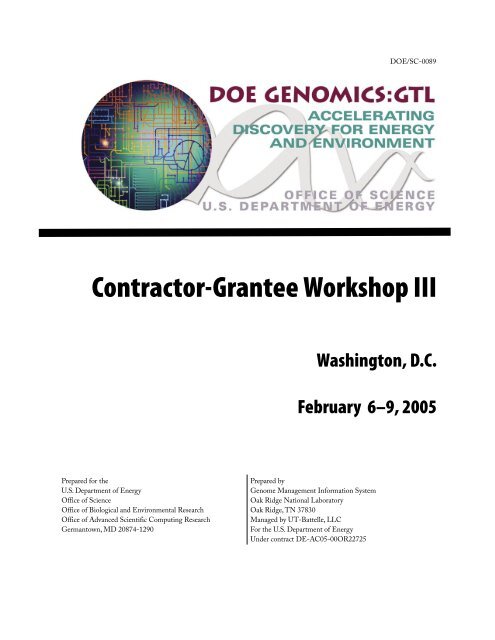
Contractor-Grantee Workshop III<br />
Prepared for the<br />
U.S. <strong>Department</strong> <strong>of</strong> <strong>Energy</strong><br />
Office <strong>of</strong> Science<br />
Office <strong>of</strong> Biological and Environmental Research<br />
Office <strong>of</strong> Advanced Scientific Computing Research<br />
Germantown, MD 20874-1290<br />
Washington, D.C.<br />
February 6–9, <strong>2005</strong><br />
Prepared by<br />
Genome Management Information System<br />
Oak Ridge National Laboratory<br />
Oak Ridge, TN 37830<br />
Managed by UT-Battelle, LLC<br />
For the U.S. <strong>Department</strong> <strong>of</strong> <strong>Energy</strong><br />
Under contract DE-AC05-00OR22725<br />
DOE/SC-0089
Welcome to <strong>Genomics</strong>:GTL Workshop III<br />
<strong>Genomics</strong>:GTL Program Projects<br />
Harvard Medical School<br />
* Presenting author<br />
Contents<br />
1 Metabolic Network Modeling <strong>of</strong> Prochlorococcus marinus ..........................................................3<br />
George M. Church* (g1m1c1@arep.med.harvard.edu), Xiaoxia Lin, Daniel Segrè, Aaron Brandes, and<br />
Jeremy Zucker<br />
2 Quantitative Proteomics <strong>of</strong> Prochlorococcus marinus ..................................................................4<br />
Kyriacos C. Leptos* (leptos@fas.harvard.edu), Jacob D. Jaffe, Eric Zinser, Debbie Lindell,<br />
Sallie W. Chisholm, and George M. Church<br />
3 Genome Sequencing from Single Cells with Ploning ...............................................................5<br />
Kun Zhang* (kzhang@genetics.med.harvard.edu), Adam C. Martiny, Nikkos B. Reppas,<br />
Sallie W. Chisholm, and George M. Church<br />
Lawrence Berkeley National Laboratory<br />
4 VIMSS Computational Microbiology Core Research on Comparative and Functional<br />
<strong>Genomics</strong> .................................................................................................................................6<br />
Adam Arkin* (aparkin@lbl.gov), Eric Alm, Inna Dubchak, Mikhail Gelfand, Katherine Huang, Vijaya<br />
Natarajan, Morgan Price, and Yue Wang<br />
5 The Virtual Institute <strong>of</strong> Microbial Stress and Survival (VIMSS): Deduction <strong>of</strong> Stress<br />
Response Pathways in Metal/Radionuclide Reducing Microbes ..............................................8<br />
Carl Abulencia, Eric Alm, Gary Andersen, Adam Arkin* (APArkin@lbl.gov), Kelly Bender, Sharon Borglin,<br />
Eoin Brodie, Swapnil Chhabra, Steve van Dien, Inna Dubchak, Matthew Fields, Sara Gaucher, Jil Geller,<br />
Masood Hadi, Terry Hazen, Qiang He, Zhili He, Hoi-Ying Holman, Katherine Huang, Rick Huang,<br />
Janet Jacobsen, Dominique Joyner, Jay Keasling, Keith Keller, Martin Keller, Aindrila Mukhopadhyay,<br />
Morgan Price, Joseph A. Ringbauer, Jr., Anup Singh, David Stahl, Sergey Stolyar, Jun Sun,<br />
Dorothea Thompson, Christopher Walker, Judy Wall, Jing Wei, Denise Wolf, Denise Wyborski,<br />
Huei-che Yen, Grant Zane, Jizhong Zhou, and Beto Zuniga<br />
i
Poster<br />
Page<br />
6 VIMSS Applied Environmental Microbiology Core Research on Stress Response<br />
Pathways in Metal-Reducers .................................................................................................. 11<br />
ii<br />
Terry C. Hazen* (tchazen@lbl.gov), Carl Abulencia, Gary Andersen, Sharon Borglin, Eoin Brodie,<br />
Steve van Dien, Matthew Fields, Jil Geller, Hoi-Ying Holman, Rick Huang, Janet Jacobsen,<br />
Dominique Joyner, Martin Keller, Aindrila Mukhopadhyay, David Stahl, Sergey Stolyar, Jun Sun,<br />
Dorothea Thompson, Judy Wall, Denise Wyborski, Huei-che Yen, Grant Zane, Jizhong Zhou, and<br />
Beto Zuniga<br />
7 VIMSS Functional <strong>Genomics</strong> Core: Analysis <strong>of</strong> Stress Response Pathways in<br />
Metal-Reducing Bacteria ....................................................................................................... 14<br />
Aindrila Mukhopadhyay, Steven Brown, Swapnil Chhabra, Brett Emo, Weimin Gao, Sara Gaucher,<br />
Masood Hadi, Qiang He, Zhili He, Ting Li, Yongqing Liu, Alyssa Redding, Joseph Ringbauer, Jr.,<br />
Dawn Stanek, Jun Sun, Lianhong Sun, Jing Wei, Liyou Wu, Huei-Che Yen, Wen Yu, Grant Zane,<br />
Matthew Fields, Martin Keller (mkeller@diversa.com), Anup Singh (aksingh@sandia.gov),<br />
Dorothea Thompson, Judy Wall (wallj@missouri.edu), Jizhong Zhou (zhouj@ornl.gov), and Jay Keasling*<br />
(keasling@socrates.berkeley.edu)<br />
Oak Ridge National Laboratory and Pacific Northwest National Laboratory<br />
8 Center for Molecular and Cellular Systems: High-Throughput Identification and<br />
Characterization <strong>of</strong> Protein Complexes .................................................................................. 15<br />
Michelle Buchanan, Frank Larimer, Steven Wiley, Steven Kennel, Dale Pelletier, Brian Hooker, Gregory<br />
Hurst, Robert Hettich, Hayes McDonald* (mcdonaldwh@ornl.gov), Vladimir Kery, Mitchel Doktycz,<br />
Jenny Morrell, Bob Foote, Denise Schmoyer, Manesh Shah, and Bill Cannon<br />
9 High-Throughput Analysis <strong>of</strong> Protein Complexes in the Center for Molecular and Cellular<br />
Systems ................................................................................................................................... 16<br />
Vladimir Kery* (vladimir.kery@pnl.gov), Dale A. Pelletier, Joshua N. Adkins, Deanna L. Auberry, Frank<br />
R. Collart, Linda J. Foote, Brian S. Hooker, Peter Hoyt, Gregory B. Hurst, Stephen J. Kennel, Trish K.<br />
Lankford, Chiann-Tso Lin, Eric A. Livesay, Tse-Yuan S. Lu, Cathy K. McKeown, Priscilla A. Moore, Ronald J.<br />
Moore, and Kristin D. Victry<br />
10 Investigating Gas Phase Dissociation Pathways <strong>of</strong> Crosslinked Peptides: Application to Protein<br />
Complex Determination ......................................................................................................... 17<br />
Sara P. Gaucher* (spgauch@sandia.gov), Masood Z. Hadi, and Malin M. Young<br />
11 Center for Molecular and Cellular Systems: Statistical Screens for Datasets from High-<br />
Throughput Protein Pull-Down Assays ................................................................................. 18<br />
Frank W. Larimer* (larimerfw@ornl.gov), Kenneth K. Anderson, Deanna L. Auberry, Don S. Daly, Vladimir<br />
Kery, Denise D. Schmoyer, Manesh B. Shah, and Amanda M. White<br />
12 Center for Molecular and Cellular Systems: Analysis and Visualization <strong>of</strong> Data from a High-<br />
Throughput Protein Complex Identification Pipeline Using Modular and Automated Tools .. 19<br />
W. Hayes McDonald (mcdonaldwh@ornl.gov), Joshua N. Adkins, Deanna L. Auberry, Kenneth J.<br />
Auberry, Gregory B. Hurst, Vladimir Kery, Frank W. Larimer, Manesh B. Shah, Denise D. Schmoyer, Eric F.<br />
Strittmatter, and Dave L. Wabb<br />
* Presenting author
Poster<br />
Sandia National Laboratories<br />
13 Carbon Sequestration in Synechococcus: A Computational Biology Approach to Relate<br />
the Genome to Ecosystem Response .......................................................................................20<br />
Grant S. Heffelfinger* (gsheffe@sandia.gov)<br />
14 Integrating Heterogeneous Databases and Tools for High Throughput Microbial Analysis ... 21<br />
Nagiza Samatva* (samatovan@ornl.gov), Al Geist, Praveen Chandramohan, and Ramya Krishnamurthy<br />
15 Toward Comprehensive Analysis <strong>of</strong> MS/MS Data Flows .......................................................22<br />
Andrey Gorin* (agor@ornl.gov), Nikita D. Arnold, Robert M. Day, and Tema Fridman<br />
16 The Transcriptome <strong>of</strong> a Marine Cyanobacterium—Analysis Through Whole Genome<br />
Microarray Analyses ...............................................................................................................24<br />
Brian Palenik* (bpalenik@ucsd.edu), Ian Paulsen* (ipaulsen@tigr.org), Bianca Brahamsha, Rob Herman,<br />
Katherine Kang, Ed Thomas, Jeri Timlin, and Dave Haaland<br />
17 DEB: A Data Entry and Browsing Tool for Entering and Linking Synechococcus sp. WH8102<br />
Whole Genome Microarray Metadata from Multiple Data Sources ....................................... 25<br />
Arie Shoshani* (Shoshani@lbl.gov), Victor Havin, Vijaya Natarajan, Tony Martino, Jerilyn A. Timlin,<br />
Katherine Kang, Ian Paulsen, Brian Palenik, and Thomas Naughton<br />
18 Microarray Analysis using VxInsight and PAM ......................................................................28<br />
George S. Davidson* (GSDAVID@sandia.gov), David Hanson, Shawn Martin, Margaret Werner-<br />
Washburne, and Mark D. Rintoul<br />
19 Mapping <strong>of</strong> Biological Pathways and Networks across Microbial Genomes ............................30<br />
F. Mao, V. Olman, Z. Su, P. Dam, and Ying Xu* (xyn@bmb.uga.edu)<br />
20 Proteomic Analysis <strong>of</strong> the Synechococcus WH8102 CCM with Varying CO 2 Concentrations ... 31<br />
Arlene Gonzales, Yooli K. Light, Zhaoduo Zhang, Michael D. Leavell, Rajat Sapra, Tahera Iqbal,<br />
Todd W. Lane, and Anthony Martino* (martino@sandia.gov)<br />
21 Predicting Protein-Protein Interactions Using Signature Products with an Application<br />
to β-Strand Ordering ............................................................................................................. 32<br />
Shawn Martin (smartin@sandia.gov), W. Michael Brown, Charlie Strauss, Mark D. Rintoul*, and<br />
Jean-Loup Faulon<br />
22 In Vivo Observation <strong>of</strong> the Native Pigments in Synechocystis sp. PCC 6803 Using a New<br />
Hyperspectral Confocal Microscope ......................................................................................33<br />
Michael B. Sinclair* (mbsincl@sandia.gov), Jerilyn A. Timlin, David M. Haaland, Sawsan Hamad, and<br />
Wim F.J. Vermaas<br />
* Presenting author<br />
Page<br />
iii
Poster<br />
Page<br />
23 Connecting Temperature and Metabolic Rate to Population Growth Rates in Marine<br />
Picophytoplankton .................................................................................................................34<br />
iv<br />
Andrea Belgrano* (ab@ncgr.org) and Damian Gessler<br />
24 Deciphering Response Networks in Microbial Genomes through Data Mining and<br />
Computational Modeling .......................................................................................................34<br />
Z. Su, P. Dam, V. Olman, F. Mao, H. Wu, X. Chen, T. Jiang, B. Palenik, and Ying Xu* (xyn@bmb.uga.edu)<br />
25 BiLab – A New Tool that Combines the Ease-<strong>of</strong>-Use <strong>of</strong> MatLab and the Power <strong>of</strong> Multiple<br />
Computational Biology Libraries ........................................................................................... 37<br />
Al Geist* (gst@ornl.gov) and David Jung<br />
26 Microbial Cell Modeling via Reacting/Diffusing Particles .................................................... 38<br />
Steve Plimpton* (sjplimp@sandia.gov) and Alex Slepoy<br />
27 Modeling RuBisCO’s Gating Mechanism Using Targeted Molecular Dynamics ................... 38<br />
Paul S. Crozier (pscrozi@sandia.gov), Steven J. Plimpton, Mark D. Rintoul*, Christian Burisch, and<br />
Jürgen Schlitter<br />
28 Selection <strong>of</strong> Ligands by Panning <strong>of</strong> Phage Display Peptide Libraries Reveals Potential<br />
Partners for TPR Domain and rbcS in Synechococcus WH8102 ...............................................40<br />
Zhaoduo Zhang* (zzhang@sandia.gov), Arlene D. Gonzales, Todd W. Lane, and Anthony Martino<br />
University <strong>of</strong> Massachusetts, Amherst<br />
29 Progress Toward Genome-Scale Monitoring <strong>of</strong> In Situ Gene Expression During Uranium<br />
Bioremediation and Electricity Harvesting ............................................................................40<br />
Dawn Holmes* (dholmes@microbio.umass.edu), Kelly Nevin, Regina O’ Neil, Zhenya Shelbolina,<br />
Martin Lanthier, Jonathan Kaye, Brad Postier, and Derek Lovley<br />
30 Integrating Phenotypic and Expression Data to Characterize Metabolism in<br />
G. sulfurreducens .....................................................................................................................42<br />
R. Mahadevan, C. H. Schilling, D. Segura, B. Yan, J. Krushkal, and D. R. Lovley*<br />
(dlovley@microbio.umass.edu)<br />
31 Novel Regulatory Systems and Adaption <strong>of</strong> Some Well-Known Systems Controlling<br />
Respiration, Growth, and Chemotaxis <strong>of</strong> Geobactor Species ...................................................44<br />
Maddalena Coppi* (mcoppi@microbio.umass.edu), Byoung-Chan Kim, Laurie DiDonato, Julia Krushkal,<br />
Bin Yan, Richard Glaven, Regina O’ Neil, Suphan Bakkal, Allen Tsang, Hoa Tran, Abraham Esteve-Nunez,<br />
Cinthia Nunez, Ching Leang, Kuk-Jeong Chin, Barbara Methe , Robert Weis, Pablo Pomposiello, Kelly<br />
Nevin, and Derek Lovley<br />
* Presenting author
Poster<br />
Page<br />
32 Nanowires, Capacitors, and Other Novel Electron Transfer Mechanisms in Geobacter<br />
Species Elucidated from Genome-Scale Investigations ..........................................................46<br />
Gemma Reguera* (greguera@microbio.umass.edu), Teena Mehta, Dawn E. Holmes, Abraham Esteve-<br />
Núñez, Jessica Butler, Barbara Methe, Kelly Nevin, Swades K. Chaudhuri, Richard Glaven, Tunde Mester,<br />
Raymond DiDonato, Kevin McCarthy, Mark T. Tuominen, and Derek Lovley<br />
33 Continued Progress in the use <strong>of</strong> Microarray Technology to Predict Gene Regulation and<br />
Function in Geobacter sulfurreducens ......................................................................................48<br />
Barbara Methé*(bmethe@tigr.org), Jennifer Webster, Kelly Nevin, and Derek Lovley<br />
Shewanella Federation<br />
34 The Shewanella Federation: Functional Genomic Investigations <strong>of</strong> Dissimilatory Metal-<br />
Reducing Shewanella ..............................................................................................................50<br />
James K. Fredrickson* (jim.fredrickson@pnl.gov), Carol S. Giometti, Eugene Kolker*,<br />
Kenneth H. Nealson, James M. Tiedje, Jizhong Zhou, Monica Riley, Shimon Weiss, James J. Collins,<br />
Frank Larimer, Frank Collart, Lee Ann McCue, Chip Lawrence, and Timothy S. Gardner<br />
35 Global Pr<strong>of</strong>iling <strong>of</strong> Shewanella oneidensis MR-1: Expression <strong>of</strong> ‘Hypothetical’ Genes and<br />
Improved Functional Annotations ......................................................................................... 51<br />
Eugene Kolker* (ekolker@biatech.org), Alex F. Picone, Michael Y. Galperin, Margaret F. Romine,<br />
Roger Higdon, Kira S. Makarova, Natali Kolker, Gordon A. Anderson, Xiaoyun Qiu, Kenneth J. Auberry,<br />
Gyorgy Babnigg, Alex S. Beliaev, Paul Edlefsen, Dwayne A. Elias, Yuri Gorby, Ted Holzman,<br />
Joel Klappenbach, Konstantinos T. Konstantinidis, Miriam L. Land, Mary S. Lipton, Lee-Ann McCue,<br />
Matthew Monroe, Ljiljana Pasa-Tolic, Grigoriy Pinchuk, Samuel Purvine, Margaret Serres,<br />
Sasha Tsapin, Brian A. Zakrajsek, Wenhong Zhu, Jizhong Zhou, Frank W. Larimer, Charles Lawrence,<br />
Monica Riley, Frank R. Collart, John R. Yates, III, Richard D. Smith, Carol Giometti, Kenneth Nealson,<br />
James K. Fredrickson, and James M. Tiedje<br />
36 Respiratory Pathways and Regulatory Networks <strong>of</strong> Shewanella oneidensis Involved in<br />
<strong>Energy</strong> Metabolism and Environmental Sensing ................................................................... 52<br />
Alex Beliaev*, Yuri Gorby, Margie Romine, Jeff McLean, Grigoriy Pinchuk, Eric Hill, Jim Fredrickson,<br />
Jizhong Zhou, and Daad A. Saffarini<br />
37 Functional Analysis <strong>of</strong> Shewanella, A Cross Genome Comparison .........................................54<br />
Margrethe H. Serres* (mserres@mbl.edu) and Monica Riley<br />
38 Optical Methods for Characterization <strong>of</strong> Expression Levels and Protein-Protein<br />
Interactions in Shewanella oneidensis MR-1 ............................................................................ 55<br />
Natalie R. Gassman* (ngassman@chem.ucla.edu), Xiangxu Kong, Gopal Iyer, Younggyu Kim, and<br />
Shimon Weiss<br />
* Presenting author<br />
v
Poster<br />
Page<br />
39 Reverse-Engineering Microbial Networks in Escherichia coli and Shewanella oneidensis<br />
MR-1 via Large-Scale Perturbation Studies ........................................................................... 56<br />
vi<br />
G. Cottarel, M.E. Driscoll, J. Faith, M.K. Kohanski, J. Wierzbowski, C.B. Cantor, J.J. Collins, and<br />
T.S. Gardner* (tgardner@bu.edu)<br />
40 Comparative Analysis <strong>of</strong> Gene Expression Pr<strong>of</strong>iles <strong>of</strong> Shewanella oneidensis MR-1<br />
Following Exposure to Ionizing Radiation and Ultraviolet Radiation ................................... 57<br />
Xiaoyun Qiu* (qiuxiaoy@msu.edu), George Sundin, Michael J. Daly, Alexander Vasilenko,<br />
Marina V. Omelchenko, Jizhong Zhou, Liyou Wu, Mary S. Lipton, and James M. Tiedje<br />
41 The Microbial Proteome Project: A Database <strong>of</strong> Microbial Protein Expression in the<br />
Context <strong>of</strong> Genome Analysis .................................................................................................. 58<br />
Carol S. Giometti * (csgiometti@anl.gov), Gyorgy Babnigg, Sandra L. Tollaksen, Tripti Khare,<br />
Angela Ahrendt, Wenhong Zhu, Derek R. Lovley, James K. Fredrickson, and John R. Yates III<br />
J. Craig Venter Institute<br />
42 Estimation <strong>of</strong> the Minimal Mycoplasma Gene Set Using Global Transposon Mutagenesis<br />
and Comparative <strong>Genomics</strong> ...................................................................................................60<br />
John I. Glass* (JGlass@venterinstitute.org), Nina Alperovich, Nacyra Assad-Garcia, Shibu Yooseph,<br />
Mahir Maruf, Carole Lartigue, Cynthia Pfannkoch, Clyde A. Hutchison III, Hamilton O. Smith, and<br />
J. Craig Venter<br />
43 Progress toward a Synthetic Cellular Genome ........................................................................ 61<br />
Hamilton O. Smith* (hsmith@venterinstitute.org), Cynthia Pfannkoch, Holly A. Baden-Tillson,<br />
Clyde A. Hutchison III, and J. Craig Venter<br />
44 Development <strong>of</strong> a Deinococcus radiodurans Homologous Recombination System ....................62<br />
Sanjay Vashee*, Ray-Yuan Chuang* (RChuang@venterinstitute.org), Christian Barnes, Hamilton O.<br />
Smith, and J. Craig Venter<br />
45 Development <strong>of</strong> a Novel Recombinant Cyanobacterial System for Hydrogen Production<br />
from Water .............................................................................................................................63<br />
Qing Xu, Shibu Yooseph, Hamilton O. Smith, and J. Craig Venter (jcventer@tcag.org)<br />
46 Biotechnology For the Production <strong>of</strong> Ethanol and Butanol from Cellulose ............................64<br />
Prabha P. Iyer* (piyer@venterinstitute.org), Hamilton O. Smith, and J. Craig Venter<br />
* Presenting author
Poster<br />
Communication<br />
47 Communicating <strong>Genomics</strong>:GTL ........................................................................................... 65<br />
Anne E. Adamson, Shirley H. Andrews, Jennifer L. Bownas, Denise K. Casey, Sherry A. Estes,<br />
Sheryl A. Martin, Marissa D. Mills, Kim Nylander, Judy M. Wyrick, Anita J. Alton, and Betty K. Mansfield*<br />
(mansfieldbk@ornl.gov)<br />
Bioinformatics, Modeling, and Computation<br />
48 SimPheny: A Computational Infrastructure for Systems Biology ....................................... 67<br />
Christophe H. Schilling* (cschilling@genomatica.com), Sean Kane, Martin Roth, Jin Ruan,<br />
Kurt Stadsklev, Rajendra Thakar, Evelyn Travnik, Steve van Dien, and Sharon Wiback<br />
49 Hybrid Bacterial Cell Models: Linking <strong>Genomics</strong> to Physiological Response ........................68<br />
Jordan C. Atlas* (jca33@cornell.edu), Mariajose Castellanos, Anjali Dhiman, Bruce Church,<br />
and Michael L. Shuler<br />
50 Identification <strong>of</strong> the Most Probable Biological Network Using Model Discrimination<br />
Analysis .................................................................................................................................. 69<br />
Andrea L. Knorr and Ranjan Srivastava* (srivasta@engr.uconn.edu)<br />
51 Rhodopseudomonas palustris Regulons Detected by a Cross-Species Analysis <strong>of</strong> the<br />
α-Proteobacteria ..................................................................................................................... 70<br />
Sean Conlan* (sconlan@wadsworth.org), Charles E. Lawrence, and Lee Ann McCue<br />
52 Exploring Evolutionary Space ................................................................................................ 72<br />
Timothy G. Lilburn* (tlilburn@atcc.org), Yun Bai, Yuan Zhang, James R. Cole, and George M. Garrity<br />
53 PhyloScan: A New Tool for Identifying Statistically Significant Transcription Factor<br />
Binding Sites by Combining Cross-Species Evidence ............................................................. 72<br />
Lee A. Newberg*, C. Steven Carmack, Lee Ann McCue (mccue@wadsworth.org), and<br />
Charles E. Lawrence<br />
54 Predicting Protein Interactions via Docking Mesh Evaluator ................................................ 74<br />
Roummel F. Marcia, Susan D. Lindsey, Erick A. Butzlaff, and Julie C. Mitchell* (mitchell@math.wisc.edu)<br />
55 UC Merced Center for Computational Biology ....................................................................... 75<br />
Michael Colvin* (mcolvin@ucmerced.edu), Arnold Kim, and Felice Lightstone<br />
56 Biomic Approach to Predictive Cell Modeling ...................................................................... 76<br />
P. J. Ortoleva* (ortoleva@indiana.edu), L. Ensman, J. Fan, K. Hubbard, A. Sayyed-Ahmad, F. Stanley,<br />
K. Tuncay, and K. Varala<br />
* Presenting author<br />
Page<br />
vii
Poster<br />
Page<br />
57 The BioWarehouse System for Integration <strong>of</strong> Bioinformatics Databases ................................. 78<br />
viii<br />
Tom Lee, Valerie Wagner, Yannick Pouliot, and Peter D. Karp* (pkarp@ai.sri.com)<br />
58 Building Large Biological Dynamic Models <strong>of</strong> Shewanella oneidensis from Incomplete Data . 79<br />
Ravishankar R. Vallabhajosyula* (rrao@kgi.edu), Sri Paladugu, Klaus Maier, and Herbert M. Sauro<br />
59 A Bayesian Method for Identifying Missing Enzymes in Predicted Metabolic Pathway<br />
Databases ............................................................................................................................... 81<br />
Michelle L. Green* (green@ai.sri.com) and Peter D. Karp<br />
60 Does EcoCyc or KEGG Provide a Preferable Gold Standard for Training and Evaluation<br />
<strong>of</strong> Genome-Context Methods? ...............................................................................................82<br />
Peter D. Karp* (pkarp@ai.sri.com) and Michelle L. Green<br />
61 Towards a Physics and Systems Understanding <strong>of</strong> Ion Transport in Prokaryotes ....................83<br />
Shreedhar Natarajan, Asba Tasneem*, Sameer Varma, Lakshminarayan Iyer, L. Aravind, and Eric<br />
Jakobsson* (jake@ncsa.uiuc.edu)<br />
62 OptStrain: A Computational Framework for Redesign Microbial Production Systems ..........84<br />
Priti Pharkya and Costas D. Maranas* (costas@psu.edu)<br />
63 DEMSIM: A Discrete Event Based Mechanistic Simulation Platform for Gene Expression<br />
and Regulation Dynamics ......................................................................................................84<br />
Madhukar Dasika and Costas D. Maranas* (costas@psu.edu)<br />
64 On the Futility <strong>of</strong> Optima in Network Inferences and What Can Be Done About It .............. 86<br />
Charles (Chip) E. Lawrence* (lawrence@dam.brown.edu)<br />
Environmental <strong>Genomics</strong><br />
65 Whole Community Proteomics Study <strong>of</strong> an Acid Mine Drainage Bi<strong>of</strong>ilm Reveals Key Roles<br />
for “Hypothetical” Proteins in a Natural Microbial Bi<strong>of</strong>ilm ...................................................87<br />
Jill Banfield* (jill@eps.berkeley.edu), Rachna J. Ram, Gene W. Tyson, Eric Allen, Nathan VerBerkmoes,<br />
Michael P. Thelen, Brett J. Baker, Manesh Shah, Robert Hettich, and Robert C. Blake II<br />
66 Application <strong>of</strong> High Throughput Microcapsule Culturing to Develop a Novel <strong>Genomics</strong><br />
Technology Platform .............................................................................................................88<br />
Martin Keller* (mkeller@diversa.com), Karsten Zengler, Marion Walcher, Carl Abulencia, Denise<br />
Wyborski, Sherman Chang, Imke Haller, Trevin Holland, Fred Brockman, Cheryl Kuske, and Susan Barns<br />
* Presenting author
Poster<br />
Page<br />
67 Environmental Bacterial Diversity from Communities to Genomes ......................................89<br />
Janelle R. Thompson*, Silvia G. Acinas, Vanja Klepac-Ceraj, Sarah Pacocha, Chanathip Pharino,<br />
Dana E. Hunt, Luisa A. Marcelino, Jennifer Benoit, Ramahi Sarma-Rupavtarm, Daniel L. Distel, and<br />
Martin F. Polz (mpolz@mit.edu)<br />
68 Distribution and Variation <strong>of</strong> Prochlorococcus Genotypes Across Multiple Oceanic Habitats ..90<br />
Adam C. Martiny* (martiny@mit.edu), P. K. Amos Tai, Anne W. Thompson, and Sallie W. Chisholm<br />
69 From Perturbation Analysis to the Genomic Regulatory Code: the Sea Urchin<br />
Endomesoderm GRN ............................................................................................................. 91<br />
Paola Oliveri* (poliveri@caltech.edu), Pei-Yun Lee, Takuya Minokawa, Joel Smith, Qiang Tu,<br />
Meredith Howard, David McClay, and Eric H. Davidson<br />
Microbial <strong>Genomics</strong><br />
70 The Genome <strong>of</strong> the Ammonia Oxidizing Bacterium Nitrosomonas europaea: Iron Metabolism<br />
and Barriers to Heterotrophy .................................................................................................. 93<br />
Xueming Wei, Neeraja Vajrala, Norman Hommes, Luis Sayavedra-Soto*, and Daniel Arp<br />
(arpd@science.oregonstate.edu)<br />
71 Pelagibacter ubique: A Post-Genomic Investigation <strong>of</strong> Carbon Metabolism and<br />
Photochemistry in an Extraordinarily Abundant Oceanic Bacterium .................................... 95<br />
Stephen J. Giovannoni * (steve.giovannoni@oregonstate.edu), Lisa Bibbs, James Tripp, Scott Givan,<br />
Jang-Cheon Cho, Martha D. Stapels, Russell Desiderio, Mercha Podar, Kevin L. Vergin, Mick Noordeweir,<br />
Michael S. Rappé, Samuel Laney, Douglas F. Bar<strong>of</strong>sky, and Eric Mathur<br />
72 Does the Three Dimensional Organization <strong>of</strong> the Nucleoid <strong>of</strong> the Deinococcaceae<br />
Contribute to their Ionizing Radiation Resistance? ................................................................ 96<br />
J. M. Zimmerman and J. R. Battista* (jbattis@lsu.edu)<br />
73 Large Scale Genomic Analysis for Understanding Hydrogen Metabolism in Chlamydomonas<br />
reinhardtii ...............................................................................................................................97<br />
Michael Seibert* (mike_seibert@nrel.gov), Arthur R. Grossman, Maria L. Ghirardi, and<br />
Matthew C. Posewitz<br />
74 Exploring the Genome and Proteome <strong>of</strong> Desulfitobacterium hafniense DCB2 for its<br />
Protein Complexes Involved in Metal Reduction and Dechlorination ....................................99<br />
James M. Tiedje*, Sang-Hoon Kim, Christina Harzman, John Davis, Brett Phinney, Michael Ngowe,<br />
Washington Mutatu, William Broderick, David DeWitt, Joan Broderick, and Terence L. Marsh<br />
(marsht@msu.edu)<br />
* Presenting author<br />
ix
Poster<br />
Page<br />
75 An Integrative Approach to <strong>Energy</strong>, Carbon, and Redox Metabolism in the<br />
Cyanobacterium Synechocystis sp. PCC 6803 .........................................................................100<br />
x<br />
Wim Vermaas* (wim@asu.edu), Robert Roberson, Allison van de Meene, Bing Wang, Sawsan Hamad,<br />
Zhi Cai, Julian Whitelegge, Kym Faull, Sveta Gerdes, Andrei Osterman, and Ross Overbeek<br />
76 Role <strong>of</strong> Cellulose Binding Modules in Cellulose Hydrolysis ................................................. 102<br />
David B. Wilson* (dbw3@cornell.edu) and Shaolin Chen<br />
77 Three Prochlorococcus Cyanophage Genomes: Signature Features and Ecological<br />
Interpretation ...................................................................................................................... 103<br />
Matthew B. Sullivan* (mbsulli@mit.edu), Maureen Coleman, Peter Weigele, Forest Rohwer, and<br />
Sallie W. Chisholm<br />
78 The Alternative Sigma Factor RpoN Regulon <strong>of</strong> Rhodopseudomonas palustris .......................104<br />
Yasuhiro Oda* (yasuhiro-oda@uiowa.edu), Sudip K. Samanta, Frank W. Larimer, and<br />
Caroline S. Harwood<br />
79 Integrative Control <strong>of</strong> Key Metabolic Processes in Rhodopseudomonas palustris for the<br />
Enhancement <strong>of</strong> Carbon Sequestration and Biohydrogen Production .................................. 105<br />
F. Robert Tabita* (Tabita.1@osu.edu), Janet L. Gibson, Caroline S. Harwood, Frank Larimer,<br />
J. Thomas Beatty, James C. Liao, and Jizhong (Joe) Zhou<br />
80 Whole Genome Transcriptional Analysis <strong>of</strong> Toxic Metal Stresses in Caulobacter crescentus .. 107<br />
Gary L. Andersen* (GLAndersen@lbl.gov), Ping Hu, Eoin L. Brodie, and Harley H. McAdams<br />
81 Systematic Analysis <strong>of</strong> Two-Component Signal Transduction Systems Regulating Cell<br />
Cycle Progression in Caulobacter crescentus ........................................................................... 108<br />
Michael Laub* (Laub@CGR.Harvard.edu)<br />
82 The U.S. DOE Joint Genome Institute Microbial Program ................................................. 109<br />
David Bruce* (dbruce@lanl.gov), Alla Lapidus, Patrick Chain, Jeremy Schmutz, Frank Larimer,<br />
Nikos Kyrpides, Paul Gilna, Eddy Rubin and Paul Richardson<br />
83 Identification <strong>of</strong> Genes that are Required for Recycling Reducing Power during<br />
Photosynthetic Growth ....................................................................................................... 110<br />
Christine L. Tavano, Angela M. Podevels, and Timothy J. Donohue* (tdonohue@bact.wisc.edu)<br />
84 A Tightly-Regulated Oscillatory Circuit Formed by Conserved Master Regulator<br />
Proteins Controls the Caulobacter Cell Cycle ........................................................................ 110<br />
Harley McAdams* (hmcadams@stanford.edu) and Lucy Shapiro<br />
85 Dynamics and Control <strong>of</strong> Bi<strong>of</strong>ilms <strong>of</strong> the Oligotrophic Bacterium Caulobacter crescentus ..... 112<br />
Alfred M. Spormann (spormann@stanford.edu ) and Plamena Entcheva-Dimitrov<br />
* Presenting author
Poster<br />
Page<br />
86 Widespread and Abundant CelM Endoglucanases <strong>of</strong> Marine Cytophaga-like Bacteria<br />
Revealed by Whole Genome Shotgun Sequencing and Fosmid Cloning .............................. 112<br />
Matthew T. Cottrell and David L. Kirchman* (kirchman@cms.udel.edu)<br />
87 Data Analysis and Protein Identification Strategy for the Systems-Level Protein-Protein<br />
Interaction Networks <strong>of</strong> Shewanella oneidensis MR-1 ............................................................ 114<br />
Gordon A. Anderson* (gordon@pnl.gov), James E. Bruce, Xiaoting Tang, Gerhard Munske, and<br />
Nikola Tolic<br />
88 A Protein Interaction Reporter Strategy for Systems-Level Protein Interaction Networks<br />
<strong>of</strong> Shewanella oneidensis MR-1 .............................................................................................. 115<br />
James E. Bruce* (james_bruce@wsu.edu), Xiaoting Tang, Harry Zhu, Saiful Chowdhury, Devi Adhikari,<br />
Gerhard Munske, Gordon A. Anderson, and Nikola Tolic<br />
Technology Development and Use<br />
Imaging, Molecular, and Cellular Analysis<br />
89 Probing Single Microbial Proteins and Multi-Protein Complexes with Bioconjugated<br />
Quantum Dots ..................................................................................................................... 117<br />
Gang Bao* (gang.bao@bme.gatech.edu), Grant Jensen, Shuming Nie, and Phil LeDuc<br />
90 Single-Molecule Imaging <strong>of</strong> Macromolecular Dynamics in a Cell ....................................... 119<br />
Jamie H. D. Cate (jcate@lbl.gov) and Haw Yang* (hawyang@berkeley.edu)<br />
91 Developing a High Resolution Method for Protein Localization in Whole Bacterium .........120<br />
Huilin Li* (hli@bnl.gov) and James Hainfeld (hainfeld@bnl.gov)<br />
92 Novel Vibrational Nanoprobes for Microbiology at the Single Cell Level ............................. 121<br />
Thomas Huser* (huser1@llnl.gov), Chad E. Talley, James W. Chan, Heiko Winhold, Ted Laurence,<br />
Anthony Esposito, Christopher W. Hollars, Christine A. Hara, Allen T. Christian, Michele H. Corzett,<br />
Rod Balhorn, and Stephen M. Lane<br />
93 Instrumented Cell for Characterization <strong>of</strong> Mammalian and Microbial Cells ...................... 122<br />
Jane Bearinger* (bearinger1@llnl.gov), Graham Bench, Jackie Crawford, Lawrence Dugan,<br />
Amy Hiddessen, Angela Hinz, Thomas Huser, Robin Miles, Magnus Palmblad, Chad Talley,<br />
Elizabeth Wheeler, and Allen Christian<br />
94 Chemical Imaging <strong>of</strong> Biological Materials by NanoSIMS ....................................................123<br />
Peter K. Weber* (weber21@llnl.gov), Ian D. Hutcheon, Radu Popa, and Ken Nealson<br />
* Presenting author<br />
xi
Poster<br />
Page<br />
95 Direct Determination <strong>of</strong> Affinity in Individual Protein-Protein Complexes in Mono<br />
and Multivalent Configurations Using Dynamic Force Spectroscopy ..................................124<br />
xii<br />
Todd A. Sulchek, Kevin Langry, Raymond W. Friddle, Timothy V. Ratto, Sally DeNardo,<br />
Huguette Albrecht, Michael Colvin, and Aleksandr Noy* (noy1@llnl.gov)<br />
96 Electron Tomography <strong>of</strong> Intact and Sectioned Microbial Cells ............................................ 126<br />
Kenneth H. Downing* (khdowning@lbl.gov), Luis Comolli, Haixin Sui, Hoi-Ying Holman, Ellen Judd,<br />
and Harley McAdams<br />
97 Probing the High-Resolution Architecture and Environmental Dynamics <strong>of</strong> Microbial<br />
Surfaces by in vitro Atomic Force Microscopy ...................................................................... 127<br />
Alexander J. Malkin* (malkin1@llnl.gov), Marco Plomp, Terrance J. Leighton, and Katherine E. Wheeler<br />
98 Real-Time Gene Expression Pr<strong>of</strong>iling <strong>of</strong> Single Live Cells <strong>of</strong> Shewanella oneidensis ............ 128<br />
X. Sunney Xie*, Jie Xiao, Ji Yu, Long Cai, Paul Choi*, Nir Friedman, Xiajia Ren, and Luying Xun*<br />
99 High Throughput Fermentation and Cell Culture Device .................................................... 130<br />
David Klein (dklein@gener8.net), David Laidlaw, Gregory Andronaco, and Stephen Boyer<br />
100 Immobilized Enzymes in Nanoporous Materials Exhibit Enhanced Stability and Activity .. 132<br />
Chenghong Lei, Yongsoon Shin, Jun Liu, and Eric J. Ackerman* (eric.ackerman@pnl.gov)<br />
Protein Production and Molecular Tags<br />
101 Towards High Throughput Selection <strong>of</strong> Binding Ligands: Using Flow Cytometry ............... 133<br />
Peter Pavlik, Milan Ovecka, Nileena Velappan, and Andrew Bradbury* (amb@lanl.gov)<br />
102 Efficient Chemical Methods for the Total Synthesis <strong>of</strong> Small Proteins: The First<br />
Crystallographic Structure <strong>of</strong> a Protein Diastereomer, [D-Gln35]-ubiquitin ......................134<br />
Duhee Bang* (duhee@uchicago.edu), George I. Makhatadze, and Stephen B. Kent<br />
(skent@uchicago.edu)<br />
103 Development and Application <strong>of</strong> Multipurpose Affinity Probes to Isolate Intact Protein<br />
Complexes Associated with Metal Reduction from Shewanella oneidensis MR-1 ................... 135<br />
Liang Shi*, Thomas C. Squier* (thomas.squier@pnl.gov), M. Uljana Mayer*, Haishi Cao, Baowei Chen,<br />
Yuri A. Gorby, David F. Lowry, Jeff Mclean, Seema Verma, and Ping Yan<br />
104 A Combined Informatics and Experimental Strategy for Improving Protein Expression ...... 137<br />
Osnat Herzberg, John Moult* (moult@umbi.umd.edu), Fred Schwarz, and Harold Smith<br />
105 High-Throughput Production and Analyses <strong>of</strong> Purified Proteins ......................................... 138<br />
F. William Studier* (studier@bnl.gov), John C. Sutherland, Lisa M. Miller, and Lin Yang<br />
* Presenting author
Poster<br />
Page<br />
106 Development <strong>of</strong> Genome-Scale Expression Methods ........................................................... 139<br />
Sarah Fey, Elizabeth Landorf, Yuri Londer, Terese Peppler, and Frank Collart* (fcollart@anl.gov)<br />
107 Plate-Based Methods for Expression <strong>of</strong> Cytoplasmic Proteins from Shewanella oneidensis .... 140<br />
Elizabeth Landorf, Terese Peppler, Sarah Fey, Alexander Iakounine, Eugene Kolker, and Frank Collart*<br />
(fcollart@anl.gov)<br />
108 Generating scFv and Protein Scaffolds to Protein Targets .................................................... 141<br />
Brian K. Kay* (bkay@anl.gov), Michael Scholle, Ushma Kriplani, John Kehoe, and Frank Collart<br />
109 Cell Free Approaches for Protein Production ....................................................................... 141<br />
Gerald W. Becker*, Pavel Shiyanov, Yifei Wu, Sarah Fey, Elizabeth Landorf, Terese Peppler, and<br />
Frank Collart (fcollart@anl.gov)<br />
110 Rapid Synthesis <strong>of</strong> Peptidic and Peptidomimetic Ligands for High-Throughput Protein<br />
Purification and Labeling ..................................................................................................... 142<br />
Jeffrey B.-H. Tok* (tok2@llnl.gov), Priscilla Chan, David Smithson, Ted Tarasow, and Rod Balhorn<br />
Proteomics and Metabolomics<br />
111 Development and Application <strong>of</strong> New Technologies for Comprehensive and Quantitative<br />
High Throughput Microbial Proteomics .............................................................................. 143<br />
Richard D. Smith* (rds@pnl.gov), Mary S. Lipton, James K. Fredrickson, Matthew Monroe, Eric Livesay,<br />
Konstantinos Petritis, Joshua Adkins, Gordon A. Anderson, Kim Hixson, Ruihua Fang, Rui Zhao,<br />
Ronald J. Moore, and Yufeng Shen<br />
112 Characterization <strong>of</strong> Rhodobacter sphaeroides by High Resolution Proteomic Measurements .144<br />
Mary S. Lipton* (Mary.Lipton@pnl.gov), Timothy Donohue* (tdonohue@bact.wisc.edu), Samuel<br />
Kaplan* (Samuel.Kaplan@uth.tmc.edu), Stephen Callister, Matthew E. Monroe, Margie F. Romine,<br />
Ruihua Fang, Carrie D. Goddard, Nikola Tolic, Gordon A. Anderson, Richard D. Smith, Jim K. Fredrickson,<br />
Miguel Dominguez, Christine Tavano, Xiaihua Zeng, and Jung Hyeob Roh<br />
113 Quantitative Metalloproteomics .......................................................................................... 146<br />
Patrick G. Grant* (pggrant@llnl.gov), Sharon Shields, Magnus Palmblad, and Graham Bench<br />
114 New Technologies for Metabolomics .................................................................................... 147<br />
Jay D. Keasling* (jdkeasling@lbl.gov), Carolyn Bertozzi, Julie Leary, Michael Marletta, and David<br />
Wemmer<br />
115 Characterization <strong>of</strong> Metal Reducing Microbial Systems by High Resolution Proteomic<br />
Measurements ...................................................................................................................... 148<br />
Mary S. Lipton* (Mary.Lipton@pnl.gov), Ruihua Fang, Dwayne A. Elias, Margie F. Romine, Alex Beliaev,<br />
Matthew E. Monroe, Kim K. Hixson, Yuri A. Gorby, Ljiljana Pasa-Tolic, Heather M. Mottaz,<br />
Gordon A. Anderson, Richard D. Smith, Jim K. Fredrickson, Derek Lovley, and Yanhuai R. Ding<br />
* Presenting author<br />
xiii
Poster<br />
Page<br />
116 Protein Complexes and Pathways ......................................................................................... 150<br />
xiv<br />
David Eisenberg* (david@mbiucla.edu), Peter Bowers, Michael Strong, Huiying Li, Lukasz Salwinski,<br />
Robert Riley, Richard Llwellyn, Einat Sprinzak, Debnath Pal, and Todd Yeates<br />
117 Metabolomic Functional Analysis <strong>of</strong> Bacterial Genomes ..................................................... 151<br />
Clifford J. Unkefer* (cju@lanl.gov)<br />
118 Dynameomics: Mass Annotation <strong>of</strong> Protein Dynamics through Molecular Dynamics<br />
Simulations <strong>of</strong> Fold-Space Representatives .......................................................................... 152<br />
David A. C. Beck* (dacb@u.washington.edu), Ryan Day, Kathryn A. Scott, R. Dustin Schaeffer,<br />
Robert E. Steward, Amanda L. Jonsson, Darwin O. V. Alonso, and Valerie Daggett<br />
Ethical, Legal, and Societal Issues<br />
119 The DNA Files® .................................................................................................................... 153<br />
Bari Scott* (bariscot@aol.com)<br />
120 Science Literacy Training for Public Radio Journalists ......................................................... 155<br />
Bari Scott* (bariscot@aol.com)<br />
Appendix 1: Attendees 157<br />
Appendix 2: Web Sites 167<br />
Author Index 169<br />
Institution Index 177<br />
* Presenting author
Welcome to <strong>Genomics</strong>:GTL Workshop III<br />
Welcome to the third <strong>Genomics</strong>:GTL Contractor-Grantee workshop. GTL continues to grow—scientifically,<br />
in DOE relevance, and as a program that needs all your diverse scientific, technical, and<br />
intellectual efforts to make it a success. GTL is attracting broad and enthusiastic interest and support from<br />
scientists at universities, national laboratories, and industry; colleagues at other federal agencies; <strong>Department</strong><br />
<strong>of</strong> <strong>Energy</strong> leadership; and Congress.<br />
GTL’s challenge to the scientific community is to further develop and use a broad array <strong>of</strong> innovative technologies<br />
and computational tools to systematically leverage the knowledge and capabilities brought to us by<br />
DNA sequencing projects. The goal is to seek a broad and predictive understanding <strong>of</strong> the functioning and<br />
control <strong>of</strong> complex systems in individual microbes and microbial communities. GTL’s prominent position<br />
at the interface <strong>of</strong> the physical, computational, and biological sciences is both a strength and a challenge.<br />
Microbes remain GTL’s principal biological focus. In the complex “simplicity” <strong>of</strong> microbes, we find capabilities<br />
needed by DOE and the nation for clean and secure energy, cleanup <strong>of</strong> environmental contamination,<br />
and sequestration <strong>of</strong> atmospheric carbon dioxide that contributes to global warming. An ongoing<br />
challenge for the entire GTL community is to demonstrate that the fundamental science conducted in<br />
each <strong>of</strong> your research projects brings us a step closer to biology-based solutions for these important national<br />
energy and environmental needs.<br />
This year brings two important milestones for GTL. First is the development <strong>of</strong> a roadmap that will help<br />
guide and justify the GTL program to a broad audience <strong>of</strong> scientists, policymakers, and the public. In the<br />
coming weeks we will be calling on many <strong>of</strong> you to provide critical review <strong>of</strong> this important document.<br />
Second is an important step forward in developing GTL user facilities: we are beginning the process <strong>of</strong><br />
engineering and designing the Facility for Production and Characterization <strong>of</strong> Proteins and Molecular<br />
Tags.<br />
GTL workshops are high-energy events that provide an opportunity for all <strong>of</strong> us to discuss, listen, and<br />
learn about exciting new advances in science; identify research needs and opportunities; form research<br />
partnerships; and share the excitement <strong>of</strong> this program with the broader scientific community. We look<br />
forward to a stimulating and productive meeting and <strong>of</strong>fer our sincere thanks to all the organizers and to<br />
you, the scientists, whose vision and efforts will help us all to realize the promise <strong>of</strong> this exciting venture.<br />
Ari Patrinos<br />
Associate Director <strong>of</strong> Science for<br />
Biological and Environmental Research<br />
Office <strong>of</strong> Science<br />
U.S. <strong>Department</strong> <strong>of</strong> <strong>Energy</strong><br />
ari.patrinos@science.doe.gov<br />
Ed Oliver<br />
Associate Director <strong>of</strong> Science for<br />
Advanced Scientific Computing Research<br />
Office <strong>of</strong> Science<br />
U.S. <strong>Department</strong> <strong>of</strong> <strong>Energy</strong><br />
ed.oliver@science.doe.gov
<strong>Genomics</strong>:GTL Program Projects<br />
Harvard Medical School<br />
1<br />
Metabolic Network Modeling <strong>of</strong> Prochlorococcus marinus<br />
George M. Church* (g1m1c1@arep.med.harvard.edu), Xiaoxia Lin, Daniel Segrè, Aaron Brandes,<br />
and Jeremy Zucker<br />
Harvard Medical School, Boston, MA<br />
The marine cyanobacterium Prochlorococcus marinus dominates the phytoplankton in the tropical<br />
and subtropical oceans and contributes to a significant fraction <strong>of</strong> the global photosynthesis. Several<br />
strains <strong>of</strong> Prochlorococcus have been sequenced, which provides us a promising starting point for investigating<br />
the relationship between genotype and phenotype at a genome scale and with a comparative<br />
approach. To achieve the ultimate goal <strong>of</strong> understanding the metabolism at a systems level, we are<br />
developing and utilizing new metabolic network models in several directions.<br />
Comparison and connection <strong>of</strong> day-night metabolisms<br />
Day-night cycles are known to play a central role in the metabolism <strong>of</strong> Prochlorococcus. We are exploring<br />
two approaches to model the difference and connection between day and night. One is to take<br />
the full metabolic network and formulate two separate models assuming different nutrient conditions<br />
and optimality criteria. Then the flux predictions can be compared to mRNA and protein expression<br />
data. In the other approach, we make use <strong>of</strong> the protein expression data, which helps to reduce the<br />
feasible flux space and leads to finer flux predictions.<br />
Construction <strong>of</strong> metabolic networks<br />
One major challenge in constructing complete and accurate in silico metabolic networks for quantitative<br />
analysis such as flux balance analysis (FBA) is to identify reactions that are “missed” in the annotation.<br />
We have been mainly using Pathway Tools s<strong>of</strong>tware suite developed by SRI to identify metabolic<br />
reactions and are developing new algorithms to construct the “functional” metabolic network<br />
from a network perspective. Biochemical reactions with identified enzymes are included and then an<br />
“optimal” set <strong>of</strong> reactions are added such that the network produces the specified growth phenotype<br />
given corresponding nutrient conditions. Identification <strong>of</strong> the missing links will also help to refine<br />
the genome annotation. Another problem is that there exist “orphaned enzymes” — experimentally<br />
elucidated biochemical reactions whose enzyme has never been sequenced. To address this problem,<br />
we are utilizing a pathway hole-filling algorithm developed by SRI and developing bioinformatics<br />
techniques to identify candidate genes for these orphaned enzymes.<br />
Analysis <strong>of</strong> metabolic networks with mass balance and energy balance<br />
Conventional flux balance analysis (FBA) only considers mass balance. We are incorporating constraints<br />
representing the second law <strong>of</strong> thermodynamics, which eliminates thermodynamically infea-<br />
* Presenting author<br />
3
<strong>Genomics</strong>:GTL Program Projects<br />
sible fluxes. A subset <strong>of</strong> the additional constraints exhibits non-convexity, giving rise to substantial<br />
difficulty in the solution <strong>of</strong> the resulting optimization problem. We are developing new methods to<br />
overcome this challenge to make full use <strong>of</strong> combined FBA and EBA (energy balance analysis).<br />
Construction and comparative study <strong>of</strong> whole-cell metabolic networks <strong>of</strong> MED4 and other strains<br />
By combining a bioinformatics pipeline for generating metabolic network models from genome<br />
annotations and manual inspection/modification, we have constructed the in silico metabolic network<br />
<strong>of</strong> central carbon metabolism and amino acid biosynthesis for Prochlorococcus MED4, a highlight-adapted<br />
strain. We are extending it towards the genome-wide network. In addition, we will<br />
construct metabolic network models for the other sequenced strains, including the low-light-adapted<br />
MIT9313. Comparison <strong>of</strong> the structures <strong>of</strong> their metabolic networks and the calculated flux distributions<br />
under varying conditions will enable us to understand at a systems level how these different<br />
strains adapt their metabolisms to the different environments.<br />
Project Web site: http://arep.med.harvard.edu/DOEGTL/<br />
2<br />
Quantitative Proteomics <strong>of</strong> Prochlorococcus marinus<br />
Kyriacos C. Leptos 1 * (leptos@fas.harvard.edu), Jacob D. Jaffe 1 , Eric Zinser 2 , Debbie Lindell 2 , Sallie W.<br />
Chisholm 2 , and George M. Church 1<br />
1 Harvard Medical School, Boston, MA and 2 Massachusetts Institute <strong>of</strong> Technology, Cambridge, MA<br />
With the capability <strong>of</strong> performing whole-cell proteome analysis, a need to extent the above capability<br />
to whole-cell protein quantitation has proven to be a necessity. For this purpose we developed<br />
MapQuant, a platform-independent open-source s<strong>of</strong>tware, which given large amounts <strong>of</strong> mass-spectrometry<br />
data, outputs quantitation for any organic species in the sample. We have previously applied<br />
MapQuant in the study <strong>of</strong> standardization samples at different concentrations on both LCQ and<br />
LTQ-FT spectrometers and also in the content <strong>of</strong> protein mixture <strong>of</strong> medium complexity and have<br />
showed linearity <strong>of</strong> signal with respect to the quantity <strong>of</strong> protein introduced.<br />
The Prochlorococcus species is an abundant marine cyanobacterium that contributes significantly to<br />
the primary production <strong>of</strong> the ocean and whose life cycle is synchronized to the solar day (the “diel<br />
cycle”). In this study we leverage previously obtained protein identification data and the capabilities<br />
<strong>of</strong> MapQuant to quantify the proteins in a time-series dataset which includes 25 time points<br />
distributed along a 48-hour period (two diel cycles) <strong>of</strong> the strain MED4 <strong>of</strong> Prochlorococcus marinus.<br />
Protein samples from the growing culture were collected in duplicate and digested into peptides<br />
using trypsin, each time-point sample subjected to liquid chromatography coupled to hybrid linear<br />
ion trap-FTICR mass spectrometry, giving rise to a total 150 LC/MS experiments. The data<br />
acquisition took place on a Finnigan LTQ-FT mass spectrometer and it involved the acquisition<br />
<strong>of</strong> maximum two MS/MS spectra per MS spectrum. MS/MS spectra were interpreted using the<br />
program SEQUEST. The cross-correlation scores assigned to peptides that scored were filtered using<br />
thresholds to take into account false-positive results and the peptides were compiled into a summary<br />
list. This list <strong>of</strong> highly scored peptides was used as landmarks for evaluating MapQuant performance.<br />
MapQuant algorithms include morphological operations, noise filtering, watershed segmentation,<br />
peak finding and fitting, peak clustering and isotopic-cluster deconvolution and fitting using binomially<br />
distributed clusters <strong>of</strong> gaussioid peaks.<br />
4 * Presenting author
<strong>Genomics</strong>:GTL Program Projects<br />
MapQuant outputs a list <strong>of</strong> potential organic species, by reporting four physical attributes for each<br />
isotopic cluster that it deconvolves. Those attributes are the m/z and the retention time (RT) <strong>of</strong> the<br />
monoisotopic peak, its charge and its carbon content. We have employed an m/z, RT and charge<br />
matching approach to assigning MapQuant Isotopic Clusters (MQIC) to the landmark peptides<br />
identified by SEQUEST in the same run with 91% success. However, MQICs that were assigned<br />
to a peptide using SEQUEST constitute 3% <strong>of</strong> the total MQIC found in a 2-D map. We are in the<br />
process <strong>of</strong> developing a matching algorithm that will be able to assign identities to unassigned MQ-<br />
ICs. This approach will utilize SEQUEST peptides identified in the same organism Prochlorococcus<br />
marinus MED4 in five LC/LC/MS/MS experiments performed in the past, which correspond to<br />
five different environmental conditions. The matching algorithm should enable mapping <strong>of</strong> many <strong>of</strong><br />
the remaining (97%) <strong>of</strong> the unidentified MQICs.<br />
Our end goal is to be able to perform quantitation for most peptides found in the 25 time-points <strong>of</strong><br />
the two diel cycles and hope to understand how carbon fixation, light-response and cell division are<br />
coordinated throughout the daily cycle.<br />
Project Web site: http://arep.med.harvard.edu/DOEGTL/<br />
3<br />
Genome Sequencing from Single Cells with Ploning<br />
Kun Zhang 1 * (kzhang@genetics.med.harvard.edu), Adam C. Martiny 2 , Nikkos B. Reppas 1 , Sallie W.<br />
Chisholm 2 , and George M. Church 1<br />
1 Harvard Medical School, Boston, MA and 2 Massachusetts Institute <strong>of</strong> Technology, Cambridge, MA<br />
Currently genome sequencing is performed on cell populations because <strong>of</strong> the difficulty in preparing<br />
sequencing template from single cells. This makes the genome sequences <strong>of</strong> many difficult-to-culture<br />
organisms inaccessible or poorly assembled. We have developed a method that enables genome sequencing<br />
from a single cell by performing polymerase cloning (ploning). In this method, we prepare<br />
sequencing templates from single cells with real-time multiple displacement amplification (rtMDA),<br />
which allows us to tackle the big technical challenge in single-cell whole genome analysis: to detect<br />
and suppress spurious amplification while targeting a single molecule <strong>of</strong> a microbial chromosome.<br />
Experiments on Escherichia coli show that, (1) an amplification magnitude <strong>of</strong> 10 9 was achieved by<br />
rtMDA, (2) strain-specific genetic signatures were preserved, (3) neither spurious amplification<br />
product nor chimeric sequence was detected, (4) an estimated 97% <strong>of</strong> the target genome could be<br />
recovered from a polymerase clone (plone) at the 10X sequencing depth. The remaining regions are<br />
not missing, but present at lower copy numbers, and easily recovered by PCR. Since the low-coverage<br />
regions seem random, genome coverage can be improved by pooling the sequencing reads from two<br />
or more plones <strong>of</strong> the same type <strong>of</strong> cells during the assembly stage. Furthermore, we successfully performed<br />
ploning on both fresh and frozen Prochlorococcus cells, and obtained nearly complete coverage<br />
on both strains (MED4 and MIT9312) we tested. Plones <strong>of</strong> single cells from an ocean sample (from<br />
the Hawaii Ocean Time-series) are being screened for Prochlorococcus cells for genome sequencing.<br />
Initial results indicate successful amplification <strong>of</strong> single Prochlorococcus cells from this sample. After<br />
further screening <strong>of</strong> genome coverage, whole genome shot-gun sequencing will be performed on a<br />
few selected plones.<br />
* Presenting author<br />
5
<strong>Genomics</strong>:GTL Program Projects<br />
Lawrence Berkeley National Laboratory<br />
4<br />
VIMSS Computational Microbiology Core Research on Comparative<br />
and Functional <strong>Genomics</strong><br />
Adam Arkin* 1,2,3 (aparkin@lbl.gov), Eric Alm 1 , Inna Dubchak 1 , Mikhail Gelfand, Katherine Huang 1 ,<br />
Vijaya Natarajan 1 , Morgan Price 1 , and Yue Wang 2<br />
1 Lawrence Berkeley National Laboratory, Berkeley, CA; 2 University <strong>of</strong> California, Berkeley, CA; and<br />
3 Howard Hughes Medical Institute, Chevy Chase, MD<br />
Background. The VIMSS Computational Core group is tasked with data management, statistical<br />
analysis, and comparative and evolutionary genomics for the larger VIMSS effort. In the early<br />
years <strong>of</strong> this project, we focused on genome sequence analysis including development <strong>of</strong> an operon<br />
prediction algorithm which has been validated across a number <strong>of</strong> phylogenetically diverse species.<br />
Recently, the Computational Core group has expanded its efforts, integrating large amounts <strong>of</strong> functional<br />
genomic data from several species into its comparative genomic framework.<br />
Operon Prediction. To understand how bacteria work from genome sequences, before considering<br />
experimental data, we developed methods for identifying groups <strong>of</strong> functionally related genes. Many<br />
bacterial genes are organized in linear groups called operons. The problem <strong>of</strong> identifying operons had<br />
been well studied in model organisms such as E. coli, but we wished to predict operons in less studied<br />
bacteria such as D. vulgaris, where data to train the prediction method is not available. We used<br />
comparisons across dozens <strong>of</strong> genomes to identify likely conserved operons, and used these conserved<br />
operons instead <strong>of</strong> training data. The predicted operons from this approach show good agreement<br />
with known operons in model organisms and with gene expression data from diverse bacteria.<br />
Statistical Modeling <strong>of</strong> Functional <strong>Genomics</strong> Experiments. These operon predictions give hints<br />
to the function and regulation <strong>of</strong> many genes, but they can also aid the analysis <strong>of</strong> gene expression<br />
data. Genes in the same operon generally have similar expression patterns, so the degree to which<br />
genes in the same operon have correlated measurements gives an indication <strong>of</strong> the reliability <strong>of</strong> the<br />
data. Although most analyses <strong>of</strong> gene expression data have assumed that there are no systematic<br />
biases, we found that many data sets have systematic biases – biases that can not be corrected simply<br />
by increasing the number <strong>of</strong> experimental replicates. Using a priori knowledge <strong>of</strong> operon structure<br />
from our predictions, we can measure and account for these systematic biases, and more accurately<br />
assign confidence levels to experimental measurements. Furthermore, if several genes in an operon<br />
have consistent measurements, we have developed novel statistical models that assign much higher<br />
confidence to those measurements.<br />
Evolution <strong>of</strong> Microbial Genomes. Our analysis <strong>of</strong> operons also led us to discoveries about how<br />
bacteria evolve. First, a popular theory has been that operons are assembled by horizontal gene<br />
transfer, and that operons exist, in part, to facilitate such transfers. We showed that such transfers<br />
are not involved in operon formation, and instead argue that operons evolve because they improve<br />
gene regulation. Second, we discovered that operons are preferentially found on the leading strand <strong>of</strong><br />
DNA replication. (In most bacteria, a majority <strong>of</strong> genes are on the leading strand.) This observation<br />
6 * Presenting author
<strong>Genomics</strong>:GTL Program Projects<br />
is not explained the leading theories for strand bias. Instead, we note that genes, and especially long<br />
operons, are turned <strong>of</strong>f during DNA replication, and these disruptions are shorter for operons on the<br />
leading strand. We believe that this mechanism can explain the known patterns <strong>of</strong> strand bias.<br />
Metabolic Reconstruction <strong>of</strong> Delta-Proteobacteria. Species in the delta subgroup <strong>of</strong> the<br />
proteobacteria represent an important constituent <strong>of</strong> natural environmental diversity with key properties<br />
such as the ability to reduce heavy metals that make them <strong>of</strong> particular relevance to DOE core<br />
missions. Recently, a number <strong>of</strong> delta-proteobacterial genomes were sequenced, yet little is known<br />
about the physiology and regulation <strong>of</strong> key pathways. We have completed a comprehensive survey <strong>of</strong><br />
regulatory signals and metabolic reconstruction <strong>of</strong> metal-reducing delta-proteobacterial species using<br />
comparative genomic analysis. In our survey, we characterized the evolution <strong>of</strong> 15 distinct regulons<br />
across six species. Interestingly, these species shared as many regulatory pathways in common with B.<br />
subtilis, a gram-positive bacterium, as they did with E. coli, itself a member <strong>of</strong> the proteobacteria. In<br />
addition to previously characterized regulons, we discovered a new CRP-like transcription factor that<br />
controls the sulfate-reduction machinery in Desulfovibrio spp., and is generally present across anaerobic<br />
species, which we have named HcpR.<br />
Data Analysis. The Computational Core group also played a role in the interpretation <strong>of</strong> experimental<br />
data generated by the Functional <strong>Genomics</strong> Core group. In a recent experiment in which D.<br />
vulgaris cells were subject to nitrite stress, the Computation Core group developed a detailed biological<br />
model that explains the observed transcriptional responses at a molecular level. In particular, enzymes<br />
involved in nitrite reduction to ammonia and incorporation <strong>of</strong> ammonia into glutamate were<br />
up-regulated, while the sulfate reduction machinery was down-regulated. In addition, iron uptake<br />
and oxidative stress genes were found to be up-regulated. Individual transcription factors along with<br />
their cognate DNA motifs were identified for each <strong>of</strong> these responses, and a model was proposed<br />
in which nitrite or other nitrogen intermediates play a role in oxidizing Fe(II), which in turn de-represses<br />
transcription from both the iron uptake and oxidative stress regulons.<br />
Data Management. To support the larger VIMSS effort, the computational core group has deployed<br />
several new databases: the Bi<strong>of</strong>iles database for rapid upload <strong>of</strong> arbitrary data types; the Experimental<br />
Data Staging and Experiment/Data Reporting Systems (EDSS/EDR) to automate the processing <strong>of</strong><br />
key data types such as gene expression experiments; and the MicrobesOnline database which features<br />
a suite <strong>of</strong> analysis and visualization tools.<br />
The EDSS database contains information and data from biomass production experiments (time<br />
points, stressor, direct cells counts, micrographs) and growth curve experiments. Several Web interfaces<br />
have been developed to access the EDSS database, including, details about the biomass production<br />
experiments (lab procedures, sample allocations, shipping conditions), tables <strong>of</strong> QA data (direct<br />
counts), and plots <strong>of</strong> growth curve data. In addition, time points and information about stressors<br />
stored in EDSS are accessed when the results <strong>of</strong> other experiments (e.g., microarray experiments)<br />
are analyzed and results compared. The EDR database and Web interface were developed to provide<br />
a reporting system to track data generation from the starting point <strong>of</strong> biomass production through<br />
the entire suite <strong>of</strong> laboratory analyses performed on the biomass. The reporting system allows PIs to<br />
document each step in the experimental pipeline (e.g., sample preparation, QA measurements, etc.).<br />
A major component <strong>of</strong> the EDR system is a Web interface for writing and submitting reports about<br />
data being uploaded to the VIMSS file server. The interface requires users to describe the laboratory<br />
analysis that generated the data (type <strong>of</strong> analysis, dates data were generated, biomass source, etc.),<br />
content <strong>of</strong> the uploaded data, the file format and the format <strong>of</strong> the data within the file(s), and any<br />
reference information needed to fully understand the data file(s).<br />
* Presenting author<br />
7
<strong>Genomics</strong>:GTL Program Projects<br />
The MicrobesOnline Database. The MicrobesOnline database currently hosts 180 genomes and<br />
features a full suite <strong>of</strong> s<strong>of</strong>tware tools for browsing and comparing microbial genomes. Highlights<br />
include operon and regulon predictions, a multi-species genome browser, a multi-species Gene<br />
Ontology browser, a comparative KEGG metabolic pathway viewer and the VIMSS Bioinformatics<br />
Workbench for more in-depth sequence analysis. In addition, we provide an interface for genome<br />
annotation, which like all <strong>of</strong> the tools reported here, is freely available to the scientific community.<br />
To keep up with the ever-increasing rate at which microbial genomes are being sequenced, we have<br />
established an automated genome import pipeline. Since August 2004 this automated pipeline has<br />
allowed us to increase the number <strong>of</strong> hosted genomes from 100 to 180.<br />
A number <strong>of</strong> outside groups are currently using the MicrobesOnline database for genome annotation<br />
projects. To facilitate the use <strong>of</strong> this community resource we are developing a sophisticated access<br />
control system, so individual research groups can use the power <strong>of</strong> the VIMSS annotation tools,<br />
while keeping data from their own particular genome project private until their analyses are ready to<br />
be made public.<br />
Addition <strong>of</strong> Functional <strong>Genomics</strong> to MicrobesOnline. In addition to browsing comparative<br />
genomics, the MicrobesOnline database and website now allows users to browse and compare functional<br />
genomics data. In particular we have started with gene expression microarray data as a test case<br />
for high-throughput functional genomics measurements. Currently gene expression data from 262<br />
experiments across four different species are hosted in the database. S<strong>of</strong>tware tools available from the<br />
MicrobesOnline functional genomics web portal allow users to overlay expression data on predicted<br />
operon structure or metabolic pathways. In addition, an operon-based estimate <strong>of</strong> microarray accuracy<br />
has proven useful in determining the quality <strong>of</strong> experimental measurements.<br />
5<br />
The Virtual Institute <strong>of</strong> Microbial Stress and Survival (VIMSS):<br />
Deduction <strong>of</strong> Stress Response Pathways in Metal/Radionuclide<br />
Reducing Microbes<br />
Carl Abulencia 4 , Eric Alm 1 , Gary Andersen 1 , Adam Arkin 1 * (APArkin@lbl.gov), Kelly Bender 5 , Sharon<br />
Borglin 1 , Eoin Brodie 1 , Swapnil Chhabra 3 , Steve van Dien 6 , Inna Dubchak 1 , Matthew Fields 7 , Sara<br />
Gaucher 3 , Jil Geller 1 , Masood Hadi 3 , Terry Hazen 1 , Qiang He 2 , Zhili He 2 , Hoi-Ying Holman 1 , Katherine<br />
Huang 1 , Rick Huang 1 , Janet Jacobsen 1 , Dominique Joyner 1 , Jay Keasling 1 , Keith Keller 1 , Martin<br />
Keller 4 , Aindrila Mukhopadhyay 1 , Morgan Price 1 , Joseph A. Ringbauer, Jr. 5 , Anup Singh 3 , David<br />
Stahl 6 , Sergey Stolyar 6 , Jun Sun 4 , Dorothea Thompson 2 , Christopher Walker 6 , Judy Wall 5 , Jing Wei 4 ,<br />
Denise Wolf 1 , Denise Wyborski 4 , Huei-che Yen 5 , Grant Zane 5 , Jizhong Zhou 2 , and Beto Zuniga 6<br />
1 Lawrence Berkeley National Laboratory, Berkeley, CA; 2 Oak Ridge National Laboratory, Oak Ridge,<br />
TN; 3 Sandia National Laboratories, Livermore, CA; 4 Diversa, Inc., San Diego, CA; 5 University <strong>of</strong><br />
Missouri, Columbia, MO; 6 University <strong>of</strong> Washington, Seattle, WA; and 7 Miami University, Oxford,<br />
OH<br />
Introduction<br />
The mission <strong>of</strong> the Virtual Institute <strong>of</strong> Microbial Stress and Survival, is to understand the molecular<br />
basis for the survival and growth <strong>of</strong> microbes in the environment. Towards this end VIMSS has<br />
8 * Presenting author
<strong>Genomics</strong>:GTL Program Projects<br />
designed a series <strong>of</strong> key protocols, experimental pipelines and computational analysis to support and<br />
coordinate research in this area. Our flagship project aims to elucidate the pathways and community<br />
interactions which underlie the ability <strong>of</strong> Desulfovibrio vulgaris Hildenborough (DvH) to survive in<br />
diverse, possibly contaminated environments and reduce metals. Their ability to reduce toxic Uranium<br />
and Chromium, major contaminants <strong>of</strong> industrial and DOE waste sites, to a less soluble form<br />
has made them attractive from the perspective <strong>of</strong> bioremediation.<br />
We are discovering the molecular basis for the physiology <strong>of</strong> these organisms first through characterization<br />
<strong>of</strong> the biogeochemical environment in which these microbes live and how different features<br />
<strong>of</strong> these environments affect their growth and reductive potential. We have created an integrated program<br />
through the creation <strong>of</strong> an experimental pipeline for the physiological and functional genomic<br />
characterization <strong>of</strong> microbes under diverse perturbations. This pipeline produced controlled biomass<br />
for a plethora <strong>of</strong> analyses as described below and is managed through workflow tools and a data management<br />
and analysis system. The effort is broken into three interacting core activities: The Applied<br />
Environmental Microbiology Core; the Functional <strong>Genomics</strong> Core; and the Computational Core.<br />
Accomplishments <strong>of</strong> the Applied Environmental Microbiology Core (AEMC)<br />
Characterization <strong>of</strong> the Environment. The AEMC has collected or completed basic analysis <strong>of</strong><br />
the stressors present at a number <strong>of</strong> NABIR FRC site, and characterized the microbial community<br />
before and after stimulation using 16SRNA microarrays. Large insert cloning was used to characterize<br />
the enrichment <strong>of</strong> genomic functions in these environments. Diversity analysis <strong>of</strong> library clones<br />
revealed genes used in transport, small molecule binding, toxicity response and DNA synthesis,<br />
among others. We are now targeting primers for enrichment <strong>of</strong> signal transduction pathway components.<br />
In addition, nine D. vulgaris-like bacteria (DP1-9) were isolated from a metal impacted field<br />
site (Lake DePue, Illinois). All had identical 16S rRNA and dsrAB genes that were virtually identical<br />
to the orthologous genes <strong>of</strong> DvH. Complementary whole-genome microarray hybridization revealed<br />
that approximately 300 deleted genes were distributed in six regions <strong>of</strong> the chromosome, annotated<br />
as conserved/ hypothetical or phage related genes in DvH. We are now following up characterization<br />
<strong>of</strong> these phageless strains.<br />
Biomass Production and Characterization: In the core pipeline experiments each microbe is first<br />
characterized physiologically using Omnilog phenotypic microarrays. A stressor condition is then<br />
applied to a large set <strong>of</strong> batch cultures and samples are collected periodically to obtain a time-series<br />
<strong>of</strong> cellular response. Each time-point is split so that the cells can be imaged, analyzed through synchrotron<br />
IR microscopy to measure the bulk physiological changes <strong>of</strong> the cells during their response,<br />
and determine the optimal time points to send to the functional genomics core (FGC) for transcript,<br />
protein and metabolite analysis. Response to oxygen stress, salt stress (shock and adaptation) and nitrate<br />
have been fully characterized in this way. In related work, we are developing laboratory systems<br />
that simulate environmental conditions than can not be achieved in pure culture, initially focusing<br />
on co-cultures <strong>of</strong> two different Desulfovibrio species (DvH and Desulfovibrio sp. PT2) syntrophically<br />
coupled to a hydrogenotrophic methanogen (Methanococcus maripaludis). Transcriptional dynamics<br />
<strong>of</strong> the co-culture has been measured by the FGC. In addition, a metabolic stoichiometric model has<br />
been constructed using flux balance analysis (FBA) to complement and direct experimental studies<br />
on the physiology <strong>of</strong> DvH growing either alone or in co-culture.<br />
Accomplishments <strong>of</strong> the Functional <strong>Genomics</strong> Core<br />
Genetics: To improve the genetic accessibility <strong>of</strong> DvH, we found the cells to be sensitive to the antibiotic<br />
Geneticin or G418, therefore, allowing kanamycin resistance to be used as a genetic marker.<br />
* Presenting author<br />
9
<strong>Genomics</strong>:GTL Program Projects<br />
Using the modified mini-Tn5 from Bill Metcalf, we have been able to generate a library <strong>of</strong> transposon<br />
mutants that appear to be randomly inserted throughout the genome. Several putative regulatory<br />
genes were among those mutated and we are screening for mutants <strong>of</strong> specific phenotypes. We have<br />
generated tagged hspC and rpoB genes in single copy controlled by their native promoters to use<br />
for development <strong>of</strong> assays for protein complexes. We have established a procedure for making gene<br />
deletions in non-essential genes that introduces a unique oligonucleotide that can be used for mutant<br />
identification. With this procedure, we have generated a putative fur deletion that is increased four<br />
fold over the wild type in its resistance to manganese. We are also generating a library <strong>of</strong> histidine<br />
kinase (HK) knockouts. DvH has 69 HKs that govern signal transduction. A suicide vector has been<br />
designed and created to enable gene deletion and concurrent “bar-coding” <strong>of</strong> the chromosome. Our<br />
preliminary results include 6 potential knock-out mutants.<br />
Transcriptomics: We have, to date, characterized five stresses in DvH and five in S. oneidensis and<br />
results are integrated with the VIMSS MicrobesOnline Database. New regulons and their cis-regulatory<br />
sequences have been discovered along with new hypotheses <strong>of</strong> the pathways by which both<br />
organisms respond to these different stressors. A number <strong>of</strong> papers are in press, submitted or are in<br />
preparation around this topic.<br />
Proteomics: We have developed three complementary proteomics methods to characterize protein<br />
expression in our microbes Differential In Gel Electrophoresis followed by MALDI-TOF and<br />
nanLC-ISI-QTOF, Isotope coded affinity tagging with tandem LC mass spec, and direct MS-MS.<br />
In addition, to characterize protein complexes we have developed both a high throughput cloning &<br />
expression <strong>of</strong> DvH proteins in E. coli and methods for expression <strong>of</strong> genetically-modified proteins at<br />
their native levels in the host organism. These proteins are then used as bait proteins to enable “pulldown”<br />
<strong>of</strong> associated proteins.<br />
Metabolomics: We have set up and optimized both Capillary electrophoresis (CE) and Liquid<br />
chromatography (LC) coupled with Mass spectrometry (MS) methods for characterization <strong>of</strong> metabolites.<br />
Metabolite extraction protocols have been developed for DvH.<br />
Accomplishments <strong>of</strong> the Computational Core<br />
During the past year the computational core has focused on building the comparative and functional<br />
genomic analysis tools to aid in the prediction <strong>of</strong> regulatory networks in microbes, elucidate<br />
their evolutionary relationships and extract the most meaning from the functional genomics and<br />
phenotypic data described in the last two sections. We have developed an increasingly sophisticated<br />
experimental and data management system that centralizes and serves all VIMSS data and tracks the<br />
progress through experimental runs <strong>of</strong> the pipeline. One <strong>of</strong> the key technologies we have developed<br />
is a set <strong>of</strong> web-accessible comparative genomic tools (http://vimss.org) designed to facilitate multispecies<br />
comparison among prokaryotes. Highlights <strong>of</strong> the system accessible through the VIMSS<br />
website include operon and regulon predictions based on novel methods we have proven to work on<br />
a wide diversity <strong>of</strong> micro-organisms, a multi-species genome browser, a multi-species Gene Ontology<br />
browser, a comparative KEGG metabolic pathway viewer and the VIMSS Bioinformatics Workbench<br />
for in-depth sequence analysis. In addition, we provide an interface for genome annotation,<br />
which like all <strong>of</strong> the tools reported here, is freely available to the scientific community. This tool has<br />
been used successfully by a number <strong>of</strong> projects. In particular, an Joint Genome Institute Annotation<br />
Jamboree we ran to annotate D. desulfuricans G20 which will likely be reclassified as D. alaskensis. We<br />
have also been working on tools for modeling pathways and understanding how the molecular strategies<br />
we measure in the lab confer the ability to survive in the environment.<br />
10 * Presenting author
<strong>Genomics</strong>:GTL Program Projects<br />
6<br />
VIMSS Applied Environmental Microbiology Core Research on Stress<br />
Response Pathways in Metal-Reducers<br />
Terry C. Hazen* 1 (tchazen@lbl.gov), Carl Abulencia 3 , Gary Andersen 1 , Sharon Borglin 1 , Eoin Brodie 1 ,<br />
Steve van Dien 5 , Matthew Fields 6 , Jil Geller 1 , Hoi-Ying Holman 1 , Rick Huang 1 , Janet Jacobsen 1 ,<br />
Dominique Joyner 1 , Martin Keller 3 , Aindrila Mukhopadhyay 1 , David Stahl 5 , Sergey Stolyar 5 , Jun Sun 3 ,<br />
Dorothea Thompson 2 , Judy Wall 4 , Denise Wyborski 3 , Huei-che Yen 4 , Grant Zane 4 , Jizhong Zhou 2 , and<br />
Beto Zuniga 5<br />
1 Lawrence Berkeley National Laboratory, Berkeley, CA; 2 Oak Ridge National Laboratory, Oak<br />
Ridge, TN; 3 Diversa, Inc., San Diego, CA; 4 University <strong>of</strong> Missouri, Columbia, MO; 5 University <strong>of</strong><br />
Washington, Seattle, WA; and 6 Miami University, Oxford, OH<br />
Field Studies<br />
Identification <strong>of</strong> Different Relationships Between Contaminated Groundwater Samples Based Upon<br />
Geochemical Data or Multiple Gene Sequences from Microbial Communities. Factor analysis was used<br />
to identify a subset <strong>of</strong> variables that may explain a majority <strong>of</strong> the observed variance between the<br />
contaminated groundwater sites, and principal components analyses were then used to compare<br />
the sites based upon geochemistry, phylogenetic markers (n=353), and functional markers (n=432).<br />
The clonal libraries <strong>of</strong> the multiple genes (SSU rRNA gene, nirK, nirS, amoA, pmoA, and dsrAB)<br />
were constructed from groundwater samples (n=6) that varied in degrees <strong>of</strong> contamination. When<br />
geochemical characteristics were analyzed, the data suggested that the samples could be differentiated<br />
based upon pH, nitrate, sulfate, nickel, aluminum, and uranium. Similar relationships between<br />
the sites were observed when 107 analytes were used, but more resolution was achieved between the<br />
more contaminated sites. In addition, a majority <strong>of</strong> the variance between the acidic samples could be<br />
accounted for by tetrachloroethene, 99 Tc, SO 4 , Al, Th and 1,1,2-trichloro-1,2,2-trifluoroethane. The<br />
analysis based on a phylogenetic marker resulted in different groupings for background and the two<br />
circumneutral sites compared to the geochemical analysis, and analyses <strong>of</strong> the OTU distributions<br />
for the functional genes each predicted different relationships between the sites. A tripartite PCA<br />
explained 76% <strong>of</strong> the variance and grouped the background sample with the three, heavily contaminated<br />
sites. When all gene OTUs were used in the analyses, the sites were more similar than in any<br />
other comparison, 94% <strong>of</strong> the observed variance cold be explained, the background site was grouped<br />
with the contaminated sites, and possible key populations were identified by factor analysis. The data<br />
suggested that even though the background site was phylogenetically and geochemically distinct<br />
from the acidic sites, the extreme conditions <strong>of</strong> the acidic samples might be more analogous to the<br />
limited-nutrient conditions <strong>of</strong> the background site.<br />
Biopanning/Clone libraries. Diversa extracted high molecular weight DNA from organisms present<br />
in contaminated soil sediment samples using a method that preserves the integrity <strong>of</strong> the DNA.<br />
Because the number <strong>of</strong> organisms in these samples was low, the genomic DNA was amplified using a<br />
phage polymerase amplification system. 16S rRNA analysis was then used to examine the microbial<br />
diversity <strong>of</strong> the samples. The amplified DNA was also used in the construction <strong>of</strong> large and small<br />
insert DNA libraries. These libraries were then screened for the presence <strong>of</strong> histidine kinase genes<br />
with homology to a subfamily <strong>of</strong> Desulfovibrio vulgaris histidine kinases. Genomic DNA has been<br />
extracted and amplified from nine different sites at the NABIR field research center. 16S rRNA<br />
analysis revealed the presence <strong>of</strong> distinct bacterial phyla, including proteobacteria, acidobacteria,<br />
* Presenting author<br />
11
<strong>Genomics</strong>:GTL Program Projects<br />
and planctomycetes. Small and large insert libraries were constructed for all samples and examined<br />
for clonal diversity. Plaque hybridization <strong>of</strong> these libraries to histidine kinase homologous probes<br />
resulted in multiple positive clones. These clones will be compared and used to develop a better<br />
understanding <strong>of</strong> cellular responses to different environmental factors. These experiments have furthered<br />
the understanding <strong>of</strong> how the biological organisms in a contaminated system are organized,<br />
regulated and linked.<br />
Enrichments. Nine D. vulgaris-like bacteria (DP1-9) were isolated from a metal impacted field site<br />
(Lake DePue, Illinois) as an additional reference set for comparative stress analyses. All had identical<br />
16S rRNA and dsrAB genes that were virtually identical to the orthologous genes <strong>of</strong> D. vulgaris<br />
Hildenborough (DvH). However, pulse field gel electrophoretic analysis <strong>of</strong> I-CeuI digests identified<br />
a large deletion in the genomes <strong>of</strong> all isolates. Complementary whole-genome microarray hybridization<br />
revealed that approximately 300 deleted genes were distributed in six regions <strong>of</strong> the chromosome,<br />
annotated as conserved/ hypothetical or phage related genes in DvH. These deletions were<br />
also confirmed by PCR analysis, using primers complementary to regions flanking the deletions.<br />
Continuing collaboration with Judy Wall (U Missouri) has shown that one <strong>of</strong> the “phage-deficient”<br />
D. vulgaris strains (DP4) serves as host for latent viruses <strong>of</strong> D. vulgaris Hildenborough, identifying<br />
two phage morphotypes by EM. MPN enrichments from FRC area 2 sediments were developed<br />
using a PIPES buffered B2 medium supplemented with: 1) lactate, 2) lactate plus ethanol, 3) acetate,<br />
4) propionate 5) pyruvate or 6) hydrogen plus carbon dioxide. All showed sulfate reducing activity<br />
within a range <strong>of</strong> 10 -1 to 10 -4 dilutions. Thirty isolates from the lactate medium were shown by 16S<br />
rRNA sequence to be affiliated with the “Firmicutes”. A Gram-negative sulfate reducer (curved-rod<br />
morphology) maintained on an H 2 /CO 2 plus acetate medium was also isolated.<br />
Dual culture systems. The kinetics and stoichiometry <strong>of</strong> syntrophic growth were determined in batch<br />
culture by quantifying each population, substrate consumption (lactate), evolution <strong>of</strong> metabolic<br />
intermediates (H 2 and acetate), and end-product accumulation (CO 2 and methane). D. vulgaris<br />
monocultures were grown at generation times comparable to syntrophic batch cultures (24 and 36<br />
hours) in sulfate-limited chemostats for comparative transcription analyses. Fermentative growth D.<br />
vulgaris on a lactate medium (sulfate minus) with continuous headspace purging was also developed<br />
for comparison. Transcription analyses <strong>of</strong> co-cultures identified a preliminary set <strong>of</strong> D. vulgaris genes<br />
either up or down regulated with syntrophic association, including periplasmic and cytoplasmic<br />
hydrogenases. These analyses are now being replicated at ORNL. A metabolic stoichiometric model<br />
was constructed using flux balance analysis (FBA) to complement and direct experimental studies on<br />
the physiology <strong>of</strong> D. vulgaris growing either alone or in co-culture. The network for each organism<br />
was based primarily on the annotated genome sequences, supplemented by available biochemical<br />
knowledge. The Desulfovibrio model consists <strong>of</strong> 86 reactions and 73 internal metabolites, while that<br />
<strong>of</strong> the methanogen contains 84 reactions and 72 metabolites.<br />
Stress Experiments<br />
High Throughput Biomass Production. Producing large quantities <strong>of</strong> high quality and defensibly reproducible<br />
cells that have been exposed to specific environmental stressors is critical to high throughput<br />
and concomitant analyses using transcriptomics, proteomics, metabolomics, and lipidomics. Culture<br />
<strong>of</strong> D. vulgaris is made even more difficult because it is an obligate anaerobe and sulfate reducer. For<br />
the past two years, our <strong>Genomics</strong>:GTL VIMSS project has developed defined media, stock culture<br />
handling, scale-up protocols, bioreactors, and cell harvesting protocols to maximize throughput for<br />
simultaneous sampling for lipidomics, transcriptomics, proteomics, and metabolomics. All cells for<br />
every experiment, for every analysis are within two subcultures <strong>of</strong> the original ATCC culture <strong>of</strong> D.<br />
12 * Presenting author
<strong>Genomics</strong>:GTL Program Projects<br />
vulgaris. In the past two years we have produced biomass for 38 integrated experiments (oxygen,<br />
NaCl, NO 3 , NO 2 , heat shock, cold shock, pH) each with as much as 30 liters <strong>of</strong> mid-log phase cells<br />
(3 x 10 8 cells/ml). In addition, more than 40 adhoc experiments for supportive studies have been<br />
done each with 1-6 liters <strong>of</strong> culture. All cultures, all media components, all protocols, all analyses, all<br />
instruments, and all shipping records are completely documented using QA/QC level 1 for every<br />
experiment and made available to all investigators on the VIMSS Bi<strong>of</strong>iles database (http://vimss.<br />
lbl.gov/perl/bi<strong>of</strong>iles). To determine the optimal growth conditions and determine the minimum<br />
inhibitory concentration (MIC) <strong>of</strong> different stressors we adapted plate reader technology using<br />
Biolog and Omnilog readers using anaerobic bags and sealed plates. Since each well <strong>of</strong> the 96-well<br />
plate produces an automated growth curve, over more than 200 h, this has enabled us to do more<br />
than 4,000 growth curves over the last two years. Since the Omnilog can monitor 50 plates at a time,<br />
this allows us to do more than 5,000 growth curves in a year. We have also developed chemostat<br />
techniques using a specially made extremophile fermentor (FairManTech) that has no internal metal<br />
parts. With this system we can get D. vulgaris to steady state from the freezer in less than 80 h in<br />
turbidostat mode, with a dilution rate <strong>of</strong> 0.25 l/h. Each reactor has a useable volume <strong>of</strong> 3 liters, with<br />
our current two reactors this enables production <strong>of</strong> 6 liters <strong>of</strong> steady state culture twice a week. We<br />
have also developed new harvesting techniques to minimize the stress caused by sample preparation<br />
for shipping. Since the volumes being centrifuged are large, the cells were not cooling fast enough to<br />
ensure high quality samples, so we devised a sampling apparatus that draws the cells from the culture<br />
vessel through capillary tubing in a MgCl ice bath that lowers the sample to 4°C in less than 20 sec.<br />
These procedures have maximized our reproducibility and throughput for the 8 labs involved.<br />
Phenotypic Responses. Phenotypic Microarray analysis is a recently developed analytical tool to determine<br />
the phenotype <strong>of</strong> an organism. The plates, which are commercially available from Biolog<br />
(Hayward, CA), consist <strong>of</strong> 20 96-well plates. The first eight plates test a variety <strong>of</strong> metabolic agents,<br />
including electron donors, acceptors, and amino acids. Plates 9 and 10 cover a pH and osmotic stressors,<br />
while plates 11-20 contain a variety <strong>of</strong> inhibitors, including toxic agents and antibiotics. We have<br />
developed the ability use these plates under anaerobic conditions by inoculating plates in an anaerobic<br />
chamber, and heat-sealing them in polyethylene bags containing an anaerobic sachet. Using this<br />
technique, anaerobic conditions were maintained for up to a week. It was found that preconditioning<br />
<strong>of</strong> the cells in specialized media was required for the different types <strong>of</strong> plates in order to get a valid<br />
phenotype. The plates have been successfully used to characterize the phenotype <strong>of</strong> the D. vulgaris<br />
ATCC strain and are currently being applied to mutant strains to provide rapid screening <strong>of</strong> mutant<br />
phenotypic changes, for rapid pathway analyses and modeling. See (https://vimss.lbl.gov/~jsjacobsen/<br />
cgi-bin/Test/HazenLab/Omnilog/home.cgi) for sample data sets and analyses.<br />
Synchrotron FTIR Spectromicroscopy for Real-Time Stress Analysis. LBNL’s newly developed synchrotron<br />
radiation-based (SR) Fourier-transform infrared (FTIR) spectromicroscopy beamline allows<br />
the study <strong>of</strong> many biochemical and biophysical phenomena non-invasively as the processes are<br />
happening. It has been enabling for our high throughput determinations <strong>of</strong> optimal sampling times<br />
stress experiments. We can observe real-time changes <strong>of</strong> major biomolecule pools within cells as they<br />
are exposed to different stressors. This allows us to pick optimal sampling points during the stress<br />
response for transcriptome, protemome, meatabolome, and lipidome analyses. It also allows us to<br />
verify the purity and state <strong>of</strong> the culture for QA/QC.<br />
* Presenting author<br />
13
<strong>Genomics</strong>:GTL Program Projects<br />
7<br />
VIMSS Functional <strong>Genomics</strong> Core: Analysis <strong>of</strong> Stress Response<br />
Pathways in Metal-Reducing Bacteria<br />
Aindrila Mukhopadhyay 1 , Steven Brown 4 , Swapnil Chhabra 2 , Brett Emo 3 , Weimin Gao 4 , Sara<br />
Gaucher 2 , Masood Hadi 2 , Qiang He 4 , Zhili He 4 , Ting Li 4 , Yongqing Liu 4 , Alyssa Redding 1 , Joseph<br />
Ringbauer, Jr. 3 , Dawn Stanek 4 , Jun Sun 5 , Lianhong Sun 1 , Jing Wei 5 , Liyou Wu 4 , Huei-Che Yen 3 ,<br />
Wen Yu 5 , Grant Zane 3 , Matthew Fields 4 , Martin Keller 5 (mkeller@diversa.com), Anup Singh 2<br />
(aksingh@sandia.gov), Dorothea Thompson 4 , Judy Wall 3 (wallj@missouri.edu), Jizhong Zhou 4<br />
(zhouj@ornl.gov), and Jay Keasling 1 * (keasling@socrates.berkeley.edu)<br />
1 Lawrence Berkeley National Laboratory, Berkeley, CA; 2 Sandia National Laboratories, Livermore,<br />
CA; 3 University <strong>of</strong> Missouri, Columbia, MO; 4 Oak Ridge National Laboratory, Oak Ridge, TN; and<br />
5 Diversa Inc., San Diego, CA<br />
The Functional <strong>Genomics</strong> Core is part <strong>of</strong> the VIMSS project and there is a separate overview<br />
presentation <strong>of</strong> the VIMSS project. Environmental contamination by metals and radionuclides constitutes<br />
a serious problem in many ecosystems. Bioremediation schemes involving dissimilatory metal<br />
ion-reducing bacteria are attractive for their cost-effectiveness and limited physical detriment and<br />
disturbance on the environment. Desulfovibrio vulgaris, Shewanella oneidensis, and Geobacter metallireducens<br />
represent three different groups <strong>of</strong> organisms capable <strong>of</strong> metal and radionuclide reduction<br />
whose complete genome sequences were determined under the support <strong>of</strong> DOE-funded projects.<br />
Utilizing the available genome sequence information, we have focused our efforts on the experimental<br />
analysis <strong>of</strong> various stress response pathways in D. vulgaris Hildenborough using a repertoire <strong>of</strong><br />
functional genomic tools and mutational analysis.<br />
Originally isolated in 1946, from the clay soils in Hildenborough, Kent (UK), Desulfovibrio vulgaris<br />
Hildenborough belongs to a class <strong>of</strong> sulfate reducing bacteria (SRB) that are found ubiquitously<br />
in nature. As with most soil bacteria that do not live permanently in hyperosmotic environments, a<br />
NaCl salt stress in a D .vulgaris can be expected to result in at least two primary responses, osmotic<br />
and that towards Na+ ions. The genomic sequence <strong>of</strong> D. vulgaris indicates that a variety <strong>of</strong> mechanisms<br />
may be employed to counter these two stresses. In order to understand these mechanisms at<br />
the physiological level an integrated functional genomics analysis was conducted. Data from microarray<br />
analysis <strong>of</strong> the transcriptome, quantitative and qualitative proteomic analysis, PLFA pr<strong>of</strong>iling<br />
and IR studies reveal many interesting responses to stress. For example, with respect to hyperosmotic<br />
stress, the three gene operon that regulates the uptake <strong>of</strong> the osmoprotectant, glycine betaine is<br />
highly up-regulated. With respect to Na+ stress, Na+/H+ antiporters such as the dehydrogenase<br />
mnhA show upregulation in mRNA levels. As might be expected with cellular physiology, a myriad<br />
<strong>of</strong> other relevant responses were observed such as upregulation in ATP synthesis, down-regulation in<br />
flagellar systems. IR studies also indicate changes in cell wall composition. Moreover, several genes <strong>of</strong><br />
unknown function were observed to be significantly and reproducibly changing, and may lead to the<br />
annotation <strong>of</strong> additional candidates involved in Salt stress. The study also attempts to understand the<br />
general correlation <strong>of</strong> proteomics vs. transcriptomics data. Results from this work will lead to further<br />
studies with metabolic pr<strong>of</strong>iling, and gene deletion mutants.<br />
14 * Presenting author
Oak Ridge National Laboratory and<br />
Pacific Northwest National Laboratory<br />
8<br />
Center for Molecular and Cellular Systems: High-Throughput<br />
Identification and Characterization <strong>of</strong> Protein Complexes<br />
<strong>Genomics</strong>:GTL Program Projects<br />
Michelle Buchanan 1 , Frank Larimer 1 , Steven Wiley 2 , Steven Kennel 1 , Dale Pelletier 1 , Brian Hooker 2 ,<br />
Gregory Hurst 1 , Robert Hettich 1 , Hayes McDonald* 1 (mcdonaldwh@ornl.gov), Vladimir Kery 2 ,<br />
Mitchel Doktycz 1 , Jenny Morrell 1 , Bob Foote 1 , Denise Schmoyer 1 ,Manesh Shah 1 , and Bill Cannon 2<br />
1 Oak Ridge National Laboratory, Oak Ridge, TN and 2 Pacific Northwest National Laboratory,<br />
Richland, WA<br />
The Center for Molecular and Cellular Systems (CMCS) focuses on Goal 1 <strong>of</strong> the <strong>Genomics</strong>:GTL<br />
program, which aims to identify and characterize the complete set <strong>of</strong> protein complexes within a<br />
cell to provide a mechanistic basis <strong>of</strong> biochemical functions. Over the past two years, the CMCS<br />
has emphasized developing technologies that can be incorporated into a high throughput “pipeline”<br />
for the robust analysis <strong>of</strong> protein complexes. Several approaches for the isolation and identification<br />
<strong>of</strong> protein complexes from microbial cells were evaluated. Our experience has demonstrated that no<br />
single approach will be sufficient to handle the diverse types <strong>of</strong> complexes present in a cell. Thus,<br />
an integrated pipeline has been developed that uses two affinity-based approaches to isolate protein<br />
complexes in which tagged proteins are either expressed endogenously or exogenously. The individual<br />
technologies have been refined, validated and assembled into a semi-automated pipeline has been<br />
in operation for over 30 continuous weeks. A comprehensive laboratory information management<br />
system has been developed for sample tracking, process management, and data control. Experiments<br />
using over 200 tagged proteins have been conducted using Rhodopseudomonas palustris and Shewanella<br />
oneidensis cultures grown under different states. Data from the two types <strong>of</strong> pull-down approaches<br />
have been compared and have been found to provide complementary information. This suggests that<br />
both approaches are needed for comprehensive identification <strong>of</strong> protein complexes.<br />
During the past year research tasks have been designed to improve the analysis pipeline. “Top-down”<br />
mass spectrometry has been used to identify modifications <strong>of</strong> constituent protein in the complexes.<br />
Several types <strong>of</strong> imaging tools have been employed to observe the complexes in live cells, including<br />
co-localization assays and fluorescence resonance energy transfer (FRET)-based assays. Additional<br />
effort has been placed on identifying new approaches for minimizing sample handling, such as<br />
micr<strong>of</strong>luidic devices and automation. All <strong>of</strong> these research efforts have focused on development and<br />
validation <strong>of</strong> approaches to provide improved confidence <strong>of</strong> complex identification, increased sample<br />
throughput, and enhanced complex characterization.<br />
* Presenting author<br />
15
<strong>Genomics</strong>:GTL Program Projects<br />
9<br />
High-Throughput Analysis <strong>of</strong> Protein Complexes in the Center for<br />
Molecular and Cellular Systems<br />
Vladimir Kery* 2 (vladimir.kery@pnl.gov), Dale A. Pelletier 1 , Joshua N. Adkins 2 , Deanna L. Auberry 2 ,<br />
Frank R. Collart 3 , Linda J. Foote 1 , Brian S. Hooker 2 , Peter Hoyt 1 , Gregory B. Hurst 1 , Stephen J.<br />
Kennel 1 , Trish K. Lankford 1 , Chiann-Tso Lin 2 , Eric A. Livesay 2 , Tse-Yuan S. Lu 1 , Cathy K. McKeown 1 ,<br />
Priscilla A. Moore 2 , Ronald J. Moore 2 , and Kristin D. Victry 2<br />
1 Oak Ridge National Laboratory, Oak Ridge, TN; 2 Pacific Northwest National Laboratory, Richland,<br />
WA; and 3 Argonne National Laboratory, Argonne, IL<br />
The <strong>Genomics</strong>:GTL Center for Molecular and Cellular Systems has implemented an integrated<br />
high-throughput “pipeline” for identifying the components <strong>of</strong> protein complexes from two bacterial<br />
species <strong>of</strong> interest to the DOE: Shewanella oneidensis, and Rhodopseudomonas palustris. This integrated<br />
pipeline uses two complementary approaches to isolate and identify protein complexes using affinitytagged<br />
proteins—an endogenous approach, and an exogenous approach. In the exogenous approach,<br />
the targets <strong>of</strong> interest are cloned in a high-throughput procedure. Proteins are then expressed in<br />
E. coli and purified on Ni 2+ agarose. Dialyzed purified tagged proteins are reattached to fresh Ni 2+<br />
agarose and exposed to lysate from the host cell <strong>of</strong> interest, thus forming protein complexes with<br />
host target proteins in vitro. In the endogenous approach, plasmids expressing the tagged protein <strong>of</strong><br />
interest are transformed into the native host, and complexes are purified by tandem affinity purification<br />
using resins selective for the hexahistidine tag and the V5 epitope. In both approaches, the<br />
complexes are eluted from the beads under denaturing conditions, digested with trypsin and identified<br />
using automated liquid chromatography/electrospray tandem mass spectrometry in combination<br />
with SEQUEST TM analysis <strong>of</strong> the data. All main liquid handling procedures in protein and protein<br />
complex purification as well as MS sample preparation and MS measurement are automated. We are<br />
completing automation <strong>of</strong> data processing and bioinformatics. A laboratory information management<br />
system (LIMS) has been implemented for integrating all aspects <strong>of</strong> sample tracking, analysis<br />
and data flow. Between ORNL and PNNL, we have to date attempted expression <strong>of</strong> nearly 400<br />
different genes as affinity-tagged fusion proteins, completed over 5000 “pulldown” experiments on<br />
these genes (including replicates) and identified several hundred different proteins in the pulldown<br />
samples. Distinguishing authentic interactors from non-specific interactors in the identified proteins<br />
has been an important aspect <strong>of</strong> this work. Our process was validated on a number <strong>of</strong> well known<br />
bacterial protein complexes; RNA polymerase, RNA degradosome, F1F0-ATP synthase, GroESL,<br />
and others. Some interesting findings on composition <strong>of</strong> other newly identified protein complexes<br />
are being further validated and investigated (e. g. peptidoglycan biosynthesis complexes involving<br />
genes Mur A, Mur C and Mur E <strong>of</strong> S. oneidensis etc.). While we have initially focused our efforts on<br />
R. palustris and S. oneidensis, the processes that we have developed are universally applicable to any<br />
organism <strong>of</strong> interest. Our aim is to scale up this process to provide a fully automated capability for<br />
high-throughput analysis <strong>of</strong> protein complexes with the goal <strong>of</strong> increasing throughput that would<br />
allow characterization <strong>of</strong> greater than 5,000 complexes per year.<br />
16 * Presenting author
10<br />
Investigating Gas Phase Dissociation Pathways <strong>of</strong> Crosslinked<br />
Peptides: Application to Protein Complex Determination<br />
Sara P. Gaucher* (spgauch@sandia.gov), Masood Z. Hadi, and Malin M. Young<br />
Sandia National Laboratories, Livermore, CA<br />
<strong>Genomics</strong>:GTL Program Projects<br />
Chemical crosslinking is an important tool for probing protein structure 1 and protein-protein<br />
interactions. 2-3 The approach usually involves crosslinking <strong>of</strong> specific amino acids within a folded<br />
protein or protein complex, enzymatic digestion <strong>of</strong> the crosslinked protein(s), and identification<br />
<strong>of</strong> the resulting crosslinked peptides by liquid chromatography/mass spectrometry (LC/MS). In<br />
this manner, distance constraints are obtained for residues that must be in close proximity to one<br />
another in the native structure or complex. As the complexity <strong>of</strong> the system under study increases,<br />
for example, a large multi-protein complex, simply measuring the mass <strong>of</strong> a crosslinked species will<br />
not always be sufficient to determine the identity <strong>of</strong> the crosslinked peptides. In such a case, tandem<br />
mass spectrometry (MS/MS) could provide the required information if the data can be properly<br />
interpreted. In MS/MS, a species <strong>of</strong> interest is isolated in the gas phase and allowed to undergo<br />
collision induced dissociation (CID). Because the gas-phase dissociation pathways <strong>of</strong> peptides have<br />
been well studied, methods are established for determining peptide sequence by MS/MS. However,<br />
although crosslinked peptides dissociate through some <strong>of</strong> the same pathways as isolated peptides, the<br />
additional dissociation pathways available to the former have not been studied in detail. S<strong>of</strong>tware<br />
such as MS2Assign 4 has been written to assist in the interpretation <strong>of</strong> MS/MS from crosslinked<br />
peptide species, but it would be greatly enhanced by a more thorough understanding <strong>of</strong> how these<br />
species dissociate. We are thus systematically investigating the dissociation pathways open to crosslinked<br />
peptide species. A series <strong>of</strong> polyalanine and polyglycine model peptides have been synthesized<br />
containing one or two lysine residues to generate defined inter- and intra-molecular crosslinked species,<br />
respectively. Each peptide contains 11 total residues, and one arginine residue is present at the<br />
carboxy terminus to mimic species generated by tryptic digestion. The peptides have been allowed to<br />
react with a series <strong>of</strong> commonly used crosslinkers such as DSS, DSG, and DST. The tandem mass<br />
spectra acquired for these crosslinked species are being examined as a function <strong>of</strong> crosslinker identity,<br />
site(s) <strong>of</strong> crosslinking, and precursor charge state. Results from these model studies and observations<br />
from actual experimental systems are being incorporated into the MS2Assign s<strong>of</strong>tware to enhance<br />
our ability to effectively use chemical crosslinking in protein complex determination.<br />
This project is funded by the DOE <strong>Genomics</strong>:GTL Program Project lead by ORNL/PNNL.<br />
References<br />
1. Young, MM; Tang, N; Hempel, JC; Oshiro, CM; Taylor, EW; Kuntz, ID; Gibson, BW; Dollinger,<br />
G. “High throughput protein fold identification by using experimental constraints derived from<br />
intramolecular crosslinks and mass spectrometry.” PNAS 2000, 97, 5802-5806.<br />
2. Lanman, J; Lam, TT; Barnes, S; Sakalian, M; Emmett, MR; Marshall, AG; Prevelige, PE. “Identification<br />
<strong>of</strong> novel interactions in HIV-1 capsid protein assembly by high-resolution mass spectrometry.”<br />
J. Mol. Biol. 2003, 325, 759-772.<br />
3. Rappsilber, J.; Siniossoglou, S; Hurt, EC; Mann, M. “A generic strategy to analyze the spatial organization<br />
<strong>of</strong> multi-protein complexes by cross-linking and mass spectrometry.” Anal. Chem. 2000, 72,<br />
267-275.<br />
4.<br />
Schilling, B; Row, RH; Gibson, BW; Guo, X; Young, MM. “MS2Assign, automated assignment<br />
and nomenclature <strong>of</strong> tandem mass spectra <strong>of</strong> chemically crosslinked peptides.” J. Am. Soc. Mass<br />
Spectrom. 2003, 14, 834-850.<br />
* Presenting author<br />
17
<strong>Genomics</strong>:GTL Program Projects<br />
11<br />
Center for Molecular and Cellular Systems: Statistical Screens for<br />
Datasets from High-Throughput Protein Pull-Down Assays<br />
Frank W. Larimer* 1 (larimerfw@ornl.gov), Kenneth K. Anderson 2 , Deanna L. Auberry 2 , Don S. Daly 2 ,<br />
Vladimir Kery 2 , Denise D. Schmoyer 1 , Manesh B. Shah 1 , and Amanda M. White 2<br />
1 Oak Ridge National Laboratory, Oak Ridge, TN and 2 Pacific Northwest National Laboratory,<br />
Richland, WA<br />
The large-scale analysis pipeline developed at ORNL and PNNL to identify protein complexes<br />
uses a high-throughput affinity-tag “pulldown” isolation step followed by denaturing elution, trypsin<br />
digestion and combined liquid chromatography tandem mass spectrometry analysis. Each pulldown<br />
experiment identifies potential associations between the target proteins and the tagged protein in the<br />
isolation step. Any protein complex analysis method may miss some protein:protein interactions and<br />
identify artifactual associations.<br />
We are developing informatics-based criteria for assigning significance to protein identifications and<br />
associations. Data from blanks and quality assurance standards are used identify analysis problems<br />
such as carry-over between samples. Wild-type controls and replicate pull-downs are used to estimate<br />
repeatability. Proteins that show up with statistically significant frequency in a large number <strong>of</strong><br />
experiments are used to establish background pr<strong>of</strong>iles. As our Mass Spec dataset <strong>of</strong> pulldown experiments<br />
grows, it will facilitate validation <strong>of</strong> this approach. With an understanding <strong>of</strong> the experimental<br />
noise, a quantitative estimate <strong>of</strong> the significance <strong>of</strong> specific pulldown results can be estimated.<br />
We are also evaluating published statistical frameworks for interpreting protein association data. In<br />
tandem, we have developed a statistical screen for high-throughput pulldown experiments to reduce<br />
labeling spurious associations and strengthen identification <strong>of</strong> true associations. Initial results, though<br />
promising, emphasize the difficulties in developing a valid estimator <strong>of</strong> the probability <strong>of</strong> association<br />
between two proteins.<br />
18 * Presenting author
<strong>Genomics</strong>:GTL Program Projects<br />
12<br />
Center for Molecular and Cellular Systems: Analysis and Visualization<br />
<strong>of</strong> Data from a High-Throughput Protein Complex Identification<br />
Pipeline Using Modular and Automated Tools<br />
W. Hayes McDonald 1 (mcdonaldwh@ornl.gov), Joshua N. Adkins 2 , Deanna L. Auberry 2 , Kenneth<br />
J. Auberry 2 , Gregory B. Hurst 1 , Vladimir Kery 2 , Frank W. Larimer 1 , Manesh B. Shah 1 , Denise D.<br />
Schmoyer 1 , Eric F. Strittmatter 2 , and Dave L. Wabb 1<br />
1 Oak Ridge National Laboratory, Oak Ridge, TN and 2 Pacific Northwest National Laboratory,<br />
Richland, WA<br />
Global or systems level analysis <strong>of</strong> biological processes is becoming increasingly common and some<br />
<strong>of</strong> the best examples are emerging out the field <strong>of</strong> proteomics. The Center for Molecular and Cellular<br />
systems is focused on high-throughput isolation and characterization <strong>of</strong> protein complexes. The<br />
core experimental pipeline <strong>of</strong> this effort uses the parallel and complementary approaches <strong>of</strong> affinity<br />
purification <strong>of</strong> endogenously expressed tagged proteins (endogenous pulldown) and heterologously<br />
expressed tagged proteins which are then used to isolate interacting proteins out <strong>of</strong> a cell lysate<br />
(exogenous pulldown). This integrated pipeline is currently being applied to the study <strong>of</strong> protein<br />
complexes from R. palustris and S. oneidensis. After isolation, constituents <strong>of</strong> these complexes are<br />
identified using high performance liquid chromatography coupled to either tandem mass spectrometry<br />
(LC-MS/MS) or high resolution mass spectrometry (LC-MS).<br />
The two affinity isolation and the two mass spectrometry protocols have differing analysis requirements,<br />
therefore we have modularized our data analysis and visualization tools. This gives us not<br />
only the capabilities to automate and integrate data from these different sources, but also to “plug<br />
in” and evaluate new tools readily. Each <strong>of</strong> the following modules uses one or more s<strong>of</strong>tware tools to<br />
accomplish its task: (a) MS data extraction and preparation - including extraction <strong>of</strong> data, filtering,<br />
and output to necessary format; (b) MS search – MS/MS database searches using SEQUEST or<br />
DBDigger or MS searches against an Accurate Mass Tag (AMT) database; (c) Search result filtering<br />
and summarization – protein identification and confidence; (d) Experimental filtering – reproducibility<br />
and background subtraction built on statistical evaluation and expert analysis; (e) Network visualization<br />
– using Cytoscape to view both simple and weighted networks <strong>of</strong> interactions. Currently,<br />
we have major modules automated; future work will require the seamless integration across modules<br />
and across PNNL and ORNL. Taken together this tool set gives us not only the ability to automate<br />
our data analysis, but also to quickly explore and compare relative strengths and utilities <strong>of</strong> both the<br />
experimental and data analysis pipelines.<br />
* Presenting author<br />
19
<strong>Genomics</strong>:GTL Program Projects<br />
Sandia National Laboratories<br />
13<br />
Carbon Sequestration in Synechococcus: A Computational Biology<br />
Approach to Relate the Genome to Ecosystem Response<br />
Grant S. Heffelfinger* (gsheffe@sandia.gov)<br />
Sandia National Laboratories, Albuquerque, NM<br />
This talk will provide an update on the progress to date <strong>of</strong> the <strong>Genomics</strong>:GTL project led by Sandia<br />
National Laboratories: “Carbon Sequestration in Synechococcus sp.: From Molecular Machines to<br />
Hierarchical Modeling.” This effort is focused on developing, prototyping, and applying new computational<br />
tools and methods to elucidate the biochemical mechanisms <strong>of</strong> the carbon sequestration<br />
<strong>of</strong> Synechococcus sp., an abundant marine cyanobacteria known to play an important role in the global<br />
carbon cycle. While much <strong>of</strong> our recent progress and results will be presented in detail in the seven<br />
or more posters submitted to this meeting (see Davidson et al., Geist et al., Martino et al., Plimpton<br />
et al., Samatova et al. Sinclair et al., Xu et al. and others), this talk will recap the larger focus and<br />
recent results <strong>of</strong> the project. Our project’s results include experimental data on the Synechococcus<br />
carboxysome and CO 2 levels on growth rate and protein expression patterns in Synechococcus (sp.<br />
WH8102), the first characterizations <strong>of</strong> components <strong>of</strong> the proteome, and characterizations <strong>of</strong> the<br />
phosphorus and nitrogen regulatory pathways in conjunction with computationally derived predictions<br />
<strong>of</strong> these pathways. Our computational tool development efforts relative to processing high<br />
throughput experimental data have yielded new methods and algorithms for gene expression array<br />
analysis and a radically new tandem MS, MS/MS data analysis method which enables prototype<br />
assignment for large and diverse data sets (~60,000 spectra) with a surprising level <strong>of</strong> confidence. We<br />
have also developed and prototyped new computational tools for microbial systems biology, including<br />
methods for multi-scale characterization <strong>of</strong> protein interactions, methods for recognizing protein<br />
functional sites, and an integrating framework such tools. In addition to our work with Synechococcus<br />
Sp., we have applied these tools to other microbes in collaboration with other <strong>Genomics</strong>:GTL projects<br />
including the ORNL-PNNL microbial proteomics effort for Rhodopseudomonas palustris (with<br />
F. Larimer and H. McDonald). Our efforts to develop and apply modeling and simulation tools<br />
have yielded structural insight into the specificity <strong>of</strong> the carbon fixing enzyme RuBisCO as well as<br />
a computational capability to track spatial and temporal variations in protein species concentrations<br />
in realistic cellular geometries for important cyanobacterial subcellular processes. Finally, we have<br />
constructed an integrated data infrastructure which allows advanced search and queries across a large,<br />
diverse set <strong>of</strong> data sources (e.g. databases <strong>of</strong> sequence, structure, pathway, protein interaction, and raw<br />
mass spectra and microarray data). Our “Synechococcus Encyclopedia,” contains all currently available<br />
database knowledge about this microbe and we are working now to create an encyclopedia for Rhodopseudomonas<br />
palustris and Shewanella for use by the GTL Microbial Complexes Pipeline project<br />
and Shewanella Federation respectively. More detailed discussions <strong>of</strong> our results may be found in our<br />
project’s quarterly reports, available at www.genomes-to-life.org.<br />
20 * Presenting author
<strong>Genomics</strong>:GTL Program Projects<br />
14<br />
Integrating Heterogeneous Databases and Tools for High Throughput<br />
Microbial Analysis<br />
Nagiza Samatva* (samatovan@ornl.gov), Al Geist, Praveen Chandramohan, and Ramya<br />
Krishnamurthy<br />
Oak Ridge National Laboratory, Oak Ridge, TN<br />
Going beyond simple data archiving and retrieval <strong>of</strong> diverse data sets, we will describe a knowledge<br />
infrastructure that provides capabilities far beyond what has been available before. As part <strong>of</strong> the<br />
<strong>Genomics</strong>:GTL Synechococcus: From Molecular Machines to Hierarchical Modeling project, we have<br />
developed the technology needed to construct an integrated data infrastructure that allows advanced<br />
search and queries across a large, diverse set <strong>of</strong> data sources including sequence databases (COG, IN-<br />
TERPRO, SWISS PROT, TIGR, JGI, PFAM, PRODOM, SMART), structure databases (PDB,<br />
COILS, SOSUI, PROSPECT), pathway databases (KEGG), protein interaction databases (BIND,<br />
DIP, MIPS), and databases <strong>of</strong> raw mass spec and microarray data. Both a query language and integrated<br />
schema technology were developed to allow search and queries across these diverse databases.<br />
We used our integrated data infrastructure to create a Synechococcus Encyclopedia (see Figure 1) containing<br />
all the database knowledge in the world about this microbe. This knowledgebase involves the<br />
integration <strong>of</strong> 23 different databases and is being used to do protein complex predictions, and pathway<br />
predictions. The technology can be used to create knowledgebases for other organisms and we<br />
have begun discussions with other GTL projects about setting up encyclopedias for their microbes.<br />
The encyclopedia not only has data but also tools to analyze the data. This past year we have added a<br />
suite <strong>of</strong> easy-to-use web-based analysis tools to the encyclopedia. These tools, which are being developed<br />
within our GTL project, include protein function characterization, protein structure prediction,<br />
comparative analysis <strong>of</strong> protein-protein interfaces, metadata entry and browsing, pathway prediction,<br />
and electronic notebooks. Several <strong>of</strong> these tools provide transparent access to supercomputers at<br />
ORNL and around the nation. We will describe how the encyclopedia data and analysis tools were<br />
used to correctly predict the proteins making up a known membrane complex – including the membrane<br />
proteins involved – a task that is presently impossible by experimental mass spec analysis alone.<br />
The new ability to rapidly construct advanced queries that require correlating and combining data<br />
from sequence annotations, protein structure, and interaction databases and to use the results in colocated<br />
analysis tools allows biologists to combine knowledge and see relationships that were previously<br />
obscured by the distributed nature and diverse data types in the biological databases.<br />
The presentation will include “live” demonstrations <strong>of</strong> advanced queries <strong>of</strong> the Synechococcus Encyclopedia.<br />
Acknowledgement: This project is supported by the U.S.<strong>Department</strong> <strong>of</strong> <strong>Energy</strong>’s <strong>Genomics</strong>:GTL Program under<br />
project “Carbon Sequestration in Synechococcus sp: From Molecular Machines to Hierarchical Modeling” (http://www.<br />
genomes-to-life.org)<br />
* Presenting author<br />
21
<strong>Genomics</strong>:GTL Program Projects<br />
Figure 1. Synechococcus sp. Encyclopedia. Advanced query and analysis interface. Search all Synechococcus<br />
databases. Browse experimental and analysis data. Download datasets.<br />
15<br />
Toward Comprehensive Analysis <strong>of</strong> MS/MS Data Flows<br />
Andrey Gorin* (agor@ornl.gov), Nikita D. Arnold, Robert M. Day, and Tema Fridman<br />
Oak Ridge National Laboratory, Oak Ridge, TN<br />
Tandem mass spectrometry (MS/MS) is a powerful tool applied across several <strong>Genomics</strong>:GTL projects<br />
for a variety <strong>of</strong> challenging proteomics projects: search for modified proteins, characterization<br />
<strong>of</strong> whole cell proteome, and identification <strong>of</strong> components <strong>of</strong> protein molecular machines. Despite<br />
great variety <strong>of</strong> the biological drivers, computational algorithms used “under the hood” face exactly<br />
the same challenges, and existing limitations <strong>of</strong> such algorithms are reproduced across many experimental<br />
designs. in ion trap devices under common conditions only ~20% <strong>of</strong> MS/MS spectra lead<br />
to peptide identifications that are worth to be considered, and misidentification rates remains to be<br />
high.<br />
In certain range <strong>of</strong> score values the problem presents the tug-<strong>of</strong>-war alternative — boost <strong>of</strong> the reliability<br />
threshold (e.g. SEQUEST x-correlation value) rapidly decreases fraction <strong>of</strong> spectra that could<br />
be identified, while lowering it produces identifications from the “grey area”, which are <strong>of</strong> dubious<br />
quality. Algorithmically, the only way out is to increase information extraction from tandem MS data.<br />
If we could somehow retrieve total information content <strong>of</strong> a given spectrum, its fate can be decided<br />
unambiguously depending on our capacity to learn from it. Such capability could be useful for a<br />
22 * Presenting author
<strong>Genomics</strong>:GTL Program Projects<br />
number <strong>of</strong> other interesting proteomics applications. The difficulties, <strong>of</strong> course, start with the definition<br />
<strong>of</strong> something as unusual as information content <strong>of</strong> peptide spectrum.<br />
Recently we proposed Probability Pr<strong>of</strong>ile Method (PPM) — classification algorithm that infers<br />
identities <strong>of</strong> the individual spectral peaks examining their spectral neighborhoods under the “microscope”<br />
<strong>of</strong> Bayesian statistics. PPM results have the form <strong>of</strong> probabilistic statements, like peak number<br />
123 is a b-ion with a 0.85 probability. Efficient identification <strong>of</strong> “noble” b- and y-ion peaks dramatically<br />
simplifies construction <strong>of</strong> de novo tags (partial peptides) for a particular spectrum. Relatively<br />
simple algorithmic advances allowed us to build PPM-chain – tool for de novo protein tagging based<br />
on our methodology. During this study we have realized that traditional separation <strong>of</strong> MS computational<br />
algorithms into database search and de novo is very misleading. Our PPM-chain can be used<br />
in SEQUEST-emulation mode, taking full advantage <strong>of</strong> the known protein database, but at the same<br />
time has quite unique algorithmic capabilities, which include classical full-length de novo sequencing<br />
(it is not very good at the later task yet).<br />
While capable <strong>of</strong> emulating SEQUEST our program works on entirely different mathematical and<br />
algorithmic principles. The laborious comparison between theoretical spectra and experimental spectra<br />
is the main CPU time consumer in database look-up algorithms, and correspondingly the performance<br />
typically scales linearly with the size <strong>of</strong> the search space, which grows exponentially in many<br />
situations (e.g., with the number <strong>of</strong> PTMs considered for each peptide). In de novo approach, almost<br />
all work is done up front, on the experimental spectra: peak labeling, finding <strong>of</strong> the tags, tag scoring.<br />
The need for the database comes very late in the process, involves very few candidate sequences and<br />
very simple procedures, which could be skipped all together for spectra with too little (no connectable<br />
peaks) or a lot (direct de novo identification) in terms <strong>of</strong> the informational content.<br />
De novo identification also has inherent flexibility, which is reflected in the suppleness <strong>of</strong> its output.<br />
For a given spectrum and given specifications for de novo tag (e.g. 3 residues are set as a minimum<br />
length) PPM-chain has three possible outcomes: (1) “no tag” - no satisfactory de novo tag could be<br />
constructed for the spectrum; (2) “tag-no-match” – there are good de novo tags, but they do not conform<br />
to the available database; (3) “answer” – satisfactory de novo tag is found and mapped to a protein<br />
in the database. In contrast, database look-up programs return the best match with an attached<br />
score, which slowly decreases from the confidently identified spectra toward definite identification<br />
failures. In this case the bad quality <strong>of</strong> the match (e.g., because the database protein contains sequencing<br />
error) is hard to distinguish from the mediocre informational content <strong>of</strong> the spectrum (e.g.,<br />
due to poor fragmentation). Such mix-up leads to all kinds <strong>of</strong> “grey area” situations, where valuable<br />
information - <strong>of</strong>ten indicative <strong>of</strong> unusual and interesting biological events - can be irrecoverably lost<br />
We compared PPM-chain and SEQUEST using data sample obtained on the 54 ribosomal proteins<br />
<strong>of</strong> the Rhodopseudomonas palustris produced by Dr. Michael Strader at ORNL Center for Molecular<br />
and Cellular System. We have explored results <strong>of</strong> both programs for three spectral sets separated<br />
by SEQUEST X-correlation score: “high quality” (>3.2), “medium” (from 2.2 to 3.2) and “low<br />
quality” (
<strong>Genomics</strong>:GTL Program Projects<br />
is feasible that a large fraction <strong>of</strong> such errors is due to the absence <strong>of</strong> the underlying correct<br />
answer in the database. In such cases the returned match bound to be an incorrect one, but still<br />
may have relatively high X-correlation value. In our approach such spectra immediately become<br />
candidates for further study, such as Post Translational Modification (PTM) search or further<br />
de novo processing.<br />
Summarizing, our testing indicates the following:<br />
24 * Presenting author<br />
•<br />
•<br />
•<br />
Even with the existing technology (which certainly could be improved) reliable de novo tags<br />
can be constructed for a large majority <strong>of</strong> MS/MS spectra – and virtually for all high quality<br />
spectra.<br />
When de novo solution is compatible with the database, it is almost always the same as provided<br />
by SEQUEST. This conclusion confirms a high reliability <strong>of</strong> the SEQUEST identifications<br />
in the cases when the expected peptides are present in the protein database.<br />
There is a significant fraction <strong>of</strong> spectra (~33% for medium X-correlation values, ~50% low<br />
X-corr values), where the PPM-Chain finds good de novo tags not compatible with anything<br />
in the target database. Some <strong>of</strong> these tags definitely reflect complex and interesting biological<br />
phenomena, where PTMs and point mutations are blocking the possibility <strong>of</strong> finding the correct<br />
answers in the “plain vanilla” database searches.<br />
For a future work we plan to apply PPM-chain for a comprehensive data extraction from the<br />
proteomics samples aiming at low abundance proteins as well as interesting biological facts, such as<br />
PTMs and mutated proteins. Our results strongly suggest that this approach will not only increase<br />
the output <strong>of</strong> useful information, but will also eliminate significant part <strong>of</strong> incorrect identification,<br />
further improving quality <strong>of</strong> the corresponding proteomics studies.<br />
This work was funded in part by the US <strong>Department</strong> <strong>of</strong> <strong>Energy</strong>'s <strong>Genomics</strong>:GTL program (www.doegenomestolife.<br />
org) under two projects, “Carbon Sequestration in Synechococcus Sp.: From Molecular Machines to Hierarchical Modeling”<br />
(www.genomes-to-life.org) and “Center for Molecular and Cellular Systems” (www.ornl.gov/GenomestoLife).<br />
16<br />
The Transcriptome <strong>of</strong> a Marine Cyanobacterium—Analysis Through<br />
Whole Genome Microarray Analyses<br />
Brian Palenik 1 * (bpalenik@ucsd.edu), Ian Paulsen 2 * (ipaulsen@tigr.org), Bianca Brahamsha 1 , Rob<br />
Herman 1 , Katherine Kang 2 , Ed Thomas 3 , Jeri Timlin 3 , and Dave Haaland 3<br />
1 Scripps Institution <strong>of</strong> Oceanography, La Jolla, CA; 2 The Institute for Genomic Research, Rockville,<br />
MD; and 3 Sandia National Laboratories, Albuquerque, NM<br />
Nitrogen and phosphorus abundance and type are thought to control photosynthesis and carbon sequestration<br />
in large areas <strong>of</strong> the world’s oceans. Little is known about the regulation in cyanobacteria<br />
<strong>of</strong> nitrogen and phosphorus metabolism and their interaction with other environmental variables<br />
such as light and micronutrients. We are using a whole genome microarray <strong>of</strong> Synechococcus sp.<br />
WH8102 to examine these issues.<br />
We have used whole genome microarrays in a number or experiments, initially to compare cells<br />
grown with nitrate and cells grown with ammonia. We found that 247 genes were down-regulated
<strong>Genomics</strong>:GTL Program Projects<br />
during growth under ammonia compared to nitrate. This included the NtcA transcriptional regulator<br />
known to control growth when ammonia is low and nitrogen sources other than ammonia are used.<br />
We also found that the use <strong>of</strong> a number <strong>of</strong> alternative nitrogen sources were down regulated (e.g.<br />
nitrate metabolism, cyanate transport, and urea metabolism). Some <strong>of</strong> these clearly have ntcA binding<br />
sites upstream although a complete comparison <strong>of</strong> the microarray results with ntcA binding site<br />
predictions are still in progress by Zhengchang Su and Ying Xu (UGA) as part <strong>of</strong> our GTL project.<br />
In addition, a number <strong>of</strong> genes associated with stress conditions are down regulated. These include<br />
glutathione peroxidase and a number <strong>of</strong> proteases and heat shock like proteins. This supports our hypothesis<br />
that growth under nitrate is actually more stressful than on ammonia due to the requirement<br />
<strong>of</strong> additional electron transport activity (and electron leakage) to reduce nitrate to nitrite to ammonia.<br />
Thus, we currently hope to analyze and model these results as a combination <strong>of</strong> NtcA regulation and<br />
stress response regulation. Interestingly a number <strong>of</strong> hypotheticals and conserved hypotheticals are<br />
down regulated, giving us an initial clue as to their possible functions in the cell.<br />
In addition, we have also characterized phosphate limitation in WH8102 and made knockout<br />
mutants in a number <strong>of</strong> the two-component regulatory systems <strong>of</strong> the cell. We are also examining<br />
these phosphate limitation experiments with the wild type and mutant cells using whole genome<br />
microarray analyses. Because <strong>of</strong> its relatively small number <strong>of</strong> regulatory systems compared to many<br />
microbes, Synechococcus sp. WH8102 is an ideal model system for preparing a complete picture <strong>of</strong> the<br />
regulatory networks <strong>of</strong> an environmentally significant microbe.<br />
Sandia is a multi-program laboratory operated by Sandia Corporation, a Lockheed Martin Company, for the United<br />
States <strong>Department</strong> <strong>of</strong> <strong>Energy</strong> under Contract DE-ACO4-94AL85000.<br />
This work was funded by the U.S.<strong>Department</strong> <strong>of</strong> <strong>Energy</strong>’s <strong>Genomics</strong>: GTL program (www.doegenomestolife.org)<br />
under project, “Carbon Sequestration in Synechococcus Sp.: From Molecular Machines to Hierarchical Modeling,”<br />
(www.genomes-to-life.org) and by its Microbial Genome Project under “Transport and its regulation in marine<br />
microorganisms—a genomics-based approach.”<br />
17<br />
DEB: a Data Entry and Browsing Tool for Entering and Linking<br />
Synechococcus sp. WH8102 Whole Genome Microarray Metadata from<br />
Multiple Data Sources<br />
Arie Shoshani 1 * (Shoshani@lbl.gov), Victor Havin 1 , Vijaya Natarajan 1 , Tony Martino 2 , Jerilyn A.<br />
Timlin 2 , Katherine Kang 3 , Ian Paulsen 3 , Brian Palenik 4 , and Thomas Naughton 5<br />
1 Lawrence Berkeley National Laboratory, Berkeley, CA; 2 Sandia National Laboratories, Albuquerque,<br />
NM; 3 The Institute for Genomic Research, Rockville, MD; 4 Scripps Institution <strong>of</strong> Oceanography, La<br />
Jolla, CA; and 5 Oak Ridge National Laboratory, Oak Ridge, TN<br />
The process <strong>of</strong> generating and analyzing microarray data for Synechococcus sp. WH8102 whole<br />
genome in the Sandia-led GTL project involves three collaborators, where each generates metadata<br />
about their operation as well as data files. The Synechococcus sp. microbes are cultured in the Scripps<br />
Institution <strong>of</strong> Oceanography in San Diego, then the sample pool is sent to TIGR in Rockville,<br />
Maryland for microarray hybridization, 2-color scanning, and analysis. The scanned files and slides<br />
are then sent to Sandia Lab in Albuquerque, New Mexico for analysis and additional scanning with<br />
* Presenting author<br />
25
<strong>Genomics</strong>:GTL Program Projects<br />
a Hyperspectral Imaging instrument. Each <strong>of</strong> the institutes has an independent system for keeping<br />
track <strong>of</strong> metadata about their part <strong>of</strong> the operation and unfortunately these systems do not facilitate<br />
easy transfer <strong>of</strong> metadata details between institutions. This situation is typical <strong>of</strong> many biology projects,<br />
and it begs for a solution.<br />
In this sub-project we set out to develop a single system where such metadata can be collected and<br />
linked in an orderly fashion. We developed a web-based Data Entry and Browsing (DEB) tool<br />
that can capture the metadata from experiments and laboratories and store them in a database in a<br />
computer searchable form. The key need is to have an easy-to-use intuitive system that integrates the<br />
metadata <strong>of</strong> all the related activities in this project. The design <strong>of</strong> the DEB tool is based on inputs<br />
and insights from the biologists on the project and as such contains features that a biologist will find<br />
useful. The interface design mimics the familiar laboratory notebook format. The system is built on<br />
top <strong>of</strong> the Oracle database system. The main concept <strong>of</strong> the interface design is to expose the biologist<br />
to a single “object” and its attributes at a time, and presenting objects as pages in a notebook that can<br />
be “turned”, yet provide links between the objects in a simple intuitive fashion. An example <strong>of</strong> such a<br />
web-based screen is shown in the figure below.<br />
The most powerful capability <strong>of</strong> the DEB system is that it is schema-driven, that is, all the interfaces<br />
to support all <strong>of</strong> the above features are generated automatically from the schema definition. Therefore,<br />
new metadata schemas can quickly be used to generate DEB interfaces as well as the underlying<br />
Oracle database for them. This feature makes this tool immediately applicable to new and/or changing<br />
databases. This allowed us to generate databases based on schemas designed by the biologists.<br />
Specifically, the scientists from the three sites have defined schemas for the Nucleotide Pool <strong>of</strong><br />
microbes, for the Microarray Hybridization (based on the MIAME concepts), and the Hyperspectral<br />
Imaging and analysis system. The design included the ability to link these schemas and thus allow<br />
a researcher from any area <strong>of</strong> the project to extract metadata from the various parts <strong>of</strong> the experiment.<br />
For example the microarray hybridization schema has “probe_source” that links (points to) the<br />
“nucleotide_pool_id” in the Nucleotide Pool schema.<br />
Data entry to the databases is done in two different modes: 1) on-line web-based data entry, and 2)<br />
automated data uploading from another database source. The on-line mode is used by the people<br />
who culture the Nucleotide Pools (Scripps), and the people running the Hyperspectral Imaging<br />
(Sandia). The automated data uploading is used for the Microarray Hybridization metadata (TIGR)<br />
because they have their own well-developed internal database system. The automated metadata<br />
loading is performed by dumping the metadata into simple formatted files (similar the spreadsheet<br />
output format) and have schema-driven tools for loading the data into the common database.<br />
The main features <strong>of</strong> the DEB system are:<br />
26 * Presenting author<br />
•<br />
•<br />
•<br />
•<br />
It supports multiple inter-related object-classes, such as “experiment, materials, nucleotidepool,<br />
samples, arrays, etc.<br />
For each object-class, it displays a page that mimics a notebook, with pages that can be<br />
“turned” (i.e. selected by previous-next, or by number)<br />
Objects can be linked to each other by simple connectors, such as a “sample” object linked to<br />
its “nucleotide-pool”.<br />
Any file types (document, images, excel, etc.) can be uploaded to the system, and related to<br />
the metadata
•<br />
•<br />
•<br />
•<br />
<strong>Genomics</strong>:GTL Program Projects<br />
Pages <strong>of</strong> the metadata can be printed for entry into a physical notebook – a requirement that<br />
makes sure the information is physically recorded<br />
Recording a new entry can be based on a previous entry, thus avoiding the re-entry <strong>of</strong> existing<br />
entries<br />
Security features to protect the metadata can be controlled by users and groups; each experiment<br />
or related objects can be assigned read, write, and delete permission to other users/groups.<br />
A query feature to search the metadata based on conditions on the attributes <strong>of</strong> the objects.<br />
The importance <strong>of</strong> an easy-to-use system for capturing metadata in GTL cannot be overlooked,<br />
especially as an ever growing number <strong>of</strong> experiments are conducted and a large number <strong>of</strong> datasets<br />
are collected. The ability to quickly and automatically generate metadata systems from a schema<br />
description is essential for this evolving field with multiple sources <strong>of</strong> data gathered independently.<br />
DEB is currently running on LBNL’s development server and at ORNL’s GTL project operational<br />
server. While this system is designed for this project, its schema-driven architecture means that it can<br />
be applied to other GTL efforts.<br />
Sandia is<br />
a multiprogram<br />
laboratory<br />
operated by<br />
San- dia<br />
Corpo- ra-<br />
* Presenting author<br />
27
<strong>Genomics</strong>:GTL Program Projects<br />
tion, a Lockheed Martin Company, for the United States <strong>Department</strong> <strong>of</strong> <strong>Energy</strong> under Contract DE-ACO4-<br />
94AL85000.<br />
18<br />
Microarray Analysis using VxInsight and PAM<br />
George S. Davidson* 1 (GSDAVID@sandia.gov), David Hanson 2 , Shawn Martin 1 , Margaret Werner-<br />
Washburne 2 , and Mark D. Rintoul 1<br />
1 Sandia National Laboratories, Albuquerque, NM and 2 University <strong>of</strong> New Mexico, Albuquerque, NM<br />
In 2001 Hihara et al. [1] published a series <strong>of</strong> microarray experiments describing gene expression<br />
changes in the cyanobacterium Synechocystis sp. PCC 6803 in response to acclimation from a low<br />
light level (20 µmol photons m -2 sec -1 to 300 µmol photons m -2 sec -1 ). These data, which are publicly<br />
available from the KEGG database [2], have been reanalyzed using the VxInsight genome tools [3]<br />
from Sandia National Laboratories, and PAM [4] from Stanford. The analysis served as a test-bed<br />
for preparing the VxInsight tools to work with bacterial microarray data and associated databases.<br />
Here we present the results <strong>of</strong> clustering the individual experiments and the genes by co-expression.<br />
Interestingly, a number <strong>of</strong> experimental design issues are also raised. We examined lists <strong>of</strong> genes<br />
generated by PAM and by VxInsight that include significant differences in expression under the low<br />
light (LL) and the high light (HL) experimental conditions. We discuss the methodologies and the<br />
visual user interface linking these genes to online annotations and regulatory networks.<br />
Hihara et al. measured total mRNA from HL conditions at 15 min, 1 hr, 6 hr, and 15 hr. These were<br />
compared to the mRNA levels measured under LL conditions, which served as control data. Expression<br />
levels were measured with CyanoCHIP version 0.8 from TaKaRa. These experiments revealed<br />
84 ORFs with up regulated expression and 80 ORFs with down regulated expression after exposure<br />
to HL. Almost all <strong>of</strong> the photosystem I genes were immediately down regulated, while genes associated<br />
with photosystem II showed more complicated patterns. Both observations are consistent with<br />
the increasing PSII/PSI ratio in response to HL which involves an initial shift away from <strong>of</strong> PSI and<br />
the gradual construction <strong>of</strong> PSII (generally completed within 60 min.).<br />
VxInsight found three groups <strong>of</strong> experiments, as shown in Figure 1, the first <strong>of</strong> which clearly reflects<br />
the stable, ongoing response to HL. The second captures the intermediate state when many <strong>of</strong><br />
the PSII proteins are being synthesized, or have largely become available as the cells shift toward<br />
a higher metabolic plane. The third group contains a random mixture <strong>of</strong> arrays from time points<br />
throughout the experiment, which suggests that technical problems were confounding the measurements<br />
(something that is not uncommon in microarray experiments). We identified genes with<br />
significantly different expressions between late experimental conditions (first group) and the second<br />
group, which consisted <strong>of</strong> an equal number <strong>of</strong> measurements made at 15 minutes and at 60 minutes.<br />
VxInsight and PAM identified many <strong>of</strong> the same genes that Hihara et al. found; however the differences<br />
<strong>of</strong>fer opportunities for deeper study. We present these genes with their scores and demonstrate<br />
the interactive analysis <strong>of</strong> these lists, including the use <strong>of</strong> KEGG pathways.<br />
28 * Presenting author
<strong>Genomics</strong>:GTL Program Projects<br />
Figure 1. The three clusters <strong>of</strong> arrays in the Hihara et al. experiment (top left), together with the gene<br />
clusters (top right). The top 15 genes relevant to the shift from HL to LL are listed on the bottom left,<br />
where the list contains links to KEGG networks, as shown on the bottom right.<br />
References<br />
1.<br />
2.<br />
3.<br />
4.<br />
Hihara, Y., et al., DNA microarray analysis <strong>of</strong> cyanobacterial gene expression during acclimation to high<br />
light. The Plant Cell, 2001. 13: p. 793-806.<br />
Hihara, Y., http://www.genome.jp/kegg/expression/.<br />
Davidson, G.S., et al., High throughput instruments, methods, and informatics for systems biology (also<br />
to appear as: Robust Methods for Microarray Analysis, in <strong>Genomics</strong> and Proteomics Engineering in<br />
Medicine and Biology, IEEE/Wiley Press, Metin Akay, editor, in press). 2003, Sandia National<br />
Laboratories SAND2003-4664: Albuquerque, New Mexico 87185.<br />
Tibshirani, R., et al., Diagnosis <strong>of</strong> multiple cancer types by shrunken centroids <strong>of</strong> gene expression. PNAS,<br />
2002. 99(10): p. 6567-6572.<br />
Sandia is a multiprogram laboratory operated by Sandia Corporation, a Lockheed Martin Company, for the United<br />
States <strong>Department</strong> <strong>of</strong> <strong>Energy</strong>’s National Nuclear Security Administration under contract DE-AC04-94AL85000.<br />
This work was supported by the U.S.<strong>Department</strong> <strong>of</strong> <strong>Energy</strong>’s <strong>Genomics</strong>: GTL program (www.doegenomestolife.org)<br />
under project, “Carbon Sequestration in Synechococcus sp.: From Molecular Machines to Hierarchical<br />
Modeling,”(www.genomes-to-life.org).<br />
* Presenting author<br />
29
<strong>Genomics</strong>:GTL Program Projects<br />
19<br />
Mapping <strong>of</strong> Biological Pathways and Networks across Microbial<br />
Genomes<br />
F. Mao, V. Olman, Z. Su, P. Dam, and Ying Xu* (xyn@bmb.uga.edu)<br />
University <strong>of</strong> Georgia, Athens, GA and Oak Ridge National Laboratory, Oak Ridge, TN<br />
Homology exists beyond the individual gene level, and it could exist at the biological pathway and<br />
network level. There are a number <strong>of</strong> databases consisting <strong>of</strong> all experimentally validated and reliably<br />
predicted pathways/networks, providing a rich source <strong>of</strong> information for genome annotation and biological<br />
studies at a systems level. A key to effectively use such information is to identify orthologous<br />
genes accurately. However existing methods for mapping these known pathways and networks have<br />
serious limitations, greatly limiting the utility <strong>of</strong> such very useful information. Virtually all existing<br />
mapping methods are based on sequence similarity information, using tools such as reciprocal<br />
BLAST search or COG mapping. A fundamental problem with such methods is that sequence similarity<br />
information alone does NOT contain all the information needed to identify true orthologous<br />
genes!<br />
We have recently developed a computational method and s<strong>of</strong>tware, called P-MAP, for mapping<br />
a known pathway/network from one microbial organism to another by combining homology<br />
information and genomic structure information. The basic idea is that in microbes, genes working<br />
in the same pathway can generally be decomposed into a few operons or, in case <strong>of</strong> complex pathways/networks,<br />
regulons. Such information has not been effectively used in pathway mapping. When<br />
mapping known pathways, we first predict all the operons in a genome using our operon prediction<br />
program. The predictions are then validated through comparing microarray data mainly to check<br />
for consistency between gene expression patterns for genes predicted to be in the same operons or<br />
adjacent operons. Our evaluation has indicated that our prediction accuracy is close to 90%. With<br />
such information, we then map genes in a pathway template to the target genome that simultaneously<br />
gives relatively high sequence similarity between predicted orthologous gene pairs and has all<br />
the mapped genes grouped into a number <strong>of</strong> operons, preferably co-regulated operons based on the<br />
predicted cis regulatory elements and available microarray data. We have formulated the mapping<br />
problem as a linear integer programming (LIP) problem, and solved the problem using a commercial<br />
LIP solver, called COIN.<br />
We have applied the P-MAP program to map known biological pathways in KEGG and MetaCyc<br />
to the cyanobacterial genomes and currently are mapping them to the Shewanella oneidensis MR-1<br />
genome. Some <strong>of</strong> the mapping results could be found at http://csbl.bmb.uga.eddu/WH8102.<br />
Acknowledgement: This project is supported by the U.S.<strong>Department</strong> <strong>of</strong> <strong>Energy</strong>’s <strong>Genomics</strong>:GTL Program under<br />
project “Carbon Sequestration in Synechococcus sp: From Molecular Machines to Hierarchical Modeling” (http://www.<br />
genomes-to-life.org).<br />
30 * Presenting author
<strong>Genomics</strong>:GTL Program Projects<br />
20<br />
Proteomic Analysis <strong>of</strong> the Synechococcus WH8102 CCM with Varying<br />
CO 2 Concentrations<br />
Arlene Gonzales, Yooli K. Light, Zhaoduo Zhang, Michael D. Leavell, Rajat Sapra, Tahera Iqbal,<br />
Todd W. Lane, and Anthony Martino* (martino@sandia.gov)<br />
Sandia National Laboratories, Livermore, CA<br />
The genera Synechococcus and Prochlorococcus are oxygenic photoautotroph cyanobacteria. They are the<br />
most abundant picophytoplankton in the world’s oceans where they form the foundation <strong>of</strong> the marine<br />
food web and are likely the largest contributors to primary production. Whole genome sequences<br />
are now available for a number <strong>of</strong> cyanobacteria including Synechococcus WH8102, Prochlorococcus<br />
MED4, and Prochlorococcus MIT9313. The sequences make it possible to use comparative analysis<br />
and high-throughput functional genomics and proteomics experiments to help better understand<br />
global diversity involved in carbon fixation.<br />
Synechococcus WH8102’s 2.4 Mb genome has yielded a number <strong>of</strong> interesting results regarding the<br />
carbon concentrating mechanism (CCM) in this organism. The carboxysome encoding operon<br />
in 8102 resembles that <strong>of</strong> β-proteobacteria rather than cyanobacteria. The operon most likely was<br />
acquired through horizontal gene transfer from phage. Carbonic anhydrase (CA) activity in the<br />
carboxysome shell protein csoS3 has been determined experimentally. Genome analysis indicates a<br />
putative β-CA and a ferripyochelin binding protein CA may also exist. Finally, transport <strong>of</strong> inorganic<br />
carbon in 8102 may occur through the low affinity CO 2 uptake genes ndhD4, ndhF4, and chpX. In<br />
Prochlorococcus, uptake genes have not been observed. Perhaps a unique transport mechanism exists in<br />
oceanic cyanobacteria.<br />
We will present a high-throughput proteomic approach using mass spectrometry (MS), 2-hybrid<br />
analysis, and phage display to deconstruct components <strong>of</strong> the CCM and determine the effect <strong>of</strong><br />
changing CO 2 levels in Synechococcus 8102. Protein expression levels <strong>of</strong> CCM components and protein-protein<br />
interactions within the carboxysome will be presented. Protein fractions were separated<br />
in to particulate and soluble fractions, and western blots <strong>of</strong> the fractions indicated rbcL and carboxysome<br />
shell proteins partitioned exclusively with the particulate fractions. Developing and fully<br />
mature carboxysomes were observed in the particulate fractions using electron microscopy. The two<br />
putative CAs partitioned separately in the particulate and soluble fractions. Changes in expression <strong>of</strong><br />
specific proteins in cultures bubbled under different CO 2 levels were determined using 2D electrophoresis/MALDI-TOF<br />
MS. A synergistic whole proteome approach using capillary LC-MS/MS<br />
continues. Protein-protein interactions within the carboxysome have been determined using bacterial<br />
2-hybrid techniques, and a number <strong>of</strong> pair wise interactions with be presented. Finally, rbcS-peptide<br />
interactions are being studied using phage display techniques.<br />
* Presenting author<br />
31
<strong>Genomics</strong>:GTL Program Projects<br />
21<br />
Predicting Protein-Protein Interactions Using Signature Products with<br />
an Application to β-Strand Ordering<br />
Shawn Martin 1 (smartin@sandia.gov), W. Michael Brown 1 , Charlie Strauss 2 , Mark D. Rintoul* 1 , and<br />
Jean-Loup Faulon 3<br />
1 Sandia National Laboratories, Albuquerque, NM; 2 Los Alamos National Laboratory, Los Alamos,<br />
NM; and 3 Sandia National Laboratories, Livermore, CA<br />
As a part <strong>of</strong> the project entitled “Carbon Sequestration in Synechococcus sp.: From Molecular<br />
Machines to Hierarchical Modeling,” we have developed a computational method for predicting<br />
protein-protein interactions from amino acid sequence and experimental data (Martin, Roe et<br />
al. 2004). This method is based on the use <strong>of</strong> symmetric tensor products <strong>of</strong> amino acid sequence<br />
fragments, which we call signature products. These products occur with different frequencies when<br />
considering interacting versus non-interacting protein pairs. We can therefore predict when a protein<br />
pair interacts by comparing the frequency <strong>of</strong> the signature products in that pair to the corresponding<br />
frequencies in protein pairs known to interact. Computationally, these comparisons are encoded<br />
into a Support Vector Machine (SVM) framework (Cristianini and Shawe-Taylor 2000), where the<br />
signature products are implemented using kernel functions. The final result is an automated classification<br />
system which can extrapolate from experimental results to complexes to entire proteomes,<br />
based only on experiment and primary sequence.<br />
We have expended significant effort in benchmarking our method against competing techniques.<br />
In particular, we have compared the signature product method with methods based on products <strong>of</strong><br />
InterPro signatures (Sprinzak and Margalit 2001; Mulder, Apweiler et al. 2003), concatenation <strong>of</strong><br />
full-length amino acid sequences (Bock and Gough 2001), and a method combining multiple data<br />
sources ( Jansen, Yu et al. 2003). In all cases our method performed as well as or better than the<br />
competing methods, as measured by 10-fold cross validation. We have applied our method to the<br />
prediction <strong>of</strong> protein-protein interactions in the case <strong>of</strong> yeast SH3 domains (Tong, Drees et al. 2002),<br />
where we achieved 80.7% accuracy, the full yeast proteome (69% accuracy), and H. Pylori (Rain, Selig<br />
et al. 2001) (83.4% accuracy). In addition to these results, which appear in (Martin, Roe et al. 2004),<br />
we have applied our method using COG networks (Tatusov, Koonin et al. 1997) to Synechocystis sp.<br />
(91% accuracy), and Nostoc sp. (69% accuracy).<br />
Using our signature product approach, we have gone on to develop a method for ordering β-strands,<br />
which can in turn be used to improve the results <strong>of</strong> ab initio protein folding. The first step in our<br />
method is to train a signature product model for predicting β-strand interactions. This model was<br />
trained by extracting all β-strands from the Protein Data Bank (PDB) (Berman, Westbrook et al.<br />
2000) using the dictionary <strong>of</strong> protein secondary structure (DPSS) method (Kabsch and Sander,<br />
1983). After removing identical sequences from the ~20,000 structures available in the PDB, we obtained<br />
~90,000 β-strands with more than 3 residues. Using the product signature method, we trained<br />
a β-strand interaction predictor which achieved 77% accuracy on a randomly selected training/test set<br />
combination with 80% <strong>of</strong> the β-strands in the training set and the remaining 20% <strong>of</strong> the β-strands in<br />
the test set. The next step in our method, as yet unimplemented, applies to individual proteins. For a<br />
given protein, we will apply our model to every possible pair <strong>of</strong> β-strands within the protein, and then<br />
consider every possible ordering <strong>of</strong> these strands. We will use the ordering which gives the highest<br />
32 * Presenting author
<strong>Genomics</strong>:GTL Program Projects<br />
average interaction score, as measured using our β-strand interaction predictor. We will validate our<br />
results by comparing our predicted ordering to the ordering actually present in the PDB.<br />
Sandia is a multiprogram laboratory operated by Sandia Corporation, a Lockheed Martin Company, for the U.S.<br />
<strong>Department</strong> <strong>of</strong> <strong>Energy</strong>’s National Nuclear Security Administration under Contract DE-AC04-94AL85000.<br />
22<br />
In Vivo Observation <strong>of</strong> the Native Pigments in Synechocystis sp. PCC<br />
6803 Using a New Hyperspectral Confocal Microscope<br />
Michael B. Sinclair 1 * (mbsincl@sandia.gov), Jerilyn A. Timlin 1 , David M. Haaland 1 , Sawsan Hamad 2 ,<br />
and Wim F.J. Vermaas 2<br />
1 Sandia National Laboratories, Albuquerque, NM and 2 Arizona State University, Tempe, AZ<br />
We have developed a new hyperspectral confocal microscope that combines the attributes <strong>of</strong> high<br />
spatial resolution (8 MB/s) and single photon sensitivity. The<br />
new instrument records the emission spectrum from 500 nm to 800 nm for each voxel within the<br />
3-dimensional sample. The acquisition <strong>of</strong> full spectral information, when coupled with modern<br />
multivariate data analysis techniques allows for quantification <strong>of</strong> the contribution <strong>of</strong> each <strong>of</strong> the<br />
emitting components present within any voxel. To demonstrate the advantages <strong>of</strong> this approach, we<br />
have obtained and analyzed in vivo hyperspectral images <strong>of</strong> wild type Synechocystis sp. PCC 6803, as<br />
well as two mutant strains: chlL–, which is incapable <strong>of</strong> light-independent synthesis <strong>of</strong> chlorophyll,<br />
and PS1-less/chlL–, which in addition to being incapable <strong>of</strong> light-independent synthesis <strong>of</strong> chlorophyll<br />
does not assemble photosystem I. The raw emission spectra obtained from these specimens are<br />
quite complex, containing overlapping signatures from many pigments. Multivariate curve resolution<br />
analysis <strong>of</strong> the spectral images shows that each spectrum can be decomposed into independently<br />
varying contributions from phycocyanin, allophycocyanin, chlorophyll, a specific pool <strong>of</strong> “low-energy”<br />
chlorophyll associated with photosystem I, and protochlorophyllide. The relative contribution <strong>of</strong> each<br />
<strong>of</strong> these components varies from species to species in a manner consistent with expectations based on<br />
the genetic composition <strong>of</strong> the mutant strains. For example, we observe significant protochlorophyllide<br />
emission from the chlL– mutant which was grown in virtual darkness, while this emission is<br />
absent in the wild type. To our knowledge, this is the first ever demonstration <strong>of</strong> the coupling <strong>of</strong> rigorous<br />
deconvolution methods with hyperspectral confocal microscopy to reveal multiple overlapping<br />
native pigment emissions from in vivo specimens. We have also observed evidence for an inhomogeneous<br />
distribution <strong>of</strong> the emitting compounds within the cyanobacterium and are currently quantitatively<br />
exploring this inhomogeneity in the concentration distributions both within and between<br />
cells. This presentation will describe the design, construction and performance <strong>of</strong> the hyperspectral<br />
confocal microscope. Our results for Synechocystis will be described in detail.<br />
Sandia is a multi-program laboratory operated by Sandia Corporation, a Lockheed Martin Company, for the<br />
United States <strong>Department</strong> <strong>of</strong> <strong>Energy</strong> under Contract DE-ACO4-94AL85000. This work was funded in part by the<br />
U.S.<strong>Department</strong> <strong>of</strong> <strong>Energy</strong>’s <strong>Genomics</strong>: GTL program (www.doegenomestolife.org) under project, “Carbon Sequestration<br />
in Synechococcus sp: From Molecular Machines to Hierarchical Modeling,” (www.genomes-to-life.org).<br />
* Presenting author<br />
33
<strong>Genomics</strong>:GTL Program Projects<br />
23<br />
Connecting Temperature and Metabolic Rate to Population Growth<br />
Rates in Marine Picophytoplankton<br />
Andrea Belgrano* (ab@ncgr.org) and Damian Gessler<br />
National Center for Genome Resources, Santa Fe, NM<br />
Photosynthetic picophytoplankton bacteria such as Synechococcus can contribute up to more then 50%<br />
<strong>of</strong> the total water column primary production, thus playing an important role in controlling the net<br />
flux <strong>of</strong> CO 2 between the atmosphere and the ocean, by the sequestration <strong>of</strong> carbon from the atmosphere<br />
via photosynthesis, and to global carbon cycling in marine systems.<br />
We present how the rate <strong>of</strong> photosynthesis is regulated by changes in CO 2 concentration, irradiance<br />
and temperature, including allometric scaling theory and Rubisco activity as the primary catalyst, for<br />
the fixation <strong>of</strong> carbon by picophytoplankton.<br />
To integrate physiological, biogeochemical, and environmental data in a single model, we integrate<br />
¾-power scaling laws, along with irradiance, temperature, and nutrient uptake functions. Body<br />
mass scaling provides the theoretical and empirical model for constraints on the supply and use <strong>of</strong><br />
energetic resources for picophytoplankton. We use a Boltzamann factor approach to capture the<br />
temperature-dependence <strong>of</strong> metabolism, and additionally introduce a Michaelis-Menten approach<br />
for nutrient uptake that includes cell quotas in a single growth model for Synechococcus.<br />
We present evidence that a rise in oceanic temperature may reduce the relative contribution that<br />
picophytoplankton plays in the ocean as a carbon pathway. This highlights the importance <strong>of</strong> understanding<br />
shifts in the size composition <strong>of</strong> phytoplankton assemblages in relation to oceanic primary<br />
production <strong>of</strong> biomass, cell density, and nutrient status in the northwestern North Atlantic Ocean.<br />
24<br />
Deciphering Response Networks in Microbial Genomes through Data<br />
Mining and Computational Modeling<br />
Z. Su 3 , P. Dam 3 , V. Olman 3 , F. Mao 3 , H. Wu 3 , X. Chen 1 , T. Jiang 1 , B. Palenik 2 , and Ying Xu 3 *<br />
(xyn@bmb.uga.edu)<br />
1 University <strong>of</strong> California, Riverside, CA; 2 Scripps Institution <strong>of</strong> Oceanography, San Diego, CA; and<br />
3 University <strong>of</strong> Georgia, Athens, GA and Oak Ridge National Laboratory, Oak Ridge, TN<br />
Deciphering <strong>of</strong> the “wiring diagrams” <strong>of</strong> biological networks (including metabolic, signaling and regulatory<br />
networks) represents a highly challenging problem, due to our lack <strong>of</strong> general understanding<br />
about the conceptual framework <strong>of</strong> how biomolecules work together as a system and an insufficient<br />
amount <strong>of</strong> experimental data. The majority <strong>of</strong> on-going computational research has been focusing on<br />
developing general methodologies for deriving “functionally equivalent” networks that are consistent<br />
with the limited experimental data such as microarray kinetics data, possibly leading to network<br />
topologies that are not biologically meaningful.<br />
34 * Presenting author
<strong>Genomics</strong>:GTL Program Projects<br />
We have been developing a computational framework, attempting to systematically derive network<br />
topologies that are most consistent with (a) information derived through mining genomic sequences<br />
and various genomic and proteomic data and (b) the kinetics data derived from microarray gene<br />
expression experiments. The framework consists <strong>of</strong> the following three key components:<br />
1.<br />
2.<br />
Identification <strong>of</strong> genes involved in a particular biological process: To facilitate identification <strong>of</strong><br />
genes possibly involved in a particular biological network, we first made genome-scale predictions<br />
<strong>of</strong> (a) gene functions, (b) operon structures and (c) cis regulatory elements at the genome scale.<br />
Gene function prediction is based on available genome annotation plus our own function prediction<br />
pipeline using additional information, including motif search and structure-based function<br />
prediction. Operons are predicted using our own program (see Section on operon/regulon predictions)<br />
Cis regulatory elements are predicted using our prediction program CUBIC, in conjunction<br />
with microarray data when available, through identification <strong>of</strong> conserved sequence motifs<br />
and similar gene expression patterns. Based on the initial identification <strong>of</strong> genes possibly involved<br />
in a particular biological network, we then refine/expend the gene candidate list through comparing<br />
to the information collected in (a), (b) and (c) described above.<br />
Prediction <strong>of</strong> interaction relationships among these candidate genes: Currently we attempt<br />
to predict two types <strong>of</strong> interactions: (a) protein-protein interactions, both physical interactions<br />
and functional associations, and (b) protein-DNA interactions. Protein-protein interactions are<br />
predicted using homology search against protein interaction databases such as DIP & BIND and<br />
also based on prediction methods such as gene fusion/fission analysis, and phylogenetic pr<strong>of</strong>ile<br />
analysis. We have developed our own methods for protein (transcription factors)-DNA interactions,<br />
based on both sequence and structural information. The sequence-based method is mainly<br />
based on (a) homology search against known protein-DNA complexes, (b) identification <strong>of</strong><br />
self-regulation events, and (c) co-evolution information <strong>of</strong> transcription factors and operons they<br />
regulate. When the 3D structures <strong>of</strong> transcription factors are available, our method can accurately<br />
predict the binding affinity between the structure and its predicted DNA binding motifs, providing<br />
a highly effective tool for protein-DNA interaction prediction. Another piece <strong>of</strong> information<br />
for bio-molecular interactions comes from mapping known pathways from related organisms<br />
to the target genomes. Though not all such pathway mappings will provide complete pathway<br />
models in the target genome, the molecular interactions in the predicted pathways are useful and<br />
could be used for piecing together the “complete” network model in (3).<br />
3.<br />
Prediction <strong>of</strong> wiring diagrams through computational optimization: We have developed two<br />
complementary methods for prediction <strong>of</strong> “complete” wiring diagrams <strong>of</strong> a target network, based<br />
on the predicted gene candidates and their (partial) interaction relationships and additional<br />
information. The first method connects the partially connected pieces predicted in (2) through<br />
mapping them to a genome-scale protein-protein interaction map we predicted in (2). The idea is<br />
to find the biologically most meaningful “paths” to connect the unconnected pieces (made <strong>of</strong> protein-protein<br />
and protein-DNA interactions). An algorithm has been developed for accomplishing<br />
this. In addition, we are currently developing a new algorithm that connects all the interaction<br />
components that are most consistent with the available microarray kinetics data, generalizing the<br />
current popular methods. By doing so, we can get wiring diagrams that are consistent with both<br />
molecular interaction information derived through data mining and microarray kinetics data.<br />
We now describe two key procedures needed to implement the above computational prediction protocol<br />
because <strong>of</strong> the significance by their right.<br />
* Presenting author<br />
35
<strong>Genomics</strong>:GTL Program Projects<br />
Prediction <strong>of</strong> operons and regulons: We have recently developed a computational capability for<br />
prediction <strong>of</strong> operons in microbes, using multiple sources <strong>of</strong> information including (a) conserved<br />
gene neighborhoods across closely related organisms, (b) detected co-evolutionary information <strong>of</strong><br />
genes, (c) functional relatedness <strong>of</strong> genes, (d) inter-genic distance information plus various types<br />
<strong>of</strong> other information. The overall prediction accuracy has reached 80% based on our test results on<br />
known operons in E. coli. We have applied this prediction program, in conjunction with available<br />
microarray data, to a number <strong>of</strong> genomes including E. coli, Shewanella, Pyrococcus and Synechococcus.<br />
The prediction procedure can be outlined as follows. We run the program to produce the initial<br />
operon candidate list and then we compare the predicted operons with available microarray data to<br />
check for consistency. Corrections will be made on the initial predictions if genes <strong>of</strong> the same operon<br />
exhibit significantly different expression patterns under any experimental condition, or genes from<br />
the neighboring operons have highly similar expression patterns under all known conditions and<br />
these operons are very close in the genomic sequence. In general about 5-10% <strong>of</strong> the original predictions<br />
are corrected based on the microarray data. We expect that the prediction accuracy could reach<br />
close to or even beyond 90% when sufficient microarray data is available. Based on the predicted<br />
operon structures, we then predicted regulons, based on available microarray data and genome-scale<br />
prediction <strong>of</strong> cis regulatory elements. The prediction procedure identifies operons that share similar<br />
expression patterns under the given experimental conditions and share conserved (predicted) binding<br />
motifs, and then clusters them into regulons. While this work is still in its early stage, we have identified<br />
a number <strong>of</strong> interesting regulons in the genomes we have applied prediction programs.<br />
Pathway mapping: We have recently developed a computational method and s<strong>of</strong>tware P-MAP<br />
for mapping a known pathway/network from one microbial organism to another by combining<br />
homology information and genomic structure information. The basic idea is that in microbes, genes<br />
working in the same pathway can generally be decomposed into a few operons or, in case <strong>of</strong> complex<br />
pathways/networks, regulons. Such information has not been effectively used in pathway mapping.<br />
When mapping known pathways, we first predict all the operons in a genome using our operon<br />
prediction program. The predictions are then validated through comparing microarray data mainly to<br />
check for consistency between gene expression patterns for genes predicted to be in the same operons<br />
or adjacent operons. Our evaluation has indicated that our prediction accuracy is close to 90%. With<br />
such information, we then map genes in a pathway template to the target genome that simultaneously<br />
gives relatively high sequence similarity between predicted orthologous gene pairs and has<br />
all the mapped genes grouped into a number <strong>of</strong> operons, preferably co-regulated operons based on<br />
the predicted cis regulatory elements and available microarray data. We have applied the P-MAP<br />
program to map known biological pathways in KEGG and MetaCyc to the cyanobacterial genomes<br />
and currently are mapping them to the Shewanella oneidensis MR-1 genome. Some <strong>of</strong> the mapping<br />
results could be found at http://csbl.bmg.uga.edu/WH8102.<br />
Applications: We have applied this computational framework to predict the wiring diagrams <strong>of</strong><br />
various response networks, which consists <strong>of</strong> signaling, regulatory and metabolic components. These<br />
include the carbon fixation, phosphorus assimilation and nitrogen assimilation networks in cyanobacterial<br />
genomes. Research is on going to apply the framework to Shewanella oneidensis MR-1.<br />
Acknowledgement: This project is supported by the U.S.<strong>Department</strong> <strong>of</strong> <strong>Energy</strong>’s <strong>Genomics</strong>:GTL Program under<br />
project “Carbon Sequestration in Synechococcus sp: From Molecular Machines to Hierarchical Modeling” (http://www.<br />
genomes-to-life.org)<br />
36 * Presenting author
<strong>Genomics</strong>:GTL Program Projects<br />
25<br />
BiLab – A New Tool that Combines the Ease-<strong>of</strong>-Use <strong>of</strong> MatLab and the<br />
Power <strong>of</strong> Multiple Computational Biology Libraries<br />
Al Geist* (gst@ornl.gov) and David Jung<br />
Oak Ridge National Laboratory, Oak Ridge, TN<br />
As part <strong>of</strong> the <strong>Genomics</strong>:GTL Synechococcus: From Molecular Machines to Hierarchical Modeling<br />
project, we are developing a new tool called BiLab that we hope will revolutionize computational<br />
biology the way the MatLab revolutionized numerical linear algebra. MatLab is widely used to do<br />
analysis, to develop new algorithms, and to teach students. MatLab is easy enough for new users and<br />
powerful enough for sophisticated users. Yet under the covers it is just a scripting language that provides<br />
easy access to the robust linear algebra functions in the LAPACK library. BiLab takes a similar<br />
approach except instead <strong>of</strong> only understanding matrices and doing linear algebra, BiLab understands<br />
biological objects such as DNA, proteins, and molecules and is able to manipulate them through<br />
any <strong>of</strong> the functions in a half-dozen standard computational biology libraries. And BiLab is able to<br />
display results in biologically relevant form, for example, a protein may be displayed as a molecule, a<br />
sequence alignment as stacked sequences.<br />
We developed the BiLab scripting language to cater to different level <strong>of</strong> expertise in the user, from<br />
biologists who just want a quick way to use existing functions to bioinformatics programmers who<br />
want to write sophisticated programs in the BiLab scripting language. We have developed the tool<br />
to allow the easy addition <strong>of</strong> new biological objects and functions. Today BiLab provides access to all<br />
the functions in bioJava, bioPython, the text based NCBI tools, Jmol, JalView, and CDK. Developers<br />
can extend the scripting language to understand new biology. Thus the tool is designed to evolve<br />
with the <strong>Genomics</strong>: GTL program.<br />
Like MatLab, data can be typed in manually or read in from files. BiLab understands the concept <strong>of</strong><br />
remote biological databases and is able to dynamically load data from SwissProt, GenBank, FASTA,<br />
Protein Data Bank, EMBL, and other databases for analysis and study.<br />
This presentation will describe how BiLab is built, how it can be extended, and hands-on demonstrations<br />
<strong>of</strong> the capabilities <strong>of</strong> the BiLab prototype.<br />
Acknowledgement: This project is supported by the U.S.<strong>Department</strong> <strong>of</strong> <strong>Energy</strong>’s <strong>Genomics</strong>:GTL Program under<br />
project “Carbon Sequestration in Synechococcus sp: From Molecular Machines to Hierarchical Modeling” (http://www.<br />
genomes-to-life.org)<br />
* Presenting author<br />
37
<strong>Genomics</strong>:GTL Program Projects<br />
26<br />
Microbial Cell Modeling via Reacting/Diffusing Particles<br />
Steve Plimpton* (sjplimp@sandia.gov) and Alex Slepoy<br />
Sandia National Laboratories, Albuquerque, NM<br />
We have developed a simulator called ChemCell [1] that tracks protein interactions within cells and<br />
can be used to model signaling, metabolic, or regulatory response. Cell features for microbial cells are<br />
represented realistically by triangulated membrane surfaces. Particles represent proteins, complexes, or<br />
other biomolecules <strong>of</strong> interest. They diffuse via 3d Brownian motion within the cytoplasm, or in 2d<br />
within membrane surfaces. When particles are near each other, they interact in accord with Monte<br />
Carlo rules to perform biochemical reactions, which can represent protein complex formation and<br />
dissociation events, ligand binding, etc. ChemCell is similar in spirit to MCell [2] and Smoldyn [3].<br />
In this poster, we focus on the underlying algorithms used for reaction rules. We have recently developed<br />
a spatial version <strong>of</strong> the stochastic simulation algorithm (SSA) due to Gillespie [4] and discuss<br />
it’s implementation in ChemCell. We compare it to alternative approaches including the original SSA<br />
and the interaction rules recently developed by Andrews and Bray [3]. We also highlight issues with<br />
various reaction/diffusion algorithms relevant to parallel implementation within ChemCell, with the<br />
eventual goal <strong>of</strong> enabling whole-cell models <strong>of</strong> realistic numbers <strong>of</strong> proteins and other biomolecules.<br />
References:<br />
1.<br />
2.<br />
3.<br />
4.<br />
38 * Presenting author<br />
S. J. Plimpton and A. Slepoy, SAND Report 2003-4509 (2003).<br />
J. R. Stiles and T. M. Bartol, in Computational Neuroscience: Realistic modeling for experimentalists,<br />
edited by E. De Schutter, published by CRC Press, 87-127 (2001).<br />
S. S. Andrews and D. Bray, Phys Biology, 1, 137-151 (2004).<br />
D. T. Gillespie, J Comp Phys, 22, 403-434 (1976).<br />
27<br />
Modeling RuBisCO’s Gating Mechanism Using Targeted Molecular<br />
Dynamics<br />
Paul S. Crozier 1 (pscrozi@sandia.gov), Steven J. Plimpton 1 , Mark D. Rintoul 1 *, Christian Burisch 2 , and<br />
Jürgen Schlitter 2<br />
1 Sandia National Laboratories, Albuquerque, NM and 2 Ruhr-Universität Bochum, Bochum, Germany<br />
RuBisCO is the enzymatic bottleneck <strong>of</strong> carbon sequestration in Synechococcus, which is partly due to<br />
its catalysis <strong>of</strong> a competing oxygenase reaction that limits its specificity and efficiency. The binding<br />
niche residues are highly conserved across RuBisCO species, yet experimentally-measured specificities<br />
and carboxylation rates vary widely. The residues that make up the gate to the binding niche<br />
affect the gate’s opening and closing rates, and in turn, RuBisCO specificities and reaction rates. We
<strong>Genomics</strong>:GTL Program Projects<br />
have performed molecular dynamics (MD) simulations <strong>of</strong> RuBisCO’s gating mechanism to gain<br />
insight into how residue-level changes in RuBisCO’s primary sequence affect enzyme performance.<br />
Traditional MD is currently limited to the sub-microsecond timescale, but hardware and algorithm<br />
improvements continue to push the attainable timescales upward. We have recently developed several<br />
upgrades for our open-source parallel MD simulation package, LAMMPS (http://www.cs.sandia.<br />
gov/~sjplimp/lammps.html), which essentially double the algorithm’s performance on typical biomolecular<br />
simulations. Performance enhancements have come through implementation <strong>of</strong> the rRESPA<br />
hierarchical time-stepping method and a high-performance tabulation algorithm for rapid evaluation<br />
<strong>of</strong> CPU-intensive coulombic interatomic forces. The LAMMPS simulation package was <strong>of</strong>ficially<br />
released as an open-source parallel MD code available for download on the first <strong>of</strong> September. Since<br />
then, it has been downloaded 1,075 times.<br />
In addition to improving the traditional MD capabilities in LAMMPS, we have implemented<br />
advanced MD methods that allow simulation <strong>of</strong> events that occur on much longer timescales. One<br />
such method, targeted molecular dynamics (TMD), allows simulation <strong>of</strong> user-specified transition<br />
events, like the RuBisCO gating mechanism, by imposing a dynamic holonomic constraint on the<br />
macromolecular complex. TMD yields the free energy pr<strong>of</strong>ile <strong>of</strong> the transition event, which is related<br />
to the rate <strong>of</strong> the transition event.<br />
We performed TMD simulations <strong>of</strong> the gating event <strong>of</strong> spinach and Synechococcus RuBisCO, each<br />
for WT and for mutant D473A. Our simple implicit solvent reduced-model predictions <strong>of</strong> gating<br />
free energy pr<strong>of</strong>iles have been encouraging since they have demonstrated the ability to discriminate<br />
between RuBisCO structural differences, and are in qualitative agreement with expected trends. For<br />
example, our TMD prediction shows a much higher gate opening barrier for Synechococcus than for<br />
spinach, which indicates more time in the closed state, more photorespiration, and lower specificity<br />
for Synechococcus RuBisCO. This is in qualitative agreement with the experimentally-measured<br />
specificities <strong>of</strong> Synechococcus RuBisCO (47) and spinach RuBisCO (92). Likewise, D473A mutations<br />
performed in silico for both RuBisCO species show a much lower free energy barrier for gate<br />
opening than do wild type RuBisCOs. Experiments show that D473A mutants are not catalytically<br />
competent, which is probably due to the fact that the binding niche gate can not properly close (and<br />
rapidly opens), without the D473 – R134 salt bridge.<br />
This project is supported by the U.S.<strong>Department</strong> <strong>of</strong> <strong>Energy</strong>’s <strong>Genomics</strong>:GTL Program under project “Carbon Sequestration<br />
in Synechococcus sp: From Molecular Machines to Hierarchical Modeling” (http://www.genomes2life.org/).<br />
Sandia is a multiprogram laboratory operated by Sandia Corporation, a Lockheed Martin Company, for the United<br />
States <strong>Department</strong> <strong>of</strong> <strong>Energy</strong>’s National Nuclear Security Administration under contract DE-AC04-94AL85000.<br />
* Presenting author<br />
39
<strong>Genomics</strong>:GTL Program Projects<br />
28<br />
Selection <strong>of</strong> Ligands by Panning <strong>of</strong> Phage Display Peptide Libraries<br />
Reveals Potential Partners for TPR Domain and rbcS in Synechococcus<br />
WH8102<br />
Zhaoduo Zhang* (zzhang@sandia.gov), Arlene D. Gonzales, Todd W. Lane, and Anthony Martino<br />
Sandia National Laboratories, Livermore, CA<br />
One <strong>of</strong> the goals <strong>of</strong> functional genomics is the identification <strong>of</strong> reliable protein interaction partners.<br />
The oceanic cyanobacterium Synechococcus WH8102 is an abundant marine microorganism<br />
important to global CO 2 fixation. We have cloned, expressed and purified two TPR domains <strong>of</strong><br />
a conserved hypothetical protein and the RuBiscO small subunit protein rbcS from Synechococcus<br />
WH8102. After immobilizing TPR domains and rbcS, selection <strong>of</strong> ligands were carried out by panning<br />
<strong>of</strong> two phage libraries displayed random peptides. Peptides specifically binding to TPR domain<br />
or rbcS were selected and enriched after three panning processes from a 7-mer and a 12-mer library.<br />
A sequence <strong>of</strong> three amino acids TPR or TPS forms a consensus peptide specific for TPR domains,<br />
and APL or APR forms a consensus specific for rbcS. The binding <strong>of</strong> clones to the target protein<br />
was further confirmed by ELISA assay. Peptides specifically binding to rbcS were found in carboxysome<br />
protein ccmK2, orfA, csoS3, csoS2 and rbcL, potential partners for rbcS.<br />
University <strong>of</strong> Massachusetts, Amherst<br />
29<br />
Progress Toward Genome-Scale Monitoring <strong>of</strong> In Situ Gene<br />
Expression During Uranium Bioremediation and Electricity<br />
Harvesting<br />
Dawn Holmes* (dholmes@microbio.umass.edu), Kelly Nevin, Regina O’ Neil, Zhenya Shelbolina,<br />
Martin Lanthier, Jonathan Kaye, Brad Postier, and Derek Lovley<br />
University <strong>of</strong> Massachusetts, Amherst, MA<br />
The first two goals <strong>of</strong> our <strong>Genomics</strong>:GTL project are: 1) to determine the genome sequences <strong>of</strong><br />
the Geobacteraceae that predominate during in situ bioremediation <strong>of</strong> uranium-contaminated waters<br />
and on the surface <strong>of</strong> electrodes harvesting electricity from waste organic matter and 2) to comprehensively<br />
determine genome-wide patterns <strong>of</strong> the expression <strong>of</strong> these Geobacteraceae genes in the<br />
environments <strong>of</strong> interest. This data will be used to modify in silico models <strong>of</strong> Geobacteraceae, that are<br />
initially being developed with data from well-studied pure cultures <strong>of</strong> Geobacteraceae so that these<br />
models will be able to more accurately predict the growth and metabolism under a variety <strong>of</strong> conditions<br />
in subsurface environments or on electrodes.<br />
40 * Presenting author
<strong>Genomics</strong>:GTL Program Projects<br />
Substantial progress was made in 2004 in field-scale genomic studies <strong>of</strong> in situ uranium<br />
bioremediation and harvesting electricity from aquatic sediments. The in situ uranium<br />
bioremediation studies were conducted at the DOE-NABIR UMTRA field study site in Rifle,<br />
Colorado. Acetate was added to the groundwater in order to stimulate the activity <strong>of</strong> dissimilatory<br />
metal-reducing microorganisms. As previously observed, uranium was rapidly removed from the<br />
groundwater as soluble U(VI) was reduced to insoluble U(IV). This is important because it demonstrates<br />
the ability to conduct reproducible field experiments at the site.<br />
The composition <strong>of</strong> the microbial community and groundwater chemistry were monitored daily<br />
during the 27 day experiment. The expression <strong>of</strong> a suite <strong>of</strong> key genes was monitored every other day.<br />
Furthermore, about every 5 days large quantities <strong>of</strong> groundwater (> 500 liters) were collected for<br />
analysis <strong>of</strong> the genome sequences <strong>of</strong> the predominant microorganisms. This level <strong>of</strong> molecular analysis<br />
<strong>of</strong> a subsurface environment is unprecedented.<br />
Analysis <strong>of</strong> 16S rRNA gene sequences demonstrated that within 13 days Geobacteraceae increased<br />
from less than 5% <strong>of</strong> the microbial community when acetate was first injected to over 99% <strong>of</strong> the<br />
community. Quantitative analysis <strong>of</strong> multiple highly conserved Geobacteraceae genes indicated that<br />
the number <strong>of</strong> Geobacteraceae increased more than 4 orders <strong>of</strong> magnitude in this short time. Furthermore,<br />
the diversity <strong>of</strong> Geobacteraceae was extremely low. For example, during the height <strong>of</strong> uranium<br />
removal, 82% <strong>of</strong> the Geobacteraceae had 16S rRNA gene sequences that were 97.5-100% identical<br />
with 35% having an identical sequence within sequencing error. This is an incredible enrichment <strong>of</strong><br />
closely related microorganisms in a natural environment. The finding that field experiments can be<br />
reproducibly conducted at the Rifle site and that the environment is highly enriched in a small cluster<br />
<strong>of</strong> highly related organisms demonstrates that this environment is ideal for the genome-enabled<br />
in silico environmental modeling we have proposed.<br />
Furthermore, as the result <strong>of</strong> our detailed investigations <strong>of</strong> pure culture Geobacteraceae genomes in<br />
the first two years <strong>of</strong> our <strong>Genomics</strong>:GTL project, it was possible to use newly discovered Geobacteraceae-specific<br />
gene sequences to conduct detailed studies on levels <strong>of</strong> in situ gene expression in the<br />
subsurface. For example, from the analysis <strong>of</strong> the six available Geobacteraceae genomes it was possible<br />
to identify Geobacteraceae-specific sequences for unique genes such as OmpB, a unique Geobacteraceae-specific<br />
outer-membrane protein and GltA, a eukaryotic-like citrate synthase unique to Geobacteraceae,<br />
as well as for genes involved in nutrient uptake and a diversity <strong>of</strong> housekeeping genes.<br />
Studies on expression <strong>of</strong> gltA in chemostat cultures demonstrated a positive correlation between<br />
rates <strong>of</strong> acetate metabolism and levels <strong>of</strong> gltA transcripts, suggesting that levels <strong>of</strong> gltA transcripts<br />
might provide an indication <strong>of</strong> rates <strong>of</strong> metabolism. In the field experiment there was a remarkable<br />
correspondence between acetate levels in the groundwater and levels <strong>of</strong> gltA transcripts. As acetate<br />
rose gltA transcript levels increased. Both acetate and gltA transcript levels dropped during a rain<br />
event that diluted the acetate with rainwater recharge, and then gltA levels increased concurrent with<br />
a renewed increase in acetate over time. This contrasted with the constant expression, relative to total<br />
RNA, <strong>of</strong> Geobacteraceae housekeeping genes such as recA, rpoD, and proC. The pattern <strong>of</strong> expression<br />
<strong>of</strong> ompB was similar to that <strong>of</strong> the housekeeping genes, consistent with pure culture results indicating<br />
that this gene is constitutively expressed and its transcript levels are not correlated with rates <strong>of</strong> metabolism.<br />
Expression <strong>of</strong> Geobacteraceae nifD, which encodes for a portion <strong>of</strong> the nitrogenase complex,<br />
followed a pattern similar to that <strong>of</strong> gltA. This provided further evidence that the metabolism <strong>of</strong> the<br />
Geobacteraceae was controlled by the availability <strong>of</strong> acetate and also demonstrated that the growth <strong>of</strong><br />
Geobacteraceae during bioremediation was limited by the availability <strong>of</strong> fixed nitrogen.<br />
* Presenting author<br />
41
<strong>Genomics</strong>:GTL Program Projects<br />
These results demonstrate that high quality mRNA can be recovered from the subsurface and used<br />
to monitor the activity and metabolic state <strong>of</strong> Geobacteraceae during in situ uranium bioremediation.<br />
In order to monitor gene expression on a genome-wide basis a microarray approach is required. In<br />
order to develop this technique, pure cultures <strong>of</strong> Geobacter metallireducens were inoculated into sterilized<br />
sediments from the Rifle site which were amended with acetate in order to simulate growth <strong>of</strong><br />
Geobacter species during in situ uranium bioremediation. High quality mRNA in sufficient quantities<br />
for microarray analysis could be extracted from the sediments and are currently being analyzed with<br />
a whole-genome microarray.<br />
In order to construct microarrays that represent the genomes <strong>of</strong> the Geobacteraceae that predominate<br />
in the subsurface the genome sequences <strong>of</strong> these organisms are being determined with three approaches.<br />
Isolates with 16S rRNA gene sequences that are identical to those that predominate<br />
during in situ uranium bioremediation have been recovered and their genomes are being sequenced.<br />
High quality genomic DNA was extracted from the subsurface during the 2004 field experiment and<br />
is being used to construct both BAC and small insert libraries in order to obtain additional sequence<br />
from any Geobacteraceae that might not be isolated. Furthermore, samples have been preserved for<br />
single-cell genome sequencing. Sequence data from all three approaches should be available at the<br />
time <strong>of</strong> the meeting. This sequence data will be used to construct arrays for genome-scale analysis <strong>of</strong><br />
gene expression using mRNA extracted for this purpose in the 2004 field experiment.<br />
Field experiments on electricity harvesting were carried out in freshwater and marine sediments at<br />
the UMASS field station on Nantucket Island as well as with a swine waste digestor. Electrodes<br />
harvesting energy were highly enriched with Geobacteraceae <strong>of</strong> low diversity and representatives <strong>of</strong><br />
the predominant Geobacteraceae were recovered in culture. Studies on gene expression and sequencing<br />
genomic DNA similar to those described above for the uranium bioremediation field experiment<br />
were carried out and will be presented.<br />
30<br />
Integrating Phenotypic and Expression Data to Characterize<br />
Metabolism in G. sulfurreducens<br />
R. Mahadevan 1 , C. H. Schilling 1 , D. Segura 2 , B. Yan 3 , J. Krushkal 3 , and D. R. Lovley 2 *<br />
(dlovley@microbio.umass.edu)<br />
1 Genomatica, Inc., San Diego, CA; 2 University <strong>of</strong> Massachusetts, Amherst, MA; and 3 University <strong>of</strong><br />
Tennessee, Memphis, TN<br />
Geobacteraceae have been shown to be important in bioremediation <strong>of</strong> uranium contaminated subsurface<br />
environments, and in harvesting electricity from waste organic matter. These applications are<br />
intricately linked to cellular metabolism, and hence, motivating the need to understand metabolism<br />
in these metal reducing bacteria. An iterative approach <strong>of</strong> mathematical modeling followed by experimentation<br />
was adopted to understand metabolism in these organisms.<br />
A genome-scale metabolic model has been developed using the constraint-based modeling approach.<br />
Model-based analysis has revealed significant insights on the effect <strong>of</strong> global proton balance on the<br />
physiology <strong>of</strong> G. sulfurreducens and has provided explanation for the reduced yields during Fe (III)<br />
reduction. In addition, the comparison <strong>of</strong> the model predictions <strong>of</strong> the flux distributions with gene<br />
42 * Presenting author
<strong>Genomics</strong>:GTL Program Projects<br />
expression data was valuable in elucidating the function <strong>of</strong> genes putatively annotated as encoding<br />
for NADPH dehydrogenase. The in silico analysis <strong>of</strong> the energetics <strong>of</strong> menaquinone secretion indicated<br />
a substantial reduction in the growth rate and suggested an explanation for why Geobacteraceae<br />
predominate over other bacteria that require such electron shuttles. The initial metabolic model provided<br />
important physiological and ecological insights on the metabolism <strong>of</strong> Geobacteraceae. However,<br />
the analysis <strong>of</strong> metabolism revealed several redundant pathways in central metabolism around acetate<br />
utilization and pyruvate metabolism.<br />
In order to further understand the role <strong>of</strong> these redundant pathways and their contribution to the<br />
overall robustness <strong>of</strong> metabolism, a combined computational and experimental approach was utilized<br />
to unravel the activity <strong>of</strong> the redundant pathways under different environmental conditions. The<br />
computational analysis <strong>of</strong> the metabolic network identified all the conditionally dependent metabolic<br />
pathways. A series <strong>of</strong> metabolic mutants in pyruvate oxidoreductase (POR), malate dehydrogenase,<br />
phosphoenol pyruvate carboxykinase (PPCK), phosphotransacetylase, was designed based on the<br />
computational analysis to resolve the activity <strong>of</strong> the redundant pathways. These mutants were characterized<br />
phenotypically under different growth conditions and the experimental data was compared<br />
with model predictions. This comparison revealed several interesting aspects <strong>of</strong> how central metabolism<br />
is regulated: POR is the only mechanism for the synthesis <strong>of</strong> pyruvate from acetate, PPCK is<br />
essential for growth on Fe(III) suggesting a potential for this enzyme to be regulated during Fe(III)<br />
reduction. These studies indicated the importance <strong>of</strong> incorporating the mechanism corresponding to<br />
the regulation <strong>of</strong> metabolism to refine the conceptual and in silico model.<br />
Gene expression data corresponding to several environmental and genetic perturbations in G. sulfurreducens<br />
represents information that captures the activity <strong>of</strong> the regulatory network. Hence, gene<br />
expression data derived from several experiments were processed and assembled for further analysis.<br />
This expression data was filtered and then clustered based on expression similarities to identify coexpressed<br />
genes across the different perturbations. This was followed by sequence analysis including<br />
the searching the upstream regions <strong>of</strong> these co-expressed genes and operons for known transcription<br />
factor binding sites, and aligning the upstream regions to identify motifs that correspond to novel<br />
sites. This analysis revealed several potential regulatory interactions including a mechanism for<br />
regulating heat shock response, and motifs for regulation <strong>of</strong> sulfate metabolism. Further analysis with<br />
additional expression data that incorporates metabolic perturbations is expected to derive regulatory<br />
constraints for the metabolic model.<br />
These studies reveal the potential <strong>of</strong> a combined computational and experimental strategy to iteratively<br />
characterize metabolism and the associated regulatory network. Such highly refined conceptual<br />
and in silico models <strong>of</strong> cellular metabolism will be important to design and optimize efficient strategies<br />
for bioremediation and harvesting energy from organic substrates.<br />
* Presenting author<br />
43
<strong>Genomics</strong>:GTL Program Projects<br />
31<br />
Novel Regulatory Systems and Adaption <strong>of</strong> Some Well-Known Systems<br />
Controlling Respiration, Growth, and Chemotaxis <strong>of</strong> Geobactor Species<br />
Maddalena Coppi 1 * (mcoppi@microbio.umass.edu), Byoung-Chan Kim 1 , Laurie DiDonato 1 , Julia<br />
Krushkal 2 , Bin Yan 2 , Richard Glaven 1 , Regina O’ Neil 1 , Suphan Bakkal 1 , Allen Tsang 1 , Hoa Tran 1 ,<br />
Abraham Esteve-Nunez 1 , Cinthia Nunez 1 , Ching Leang 1 , Kuk-Jeong Chin 1 , Barbara Methe 3 , Robert<br />
Weis 1 , Pablo Pomposiello 1 , Kelly Nevin 1 , and Derek Lovley 1<br />
1 University <strong>of</strong> Massachusetts, Amherst, MA; 2 University <strong>of</strong> Tennessee Health Science Center,<br />
Memphis, TN; and 3 The Institute for Genomic Research, Rockville, MD<br />
The goal <strong>of</strong> our <strong>Genomics</strong>:GTL project is to be able to predicatively model the growth and activity<br />
<strong>of</strong> the Geobacter species that predominate during in situ bioremediation <strong>of</strong> uranium and on the<br />
surface <strong>of</strong> energy-harvesting electrodes in order to better understand these processes and to have<br />
the ability to predict the likely outcome <strong>of</strong> optimization strategies. This requires an understanding<br />
not only <strong>of</strong> the physiological capabilities <strong>of</strong> Geobacter species, but also <strong>of</strong> how the expression <strong>of</strong> those<br />
physiological capabilities is regulated under various environmental conditions. At last year’s meeting<br />
we reported on elucidation <strong>of</strong> global transcriptional regulatory systems in G. sulfurreducens, such as<br />
the RpoS, RpoE, and Fur regulons. In the past year studies have focused on genome-scale computational<br />
and microarray analysis <strong>of</strong> transcriptional regulation as well as more in depth studies on the<br />
regulation <strong>of</strong> expression <strong>of</strong> genes known to encode for proteins important in extracellular electron<br />
transfer to metals and electrodes.<br />
One <strong>of</strong> the most surprising findings was that some <strong>of</strong> the c-type cytochromes in G. sulfurreducens<br />
have a regulatory function and play a role in regulating the production <strong>of</strong> other c-type cytochromes<br />
via either transcriptional or post-translation regulatory functions. For example, the omcF gene is<br />
predicted to encode for a small outer-membrane mono-heme c-type cytochrome. Thus, its function<br />
as a potential electron transfer protein was evaluated. An OmcF-deficient mutant was deficient in<br />
its ability to reduce Fe(III) and this was associated with an absence <strong>of</strong> the outer-membrane c-type<br />
cytochrome, OmcB, which is known to be required for Fe(III) reduction. Reverse transcriptase PCR<br />
and northern blot analysis revealed that the omcB was not transcribed in the OmcF-deficient mutant.<br />
Expression <strong>of</strong> omcF in trans restored the expression <strong>of</strong> omcB as well as the ability <strong>of</strong> the mutant to<br />
reduce Fe(III). These results suggest that OmcF may play a role in transcriptional regulation <strong>of</strong><br />
omcB. Deletion <strong>of</strong> another outer-membrane c-type cytochrome gene, designated omcG, which is<br />
predicted to contain 13 hemes, also greatly diminished levels <strong>of</strong> OmcB. However, unlike the omcF<br />
mutant, omcB transcription was not affected. These results indicate that OmcG is specifically involved<br />
in either the modification, stabilization, or maturation <strong>of</strong> OmcB. These are the first reports <strong>of</strong><br />
cytochromes that are necessary for electron transfer to metals, but not directly involved in electron<br />
transfer process. Their more likely role is to serve as sensors that regulate cytochrome expression.<br />
Expression <strong>of</strong> omcB is also controlled by RpoS and Rel Gsu , the G. sulfurreducens homolog <strong>of</strong> RelA,<br />
which are important in response to growth under nutrient-limited or stressful conditions. Another<br />
transcriptional regulator <strong>of</strong> omcB expression appears to be the product <strong>of</strong> the gene <strong>of</strong>rR, which is<br />
immediately upstream <strong>of</strong> the operon that includes omcB. Levels <strong>of</strong> omcB transcripts increased orders<br />
<strong>of</strong> magnitude in response to a limitation in electron-acceptor availability or as rates <strong>of</strong> growth on<br />
Fe(III) increased. In a similar manner, expression <strong>of</strong> omcS, which encodes for an outer-membrane<br />
c-type cytochrome that is required for electricity production is regulated via multiple mechanisms.<br />
44 * Presenting author
<strong>Genomics</strong>:GTL Program Projects<br />
Microarray studies have identified two-component systems that subsequent genetic studies have<br />
demonstrated control cytochrome production in response to changing environmental conditions.<br />
These results demonstrate that extracellular electron transfer is highly regulated in G. sulfurreducens.<br />
As outlined in a companion abstract, it has recently been determined that the pili <strong>of</strong> G. sulfurreducens<br />
function as nanowires that are required for electron transfer to Fe(III) oxides. Genome-scale studies<br />
<strong>of</strong> the regulation <strong>of</strong> pilin formation suggested that expression <strong>of</strong> pilA, the gene for the structural pilin<br />
protein, is regulated in response to electron acceptor availability, as well as redox and nutrient status.<br />
For example, levels <strong>of</strong> pilA transcripts were significantly higher in mutants in which one <strong>of</strong> the two<br />
Fnr-like genes was deleted or when cells were grown under electron-acceptor limiting conditions.<br />
Deleting the rel Gsu lowered pilA transcript levels. Genome analysis suggests that pilA expression<br />
is also controlled by a two component regulatory system and the sigma factor, RpoN. Additional<br />
mechanisms for pilin production will be reported.<br />
It is expected that regulation <strong>of</strong> cell behavior in the form <strong>of</strong> chemotaxis plays an important role in<br />
the predominance <strong>of</strong> Geobacter species in subsurface environments. Previous studies have demonstrated<br />
that chemotaxis to iron is an important aspect <strong>of</strong> the reduction <strong>of</strong> Fe(III) oxides by Geobacter<br />
species. The genome sequence <strong>of</strong> G. sulfurreducens, contains multiple homologs <strong>of</strong> chemotaxis genes,<br />
including cheW (10), cheA (4), cheY (7), cheR (5), cheB (3), cheC (3), cheD (3) and cheV (1). This<br />
contrasts with the genome <strong>of</strong> E. coli which only contains a single copy <strong>of</strong> a subset <strong>of</strong> these genes. In<br />
order to elucidate factors controlling chemotaxis in Geobacter species the gene for a methyl-accepting<br />
chemotaxis protein (MCP), from Geobacter metallireducens was expressed in a strain <strong>of</strong> E. coli<br />
(HCB429) that lacks MCPs. This restored chemotaxis-like in the E. coli strain grown in s<strong>of</strong>t agar.<br />
Evaluating the function <strong>of</strong> Geobacter chemotaxis proteins in E. coli shows promise as a versatile high<br />
throughput approach.<br />
Computational analyses that integrated whole genome analyses, comparative genomics, and gene<br />
expression microarray data have identified thousands <strong>of</strong> sites potentially related to gene regulation in<br />
Geobacter species. A comprehensive resource has been developed that provides the predicted operon<br />
organization <strong>of</strong> the genomes and contig assemblies <strong>of</strong> Geobacter species, potential transcriptional<br />
regulatory motifs in the upstream regions <strong>of</strong> every predicted operon, the results <strong>of</strong> bi-directional<br />
similarity comparisons between E. coli regulatory proteins and proteins <strong>of</strong> G. sulfurreducens, and the<br />
genome locations <strong>of</strong> predicted transcription regulatory elements, palindromic motifs, and conserved<br />
bacterial elements. This is an invaluable resource for ongoing experimental studies <strong>of</strong> regulation<br />
<strong>of</strong> respiration and more recently initiated studies on regulation <strong>of</strong> central metabolism. As will be<br />
detailed at the meeting, many <strong>of</strong> the binding sites for transcriptional regulators that were predicted<br />
from analysis <strong>of</strong> multiple whole-genome gene expression studies with microarrays are in agreement<br />
with sequence-based predictions and with experimental results.<br />
Details will also be provided on studies <strong>of</strong> other forms <strong>of</strong> regulation such as the response to oxidative<br />
stress and the coordinated regulation <strong>of</strong> the expression <strong>of</strong> central metabolism and respiratory genes<br />
under conditions simulating those expected during in situ uranium bioremediation as well as continued<br />
studies <strong>of</strong> the global regulatory systems first described at last years meeting.<br />
* Presenting author<br />
45
<strong>Genomics</strong>:GTL Program Projects<br />
32<br />
Nanowires, Capacitors, and Other Novel Electron Transfer<br />
Mechanisms in Geobacter Species Elucidated from Genome-Scale<br />
Investigations<br />
Gemma Reguera 1 * (greguera@microbio.umass.edu), Teena Mehta 1 , Dawn E. Holmes 1 , Abraham<br />
Esteve-Núñez 1 , Jessica Butler 1 , Barbara Methe 2 , Kelly Nevin 1 , Swades K. Chaudhuri 1 , Richard<br />
Glaven 1 , Tunde Mester 1 , Raymond DiDonato 1 , Kevin McCarthy 1 , Mark T. Tuominen 1 , and Derek<br />
Lovley 1<br />
1 University <strong>of</strong> Massachusetts, Amherst, MA and 2 The Institute for Genomic Research, Rockville, MD<br />
Molecular ecology studies have demonstrated that Geobacteraceae are the predominant microorganisms<br />
involved in in situ bioremediation <strong>of</strong> uranium-contaminated groundwater and on the surface <strong>of</strong><br />
electrodes harvesting electricity from waste organic matter. However, there has been little information<br />
on how these organisms transfer electrons outside the cell onto insoluble electron acceptors, such<br />
as metals and electrodes, or alternative mechanisms for respiration.<br />
Insoluble Fe(III) oxides are the primary electron acceptor supporting the growth <strong>of</strong> Geobacter species<br />
in subsurface environments, including during uranium bioremediation. Previous studies on electron<br />
transfer to Fe(III) oxides in metal-reducing microorganisms have primarily focused on outer-membrane<br />
c-type cytochromes functioning as the terminal electron carriers that transfer electrons onto<br />
Fe(III) oxides. However, analysis <strong>of</strong> the genomes <strong>of</strong> two Pelobacter species, which are also members <strong>of</strong><br />
the Geobacteraceae indicated that these organisms lacked genes for the outer-membrane cytochromes<br />
that are prevalent in Geobacter species. Yet, Pelobacter species can reduce Fe(III) oxide. If it is assumed<br />
that the same mechanism for extracellular electron transfer to Fe(III) oxide is conserved within the<br />
Geobacteraceae, then these results suggested that outer-membrane c-type cytochromes are not the<br />
Fe(III) oxide reductase.<br />
Comparison <strong>of</strong> the available Geobacteraceae genomes indicated that the genes for pili, are highly<br />
conserved and expression studies indicated that PilA, the structural pilin protein, was expressed during<br />
growth on Fe(III) oxide, but not soluble, chelated Fe(III). A mutant <strong>of</strong> Geobacter sulfurreducens<br />
in which pilA was deleted could reduce soluble electron acceptors, including Fe(III) citrate, as well as<br />
wild-type but could not reduce Fe(III) oxide. Complementation with a functional pilA restored the<br />
capacity for Fe(III) oxide reduction. Based on the role <strong>of</strong> pili in other organisms, it was hypothesized<br />
that the pili were required for G. sulfurreducens to attach to Fe(III) oxides, but the pilA mutant attached<br />
to Fe(III) oxide as well as the wild-type, suggesting a novel role for pili in Fe(III) oxide reduction.<br />
Conducting-probe atomic force microscopy revealed that the pili were highly conductive. In<br />
contrast, non-pilin proteins had no detectable conductivity and in instances in which the non-pilin<br />
proteins covered the pili filaments, they insulated the pili from the conductive tip. No conductivity<br />
was detected in the pili <strong>of</strong> Shewanella oneidensis, which, unlike Geobacter species, does not need to<br />
contact Fe(III) oxides in order to reduce them. These results suggest that the pili <strong>of</strong> G. sulfurreducens<br />
serve as biological nanowires, transferring electrons from the cell surface to the surface <strong>of</strong> Fe(III)<br />
oxides. Electron transfer via pili suggests possibilities for other unique cell-surface and cell-cell interactions,<br />
and for bioengineering <strong>of</strong> novel conductive materials.<br />
The finding that pili, rather than c-type cytochromes, are likely to be responsible for the final electron<br />
transfer to Fe(III) oxides leads to the question <strong>of</strong> why c-type cytochromes are so abundant in<br />
46 * Presenting author
<strong>Genomics</strong>:GTL Program Projects<br />
Geobacter species, both in quantity and number <strong>of</strong> genes in the genomes. Some c-type cytochromes,<br />
such as the small, periplasmic PpcA, which are highly conserved in Geobacter and Pelobacter genomes,<br />
may be intermediaries in electron transfer to Fe(III). However, for many outer-membrane cytochromes,<br />
there is little similarity in gene sequences in even closely related Geobacter species. Whole<br />
genome gene microarray and proteomic studies revealed much higher expression <strong>of</strong> multiple cytochrome<br />
genes under electron-acceptor limiting conditions. Further investigation suggested that the<br />
cytochromes behave as a capacitor capable <strong>of</strong> accepting electrons from energy-generating electron<br />
transfer reactions in the inner membrane and storing these electrons until a suitable electron acceptor<br />
is available. This explains how Geobacter species are able to thrive in subsurface environments<br />
in which insoluble Fe(III) oxides are heterogeneously dispersed because the capacitor cytochromes<br />
permit continued electron transfer during the search for new Fe(III) oxides followed by discharge to<br />
the Fe(III) oxide once a suitable source is found.<br />
Surprisingly, the pilA mutant produced electricity as well as the wild-type, suggesting that electron<br />
transfer to electrodes proceeded via different mechanisms than electron transfer to Fe(III) oxides.<br />
Global analysis <strong>of</strong> gene expression in G. sulfurreducens with a whole-genome DNA microarray indicated<br />
that the outer-member c-type cytochrome gene, omcS, and its co-transcribed homolog, omcT,<br />
were the only genes coding for likely electron transfer proteins that were consistently up-regulated<br />
during growth on electrodes versus growth with Fe(III) as the electron acceptor. Quantitative PCR<br />
demonstrated that mRNA levels for these cytochromes increased as the amount <strong>of</strong> current harvested<br />
with the electrode increased. When omcS and omcT were deleted, current production decreased to<br />
ca. a third <strong>of</strong> the wild type and the potential <strong>of</strong> the anode went from –0.5 V in the wild type to only<br />
–0.15 in the mutant. Complementation <strong>of</strong> the omcS gene in the mutant restored current production<br />
and anode potential to values comparable to wild type. These results suggest that OmcS is likely to<br />
be the primary protein mediating electrical contact between the cell and the electrode surface. This<br />
finding <strong>of</strong>fers several possibilities for engineering electrode surfaces and/or microorganisms to improve<br />
the function <strong>of</strong> microbial fuel cells.<br />
Fumarate is an electron acceptor in some Geobacter species, such as G. sulfurreducens, but not in others,<br />
such as G. metallireducens. Yet the genomes <strong>of</strong> both organisms contained what appeared to be a<br />
heterotrimic type <strong>of</strong> fumarate reductase, frdCAB, homologous to the fumarate reductase <strong>of</strong> Wolinella<br />
succinogenes. Mutation <strong>of</strong> the putative catalytic subunit in G. sulfurreducens resulted in a strain that<br />
lacked fumarate reductase activity and was unable to respire fumarate. Furthermore, the mutant<br />
strain could not grow with acetate as the electron donor, regardless <strong>of</strong> electron acceptor, and lacked<br />
succinate dehydrogenase activity. Oxidation <strong>of</strong> acetate coupled to Fe(III) reduction was possible in<br />
the mutant strain if exogenous fumarate was provided, as fumarate could be converted to succinate<br />
through TCA cycle reactions and excreted. Highly similar genes were present in Geobacter metallireducens,<br />
which cannot respire fumarate. When a putative dicarboxylic acid transporter from G.<br />
sulfurreducens was expressed in G. metallireducens, growth with fumarate as the sole electron acceptor<br />
was possible. These results demonstrate that, unlike previously described organisms, Geobacter species<br />
use the same enzyme for both fumarate reduction and succinate oxidation in vivo. This also represents<br />
the first example <strong>of</strong> genetic engineering <strong>of</strong> a Geobacter species for novel respiratory abilities.<br />
Significant progress was also made in genome-enabled studies <strong>of</strong> oxygen respiration and novel outermembrane<br />
proteins that were first reported at last year’s meeting and updates will be provided.<br />
* Presenting author<br />
47
<strong>Genomics</strong>:GTL Program Projects<br />
33<br />
Continued Progress in the use <strong>of</strong> Microarray Technology to Predict<br />
Gene Regulation and Function in Geobacter sulfurreducens<br />
Barbara Methé 1 *(bmethe@tigr.org), Jennifer Webster 1 , Kelly Nevin 2 , and Derek Lovley 2<br />
1 The Institute for Genomic Research, Rockville, MD and 2 University <strong>of</strong> Massachusetts, Amherst, MA<br />
Geobacter species represent a rare example in environmental microbiology in which microorganisms<br />
closely related to those which predominate in the environment and carry out environmental<br />
processes <strong>of</strong> interest can be readily cultivated in the laboratory. Molecular analyses designed to avoid<br />
culture bias, have demonstrated that microorganisms in the family Geobacteraceae are the dominant<br />
dissimilatory metal-reducing microorganisms in subsurface environments in which organic contaminants<br />
are being degraded with the reduction <strong>of</strong> Fe(III) and in aquatic sediments where dissimilatory<br />
metal reduction is important. In addition to their importance in global carbon, nutrient and metal<br />
cycles interest in Geobacter spp. stems from their potential as agents <strong>of</strong> bioremediation and capacity<br />
to create electricity.<br />
Since completion <strong>of</strong> the G. sulfurreducens genome sequence, global gene expression pr<strong>of</strong>iling has been<br />
undertaken through the application <strong>of</strong> microarray technology. Experiments for querying whole genome<br />
PCR-based arrays currently being pursued include the examination <strong>of</strong> wild type G. sulfurreducens<br />
gene expression pr<strong>of</strong>iles under relevant physiological conditions and the testing <strong>of</strong> mutant strains<br />
in which a selected gene has been knocked out versus their wild type counterpart. Various data<br />
mining techniques including cluster analyses and analysis <strong>of</strong> variance are being employed to examine<br />
results from individual experiments and collectively across multiple experiments. These efforts have<br />
provided new insights into Geobacter physiology and regulatory networks.<br />
For example, cells grown with chelated Fe(III) as the electron acceptor had higher levels <strong>of</strong> transcripts<br />
for omcB (GSU2737), an outer-membrane c-type cytochrome that is essential for Fe(III) reduction.<br />
Several other c-type cytochrome genes also appeared to be up regulated including a putative<br />
c-type cytochrome (GSU1334) which based on current genome comparative analyses is unique to<br />
Geobacter lineages. A substantial proportion (30%) <strong>of</strong> the significantly expressed genes during Fe(III)<br />
reduction were genes <strong>of</strong> unknown function, or hypothetical proteins. These results suggest differences<br />
in the physiology <strong>of</strong> Fe(III) reduction among microorganisms which perform this metabolic process.<br />
An unexpected result was significantly higher levels <strong>of</strong> transcripts for genes which have a role in<br />
metal efflux, potentially suggesting the importance <strong>of</strong> maintaining metal homeostasis during release<br />
<strong>of</strong> soluble metals when reducing Fe(III). This includes at least six transporter genes that are members<br />
<strong>of</strong> the resistance-nodulation-division (RND) superfamily <strong>of</strong> efflux transporters such as representatives<br />
<strong>of</strong> the transmembrane spanning heavy metal efflux pump czcA family (GSU0830, GSU1332)<br />
and one gene encoding for the membrane fusion protein <strong>of</strong> the czcB family (GSU0829). In contrast,<br />
transcript levels for other members <strong>of</strong> the czcA and czcB families in the G. sulfurreducens genome<br />
(GSU2135, GSU2136, GSU3400) were comparable during growth on Fe(III) and fumarate. This<br />
suggests that these czc family members are paralogs with different physiological roles and regulation.<br />
Common themes appearing across multiple experiments include the importance <strong>of</strong> transporter<br />
expression and the expression <strong>of</strong> a group <strong>of</strong> genes related to protein folding which for example are<br />
down regulated under conditions such as growth <strong>of</strong> Geobacter as a bi<strong>of</strong>ilm and with Fe(III) as an<br />
electron acceptor, in contrast they are up regulated in a mutant strain in which the rpoE sigma factor<br />
has been knocked out.<br />
48 * Presenting author
<strong>Genomics</strong>:GTL Program Projects<br />
The next phase <strong>of</strong> the microarray component is building upon these previous successes while<br />
extending the flexibility and power <strong>of</strong> this technique. One example is the adaptation <strong>of</strong> methods<br />
to effect linear amplification <strong>of</strong> total RNA. Development <strong>of</strong> this protocol is important to producing<br />
high quality hybridizations from samples where the quantity <strong>of</strong> RNA that can ultimately be<br />
obtained from that sample is limited. High quality microarray hybridizations typically require several<br />
micrograms <strong>of</strong> total RNA per replicate. In order to obtain sufficient statistical power for meaningful<br />
analyses <strong>of</strong> microarray data it is necessary to replicate both biological samples as well as within<br />
sample replication (technical replication). Cell growth under conditions such as on poorly crystalline<br />
iron oxide media (an environmentally relevant growth condition <strong>of</strong> G. sulfurreducens) produces less<br />
total RNA upon extraction which can potentially hamper microarray efforts. Linear amplification<br />
(as opposed to geometric amplification with traditional PCR) <strong>of</strong> the total RNA allows the production<br />
<strong>of</strong> sufficient RNA quantities representative <strong>of</strong> the original proportions <strong>of</strong> the mRNA transcript<br />
population to facilitate the necessary replication <strong>of</strong> hybridizations to ensure a meaningful outcome<br />
post data analysis. The protocol described briefly here is currently being successfully tested on the G.<br />
sulfurreducens microarray and is a modification <strong>of</strong> the work <strong>of</strong> the classic “Eberwine” T7-Amplification<br />
method.<br />
Amplified sense RNA is produced using random hexamers in a standard manner for first strand<br />
cDNA synthesis to create antisense cDNA. This product (antisense cDNA) is now used as the<br />
template in a second strand synthesis along with random nonamers to which a viral T3 promoter is<br />
attached. This results in a double-stranded cDNA in which the T3 promoter has been incorporated<br />
into the second strand. This second strand is in the sense orientation. An in vitro transcription (IVT)<br />
reaction can then be used to transcribe copious quantities <strong>of</strong> sense RNA from the T3 promoter sites.<br />
The resulting IVT product now serves as the template for standard cDNA synthesis and indirect<br />
fluorescent (Cy-Dye) labeling. These resulting targets can be used for hybridization to both PCR<br />
and oligonucleotide-based arrays.<br />
One example where linear amplification has been applied is to the examination <strong>of</strong> gene expression<br />
patterns <strong>of</strong> G. sulfurreducens when grown using (insoluble) iron oxide as a sole electron acceptor. An<br />
overall improvement in hybridization intensity was realized with targets prepared from linear amplified<br />
RNA in comparison to unamplified RNA targets. Further, quantitative RT-PCR <strong>of</strong> a subset <strong>of</strong><br />
genes from each RNA population (amplified and unamplified) have revealed that the trend <strong>of</strong> each<br />
gene (either significant elevation, depression or no change in gene expression) was the same in both<br />
RNA populations. The use <strong>of</strong> this linear RNA amplification technique in future will include the<br />
examination <strong>of</strong> global gene expression patterns from RNA extracted directly from environments in<br />
which Geobacter spp. are dominant members <strong>of</strong> the microbial community.<br />
* Presenting author<br />
49
<strong>Genomics</strong>:GTL Program Projects<br />
Shewanella Federation<br />
34<br />
The Shewanella Federation: Functional Genomic Investigations <strong>of</strong><br />
Dissimilatory Metal-Reducing Shewanella<br />
James K. Fredrickson* 1 (jim.fredrickson@pnl.gov), Carol S. Giometti 2 , Eugene Kolker 3 *, Kenneth H.<br />
Nealson 4 , James M. Tiedje 5 , Jizhong Zhou 6 , Monica Riley 7 , Shimon Weiss 8 , James J. Collins 9 , Frank<br />
Larimer 6 , Frank Collart 2 , Lee Ann McCue 10 , Chip Lawrence 10 , and Timothy S. Gardner 9<br />
1 Pacific Northwest National Laboratory, Richland, WA; 2 Argonne National Laboratory, Argonne, IL;<br />
3 BIATECH, Bothell, WA; 4 University <strong>of</strong> Southern California, Los Angeles, CA; 5 Michigan State<br />
University, East Lansing, MI; 6 Oak Ridge National Laboratory, Oak Ridge, TN; 7 Marine Biological<br />
Laboratory, Woods Hole, MA; 8 University <strong>of</strong> California, Los Angeles, and 9 Boston University, Boston,<br />
MA; and 10 Wadsworth Center, Troy, NY<br />
Shewanella oneidensis MR-1 is a motile, facultative γ-Proteobacterium with remarkable metabolic<br />
versatility in regards to electron acceptor utilization; it can utilize O 2 , nitrate, fumarate, TMAO,<br />
DMSO, Mn, Fe, and S 0 as terminal electron acceptors during anaerobic respiration. The ability to<br />
effectively reduce nitrate, polyvalent metals including solid phase Fe and Mn oxides and radionuclides<br />
such as uranium and technetium has generated considerable interest in the potential role <strong>of</strong><br />
this organism in metal biogeochemical cycling and bioremediation. The Shewanella Federation (SF),<br />
a collaborative scientific team assembled by DOE, is applying these approaches to achieve a systemlevel<br />
understanding <strong>of</strong> how Shewanella regulates energy and material flow and to utilize its versatile<br />
electron transport system to reduce metals and nitrate. The SF has developed an integrated approach<br />
to Shewanella functional genomics that capitalizes on the relative strengths, capabilities, and expertise<br />
<strong>of</strong> each group. SF members share information and resources and collaborate on projects that range<br />
from a few investigators focused on a defined topic to more complex “Federation-level” efforts. These<br />
intend to utilize combined SF capabilities in addressing more general scientific questions.<br />
The SF is organized into various working groups that are engaged in a series <strong>of</strong> Shewanella-based<br />
collaborative sub-projects. The ongoing investigations include: (i) generation <strong>of</strong> deletion mutants<br />
in each <strong>of</strong> the 40 predicted c-type cytochromes in the MR-1 genome and characterization <strong>of</strong> their<br />
phenotype; (ii) global expression pr<strong>of</strong>iling using whole genome microarray and proteomic analyses<br />
and network modeling <strong>of</strong> steady-state and transitions between growth on various electron acceptors;<br />
and (iii) analyses <strong>of</strong> global regulatory mutants involved in carbon and energy metabolism; and<br />
development <strong>of</strong> common standards and statistical models for numerous SF-wide studies. In addition,<br />
coordinate work is underway to revise the annotation <strong>of</strong> MR-1 based on new information generated<br />
by the SF and available in public databases. Supported by the database development, this re-annotation<br />
effort will provide the foundation for further functional experimentation, analysis, and modeling<br />
<strong>of</strong> MR-1. Additionally, it will facilitate comparative genomic <strong>of</strong> analyses <strong>of</strong> seven new strains <strong>of</strong><br />
Shewanella recently completed by DOE’s Joint Genome Institute. These include S. amazonensis, S.<br />
putrefaciens strains CN32 and 200, S. denitrificans OS217T, S. baltica OS155, S. frigidimarina 400, S.<br />
sp. str. PV-4. In parallel, detailed physiological analyses are also being conducted with the sequenced<br />
strains to provide a basis for genomic comparisons and predictions. The overall goal <strong>of</strong> this collaborative<br />
effort is to develop an evolutionary model for speciation in Shewanella as well as providing<br />
additional insights into electron transport and carbon metabolism.<br />
50 * Presenting author
35<br />
Global Pr<strong>of</strong>iling <strong>of</strong> Shewanella oneidensis MR-1: Expression <strong>of</strong><br />
‘Hypothetical’ Genes and Improved Functional Annotations<br />
<strong>Genomics</strong>:GTL Program Projects<br />
Eugene Kolker* 1 (ekolker@biatech.org), Alex F. Picone 1 , Michael Y. Galperin 2 , Margaret F. Romine 3 ,<br />
Roger Higdon 1 , Kira S. Makarova 2 , Natali Kolker 1 , Gordon A. Anderson 3 , Xiaoyun Qiu 4 , Kenneth<br />
J. Auberry 3 , Gyorgy Babnigg 5 , Alex S. Beliaev 3 , Paul Edlefsen 1 , Dwayne A. Elias 3 , Yuri Gorby 3 , Ted<br />
Holzman 1 , Joel Klappenbach 4 , Konstantinos T. Konstantinidis 4 , Miriam L. Land 6 , Mary S. Lipton 3 ,<br />
Lee-Ann McCue 7 , Matthew Monroe 3 , Ljiljana Pasa-Tolic 3 , Grigoriy Pinchuk 3 , Samuel Purvine 1,3 ,<br />
Margaret Serres 8 , Sasha Tsapin 9 , Brian A. Zakrajsek 3 , Wenhong Zhu 10 , Jizhong Zhou 6 , Frank W.<br />
Larimer 6 , Charles Lawrence 7 , Monica Riley 8 , Frank R. Collart 5 , John R. Yates, III 10 , Richard D. Smith 3 ,<br />
Carol Giometti 5 , Kenneth Nealson 9 , James K. Fredrickson 3 , and James M. Tiedje 4<br />
1 BIATECH, Bothell, WA; 2 National Institutes <strong>of</strong> Health, Bethesda, MD; 3 Pacific Northwest<br />
National Laboratory, Richland, WA; 4 Michigan State University, East Lansing, MI; 5 Argonne<br />
National Laboratory, Argonne, IL; 6 Oak Ridge National Laboratory, Oak Ridge, TN; 7 Wadsworth<br />
Center, Albany, NY; 8 Marine Biological Laboratory, Woods Hole, MA; 9 University <strong>of</strong> Southern<br />
California, Los Angeles, CA; and 10 Scripps Research Institute, La Jolla, CA<br />
The γ-proteobacterium Shewanella oneidensis strain MR-1 is a metabolically versatile organism that<br />
can reduce a wide range <strong>of</strong> organic compounds, metal ions, and radionuclides. Similar to most other<br />
sequenced organisms, approximately 40% <strong>of</strong> the predicted ORFs in the S. oneidensis genome were<br />
annotated as uncharacterized ‘hypothetical’ genes. We implemented an integrative approach using<br />
experimental and computational analyses to provide more detailed insight into gene function. Global<br />
expression pr<strong>of</strong>iles were determined for cells following UV irradiation and under aerobic and suboxic<br />
growth conditions. Transcriptomic and proteomic analyses confidently identified 538 ‘hypothetical’<br />
genes as expressed in S. oneidensis cells both as mRNAs and proteins (33% <strong>of</strong> all predicted ‘hypothetical’<br />
proteins). Publicly available analysis tools and databases and the expression data were applied<br />
to improve the annotation <strong>of</strong> these genes. The annotation results were scored using a seven-category<br />
schema that ranked both confidence and precision <strong>of</strong> the functional assignment. We were able to<br />
identify homologs for nearly all <strong>of</strong> these ‘hypothetical’ proteins (97%), but could confidently assign<br />
exact biochemical functions for only 16 proteins (category 1; 3%). Altogether, computational and<br />
experimental evidence provided functional assignments or insights for 240 more genes (categories<br />
2-5; 45%). These functional annotations advance our understanding <strong>of</strong> genes involved in vital cellular<br />
processes including energy conversion, ion transport, secondary metabolism, and signal transduction.<br />
We propose that this integrative approach <strong>of</strong>fers a valuable means to undertake the enormous<br />
challenge <strong>of</strong> characterizing the rapidly growing number <strong>of</strong> ‘hypothetical’ proteins with each newly<br />
sequenced genome.<br />
This work was carried out by the Shewanella Federation supported by the <strong>Genomics</strong>: GTL program <strong>of</strong> the <strong>Department</strong><br />
<strong>of</strong> <strong>Energy</strong>’s Offices <strong>of</strong> Biological and Environmental Research and Advanced Scientific Computing Research (Grants<br />
DE-FG02-03ER63527, DE-FG02-02ER63427, and DE-FG02-02ER63342). Argonne, Oak Ridge, and Pacific<br />
Northwest National Laboratories are managed for the <strong>Department</strong> <strong>of</strong> <strong>Energy</strong> by University <strong>of</strong> Chicago (Contract W-<br />
31-109-ENG-38), University <strong>of</strong> Tennessee-Battelle LLC (Contract DE-AC05-00OR22725), and Battelle Memorial<br />
Institute (Contract DE-ACO6-76RLO1830), respectively.<br />
* Presenting author<br />
51
<strong>Genomics</strong>:GTL Program Projects<br />
36<br />
Respiratory Pathways and Regulatory Networks <strong>of</strong> Shewanella<br />
oneidensis Involved in <strong>Energy</strong> Metabolism and Environmental Sensing<br />
Alex Beliaev* 1 , Yuri Gorby 1 , Margie Romine 1 , Jeff McLean 1 , Grigoriy Pinchuk 1 , Eric Hill 1 , Jim<br />
Fredrickson 1 , Jizhong Zhou 2 , and Daad A. Saffarini 3<br />
1 Pacific Northwest National Laboratory, Richland, WA; 2 Oak Ridge National Laboratory, Oak Ridge,<br />
TN; and 3 University <strong>of</strong> Wisconsin, Milwaukee, WI<br />
Shewanella oneidensis MR-1 is a facultative γ-Proteobacterium with remarkable metabolic versatility<br />
in regards to electron acceptor utilization; it can utilize O 2 , nitrate, fumarate, Mn, Fe, and S 0 as<br />
terminal electron acceptors during respiration. This versatility allows MR-1 to efficiently compete<br />
for resources in environments where electron acceptor type and concentration fluctuate in space<br />
and time. The ability to effectively reduce polyvalent metals and radionuclides, including solid<br />
phase Fe and Mn oxides, has generated considerable interest in the potential role <strong>of</strong> this organism<br />
in biogeochemical cycling and in the bioremediation <strong>of</strong> contaminant metals and radionuclides. The<br />
entire genome sequence <strong>of</strong> MR-1 has been determined and high throughput methods for measuring<br />
gene expression are being developed and applied. This project is part <strong>of</strong> the Shewanella Federation, a<br />
multi-investigator and cross-institutional consortium formed to achieve a systems level understanding<br />
<strong>of</strong> how S. oneidensis MR-1 senses and responds to its environment.<br />
Electron Acceptor-Induced Shifts in S. oneidensis MR-1 Gene Expression Pr<strong>of</strong>iles. To define the<br />
repertoire <strong>of</strong> genes responding to both metal and non-metal electron acceptors and identify basic<br />
regulatory mechanisms governing anaerobic respiration in S. oneidensis, we compared mRNA expression<br />
patterns <strong>of</strong> anaerobic cultures incubated with fumarate to those exposed to nitrate, thiosulfate,<br />
DMSO, TMAO, ferric citrate, hydrous HFO, manganese dioxide, colloidal manganese, and cobalt<br />
using whole-genome arrays. The extent <strong>of</strong> S. oneidensis transcriptome response to metal electron<br />
acceptors was revealed by hierarchical clustering analysis, where a high degree <strong>of</strong> similarity in global<br />
expression pr<strong>of</strong>iles was exhibited throughout all metal-reducing conditions which resulted in metals<br />
grouping separately from non-metal electron acceptors and forming a tight, well-defined branch. In<br />
accordance with the results <strong>of</strong> a principal component analysis, we identified two major expression<br />
groups that displayed activation and repression in the presence <strong>of</strong> metals and accounted for over 60%<br />
<strong>of</strong> the differentially expressed genes. While genes encoding hypothetical and conserved hypothetical<br />
proteins dominated both clusters, there were several functional subgroups encoding putative<br />
components <strong>of</strong> electron transport chain, transcriptional regulators and detoxification/toxin resistance<br />
proteins that were characterized by their non-specific upregulation to all metal electron acceptors.<br />
Contrary to what was expected, the mtrCAB operon which encodes two deca-heme c-type cytochromes<br />
and an outer membrane protein essential for Fe(III) and Mn(IV) respiration in S. oneidensis<br />
showed 2- to 8- fold decrease in mRNA levels under metal-reducing conditions. In contrast, S.<br />
oneidensis demonstrated specific transcriptome responses to individual non-metal electron acceptors<br />
producing unique clusters <strong>of</strong> nitrate, thiosulfate and TMAO induced genes. While these observations<br />
undoubtedly reflect the nature <strong>of</strong> metal and non-metal electron acceptors, the diversification<br />
and tighter regulatory control <strong>of</strong> the non-metal respiratory systems may be indicative <strong>of</strong> different<br />
evolutionary pathways taken by these respiratory systems. Moreover, the absence <strong>of</strong> upregulation for<br />
known genes involved in metal reduction may be due to the low-specificity and the opportunistic<br />
nature <strong>of</strong> the metal reduction pathways in S. oneidensis. This work represents an important step to-<br />
52 * Presenting author
<strong>Genomics</strong>:GTL Program Projects<br />
wards understanding the anaerobic respiratory system <strong>of</strong> S. oneidensis MR-1 on a genomic scale and<br />
has yielded numerous candidate genes for more detailed functional analysis.<br />
Autoaggregation <strong>of</strong> Shewanella oneidensis in Response to High Oxygen Concentrations. Despite<br />
the potential environmental importance <strong>of</strong> this phenomenon, little is known about the mechanisms<br />
inducing aggregate formation and subsequent impacts on cells inside the aggregates. Under aerobic<br />
conditions, S. oneidensis cells are highly adhesive to glass and in the presence <strong>of</strong> CaCl 2 the cells aggregate<br />
into large multi-cellular structures. Microscopic analyses <strong>of</strong> these aggregates identified a<br />
presence <strong>of</strong> DNA, proteins and carbohydrate-like material in the extracellular matrix. In contrast,<br />
cells grown under suboxic conditions did not display any autoaggregation while their adhesion to<br />
surfaces was significantly reduced. Microarray expression analysis comparing samples <strong>of</strong> suboxically-<br />
vs. aerobically-grown cells identified a set <strong>of</strong> genes encoding cell-to-cell and cell-to-surface adhesion<br />
and colonization factors that positively responded to increased O 2 concentrations. Of particular<br />
interest was the O 2 -dependent upregulation <strong>of</strong> S. oneidensis csgAB and csgDEF operons which are<br />
putatively involved in curli fimbrae formation. In other organisms, such Escherichia coli, these structures<br />
confer attachment to inert surfaces such as glass and have also been implicated in cell-cell attachment.<br />
Although, when compared to suboxic conditions, both flocculated and unflocculated cells<br />
displayed some similarities in gene expression in response to elevated levels <strong>of</strong> O 2 , autoaggregation<br />
had a significant impact on gene expression in S. oneidensis. Direct comparison <strong>of</strong> aggregated versus<br />
unagreggated cells grown under 50% dissolved O 2 tension (DOT) revealed remarkable differences<br />
in mRNA patterns between these two states. Unflocculated cells displayed significant increase <strong>of</strong><br />
mRNA levels <strong>of</strong> genes involved in aerobic energy metabolism, intermediary carbon metabolism and<br />
gluconeogenesis as well as chemotaxis and motility. In contrast, several genes putatively involved<br />
in anaerobic metabolism, gene cluster encoding outer membrane proteins and cytochromes, and<br />
transcriptional regulation were upregulated under 50% DOT aggregated conditions. Remarkably, the<br />
majority (~90%) <strong>of</strong> genes located on the 50-kb megaplasmid <strong>of</strong> MR-1 displayed substantial levels <strong>of</strong><br />
upregulation in flocculated cells. It is currently unclear whether this phenomenon is due to a global<br />
regulatory effect or to an increase in plasmid copy number. Although further studies are required for<br />
resolution, we speculate that autoaggregation in S. oneidensis MR-1 may serve as a mechanism to<br />
facilitate reduced O 2 tensions within aggregate, leading to the expression <strong>of</strong> anaerobic genes under<br />
bulk aerobic conditions.<br />
Cyclic AMP Signaling and cAMP Receptor Protein-Dependent Regulation <strong>of</strong> Anaerobic <strong>Energy</strong><br />
Metabolism in Shewanella oneidensis MR-1. Unlike many bacteria studied to date, the ability<br />
<strong>of</strong> S. oneidensis to grow anaerobically with several electron acceptors is regulated by the cAMP-receptor<br />
protein (CRP). CRP-deficient mutants <strong>of</strong> MR-1 are impaired in anaerobic reduction and growth<br />
with Fe(III), Mn(IV), fumarate, nitrate, and DMSO. Loss <strong>of</strong> anaerobic respiration in crp mutants<br />
is due to loss <strong>of</strong> terminal anaerobic reductases and not due to deficiency in carbon metabolism. To<br />
further elucidate the role <strong>of</strong> CRP and to understand the mechanisms <strong>of</strong> cAMP-dependent gene<br />
expression under anaerobic conditions in S. oneidensis, DNA microarray analyses were performed.<br />
Comparison <strong>of</strong> mRNA expression pr<strong>of</strong>iles <strong>of</strong> wild-type and crp mutant cells grown anaerobically<br />
with different electron acceptors indicated that CRP positively regulates the expression <strong>of</strong> genes<br />
involved in energy generation and transcriptional regulation. These include the periplasmic nitrate<br />
reductase, the polysulfide reductase, anaerobic DMSO reductase genes, as well as the nitrate/nitrite<br />
sensor protein narQ. To identify the mechanisms and proteins that lead to CRP activation under<br />
anaerobic conditions, genes predicted to encode adenylate cyclases were analyzed. The genome<br />
sequence <strong>of</strong> S. oneidensis contains three putative adenylate cyclase genes, designated cyaA, cyaB, and<br />
cyaC. Deletion <strong>of</strong> cyaA or cyaB did not affect anaerobic growth with any <strong>of</strong> the electron acceptors<br />
* Presenting author<br />
53
<strong>Genomics</strong>:GTL Program Projects<br />
tested while deletion <strong>of</strong> cyaC resulted in growth deficiency with DMSO. Surprisingly, deletions <strong>of</strong><br />
both cyaA and cyaC resulted in anaerobic growth deficiency with DMSO, nitrate, Fe(III), Mn(IV),<br />
and fumarate. These phenotypes are similar to the phenotypes <strong>of</strong> the CRP-deficient mutants. Our<br />
results indicate that both CyaA and CyaC are needed for the production <strong>of</strong> cAMP under anaerobic<br />
conditions, and for activation <strong>of</strong> CRP. Further work to identify the cAMP signaling pathways in S.<br />
oneidensis is underway.<br />
37<br />
Functional Analysis <strong>of</strong> Shewanella, A Cross Genome Comparison<br />
Margrethe H. Serres* (mserres@mbl.edu) and Monica Riley<br />
Marine Biological Laboratory, Woods Hole, MA<br />
The genome sequence <strong>of</strong> Shewanella oneidensis MR-1, a microbe with unique metabolic and respiratory<br />
properties including the use <strong>of</strong> metals as electron acceptors, has been published (Heidelberg<br />
et al. 2002). Recently six additional Shewanella genomes; S. putrefaciens CN-32, S. alga PV-4, S.<br />
amazonensis SB2B, S. baltica OS155, S. frigidimarina NCIMB400, and S. denitrificans OS2717<br />
have been sequenced at the Joint Genome Institute. The new strains were selected as representatives<br />
<strong>of</strong> different ecological niches and redox environments ranging from terrestrial to marine and<br />
freshwater sediments. We are making use <strong>of</strong> the new sequences to do a comparative analysis between<br />
the protein coding sequences <strong>of</strong> S. oneidensis and MR-1 and those <strong>of</strong> the newly available Shewanella<br />
strains. In addition we are including the genomes <strong>of</strong> Escherichia coli, an experimentally well studied<br />
organism, and Geobacter sulfurreducens, an organism with metal reducing capabilities. Our aim is to<br />
detect variations in the protein content that may be related to differences in metabolic properties and<br />
environmental adaptation for the organisms.<br />
In our previous work we have assembled groups <strong>of</strong> sequence similar proteins <strong>of</strong> S. oneidensis MR-1.<br />
We initially identified fused proteins (155) as these complicate the grouping process and separated<br />
them into stand-alone functional entities. The Darwin algorithm was used to detect sequence similarity<br />
among the protein sequences. A transitive grouping process was applied to generate sequence<br />
similar groups containing both closely related as well as more distantly related members. We also<br />
restrict the membership <strong>of</strong> a protein to one sequence similar group. As a result we identified 406<br />
paralogous or sequence similar groups with memberships ranging from 2 to 64. The largest paralogous<br />
groups were found to encode for response regulators, ATP-binding component <strong>of</strong> the ABC superfamily<br />
transporters, transcriptional regulators <strong>of</strong> the LysR family, regulatory proteins, and sensory<br />
histidine kinases. The group sizes show a power-law distribution with most <strong>of</strong> the groups having few<br />
members and few groups having many members. The paralogous groups represent ancestral genes<br />
which have gone through duplication and divergence to generate today’s gene families. These gene<br />
families encode for proteins with related functions. Gene duplication and divergence is thought <strong>of</strong> as<br />
an important means by which an organism may specialize or generate functions.<br />
We are making use <strong>of</strong> the paralogous group data for the S. oneidensis MR-1 genome to search for<br />
sequence matches in the newly sequenced Shewanella genomes and in E. coli and G. sulfurreducens.<br />
Data will be presented on the distribution <strong>of</strong> homologs to the members <strong>of</strong> the S. oneidensis MR-1<br />
paralogous groups. The data will be analyzed to identify differences or similarities between the or-<br />
54 * Presenting author
<strong>Genomics</strong>:GTL Program Projects<br />
ganisms and further use this information to shed light on functions that may be <strong>of</strong> importance to the<br />
metabolic properties and environmental fitness <strong>of</strong> the organisms.<br />
One <strong>of</strong> the larger S. oneidensis MR-1 paralogous groups was found to contain a family <strong>of</strong> 27 methylaccepting<br />
chemotaxis proteins (MCPs). These proteins are located in the membrane where they<br />
serve as signal receptors for the chemotaxis apparatus. Five MCPs are encoded in the E. coli genome<br />
and they are known to bind specific attractants or repellants. The expanded repertoire <strong>of</strong> MCPs in S.<br />
oneidensis MR-1 suggests that the chemotactic response and environmental sensing is highly specialized<br />
in this organism. Sequence similarity between the S. oneidensis MR-1 MCPs and the proteins<br />
encoded in the newly sequenced Shewanella strains shows an expansion <strong>of</strong> the MCPs versus that <strong>of</strong><br />
E. coli for all the strains. Interestingly the number <strong>of</strong> proteins with sequence matches varies widely<br />
in the different genomes from 19 to 40. G. sulfurreducens appears to contain 33 homologs <strong>of</strong> the S.<br />
oneidensis MR-1 MCP group. The sequence similarity and distribution <strong>of</strong> other chemotaxis related<br />
proteins will be presented.<br />
38<br />
Optical Methods for Characterization <strong>of</strong> Expression Levels and<br />
Protein-Protein Interactions in Shewanella oneidensis MR-1<br />
Natalie R. Gassman 1 * (ngassman@chem.ucla.edu), Xiangxu Kong 1 , Gopal Iyer 1 , Younggyu Kim 1 ,<br />
and Shimon Weiss 1,2<br />
1 University <strong>of</strong> California, Los Angeles, CA and 2 University <strong>of</strong> California, Santa Barbara, CA<br />
Biological networks are dependent on a delicate balance <strong>of</strong> cellular signaling and dynamic transcriptional<br />
response, and it has become increasingly important to unravel these networks by accurately<br />
quantifying gene expression levels and mapping protein-protein interactions. We have developed a<br />
single optical technique, Alternating Laser Excitation (ALEX), which can integrate the analysis <strong>of</strong><br />
protein-protein interactions and gene expression levels in a sensitive and potentially high-throughput<br />
manner.<br />
A protein-protein interaction rich system in MR-1 is transcriptional regulation. Using the sigma<br />
factor, σ 24 , and the DNA bending protein IHF, we are currently reconstructing an active transcription<br />
system from MR-1 and creating fluorescently labeled proteins for interaction analysis. When fluorescently<br />
labeled proteins are characterized, the ALEX method can be used to examine the mechanistic<br />
process <strong>of</strong> gene regulation by σ 24 -RNA polymerase (RNAP) from the formation <strong>of</strong> open<br />
complex to transcription elongation, and the dynamics <strong>of</strong> DNA bending by IHF for transcriptional<br />
initiation can be resolved.<br />
Additionally, we are currently expanding the capabilities <strong>of</strong> the ALEX technique for gene expression<br />
analysis. To demonstrate advances in this effort, we examined a DNA model system using two-color<br />
coincident detection. By hybridizing two spectrally distinct fluorophores to the target, we detected<br />
and quantified individual DNA molecules. Hybridized complexes were detected by colocalization<br />
<strong>of</strong> the probes to the target and specificity <strong>of</strong> the probes was determined by Forster resonance energy<br />
transfer (FRET) between the probes. With ALEX, we have detected and quantified interactions at<br />
the DNA level and are currently focusing our efforts at the mRNA level. Progress in both quantification<br />
<strong>of</strong> gene expression and protein-protein interactions will be reported.<br />
* Presenting author<br />
55
<strong>Genomics</strong>:GTL Program Projects<br />
39<br />
Reverse-Engineering Microbial Networks in Escherichia coli and<br />
Shewanella oneidensis MR-1 via Large-Scale Perturbation Studies<br />
G. Cottarel, M.E. Driscoll, J. Faith, M.K. Kohanski, J. Wierzbowski, C.B. Cantor, J.J. Collins, and T.S.<br />
Gardner* (tgardner@bu.edu)<br />
Boston University, Boston, MA<br />
The impressive capabilities <strong>of</strong> microbes, ranging from energy transduction to signal processing,<br />
rival those <strong>of</strong> any engineered system. Functions <strong>of</strong> respiration, growth, and environmental sensing<br />
are principally regulated by transcriptional gene networks. Identifying the large-scale structure and<br />
dynamics <strong>of</strong> such networks is an important first step towards engineering microbes for applications<br />
in bioremediation and energy production.<br />
Towards this goal, we have recently completed a pilot study validating an approach for rapid cellwide<br />
reverse-engineering <strong>of</strong> transcriptional gene networks. In an extended study <strong>of</strong> the DNA-damage<br />
response network <strong>of</strong> Escherichia coli, we generated genome expression pr<strong>of</strong>iles <strong>of</strong> cells under 65<br />
experimental conditions, encompassing both time-series pr<strong>of</strong>iles and genetic perturbations, in a<br />
background <strong>of</strong> antibiotic-induced DNA damage.<br />
We succeeded in reconstructing a network map comprising over one hundred genes using an inference<br />
algorithm developed previously in our lab [1,2]. This network provides a comprehensive picture<br />
<strong>of</strong> a major stress-response system in prokaryotes, buttressing and unifying evidence from previous<br />
studies. In addition, we have also identified several novel regulators in the network, for which we<br />
are pursuing further experimental validation. Our results establish the feasibility and scalability <strong>of</strong><br />
our reverse-engineering approach, and have laid the groundwork for a similar study in Shewanella<br />
oneidensis MR-1.<br />
S. oneidensis is a gram-negative microbe whose ability to reduce heavy-metals and other organic toxins<br />
has made it a promising candidate for use in environmental remediation. Using our reverse-engineering<br />
methods, we are conducting a broad series <strong>of</strong> growth-condition perturbations to reconstruct<br />
the transcriptional networks governing Shewanella’s respiratory system.To facilitate these studies, we<br />
have designed the first high-density Affymetrix oligonucleotide microarray for S. oneidensis. Beyond<br />
its use for genome-wide expression pr<strong>of</strong>iling, this microarray can also be used in chromatin immunoprecipitation<br />
studies, which will complement our expression-based inference techniques.<br />
The knowledge we are deriving from our work both in E. coli and S. oneidensis has a variety <strong>of</strong> applications,<br />
including the improvement <strong>of</strong> antibiotics, environmental remediation, and the prospect <strong>of</strong><br />
biologically-derived energy sources.<br />
References<br />
1.<br />
2.<br />
56 * Presenting author<br />
Gardner TS, di Bernardo D, Lorenz D, Collins JJ. Inferring genetic networks and identifying<br />
compound mode <strong>of</strong> action via expression pr<strong>of</strong>iling. Science. 2003 Jul 4;301(5629):102-5.<br />
Tegner J, Yeung MK, Hasty J, Collins JJ. Reverse engineering gene networks: Integrating genetic<br />
perturbations with dynamical modeling. Proc Natl Acad Sci U S A. 2003 May 13;100(10):5944-9.
40<br />
Comparative Analysis <strong>of</strong> Gene Expression Pr<strong>of</strong>iles <strong>of</strong> Shewanella<br />
oneidensis MR-1 Following Exposure to Ionizing Radiation and<br />
Ultraviolet Radiation<br />
<strong>Genomics</strong>:GTL Program Projects<br />
Xiaoyun Qiu 1 * (qiuxiaoy@msu.edu), George Sundin 1 , Michael J. Daly 2 , Alexander Vasilenko 2 , Marina<br />
V. Omelchenko 3 , Jizhong Zhou 4 , Liyou Wu 4 , Mary S. Lipton 5 , and James M. Tiedje 1<br />
1 Michigan State University, East Lansing, MI; 2 Uniformed Services University <strong>of</strong> the Health Sciences,<br />
Bethesda, MD; 3 National Institutes <strong>of</strong> Health, Bethesda, MD; 4 Oak Ridge National Laboratory, Oak<br />
Ridge, TN; and 5 Pacific Northwest National Laboratory, Richland, WA<br />
Shewanella oneidensis MR-1, a Gamma proteobacterium, is notable in the terminal electron acceptors<br />
it uses including some toxic metal ions and radionuclides. Thus it has potential for bioremediation.<br />
However, MR-1 is highly sensitive to both ionizing radiation (IR) and ultraviolet radiation (UVR).<br />
We delineated the genomic response <strong>of</strong> Shewanella oneidensis MR-1 to gamma ray, UVC (254 nm),<br />
UVB (290-320 nm), UVA (320-400 nm) and natural solar radiation. A total <strong>of</strong> 5.9-, 4.2-, 3.9-, 8.1-,<br />
and 28.0% <strong>of</strong> the MR-1 genome showed differential expression (P2), respectively,<br />
at a dose that yielded about 20% survival rate. The gene expression pr<strong>of</strong>ile <strong>of</strong> MR-1 in response to<br />
ionizing radiation is more similar to that <strong>of</strong> UVC, which is characterized by the induction <strong>of</strong> SOS<br />
response and prophage synthesis, plus a strong induction <strong>of</strong> antioxidant enzymes. Genomic response<br />
to UVB is a combination <strong>of</strong> the UVC and UVA patterns, which represents a shift from shorter<br />
wavelength <strong>of</strong> UVR-induced direct DNA damage and activation <strong>of</strong> prophages to longer wavelength<br />
<strong>of</strong> UVR-induced global photo-oxidative damage. We observed the traditional UVA-induced stress<br />
responses in MR-1 such as induction <strong>of</strong> antioxidant enzymes and proteins, sequestration <strong>of</strong> the transition<br />
metals and activation <strong>of</strong> the degradative pathways, however, the induction <strong>of</strong> heavy metal and<br />
multidrug efflux pumps is a previously unknown phenotype for this stress. Consistent with natural<br />
solar UV radiation composition (about 95% UVA and 5% UVB), genomic response to solar radiation<br />
is more similar to that <strong>of</strong> UVA but with more genes induced for detoxification. In addition, the<br />
number <strong>of</strong> differentially expressed genes from most functional categories was much greater than for<br />
UVB or UVA or their sum. This unique gene expression pr<strong>of</strong>ile indicates that natural solar radiation<br />
impacts biological processes in a much more complex way than previously thought.<br />
Comparative genome analysis indicates that S. oneidensis MR-1 encodes a complex set <strong>of</strong> DNA repair<br />
and detoxification genes. For example, about 2.8% <strong>of</strong> the MR-1 genome is dedicated to DNA repair,<br />
replication and recombination, which is very similar to that <strong>of</strong> Escherichia coli K12 (2.7%) and the extremely<br />
radiation resistant Deinococcus radiodurans R1 (3.1%). However, only about 5.8- and 13.9% <strong>of</strong><br />
those genes were induced in MR-1 by UVC and gamma ray, respectively, which is much lower than<br />
in E. coli K12 (15.7% were induced by UVC) and in D. radiodurans (22.0% were induced by gamma<br />
ray). This result indicates that alteration in gene content and gene regulation, which may be the consequence<br />
<strong>of</strong> lack <strong>of</strong> recent natural selection, contribute to the high radiation sensitivity <strong>of</strong> MR-1.<br />
Although we observed a strong induction <strong>of</strong> the SOS response in MR-1 following exposure to IR<br />
or short wavelength UVR (UVC and UVB), DNA damage caused during irradiation itself might<br />
not be the primary cause <strong>of</strong> cell death since there is relatively little DNA damage in MR-1 following<br />
40 Gy or 3.3 J m -2 <strong>of</strong> UVC. MR-1 is a respiratory generalist and is very rich in cytochromes, which<br />
together with other respiratory chain components such as flavins and quinones are an important<br />
* Presenting author<br />
57
<strong>Genomics</strong>:GTL Program Projects<br />
source <strong>of</strong> oxidative stress. A recent study by Daly et al. showed that in contrast to D. radiodurans,<br />
MR-1 accumulates exceptionally high Fe and low Mn levels. Accumulation <strong>of</strong> Mn in bacteria has<br />
been proposed to serve as non-enzymatic antioxidants during recovery after radiation exposure.<br />
Collectively, the results support that irradiated S. oneidensis is responding to oxidative stress elicited<br />
by metabolism-induced free radicals produced during recovery. For Fe-rich, Mn-poor cells such as<br />
S. oneidensis, death at low doses <strong>of</strong> IR might be caused by a combination <strong>of</strong> oxidative stress and the<br />
induction <strong>of</strong> lytic prophages, as proposed following recovery from UV radiation.<br />
41<br />
The Microbial Proteome Project: A Database <strong>of</strong> Microbial Protein<br />
Expression in the Context <strong>of</strong> Genome Analysis<br />
Carol S. Giometti 1 * (csgiometti@anl.gov), Gyorgy Babnigg 1 , Sandra L. Tollaksen 1 , Tripti Khare 1 ,<br />
Angela Ahrendt 1 , Wenhong Zhu 2 , Derek R. Lovley 3 , James K. Fredrickson 4 , and John R. Yates III 2<br />
1 Argonne National Laboratory, Argonne, IL; 2 Scripps Research Institute, La Jolla, CA; 3 University <strong>of</strong><br />
Massachusetts, Amherst, MA; and 4 Pacific Northwest National Laboratory, Richland, WA<br />
This project, funded through the Microbial Genome and <strong>Genomics</strong>:GTL programs, is focused on<br />
the detection and characterization <strong>of</strong> differential protein expression in microbial systems relevant to<br />
the goals <strong>of</strong> the Office <strong>of</strong> Biological and Environmental Research. As part <strong>of</strong> this effort, relational<br />
databases are being used to assimilate and integrate the data collected. This growing knowledgebase<br />
can serve as a resource for comparison <strong>of</strong> proteomes across species and assessment <strong>of</strong> differential<br />
cellular responses to a variety <strong>of</strong> growth conditions. The Microbial Proteome Project knowledgebase<br />
currently includes experimental details and proteome data for 11 different microbes (Deinococcus<br />
radiodurans, Geobacter sulfurreducens, Geobacter metallireducens, Methanococcus jannaschii, Prochlorococcus<br />
marinus, Pyrococcus furiosus, Psychrobacter sp.5, Rhodopseudomonas palustris, Rhodobacter sphaeroides,<br />
Shewanella oneidensis, and Synechocystis sp. PC) and includes over 8000 protein pattern images that are<br />
accessible to authenticated users.<br />
As partners in the Shewanella Federation and University <strong>of</strong> Massachusetts <strong>Genomics</strong>:GTL projects,<br />
our laboratory efforts in the past year have been focused on the proteomes <strong>of</strong> S. oneidensis MR-1,<br />
Geobacter sulfurreducens, and Geobacter metallireducens. Using two-dimensional electrophoresis (2DE)<br />
analyses, we have been able to complement the results generated using LC/MS-MS proteomics and<br />
microarray mRNA analysis methods, providing “snapshots” <strong>of</strong> protein components to confirm the<br />
expression <strong>of</strong> specific proteins, measure their relative abundance, and detect post-translational modifications.<br />
In addition to traditional 2DE methods, we have introduced approaches that capitalize on<br />
the sensitivity <strong>of</strong> fluorescence and chemiluminescence. Using these approaches, we have been able to<br />
detect differences in protein phosphorylation patterns and in heme protein expression. By combining<br />
affinity chromatography to enrich samples for phosphoproteins with detection using phosphate-specific<br />
fluorescent dyes and immunoblotting with antibodies against specific phosphoamino acids, we<br />
have detected differences in the phosphoproteome <strong>of</strong> S. oneidensis when cells are grown aerobically<br />
compared to anaerobically. To optimize our detection <strong>of</strong> c-type cytochromes, we have used an isoelectric<br />
focusing fractionation protocol to concentrate G. sulfurreducens and G. metallireducens protein<br />
samples prior to electrophoresis and then detected the heme-positive proteins using a chemiluminescence<br />
assay. This latter method has increased the number <strong>of</strong> heme-positive proteins detectable from<br />
58 * Presenting author
<strong>Genomics</strong>:GTL Program Projects<br />
only four or five up to approximately 25 for each Geobacter species, and has provided the opportunity<br />
to do comparative analysis <strong>of</strong> the c-type cytochromes expressed by each <strong>of</strong> these microbes grown<br />
under different conditions.<br />
In parallel with our investigation <strong>of</strong> subsets <strong>of</strong> the microbial proteomes, we continued our efforts to<br />
identify both constitutively expressed and induced proteins in S. oneidensis, G. sulfurreducens, and G.<br />
metallireducens. Our knowledgebase currently includes the identifications <strong>of</strong> 575 S. oneidensis MR-1,<br />
400 G. sulfurreducens PCA, and 77 G. metallireducens proteins together with the peptide mass data<br />
used to obtain these identifications. These identifications represent the most abundant protein spots<br />
detected in 2DE patterns with either Coomassie Blue R250 or silver nitrate and include proteins expressed<br />
in S. oneidensis cells grown with 50% dissolved oxygen (i.e., aerobic conditions), 0% dissolved<br />
oxygen (i.e., suboxic or hypoxic), Fe(III), nitrate, or fumarate, G. sulfurreducens cells grown with<br />
either fumarate or Fe(III), and G. metallireducens cells grown with either nitrate or Fe(III). When<br />
the functional categories <strong>of</strong> the identified S. oneidensis and G. sulfurreducens proteins are compared,<br />
the relative number <strong>of</strong> identified proteins in each category is quite similar. Exceptions are found in<br />
the categories <strong>of</strong> cellular processes and protein fate with a greater percentage <strong>of</strong> S. oneidensis proteins<br />
identified than G. sulfurreducens proteins. More proteins associated with regulatory functions, on<br />
the other hand, have been identified among the G. sulfurreducens proteins detected in 2DE patterns<br />
than among the S. oneidensis proteins. Also, a larger number <strong>of</strong> G. sulfurreducens proteins with no<br />
annotation or annotated as having unknown function have been identified whereas more proteins<br />
annotated as hypothetical proteins has been identified in the 2DE patterns <strong>of</strong> S. oneidensis. The latter<br />
observation could reflect the less mature annotation <strong>of</strong> the G. sulfurreducens genome compared to the<br />
S. oneidensis genome, i.e., proteins currently having no functional annotation could be reannotated as<br />
proteins with known function as improved methods for functional annotations become available. Of<br />
the 77 G. metallireducens proteins identified, 25 are currently annotated as unknown, hypothetical, or<br />
conserved hypothetical proteins.<br />
Since the overall goal <strong>of</strong> this project is to provide a public resource <strong>of</strong> protein expression information<br />
for microbes in the context <strong>of</strong> genome sequence, in addition to a secure website shared by Shewanella<br />
and Geobacter co-investigators, public websites have been designed to provide access to proteome<br />
analysis results as they become validated and published. ProteomeWeb (http://ProteomeWeb.anl.<br />
gov) is an interactive public site that currently provides the identification <strong>of</strong> M. jannaschii proteins<br />
detected by 2DE with links to genome sequence information, tools for mining the proteome data,<br />
and links to metabolic pathways. GelBank (http://GelBank.anl.gov) includes the complete genome<br />
sequences <strong>of</strong> approximately 205 microbes and is designed to allow queries <strong>of</strong> proteome information.<br />
Numerous tools are provided, including the capability to search available sequence databases for<br />
specific protein functions and amino acid sequences. Web applications pertinent to 2DE analysis are<br />
provided on this site (e.g., titration curves for collections <strong>of</strong> proteins, 2DE pattern animations). The<br />
database is currently populated with protein identifications from the Argonne Microbial Proteomics<br />
studies and will accept data input from outside users interested in sharing and comparing results<br />
from proteome experiments.<br />
This work is supported under Contract No. W-31-109-ENG-38 with the U.S. <strong>Department</strong> <strong>of</strong> <strong>Energy</strong>.<br />
* Presenting author<br />
59
<strong>Genomics</strong>:GTL Program Projects<br />
J. Craig Venter Institute<br />
42<br />
Estimation <strong>of</strong> the Minimal Mycoplasma Gene Set Using Global<br />
Transposon Mutagenesis and Comparative <strong>Genomics</strong><br />
John I. Glass* (JGlass@venterinstitute.org), Nina Alperovich, Nacyra Assad-Garcia, Shibu Yooseph,<br />
Mahir Maruf, Carole Lartigue, Cynthia Pfannkoch, Clyde A. Hutchison III, Hamilton O. Smith, and<br />
J. Craig Venter<br />
J. Craig Venter Institute, Rockville, MD<br />
The Venter Institute aspires to make bacteria with specific metabolic capabilities encoded by artificial<br />
genomes. To achieve this we must develop technologies and strategies for creating bacterial cells<br />
from constituent parts <strong>of</strong> either biological or synthetic origin. Determining the minimal gene set<br />
needed for a functioning bacterial genome in a defined laboratory environment is a necessary step<br />
towards our goal. For our initial rationally designed cell we plan to synthesize a genome based on a<br />
mycoplasma blueprint (mycoplasma being the common name for the class Mollicutes). We chose this<br />
bacterial taxon because its members already have small, near minimal genomes that encode limited<br />
metabolic capacity and complexity. We took two approaches to determine what genes would need<br />
to be included in a truly minimal synthetic chromosome <strong>of</strong> a planned Mycoplasma laboratorium:<br />
determination <strong>of</strong> all the non-essential genes through random transposon mutagenesis <strong>of</strong> model<br />
mycoplasma species, Mycoplasma genitalium, and comparative genomics <strong>of</strong> a set <strong>of</strong> 15 mycoplasma<br />
genomes in order to identify genes common to all members <strong>of</strong> the taxon.<br />
Global transposon mutagenesis has been used to predict the essential gene set for a number <strong>of</strong> bacteria.<br />
In Bacillus subtilis all but 271 <strong>of</strong> bacterium’s ~4100 genes could be knocked out. Because <strong>of</strong> the<br />
redundancy <strong>of</strong> many genes and systems in B. subtilis and other conventional bacteria, in these bacteria<br />
disruptions <strong>of</strong> genes involved in essential functions are <strong>of</strong>ten not lethal. M. genitalium is a slow-growing<br />
human urogenital pathogen that has the smallest known genome <strong>of</strong> any free-living cell at 580 kb.<br />
There is almost no redundancy in this genome, and as such M. genitalium is <strong>of</strong>ten used as a model<br />
<strong>of</strong> a minimal cell. In 1999 we published preliminary estimate <strong>of</strong> the M. genitalium minimal gene set.<br />
Global transposon mutagenesis identified 130 <strong>of</strong> the 484 M. genitalium protein-coding genes not essential<br />
for cell growth under laboratory conditions, and that work predicted that in a complete study<br />
still additional genes would likely be disrupted. In our current effort we have improved the technique<br />
to allow isolation and characterization <strong>of</strong> disruption mutants. Surprisingly, after attaining saturation<br />
mutagenesis <strong>of</strong> the M. genitalium genome we could only identify 98 disrupted genes, suggesting<br />
that for growth under our laboratory conditions the minimal mycoplasma essential gene set is ~386<br />
protein coding genes. Some genes were disrupted that are involved in presumably critical metabolic<br />
processes, such as lactate, pyruvate and glycerol-3-phosphate dehydrogenases. This suggests that as<br />
has already been shown for some M. genitalium kinases, these dehydrogenases may be somewhat<br />
redundant due to less than stringent substrate specificity.<br />
The 15 mycoplasma genomes comprise an excellent comparative genomics virtual laboratory. Previous<br />
similar computational comparisons <strong>of</strong> genomes across diverse phyla <strong>of</strong> the eubacteria are <strong>of</strong><br />
limited value. Because <strong>of</strong> non-orthologous gene displacement, pan-bacterial comparisons identified<br />
60 * Presenting author
<strong>Genomics</strong>:GTL Program Projects<br />
less than 100 genes common to all bacteria; however determination <strong>of</strong> conserved genes within the<br />
narrow mycoplasma taxon is much more instructive. We identified 169 protein coding genes present<br />
in all <strong>of</strong> the complete mycoplasma genome sequences, and an expanded core set <strong>of</strong> 310 genes that<br />
are encoded in almost every member <strong>of</strong> our set <strong>of</strong> genomes. The additional genes in the expanded<br />
core gene set take into account that non-glycolytic mycoplasmas do not encode some glycolysis genes<br />
for instance, and that the obligate intracellular plant parasite, Phytoplasma asteris, has dispensed with<br />
many genes because the functions are provided by its host. At least 36 elements <strong>of</strong> the expanded<br />
core gene set are non-essential based on the gene disruption study. The combination <strong>of</strong> comparative<br />
genomics with the gene disruption data, and reports <strong>of</strong> specific enzymatic activities in different mycoplasma<br />
species enabled us to predict what elements are critical for this bacterial taxon. In addition<br />
to determining the consensus set <strong>of</strong> genes involved in different cellular functions, we identified 10<br />
hypothetical genes conserved in almost all the genomes, and paralogous gene families likely involved<br />
in antigenic variation that comprise significant fractions <strong>of</strong> each genome and are presumably unnecessary<br />
for cell viability under laboratory conditions.<br />
43<br />
Progress toward a Synthetic Cellular Genome<br />
Hamilton O. Smith* (hsmith@venterinstitute.org), Cynthia Pfannkoch, Holly A. Baden-Tillson,<br />
Clyde A. Hutchison III, and J. Craig Venter<br />
J. Craig Venter Institute, Rockville, MD<br />
To test our understanding <strong>of</strong> the genetic requirements for cellular life, we proposed to construct a<br />
minimal cellular genome by chemical synthesis (Hutchison, et al., 1999). A number <strong>of</strong> technical<br />
hurdles remain before this can be accomplished and we report progress on these here.<br />
We improved upon the methodology and shortened the time required for accurate assembly <strong>of</strong> 5- to<br />
6-kb segments <strong>of</strong> DNA from synthetic oligonucleotides. We first tested our methodology by assembly<br />
<strong>of</strong> infectious phiX174 genomes (Smith, et al., 2003). The methods have since been tested by<br />
assembling three segments (4.6, 5.3, and 6.5 kb) <strong>of</strong> the mouse mitochondrial genome. In each case<br />
assembly was straightforward, so we feel the methods are quite robust. We have made improvements<br />
in these assembly methods aimed at improving sequence accuracy <strong>of</strong> the assembled DNA.<br />
About one lethal error per 500 bp occurred in our phiX174 genome assembly, assuming a random<br />
distribution <strong>of</strong> errors among assembled genomes. We have approached the elimination <strong>of</strong> such errors<br />
at four levels: 1) Purifying oligonucleotides prior to assembly by high-throughput capillary electrophoresis,<br />
to remove molecules containing errors that lead to mutations 2) Modifying assembly conditions<br />
to minimize DNA damage leading to mutations, 3) Global error correction <strong>of</strong> the assembled<br />
product by biochemical methods directed at producing the consensus sequence <strong>of</strong> the assembled<br />
product, and 4) Efficient correction <strong>of</strong> errors by oligonucleotide-directed mutagenesis following<br />
cloning and sequencing <strong>of</strong> the assembled product.<br />
To drive development <strong>of</strong> improved methodology we are undertaking some larger synthetic projects.<br />
We are assembling the complete genome <strong>of</strong> the mouse mitochondrion. We are also synthesizing<br />
a region from the M. genitalium genome that encodes genes essential for translation <strong>of</strong> mRNA to<br />
produce proteins.<br />
* Presenting author<br />
61
<strong>Genomics</strong>:GTL Program Projects<br />
44<br />
Development <strong>of</strong> a Deinococcus radiodurans Homologous Recombination<br />
System<br />
Sanjay Vashee*, Ray-Yuan Chuang* (RChuang@venterinstitute.org), Christian Barnes, Hamilton O.<br />
Smith, and J. Craig Venter<br />
J. Craig Venter Institute, Rockville, MD<br />
A major goal <strong>of</strong> our Institute is to rationally design synthetic microorganisms that are capable <strong>of</strong><br />
carrying out the required functions. One <strong>of</strong> the requirements for this effort entails the packaging<br />
<strong>of</strong> the designed pathways into a cohesive genome. Our approach to this problem is to develop an<br />
efficient in vitro homologous recombination system based upon Deinococcus radiodurans (Dr).<br />
This bacterium was selected because it has the remarkable ability to survive 15,000 Gy <strong>of</strong> ionizing<br />
radiation. In contrast, doses below 10 Gy are lethal to almost all other organisms. Although hundreds<br />
<strong>of</strong> double-strand breaks are created, Dr is able to accurately restore its genome without evidence <strong>of</strong><br />
mutation within a few hours after exposure, suggesting that the bacterium has a very efficient repair<br />
mechanism. The major repair pathway is thought to be homologous recombination, mainly because<br />
Dr strains containing mutations in recA, the bacterial recombinase, are sensitive to ionizing radiation.<br />
Since the mechanism <strong>of</strong> homologous recombination is not yet well understood in Dr, we have undertaken<br />
two general approaches to study this phenomenon. First, we are establishing an endogenous<br />
extract that contains homologous recombination activity. This extract can then be fractionated to<br />
isolate and purify all proteins that perform homologous recombination. We are also utilizing information<br />
from the sequenced genome. For example, homologues <strong>of</strong> E. coli homologous recombination<br />
proteins, such as recD and ruvA, are present in Dr. Thus, another approach is to assemble the homologous<br />
recombination activity by purifying and characterizing the analogous recombinant proteins.<br />
However, not all genes that play a major role in homologous recombination have been identified by<br />
annotation.<br />
We have over-expressed and purified D. radiodurans homologues <strong>of</strong> repair proteins, including RecA,<br />
SSB, RecD, RuvB, and RuvC. We showed that the properties <strong>of</strong> DrSsb DNA binding and strandexchange<br />
properties are very similar to that <strong>of</strong> E. coli SSB. In addition, using antibodies we have<br />
raised, we have determined that the amount <strong>of</strong> both DrSsb and RecA protein increases in the cell<br />
when exposed to a DNA damaging agent, whereas the level <strong>of</strong> DrRecD protein remains the same.<br />
62 * Presenting author
45<br />
Development <strong>of</strong> a Novel Recombinant Cyanobacterial System for<br />
Hydrogen Production from Water<br />
Qing Xu, Shibu Yooseph, Hamilton O. Smith, and J. Craig Venter (jcventer@tcag.org)<br />
J. Craig Venter Institute, Rockville, MD<br />
<strong>Genomics</strong>:GTL Program Projects<br />
Hydrogen is a clean alternative to gasoline and other fossil fuels as it generates only water as a byproduct.<br />
The development <strong>of</strong> cost-effective and renewable approaches to produce hydrogen fuel will<br />
lead to a new and efficient energy system, which will address both the adverse environmental impacts<br />
<strong>of</strong> fossil fuels and the need for energy independence. Photobiological processes are attractive routes<br />
to renewable hydrogen production. With solar energy, photosynthetic microbes such as cyanobacteria<br />
can extract energy from water via oxygenic photosynthesis. The resulting energy can be coupled to a<br />
hydrogenase system that yields hydrogen. However, one major drawback <strong>of</strong> this process is that most<br />
hydrogen-evolving hydrogenases are irreversibly inhibited by oxygen, which is an inherent byproduct<br />
<strong>of</strong> oxygenic photosynthesis.<br />
The overall goal <strong>of</strong> our project is to develop a novel, O 2 -tolerant photobiological system in cyanobacteria<br />
that can produce hydrogen continuously using water as the substrate. We have undertaken two<br />
general approaches to achieve this goal. Our first approach is to transfer known O 2 -tolerant NiFehydrogenase<br />
into cyanobacteria. It is reported that purple-sulfur photosynthetic bacterium Thiocapsa<br />
roseopersicina has an oxygen-tolerant evolving NiFe-hydrogenase, but this anoxygenic microbe can<br />
not split water. It is therefore logic to transfer the hydrogenase into cyanobacteria to construct a<br />
novel hybrid system that physically combines the most desirable properties <strong>of</strong> two bacteria. Thus far,<br />
we have cloned O 2 -tolerant hydrogenase genes hydS and hydL from T. roseopersicina into a cyanobacterial<br />
expression vector to create plasmid pEX-Tr, which was then transformed into Synechococcus<br />
sp. PCC7942. Analysis <strong>of</strong> pEX-Tr Synechococcus transformants are currently in the process. Our<br />
second approach is to identify putative O 2 -tolerant NiFe-hydrogenases from marine microbes and<br />
transfer them into cyanobacteria. Screening hydrogenase genes from the environment has been a<br />
useful approach to find novel O 2 -tolerant hydrogenases. However, to date, only a few O 2 -tolerant<br />
hydrogenases have been identified. To take advantage <strong>of</strong> the environmental genetic information<br />
generated by our ongoing Global Ocean Sampling Project, we currently use probabilistic modeling<br />
methods such as HMMs to search our sequence databases for putative NiFe-hydrogenases. We will<br />
transfer them into cyanobacteria for heterologous expression, and then screen for novel O 2 -tolerant<br />
hydrogenases using a chemochromic screening method developed by scientists at NREL. Thus far,<br />
we have identified a putative NiFe-hydrogenase that shows strong homology to a known O 2 -tolerant<br />
hydrogenase. Cloning the genes is in the process.<br />
* Presenting author<br />
63
<strong>Genomics</strong>:GTL Program Projects<br />
46<br />
Biotechnology For the Production <strong>of</strong> Ethanol and Butanol from<br />
Cellulose<br />
Prabha P. Iyer* (piyer@venterinstitute.org), Hamilton O. Smith, and J. Craig Venter<br />
J. Craig Venter Institute, Rockville, MD<br />
Cellulose is the largest component <strong>of</strong> all biomass and is the most abundant organic polymer in<br />
nature. It also has the potential to be fermented into a number <strong>of</strong> useful products including ethanol<br />
and butanol. This has important implications for the production <strong>of</strong> renewable, biomass-based fuels.<br />
However, the crystalline structure <strong>of</strong> cellulose makes it a challenging substrate for most bacteria to<br />
break down. Further, most <strong>of</strong> the bacteria that do degrade cellulose are not able to produce large<br />
amounts <strong>of</strong> ethanol or butanol. The goal <strong>of</strong> this project is to construct a microorganism which can<br />
break cellulose down to glucose monomers and then ferment the glucose to appreciable quantities <strong>of</strong><br />
alcohols. This would eliminate the current requirement for large amounts <strong>of</strong> enzymes to depolymerize<br />
cellulose before it can be fermented to alcohols.<br />
We are in the process <strong>of</strong> cloning and heterologously expressing the entire cellulosome gene cluster <strong>of</strong><br />
Clostridium cellulolyticum in Clostridium acetobutylicum. C. cellulolyticum is able to degrade crystalline<br />
cellulose; however, it is not able to produce large amounts <strong>of</strong> ethanol. On the other hand, C. acetobutylicum<br />
is a very well characterized organism which has traditionally been used to produce solvents<br />
in acetone-butanol-ethanol fermentations. Production <strong>of</strong> a cellulosome in C. acetobutylicum would<br />
constitute a major advance towards consolidated bioprocessing <strong>of</strong> cellulose to ethanol.<br />
Expression <strong>of</strong> the cellulosome in organisms capable <strong>of</strong> producing butyrate such as Clostridium butyricum<br />
and Clostridium tyrobutyricum is also being explored. This would facilitate the degradation <strong>of</strong><br />
cellulose to butyrate which can then be converted into butanol via a second fermentation reaction.<br />
64 * Presenting author
47<br />
Communicating <strong>Genomics</strong>:GTL<br />
Communication<br />
Anne E. Adamson, Shirley H. Andrews, Jennifer L. Bownas, Denise K. Casey, Sherry A. Estes, Sheryl<br />
A. Martin, Marissa D. Mills, Kim Nylander, Judy M. Wyrick, Anita J. Alton, and Betty K. Mansfield*<br />
(mansfieldbk@ornl.gov)<br />
Oak Ridge National Laboratory, Oak Ridge, TN<br />
Project Web site: DOEGenomeToLife.org<br />
To help build the critical multidisciplinary research community needed to advance systems microbiology<br />
research, the Genome Management Information System (GMIS) contributes to DOE<br />
<strong>Genomics</strong>:GTL program strategies. GMIS also communicates key scientific and technical concepts<br />
emanating from GTL and related programs to the scientific community and the public. We welcome<br />
ideas for extending and improving communications and program integration to represent GTL science<br />
more comprehensively to multidisciplinary audiences.<br />
Accelerating GTL Science<br />
For the past 16 years, we have focused on presenting Human Genome Project (HGP) information<br />
and on imparting knowledge to a wide variety <strong>of</strong> audiences. Our goal always has always been to help<br />
ensure that investigators could participate in and reap the scientific bounty <strong>of</strong> the genomic revolution,<br />
new generations <strong>of</strong> students could be trained, and the public could make informed decisions<br />
regarding complicated genetics issues. Since 2000, GMIS has built on this experience to communicate<br />
about the DOE Office <strong>of</strong> Science’s <strong>Genomics</strong>:GTL program, sponsored jointly by the Office <strong>of</strong><br />
Biological and Environmental Research and the Office <strong>of</strong> Advanced Scientific Computing Research.<br />
GTL is a departure into a new territory <strong>of</strong> complexity and opportunity requiring contributions <strong>of</strong><br />
interdisciplinary teams from the life, physical, and computing sciences and necessitating an unprecedented<br />
integrative communications approach. Because each discipline has its own perspective,<br />
effective communication is highly critical to the overall coordination and success <strong>of</strong> GTL. Part <strong>of</strong> the<br />
challenge is to help groups speak the same language, from team and research-community building<br />
and strategy development through program implementation and reporting <strong>of</strong> results to technical and<br />
lay audiences. Our mission is to inform and foster participation by the greater scientific community<br />
and administrators, educators, students, and the general public.<br />
Specifically, our goals center on accelerating GTL science and subsequent applications. They include<br />
the following:<br />
•<br />
•<br />
Foster information sharing, strategy development, and communication among scientists and<br />
across disciplines to accomplish synergies, innovation, and increased integration <strong>of</strong> knowledge.<br />
A new research community centered around the advanced concepts in GTL will emerge from<br />
this effort.<br />
Help reduce duplication <strong>of</strong> work.<br />
* Presenting author<br />
65
Communication<br />
•<br />
66 * Presenting author<br />
Increase public awareness about the importance <strong>of</strong> understanding microbial systems and their<br />
capabilities. This information is critical not only to DOE missions in energy and environment<br />
but to the international community as well.<br />
For the past year we have been working with DOE staff and teams <strong>of</strong> scientists to develop the next<br />
program and facilities roadmap for GTL. This roadmap, a planning and program-management tool,<br />
will be reviewed by the National Academy <strong>of</strong> Sciences. Tasks have included helping to organize<br />
workshops, capture workshop output for the roadmap, and conduct the myriad activities associated<br />
with creating a technical document <strong>of</strong> the roadmap’s size and importance.<br />
In the last 12 months, we also overhauled the GTL website for the growing program. All GTL publications<br />
are on the public Web site, which includes an image gallery, research <strong>abstracts</strong>, and links to<br />
program funding announcements and individual researcher Web sites. Additional site enhancements<br />
will be implemented once the GTL roadmap is published.<br />
In addition to the GTL Web site, we produce such related sites as Human Genome Project Information,<br />
Microbial Genome Program, Microbial <strong>Genomics</strong> Gateway, Gene Gateway, Chromosome<br />
Launchpad, and the CERN Library on Genetics. Collectively, HGMIS Web sites receive more than<br />
15 million hits per month. Over a million text-file hits from more than 300,000 user sessions last<br />
about 13 minutes—well above the average time for Web visits. We are leveraging this Web activity to<br />
increase visibility for the GTL program.<br />
For outreach and to increase program input and grantee base, we identify venues for special GTL<br />
symposia and presentations by program managers and grantees. We present the GTL program via<br />
our exhibit at meetings <strong>of</strong> such organizations as the American Association for the Advancement <strong>of</strong><br />
Science, American Society for Microbiology, American Chemical Society, National Science Teachers<br />
Association, National Association for Biology Teachers, and Biotechnology Industry Organization.<br />
We mail some 1600 packages <strong>of</strong> educational material each month to requestors and furnish handouts<br />
in bulk to meeting organizers who are hosting genomics educational events. We continue to create<br />
and update handouts, including a primer that explores the impact <strong>of</strong> genomics on science and society<br />
and flyers on careers in genetics and on relevant issues <strong>of</strong> concern to minority communities. We supply<br />
educational materials in print and on the Web site about ethical, legal, and social issues (called<br />
ELSI) surrounding the increased availability <strong>of</strong> genetic information.<br />
This research sponsored by Office <strong>of</strong> Biological and Environmental Research and Office <strong>of</strong> Advanced Scientific<br />
Computing Research, U.S. <strong>Department</strong> <strong>of</strong> <strong>Energy</strong>. Oak Ridge National Laboratory (ORNL) is managed by UT-Battelle,<br />
LLC, for the U. S. <strong>Department</strong> <strong>of</strong> <strong>Energy</strong> under Contract No. DE-AC05-00OR22725.
Bioinformatics, Modeling, and Computation<br />
48<br />
SimPheny: A Computational Infrastructure for Systems Biology<br />
Christophe H. Schilling* (cschilling@genomatica.com), Sean Kane, Martin Roth, Jin Ruan, Kurt<br />
Stadsklev, Rajendra Thakar, Evelyn Travnik, Steve van Dien, and Sharon Wiback<br />
Genomatica, Inc., San Diego, CA<br />
The <strong>Genomics</strong>:GTL (GTL) program has clearly stated a number <strong>of</strong> overall goals that will only be<br />
achieved if we develop “a computational infrastructure for systems biology that enables the development<br />
<strong>of</strong> computational models for complex biological systems that can predict the behavior <strong>of</strong> these<br />
complex systems and their responses to the environment.” At Genomatica we have developed the<br />
SimPheny TM (for Simulating Phenotypes) platform as the computational infrastructure to support<br />
a model-driven systems biology research paradigm. SimPheny enables the efficient development <strong>of</strong><br />
genome-scale metabolic models <strong>of</strong> microbial organisms and their simulation using a constraint-based<br />
modeling approach.<br />
With SBIR grants from the DOE <strong>Genomics</strong>:GTL program we are in the process <strong>of</strong> enhancing<br />
the system-level functionality within SimPheny based on a collection <strong>of</strong> well qualified input from<br />
our pilot users. We have successfully deployed SimPheny into a series <strong>of</strong> academic laboratories and<br />
addressed key issues related to deployment strategies, collaboration requirements, training/support<br />
services, and experimental data integration needs, as well as modeling and simulation requirements.<br />
The findings that have resulted from these efforts have led to the identification <strong>of</strong> a series <strong>of</strong> high<br />
priority system-level requirements that need to be incorporated into the SimPheny platform in order<br />
to facilitate the use <strong>of</strong> such technologies to establish a model-driven research paradigm that can be<br />
broadly utilized by both expert and non-expert users.<br />
We are now focusing on the s<strong>of</strong>tware research and development activities necessary to achieve these<br />
system requirements. They include a major rework <strong>of</strong> the SimPheny underlying architecture and the<br />
related addition <strong>of</strong> functionality to improve remote connectivity for collaboration. Furthermore we<br />
are developing functionality to enable model and simulation data to be exported and imported within<br />
SimPheny. We are also developing capabilities to enable model comparison and the publishing <strong>of</strong><br />
model content.<br />
An integrated Fermentation Module has recently been added to SimPheny. This module allows<br />
users to manage, analyze, visualize, and integrate fermentation data within the existing modeling<br />
infrastructure. The module has the capability to check for the consistency <strong>of</strong> fermentation data by<br />
conducting elemental material balances and to convert raw concentration measurements into flux data<br />
that can be used to constrain and further validate simulation studies within SimPheny.<br />
We are also in the process <strong>of</strong> integrating flux measurements within the context <strong>of</strong> genome-scale metabolic<br />
models to further our ability to analyze, interpret, and predict the behavior <strong>of</strong> biological systems.<br />
Designs are currently under way to incorporate data from 13C-labeling experiments into SimPheny<br />
and interpret the resulting flux measurements in the context <strong>of</strong> the predictive genome-scale models.<br />
Such functionality in SimPheny will provide experimental verification for our constraint-based<br />
* Presenting author<br />
67
Bioinformatics, Modeling, and Computation<br />
predictions <strong>of</strong> metabolic flux distributions as well as lead to a streamlined method for isotope label<br />
tracing experiment analysis that can be implemented by the non-expert user.<br />
These enhancements and overall system improvements will create an extremely stable foundation that<br />
will enable SimPheny to be deployed to a broader range <strong>of</strong> users with more targeted functionality.<br />
These efforts will lead to the creation <strong>of</strong> a comprehensive package <strong>of</strong> s<strong>of</strong>tware, model content, and<br />
training/support for the delivery <strong>of</strong> cellular modeling technologies to the metabolic research community.<br />
49<br />
Hybrid Bacterial Cell Models: Linking <strong>Genomics</strong> to Physiological<br />
Response<br />
Jordan C. Atlas 1 * (jca33@cornell.edu), Mariajose Castellanos 1 , Anjali Dhiman 1,2 , Bruce Church 2 , and<br />
Michael L. Shuler 1<br />
1 Cornell University, Ithaca, NY and 2 Gene Network Sciences, Ithaca, NY<br />
A major challenge in the biological sciences is to relate cell physiology to genomic structure. Models<br />
that explicitly link genomic and proteomic data to physiology are necessary to take full advantage <strong>of</strong><br />
bioinformatics. We have developed a whole cell hybrid model that captures the dynamics <strong>of</strong> a single<br />
celled chemoheterotrophic prokaryote. By hybrid model we refer to inserting a genomically/molecularly<br />
detailed sub-model into a coarse-grained model which is embedded in a representation <strong>of</strong> the cell’s<br />
environment. The initial step is to construct a coarse-grained model with lumped “pseudochemical<br />
species” (lumped components <strong>of</strong> similar chemical species). All subsystems <strong>of</strong> the coarse-grained model<br />
can be “de-lumped” into genomically complete, chemically distinct subsystems with corresponding<br />
genes and gene products. Using this coarse-grained host model structure we expect to quickly build a<br />
complete coarse-grained model <strong>of</strong> any given chemoheterotrophic bacteria using data from chemostat<br />
or other growth experiments. By combining molecularly detailed modules within the coarse grained<br />
host model, we capture not only the internal details <strong>of</strong> the dynamics <strong>of</strong> the molecular subsystem, but<br />
also can evaluate that mechanism within the context <strong>of</strong> a whole cell and its environment. The whole<br />
cell modeling approach presented here is being augmented by statistical mechanics methods for parameter<br />
estimation that allow us to rapidly develop parameter sets for new modules as they are added.<br />
This framework has been applied to create Cornell’s Minimal Cell Model (MCM). The MCM is a<br />
theoretical construction that attempts to develop our understanding <strong>of</strong> the relationship between cellular<br />
function and genetics using a “bottom up” approach; the necessary model functions are selected by<br />
rationally deciding what machinery a cell needs to live and reproduce. A “minimal cell” is a hypothetical<br />
free living cell possessing the functions required for sustained growth and reproduction in a maximally<br />
supportive culture environment. The MCM simulates the growth <strong>of</strong> a minimal cell. Ultimately,<br />
we aim to model the complete functionality <strong>of</strong> a minimal cell. The Shuler group has demonstrated the<br />
“modularity” <strong>of</strong> hybrid models by constructing a genomically and chemically detailed model <strong>of</strong> nucleotide<br />
metabolism within the MCM (PNAS v. 101(17), pp. 6681-6686). The current work focuses on<br />
incorporating amino acid supplementation into the coarse grained model. Another system <strong>of</strong> interest is<br />
Shewanella oneidensis, which has the potential to help remove metal pollutants from the environment.<br />
We believe that these techniques will ultimately allow us to build a model for Shewanella that creates<br />
a connection from the organism’s genomics, to its molecular functions, to the whole cell, and to the<br />
environment.<br />
68 * Presenting author
Bioinformatics, Modeling, and Computation<br />
50<br />
Identification <strong>of</strong> the Most Probable Biological Network Using Model<br />
Discrimination Analysis<br />
Andrea L. Knorr and Ranjan Srivastava* (srivasta@engr.uconn.edu)<br />
University <strong>of</strong> Connecticut, Storrs, CT<br />
In seeking to understand the behavior <strong>of</strong> biological systems, whether at the molecular, cellular, or<br />
higher level, it is possible to develop multiple hypotheses <strong>of</strong> how the system <strong>of</strong> interest functions.<br />
These hypotheses may <strong>of</strong>ten be formulated into different network descriptions <strong>of</strong> the system. Using<br />
a Bayesian-based model discrimination technique, it is possible to determine which network, and as a<br />
consequence, which hypothesis is most probable. It is important to note that this method <strong>of</strong> network<br />
determination is not a data-mining approach, but rather is hypothesis-driven.<br />
As an illustration <strong>of</strong> model discrimination, our work evaluating and identifying the most probable<br />
model <strong>of</strong> HIV-1 viral dynamics will be presented. Four different models <strong>of</strong> viral dynamics accounting<br />
for uninfected cells, infected cells, viral level, and/or the immune response were either taken from the<br />
literature or developed by our group. Parameters for the models were estimated from a cohort <strong>of</strong> 338<br />
patients monitored for up to 2,484 days. Model discrimination was applied to determine which <strong>of</strong><br />
the models, based on how they best captured overall viral dynamics, was most probable. The model<br />
determined as most likely was overwhelmingly favored relative to the remaining three models. It accounted<br />
for uninfected cells, infected cells, and cytotoxic T lymphocyte dynamics. Interestingly it was<br />
the only model that did not explicitly account for viral load, suggesting that none <strong>of</strong> the models to<br />
date have captured the appropriate network connectivity relating to viral load.<br />
The technique <strong>of</strong> model discrimination is generic enough that it may easily be used to analyze biochemical<br />
kinetic pathways or identify the most likely genetic regulatory network in a given system <strong>of</strong><br />
interest. To make such analysis readily available to a larger user base, we are in the process <strong>of</strong> developing<br />
a s<strong>of</strong>tware package for carrying out model discrimination. Specifically, the package will allow<br />
the user to enter their models using SBML, as well as the appropriate data. The package will then<br />
determine the most probable model, presenting statistical analysis and comparative graphical output<br />
<strong>of</strong> actual and predicted network behavior.<br />
* Presenting author<br />
69
Bioinformatics, Modeling, and Computation<br />
51<br />
Rhodopseudomonas palustris Regulons Detected by a Cross-Species<br />
Analysis <strong>of</strong> the α-Proteobacteria<br />
Sean Conlan 1, * (sconlan@wadsworth.org), Charles E. Lawrence 1,2 , and Lee Ann McCue 1<br />
1 The Wadsworth Center, Albany, NY and 2 Brown University, Providence, RI<br />
The objective <strong>of</strong> this study is to elucidate transcription regulatory mechanisms <strong>of</strong> the environmentally<br />
significant bacterium, Rhodopseudomonas palustris. This α-proteobacterial species carries out three<br />
<strong>of</strong> the chemical reactions that support life on this planet: the conversion <strong>of</strong> sunlight to chemical-potential<br />
energy, the absorption <strong>of</strong> carbon dioxide which it converts to cellular material, and the fixation<br />
<strong>of</strong> atmospheric nitrogen into ammonia. We predicted regulatory signals genome-wide by applying a<br />
Gibbs sampling algorithm 1,2,3 to orthologous intergenic regions; specifically, those upstream <strong>of</strong> 2,044<br />
genes/operons from R. palustris and seven other α-proteobacterial species (Bradyrhizobium japonicum,<br />
Brucella suis, Caulobacter crescentus, Rhodobacter sphaeroides, Rhodospirillum rubrum, Hyphomonas neptunium,<br />
and Novosphingobium aromaticivorans). A Bayesian motif clustering algorithm 4 was then used<br />
to cluster the cross-species motifs to identify genes that are likely regulated by the same transcription<br />
factor (i.e., a regulon). Of the 101 putative regulons detected, several were <strong>of</strong> particular interest: an<br />
organic hydroperoxide resistance regulon, a flagellar regulon, a photosynthetic regulon, the LexA<br />
regulon, and four regulons involved in nitrogen metabolism (FixK 2 , NnrR, NtrC, σ 54 ). In addition, a<br />
highly conserved repeat sequence was detected downstream <strong>of</strong> over 100 genes.<br />
Cognate transcription factor identification: Organic hydroperoxide resistance<br />
At the core <strong>of</strong> the transcription regulatory network is the interaction between a transcription factor<br />
and its cognate binding site. Currently, there is no reliable way to infer these cis-trans connections<br />
from sequence data alone. It has been estimated,<br />
however, that ~50% <strong>of</strong> transcription<br />
factors in E. coli are auto-regulatory. Given<br />
that, auto-regulatory site(s) for a transcription<br />
factor should cluster with sites for additional<br />
genes that are regulated. We investigated eight<br />
R. palustris clusters (mentioned above) with<br />
biochemical and genetic data in the literature<br />
and found that four <strong>of</strong> those clusters contain<br />
motifs upstream <strong>of</strong> the likely cognate transcription<br />
factor. In particular, a motif upstream<br />
<strong>of</strong> rpa0828, a MarR family transcription factor<br />
<strong>of</strong> unknown specificity, clustered with motifs upstream <strong>of</strong> two genes involved in resistance to organic<br />
hydroperoxides (ohr, rpa4101). RPA0828, and its orthologs, all contain a highly-conserved cysteine<br />
residue required for the activity <strong>of</strong> the organic hydroperoxide resistance regulator, OhrR, from Xanthamonas<br />
campestris (Fig. 1). Therefore, it seems likely that RPA0828 is a regulator <strong>of</strong> hydroperoxide<br />
resistance in R. palustris. An additional OhrR homolog, RPA4102, was found in the rpa4101-4103<br />
operon and may have a similar or redundant function.<br />
70 * Presenting author<br />
Figure 1. Alignment <strong>of</strong> the oxidation sensitive regions<br />
<strong>of</strong> several OhrR orthologs.<br />
Organism Protein Alignment .<br />
X. campestris OhrR LDNQLCFALYS<br />
R. palustris RPA0828 LETQLCFALYS<br />
R. palustris RPA4102 LDRQVCFLLYA<br />
B. japonicum BLR0736 LDNQICFAVYS<br />
B. suis BRA0886 LADMLCFAVYS<br />
H. neptunium n.d. LDHALCFAIYS
Discriminating between members <strong>of</strong> a transcription factor<br />
family: FixK 2 and NnrR<br />
A difficulty with any clustering procedure is determining<br />
how many clusters are present in the data set, and many<br />
clustering approaches require this knowledge a priori. The<br />
Bayesian motif clustering algorithm (BMC), used in our<br />
work, determines the number <strong>of</strong> clusters based on sequence<br />
evidence and a tunable parameter (q), which influences<br />
whether a motif forms a cluster by itself or joins an existing<br />
cluster. This ability <strong>of</strong> the BMC algorithm to infer the<br />
number <strong>of</strong> clusters, while detecting subtle differences between<br />
motif types, is demonstrated by the FixK 2 and NnrR clusters.<br />
FixK 2 and NnrR regulate genes involved in respiration and nitric<br />
oxide metabolism, respectively, and both belong to the Fnr/Crp<br />
transcription factor family. Genes involved in these two pathways<br />
(respiration and nitric oxide metabolism) formed distinct clusters<br />
with the logos shown in Figure 2. Despite the high similarity<br />
between the binding consensus sequences, and 55% identity<br />
between the helix-turn-helix regions <strong>of</strong> FixK 2 and NnrR, BMC<br />
correctly separated the motifs <strong>of</strong> these regulons.<br />
Detection <strong>of</strong> a novel inverted repeat<br />
Bioinformatics, Modeling, and Computation<br />
A benefit <strong>of</strong> the genome-wide approach used in this study is that<br />
conserved regulatory signals, regardless <strong>of</strong> their mechanism, can<br />
be detected. By not limiting the search to a particular set <strong>of</strong> genes<br />
(e.g., a set <strong>of</strong> genes identified by a microarray experiment), or to a<br />
particular transcription factor, we were able to find a highly conserved<br />
inverted repeat downstream <strong>of</strong> over 100 genes. This repeat<br />
was found almost exclusively in intergenic regions at the 3' ends<br />
<strong>of</strong> genes. It was very rarely found between divergently transcribed<br />
genes or in coding regions. It is composed <strong>of</strong> a variable region, flanked by invariant inverted repeats.<br />
The variable region, which is 10-52 bp in length, is palindromic in 89% <strong>of</strong> the cases. Analysis <strong>of</strong> the<br />
repeats using Sfold 5 , demonstrated that many likely fold into a structure composed <strong>of</strong> an invariant<br />
helix, followed by a bulge and a variable-length hairpin (Fig. 3). While the repeat has features reminiscent<br />
<strong>of</strong> a mobile DNA element or transcriptional terminator, neither <strong>of</strong> these elements provide<br />
satisfactory explanations for the non-random distribution and perfectly conserved ends.<br />
References<br />
1.<br />
2.<br />
3.<br />
4.<br />
5.<br />
Thompson, W, Rouchka, EC and Lawrence, CE. NAR 31:566-84 (2003).<br />
McCue, LA, Thompson, W, Carmack, CS, Ryan, JS, Liu, JS, Derbyshire, V and Lawrence, CE.<br />
NAR 29: 774-782 (2001).<br />
McCue, LA, Thompson, W, Carmack, CS and Lawrence, CE. GenRes 12: 1523-32 (2002)<br />
Qin, ZS, McCue, LA, Thompson, W, Mayerh<strong>of</strong>er, L, Lawrence, CE and Liu, JS. NatBiotech 21:<br />
435-39 (2003).<br />
Ding, Y, Chan, CY and Lawrence, CE. NAR 32: W135-41 (2004).<br />
Figure 2. Sequence logos <strong>of</strong> the FixK 2<br />
(top) and NnrR (bottom) clusters.<br />
Figure 3. Putative structure <strong>of</strong><br />
a 32 bp inverted repeat. The<br />
invariant helix is boxed and the<br />
bulge is indicated with an arrow.<br />
* Presenting author<br />
71
Bioinformatics, Modeling, and Computation<br />
52<br />
Exploring Evolutionary Space<br />
Timothy G. Lilburn 1 * (tlilburn@atcc.org), Yun Bai 2 , Yuan Zhang 2 , James R. Cole 2 , and George M.<br />
Garrity 2<br />
1 American Type Culture Collection, Manassas, VA and 2 Michigan State University, East Lansing, MI<br />
The use <strong>of</strong> principal components analysis (PCA) to visualize the evolutionary relationships among<br />
thousands <strong>of</strong> sequences was developed by us as a tool for aiding in the definition <strong>of</strong> higher-level<br />
prokaryotic taxonomy. It not only helped define higher taxa by revealing naturally occurring clusters<br />
within the data, but also proved invaluable in highlighting errors in classification and annotation <strong>of</strong><br />
sequences. Such PCA projections can be viewed as maps <strong>of</strong> evolutionary space for the sequences and,<br />
by extension, the organisms (and genomes) from which the sequences are obtained. Maps based on<br />
SSU rRNA sequences show large gaps between some phylogenetic groups. Presumably, this white<br />
space is due to the constraints on the evolution <strong>of</strong> these molecules that arise from their functional<br />
requirements. Sequences that might appear there either simply cannot occur in nature or belong to<br />
extinct species. Although PCA and other projection techniques can provide a reasonable approximation<br />
<strong>of</strong> the topology hidden within a dataset, some distortion is inevitable and can be attributed to<br />
methodological biases and biases that may exist within the data. Previously, we had demonstrated<br />
that we could improve the accuracy <strong>of</strong> projections for a test case having a known topology and<br />
coordinate system by using a set <strong>of</strong> uniformly distributed external benchmarks. However, neither<br />
the true topology nor the coordinate system <strong>of</strong> the prokaryotic evolutionary space has been defined.<br />
Therefore, to understand the distortion, we would need to first define the limits <strong>of</strong> this space. In this<br />
study, we examine the use <strong>of</strong> a limited number (n=179) <strong>of</strong> internal reference points (benchmarks)<br />
on the transformation <strong>of</strong> the evolutionary distance data into the new coordinate system defined by<br />
PCA. We look at ways <strong>of</strong> making our maps <strong>of</strong> evolutionary space more accurate and explore why the<br />
white space exists. Methods explored include the generation <strong>of</strong> synthetic polychimeras, in silico random<br />
mutation, and complementation <strong>of</strong> a set <strong>of</strong> 179 proposed benchmark sequences. Our results are<br />
presented as a set <strong>of</strong> PCA plots that are evaluated in terms <strong>of</strong> their resolution and their concordance<br />
with the current taxonomy and with each other.<br />
53<br />
PhyloScan: a New Tool for Identifying Statistically Significant<br />
Transcription Factor Binding Sites by Combining Cross-Species<br />
Evidence<br />
Lee A. Newberg 1,2 *, C. Steven Carmack 1 , Lee Ann McCue 1 (mccue@wadsworth.org), and Charles E.<br />
Lawrence 3<br />
1 Wadsworth Center, Albany, NY; 2 Rensselaer Polytechnic Institute, Troy, NY; and 3 Brown University,<br />
Providence, RI<br />
If there are known transcription factor binding sites (TFBSs) for a particular transcription factor<br />
(TF), then it is possible to construct a motif model or position weight matrix with which to scan a<br />
72 * Presenting author
Bioinformatics, Modeling, and Computation<br />
sequence database for additional sites, thereby predicting a regulon. However, scanning a genome<br />
for additional TFBSs typically results in finding few statistically significant sites. Specifically, the<br />
statistical significance <strong>of</strong> a sequence match (p-value) to a motif can be assessed by comparison with<br />
the probability <strong>of</strong> observing a match with a score as good or better in a randomly generated search<br />
space <strong>of</strong> identical size and nucleotide composition -- the smaller the p-value the greater the evidence<br />
that the match is not due to chance alone. Staden [1] presented an efficient method that exactly<br />
calculates this probability, and Neuwald et al. [2] described an implementation <strong>of</strong> this method. In<br />
practice, when scanning a genome or the promoter regions <strong>of</strong> a genome, it is frequently difficult to<br />
identify (below a chosen level <strong>of</strong> statistical significance) even the known TFBSs that were used in<br />
the construction <strong>of</strong> the motif, to say nothing <strong>of</strong> additional novel sites for that TF. Essentially, given<br />
the statistical nature <strong>of</strong> this approach, only a relatively small number <strong>of</strong> TFBSs will be identified that<br />
could possibly be considered significant (low sensitivity, high specificity).<br />
With the goal <strong>of</strong> increasing the statistical power <strong>of</strong> scanning a genome sequence database with a<br />
regulatory motif, we have developed a scanning algorithm, PhyloScan, that combines evidence from<br />
matching sites found in orthologous data from several related species. Specifically, we have extended<br />
Staden’s method [1] to allow scanning <strong>of</strong> orthologous sequence data that is either multiply-aligned,<br />
unaligned or a combination there<strong>of</strong> (aligned and unaligned). PhyloScan statistically accounts for<br />
the phylogenetic dependence <strong>of</strong> the species contributing aligned data and returns a p-value for the<br />
sequence match; importantly, the statistical significance is calculated directly, without employing<br />
training sets.<br />
To evaluate this method we chose the Escherichia coli Crp and PurR motifs and gathered genome<br />
sequence data for several gamma-proteobacteria. Among the species chosen for this study (E. coli,<br />
Salmonella enterica Typhi, Yersinia pestis, Haemophilus influenzae, Vibrio cholerae, Shewanella oneidensis,<br />
and Pseudomonas aeruginosa), only E. coli and S. typhi exhibit extensive homology in the promoter<br />
regions [3]. Thus we aligned orthologous intergenic regions for these two species, and combine<br />
statistical evidence from scanning the aligned E. coli and S. typhi data with statistical evidence from<br />
scanning unaligned orthologous intergenic regions from the remaining five more distantly related<br />
species. This method enhances the identification <strong>of</strong> TFBSs in E. coli by several-fold over scanning<br />
the set <strong>of</strong> E. coli intergenic regions alone.<br />
1.<br />
2.<br />
3.<br />
Staden, R. (1989) Methods for calculating the probabilities <strong>of</strong> finding patterns in sequences. Comput<br />
Appl Biosci 5:89-96.<br />
Neuwald, A. F., Liu, J. S. and Lawrence, C. E. (1995) Gibbs motif sampling: Detection <strong>of</strong> bacterial<br />
outer membrane protein repeats. Protein Sci 4:1618-32.<br />
McCue, L. A., Thompson, W., Carmack, C. S. and Lawrence, C. E. (2002) Factors influencing<br />
the identification <strong>of</strong> transcription factor binding sites by cross-species comparison. Genome Res<br />
12:1523-32.<br />
* Presenting author<br />
73
Bioinformatics, Modeling, and Computation<br />
54<br />
Predicting Protein Interactions via Docking Mesh Evaluator<br />
Roummel F. Marcia 1 , Susan D. Lindsey 2 , Erick A. Butzlaff 1 , and Julie C. Mitchell 1 * (mitchell@math.<br />
wisc.edu)<br />
1 University <strong>of</strong> Wisconsin, Madison, WI and 2 University <strong>of</strong> California, San Diego, CA<br />
Introduction<br />
The Docking Mesh Evaluator (DoME) is a s<strong>of</strong>tware package for protein docking and energy evaluation.<br />
It uses fast energy evaluation methods and optimization algorithms to predict the docked configurations<br />
<strong>of</strong> proteins with DNA, ligands, and other macromolecules. A fully parallelized package,<br />
DoME can run on supercomputers, clusters, and linked independent workstations.<br />
Description<br />
Previously, DoME’s energy model was based on solvent effects defined implicitly using adaptive<br />
mesh solutions to the Poisson-Boltzmann equation and van der Waals energy terms. While this<br />
earlier version achieved moderate success [1], more accurate results were obtained by incorporating<br />
hydrogen bond and solvation energy terms. The hydrogen bond term uses the spatial relationship<br />
between a potential acceptor atom and a donor-hydrogen pair both to determine the existence <strong>of</strong> a<br />
hydrogen bond and to compute the bond’s potential energy. The solvation term uses a modification<br />
to the method <strong>of</strong> Zhang et al. [2] to estimate the effective atomic contact energy <strong>of</strong> a complex in<br />
water. In addition, the energy function employs weighting and switching parameters to measure the<br />
individual contribution <strong>of</strong> each energy term to the overall interaction.<br />
Global optimization methods were developed to determine the lowest function value <strong>of</strong> this energy<br />
model. In particular, the General Convex Quadratic Approximation [3] constructs a sequence <strong>of</strong><br />
convex underestimators to a collection <strong>of</strong> local minima to predict possible areas <strong>of</strong> low energies.<br />
Yukawa potentials are used as analytic solutions to the linearized Poisson-Boltzmann equations so<br />
that gradient-based local optimization can be performed. Initial scanning <strong>of</strong> the energy landscape for<br />
favorable configurations as initial seeds for underestimation improves algorithm performance. This<br />
coupled use <strong>of</strong> scanning and optimizing is more effective in determining points <strong>of</strong> low energy values<br />
than scanning or optimizing alone [4].<br />
We present results from the standard benchmarking test set <strong>of</strong> Chen et al. [5] for testing proteinprotein<br />
docking algorithms. We consider both bound and unbound crystalline structures for determining<br />
proper docked configurations. We also highlight which energy terms are significant in each<br />
test case. (Of the 59 the benchmarking test set contains, 22 are enzyme-inhibitor complexes, 19 are<br />
antibody-antigen complexes, 11 are various complexes, and 7 are difficult test cases whose solutions<br />
have considerable structural changes.)<br />
References<br />
1.<br />
2.<br />
74 * Presenting author<br />
R.F. Marcia, J.C. Mitchell, and J.B. Rosen, Iterative convex quadratic approximation for global<br />
optimization in protein docking, Comput. Optim. Appl., Accepted for publication.<br />
C. Zhang, G. Vasmatzis, J.L. Cornette, C. DeLisi, Determination <strong>of</strong> atomic desolvation energies<br />
from the structures <strong>of</strong> crystallized proteins, J. Mol. Biol, 1997 Apr. 4; 267(3); 707-26.
Bioinformatics, Modeling, and Computation<br />
J.B. Rosen and R.F. Marcia, Convex quadratic approximation, Comput. Optim. Appl., 28, pp.173-<br />
184, 2004.<br />
J.C. Mitchell, J.B. Rosen, A.T. Phillips, and L.F. Ten Eyck, Coupled optimization in protein docking,<br />
in Proceedings <strong>of</strong> the Third Annual International Conference on Computational Molecular Biology,<br />
ACM Press, 1999, pp. 280-284.<br />
R. Chen, J. Mintseris, J. Janin, and Z. Weng, A protein-protein docking benchmark, Prot. Struct.<br />
Fun. Gen., 52, pp. 88-91, 2003.<br />
55<br />
UC Merced Center for Computational Biology<br />
Michael Colvin 1 * (mcolvin@ucmerced.edu), Arnold Kim 1 , and Felice Lightstone 2<br />
1 University <strong>of</strong> California, Merced, CA and 2 Lawrence Livermore National Laboratory, Livermore, CA<br />
We are establishing a Center for Computational Biology (CCB) at the newest campus <strong>of</strong> the University<br />
<strong>of</strong> California. The CCB will sponsor multidisciplinary scientific projects in which biological<br />
understanding is guided by computational modeling. The center will also facilitate the development<br />
and dissemination <strong>of</strong> undergraduate and graduate course materials based on the latest research in<br />
computational biology. The Center is starting a number <strong>of</strong> activities that aim to recast biology as an<br />
information science:<br />
1.<br />
2.<br />
3.<br />
4.<br />
3.<br />
4.<br />
5.<br />
Host multidisciplinary research projects in computational and mathematical biology that will<br />
provide a rich environment for graduate and undergraduate research.<br />
Develop new mathematical and computational methods that are widely applicable to predictive<br />
modeling in the life sciences.<br />
Develop and disseminate computational biology course materials that translate new research<br />
results into educational resources.<br />
Extend the successes in achieving these objectives to other universities and “university-feeder”<br />
institutions such as community colleges.<br />
This project is a multi-institutional collaboration including the new University <strong>of</strong> California campus<br />
at Merced, Rice University, Rensselaer Polytechnic Institute, and Lawrence Livermore National<br />
Laboratory, as well as individual collaborators at other sites. The CCB will foster a number <strong>of</strong><br />
research projects that emphasize the role <strong>of</strong> predictive simulations in guiding biological understanding<br />
by funding graduate students and post-doctoral fellows with backgrounds in the mathematical<br />
and computational sciences to work on collaborative biology projects. Additionally, the center will<br />
work to translate this research into educational materials. UC Merced, as the first U.S. research<br />
university <strong>of</strong> the 21 st century, <strong>of</strong>fers many advantages for this new center, including an ideal venue for<br />
developing and implementing new courses and degree programs, a highly multidisciplinary faculty<br />
and organizational structure, and a strong commitment to educational outreach to diverse and underrepresented<br />
groups. New computational biology courses will be used and assessed in the new UC<br />
Merced Biological Sciences major that will be accepting freshman and junior transfers in Fall <strong>2005</strong>.<br />
Graduate courses will be implemented in the Quantitative Systems Biology Graduate Group that<br />
began accepting graduate students in Fall 2004. All course materials will be released under an open<br />
* Presenting author<br />
75
Bioinformatics, Modeling, and Computation<br />
public license using the Connexions courseware system developed at Rice University. We anticipate<br />
that this new biology curriculum will be effective in attracting students to biology who have an<br />
interest and aptitude in mathematics and computational sciences, as well as broaden the horizons <strong>of</strong><br />
students expecting a traditional biology program. The electronic, modular course materials produced<br />
by the center will facilitate linkages to feeder schools at the state university, community college, and<br />
high school levels.<br />
The long-term impact <strong>of</strong> the CCB will be to help train a new generation <strong>of</strong> biologists who bridge<br />
the gap between the computational and life sciences and to implement a new biology curriculum<br />
that can both influence and be adopted by other universities. Such scientists will be critical to the<br />
success <strong>of</strong> new approaches to biology, exemplified by the DOE <strong>Genomics</strong>:GTL program in which<br />
comprehensive datasets will be assembled with the goal <strong>of</strong> enabling predictive modeling <strong>of</strong> the<br />
behavior <strong>of</strong> microbes and microbial communities, as well as the biological components <strong>of</strong> life, such as<br />
multiprotein machines.<br />
56<br />
Biomic Approach to Predictive Cell Modeling<br />
P. J. Ortoleva* (ortoleva@indiana.edu), L. Ensman, J. Fan, K. Hubbard, A. Sayyed-Ahmad, F. Stanley,<br />
K. Tuncay, and K. Varala<br />
Indiana University, Bloomington, IN<br />
Replication, transcription, translation and metabolism, as well as physiological dynamics, are all<br />
strongly coupled and must be accounted for if predictive cell modeling is to be attained. Key barriers<br />
to doing so are that many processes are not yet understood in detail, and that the phenomenological<br />
parameters in a kinetic cell model are not yet calibrated. In this presentation we show how multiplex<br />
data (notably cDNA microarray time series) and incomplete cell models can be integrated via information<br />
theory to overcome these difficulties.<br />
A kinetic cell model is expressed as (1)<br />
where Ψ is a set <strong>of</strong> descriptive variables (RNA populations, metabolite, concentrations, etc.), Λ is a<br />
set <strong>of</strong> model parameters and Φ is a set <strong>of</strong> cell descriptive variables for which we do not have governing<br />
equations (e.g. concentrations <strong>of</strong> species for which reactions have not yet been delineated). The<br />
problem is that (1) we cannot run such an incomplete model as the timecourse Φ(t) is required; and<br />
(2) therefore we cannot calibrate the model parameters Λ.<br />
To overcome these difficulties, in our procedure we use information theory to construct the probability<br />
ρ that is a function <strong>of</strong> Λ and a functional <strong>of</strong> the timecourse Φ(t). We use available data (notably<br />
cDNA microarray, proteomics, NMR) in steady-state or time series to construct ρ. With this we seek<br />
the most probable values <strong>of</strong> Λ,Φ(t) by solving , the latter being a functional<br />
differential equation for the timecourse Φ(t). Regularization is also used to insure that there<br />
are no unphysically short timescale effects in Φ(t) created by the sparseness <strong>of</strong>, or noise in, the data.<br />
The above methodology has been implemented for the case <strong>of</strong> transcription/translation modeling<br />
integrated with cDNA microarray time series analysis. The input <strong>of</strong> our s<strong>of</strong>tware is microarray time<br />
series and a putative transcription control network (the latter being incomplete and possibly error-<br />
76 * Presenting author
Bioinformatics, Modeling, and Computation<br />
prone). Output is calibrated transcription factor (TF) binding constants and transcription rate coefficients<br />
for all genes. Degradation rate constants for each type <strong>of</strong> RNA are also provided as are the<br />
timecourse <strong>of</strong> TF thermodynamic activities.<br />
Our methodology has been tested on E. coli where we have identified some likely mistakes in existing<br />
E. coli networks and have delineated the TF timecourse that coordinate the change in transcription<br />
patterns accompanying a change in carbon source in the surroundings. The method is found to<br />
be very robust to omnipresent noise in the microarray data and to allow one to discover errors in the<br />
proposed regulatory network or to discover new TF/gene interactions. Possible interactions identified<br />
using sequence analysis are screened and their up/down regulatory function is identified.<br />
The implications <strong>of</strong> our approach are far-reaching.<br />
The approach is applicable to large systems<br />
(thousands <strong>of</strong> genes, hundreds <strong>of</strong> transcription<br />
factors). Its multiplex and automated character<br />
will greatly accelerate the delineation <strong>of</strong> the gene<br />
regulatory network. The many rate and thermodynamic<br />
constants calibrated will make cell<br />
biomic modeling feasible. As suggested in Fig. 1,<br />
our approach allows for the piecewise development<br />
and calibration <strong>of</strong> a cell biomic model.<br />
Figure 1. An extracted subsystem (here the genome)<br />
can be run and calibrated using probability<br />
functional information theory.<br />
The hierarchical nature <strong>of</strong> the organization <strong>of</strong><br />
intracellular structure and their multiple dimensional<br />
character are key to cell function. A cell<br />
must be understood in terms <strong>of</strong> its specialized<br />
zones wherein reaction and transport occur and<br />
molecules are exchanged among these zones.<br />
In summary, a cell is a very complex molecular<br />
processor that involves dynamic on fibrils (1D),<br />
membranes (2D) and with bulk medium (3D). The multiple dimensional character <strong>of</strong> intracellular<br />
dynamics is accounted for in CellX which accounts for reaction-transport dynamics along membranes<br />
embedded in bulk media, and the exchange among these zones via boundary conditions.<br />
Recent experimental studies suggest that MinC, MinD, and MinE proteins play a key role in the<br />
location <strong>of</strong> the Z-ring. The absence <strong>of</strong> Min dynamics results in location <strong>of</strong> Z-rings near the poles<br />
and imprecise cell division. MinC is an inhibitor to the formation <strong>of</strong> the Z-ring. MinC and MinD<br />
oscillations are observed to be in phase whereas MinE oscillation is coupled to MinC and MinE<br />
dynamics. Results for the autonomous localization <strong>of</strong> the division plane and the segregation <strong>of</strong> two<br />
daughter chromosomes will be presented as example applications <strong>of</strong> CellX.<br />
* Presenting author<br />
77
Bioinformatics, Modeling, and Computation<br />
57<br />
The BioWarehouse System for Integration <strong>of</strong> Bioinformatics Databases<br />
Tom Lee, Valerie Wagner, Yannick Pouliot, and Peter D. Karp* (pkarp@ai.sri.com)<br />
SRI International, Menlo Park, CA<br />
BioWarehouse [1] is an open-source toolkit for constructing bioinformatics database (DB) warehouses.<br />
It allows different users to integrate collections <strong>of</strong> DBs relevant to the problem at hand.<br />
BioWarehouse can integrate multiple public bioinformatics DBs into a common relational DB<br />
management system, facilitating a variety <strong>of</strong> DB integration tasks including comparative analysis<br />
and data mining. All data are loaded into a common schema to permit querying within a unified<br />
representation.<br />
BioWarehouse currently supports the integration <strong>of</strong> Swiss-Prot, TrEMBL, ENZYME, KEGG,<br />
BioCyc, NCBI Taxonomy, CMR, and the microbial subset <strong>of</strong> Genbank. Loader tools implemented<br />
in the C and Java languages parse and load the preceding DBs into Oracle or MySQL instances <strong>of</strong><br />
BioWarehouse.<br />
The presentation will provide an overview <strong>of</strong> BioWarehouse goals, architecture, and implementation.<br />
The BioWarehouse schema supports the following bioinformatics datatypes: chemical compounds,<br />
biochemical reactions, metabolic pathways, proteins, genes, nucleic acid sequences, features on protein<br />
and nucleic-acid sequences, organism taxonomies, and controlled vocabularies.<br />
BioWarehouse is in use by several bioinformatics projects. An SRI project is developing algorithms<br />
for predicting which genes within a sequenced genome code for missing enzymes within metabolic<br />
pathways predicted for that genome [2]. BioWarehouse fills several roles within that project: it is<br />
used to construct a complete and nonredundant dataset <strong>of</strong> sequenced enzymes by combining protein<br />
sequences from the UniProt and PIR DBs, and by removing from the resulting dataset those<br />
sequences that share a specified level <strong>of</strong> sequence similarity. Our current research involves extending<br />
the pathway hole filling algorithm with information from genome-context methods such as phylogenetic<br />
signatures, which are obtained from BioWarehouse thanks to the large all-against-all BLAST<br />
results stored within CMR. Another SRI project is comparing the data content <strong>of</strong> the EcoCyc and<br />
KEGG DBs using BioWarehouse to access the KEGG data in a computable form.<br />
BioWarehouse is supported by the <strong>Department</strong> <strong>of</strong> <strong>Energy</strong> and by DARPA through the DARPA BioSPICE program<br />
for biological simulation.<br />
1.<br />
2.<br />
78 * Presenting author<br />
BioWarehouse Home Page http://bioinformatics.ai.sri.com/biowarehouse/<br />
Green, M.L. and Karp, P.D., “A Bayesian method for identifying missing enzymes in predicted<br />
metabolic pathway databases,” BMC Bioinformatics 5(1):76 2004 http://www.biomedcentral.<br />
com/1471-2105/5/76.
Bioinformatics, Modeling, and Computation<br />
58<br />
Building Large Biological Dynamic Models <strong>of</strong> Shewanella oneidensis<br />
from Incomplete Data<br />
Ravishankar R. Vallabhajosyula 1 *(rrao@kgi.edu), Sri Paladugu 1 , Klaus Maier 2 , and Herbert M.<br />
Sauro 1<br />
1 Keck Graduate Institute, Claremont, CA and 2 University <strong>of</strong> Stuttgart, Stuttgart, Germany<br />
The objective <strong>of</strong> this study is to investigate certain issues related to building large dynamic models <strong>of</strong><br />
reaction networks. In particular, emphasis is placed on the performance <strong>of</strong> computational methods,<br />
both in terms <strong>of</strong> accuracy and computational time. In addition, we are also interested in methods for<br />
approximating reaction rate laws when kinetic information is not readily available.<br />
To investigate these questions we have constructed a hybrid model that is a first attempt at a model<br />
<strong>of</strong> Shewanella oneidensis MR-1 that includes Glycolysis and TCA cycle. Understanding how energy<br />
pathways function in S. oneidensis is very important to modelling the interaction <strong>of</strong> this organism<br />
with its environment.<br />
The SBML files for Glycolysis and TCA Cycle for Shewanella are available from KEGG [1]. These<br />
were used to assist in the construction <strong>of</strong> a test model. However, there is a lack <strong>of</strong> kinetic information<br />
for the dynamics <strong>of</strong> the underlying metabolic reactions in the KEGG database. To overcome this<br />
problem, estimates were made by comparing data from similar pathways in Escherichia Coli. While<br />
we were able to adapt a kinetic model <strong>of</strong> glycolysis from work published by [2], the data for the TCA<br />
cycle was still lacking. A hybrid model using Linlog kinetics was constructed for the unknown reaction<br />
kinetics in the TCA Cycle.<br />
Approximating Rate Laws<br />
Linlog kinetics [3] were recently developed as an attractive alternative to the commonly used Michaelis-Menten<br />
like kinetics. The underlying theoretical framework for this approach is described<br />
using an example <strong>of</strong> a branched pathway in [4]. Linlog kinetics provides a very good approximation<br />
to Michaelis-Menten kinetics and requires fewer parameters. The equations for Linear, Power-Law<br />
and Linlog approximations to Michaelis-Menten kinetics, carried out about an operating effector<br />
concentration S 0 are shown in Fig. 1.<br />
Fig. 1 Equations for Michaelis-Menten and other Approximation Methods<br />
The equation describing the Linlog kinetics is a combination <strong>of</strong> a linear term with a logarithmic<br />
component involving the effector concentrations, scaled appropriately with the respective elasticities.<br />
* Presenting author<br />
79
Bioinformatics, Modeling, and Computation<br />
A comparison <strong>of</strong> Linear, Power-law and Linlog approximations with Michaelis-Menten kinetics<br />
shows that Linlog has the least error over a wide range <strong>of</strong> effector concentrations. This comparison is<br />
shown in Fig. 2.<br />
Fig. 2 Linear, Power-Law and Linlog Approximations vs. Michaelis-Menten kinetics<br />
Performance Issues<br />
The Shewanella oneidensis model was simulated using various s<strong>of</strong>tware tools to investigate their performance<br />
given a complex network. These include publicly available simulators SCAMP and Jarnac,<br />
simulators built with languages such as FORTRAN, Java, C and C#, as well as simulators derived<br />
from commercially available s<strong>of</strong>tware packages which included Mathematica and Matlab.<br />
This study will provide useful insights into building a more powerful simulator that can handle especially<br />
complex networks necessary for carrying out large scale simulations. In this regard, it is essential<br />
to understand how existing simulators are structured internally. Since each <strong>of</strong> these simulators has<br />
a different approach to generating the solutions, one may be better than others at simulating a given<br />
network. For example, SCAMP and Jarnac generate optimized internal byte-code. Simulators for C<br />
and FORTRAN are based on compiled code. The C# and Java simulators are based on byte-code<br />
interpretation. The Java simulator code was generated in two ways, 1) The model equations were<br />
interpreted at run-time, and 2) A Java class was generated to function as a solver. Matlab performance<br />
was tested by generating a standard Matlab ODE function from the SBML <strong>of</strong> the Shewanella<br />
model. Mathematica code was similarly generated. All code other than Jarnac and SCAMP was<br />
generated using SBW SBML translator modules.<br />
The results from the simulation <strong>of</strong> the combined Glycolysis and TCA cycle pathway in Shewanella<br />
oneidensis will be presented in the poster at the next GTL conference in <strong>2005</strong>.<br />
References<br />
1.<br />
2.<br />
3.<br />
80 * Presenting author<br />
KEGG: Kyoto Encyclopedia <strong>of</strong> Genes and Genomes, http://www.genome.jp/kegg<br />
C. Chassagnole, N. Noisommit-Rizzi, J.W. Schmid, K. Mauch, M. Reuss. “Dynamic Modeling <strong>of</strong><br />
the central carbon metabolism <strong>of</strong> Escherichia Coli”. Biotechnol. Bioeng., 79(1), pp.53-73, (2002)<br />
L. Wu, W. Wang, W.A. van Winden, W.M. van Gulik and J.J. Heijnen. “A new framework for the<br />
estimation <strong>of</strong> control parameters in metabolic pathways using lin-log kinetics”, Eur. J. Biochem.,<br />
271, pp.3348-3359, (2004).
4.<br />
Bioinformatics, Modeling, and Computation<br />
D. Visser, J.J. Heijnen. “Dynamic Simulation and metabolic re-design <strong>of</strong> a branched pathway using<br />
Linlog kinetics”, Metabolic Engineering. 5, pp.164-176, (2003).<br />
59<br />
A Bayesian Method for Identifying Missing Enzymes in Predicted<br />
Metabolic Pathway Databases<br />
Michelle L. Green* (green@ai.sri.com) and Peter D. Karp<br />
SRI International, Menlo Park, CA<br />
The PathoLogic program constructs Pathway/Genome databases by using a genome’s annotation<br />
to predict the set <strong>of</strong> metabolic pathways present in an organism. PathoLogic determines the set <strong>of</strong><br />
reactions composing those pathways from the enzymes annotated in the organism’s genome. Many<br />
enzymes in a genome may be missed during the initial annotation effort or may be assigned a nonspecific<br />
function (e.g., “thiolase family protein”). These missing or incomplete annotations can result<br />
in pathway holes. Pathway holes occur when a genome appears to lack the enzymes needed to catalyze<br />
reactions in a pathway. If a protein has not been assigned a specific function during the annotation<br />
process, any reaction catalyzed by that protein will appear as a missing enzyme or pathway hole in a<br />
Pathway/Genome database.<br />
We have developed a method [1] that efficiently combines homology and pathway-based evidence<br />
using Bayesian methods to identify candidates for filling pathway holes in Pathway/Genome databases.<br />
Our program, which is now part <strong>of</strong> the Pathway Tools s<strong>of</strong>tware, identifies potential candidate<br />
sequences for pathway holes, and combines data from multiple, heterogeneous sources to assess<br />
the likelihood that a candidate has the required function. By considering not only evidence from<br />
homology searches, but also genomic and functional context (e.g., are there functionally-related<br />
genes nearby in the genome?), our algorithm emulates the manual sequence annotation process to<br />
determine the posterior belief that a candidate has the required function. The method can be applied<br />
across an entire metabolic pathway network and is generally applicable to any pathway database. We<br />
achieved 71% precision at a probability threshold <strong>of</strong> 0.9 during cross-validation using known reactions<br />
in computationally-predicted pathway/genome databases.<br />
After applying our method to 255 pathway holes in 99 pathways from the CauloCyc database, the<br />
predictions from this program completed fourteen additional pathways. The program made putative<br />
assignments to 53 pathway holes, including annotation <strong>of</strong> 2 sequences <strong>of</strong> previously unknown<br />
function. The newly completed pathways include “fatty acid oxidation pathway”, “oxidative branch<br />
<strong>of</strong> the pentose phosphate pathway”, “peptidoglycan biosynthesis”, “pyridine nucleotide biosynthesis”,<br />
“pantothenate and coenzyme A biosynthesis”, “de novo biosynthesis <strong>of</strong> pyrimidine ribonucleotides”,<br />
“de novo biosynthesis <strong>of</strong> purine nucleotides II”, “histidine biosynthesis I”, “tyrosine biosynthesis I”,<br />
“phenylalanine biosynthesis II”, and “alanine biosynthesis I”.<br />
1.<br />
Green, M.L. and Karp, P.D., “A Bayesian method for identifying missing enzymes in predicted<br />
metabolic pathway databases,” BMC Bioinformatics, 5:76 2004. http://www.biomedcentral.<br />
com/1471-2105/5/76.<br />
* Presenting author<br />
81
Bioinformatics, Modeling, and Computation<br />
60<br />
Does EcoCyc or KEGG Provide a Preferable Gold Standard for<br />
Training and Evaluation <strong>of</strong> Genome-Context Methods?<br />
Peter D. Karp* (pkarp@ai.sri.com) and Michelle L. Green<br />
SRI International, Menlo Park, CA<br />
Motivations: Genome-context methods such as phylogenetic pr<strong>of</strong>iles infer functional associations<br />
between genes and constitute a new approach to prediction <strong>of</strong> gene function. Most past developers<br />
<strong>of</strong> genome-context methods have trained and validated their algorithms against metabolic pathway<br />
DBs: primarily KEGG [1] and EcoCyc [2]. This work addresses the question <strong>of</strong> which <strong>of</strong> these<br />
two DBs is the optimal resource for training and evaluation <strong>of</strong> genome-context methods. Our<br />
hypothesis is that EcoCyc is the preferable resource to use because: (a) KEGG pathway maps contain<br />
many false-positive functional associations because KEGG maps tend to be much larger than<br />
EcoCyc pathways, and therefore contain genes from many different biological pathways, (b) KEGG<br />
pathways are less accurate because they are computationally predicted whereas EcoCyc pathways are<br />
curated from the biomedical literature, and (c) EcoCyc contains other types <strong>of</strong> functional relationships<br />
besides the metabolic and two-component signal transduction pathways that KEGG contains,<br />
such as descriptions <strong>of</strong> regulatory relationships between transcription factors and other genes.<br />
Method: We evaluated this hypothesis by randomly choosing pairs <strong>of</strong> genes from the same KEGG<br />
and EcoCyc pathways, and counting the frequency with which those gene pairs show chromosomal<br />
adjacency, or similar phylogenetic pr<strong>of</strong>iles, since these basic methods for predicting functional associations<br />
are shown to have utility by both EcoCyc and KEGG.<br />
Results: The hypothesis is validated by the following results. Two genes chosen at random from<br />
the same EcoCyc pathway were 4.7 times more likely to be adjacent on the chromosome than two<br />
genes chosen at random from the same KEGG map. Furthermore, gene pairs chosen from the same<br />
KEGG map that are not in the same EcoCyc pathway are even less likely to be chromosomally<br />
adjacent or to exhibit similar phylogenetic pr<strong>of</strong>iles. Similar results were obtained for the BioCyc and<br />
KEGG datasets for seven other organisms. In addition, two genes chosen at random from the same<br />
EcoCyc pathway were 3.0 times more likely to have similar phylogenetic pr<strong>of</strong>iles than two genes<br />
chosen at random from the same E. coli KEGG map. In addition, we find that transcription factors<br />
and the genes that they regulate show significant chromosomal adjacency and similar phylogenetic<br />
pr<strong>of</strong>iles.<br />
Summary: EcoCyc and the BioCyc DBs for other organisms are preferable resources for training<br />
and evaluation <strong>of</strong> genome-context methods because their pathways are more biologically meaningful,<br />
and because they contain a wider range <strong>of</strong> biological functional associations, such as those between<br />
transcription factors and the genes they regulate, thus allowing genome-context methods to recognize<br />
a wider set <strong>of</strong> biological relationships.<br />
1.<br />
2.<br />
82 * Presenting author<br />
M. Kanehisa, S. Goto, S. Kawashima and A. Nakaya, “The KEGG databases at GenomeNet,”<br />
Nucleic Acids Research 40:42-6 2002.<br />
I.M. Keseler, J. Collado-Vides, S. Gama-Castro, J. Ingraham, S. Paley, I.T. Paulsen, M. Peralta-Gil,<br />
and P.D. Karp, EcoCyc: a comprehensive database resource for E. coli, Nucleic Acids Research, Database<br />
Issue, <strong>2005</strong> (in press).
Bioinformatics, Modeling, and Computation<br />
61<br />
Towards a Physics and Systems Understanding <strong>of</strong> Ion Transport in<br />
Prokaryotes<br />
Shreedhar Natarajan 1 , Asba Tasneem* 1 , Sameer Varma 1 , Lakshminarayan Iyer 2 , L. Aravind 2 , and Eric<br />
Jakobsson* 2 (jake@ncsa.uiuc.edu)<br />
1 University <strong>of</strong> Illinois, Urbana, IL and 2 National Institutes <strong>of</strong> Health, Bethesda, MD<br />
Ion transport mechanisms play three fundamental roles in biological systems: 1) generation and sensing<br />
<strong>of</strong> electrochemical signals, 2) generation <strong>of</strong> osmotic force for regulating water flow, and 3) energy<br />
transduction. It is useful to study these functions in an integrated manner because: 1) Ion transporters<br />
as they perform all three functions pose similar issues in understanding the physical bases<br />
<strong>of</strong> those functions, and 2) it is common for the same transporter to be critical for more than one<br />
function. Indeed, it is universally true that transporters which are critical for one function will affect<br />
the functioning <strong>of</strong> networks <strong>of</strong> transporters critical to other functions, because every transporter in a<br />
membrane affects osmotic, chemical, and electrical driving forces for every other transporter in the<br />
same membrane.<br />
In this paper we report on our efforts along three lines: 1) By bioinformatics means we have<br />
discovered homologues <strong>of</strong> chemo-sensitive postsynaptic ion channels in prokaryotes, including in<br />
Synechococcus, which is the organism whose GTL project we are associated with. This discovery<br />
may point to a previously unknown molecular mechanism for electrochemical signaling between<br />
prokaryotes, and may also facilitate determination <strong>of</strong> structures for the ligand-gated channel family<br />
<strong>of</strong> gene proteins. 2) We have developed improved methods for assigning protonation states <strong>of</strong><br />
electrically interacting titratable residues in the lumen <strong>of</strong> bacterial porins. This is critical in order to<br />
make realistic models for ion permeation through these channels. 3) We are adapting phylogenetic<br />
pr<strong>of</strong>iling methods to infer transport and regulatory networks that govern ion and water homeostasis<br />
in prokaryotes.<br />
Supported by grant # 0235792 from the National Science Foundation, the U.S. <strong>Department</strong> <strong>of</strong> <strong>Energy</strong>’s <strong>Genomics</strong>:<br />
GTL program (www.doegenomestolife.org) under project, “Carbon Sequestration in Synechococcus Sp.: From Molecular<br />
Machines to Hierarchical Modeling,” (www.genomes-to-life.org), and the intramural program <strong>of</strong> the National<br />
Center for Biotechnology Information.<br />
* Presenting author<br />
83
Bioinformatics, Modeling, and Computation<br />
62<br />
OptStrain: A Computational Framework for Redesign Microbial<br />
Production Systems<br />
Priti Pharkya and Costas D. Maranas* (costas@psu.edu)<br />
Pennsylvania State University, University Park, PA<br />
In this talk, we will discuss the hierarchical computational framework OptStrain aimed at guiding<br />
pathways modifications, through reaction additions and deletions, <strong>of</strong> microbial networks for the<br />
overproduction <strong>of</strong> targeted compounds. A comprehensive database <strong>of</strong> biotransformations, referred<br />
to as the Universal database (with over 5,700 reactions), is compiled and regularly updated by<br />
downloading and curating reactions from multiple biopathway database sources. Combinatorial<br />
optimization is then employed to elucidate the set(s) <strong>of</strong> non-native functionalities, extracted from<br />
this Universal database, to add to the examined production host for enabling the desired product formation.<br />
Subsequently, competing functionalities are identified and removed to ensure higher product<br />
yields coupled with growth. This work establishes an integrated computational framework capable <strong>of</strong><br />
constructing stoichiometrically balanced pathways, imposing maximum product yield requirements,<br />
pinpointing the optimal substrate(s), and evaluating different microbial hosts. The range and utility<br />
<strong>of</strong> OptStrain is demonstrated by addressing a variety <strong>of</strong> product molecules and experimental verifications.<br />
63<br />
DEMSIM: A Discrete Event Based Mechanistic Simulation Platform<br />
for Gene Expression and Regulation Dynamics<br />
Madhukar Dasika and Costas D. Maranas* (costas@psu.edu)<br />
Pennsylvania State University, University Park, PA<br />
The advent <strong>of</strong> high-throughput technologies has provided a major impetus for developing sophisticated<br />
computational frameworks to unravel the underlying regulatory circuitry that governs the response<br />
<strong>of</strong> biological systems to environmental and genetic perturbations. A systems engineering view<br />
reveals that gene expression dynamics are governed by processes that are essentially event driven, i.e.,<br />
many events have to take place in a predetermined order with uncertain start and execution times<br />
to accomplish a certain task. There are many parallels between gene expression and manufacturing<br />
systems. In analogy to a manufacturing facility which produces a certain amount <strong>of</strong> finished product<br />
at a particular time with a certain probability, the transcription process produces mRNA transcripts<br />
with probability determined by the cellular environment and availability <strong>of</strong> required components.<br />
Similarly, accumulating mRNA and protein levels in the cell are akin to product inventory held in<br />
warehouses in a manufacturing system. Motivated by the numerous parallels between these two<br />
seemingly different settings, we have used discrete event simulation, which is a powerful tool employed<br />
to model and simulate supply chains and manufacturing systems, to model and simulate gene<br />
expression systems. In this talk, we will describe the DEMSIM tool that we have developed to test<br />
and hypothesize putative regulatory interactions.<br />
84 * Presenting author
Bioinformatics, Modeling, and Computation<br />
The key feature <strong>of</strong> the DEMSIM platform is the abstracting <strong>of</strong> underlying transcription, translation<br />
and decay processes as stand-alone modules. A module is further characterized by a sequence <strong>of</strong><br />
discrete events. For example, the transcription module is composed <strong>of</strong> the following sequence <strong>of</strong> discrete<br />
events: (i) binding <strong>of</strong> RNA polymerase to promoter sequence (ii) transcription elongation and<br />
(iii) transcription termination. Each module is described by a set <strong>of</strong> physical and model parameters.<br />
Physical parameters correspond to parameters which are known a priori from literature sources and<br />
are fixed within the simulation framework (e.g. length <strong>of</strong> gene, transcription rate, etc.). In contrast,<br />
model parameters are regression parameters that are fitted using the available experimental data.<br />
Subsequently, the simulation is driven by the communication between these modules in accordance<br />
with the specifics <strong>of</strong> the regulatory circuitry <strong>of</strong> the biological system being investigated. The stochasticity<br />
inherent to all events is captured using Monte Carlo based sampling.<br />
The DEMSIM s<strong>of</strong>tware implementation consists <strong>of</strong> the following three key components: (i) an<br />
event list that contains all the events that need to be executed along with their respective execution<br />
times, (ii) a global simulation clock that records the progress <strong>of</strong> simulation time as events are sequentially<br />
executed, and (iii) a set <strong>of</strong> state variables that characterize the system and which are updated<br />
every time an event is executed. At every time step, events corresponding to all active (non–terminated)<br />
modules in the system are included in the event list. Subsequently, the event list is sorted and<br />
the event having the smallest execution time is executed. The simulation clock is advanced and the<br />
execution time <strong>of</strong> all other events is updated. Such a sequential procedure prevents the occurrence<br />
<strong>of</strong> causality errors by ensuring that an event with a later time stamps is not executed before an event<br />
with an earlier time stamp. Furthermore, since the execution <strong>of</strong> certain events leads to the creation<br />
<strong>of</strong> new modules and the termination <strong>of</strong> existing ones, the number <strong>of</strong> active modules in the system is<br />
updated and new events are included in the event list. This procedure is then repeated for the duration<br />
<strong>of</strong> the simulation horizon and state variables such as number <strong>of</strong> mRNA and protein molecules<br />
are recorded.<br />
In this talk we will present the results for three biological systems <strong>of</strong> different levels <strong>of</strong> complexity<br />
that we have used to benchmark the DEMSIM platform. Simulation results for the relatively simple<br />
lac operon system <strong>of</strong> E. coli will demonstrate that the parameters embedded in the framework can<br />
indeed be trained to reproduce experimental data. Subsequently, the ability <strong>of</strong> the framework to serve<br />
as a predictive tool will be highlighted with reference to the SOS response system <strong>of</strong> E. coli. Simulation<br />
results will focus on DEMSIM’s ability to accurately predict the de novo response <strong>of</strong> the system<br />
to externally imposed perturbations. Finally, simulation studies for the araBAD system will demonstrate<br />
the framework’s ability to distinguish between different plausible regulatory mechanisms<br />
postulated to explain observed gene expression pr<strong>of</strong>iles. Overall, the presented results will highlight<br />
the broad applicability <strong>of</strong> the discrete-event paradigm, on which DEMSIM is based, to gene regulatory<br />
systems.<br />
* Presenting author<br />
85
Bioinformatics, Modeling, and Computation<br />
64<br />
On the Futility <strong>of</strong> Optima in Network Inferences and What Can Be<br />
Done About It<br />
Charles (Chip) E. Lawrence* (lawrence@dam.brown.edu)<br />
Brown University, Providence, RI<br />
Inference <strong>of</strong> metabolic and regulatory networks has become a hot topic over the last few years, and a<br />
number <strong>of</strong> reports <strong>of</strong> efforts to infer such networks using genome scale data have appeared. One factor<br />
<strong>of</strong>ten overlooked in these fledgling efforts stems from the large size <strong>of</strong> network solution spaces,<br />
which grow exponentially with the number <strong>of</strong> nodes in graphical representation <strong>of</strong> the network. The<br />
difficulty <strong>of</strong> drawing inferences in high dimensional setting is a well-recognized problem in statistical<br />
learning theory that is <strong>of</strong>ten enunciated as the “curse <strong>of</strong> dimensionality”. Most explanations <strong>of</strong> this<br />
curse don’t convey well its connection with inferences and tend to be abstract. Here I’ll use a Bayesian<br />
framework to directly illustrate how difficulties in network inference grows rapidly with network<br />
size, illustrate how the curse <strong>of</strong> dimensionality can so easily render even guaranteed optimal solutions<br />
unlikely, and show what can be done about it. Specifically, we will see that the curse stems from the<br />
large number <strong>of</strong> terms in the normalizing constant in Bayes rule, and that the individual terms define<br />
opportunities for limiting the curse’s ill effects. I’ll use a special class <strong>of</strong> planar networks, whose structure<br />
allows for guaranteed optimal solutions and a guaranteed representative sampling, to show how<br />
optimal are <strong>of</strong>ten misleading, but nevertheless likely network connections and excluded connections<br />
can be strongly inferred based on samples from the solution space. I’ll also show how incompleteness<br />
or inaccuracy <strong>of</strong> an underlying model <strong>of</strong> a network can yield optimal solutions that are not even<br />
close to right. Background material on Bayesian inference and its connection with inferences based<br />
on optimality methods will be given as a aid for those who may not be fully familiar with statistical<br />
inference methods.<br />
86 * Presenting author
Environmental <strong>Genomics</strong><br />
65<br />
Whole Community Proteomics Study <strong>of</strong> an Acid Mine Drainage<br />
Bi<strong>of</strong>ilm Reveals Key Roles for “Hypothetical” Proteins in a Natural<br />
Microbial Bi<strong>of</strong>ilm<br />
Jill Banfield* 1 (jill@eps.berkeley.edu), Rachna J. Ram 1 , Gene W. Tyson 1 , Eric Allen 1 , Nathan<br />
VerBerkmoes 2,3 , Michael P. Thelen 1 , Brett J. Baker 1 , Manesh Shah 3 , Robert Hettich 3 , and Robert C.<br />
Blake II 4<br />
1 University <strong>of</strong> California, Berkeley CA; 2 University <strong>of</strong> Tennessee-Oak Ridge National Laboratory, Oak<br />
Ridge, TN; 3 Oak Ridge National Laboratory, Oak Ridge, TN; and 4 Xavier University <strong>of</strong> Louisiana,<br />
New Orleans, LA<br />
We are studying relatively simple, low diversity microbial communities associated with extremely<br />
acidic, metal-rich mine drainage system to develop an understanding <strong>of</strong> adaptation, evolution, and<br />
the linkages between microbial activity and environmental geochemistry. In order to assess the<br />
genetic potential <strong>of</strong> an entire natural bi<strong>of</strong>ilm sample we partially reconstructed the genomes <strong>of</strong> five<br />
dominant organisms (Tyson et al. 2004). Comparative genomic analyses have revealed the structure<br />
<strong>of</strong> each population and provided insights into the processes that create and remove genome heterogeneity.<br />
The genomes contain large blocks dominated by genes that encode hypothetical proteins.<br />
These are regions inserted in one species or strain relative to others and are inferred to be <strong>of</strong> phage<br />
origin. Phage insertion appears to be the most rapid process leading to strain heterogeneity, and possibly<br />
diversification. Within strain populations, gene order is largely retained, and insertions or loss <strong>of</strong><br />
single genes are rare events. Bacterial populations are dominated by a single clonal type. Homologous<br />
recombination is a key force shaping the genomes <strong>of</strong> archaeal populations in the system, but is rare<br />
between different species.<br />
We have characterized the protein complement <strong>of</strong> a natural microbial community similar to that<br />
studied genomically in order to determine which genes are expressed and functionally important. By<br />
combining mass spectrometry-based “shotgun” proteomics with community genomics we confidently<br />
identified at least 1700 proteins from five dominant species. Proteins involved in protein refolding<br />
and response to oxidative stress were abundant, indicating that damage to biomolecules is a key<br />
challenge for survival. We validated more than 400 hypothetical proteins, a small subset <strong>of</strong> which<br />
are encoded within blocks <strong>of</strong> genes apparently acquired by lateral transfer. Entire operons encoding<br />
expressed, novel, lineage-specific proteins may be important for acid, metal and radical tolerance.<br />
26% <strong>of</strong> the detected Leptospirillum group II proteins were hypothetical. An extracellular fraction<br />
was dominated by a novel protein shown to be a cytochrome central to iron oxidation and AMD<br />
formation. Sequencing <strong>of</strong> DNA encoding cytochrome regions for which peptides were not recovered<br />
revealed two amino acid substitutions. Using the strain variant sequence, 100% peptide coverage <strong>of</strong><br />
the mature protein was achieved. Thus, an iterative genomic and proteomic approach analyses enable<br />
detailed in situ analyses <strong>of</strong> activity within natural microbial consortia.<br />
* Presenting author<br />
87
Environmental <strong>Genomics</strong><br />
66<br />
Application <strong>of</strong> High Throughput Microcapsule Culturing to Develop a<br />
Novel <strong>Genomics</strong> Technology Platform<br />
Martin Keller 1 * (mkeller@diversa.com), Karsten Zengler 1 , Marion Walcher 1 , Carl Abulencia 1 , Denise<br />
Wyborski 1 , Sherman Chang 1 , Imke Haller 1 , Trevin Holland 1 , Fred Brockman 2 , Cheryl Kuske 3 , and<br />
Susan Barns 3<br />
1 Diversa Corporation, San Diego, CA; 2 Pacific Northwest National Laboratory, Richland, WA; and<br />
3 Los Alamos National Laboratory, Los Alamos, NM<br />
Project Description<br />
The overall goal <strong>of</strong> this project is to demonstrate the combination <strong>of</strong> high-throughput cultivation<br />
in microcapsules, which gives access to previously uncultivated microorganisms, with genome sequencing<br />
from one to a few microcolony-containing microcapsules. This will allow direct access to<br />
physiological and genomic information from uncultured and/or difficult-to-culture microorganisms.<br />
This approach is fundamentally different than characterization and/or assembly <strong>of</strong> shotgun or BAC<br />
clones derived from community DNA or RNA. The units <strong>of</strong> analysis in our approach are living, pure<br />
microbial cultures in microcapsules, as opposed to the disassembled mixture <strong>of</strong> small fragments <strong>of</strong><br />
genomes and cellular networks that have lost their biological context in studies using community<br />
nucleic acids. It is envisioned that the microcapsule based, high-throughput cultivation method will<br />
also be combined with Transcriptomics and Proteomics technology in the future.<br />
Project achievements<br />
A high-throughput cultivation method based on single cell encapsulation in microcapsules in combination<br />
with flow cytometry has been applied to two soil samples. It has been demonstrated that microorganisms<br />
belonging to a variety <strong>of</strong> bacterial phyla, such as Acidobacteria, Gemmatimonadetes and<br />
candidate division TM7, were growing in the microcapsules and subsequently formed microcolonies.<br />
A fluorescent in situ hybridization (FISH) method has been optimized to selectively target and sort<br />
encapsulated microcolonies <strong>of</strong> interest using fluorescence activated cell sorting.<br />
A whole-genome amplification technique has been employed to acquire a sufficient mass <strong>of</strong> DNA<br />
from targeted, encapsulated microcolonies (E. coli) to generate libraries for shotgun sequencing <strong>of</strong><br />
entire genomes. The whole-genome amplification has been optimized so that DNA from as few as<br />
two cells can be amplified routinely. The genome coverage <strong>of</strong> amplified FISH-targeted microcolonies<br />
was evaluated using microarrays. Microarray data demonstrated a genome-coverage <strong>of</strong> 97% to 99%<br />
without high levels <strong>of</strong> bias towards certain genes. Subsequently, genomic libraries have been constructed<br />
from these amplified DNA samples. Almost 300 clones <strong>of</strong> each library have been sequenced.<br />
Sequence analysis confirmed that 70% <strong>of</strong> the clones originated from E. coli DNA. Additional gene<br />
sequences were affiliated with certain beta-Proteobacteria typically found as experimental contaminants.<br />
This work was supported by the Office <strong>of</strong> Science (BER), U.S. <strong>Department</strong> <strong>of</strong> <strong>Energy</strong>, Grant No. DE-FG02-<br />
04ER63771.<br />
88 * Presenting author
67<br />
Environmental Bacterial Diversity from Communities to Genomes<br />
Janelle R. Thompson 1,2 *, Silvia G. Acinas 1 , Vanja Klepac-Ceraj 1,2 , Sarah Pacocha 1,2 , Chanathip<br />
Pharino 1 , Dana E. Hunt 1 , Luisa A. Marcelino 1 , Jennifer Benoit 1,2 , Ramahi Sarma-Rupavtarm 1 , Daniel<br />
L. Distel 3 , and Martin F. Polz 1 (mpolz@mit.edu)<br />
1 Massachusetts Institute <strong>of</strong> Technology, Cambridge, MA; 2 Woods Hole Oceanographic Institution,<br />
Woods Hole, MA; and 3 New England Biolabs, Beverly, MA<br />
Environmental <strong>Genomics</strong><br />
We are studying the patterns <strong>of</strong> diversity among co-occurring coastal bacterioplankton from the<br />
level <strong>of</strong> the entire community to the individual genome. Our goal is to advance the understanding<br />
<strong>of</strong> structure-function relationships in microbial assemblages addressing questions including: What is<br />
the range <strong>of</strong> genomic diversity encompassed by functionally similar populations in specific environmental<br />
contexts? What mechanisms govern selection and diversification <strong>of</strong> natural microbial populations?<br />
Using environmental 16S ribosomal RNA gene sequences (ribotypes) as a proxy for bacteria,<br />
we have shown that despite a high diversity in the environment, the majority <strong>of</strong> organisms fall into<br />
closely related clusters (
Environmental <strong>Genomics</strong><br />
isolates may reflect the overall properties <strong>of</strong> populations. Indeed our results suggest that not only the<br />
gene content, but also quantitative abundance and dynamics <strong>of</strong> individual traits should be considered<br />
when evaluating the ecological significance <strong>of</strong> differences among coexisting genotypes.<br />
References:<br />
1.<br />
2.<br />
3.<br />
4.<br />
90 * Presenting author<br />
S. G. Acinas et al., Nature 430, 551-554 (2004).<br />
F. M. Cohan, Annu. Rev. Microbiol. 56, 457–487 (2002)<br />
J. R. Thompson et al., Appl. Environ. Microbiol. 70, 4103-4110 (2004).<br />
J. B. Hughes, J. J. Hellmann, T. H. Ricketts, B. J. M. Bohannan, Appl. Environ. Microbiol. 67, 4399-<br />
4406 (2001).<br />
68<br />
Distribution and Variation <strong>of</strong> Prochlorococcus Genotypes Across<br />
Multiple Oceanic Habitats<br />
Adam C. Martiny* (martiny@mit.edu), P. K. Amos Tai, Anne W. Thompson, and Sallie W. Chisholm<br />
Massachusetts Institute <strong>of</strong> Technology, Cambridge, MA<br />
The cyanobacterium Prochlorococcus is very abundant in oligotrophic regions <strong>of</strong> the world’s oceans,<br />
constituting up to 50% <strong>of</strong> the cells in the euphotic zone. Thirty-two cultures have been isolated and<br />
based on these, a phylogenetic tree has been constructed showing the presence <strong>of</strong> 6 clades, four low<br />
light adapted and two high light adapted. That opens up two questions: (i) To what extent do these<br />
cultures represent the phylogenetic space <strong>of</strong> Prochlorococcus? (ii) Are the patterns <strong>of</strong> genotypic diversity<br />
within a given clade similar in field samples collected from different geographical locations?<br />
We are using the sequence <strong>of</strong> the intergenic transcribed spacer region between the small and large<br />
subunit rRNA (ITS) as a neutral marker for genetic variation to describe the diversity <strong>of</strong> Prochlorococcus<br />
in field samples. We have cloned and sequenced 1200 ITS fragments from the North Pacific<br />
subtropical gyre (Hawaii Ocean Time Series), Sargasso Sea (Bermuda-Atlantic Time Series) at<br />
three depths – 25m, 80m and 160m and an upwelling region <strong>of</strong>f the coast <strong>of</strong> Mexico at two depths<br />
(60m and 130m). The phylogenetic analysis showed a high frequency <strong>of</strong> sequences belonging to the<br />
9312-clade from the 25m samples and 80m — consistent with the finding that the 9312 ‘ecotype’<br />
numerically dominates the upper euphotic zone in many oceanic environments (Zinser et al, in<br />
prep.). A comparison using Mantel test <strong>of</strong> the microdiversity within the 9312 clade at 25m and 80m<br />
revealed that the populations were significantly different between these two depths. In addition,<br />
a significant amount <strong>of</strong> “yet to be cultured” diversity was discovered at both 80 and 160m depth<br />
including new lineages as well as sub-lineages within the 6 known clades. In a sample collected from<br />
a sub-oxic zone <strong>of</strong>f the coast <strong>of</strong> Mexico, we found a group affiliated to the low light adapted 9313<br />
ecotype, but forming an independent lineage not yet seen in other samples. A future goal is to target<br />
such new lineages, amplify their genome using the approach described by Zhang et al. (this meeting)<br />
and thereby expand our view on the Prochlorococcus physiology and evolution through comparative<br />
genome analysis..
69<br />
From Perturbation Analysis to the Genomic Regulatory Code: the Sea<br />
Urchin Endomesoderm GRN<br />
Paola Oliveri* ,1 (poliveri@caltech.edu), Pei-Yun Lee 1 , Takuya Minokawa 2 , Joel Smith 1 , Qiang Tu 1 ,<br />
Meredith Howard 1 , David McClay 3 , and Eric H. Davidson 1<br />
1 California Institute <strong>of</strong> Technology, Pasadena, CA; 2 Tohoku University, Asamushi, Aomori, Japan;<br />
3 Duke University, Durham, NC<br />
Environmental <strong>Genomics</strong><br />
The sea urchin endomesoderm gene regulatory network (GRN) is the most comprehensively understood<br />
regulatory apparatus for control <strong>of</strong> spatial and temporal gene expression in any complex developmental<br />
system. It contains almost 50 genes, mainly encoding regulatory proteins. It was initially<br />
constructed on the basis <strong>of</strong> spatial and temporal gene expression data, interpreted through a large<br />
scale, systematic, perturbation analysis in which expression <strong>of</strong> each gene was taken out or otherwise<br />
altered, and the effects on all other relevant genes measured quantitatively and with high sensitivity.<br />
The GRN provides the essential, overall, “transformation function” by which can be solved the causal<br />
relations between the genomic regulatory code that is hardwired into the DNA sequence, and the<br />
observed events <strong>of</strong> spatial and temporal gene expression. That is, it specifies the key cis-regulatory<br />
inputs into regulatory genes, and their key outputs terminating at other regulatory genes. Hence it is<br />
directly testable at the cis-regulatory level. In the last year, we identified by computational and experimental<br />
methods, and isolated, over a dozen <strong>of</strong> the central cis-regulatory nodes <strong>of</strong> the GRN. We then<br />
require that in gene transfer experiments that these genomic fragments display the same responses to<br />
the appropriate perturbations as do the endogenous genes in the whole embryo; and that when the<br />
genomic target sites for the relevant inputs are mutated, that the cis-regulatory constructs behave in<br />
the expected ways. This system wide task will be completed this year, and to date the results indicate<br />
that the perturbation analysis indicated the real encoded linkages with perhaps surprising accuracy.<br />
In addition: we have demonstrated for the first time that knowledge <strong>of</strong> the GRN can be used to<br />
reengineer the process <strong>of</strong> development; we have found ways to identify the modular “kernels” <strong>of</strong> the<br />
GRN, which consist <strong>of</strong> multiple genes recursively “wired” to one another and which are evolutionarily<br />
resistant to change; we have developed a new theory <strong>of</strong> logic processing within cis-regulatory<br />
modules, as a start on formalization <strong>of</strong> the genomic regulatory code; we have enlarged our knowledge<br />
<strong>of</strong> the GRN and are adding into it all regulatory genes encoded in the genome that are expressed in<br />
the appropriate time and place, so that it will approximate a complete regulatory treatment; and we<br />
have begun to extend GRN analysis to later processes.<br />
* Presenting author<br />
91
Environmental <strong>Genomics</strong><br />
92 * Presenting author
Microbial <strong>Genomics</strong><br />
70<br />
The Genome <strong>of</strong> the Ammonia Oxidizing Bacterium Nitrosomonas<br />
europaea: Iron Metabolism and Barriers to Heterotrophy<br />
Xueming Wei, Neeraja Vajrala, Norman Hommes, Luis Sayavedra-Soto*, and Daniel Arp<br />
(arpd@science.oregonstate.edu)<br />
Oregon State University, Corvallis, OR<br />
Nitrosomonas europaea is an aerobic lithoautotrophic bacterium that uses ammonia (NH 3 ) as its<br />
energy source (3). As a nitrifier, it is an important participant in the N cycle, which can also influence<br />
the C cycle. The genome sequence <strong>of</strong> N. europaea has been annotated and consists <strong>of</strong> approximately<br />
2460 protein-encoding genes (1). We are continuing to use the genome sequence to explore the<br />
genetic structure and mechanisms underlying the lithoautotrophic growth style <strong>of</strong> N. europaea. Currently,<br />
we are investigating its Fe requirements and its possible barriers to utilizing carbon sources<br />
different from CO 2 .<br />
Because N. europaea has a relatively high content <strong>of</strong> hemes, sufficient Fe must be available in the medium<br />
for it to grow. The genome revealed that approximately 5% <strong>of</strong> the coding genes in N. europaea<br />
are dedicated to Fe transport and assimilation. Nonetheless, with the exception <strong>of</strong> citrate, N. europaea<br />
lacks genes for siderophore production (1). We have initiated the study on this intriguing facet by<br />
determining the Fe requirements for growth and are characterizing the expression <strong>of</strong> the putative<br />
membrane siderophore receptors.<br />
N. europaea changes its heme composition when Fe is at a relatively low concentration. Biochemical<br />
analyses showed that cytochrome and heme contents <strong>of</strong> cells grown in Fe-limited medium were 4<br />
fold lower than those from Fe-rich medium. Cellular Fe contents (in both membrane and soluble<br />
fractions) showed the same trend. The activity <strong>of</strong> hydroxylamine oxidoreductase was over three fold<br />
lower in cells grown in Fe-limited medium than that in full medium. The growth yields at 0.1 µM Fe<br />
and at 0.2 µM Fe were about 35% and 65% respectively <strong>of</strong> that observed at 10 µM Fe (full medium).<br />
N. europaea has the mechanisms to cope and grow under Fe limitation.<br />
The N. europaea genome revealed that there are over 26 sets <strong>of</strong> genes that are organized similarly to<br />
the genes in a fecR/fecI system. Through similarity searches, we have identified possible TonB-dependent<br />
receptor genes up- or downstream <strong>of</strong> these sets. Some <strong>of</strong> these are similar to genes encoding the<br />
siderophore receptors for desferrioxamine (desferal), ferrichrome, and coprogen.<br />
The addition <strong>of</strong> desferal in Fe-limiting medium promoted the growth <strong>of</strong> N. europaea, though with a<br />
longer lag phase, suggesting a necessary induction period <strong>of</strong> the corresponding receptor. A gene for<br />
the putative desferal outer membrane receptor was identified by similarity searches (NE1097, a foxA<br />
homologue). NE1097 was expressed at a higher level (>10 fold) in Fe-limiting, desferal-containing<br />
cultures than in Fe-sufficient cultures. The expression <strong>of</strong> NE1097 required the presence <strong>of</strong> desferal,<br />
since typical lag phases were observed when inoculants from desferal cultures were used. Several<br />
membrane proteins detected only in the cells grown in Fe-limited medium may be involved in Fe<br />
* Presenting author<br />
93
Microbial <strong>Genomics</strong><br />
transport. For example, a membrane peptide with the calculated MW <strong>of</strong> the putative desferal receptor<br />
was observed only in the cells grown in desferal-containing medium. Ferric citrate had an effect<br />
similar to that <strong>of</strong> desferal on N. europaea growth in Fe-limiting medium, but with a longer lag phase<br />
and a higher final cell density than that in the full medium. Ferrichrome, on the other hand, did not<br />
prolong the lag phase, yet increased total cell growth, suggesting that the genes for the ferrichrome<br />
receptors were expressed constitutively.<br />
Consistent with the genome sequence data, no siderophores were detected in N. europaea culture<br />
filtrates under either Fe-limiting or Fe-sufficient conditions using a standard siderophore assay.<br />
We considered the possibility that citrate serves as a Fe chelator/siderophore, since N. europaea has<br />
the necessary genes to produce it. Citrate was detected (2 to 5 µM) in cell-free filtrates from both,<br />
low- and full Fe cultures. Surprisingly, cell-free filtrates from full Fe cultures had relatively higher<br />
concentrations (5 µM) <strong>of</strong> citrate than in low Fe cultures (2 to 3 µM). The role <strong>of</strong> citrate in Fe acquisition,<br />
if any, is yet to be determined. N. europaea apparently expresses siderophore receptors (i.e.<br />
NE1097) under low Fe conditions to scavenge Fe more efficiently. These results reinforce the notion<br />
that N. europaea uses siderophores produced by other organisms in natural habitats.<br />
Genes encoding the putative outer membrane desferal receptor (NE1097 and NE1088, foxA homologues)<br />
have been cloned, insertional mutant constructs made, and mutant strains obtained through<br />
homologous recombination. Physiological and genetic characterization <strong>of</strong> these mutants is in progress.<br />
In addition to the Fe experiments, analysis <strong>of</strong> the N. europaea genome has led to experiments probing<br />
the possible barriers to heterotrophy in N. europaea. The genome contains genes that are similar to<br />
the genes encoding fructose transport systems (PTS-type) in other bacteria. Furthermore, N. europaea<br />
can use fructose as the only source <strong>of</strong> carbon for growth (2). However, not all the genes required<br />
for an active PTS system are present in the genome. The inactivation <strong>of</strong> the two identified PTS<br />
genes did not affect growth on fructose or cause any other growth phenotype. Fructose may enter the<br />
cells by some other means. The role <strong>of</strong> the existing PTS genes remains unclear.<br />
Historically, the activity <strong>of</strong> the enzyme 2-oxoglutarate dehydrogenase has not been detected in<br />
N. europaea. The lack <strong>of</strong> this activity was believed to be the cause for the obligate autotrophy <strong>of</strong><br />
N. europaea. However, the genomic sequence reveals that the three genes necessary to encode this<br />
enzyme are present. We inactivated the first gene (odhA) in the operon <strong>of</strong> this enzyme. The mutant<br />
strain grew similarly to wild-type cells during exponential growth. However, in late stationary phase<br />
or under ammonia starvation (i.e. energy-limiting conditions), mutant strains lost viability faster and<br />
recovered more slowly upon addition <strong>of</strong> more ammonia as compared to the wild type. This suggests<br />
that 2-oxoglutarate dehydrogenase may be involved in processes occurring during the stationary<br />
growth phase <strong>of</strong> N. europaea.<br />
A gene encoding a putative ammonia transporter (amt) is present in the genome. However, the strain<br />
with this gene inactivated showed no difference in growth to wild-type cells over a wide range <strong>of</strong><br />
ammonium concentrations. The function <strong>of</strong> amt in N. europaea is still unknown.<br />
We are exploring the idea that one barrier to heterotrophic growth in N. europaea may be due to a<br />
lack <strong>of</strong> transporters for alternative growth substrates. The genes encoding the enzymes to utilize<br />
glycerol as the carbon source are present, but the genes encoding a glycerol transporter are not. The<br />
heterologous expression <strong>of</strong> the gene for the glycerol permease from E. coli in N. europaea permits N.<br />
europaea to utilize glycerol as the carbon source.<br />
Acknowledgment: This research is funded by grants from the <strong>Department</strong> <strong>of</strong> <strong>Energy</strong> (ER63149-1017305-0007065<br />
and ER63781-1023295-0009824) and resources from the Oregon Agricultural Experiment Station.<br />
94 * Presenting author
References<br />
1.<br />
2.<br />
3.<br />
Chain, P., J. Lamerdin, F. Larimer, W. Regala, V. Lao, M. Land, L. Hauser, A. Hooper, M. Klotz, J.<br />
Norton, L. Sayavedra-Soto, D. Arciero, N. Hommes, M. Whittaker, and D. Arp. 2003. Complete<br />
genome sequence <strong>of</strong> the ammonia-oxidizing bacterium and obligate chemolithoautotroph Nitrosomonas<br />
europaea. J Bacteriol 185:2759-2773.<br />
Hommes, N. G., L. A. Sayavedra-Soto, and D. J. Arp. 2003. Chemolithoorganotrophic growth <strong>of</strong><br />
Nitrosomonas europaea on fructose. J Bacteriol 185:6809-6814.<br />
Wood, P. M. 1986. Nitrification as a bacterial energy source, p. 39-62. In J. I. Prosser (ed.), Nitrification.<br />
Society for General Microbiology, IRL Press, Oxford.<br />
71<br />
Pelagibacter ubique: A Post-Genomic Investigation <strong>of</strong> Carbon<br />
Metabolism and Photochemistry in an Extraordinarily Abundant<br />
Oceanic Bacterium<br />
Stephen J. Giovannoni 1 * (steve.giovannoni@oregonstate.edu), Lisa Bibbs 2 , James Tripp 1 , Scott<br />
Givan 1 , Jang-Cheon Cho 1 , Martha D. Stapels 3 , Russell Desiderio 1 , Mercha Podar 2 , Kevin L. Vergin 1 ,<br />
Mick Noordeweir 2 , Michael S. Rappé 4 , Samuel Laney 1 , Douglas F. Bar<strong>of</strong>sky 1 , and Eric Mathur 2<br />
1 Oregon State University, Corvallis, OR; 2 Diversa Corporation, San Diego, CA; 3 Waters Corporation,<br />
Milford, MA; and 4 Hawaii Institute <strong>of</strong> Marine Biology, Kaneohe, HI<br />
The alphaproteobacterium SAR11, now known as Pelagibacter ubique, is arguably the most abundant<br />
organism in the oceans, where it accounts for approximately 25% <strong>of</strong> all microbial plankton cells. During<br />
summer periods it may exceed 50% <strong>of</strong> the cells in the surface waters <strong>of</strong> temperate ocean gyres.<br />
Pelagibacter plays a key role in the oxidation <strong>of</strong> the oceanic dissolved organic carbon pool, which is<br />
approximately equivalent in size to the atmospheric carbon dioxide pool. The first cultured strains<br />
<strong>of</strong> Pelagibacter were isolated by high throughput methods for culturing cells by dilution into natural<br />
seawater media, and screening using a new cell array technology. Pelagibacter cultures are routinely<br />
propagated in autoclaved seawater, where they attain cell densities that are typical <strong>of</strong> native populations<br />
(ca. 10 6 cells/ml). During the sequencing <strong>of</strong> the 1.3 million base pair genome <strong>of</strong> Pelagibacter<br />
ubique, in collaboration with Diversa Corp., it was discovered that this organism has a proteorhodopsin<br />
(PR) gene. Liquid chromatography and tandem mass spectrometry were used to prove that<br />
the Pelagibacter PR gene is expressed in culture and that an identical protein is abundant in coastal<br />
Oregon seawater. Laser flash excitation experiments with whole, cultured cells revealed absorption<br />
transients with decay kinetics characteristic <strong>of</strong> retinylidene ion pumps, and light-dependent drops<br />
in pH provided confirmation that this PR is a light-dependent proton pump. Pelagibacter ubique is<br />
the first cultured bacterial isolate to exhibit the PR genes discovered by Bejá, Delong, and coworkers,<br />
and the only experimental choice at present for understanding how light-dependent proton pumps<br />
influence the efficiency <strong>of</strong> dissolved organic carbon (DOC) assimilation by heterotrophic bacteria<br />
in the ocean surface. The Pelagibacter genome is almost exactly the size <strong>of</strong> the genomes <strong>of</strong> the obligate<br />
intracellular parasites R. conorii and W. pipientis, but it appears to encode a relatively complete<br />
metabolic repertoire governed by unusually simple regulatory circuits. One objective <strong>of</strong> our current<br />
research is to predict the organic carbon sources used by Pelagibacter by metabolic reconstruction.<br />
Another major thrust <strong>of</strong> our research is the application <strong>of</strong> mass spectrometry methods to understand<br />
the regulatory responses <strong>of</strong> Pelagibacter to environmental variables, and to explore the proteome state<br />
Microbial <strong>Genomics</strong><br />
* Presenting author<br />
95
Microbial <strong>Genomics</strong><br />
<strong>of</strong> Pelagibacter cells in the oceans, so that they can be used as proxies to report the biological state <strong>of</strong><br />
the system. Metabolic modeling <strong>of</strong> Pelagibacter is an attractive long range goal because it is one <strong>of</strong> the<br />
smallest and simplest cells known. Its remarkable success may be attributable to the integration and<br />
optimization <strong>of</strong> metabolic processes for efficiency at low nutrient fluxes..<br />
72<br />
Does the Three Dimensional Organization <strong>of</strong> the Nucleoid <strong>of</strong> the<br />
Deinococcaceae Contribute to their Ionizing Radiation Resistance?<br />
J. M. Zimmerman and J. R. Battista* (jbattis@lsu.edu)<br />
Louisiana State University and A & M College, Baton Rouge, LA<br />
Transmission electron micrographs <strong>of</strong> Deinococcus radiodurans R1 suggest that the nucleoid <strong>of</strong> this<br />
species exists as a toroidal ring, and have led to speculation that this structure facilitates the extreme<br />
radioresistance <strong>of</strong> this species. However, little direct evidence supports this contention. Since extreme<br />
radioresistance is characteristic <strong>of</strong> all the members <strong>of</strong> the Deinococcaceae, we hypothesize that<br />
if nucleoid morphology contributes to radioresistance, the genomic DNA <strong>of</strong> each species should<br />
form similar structures. Using epifluorescence and deconvolution microscopy, we evaluated the<br />
nucleoid morphologies <strong>of</strong> eight <strong>of</strong> the nine validly described species <strong>of</strong> Deinococcus, the radioresistant<br />
bacterium Rubrobacter radiotolerans, and the less radioresistant Thermus aquaticus, a distant relative<br />
<strong>of</strong> the deinococci. Although the nucleoids <strong>of</strong> Deinococcus murrayi, Deinococcus proteolyticus, Deinococcus<br />
radiophilus, and Deinococcus grandis have structures similar to D. radiodurans, the nucleoids <strong>of</strong><br />
Deinococcus radiopugnans and Deinococcus geothermalis lack specific organization. The nucleoid <strong>of</strong> R.<br />
radiotolerans consists <strong>of</strong> multiple highly condensed spheres <strong>of</strong> DNA. Since only five <strong>of</strong> the seven<br />
recognized deinococcal species exhibit a structurally distinct nucleoid, we conclude there is no obvious<br />
relationship between the three dimensional organization <strong>of</strong> genomic DNA and extreme radioresistance.<br />
However, the genomic DNA <strong>of</strong> all extremely radioresistance species is highly condensed<br />
relative to the more radiosensitive species examined. We have examined nucleoid structure following<br />
the introduction <strong>of</strong> DNA double strand breaks and show that the shape <strong>of</strong> the nucleoid does not<br />
demonstrably change in radioresistant species even in strains incapable <strong>of</strong> repairing strand breaks,<br />
suggesting that DNA held in this tightly packed configuration contributes to the radioresistance <strong>of</strong><br />
these bacteria.<br />
96 * Presenting author
73<br />
Large Scale Genomic Analysis for Understanding Hydrogen<br />
Metabolism in Chlamydomonas reinhardtii<br />
Michael Seibert* 1 (mike_seibert@nrel.gov), Arthur R. Grossman 2 , Maria L. Ghirardi 1 , and Matthew<br />
C. Posewitz 3<br />
1 National Renewable <strong>Energy</strong> Laboratory, Golden, CO; 2 Carnegie Institution <strong>of</strong> Washington, Stanford,<br />
CA; and 3 Colorado School <strong>of</strong> Mines, Golden, CO<br />
Many taxonomically diverse microorganisms have the ability to produce H 2 anaerobically in pathways<br />
coupled either to dark fermentation, dark CO-oxidation, light-dependent N 2 -fixation, or<br />
photosynthetic water oxidation. However, only certain photosynthetic organisms are able to directly<br />
couple water oxidation to the photoproduction <strong>of</strong> H 2 . Among these Chlamydomonas reinhardtii, a<br />
green alga, is able to convert the low potential reductant generated from water by photosynthesis into<br />
H 2 . A more fundamental understanding <strong>of</strong> the physiology, biochemistry and genetics <strong>of</strong> this prototype<br />
alga might enable the future development <strong>of</strong> a sustainable system for H 2 production. Recently,<br />
we and our collaborators announced the discovery <strong>of</strong> a physiological switch (sulfur deprivation),<br />
which attenuates photosystem II O 2 -evolution activity (Wyk<strong>of</strong>f et al., 1998) and allows C. reinhardtii<br />
to metabolize O 2 in sealed culture vessels (Melis et al., 2000). This produces an anaerobic environment<br />
and leads to the photoproduction <strong>of</strong> volumetric amounts <strong>of</strong> H 2 over a 4-day period in batch<br />
culture. Under sulfur-deprived conditions, algal cultures are subjected to a mixed metabolic state in<br />
which anaerobic fermentation, oxygenic photosynthesis and aerobic respiration co-occur. This physiological<br />
state provides us with a unique opportunity to further explore the gene-expression patterns<br />
and protein networks that sustain algal H 2 production.<br />
With the completion <strong>of</strong> the C. reinhardtii genome sequence as a part <strong>of</strong> the DOE Office <strong>of</strong> Science’s<br />
<strong>Genomics</strong>:GTL Program, it is now possible to thoroughly explore large-scale transcript pr<strong>of</strong>iles<br />
associated with H 2 metabolism in this alga using gene microarrays. High-density DNA microarrays<br />
are being used to examine the ways in which WT and mutant strains <strong>of</strong> C. reinhardtii acclimate to<br />
conditions that allow for H 2 production. Recently, an array with approximately 3,000 elements was<br />
used to examine sulfur deprivation responses in WT and mutant C. reinhardtii strains (Zhang et al.,<br />
2004). A new array based on specific synthetic 70 mers that represents approximately 10,000 genes<br />
has been developed at the Carnegie Institution (Stephan Eberhard and Arthur Grossman, unpublished);<br />
it should be ready for use by the beginning <strong>of</strong> <strong>2005</strong>.<br />
We have isolated several independent C. reinhardtii mutants with attenuated H 2 -photoproduction<br />
activity at NREL using a rapid screening technique and will compare gene expression pr<strong>of</strong>iles from<br />
these mutants with the WT under conditions that facilitate H 2 production. One mutant lacks a<br />
functional HydEF gene (Posewitz et al., 2004), which encodes a radical SAM protein that is required<br />
to insert the metal catalytic center into the hydrogenase enzyme. The hydEF-1 mutant is the only<br />
reported C. reinhardtii strain that is unable to produce any H 2 at all. The hydEF mutation specifically<br />
disrupts hydrogenase activity, but this mutant has no discernable phenotype in comparison to the<br />
WT when grown aerobically in the light. Nevertheless, the hydrogenase structural genes are induced<br />
during anaerobiosis. These data indicate that the necessary transcriptional, regulatory and signaling<br />
pathways required for hydrogenase induction remain intact in this mutant. Consequently, the genes<br />
differentially expressed in this mutant relative to the WT, should be a consequence <strong>of</strong> the mutant’s<br />
inability to photoproduce H 2 .<br />
Microbial <strong>Genomics</strong><br />
* Presenting author<br />
97
Microbial <strong>Genomics</strong><br />
Another C. reinhardtii mutant, sta7-10 (Posewitz et al., 2004), is unable to accumulate intracellular<br />
starch. Interestingly, this mutant shows aberrant induction <strong>of</strong> hydrogenase-gene transcription and<br />
attenuated H 2 -photoproduction activity during anaerobiosis, which correlates with the redox state<br />
<strong>of</strong> its plastoquinone (PQ) pool. One reasonable hypothesis is that the redox state <strong>of</strong> the PQ pool<br />
may signal regulatory processes responsible for turning on or <strong>of</strong>f specific genes involved in anaerobic<br />
fermentative pathways and H 2 production.<br />
Previous physiological studies have linked photosynthetic electron transport and fermentation to H 2<br />
production in C. reinhardtii. However, a more general knowledge <strong>of</strong> the metabolic and regulatory<br />
context that facilitates H 2 production will be necessary to understand current limitations in H 2 -<br />
production yields. We will begin to develop a global understanding <strong>of</strong> the factors that promote H 2<br />
production during anaerobiosis by analyzing transcript pr<strong>of</strong>iles from WT and mutant cultures <strong>of</strong> C.<br />
reinhardtii. We will also investigate whether intracellular energy stores and/or redox carriers modulate<br />
this activity and/or influence the expression <strong>of</strong> genes needed for H 2 generation. This work will<br />
more fully elucidate the biochemical pathways utilized by C. reinhardtii during anaerobiosis and provide<br />
insights into how mutants altered in normal fermentative metabolism acclimate to anaerobiosis.<br />
Genome-wide expression data will also facilitate the modeling <strong>of</strong> carbon and reductant fluxes, guiding<br />
future molecular and metabolic engineering approaches to improve H 2 output by C. reinhardtii.<br />
References<br />
1.<br />
2.<br />
3.<br />
4.<br />
5.<br />
98 * Presenting author<br />
Melis, A., Zhang, L., Forestier, M., Ghirardi, M. L. and Seibert, M. (2000). Sustained photobiological<br />
hydrogen gas production upon reversible inactivation <strong>of</strong> oxygen evolution in the green alga<br />
Chlamydomonas reinhardtii. Plant Physiol 122, 127-36.<br />
Posewitz, M. C., King, P. W., Smolinski, S. L., Zhang, L., Seibert, M. and Ghirardi, M. L. (2004).<br />
Discovery <strong>of</strong> two novel radical S-adenosylmethionine proteins required for the assembly <strong>of</strong> an active<br />
[Fe] hydrogenase. J Biol Chem 279, 25711-20.<br />
Posewitz, M. C., Smolinski, S. L., Kanakagiri, S., Melis, A., Seibert, M. and Ghirardi, M. L.<br />
(2004). Hydrogen Photoproduction Is Attenuated by Disruption <strong>of</strong> an Isoamylase Gene in<br />
Chlamydomonas reinhardtii. Plant Cell 16, 2151-63.<br />
Wyk<strong>of</strong>f, D. D., Davies, J. P., Melis, A. and Grossman, A. R. (1998). The regulation <strong>of</strong> photosynthetic<br />
electron transport during nutrient deprivation in Chlamydomonas reinhardtii. Plant Physiol<br />
117, 129-39.<br />
Zhang, Z., Shrager, J., Jain, M., Chang, C.W., Vallon, O. and Grossman, A. R. (2004). Insights<br />
into the survival <strong>of</strong> Chlamydomonas reinhardtii during sulfur starvation based on microarray analysis<br />
<strong>of</strong> gene expression. Eukaryot Cell.3, 1331-48.
74<br />
Exploring the Genome and Proteome <strong>of</strong> Desulfitobacterium hafniense<br />
DCB2 for its Protein Complexes Involved in Metal Reduction and<br />
Dechlorination<br />
James M. Tiedje 1 *, Sang-Hoon Kim 1 , Christina Harzman 1 , John Davis 2 , Brett Phinney 1 , Michael<br />
Ngowe 1 , Washington Mutatu 1 , William Broderick 1 , David DeWitt 1 , Joan Broderick 1 , and Terence L.<br />
Marsh 1 (marsht@msu.edu)<br />
1 Michigan State University, East Lansing, MI and 2 Columbus State University, Columbus, GA<br />
The strictly anaerobic bacterium Desulfitobacterium hafniense DCB-2 grows by pyruvate fermentation<br />
and by alternate respiration using a wide range <strong>of</strong> electron acceptors including sulfur compounds,<br />
chlorinated compounds, and oxidized metals. This organism also grows with N 2 as the sole<br />
nitrogen source. The metabolic versatility makes this organism useful for studies <strong>of</strong> the mechanism<br />
<strong>of</strong> dechlorination and metal reduction, and potentially useful in bioremediation. The sequence <strong>of</strong> the<br />
genome has been determined and seven ORFs similar to the cprA (reductive dehalogenase or RDase)<br />
gene <strong>of</strong> D. dehalogenans and four ORFs similar to nifH <strong>of</strong> nitrogenase have been detected. To identify<br />
the RDase genes induced during alternate respiration with 3-chloro-4-hydroxybenzoate (3C4HBA),<br />
3,5-dichlorophenol (DCP), or ortho-bromophenol (o-BP), Xeotron® microarrays <strong>of</strong> the genome <strong>of</strong><br />
DCB-2 were prepared. Competitive hybridization <strong>of</strong> cDNA prepared from cultures grown by pyruvate<br />
fermentation and under three dehalorespiration conditions indicated that three RDase genes<br />
(designated as MENN, MFRS, and MSGV) were induced by 3C4HBA, and two genes (MSSA<br />
and VKMN) were induced by both DCP and o-BP. Also induced were genes within putative RDase<br />
operons, transporter/permease genes, and genes involved in electron transport systems. RT-PCR<br />
analysis targeting the seven cprA homologs revealed the same patterns <strong>of</strong> RDase gene expression.<br />
Northern hybridization assays targeting three RDase genes (MENN, MSSA, and VKMN) with<br />
RNAs from the four culture conditions showed a single mRNA species transcribed from MENN<br />
(1.8 kb) and VKMN (2 kb) that is long enough to contain genetic information for two linked genes,<br />
the RDase (cprA) and the adjacent docking protein (cprB) genes. However, Northern hybridization<br />
was not adequate as confirmation <strong>of</strong> the microarray results due to the cross-hybridization <strong>of</strong> probes<br />
to mRNAs from the multiple RDase genes that are phylogenetically related to varying degrees.<br />
The physiology <strong>of</strong> heavy metal reduction under conditions where the oxidized metal was the only<br />
available electron acceptor was investigated with D. hafniense DCB-2 growing on a defined minimal<br />
freshwater media. The ability <strong>of</strong> DCB-2 to reduce Fe(III), Cu(II), U(VI), and Se(VI) were each<br />
tested separately. Bacterial growth under these metalorespiration conditions were observed for<br />
Fe(III), Cu(II), and U(VI), but not Se(VI). Reduction <strong>of</strong> Se(VI) did occur by DCB-2 when grown<br />
by pyruvate fermentation. SEM <strong>of</strong> DCB-2 morphology under fermentative growth with Se(VI)<br />
revealed that selenium is concentrated in polyps attached to the outside <strong>of</strong> the cell. The oxidation<br />
state <strong>of</strong> this selenium is unknown. Bi<strong>of</strong>ilm formation was observed for DCB-2 under conditions <strong>of</strong><br />
fermentation (DCB-1 media) and respiration (ferric citrate media) when grown on either one <strong>of</strong> two<br />
different beads (Dupont and Siran). Preliminary morphological examination under light microscope<br />
<strong>of</strong> planktonic cells grown in different heavy metal reduction conditions revealed considerable<br />
diversity in cellular morphologies, depending on the specific growth factors. This suggests a diverse<br />
adaptive repertoire to varying environmental conditions.<br />
Microbial <strong>Genomics</strong><br />
* Presenting author<br />
99
Microbial <strong>Genomics</strong><br />
75<br />
An Integrative Approach to <strong>Energy</strong>, Carbon, and Redox Metabolism in<br />
the Cyanobacterium Synechocystis sp. PCC 6803<br />
Wim Vermaas 1, * (wim@asu.edu), Robert Roberson 1 , Allison van de Meene 1 , Bing Wang 1 , Sawsan<br />
Hamad 1 , Zhi Cai 1 , Julian Whitelegge 2 , Kym Faull 2 , Sveta Gerdes 3 , Andrei Osterman 3 , and Ross<br />
Overbeek 3<br />
1 Arizona State University, Tempe, AZ; 2 University <strong>of</strong> California, Los Angeles, CA; and 3 Fellowship<br />
for the Integration <strong>of</strong> Genomes (FIG), Argonne, IL<br />
The goal <strong>of</strong> this project is to merge knowledge from genomic, bioinformatic, proteomic, metabolic,<br />
ultrastructural and other perspectives to understand how cyanobacteria live, adapt and are regulated.<br />
This project focuses on the cyanobacterium Synechocystis sp. PCC 6803, which is spontaneously<br />
transformable and has a known genome sequence. Cyanobacteria contribute greatly to global photosynthetic<br />
CO 2 fixation and are related to the ancestors <strong>of</strong> chloroplasts.<br />
Electron tomography and structure determinations. Cyanobacteria have a comprehensive internal<br />
membrane system, the thylakoids, where photosynthetic reactions take place. Using electron tomography,<br />
we have determined the 3-D structure <strong>of</strong> cyanobacterial cells with about 4 nm resolution in<br />
all directions (also see http://lsweb.la.asu.edu/synechocystis/). Also, we have been successful with<br />
high-resolution freeze fracture demonstrating the layered character <strong>of</strong> the thylakoid membranes and<br />
the presence <strong>of</strong> particles on the outer, cytoplasmic and thylakoid membranes that represent integral<br />
and peripheral membrane proteins. The glycocalyx <strong>of</strong> the cell wall was also exposed. Together these<br />
images further expand our 3-D insights in the structure <strong>of</strong> the cyanobacterial cell and the biogenesis<br />
<strong>of</strong> its components.<br />
Studies on mutants that cannot synthesize chlorophyll in darkness show that such mutants do not<br />
have thylakoids in darkness, but will develop thylakoids within hours <strong>of</strong> illumination. Before illumination,<br />
cellular structures are observed that may be thylakoid precursors. In addition, the content<br />
<strong>of</strong> the storage compound polyhydroxyalkanoate (PHA) was determined in wild type and different<br />
mutants from 70 nm sections. Initial results were compared with the biochemical data obtained from<br />
GC/MS. Particularly in mutants that cannot respire due to the lack <strong>of</strong> terminal oxidases, polyhydroxybutyrate<br />
accumulates, suggesting that this bioplastic serves as a storable fermentation product<br />
<strong>of</strong> this organism. High-resolution, 3-D structural investigations have also led to the discovery <strong>of</strong> new<br />
structures in cyanobacteria. For example, a fine, reticulate network <strong>of</strong> filaments in the cytoplasm was<br />
observed. The nature <strong>of</strong> these filaments is being determined.<br />
Metabolic fluxes. Another important aspect that is investigated is the physiology <strong>of</strong> the organism in<br />
terms <strong>of</strong> its carbon fixation and utilization. The relative fluxes <strong>of</strong> the various central carbohydrate<br />
utilization pathways (glycolysis and the pentose phosphate pathway) are investigated by means <strong>of</strong><br />
13 C-labeling in wild type and mutants impaired in either glycolysis or the pentose phosphate pathway,<br />
and grown under various conditions. 13 C-glucose is rapidly taken up and converted. Upon isolation<br />
<strong>of</strong> metabolites at different times after 13 C-glucose addition, labeled and unlabeled metabolite<br />
products are separated by LC and analyzed by MS. The resulting patterns are then used to determine<br />
metabolic fluxes through central carbon metabolism pathways. Labeling processes occur on the timescale<br />
<strong>of</strong> 5-40 min, with different compounds showing different labeling kinetics, demonstrating the<br />
usefulness <strong>of</strong> this approach to follow the path and rate <strong>of</strong> carbon metabolism in vivo.<br />
100 * Presenting author
Proteome studies. Relative expression and protein turnover studies traditionally employ complete substitution<br />
<strong>of</strong> stable isotopes. This approach has limitations, and we are investigating the use <strong>of</strong> labeling<br />
with 2-4% 13 C to code samples for expression proteomics and turnover measurements. Altering the<br />
13 C abundance to ~2% yields a measurable effect on the peptide isotopic distribution and the inferred<br />
isotope ratio. Elevation <strong>of</strong> 13 C abundance to 4% leads to extension <strong>of</strong> isotopic distribution and background<br />
peaks across every unit <strong>of</strong> the mass range.<br />
Subtle modification <strong>of</strong> the isotope ratio (~1-2% increase in 13 C) had no effect upon either the ability<br />
<strong>of</strong> data-dependent acquisition s<strong>of</strong>tware or database searching s<strong>of</strong>tware to trigger tandem mass spectrometry<br />
or match MSMS data to peptide sequences, respectively. More severe modification <strong>of</strong> the<br />
isotope ratio caused a significant drop in performance <strong>of</strong> both functionalities. S<strong>of</strong>tware for deconvolution<br />
<strong>of</strong> isotope ratio concomitant with protein identification using LC-MSMS has been developed<br />
(Isosolv). Subtle modification <strong>of</strong> isotope ratio proteomics (SMIRP) <strong>of</strong>fers a convenient approach to<br />
in vivo isotope coding.<br />
Bioinformatics. The work <strong>of</strong> the FIG research team resulted in the deployment <strong>of</strong> a new open source<br />
genomic platform, the SEED. The SEED represents a new generation <strong>of</strong> s<strong>of</strong>tware for genome<br />
comparative analysis containing one <strong>of</strong> the largest (and permanently growing) genome collections.<br />
The complete system with annotations and tools is freely available. The SEED supports: (1) semiautomated<br />
genome comparative analysis and annotation, (2) pathway and subsystem reconstruction<br />
and analysis across multiple species, (3) community annotations and alternative assignments from<br />
major public integrations, (4) gene discovery using genome context analysis techniques, and (5) integration,<br />
comparative analysis and interpretation <strong>of</strong> functional genomics data. The current integration<br />
(http://theseed.uchicago.edu/FIG/index.cgi) contains data from 470 bacterial (<strong>of</strong> these 261 are complete<br />
or near completion), 32 archaeal (21 more or less complete), 558 (16 complete) eukaryotic, and<br />
1272 viral genomes, including complete and nearly complete genomes <strong>of</strong> 14 cyanobacteria, as well<br />
as anoxygenic phototrophic bacteria and higher plants – invaluable for comparative genomic studies<br />
<strong>of</strong> energy and carbon metabolism in Synechocystis sp. PCC 6803. An important unique feature <strong>of</strong> the<br />
SEED is the support <strong>of</strong> metabolic reconstruction and comparative genome analysis via encoding<br />
and projection <strong>of</strong> functional subsystems. The FIG research team has validated the new s<strong>of</strong>tware by<br />
developing over 150 core subsystems, covering many aspects <strong>of</strong> central metabolism. Another important<br />
SEED feature is that it is readily editable and expandable by an inexperienced user. The editing<br />
<strong>of</strong> existing subsystems and the construction <strong>of</strong> new ones is straightforward and does not require any<br />
programming skills. This provides experimental biologists with unique opportunities <strong>of</strong> fully interactive<br />
in silico analysis <strong>of</strong> metabolic pathways at a whole-genome scale and distinguishes the SEED<br />
from other valuable resources, such as KEGG.<br />
The SEED platform provided the foundation for the development <strong>of</strong> CyanoSEED (to be released in<br />
January <strong>2005</strong>), a specialized portal to comparative analysis, community-based annotation, and metabolic<br />
reconstruction <strong>of</strong> all available cyanobacterial genomes. Many new subsystems covering areas<br />
specific for cyanobacteria were added to the CyanoSEED, including: cyanobacterial photosynthetic<br />
and respiratory membrane complexes, inorganic carbon concentration and fixation mechanisms,<br />
several pigment and c<strong>of</strong>actor biosynthetic pathways, etc. A comprehensive metabolic reconstruction<br />
effort supported by the CyanoSEED provides all components required for compiling stoichiometric<br />
matrices and starting flux-balance modeling.<br />
This combination <strong>of</strong> molecular and cell biology, genomics, proteomics and metabolome analysis leads<br />
to comprehensive insight in cyanobacterial physiology and structure, and helps to elucidate the workings<br />
<strong>of</strong> this ecologically and evolutionarily important group <strong>of</strong> organisms.<br />
Microbial <strong>Genomics</strong><br />
* Presenting author<br />
101
Microbial <strong>Genomics</strong><br />
76<br />
Role <strong>of</strong> Cellulose Binding Modules in Cellulose Hydrolysis<br />
David B. Wilson* (dbw3@cornell.edu) and Shaolin Chen<br />
Cornell University, Ithaca, NY<br />
Cellulose binding modules (CBM) are found on most cellulases that catalyze the hydrolysis <strong>of</strong> crystalline<br />
cellulose, as well as on many hemicellulases produced by cellulolytic microorganisms. Removal<br />
<strong>of</strong> a CBM usually does not reduce activity on soluble substrates, such as CMC or cellulodextrins, but<br />
significantly reduces activity on crystalline cellulose (1). There is clear evidence that a major role <strong>of</strong> a<br />
CBM is to keep the catalytic domain close to its substrate, thus increasing its ability to bind to and<br />
hydrolyze individual cellulose chains (2). However, there also have been several reports that CBMs<br />
can disrupt the surface <strong>of</strong> cellulose, presumably by breaking some <strong>of</strong> the hydrogen bonds, which hold<br />
chains together (3,4). However, other workers have not seen this and thus this second role is still<br />
controversial (2, 5).<br />
We had shown that addition <strong>of</strong> a hundred fold molar excess <strong>of</strong> a family 2 CBM to the Thermobifida<br />
fusca endocellulase Cel6A catalytic domain (lacking its CBM) did not give any stimulation <strong>of</strong> its<br />
activity on filter paper and thus I did not think that CBMs could disrupt cellulose (5). However, in<br />
recent experiments, we found that while the activity <strong>of</strong> Cel6Acd on filter paper is not stimulated by<br />
free CBM, the activity <strong>of</strong> native Cel6A was doubled by a one hundred-fold molar excess <strong>of</strong> a family<br />
2 CBM. Furthermore, a twenty-fold excess <strong>of</strong> T. fusca E7 also doubled the activity <strong>of</strong> Cel6A on filter<br />
paper. E7 is a T. fusca 18K extracellular protein which is induced by growth on cellulose, is present in<br />
large amounts in the culture supernatant and that binds well to cellulose and to chitin. It has weak<br />
homology to some family 3 CBMs. These results do provide strong evidence that CBMs can alter<br />
the structure <strong>of</strong> cellulose in a way that makes it more easily hydrolyzed by an endocellulase. Experiments<br />
are under way to see if other cellulases are stimulated by E7 and if we can detect the nature <strong>of</strong><br />
the change in cellulose structure caused by E7 binding.<br />
References<br />
1.<br />
2.<br />
3.<br />
4.<br />
5.<br />
102 * Presenting author<br />
Boraston, A.B., Bolam, D.N., Gilbert, H.J., and Davies, G.J. Carbohydrate-binding modules: finetuning<br />
polysaccharide recognition. Biochem. J. 382: 769-781 (2004).<br />
Bolam, D.N., Ciruela, A., McQueen-Mason, S., et al. Pseudomonas cellulose-binding domains<br />
mediate their effects by increasing enzyme substrate proximity. Biochem. J. 331: 775-781 (1998).<br />
Din, N., Gilkes, N.R., Tekant, B., Miller, R.C. Jr., Warren, R.A.J., and Kilburn, D.G. Non–hydrolytic<br />
disruption <strong>of</strong> cellulose fibres by the binding domain <strong>of</strong> a bacterial cellulase. Bio/Technology 9:<br />
1096-1099 (1991).<br />
Levy, I., Shani, Z., and Shoseyov, O. Modification <strong>of</strong> polysaccharides and plant cell wall by endo-<br />
1,4-beta-glucanase and cellulose-binding domains. Biomol. Eng. 19: 17-30 (2002).<br />
Barr, B.K. Hydrolysis, specificity and active site binding <strong>of</strong> Thermobifida fusca cellulases. PhD<br />
Thesis Cornell University (1997)
77<br />
Three Prochlorococcus Cyanophage Genomes: Signature Features and<br />
Ecological Interpretation<br />
Matthew B. Sullivan 1 * (mbsulli@mit.edu), Maureen Coleman 1 , Peter Weigele 1 , Forest Rohwer 2 , and<br />
Sallie W. Chisholm 1<br />
1 Massachusetts Institute <strong>of</strong> Technology, Cambridge, MA and 2 San Diego State University, San Diego,<br />
CA<br />
The oceanic cyanobacteria Prochlorococcus are globally important, ecologically diverse primary producers.<br />
Their viruses (phages) are thought to mediate population sizes and affect the evolutionary<br />
trajectories <strong>of</strong> their hosts. Here we present an analysis <strong>of</strong> genomes from three Prochlorococcus phages<br />
— a podovirus and two myoviruses. Although Prochlorococcus are evolutionarily and physiologically<br />
distinct from the majority <strong>of</strong> hosts <strong>of</strong> previously sequenced phages, the morphology, overall genome<br />
features and gene content suggest these phages are quite similar to the T7-like (P-SSP7) and T4-like<br />
(P-SSM2 and P-SSM4) phages. Using the existing phage taxonomic framework as a guideline, we<br />
examined genome sequences to establish ‘core’ genes for each phage group and found our cyanophages<br />
contained 15 <strong>of</strong> 26 ‘core’ T7-like genes (P-SSP7) and 43 and 42 <strong>of</strong> 75 ‘core’ T4-like genes<br />
(P-SSM2 and P-SSM4). Outside <strong>of</strong> these ‘core’ phage genes, taxonomy <strong>of</strong> best hits analyses suggest<br />
each genome contains a significant number <strong>of</strong> ‘cyanobacterial’ genes – i.e., genes which are common<br />
in cyanobacteria, but have not been observed among phages outside the cyanophages – some <strong>of</strong><br />
which we speculate represent ‘core’ cyanophage genes. For example, all three phage genomes contain<br />
photosynthetic genes (psbA, hliP) that are thought to help maintain host photosynthetic activity during<br />
infection, as well as an aldolase family gene (talC) that suggests alternative routes <strong>of</strong> carbon metabolism<br />
are important during cyanophage infection. The podovirus P-SSP7 genome also contains<br />
an integrase gene (int) and genome features that suggest it is capable <strong>of</strong> integrating into its host. If<br />
functional, this would be the first report <strong>of</strong> a cultured temperate T7-like phage or a temperate marine<br />
cyanophage, and would have significant evolutionary and ecological implications for both phage and<br />
host. Further, both myoviruses P-SSM2 and P-SSM4 contain phosphate-inducible genes (phoH and<br />
pstS) that may be important for phage and host responses to phosphate stress, a commonly limiting<br />
nutrient for marine cyanobacterial growth. Thus, these marine cyanophages appear to be variations <strong>of</strong><br />
two well-known phages, but contain genes that, if functional, may document how cyanophages have<br />
specialized for infection <strong>of</strong> photosynthetic hosts in low nutrient oceanic environments.<br />
Microbial <strong>Genomics</strong><br />
* Presenting author<br />
103
Microbial <strong>Genomics</strong><br />
78<br />
The Alternative Sigma Factor RpoN Regulon <strong>of</strong> Rhodopseudomonas<br />
palustris<br />
Yasuhiro Oda 1 * (yasuhiro-oda@uiowa.edu), Sudip K. Samanta 1 , Frank W. Larimer 2 , and Caroline S.<br />
Harwood 1<br />
1 University <strong>of</strong> Iowa, Iowa City, IA and 2 Oak Ridge National Laboratory, Oak Ridge, TN<br />
The alternative RNA polymerase sigma factor RpoN activates a wide range <strong>of</strong> genes involved in<br />
various cellular processes in many different bacterial species. These include nitrogen and carbon<br />
metabolism, flagellar biosynthesis, and virulence. The photosynthetic bacterium Rhodopseudomonas<br />
palustris is an extremely metabolically versatile species. Under anaerobic conditions it can generate<br />
energy from light and convert nitrogen gas to ammonia and hydrogen (a bi<strong>of</strong>uel) by nitrogen<br />
fixation. It can also degrade lignin monomers. This metabolic versatility is reflected in the genome<br />
sequence <strong>of</strong> R. palustris strain CGA009. To address the question <strong>of</strong> what physiological processes are<br />
controlled by RpoN in R. palustris, we isolated an rpoN Tn5 transposon mutant. The mutant grew in<br />
medium containing ammonium as a nitrogen source with the same growth rate as wild-type, but did<br />
not grow under nitrogen-fixing conditions. In addition, rpoN appears to be involved in motility, hydrogen<br />
recycling, and bi<strong>of</strong>ilm formation. Introduction <strong>of</strong> a plasmid containing the rpoN gene into the<br />
mutant in trans complemented all <strong>of</strong> these phenotypes. To assess the RpoN regulon <strong>of</strong> R. palustris<br />
in more detail with an emphasis on nitrogen metabolism, the whole genome gene expression pr<strong>of</strong>ile<br />
<strong>of</strong> wild-type cells was compared to that <strong>of</strong> the rpoN mutant. The two strains were grown in medium<br />
containing yeast extract as the nitrogen source, which derepresses nitrogenases in wild-type cells<br />
and allows growth <strong>of</strong> the rpoN mutant. Wild-type cells expressed over 400 genes at levels <strong>of</strong> 2-fold<br />
or higher as compared to the rpoN mutant. Among these were the genes involved in nitrogen (e.g.,<br />
nitrogenases, nitrogen regulatory proteins, ammonium transporters, and glutamine synthetases) and<br />
carbon (e.g., lignin monomers, fatty acids, and dicarboxylic acids) metabolism, flagellar biosynthesis,<br />
and various transport systems. These results and a computational analysis <strong>of</strong> the RpoN regulon have<br />
elucidated the functional role <strong>of</strong> the alternative sigma factor RpoN in the successful metabolic opportunist<br />
R. palustris.<br />
104 * Presenting author
79<br />
Integrative Control <strong>of</strong> Key Metabolic Processes in Rhodopseudomonas<br />
palustris for the Enhancement <strong>of</strong> Carbon Sequestration and<br />
Biohydrogen Production<br />
F. Robert Tabita 1 * (Tabita.1@osu.edu), Janet L. Gibson 1 , Caroline S. Harwood 2 , Frank Larimer 3 , J.<br />
Thomas Beatty 4 , James C. Liao 5 , and Jizhong (Joe) Zhou 3<br />
1 Ohio State University, Columbus, OH; 2 University <strong>of</strong> Iowa, Iowa City, IA; 3 Oak Ridge National<br />
Laboratory, Oak Ridge, TN; 4 University <strong>of</strong> British Columbia, Vancouver, BC; and 5 University <strong>of</strong><br />
California, Los Angeles, CA<br />
The nonsulfur purple (NSP) photosynthetic (PS) bacteria (1) are the most metabolically versatile<br />
organisms found on Earth and they have become model organisms to understand the biology <strong>of</strong> a<br />
number <strong>of</strong> important life processes. One bacterium, Rhodopseudomonas palustris, is unique in that it is<br />
able to catalyze more processes in a single cell than any other member <strong>of</strong> this versatile group. Thus,<br />
this organism probably catalyzes more fundamentally and environmentally significant metabolic processes<br />
than any known living organism on this planet. R. palustris is a common soil and water bacterium<br />
that can make its living by converting sunlight to cellular energy and by absorbing atmospheric<br />
carbon dioxide and converting it to biomass. It is <strong>of</strong>ten the most abundant NSP PS bacterium<br />
isolated in enrichments. Its abundance is most probably related to one <strong>of</strong> its unique characteristics;<br />
i.e., unlike other NSP PS bacteria, R. palustris can degrade and recycle components <strong>of</strong> the woody<br />
tissues <strong>of</strong> plants (wood contains the most abundant polymers on earth). R. palustris can do this both<br />
aerobically in the dark and anaerobically in the light. Recent work has shown that regulation <strong>of</strong> the<br />
processes <strong>of</strong> CO 2 fixation, N 2 fixation, and H 2 metabolism is linked in NSP bacteria (2). Moreover, a<br />
different, yet uncharacterized regulatory mechanism operates under aerobic conditions (unpublished<br />
results). Now that its genome sequence is available through the efforts <strong>of</strong> the JGI and the members<br />
<strong>of</strong> this consortium (3), interactive metabolic regulation <strong>of</strong> the basic CO 2 , hydrogen, nitrogen, aromatic<br />
acid, and sulfur pathways <strong>of</strong> R. palustris, as well as other important processes, can be probed at<br />
a level <strong>of</strong> sophistication that was not possible prior to the completion <strong>of</strong> the genomic sequence. We<br />
have pooled the collective expertise <strong>of</strong> several investigators, using a global approach to ascertain how<br />
all these processes are regulated in the cell at any one time. These studies take advantage <strong>of</strong> the fact<br />
that R. palustris is phototrophic, can fix nitrogen and evolve copious quantities <strong>of</strong> hydrogen gas, and<br />
is unique in its ability to use such a diversity <strong>of</strong> substrates for both autotrophic CO 2 fixation (i.e.,<br />
2- H , H S, S O , formate) and heterotrophic carbon metabolism (i.e., sugars, dicarboxylic acids, and<br />
2 2 2 3<br />
aromatics, plus many others) under both aerobic and anaerobic conditions.<br />
With regard to the integrative control <strong>of</strong> metabolism, we have shown that the control <strong>of</strong> CO 2 fixation<br />
is superimposed on the control <strong>of</strong> nitrogen fixation and hydrogen metabolism in this organism.<br />
By interfering with the normal means by which R. palustris removes excess reducing equivalents generated<br />
from the oxidation <strong>of</strong> organic carbon, strains were constructed in which much <strong>of</strong> the electron<br />
donor material required for growth was converted to hydrogen gas. The resultant strains were shown<br />
to be derepressed for hydrogen evolution such that copious quantities <strong>of</strong> H 2 gas were produced<br />
under conditions where the wild-type would not normally do this. As R. palustris and related organisms<br />
have long been proposed to be useful for generating large amounts <strong>of</strong> hydrogen in bio-reactor<br />
systems, the advent <strong>of</strong> these newly isolated strains, in which hydrogen production is not subject to<br />
the normal control mechanisms that diminish the wild-type stain, is quite significant. Moreover,<br />
Microbial <strong>Genomics</strong><br />
* Presenting author<br />
105
Microbial <strong>Genomics</strong><br />
R. palustris is unique amongst the nonsulfur purple bacteria in that it is capable <strong>of</strong> degrading lignin<br />
monomers and other waste aromatic acids both anaerobically and aerobically. Inasmuch as the<br />
degradation <strong>of</strong> these compounds may be coupled to the generation <strong>of</strong> hydrogen gas, by combining<br />
the properties <strong>of</strong> the hydrogen- producing derepressed strains, with waste organic carbon degradation,<br />
there is much potential to apply these basic molecular manipulations to practical advances. To<br />
maximize this capability, the coordinated application <strong>of</strong> gene expression pr<strong>of</strong>iling (transcriptomics),<br />
proteomics, carbon flux analysis and bioinformatics approaches have been combined with traditional<br />
studies <strong>of</strong> mutants and physiological/biochemical characterization <strong>of</strong> cells. During the course <strong>of</strong> these<br />
studies, novel genes and regulators were identified from investigating control <strong>of</strong> specific processes by<br />
conventional molecular biology/biochemical techniques. These studies, along with the microarray<br />
studies discussed above, have shown that there are key protein regulators that control many different<br />
processes in this organism. In many instances, further surprises relative to the role <strong>of</strong> known regulators,<br />
such as the Reg system and CbbR, were noted in R. palustris. A novel phospho-relay system<br />
for controlling CO 2 fixation gene expression was also identified and biochemically characterized<br />
and the means by which this system influences other aspects <strong>of</strong> metabolism is also under study.<br />
This latter system, where key regulators contain motifs that potentially respond to diverse metabolic<br />
and environmental perturbations, suggests an exquisite means for controlling key processes such as<br />
CO 2 fixation. Likewise, interesting and important genes and proteins that control sulfur oxidation,<br />
nitrogen fixation, hydrogen oxidation, and photochemical energy generation have been identified and<br />
characterized, and the biochemistry <strong>of</strong> these systems is under intense study.<br />
In summary, functional analysis <strong>of</strong> the R. palustris proteome and transcriptome, along with traditional<br />
biochemical/physiological characterization, has led to considerable progress, placing our group<br />
in excellent position to address long term goals <strong>of</strong> computational modeling <strong>of</strong> metabolism such that<br />
carbon sequestration and hydrogen evolution might be maximized.<br />
References<br />
1.<br />
2.<br />
3.<br />
106 * Presenting author<br />
Tabita, F. R., and Hanson, T. E. Anoxygenic photosynthetic bacteria. 2004. In: Microbial <strong>Genomics</strong>.<br />
C. M. Fraser, K. E. Nelson, and T. D. Read (eds.). Humana Press, Inc., Totowa, NJ, pp. 225-<br />
243.<br />
Dubbs, J. M., and Tabita, F. R. Regulators <strong>of</strong> nonsulfur purple phototrophic bacteria and the interactive<br />
control <strong>of</strong> CO 2 assimilation, nitrogen fixation, hydrogen metabolism and energy generation.<br />
FEMS Microbiol. Rev. 28 (2004) 353-376.<br />
Larimer, F.W., Chain, P., Hauser, L., Lamerdin, J., Malfatti, S., Do, L., Land, M., Pelletier, D.A.,<br />
Beatty, J.T., Lang, A.S., Tabita, F. R., Gibson, J.L., Hanson, T. E., Bobst, C., Torres y Torres, J.,<br />
Peres, C., Harrison, F.H., Gibson, J., and Harwood, C.S. Complete genome sequence <strong>of</strong> the metabolically<br />
versatile photosynthetic bacterium Rhodopseudomonas palustris. Nature Biotechnology 22<br />
(2004) 55-61.
80<br />
Whole Genome Transcriptional Analysis <strong>of</strong> Toxic Metal Stresses in<br />
Caulobacter crescentus<br />
Gary L. Andersen* 1 (GLAndersen@lbl.gov), Ping Hu 1 , Eoin L. Brodie 1 , and Harley H. McAdams 2<br />
1 Lawrence Berkeley National Laboratory, Berkeley, CA and 2 Stanford University School <strong>of</strong> Medicine,<br />
Stanford, CA<br />
Potentially hazardous levels <strong>of</strong> heavy metals have dispersed into subsurface sediment and groundwater<br />
in a number <strong>of</strong> metal contaminated DOE sites and represent a challenge for environmental<br />
restoration. Effective bioremediation <strong>of</strong> these sites requires knowledge <strong>of</strong> genetic pathways for resistance<br />
and biotransformation by component organisms within a microbial community. The aquatic<br />
bacterium Caulobacter crescentus is a ubiquitous organism with a distinctive ability to survive in low<br />
nutrient environments. It has been selected for extensive study by DOE because <strong>of</strong> its ability to survive<br />
in broad environmental habitats where contamination may be present. The recently completed<br />
sequence <strong>of</strong> the strain CB15 has provided the information to study genome wide response to heavy<br />
metal stress. A customized 500,000-probe Affymetrix array has been designed by the McAdams<br />
laboratory at Stanford University to measure transcription levels <strong>of</strong> all 3763 putative ORFs, both<br />
strands <strong>of</strong> hypothetical proteins as well as the intervening intergenic regions. This study used this<br />
microarray to study transcriptional response to heavy metal stress.<br />
We studied the toxic effect <strong>of</strong> six heavy metals (seven compound: methylmercury chloride, cadmium<br />
sulfate, sodium selenite, lead nitrate, potassium chromate, potassium dichromate and uranyl nitrate)<br />
on growth, survival and cell morphology. We unexpectedly found that strain CB15N was not significantly<br />
affected for growth at 1 mM uranium concentration. The highest level <strong>of</strong> uranium currently<br />
observed in ground water at the Oakridge FRC is 200 µM. Under the same conditions in our laboratory,<br />
growth <strong>of</strong> E. coli K-12 was completely stopped and the growth <strong>of</strong> Pseudomonas putida (Pseudomonas<br />
spp. has been reported to accumulate uranium) was drastically reduced. We believe this is the<br />
first study to identify C. crescentus as a uranium-resistant bacterium. Whole genome transcriptional<br />
analysis using the Affymetrix C. crescentus microarray revealed groups <strong>of</strong> genes, operons and pathways,<br />
which were up regulated under different heavy metal stresses. Some <strong>of</strong> the up-regulated pathways<br />
(such as DNA repair, removal <strong>of</strong> superoxide radicals, thio-group protection) confirmed what is<br />
known about heavy metal stress on other organisms. Nine transcripts were commonly up-regulated<br />
when the cells were stressed with four different toxic metals. We also observed the up-regulation <strong>of</strong><br />
specific regulatory genes as well as genes and operons <strong>of</strong> unknown function in response to specific<br />
metal stresses. In cells stressed with uranium we observed the up-regulation <strong>of</strong> four proteins that<br />
belong to two different two-component signal transduction systems. Their involvement in uranium<br />
stress was confirmed in phenotypic studies by deletion mutants <strong>of</strong> one signaling pathway. We also<br />
identified groups <strong>of</strong> genes and operons <strong>of</strong> unknown functions, including transcripts from antisense<br />
strand <strong>of</strong> a predicted gene. Further studies may elucidate function <strong>of</strong> these transcripts and, ultimately,<br />
the mechanism used by C. crescentus to overcome uranium toxicity. Whole genome transcriptional<br />
analysis provided a powerful tool for the detection <strong>of</strong> candidate genes, with no prior knowledge, that<br />
may be involved in metal stress survival. Such analysis will be increasingly necessary as more microbial<br />
genome sequences are completed with only computational annotation to suggest function.<br />
Microbial <strong>Genomics</strong><br />
* Presenting author<br />
107
Microbial <strong>Genomics</strong><br />
81<br />
Systematic Analysis <strong>of</strong> Two-Component Signal Transduction Systems<br />
Regulating Cell Cycle Progression in Caulobacter crescentus<br />
Michael Laub* (Laub@CGR.Harvard.edu)<br />
Harvard University, Cambridge, MA<br />
Progression through the cell cycle requires the precise coordination <strong>of</strong> DNA replication, chromosome<br />
segregation, cell division, and cell growth. How these processes are coordinated and regulated<br />
can be studied in the experimentally tractable model bacterium Caulobacter crescentus. Cell cycle<br />
progression in Caulobacter is accompanied by a series <strong>of</strong> morphological transitions which culminate<br />
in the production <strong>of</strong> two asymmetric daughter cells, a sessile stalk cell that immediately initiates a<br />
new round <strong>of</strong> DNA replication after cell division and a motile swarmer cell that must differentiate<br />
into a stalked cell before initiating DNA replication. We have begun a systematic analysis <strong>of</strong><br />
the signaling and regulatory genes controlling the Caulobacter cell cycle, focusing primarily on the<br />
two-component signal transduction systems, comprised <strong>of</strong> histidine kinases and response regulators.<br />
Two-component signaling systems are ubiquitous regulatory pathways in prokaryotes that provide a<br />
versatile means <strong>of</strong> detecting and responding to changes in environmental, cellular, and developmental<br />
conditions.<br />
We systematically generated deletion strains for each <strong>of</strong> the 107 two-component signaling genes (63<br />
histidine kinases and 44 response regulators) encoded in the Caulobacter genome. The systematic<br />
phenotypic characterization <strong>of</strong> these mutants has identified four new two-component genes essential<br />
for viability and 16 others required for proper cell cycle progression. The deletion mutants generated<br />
were individually bar-coded to enable high-throughput analysis <strong>of</strong> growth and fitness under a variety<br />
<strong>of</strong> environmental conditions; use <strong>of</strong> this assay to identify the stimuli for specific two-component<br />
systems will be presented. Finally, we have developed a technique, termed kinase-substrate pr<strong>of</strong>iling,<br />
which allows the rapid and accurate delineation <strong>of</strong> phosphate flow through two-component signaling<br />
pathways. This technique has been employed to identify kinase-regulator pairs among the newly<br />
identified cell cycle regulatory proteins. Examples will be presented to demonstrate how the combination<br />
<strong>of</strong> these systematic genetic, biochemical, and genomic approaches can quickly lead to the<br />
identification <strong>of</strong> signaling pathways controlling key cellular processes and metabolic changes.<br />
108 * Presenting author
82<br />
The U.S. DOE Joint Genome Institute Microbial Program<br />
David Bruce 1 * (dbruce@lanl.gov), Alla Lapidus 2 , Patrick Chain 3 , Jeremy Schmutz 4 , Frank Larimer 5 ,<br />
Nikos Kyrpides 2 , Paul Gilna 1 , Eddy Rubin 2 and Paul Richardson 2<br />
1 JGI-Los Alamos National Laboratory, Los Alamos, NM; 2 JGI-Production <strong>Genomics</strong> Facility<br />
and Lawrence Berkeley National Laboratory, Berkeley, CA; 3 JGI-Lawrence Livermore National<br />
Laboratory, Livermore, CA; 4 JGI-Stanford Human Genome Center, Palo Alto, CA; and 5 JGI-Oak<br />
Ridge National Laboratory, Oak Ridge, TN<br />
The <strong>Department</strong> <strong>of</strong> <strong>Energy</strong> initiated the Microbial Genome Program (MGP, http://microbialgenome.org)<br />
in late 1994 as a spin-<strong>of</strong>f <strong>of</strong> the Human Genome Program. The principle goal <strong>of</strong> the<br />
MGP is to fund research into microbes related to DOE interests. The DOE Joint Genome Institute<br />
( JGI, www.jgi.doe.gov) is composed <strong>of</strong> affiliates from a number <strong>of</strong> national laboratories including<br />
Lawrence Berkeley National Laboratory, Lawrence Livermore National Laboratory, Los Alamos<br />
National Laboratory, Oak Ridge National Laboratory, as well as the Stanford Human Genome<br />
Center. The JGI’s Production <strong>Genomics</strong> Facility in Walnut Creek, California is a high throughput<br />
sequencing center and the principal engine for the JGI. Originally designed to sequence the DOE<br />
commitment to the Human Genome Program (Chromosomes 5, 16, and 19), this facility now<br />
routinely produces over 2.5 billion raw bases per month from a wide variety <strong>of</strong> organisms including<br />
microbes and microbial communities. The JGI is responsible for sequencing, assembling, and annotating<br />
microbial genomes <strong>of</strong> interest to the DOE through the MGP and GTL programs.<br />
The JGI Microbial Program was recently established to better coordinate and leverage the capabilities<br />
<strong>of</strong> its partner organizations. A work flow procedure has been formalized to process samples<br />
from DNA prep through sequencing, assembly, finishing, quality assurance, annotation and analysis.<br />
To date, the JGI has sequenced over 100 microbes to draft quality, finished over 30 and is currently<br />
working on more than 60 additional microbial projects. Most projects are now targeted for finishing<br />
at one <strong>of</strong> three JGI locations.<br />
Virtually all microbial projects are sequenced by the whole genome shotgun method. The JGI<br />
randomly shears the purified DNA under different conditions and selects for three size populations.<br />
Fragments are end repaired and selected for inserts in the range <strong>of</strong> 3kb, 8kb, and 40kb. These are<br />
cloned into different vector systems and checked for quality by pcr or sequencing. Once libraries<br />
have passed the QC step, a total coverage <strong>of</strong> approximately 8.5X sequencing is performed on colonies<br />
from the three libraries. The resulting reads are trimmed for vector sequences and assembled. This<br />
production sequencing assembly is quality checked and then released to the collaborating PI as the<br />
initial Quality Draft assembly and is automatically annotated by the group at Oak Ridge National<br />
Laboratory. At this point, the draft assembly is then assigned to a finishing group that will close all<br />
gaps, resolve repeat discrepancies, and improve low quality regions. The final assembly is then passed<br />
to the Stanford Quality Assurance group to assess the integrity and overall quality <strong>of</strong> the genome<br />
sequence. The finished sequence then receives a final annotation and this package is used as the basis<br />
for analysis and publication.<br />
Microbial <strong>Genomics</strong><br />
* Presenting author<br />
109
Microbial <strong>Genomics</strong><br />
83<br />
Identification <strong>of</strong> Genes that are Required for Recycling Reducing<br />
Power during Photosynthetic Growth<br />
Christine L. Tavano, Angela M. Podevels, and Timothy J. Donohue* (tdonohue@bact.wisc.edu)<br />
University <strong>of</strong> Wisconsin, Madison, WI<br />
Photosynthetic organisms have the unique ability to conserve the energy in light and generate<br />
reducing power. We are studying what is required for photosynthesis in the α-proteobacterium,<br />
Rhodobacter sphaeroides. Global gene expression analysis has shown that RNA levels from ~50 genes<br />
<strong>of</strong> unknown function were regulated by changes in light intensity and oxygen tension, like those <strong>of</strong><br />
known components <strong>of</strong> the R. sphaeroides photosynthetic apparatus. Several <strong>of</strong> these uncharacterized<br />
genes were located in the RSP4157-4164 gene cluster on plasmid P004. A mutant containing a<br />
polar insertion in RSP4157, CT01, was able to grow via photosynthesis under autotrophic conditions<br />
using H 2 as an electron donor and CO 2 as a carbon source. However, CT01 was unable to grow<br />
photoheterotrophically in a succinate based medium unless compounds that can be used to recycle<br />
reducing power (the external electron acceptor DMSO or CO 2 ) were present, suggesting that this<br />
mutant was defective in recycling reducing power during photosynthetic growth when a fixed carbon<br />
source was present. CT01 had decreased levels <strong>of</strong> RNA for genes encoding putative glycolate degradation<br />
functions. Exogenous glycolate also rescued photoheterotrophic growth <strong>of</strong> CT01, leading us<br />
to propose that CO 2 produced from glycolate metabolism can be used by the Calvin cycle to recycle<br />
reducing power generated in the photosynthetic apparatus. The ability <strong>of</strong> glycolate, CO 2 , or DMSO<br />
to support photoheterotrophic growth <strong>of</strong> CT01 suggests that products <strong>of</strong> the RSP4157 gene cluster<br />
serve a heret<strong>of</strong>ore unknown role in recycling reducing power under photosynthetic conditions..<br />
84<br />
A Tightly-Regulated Oscillatory Circuit Formed by Conserved Master<br />
Regulator Proteins Controls the Caulobacter Cell Cycle<br />
Harley McAdams* (hmcadams@stanford.edu) and Lucy Shapiro<br />
Stanford University School <strong>of</strong> Medicine, Stanford, CA<br />
A complex oscillatory genetic circuit controls Caulobacter crescentus cell-cycle progression and asymmetric<br />
polar morphogenesis. Two tightly regulated master regulatory proteins, CtrA and GcrA,<br />
were recently shown to form the core oscillator. 1 Two switches triggered by the progression <strong>of</strong><br />
chromosome replication and cytoplasmic compartmentalization, respectively, act to create a type <strong>of</strong><br />
escapement mechanism that paces and synchronizes progression <strong>of</strong> the cell cycle. 1,2 The intracellular<br />
concentrations <strong>of</strong> GcrA and CtrA exhibit both temporal and spatial oscillations that act to activate<br />
or repress numerous cell cycle regulated genes. Many <strong>of</strong> these genes are themselves top-level regulators<br />
<strong>of</strong> modular functions that execute the functions involved in cell cycle progression (for example,<br />
replicating the chromosome, initiating the FtsZ ring, or constructing polar structures)<br />
110 * Presenting author
The architecture <strong>of</strong> the bacterial cell’s regulatory control system is a hierarchical, modular, asynchronous,<br />
self-timed control system. 3 In this system, synchronization <strong>of</strong> the ordering <strong>of</strong> cell cycle<br />
events is dependent on cross module dependencies, that is, checkpoints, <strong>of</strong>ten involving signaling<br />
via two-component systems (see Michael Laub’s abstract). Recent results elaborating this circuit<br />
include characterization <strong>of</strong> the regulons <strong>of</strong> two additional key Caulobacter cell cycle regulatory<br />
proteins, DnaA and DivK (publications in preparation). We have also identified and characterized<br />
two long-sought proteins centrally involved in active regulation <strong>of</strong> CtrA proteolysis (publication in<br />
preparation).<br />
Caulobacter divides asymmetrically, producing daughter cells with differing polar structures, different<br />
cell fates, and asymmetric regulation <strong>of</strong> the initiation <strong>of</strong> chromosome replication. Complex intracellular<br />
signaling is required to keep the organelle developmental processes at the cell poles synchronized<br />
with other cell cycle events. Two recently characterized switch mechanisms controlling cell<br />
cycle progress are triggered by relatively large scale developmental events in the cell: the progress <strong>of</strong><br />
the DNA replication fork and the physical compartmentalization <strong>of</strong> the cell that occurs well before<br />
division. These mechanisms invoke rapid, precisely timed, and even spatially differentiated regulatory<br />
responses at important points in the cell cycle. Assays <strong>of</strong> the relative timing <strong>of</strong> cytoplasmic<br />
compartmentalization and cell division coupled with high resolution 3D tomographic images <strong>of</strong><br />
the end stages <strong>of</strong> cell division has shown how breaking <strong>of</strong> phosphor-signaling paths by blocking the<br />
cytoplasmic diffusion <strong>of</strong> phosphorylated signal proteins leads to differential regulatory programming<br />
<strong>of</strong> the daughter cells. 2,4<br />
1.<br />
2.<br />
3.<br />
4.<br />
Holtzendorff, J., Hung, D., Brende, P., Reisenauer, A., Viollier, P. H., McAdams, H. H., Shapiro,<br />
L. Oscillating global regulators control the genetic circuit driving a bacterial cell cycle. Science 304,<br />
983-7 (2004).<br />
McGrath, P.T., Viollier, P. & McAdams, H.H. Setting the Pace: Mechanisms tying<br />
Caulobacter cell-cycle progression to macroscopic cellular events. Current Opinion in Microbiology,<br />
7(2):192-7. (2004).<br />
McAdams, H.H. & Shapiro, L. A bacterial cell-cycle regulatory network operating in time and<br />
space. Science 301, 1874-7 (2003).<br />
Judd, E.M., Ryan, K., Moerner, W.E., Shapiro, L. & McAdams, H.H. Fluorescence bleaching<br />
reveals asymmetric compartment formation prior to cell division in Caulobacter. Proc. Natl. Acad.<br />
Aci. USA 100, 8235-8240 (2003).<br />
Microbial <strong>Genomics</strong><br />
* Presenting author<br />
111
Microbial <strong>Genomics</strong><br />
85<br />
Dynamics and Control <strong>of</strong> Bi<strong>of</strong>ilms <strong>of</strong> the Oligotrophic Bacterium<br />
Caulobacter crescentus<br />
Alfred M. Spormann (spormann@stanford.edu ) and Plamena Entcheva-Dimitrov<br />
Stanford University, Stanford, CA<br />
Caulobacter crescentus is an oligotrophic α-proteobacterium with a complex cell cycle involving sessile<br />
stalked and piliated, flagellated swarmer cells. Because the natural lifestyle <strong>of</strong> C. crescentus intrinsically<br />
involves a surface-associated, sessile state, we investigated the dynamics and control <strong>of</strong> C. crescentus<br />
bi<strong>of</strong>ilms developing on glass surfaces in a hydrodynamic system. In contrast to bi<strong>of</strong>ilms <strong>of</strong> the well<br />
studied Pseudomonas aeruginosa, E. coli, and Vibrio cholerae, C. crescentus CB15 cells form biphasic bi<strong>of</strong>ilms,<br />
consisting predominantly <strong>of</strong> a cell monolayer bi<strong>of</strong>ilm and a bi<strong>of</strong>ilm containing densely-packed,<br />
mushroom-shaped structures. Based on comparisons between the C. crescentus strain CB15 wild type<br />
and its holdfast (hfsA, ∆CC0095), pili (∆pilA-cpaF::Ωaac3), motility (motA), flagellum (flgH) mutants<br />
and a double mutant lacking holdfast and flagellum (hfsA; flgH), a model for bi<strong>of</strong>ilm formation in<br />
C. crescentus is proposed: For both bi<strong>of</strong>ilm forms, the holdfast structure at the tip <strong>of</strong> a stalked cell is<br />
crucial for mediating the initial attachment. Swimming motility by means <strong>of</strong> the single polar flagellum<br />
enhances initial attachment and enables progeny swarmer cells to escape from the monolayer<br />
bi<strong>of</strong>ilm. The flagellum structure contributes also to maintaining the mushroom structure. Type IV<br />
pili enhance but are not absolutely required for the initial adhesion phase. However, pili are essential<br />
for forming and maintaining the well-defined three-dimensional mushroom-shaped bi<strong>of</strong>ilm. The<br />
involvement <strong>of</strong> pili in mushroom architecture is a novel function for type IV pili in C. crescentus.<br />
These unique bi<strong>of</strong>ilm features demonstrate a spatial diversification <strong>of</strong> the C. crescentus population<br />
into a sessile, as ‘stem cells’ acting subpopulation (monolayer bi<strong>of</strong>ilm), which generates progeny cells<br />
capable <strong>of</strong> exploring the aqueous, oligotrophic environment by swimming motility, and a subpopulation<br />
accumulating in large mushroom structures.<br />
86<br />
Widespread and Abundant CelM Endoglucanases <strong>of</strong> Marine<br />
Cytophaga-like Bacteria Revealed by Whole Genome Shotgun<br />
Sequencing and Fosmid Cloning<br />
Matthew T. Cottrell and David L. Kirchman* (kirchman@cms.udel.edu)<br />
University <strong>of</strong> Delaware, Lewes, DE<br />
Culture-independent approaches for investigating the composition <strong>of</strong> marine bacterial communities<br />
have revealed that the most abundant members <strong>of</strong> natural bacterial communities differ from those<br />
that have been isolated in culture. Data obtained with a variety <strong>of</strong> approaches, including fluorescence<br />
in situ hybridization (FISH) and fosmid cloning <strong>of</strong> environmental DNA, have identified<br />
Cytophaga-like as an abundant constituent <strong>of</strong> marine bacterial consortia. In our work we have been<br />
focusing the role <strong>of</strong> Cytophaga-like bacteria in carbon cycling in the ocean and their adaptations for<br />
112 * Presenting author
the consumption <strong>of</strong> dissolved organic material (DOM) in the form <strong>of</strong> high molecular weight polysaccharides.<br />
Previous investigations revealed that in natural marine consortia Cytophaga-like bacteria<br />
are superior competitors for the consumption <strong>of</strong> radiolabeled protein, chitin and possibly other<br />
polysaccharides. In this study we sought to identify what types <strong>of</strong> endoglucanases are employed by<br />
marine Cytophaga-like bacteria to utilize high molecular weight polysaccharides. Our approach was<br />
to search contigs bearing Cytophaga-like 16S rDNA in the recently published whole genome shotgun<br />
sequence database <strong>of</strong> the Sargasso Sea for endoglucanase genes.<br />
We examined 27 contigs bearing Cytophaga-like 16S rDNA, including 11 annotated as such, plus<br />
16 additional contigs we identified as having Cytophaga-like 16S rDNA by BLAST analysis <strong>of</strong> a<br />
16S rDNA data base. BLAST analysis <strong>of</strong> the Cytophaga-like sequences revealed two contigs with<br />
open reading frames encoding proteins that are most similar to CelM in the Cytophaga hutchinsonii<br />
genome sequence. Subsequent BLAST analysis <strong>of</strong> the 1,001,987 conceptual peptides against a database<br />
<strong>of</strong> 113 cellulases (EC 3.2.1.4) obtained from Swiss-Prot revealed 30 contigs encoding CelMlike<br />
cellulases, which was the most prevalent cellulase detected. Cellulases belonging to glycosyl<br />
hydrolase family 5 were detected on 27 contigs, while 10 or fewer contigs possessed cellulases similar<br />
to those in glycosyl hydrolase families 8, 9, 10 and 12.<br />
PCR primers that amplify a 1,132 bp fragment from selected environmental CelM sequences were<br />
used to screen a fosmid library <strong>of</strong> bacterial DNA from the Arctic Ocean. The CelM-positive fosmid<br />
bears Cytophaga-like 16S rDNA that places it in a separate cluster from the Sargasso Sea Cytophagalike<br />
bacteria with CelM. Phylogenetic analysis <strong>of</strong> CelM genes from cultivated and uncultivated microbes<br />
revealed that the marine environmental and the Cytophaga hutchinsonii genes cluster together<br />
and are distinct from the CelM genes identified in a wide variety <strong>of</strong> microbes, including Archaea<br />
and Gram-positive bacteria. Sequence analysis <strong>of</strong> the Arctic CelM revealed the expected conserved<br />
domains, including COG1363 and Pfam 05343, and evidence <strong>of</strong> a signal peptide.<br />
The physiological role <strong>of</strong> CelM and related proteins is uncertain because there is experimental<br />
evidence for genes encoding similar proteins with endoglucanase activity in Clostridium thermocellum<br />
and peptidase activity in Lactococcus lactis. In order to test the hypothesis that CelM <strong>of</strong> Cytophagalike<br />
bacteria plays a role in the consumption <strong>of</strong> high molecular weight DOM by Cytophaga-like bacteria,<br />
we subcloned the Arctic Cytophaga-like CelM gene into an expression vector for endoglucanase<br />
and peptidase activity screening. We expect that the Arctic CelM will have endoglucanase activity<br />
because it is more similar to CelM in C. thermocellum gene than in L. lactis. In addition, the Arctic<br />
Cytophaga-like CelM is highly similarity to the CelM gene in C. hutchinsonii, which is an aggressive<br />
cellulose degrader. The activity <strong>of</strong> Arctic CelM is now being characterized and the complete fosmid<br />
is being sequenced by DOE-Joint Genome Institute.<br />
Microbial <strong>Genomics</strong><br />
* Presenting author<br />
113
Microbial <strong>Genomics</strong><br />
87<br />
Data Analysis and Protein Identification Strategy for the Systems-<br />
Level Protein-Protein Interaction Networks <strong>of</strong> Shewanella oneidensis<br />
MR-1<br />
Gordon A. Anderson* 1 (gordon@pnl.gov), James E. Bruce 2 , Xiaoting Tang 2 , Gerhard Munske 2 , and<br />
Nikola Tolic 1<br />
1 Pacific Northwest National Laboratory, Richland, WA and 2 Washington State University, Pullman, WA<br />
The Protein-Protein Interaction Networks research program aims to identify proteome wide protein<br />
interaction using cross-linking and high performance mass spectrometry. Cross-linking coupled with<br />
mass spectrometry is a widely used technique in protein interaction research. This technique can<br />
present a number <strong>of</strong> informatics challenges. Techniques that involve cross-linking <strong>of</strong> the proteins<br />
and then enzymatic digestion <strong>of</strong> these proteins into complex mixtures <strong>of</strong> peptides and cross linked<br />
peptides present a significant analysis challenge. The number <strong>of</strong> possible cross-linked peptides is the<br />
square <strong>of</strong> the number <strong>of</strong> tryptic peptides in the organism under study. This problem is amplified by<br />
other factors such as incomplete digestion and posttranslational modification.<br />
This research project has developed unique cross-linker molecules called Protein Interaction Reporters<br />
(PIRs) that contain bonds that can be cleaved with high specificity in the mass spectrometer.<br />
This allows the detection <strong>of</strong> the cross-linked peptide mass and then, after low energy CID, detection<br />
<strong>of</strong> the individual peptide masses. Using this mass information and the amino acid specificity <strong>of</strong> the<br />
cross-linker molecule peptide identification is possible in many cases without further mass spectrometry.<br />
An additional feature <strong>of</strong> this technique is the spacer chain <strong>of</strong> the cross-linker that is detected<br />
in the low energy CID spectrum. This provides a mass “signature” in the CID spectrum indicating<br />
cross-linked peptide data is present. This presentation will outline the data analysis steps required<br />
and outline an identification strategy.<br />
High performance mass spectrometry will be used to enable high throughput identification <strong>of</strong> cross<br />
linked proteins. The cross-linked proteins will be extracted from the organism and purified. The<br />
proteins will be digested and prepared for analysis by mass spectrometry. High performance FTICR<br />
mass spectrometry will be used because <strong>of</strong> its high mass measurement accuracy. FTICR allows peptide<br />
mass measurements <strong>of</strong> 1 to 5 ppm. This mass measurement accuracy coupled with the peptide<br />
constraints due to this PIR approach will enable peptide and protein identification based on mass<br />
measurement accuracy alone.<br />
The analysis <strong>of</strong> cross linked peptides consists <strong>of</strong> a mass spectrometry experiment that involves<br />
capturing a pre-cursor ms scan containing potential cross linked peptides. The next ms spectrum is<br />
a low energy CID <strong>of</strong> the same ions detected in the first pre-cursor spectrum. The second spectrum<br />
contains the cross-linked peptides with the cross-linker fragmented. This fragmentation occurs<br />
without affecting the peptide backbone. This pair <strong>of</strong> spectra (the pre-cursor and low energy CID)<br />
allows identification <strong>of</strong> the peptide masses that were cross linked. The low energy CID spectrum will<br />
also contain a “signature” indicating a cross linked peptide pair was in the pre-cursor spectrum. This<br />
“signature” is the mass <strong>of</strong> the cross linker’s core or spacer mass. This “signature” identifies spectra that<br />
potentially will identify a cross linked peptide pair. The two spectra can then be analyzed to identify<br />
the two peptide masses. The mass <strong>of</strong> the cross-linker core plus the two cross linked peptides must<br />
114 * Presenting author
equal the mass <strong>of</strong> a ion detected in the pre-cursor spectrum. This analysis step will insure peptide<br />
identification will only be attempted for masses resulting from cross linked peptides.<br />
The next step in the process is the identification <strong>of</strong> these peptides from their accurate masses. This<br />
identification is assisted by peptide constraints imposed by the cross-linker molecule. This presentation<br />
will show the mass measurement accuracy needed for identification with and without the peptide<br />
constrains this methodology provides. This analysis is performed using the genomic data from<br />
Shewanella oneidensis. This data will be used to calculate all <strong>of</strong> the possible cross linkable peptides.<br />
These peptide sequences will be used to calculate the peptide masses. The resultant masses will be<br />
used to predict there uniqueness at various levels <strong>of</strong> mass measurement accuracy to determine the<br />
feasibility <strong>of</strong> identifying the peptides using mass accuracy alone.<br />
This presentation, through simulation, demonstrates the analysis algorithms that will be used to<br />
identify cross-linked peptides and illustrate the mass spectrometry performance necessary for high<br />
throughput performance.<br />
This research was supported by the Office <strong>of</strong> Science (BER), U.S. <strong>Department</strong> <strong>of</strong> <strong>Energy</strong>, Grant No. DE-FG02-<br />
04ER63924.<br />
88<br />
A Protein Interaction Reporter Strategy for Systems-Level Protein<br />
Interaction Networks <strong>of</strong> Shewanella oneidensis MR-1<br />
James E. Bruce 1 * (james_bruce@wsu.edu), Xiaoting Tang 1 , Harry Zhu 1 , Saiful Chowdhury 1 , Devi<br />
Adhikari 1 , Gerhard Munske 1 , Gordon A. Anderson 2 , and Nikola Tolic 2<br />
1 Washington State University, Pullman, WA and 2 Pacific Northwest National Laboratory, Richland, WA<br />
A key challenge inherent in the utilization <strong>of</strong> genomic data is related to deciphering the network <strong>of</strong><br />
protein-protein interactions that enable biological function. Chemical cross-linking has gained increasing<br />
interest as a tool for protein interaction pr<strong>of</strong>iling, yet the examples <strong>of</strong> successful application<br />
are relatively scarce. The difficulty inherent in mass spectral interpretation and protein identification<br />
resultant from cross-linking reaction mixtures is a significant barrier that has hindered many such<br />
efforts. The analysis <strong>of</strong> complex MS and MS/MS patterns resultant from various types <strong>of</strong> crosslinker<br />
products and multiple fragmentation pathways can present levels <strong>of</strong> complexity that preclude<br />
protein and protein-protein interaction identification. However, a general technique that can identify<br />
proteins based on a physical property common to protein-protein interactions, namely, the proximity<br />
<strong>of</strong> multiple protein species within a complex mixture, is still very desirable.<br />
Our efforts have been devoted to the development <strong>of</strong> a novel approach for chemical cross-linking<br />
that can enable improved identification <strong>of</strong> protein interactions in complex systems. A key component<br />
<strong>of</strong> this research is the development <strong>of</strong> new compounds that can provide advanced features and<br />
additional information from cross-linking reaction mixtures. We call our approach that employs<br />
mass spectrometry-cleavable cross-linkers a “Protein Interaction Reporter” (PIR) strategy, since the<br />
fragments <strong>of</strong> the cross-linker themselves are encoded with additional information that enables improved<br />
analytical capabilities for protein interaction pr<strong>of</strong>iling. For example, our first-generation PIR<br />
structures have been developed with low-energy CAD cleavable bonds that, when activated, release a<br />
Microbial <strong>Genomics</strong><br />
* Presenting author<br />
115
Microbial <strong>Genomics</strong><br />
reporter ion <strong>of</strong> specific m/z. These bonds can be efficiently fragmented at energy levels that preclude<br />
fragmentation <strong>of</strong> nearly all peptide amide backbone bonds. Thus, our initial MS/MS analyses <strong>of</strong><br />
PIR-labeled products are less congested by complex multiple fragmentation pathways that are commonly<br />
observed in most cross-linked peptide MS/MS spectra. Next-generation PIR structures employ<br />
additional fragmentation schemes and features to allow even more information to be encoded in<br />
the compound. In all cases, the measured m/z’s <strong>of</strong> fragment ions resultant from PIR-peptide complex<br />
activation provide information that enables improved cross-link type and cross-linked peptide identification.<br />
This presentation will highlight our initial pro<strong>of</strong>-<strong>of</strong>-principle PIR experiments that were performed<br />
with model noncovalent complexes. These applications <strong>of</strong> PIR technology showed that MS/MS data<br />
could be used to differentiate various product types from cross-linking reactions, and help pinpoint<br />
ions that are resultant from protein interactions. Since the complexity <strong>of</strong> products normally poses a<br />
significant impediment to successful cross-linker application even for model noncovalent complexes,<br />
PIR advancements allow improved capabilities for analysis <strong>of</strong> protein interactions with mass spectrometry.<br />
This approach was able to produce protein-protein interaction structural data in excellent<br />
agreement with the known X-Ray crystal structure <strong>of</strong> Ribonuclease S, our model noncovalent complex.<br />
In addition, second-generation PIR structures were synthesized to incorporate affinity capture<br />
capabilities to allow enrichment <strong>of</strong> cross-linking products from complex mixtures. This feature will<br />
allow PIR cross-linked products to be enriched from digestion mixtures <strong>of</strong> proteins. Our initial<br />
investigations with this compound demonstrated that the sites <strong>of</strong> incorporation <strong>of</strong> a biotinylated<br />
PIR were nearly identical to those <strong>of</strong> the first generation structure. Thus, the affinity label seems to<br />
pose no major limitation to the reactivity <strong>of</strong> the PIR. We have also begun application <strong>of</strong> this PIR<br />
structure to complex protein mixtures, including Shewanella oneidensis MR-1 cell lysates in an effort<br />
to better define conditions for cross-linking studies with our compounds. The results <strong>of</strong> these initial<br />
applications, additional compound features, and our envisioned implementation <strong>of</strong> the PIR strategy<br />
with tandem accurate mass analyses for the characterization <strong>of</strong> protein interactions in Shewanella<br />
oneidensis MR-1 will be presented.<br />
This research was supported by the Office <strong>of</strong> Science (BER), U.S. <strong>Department</strong> <strong>of</strong> <strong>Energy</strong>, Grant No. DE-FG02-<br />
04ER63924.<br />
116 * Presenting author
Technology Development and Use<br />
Imaging, Molecular, and Cellular Analysis<br />
89<br />
Probing Single Microbial Proteins and Multi-Protein Complexes with<br />
Bioconjugated Quantum Dots<br />
Gang Bao 1 * (gang.bao@bme.gatech.edu), Grant Jensen 2 , Shuming Nie 1 , and Phil LeDuc 3<br />
1 Georgia Institute <strong>of</strong> Technology and Emory University, Atlanta, GA; 2 California Institute <strong>of</strong><br />
Technology, Pasadena, CA; and 3 Carnegie Mellon University, Pittsburgh, PA<br />
We have been developing quantum-dot (QD) based strategies for imaging and identification <strong>of</strong><br />
individual proteins and protein complexes in microbial cells. Currently, there is a lack <strong>of</strong> novel labeling<br />
reagents for visualizing and tracking the assembly and disassembly <strong>of</strong> multi-protein molecular<br />
machines. There is no existing method to study simultaneous co-localization and dynamics <strong>of</strong> different<br />
intra-cellular processes with high spatial resolution. As shown in Figure 1, the multifunctional<br />
quantum-dot bioconjugates we develop consisting <strong>of</strong> a quantum dot <strong>of</strong> 2-6 nm in size encapsulated<br />
in a phospholipid micelle, with delivery peptides and protein targeting ligands (adaptors) conjugated<br />
to the surface <strong>of</strong> the QD through a biocompatible polymer. After internalization into microbial cells,<br />
the adaptor molecules on the surface <strong>of</strong> QD bioconjugates bind to specific target proteins or protein<br />
complexes that are genetically tagged. Optical imaging is used to visualize the localization, trafficking<br />
and interaction <strong>of</strong> the proteins, resulting in a dynamic picture but with a limited spatial resolution<br />
(~200 nm). The same cells is imaged by EM to determine their detailed structures and localize the<br />
target proteins to ~4 nm resolution. For each protein or protein complex, selected tags are tested to<br />
optimize the specificity and signal-to-noise ratios <strong>of</strong> protein detection and localization. This innovative<br />
molecular imaging approach integrates peptide-based cellular delivery, protein targeting/tagging,<br />
light microscopy and electron microscopy.<br />
Figure 1. (A) Schematic illustration <strong>of</strong> a multifunctional quantum dot bioconjugate consisting <strong>of</strong> encapsulated<br />
QD with targeting adaptor and delivery peptide on its surface; (B) correlated optical and EM imaging<br />
<strong>of</strong> the same cell gives both temporal and spatial information on a protein complex; (C) possible conjugation<br />
and tagging strategies for optimizing detection specificity and sensitivity. Note that molecules are not drawn<br />
to the exact scale.<br />
* Presenting author<br />
117
Technology Development and Use<br />
To achieve the goals <strong>of</strong> this DoE GTL project, we have developed quantum-dot bioconjugates with<br />
QDs encapsulated in a micelle. Phospholipids conjugated to monomethoxy PEG was used to form<br />
micelles in which the hydrophobic core <strong>of</strong> a DSPE-PEG micelle provides a cavity to encapsulate<br />
individual QDs, while the dense PEG polymer layer on the outer surface facilitates conjugation <strong>of</strong><br />
linker molecules and delivery peptides. To facilitate bioconjugation for attaching adaptor molecules<br />
and delivery peptide, different functionalized PEG-lipid derivatives, such as DSPE-PEG-maleimide<br />
and DSPE-PEG-amine were used. To generate site-specificity, two tagging strategies were examined.<br />
The first used FlAsh-EDT 2 as the adaptor molecular on the QD surface and tetracysteine<br />
(Cys-Cys-Xaa-Xaa-Cys-Cys) as the tag engineered on the target protein. In the second approach,<br />
Ni-NTA was conjugated to coated QDs and the specific protein was modified to have a histidine<br />
tag. The specific targeting was demonstrated using a model system.<br />
We performed a preliminary study <strong>of</strong> peptide-based delivery <strong>of</strong> dye molecules and quantum dot<br />
bioconjugates into yeast and E. coli using specifically three different peptides, TAT, polyArg, and a<br />
peptide (ArgSerAsnAsnProPheArgAlaArg) that has been used for delivering GFP into yeast S. cerevisiae.<br />
Two yeast strains, ACY 193, a wild-type yeast strain, and ACY651, a permeable yeast strain were<br />
used. We found that the polyArg peptide was the most efficient one for yeast delivery.<br />
As part <strong>of</strong> our effort to develop QD-based technologies to identify and track individual protein<br />
complexes in microbial cells, we are advancing electron tomography as a promising new tool to image<br />
such complexes both in vitro and in vivo within small microbial cells. A new helium-cooled, 300kV,<br />
FEG, “G2 Polara” FEI TEM at Caltech was used to image purified protein complexes, viruses, and<br />
whole bacterial cells. The Polara has allowed us to record automated tilt series <strong>of</strong> a single sample<br />
cooled with either liquid nitrogen (~90K) or liquid helium (~10K). Specifically, we have recorded tilt<br />
series <strong>of</strong> purified hemocyanin and reconstructed hundreds <strong>of</strong> individual particles at various doses at<br />
each temperature, all from different “holes” <strong>of</strong> the same grid square in a single data collection session<br />
to minimize confounding variables. Surprisingly, the contrast from proteins gradually fades when<br />
they are cooled by liquid helium and iteratively imaged. Thus liquid nitrogen cooling is preferred.<br />
Using a prediction-based tracking s<strong>of</strong>tware, we performed an automatic tilt series collection without<br />
any extra tracking or focusing images, allowing a robust data collection and reducing the time<br />
required to record a large amount <strong>of</strong> data. Further, through delivery <strong>of</strong> FEI’s first “flip-flop” cryorotation<br />
stage, we have begun recording dual-axis tilt series <strong>of</strong> frozen-hydrated samples routinely.<br />
This has the advantages in improved point-spread-function; however, we found that our s<strong>of</strong>tware for<br />
merging the two tilt series is not optimal, and we are presently working to improve that. These technological<br />
advances have allowed us to visualize directly cytoskeletal elements within small microbial<br />
cells and the domain structure <strong>of</strong> purified multienzyme complexes, both are key imaging goals <strong>of</strong> the<br />
<strong>Genomics</strong>:GTL program.<br />
As a model system to study protein localization, we have been investigating the migration <strong>of</strong> Dictyostelium<br />
discoideum under defined extracellular stimuli. This organism responds quickly to changes in<br />
the direction <strong>of</strong> cyclic nucleotide, adenosine 3', 5'-cyclic monophosphate (cAMP) gradient. These<br />
highly polarized amoebas are characterized by continuous protrusion and retraction <strong>of</strong> pseudopodial<br />
extensions. Various localized structural responses occur in polarized D. discoideum. These include the<br />
localization <strong>of</strong> the β subunit <strong>of</strong> the heterotrimeric guanine nucleotide-binding proteins (G proteins)<br />
in a shallow anterior-posterior gradient, as well as the biased distribution <strong>of</strong> actin. Furthermore, the<br />
orientation <strong>of</strong> aggregating cells is dominated by the chemoattractant-induced polymerization <strong>of</strong><br />
actin.<br />
We have utilized custom-fabricated micr<strong>of</strong>luidic devices to stimulate a cell in local domains both<br />
with two-dimensional and three-dimensional control while simultaneously visualizing its response<br />
with fluorescent microscopy using quantum dots. Both peptide-based delivery and electroporation<br />
118 * Presenting author
Technology Development and Use<br />
were used to internalize QD–probes into the Dictyostelium. The dynamics <strong>of</strong> the actin cytoskeleton<br />
and the localization <strong>of</strong> β subunit <strong>of</strong> G protein in response to extracellular stimuli were studied using<br />
quantum-dot probes. Further, double labeling strategies were developed to localize extracellular<br />
cAMP receptors. This technique will be combined with high-resolution electron microscopy imaging<br />
to visualize individual proteins and protein complexes.<br />
Acknowledgement: This research is funded by a grant from from DOE (DE-FG02-04ER63785).<br />
90<br />
Single-Molecule Imaging <strong>of</strong> Macromolecular Dynamics in a Cell<br />
Jamie H. D. Cate (jcate@lbl.gov) and Haw Yang* (hawyang@berkeley.edu)<br />
Lawrence Berkeley National Laboratory, Berkeley, CA<br />
In order to monitor macromolecular dynamics optically in living cells and to relate these observations<br />
to cellular functions two sets <strong>of</strong> tools will be essential. It will be necessary to have fluorescent probes<br />
that can be used for site-specific labeling in vivo and non-bleachable probes that are biocompatible<br />
with the cellular environment. The first year <strong>of</strong> this joint research project focused on the development<br />
<strong>of</strong> these two enabling technologies.<br />
To address the need for non-bleachable and biocompatible probes, we have synthesized and characterized<br />
biocompatible gold nanoparticles, or nanotags. Nanotags will allow long-term (hours) monitoring<br />
<strong>of</strong> molecular dynamics in cells. To allow direct optical observation, the size <strong>of</strong> nanoparticles<br />
has to be greater than 10 nm. However, the surface chemistry <strong>of</strong> nanoparticles in this size range has<br />
been known to be challenging due to their propensity to aggregate. We have developed surface passivation<br />
protocols that allow stabilization <strong>of</strong> large nanoparticles and yet retain reactivity for further<br />
functionalization that is required for biological tagging.<br />
An important aspect <strong>of</strong> these nanotags is their biocompatibility. We have developed protocols to coat<br />
the stabilized nanoparticles with various passivation agents. Biocompatibility has been stringently<br />
tested against both biochemical and biological criteria. For the former, enzymes tethered to nanotags<br />
were found to retain their reactivity, whereas for the latter, mammalian cells that contain nanotags<br />
were able to survive and propagate for several generations without aggregation <strong>of</strong> nanotags. We have<br />
also developed nanotags that will allow investigation <strong>of</strong> macromolecule rotation dynamics, as well as<br />
those that will allow multiplexing.<br />
Finally, progress towards specific labeling <strong>of</strong> proteins with FRET donor and acceptor pairs that can<br />
be used in living cells and the status on instrumentation will be presented.<br />
* Presenting author<br />
119
Technology Development and Use<br />
91<br />
Developing a High Resolution Method for Protein Localization in<br />
Whole Bacterium<br />
Huilin Li* (hli@bnl.gov) and James Hainfeld (hainfeld@bnl.gov)<br />
Brookhaven National Laboratory, Upton, NY<br />
Bacteria lack intracellular membranes, yet the distribution and localization <strong>of</strong> many bacterial proteins<br />
are precisely controlled during cell cycle. Light microscopy has been used with great success to map<br />
the fluorescently labeled proteins, although the ~ 200 nm resolution is much to be desired, especially<br />
considering the extremely small size <strong>of</strong> the bacterial cells.<br />
Electron tomography reaches much higher resolution than light microscopy, to ~ 10 nm, thus can be<br />
used in principle for protein localization. However one serious problem needs to be addressed. This<br />
has to do with the currently achievable resolution. Although an order <strong>of</strong> magnitude better than light<br />
microscopy, it is still short <strong>of</strong> resolving most <strong>of</strong> the proteins in the tomograms <strong>of</strong> bacteria embedded<br />
in vitreous ice. One may either strive to improve the tomogram resolution, which is the route taken<br />
by other groups, or as we decided, to specifically tag the proteins for their identification with electron<br />
dense labels, such as nanogold.<br />
We have synthesized 3 nm diameter nanogold particles with the functionalized chemical group<br />
Ni-NTA, to be used for labeling 6X-His tagged proteins. This is being tested and optimized on<br />
expressed proteins with and without the His tag to improve specificity and efficiency <strong>of</strong> labeling.<br />
The bacteria we choose to study, Ralstonia metallidurans, is 0.3-0.5 µm in thickness. To improve the<br />
contrast <strong>of</strong> the small gold label in the relatively thick transmission electron microscopy (TEM) tomograms,<br />
we will record additional scanning transmission electron microscopy (STEM) tomograms,<br />
which is known to provide enhanced contrast for high atomic number elements, such as the nanogold<br />
particles. In order to make our Jeol 2010F FasTEM/STEM microscope capable <strong>of</strong> performing<br />
tomography in both TEM and STEM modes, we have replaced the existing objective lens pole piece<br />
with a large gap one that has no limit on tilt angle. The microscope operating s<strong>of</strong>tware FasTEM has<br />
been patched to work with the new large gap pole piece in STEM mode. We have also acquired and<br />
successfully installed a Gatan Digiscan for recording digital STEM images. The automatic tomography<br />
procedure has been implemented in TEM mode, and we are in the process <strong>of</strong> developing a<br />
Gatan Digital Micrograph-based script for automatic tomography in STEM mode. STEM tomography<br />
has been demonstrated previously in manual operation mode with plastic or inorganic samples.<br />
It is essential to record STEM tomographic tilt series in an automatic mode to minimize the radiation<br />
damage for frozen hydrated bacterial cells. We expect this to be done within a few months, and<br />
we will then proceed to image the labeled bacterial cells.<br />
120 * Presenting author
92<br />
Novel Vibrational Nanoprobes for Microbiology at the Single Cell<br />
Level<br />
Technology Development and Use<br />
Thomas Huser* (huser1@llnl.gov), Chad E. Talley, James W. Chan, Heiko Winhold, Ted Laurence,<br />
Anthony Esposito, Christopher W. Hollars, Christine A. Hara, Allen T. Christian, Michele H. Corzett,<br />
Rod Balhorn, and Stephen M. Lane<br />
Lawrence Livermore National Laboratory, Livermore, CA<br />
The measurement <strong>of</strong> intracellular chemical concentrations and molecular fluxes provides essential<br />
information for systems-biological models <strong>of</strong> cells. This information, however, is difficult to obtain at<br />
the single cell level – especially in living cells where chemical levels can change rapidly in response to<br />
external or internal events.<br />
We are studying individual microbes by a combination <strong>of</strong> optical spectroscopy techniques to obtain<br />
dynamic chemical pr<strong>of</strong>iles at the single cell level. Raman spectroscopy in combination with optical<br />
tweezers is used to non-destructively capture individual microbes in their native environment and<br />
assess their chemical composition within seconds. We have used this technique to dynamically monitor<br />
changes in the total protein concentration <strong>of</strong> individual cells due to increased expression after<br />
external stimulation. By focusing on changes in particular Raman peaks <strong>of</strong> a microbe we can follow<br />
trends in the overall intensity <strong>of</strong> specific peaks on an even faster timescale – down to milliseconds<br />
– without the need for exogenous probes. We will present examples and applications <strong>of</strong> these powerful<br />
vibrational spectroscopy techniques.<br />
To monitor chemicals at low concentrations or chemicals that cannot typically be measured by Raman<br />
spectroscopy we also present the development <strong>of</strong> nanoscale sensors based on functionalized<br />
metal nanoparticles and surface-enhanced Raman scattering (SERS). As an example, the SERS<br />
spectrum from individual silver nanoparticle (50-80 nm in diameter) clusters functionalized with<br />
4-mercaptobenzoic acid (4-MBA) is shown to exhibit a characteristic response to the pH <strong>of</strong> the<br />
surrounding solution, and is sensitive to pH changes in the range <strong>of</strong> 6 to 8. Measurements from<br />
nanoparticles incorporated into individual cells demonstrate that these nanoparticle sensors retain<br />
their robust signal and sensitivity to pH when incorporated into a cell. These sensors can be probed<br />
almost entirely background-free and their signals do not suffer from photobleaching, which makes<br />
them attractive long-term probes for chemical concentrations that cannot be probed by conventional<br />
Raman spectroscopy.<br />
This work was supported by the Office <strong>of</strong> Science <strong>of</strong> the U.S. <strong>Department</strong> <strong>of</strong> <strong>Energy</strong>. Work performed under the<br />
auspices <strong>of</strong> the U.S. <strong>Department</strong> <strong>of</strong> <strong>Energy</strong> by the University <strong>of</strong> California, Lawrence Livermore National Laboratory<br />
under contract number W-7405-ENG-48.<br />
* Presenting author<br />
121
Technology Development and Use<br />
93<br />
Instrumented Cell for Characterization <strong>of</strong> Mammalian and Microbial<br />
Cells<br />
Jane Bearinger* (bearinger1@llnl.gov), Graham Bench, Jackie Crawford, Lawrence Dugan, Amy<br />
Hiddessen, Angela Hinz, Thomas Huser, Robin Miles, Magnus Palmblad, Chad Talley, Elizabeth<br />
Wheeler, and Allen Christian<br />
Lawrence Livermore National Laboratory, Livermore, CA<br />
Scientists at Lawrence Livermore National Laboratory are developing novel methodologies for high<br />
throughput cell and bacterial analysis through an internally funded Laboratory Directed Research<br />
and Development project, Instrumented Cell. The project parallels work to be conducted in the<br />
Facility for Analysis and Modeling <strong>of</strong> Cellular Systems, the final step <strong>of</strong> which requires the ability<br />
to measure and predict dynamic events within individual cells in order to achieve a comprehensive<br />
understanding <strong>of</strong> living systems. Like the Cellular Systems Facility, work focuses on experimental<br />
capabilities for the integrated observation, measurement, and analysis <strong>of</strong> the spatial and temporal<br />
variations in cellular and microbial systems.<br />
There is a growing interest in the prospects for a new “quantitative biology”, in which experimental<br />
data is integrated into predictive models <strong>of</strong> biological systems that can in turn be used to design and<br />
evaluate new experiments. There are many challenges to realizing this vision <strong>of</strong> quantitative biology;<br />
perhaps the most significant being the need to get precise data on the concentration and distribution<br />
<strong>of</strong> cellular components that will be required to develop such models. There are many analytical capabilities<br />
at LLNL that show promise for enabling quantitative measurements <strong>of</strong> cellular processes in<br />
individual cells. Instrumented Cell is developing and applying technical capabilities to measure and<br />
manipulate biochemical concentrations at the single-cell level with the ultimate goal <strong>of</strong> developing<br />
quantitative models <strong>of</strong> cellular processes. With the LLNL Micr<strong>of</strong>abrication Facility, microstructures<br />
are being developed to isolate cells and make single cell measurements with the analytical tools possessed<br />
by the Chemistry and <strong>Energy</strong> and Environment directorates.<br />
The first year <strong>of</strong> Instrumented Cell focused on production <strong>of</strong> tools capable <strong>of</strong> manipulating the<br />
environment <strong>of</strong> individual cells, and production <strong>of</strong> analytical measurements <strong>of</strong> the cells’ reaction to<br />
stimuli. These capabilities provide quantitative data on an individual cell’s responses to stimuli, such<br />
as characterization <strong>of</strong> uptake and effect <strong>of</strong> the new siHybrid gene silencing technology as well as the<br />
uptake <strong>of</strong> nanoparticle sensors.<br />
Cultivation and maintenance <strong>of</strong> microbes and microbial communities under controlled conditions,<br />
including the ability to interrogate the function <strong>of</strong> individual microbial cells in the context <strong>of</strong> a characterized<br />
physicochemical environment, is another key technology needed by GTL. We are developing<br />
highly controlled systems for growing and maintaining microbial populations and communities<br />
via chemical and micr<strong>of</strong>abrication-based platforms, with an emphasis on development <strong>of</strong> robust<br />
protocols that are adaptable to different strains <strong>of</strong> bacteria. We have made considerable progress in<br />
isolation <strong>of</strong> discreet bacterial communities, which will assist in high throughput analysis. Our projected<br />
plan consists <strong>of</strong> on-chip analysis <strong>of</strong> gene silencing via fluorescence experiments and <strong>of</strong>f-chip<br />
cloning and mass spectrometry experiments.<br />
122 * Presenting author
Technology Development and Use<br />
This work was performed under the auspices <strong>of</strong> the U. S. <strong>Department</strong> <strong>of</strong> <strong>Energy</strong> by the University <strong>of</strong> California,<br />
Lawrence Livermore National Laboratory under Contract No. W-7405-Eng-48 with support from the LLNL <strong>of</strong>fice<br />
<strong>of</strong> Laboratory Directed Research and Development.<br />
94<br />
Chemical Imaging <strong>of</strong> Biological Materials by NanoSIMS<br />
Peter K. Weber 1 * (weber21@llnl.gov), Ian D. Hutcheon 1 , Radu Popa 2 , and Ken Nealson 2<br />
1 Lawrence Livermore National Laboratory, Livermore, CA and 2 University <strong>of</strong> Southern California,<br />
Los Angeles, CA<br />
The NanoSIMS 50 represents the state-<strong>of</strong>-the-art for in situ microanalysis for secondary ion mass<br />
spectrometry (SIMS), combining unprecedented spatial resolution (as good as 50 nm) with ultrahigh<br />
sensitivity (minimum detection limit <strong>of</strong> ~200 atoms). The NanoSIMS incorporates an array <strong>of</strong><br />
detectors, enabling simultaneous collection <strong>of</strong> 5 species originating from the same sputtered volume<br />
<strong>of</strong> a sample. The primary ion beam (Cs + or O - ) can be scanned across the sample to produce quantitative<br />
secondary ion images. This capability for multiple isotope imaging with high spatial resolution<br />
is unique to the NanoSIMS and provides a novel new approach to the study <strong>of</strong> biological materials.<br />
Studies can be made <strong>of</strong> sub-regions <strong>of</strong> tissues, mammalian cells, ad bacteria. An example <strong>of</strong> the<br />
detail afforded by NanoSIMS imaging is depicted in Fig. 1, showing the distributions <strong>of</strong> N and P in<br />
individual cancer cells. Major, minor and trace element distributions can be mapped on a submicron<br />
scale, growth and metabolism can be tracked using stable isotope labels, and biogenic origin can be<br />
determined based on composition. We have applied this technique extensively to mammalian cells<br />
(Fig. 1) and bacterial spores (Fig. 2), and we are initiating a study <strong>of</strong> growth and metabolism <strong>of</strong> bacteria.<br />
Results from these studies will be discussed.<br />
Fig. 1. NanoSIMS secondary ion images showing the distributions <strong>of</strong> N (measured as CN) and P in<br />
sectioned cancer cells.<br />
* Presenting author<br />
123
Technology Development and Use<br />
Fig. 2. NanoSIMS (a) sulfur, (b) phosphorus, (c) chlorine and (d) fluorine images <strong>of</strong> a sectioned<br />
Bacillus thuringiensis israelensis spore showing chemical zonation. The S and P images were<br />
collected simultaneously, and then the Cl and F images. The color bars show the total counts<br />
collected for each species, and the scale bar is 500 nm.<br />
95<br />
Direct Determination <strong>of</strong> Affinity in Individual Protein-Protein<br />
Complexes in Mono and Multivalent Configurations Using Dynamic<br />
Force Spectroscopy<br />
Todd A. Sulchek 1 , Kevin Langry 1 , Raymond W. Friddle 1 , Timothy V. Ratto 1 , Sally DeNardo 2 , Huguette<br />
Albrecht 2 , Michael Colvin 1 , and Aleksandr Noy 1, * (noy1@llnl.gov)<br />
1 Lawrence Livermore National Laboratory, Livermore, CA and 2 University <strong>of</strong> California, Davis, CA<br />
Our laboratory at LLNL has been developing techniques for direct determination <strong>of</strong> the energy<br />
landscapes for biological molecule interactions. Interactions between proteins drive a vast variety<br />
<strong>of</strong> cellular events, and direct determination <strong>of</strong> the strength <strong>of</strong> these interactions is important to the<br />
efforts in understanding cellular metabolism and high-throughput characterization <strong>of</strong> protein complexes.<br />
Recent advances in single biological molecule manipulation and measurement have enabled<br />
direct measurements <strong>of</strong> interaction forces between individual biological molecules. We have been<br />
using atomic force microscopy (AFM) to determine energy barriers and kinetic parameters for the<br />
dissociation <strong>of</strong> individual protein-protein complexes.<br />
124 * Presenting author
We used the atomic force microscope (AFM) to measure<br />
the binding forces between single molecule mucin1<br />
(Muc1) protein and an antibody screened against Muc1.<br />
Muc1 is overexpressed on cell surfaces in a number<br />
<strong>of</strong> human cancers. Our collaborators at the UC Davis<br />
Cancer Center use antibodies to Muc1 as the targeting<br />
mechanism for delivery <strong>of</strong> radioimmunotherapeutic drugs,<br />
which consist <strong>of</strong> several such antibodies tethered to a common<br />
radioactive payload. Direct determination <strong>of</strong> binding<br />
affinities for mono and multivalent configurations <strong>of</strong> such<br />
drugs is critical for their optimization.<br />
Technology Development and Use<br />
Our measurements utilized the proteins linked to the<br />
E<br />
surfaces <strong>of</strong> the AFM tip and sample by flexible tethers<br />
(Figure 1). This is a versatile and general approach that<br />
spatially separates specific interactions and allows quick rejection<br />
<strong>of</strong> non-specific binding events. We have confirmed<br />
measurement <strong>of</strong> specific interactions by blocking it in a<br />
competition assay. Moreover, we were able to identify and discriminate between single and multiple<br />
rupture events by monitoring the interaction force and the nature <strong>of</strong> the tether stretch.<br />
Measurements <strong>of</strong> the binding strength as the function <strong>of</strong> the bond loading rate (dynamic force spectra)<br />
allowed us to determine energy barriers, thermodynamic <strong>of</strong>f-rates and the distance to the transition<br />
state for simultaneous dissociation <strong>of</strong> one, two, and three protein-protein pairs (Figure 2). Remarkably,<br />
the dynamic force spectra for single and multiple bonds show very similar slopes corresponding<br />
to the bond width for individual protein complex. These experimental observations confirm the<br />
theoretical prediction for unbinding <strong>of</strong> molecular bonds in parallel configuration. We also show that<br />
although our measured bond strength scales linearly with the number <strong>of</strong> molecule pairs, multivalent<br />
configuration leads to a precipitous decrease in the thermodynamic <strong>of</strong>f-rates for the complex dissociation.<br />
Finally, we will discuss approaches for performing these measurements in high-throughput<br />
manner for potential<br />
end-line characterization<br />
<strong>of</strong> protein<br />
complexes and affinity<br />
tags.<br />
This work was funded<br />
by the LLNL LDRD<br />
program.<br />
Figure 1: Schematic <strong>of</strong> the measurement<br />
setup. (A) gold coated tip, (B) thiol surfactant,<br />
(C,E) PEG tethers, (D) Muc1<br />
antibody and Muc1 peptide complex.<br />
Figure 2: A dynamic force spectrum showing rupture events for one (☐,☐), two<br />
(◊), and three (∆) bonds. The blue square points (☐) correspond to stepwise<br />
ruptures <strong>of</strong> single bonds in quick succession, and red square points (☐) correspond<br />
to individual single bond rupture events. The least squares line fits<br />
predict the thermodynamic <strong>of</strong>f-rates <strong>of</strong> 7∙10 -3 s -1 , 7∙10 -5 s -1 , and 4∙10 -9 s -1 for the<br />
rupture <strong>of</strong> one, two, and three bonds respectively.<br />
Rupture Force (pN)<br />
400<br />
300<br />
200<br />
100<br />
0<br />
6.0<br />
6.5<br />
7.0<br />
7.5<br />
8.0<br />
Ln(Loading Rate)<br />
A<br />
D<br />
8.5<br />
C<br />
B<br />
9.0<br />
* Presenting author<br />
125
Technology Development and Use<br />
96<br />
Electron Tomography <strong>of</strong> Intact and Sectioned Microbial Cells<br />
Kenneth H. Downing* 1 (khdowning@lbl.gov), Luis Comolli 1 , Haixin Sui 1 , Hoi-Ying Holman 1 , Ellen<br />
Judd 2 , and Harley McAdams 2<br />
1 Lawrence Berkeley National Laboratory, Berkeley, CA and 2 Stanford University School <strong>of</strong> Medicine,<br />
Stanford, CA<br />
Electron tomography is an effective tool for the study <strong>of</strong> subcellular structure at a range <strong>of</strong> resolutions.<br />
In many labs tomography is being used to understand the overall structure and interplay <strong>of</strong><br />
sub-cellular organelles <strong>of</strong> eukaryotic cells. Such work is generally carried out on plastic-embedded,<br />
stained and sectioned samples. The resolution can be high enough to identify individual molecular<br />
complexes and even to understand conformational changes associated with their functions. When<br />
cells are suitably thin, they can also be examined without sectioning. The best preservation <strong>of</strong> intact<br />
cells is obtained by rapid freezing, which forms a vitreous embedding medium that maintains the<br />
cell in a lifelike state. Although this type <strong>of</strong> preparation generally preludes the use <strong>of</strong> contrast agents,<br />
the resolution is, in principal, sufficient to identify many <strong>of</strong> the major macromolecular complexes<br />
within the cell. Such information can give insights on localization and distribution <strong>of</strong> protein complexes<br />
and will be essential for the ultimate goals <strong>of</strong> understanding and building complete computational<br />
models <strong>of</strong> the microbes.<br />
We have been studying several microbial cells,<br />
including Caulobacter crescentus, Magnetospirillum<br />
and Deinococcus radiodurans, in frozen-hydrated<br />
preparations. The work with Caulobacter provides<br />
a particularly interesting example <strong>of</strong> the type <strong>of</strong><br />
information one can obtain by straightforward<br />
interpretation <strong>of</strong> the 3-D data in a tomogram.<br />
The cell membranes are very well resolved in thin<br />
slices extracted from the reconstructed volume,<br />
as shown in fig. 1. Using manual or automated<br />
procedures one can segment the volume to more<br />
clearly represent features such as the membranes,<br />
as shown in fig. 2. These features could not be<br />
unambiguously interpreted from simple projection<br />
images <strong>of</strong> the cells without having seen the<br />
structure in three dimensions. This work involved<br />
examining cells close to the time at which they<br />
completed division and revealed the differential<br />
closure <strong>of</strong> the inner and outer membranes.<br />
Cell membranes are among the easiest features<br />
to identify in these reconstructions because they<br />
are continuous, extended structures in three dimensions. Our goal <strong>of</strong> identifying the major macromolecular<br />
complexes will require more sophisticated template matching tools as well as the best resolution<br />
we can achieve. As a test <strong>of</strong> the achievable resolution in such work, we have been investigating<br />
the structure <strong>of</strong> a large rhabdovirus, sonchus yellow net virus. Tomographic reconstructions show<br />
126 * Presenting author<br />
Figure 1. A slice 1 nm thick through the 3-D<br />
tomographic reconstruction <strong>of</strong> a Caulobacter cell<br />
that has almost finished dividing. The membranes<br />
are well resolved, along with parts <strong>of</strong> the periodic<br />
S-layer and subcellular densities that correspond to<br />
large protein complexes.
very clearly the 5-nm period structure <strong>of</strong> the coiled<br />
nucleoprotein core and even the trimeric structure<br />
<strong>of</strong> the ~70 kD glycoprotein that studs the surface<br />
<strong>of</strong> the virus. This work, along with results from a<br />
number <strong>of</strong> other labs, support the projection that<br />
we will be able to identify complexes within bacteria<br />
that have a molecular weight with a lower limit<br />
<strong>of</strong> 500 – 750 kD.<br />
In other work, we have been using conventionally<br />
embedded microbial cells to monitor changes<br />
following a change in culture conditions. Desulfovibrio<br />
vulgaris cells, exposed to oxygen stress, were<br />
monitored by FTIR spectroscopy. At times where<br />
the IR spectra showed interpretable changes, cells<br />
were prepared for microscopy. Changes in the 3-D<br />
structure <strong>of</strong> the cells can be correlated with apparent<br />
metabolic changes indicated by spectroscopy.<br />
Technology Development and Use<br />
97<br />
Probing the High-Resolution Architecture and Environmental<br />
Dynamics <strong>of</strong> Microbial Surfaces by in vitro Atomic Force Microscopy<br />
Alexander J. Malkin 1 * (malkin1@llnl.gov), Marco Plomp 1 , Terrance J. Leighton 2 , and Katherine E.<br />
Wheeler 2<br />
1 Lawrence Livermore National Laboratory, Livermore, CA and 2 Children’s Hospital Oakland<br />
Research Institute, Oakland, CA<br />
Figure 2. Surface representation <strong>of</strong> the outer<br />
membrane <strong>of</strong> a different Caulobacter cell in a<br />
tomographic reconstruction, also showing some<br />
<strong>of</strong> the cell contents.<br />
The capability to image single microbial cell surfaces at nanometer scale under native conditions<br />
would pr<strong>of</strong>oundly impact our understanding <strong>of</strong> specific cellular processes, environmental response<br />
and bioremediation. Even though complete genome sequences are available for various microbes,<br />
the relationships between the organization and function <strong>of</strong> protein complexes within bacterial membranes<br />
and how these protein complexes respond to the change in the environment and chemical<br />
stimulants are not understood.<br />
We have recently demonstrated that in vitro atomic force microscopy (AFM) can address spatially<br />
explicit bacterial spore coat protein interactions and their structural consequences at near-molecular<br />
resolution under physiological conditions. The direct visualization <strong>of</strong> the environmental response <strong>of</strong><br />
individual B. atrophaeus spores revealed that upon dehydration, spore dimensions decreased by ~12%,<br />
followed by a nearly complete recovery in size upon rehydration. The observed decrease in the size <strong>of</strong><br />
bacterial spores and concomitant change in spore coat surface morphology following dehydration are<br />
due to the contraction <strong>of</strong> the internal spore core and/or cortex. These studies establish that the dormant<br />
spore is a dynamic physical structure and provide an experimental platform for the elucidation<br />
<strong>of</strong> molecular scale bacterial spore processes, including germination, under native conditions. For the<br />
first time, species-specific high-resolution native structures <strong>of</strong> bacterial endospores including the exo-<br />
* Presenting author<br />
127
Technology Development and Use<br />
sporium and crystalline layers <strong>of</strong> the spore coat <strong>of</strong> four Bacillus species were visualized in their natural<br />
environment, namely air and fluid. We found that strikingly different species-dependent structures <strong>of</strong><br />
the spore coat appear to be a consequence <strong>of</strong> nucleation and crystallization mechanisms that regulate<br />
the assembly <strong>of</strong> the outer spore coat and proposed a unifying mechanism for outer spore coat surface<br />
self-assembly.<br />
These studies establish in vitro AFM as a powerful tool capable <strong>of</strong> providing a direct insight into<br />
molecular architecture and structural variability <strong>of</strong> microbial surfaces as a function <strong>of</strong> spatial, temporal,<br />
developmental and environmental organizational scales. We are currently developing approaches<br />
to utilize AFM for probing <strong>of</strong> the ultra-structure and environmental dynamics <strong>of</strong> several bacteria<br />
including Arthrobacter oxydans and Thiobacillus denitrificans.<br />
1.<br />
128 * Presenting author<br />
M. Plomp, T.J. Leighton, K.E. Wheeler and A.J. Malkin (<strong>2005</strong>), Biophysical J., 88.<br />
98<br />
Real-Time Gene Expression Pr<strong>of</strong>iling <strong>of</strong> Single Live Cells <strong>of</strong><br />
Shewanella oneidensis<br />
X. Sunney Xie*, Jie Xiao, Ji Yu, Long Cai, Paul Choi*, Nir Friedman, Xiajia Ren, and Luying Xun*<br />
Harvard University, Cambridge, MA<br />
Our objective is to make real-time observations <strong>of</strong> gene expression in live Shewanella oneidensis MR1<br />
cells with high sensitivity and high throughput. Available technology is sufficient for the detection <strong>of</strong><br />
gene expression at high levels; whereas, new techniques need to be developed to study expression that<br />
produces only a few protein molecules. Our efforts are divided into three areas: Developing sensitive<br />
protein reporters, adapting and fine-tuning a practical cloning strategy for library construction, and<br />
testing automation techniques for high throughput measurements with single-molecule microscopes.<br />
Modified β-Galactosidase and green fluorescence protein (GFP) are used for reporters. Each<br />
β-galactosidase molecule generates hundreds <strong>of</strong> fluorescent molecules per second, and the product<br />
formation can be monitored in real-time. On the flipside, it is hard to observe single GFP molecules<br />
in live cells. Among the available GFP reporters, Venus GFP was chosen because <strong>of</strong> its enhanced<br />
fluorescence and short maturation time. We have detected single Venus GFP in bacterial cells after<br />
the reporter protein is attached to relatively immobile cellular components (Fig. 1). This is a major<br />
step, enabling us to follow gene expression at low signal levels. N-Terminal fusion with ubiquitin (an<br />
eukaryotic tag and degradation system) or C-terminal fusion with SsrA (a bacterial tag and degradation<br />
system) have been constructed to shorten the cellular lifetime <strong>of</strong> reporter proteins, so that cell<br />
cycle related gene expression can be monitored with single cells.
Technology Development and Use<br />
Fig. 1. Differential interface contrast (left) and fluorescence (right) images <strong>of</strong> E. coli cells generating Venus<br />
GFP. The fusion genes are expressed at very low levels. Upon cellular immobilization, single GFP molecules<br />
are detectable as individual dots above the cell auto-fluorescence background.<br />
A powerful cloning method that uses λ phage integrase was adapted for the construction <strong>of</strong> reporter<br />
libraries for Shewanella oneidensis MR-1. Cloning genes into entry vector is essentially the same<br />
as described by the commercial Gateway cloning method; however, the subsequent transfer <strong>of</strong> the<br />
cloned genes to destination vector with desired reporters is done by conjugation, an economic in<br />
vivo approach in Escherichia coli. The library is then transferred into S. oneidensis by conjugation.<br />
Since the destination vector cannot replicate in S. oneidensis, the plasmids integrate into the genome<br />
by homologous recombination between the cloned gene and the original gene on the chromosome.<br />
This creates two copies <strong>of</strong> the same gene on the chromosome separated by the reporter and vector<br />
DNA. Thus, every S. oneidensis gene can be tagged by a reporter to monitor gene expression. We<br />
have begun library construction <strong>of</strong> S. oneidensis into the entry vector. The clones can be transferred<br />
into different destination reporter vectors before being conjugated into S. oneidensis.<br />
To conduct global studies with reporter libraries, we are combining micr<strong>of</strong>ludics with single-molecule<br />
microscopy. Micr<strong>of</strong>luidic chambers, including channels and values, have been fabricated and<br />
tested with single cells carrying β-galactosidase. When fluorogenic substrate DDAO-gal is provided,<br />
the cells release the fluorescent DDAO into the chamber. Due to the fast catalysis <strong>of</strong> β-galactosidase,<br />
even a single copy <strong>of</strong> the enzyme in a cell can generate enough fluorescent signals for detection.<br />
Experiments are in progress to improve the use <strong>of</strong> micr<strong>of</strong>luidics for microscopy studies and automation.<br />
Though preliminary, our first-year results are encouraging. Complementary to DNA microarrays<br />
and mass spectrometry, our experiments will allow continuous measurements <strong>of</strong> gene expression pr<strong>of</strong>iling<br />
in live cells. Pushing the detection limit will enable the observation <strong>of</strong> gene expression at low<br />
signal levels, providing a complete picture <strong>of</strong> global gene expression.<br />
* Presenting author<br />
129
Technology Development and Use<br />
99<br />
High Throughput Fermentation and Cell Culture Device<br />
David Klein (dklein@gener8.net), David Laidlaw, Gregory Andronaco, and Stephen Boyer<br />
Gener8, Inc., Mountain View, CA<br />
The <strong>Genomics</strong>:GTL (GTL) program requires that multiple microorganisms be grown with high<br />
throughput under a variety <strong>of</strong> carefully controlled-state conditions. Additionally, recombinant clones<br />
will also require culture under controlled conditions at high levels <strong>of</strong> expression, high throughput<br />
and fast production turn-around. These endeavors require technology to a) grow specific biomass<br />
under well-characterized states b) rapidly identify optimal culture conditions for expression <strong>of</strong> tagged<br />
proteins and complexes, c) rapid scale-up to obtain necessary protein samples, d) express intact<br />
protein complexes, and e) grow microbial cells in nonstandard conditions. Towards the creation <strong>of</strong><br />
high thoughput, controlled-environment instrumentation to meet these challenges, we will present<br />
the design and function <strong>of</strong> a microreactor system with parametric controls comparable to stirred<br />
vessel bioreactors. We will further demonstrate that this type <strong>of</strong> system can be used to enhance the<br />
throughput in complex, but routine, workflows.<br />
The system designed is a bench-top, computer-controlled microreactor system. The microreactor<br />
uses a disposable cassette (SBS standard) system with 24 individually controlled 10 ml reactors. Each<br />
reactor has independent control <strong>of</strong> temperature, pH, dissolved oxygen. The current configuration<br />
covers the following range <strong>of</strong> operation:<br />
130 * Presenting author<br />
•<br />
•<br />
•<br />
•<br />
Temperature (control range <strong>of</strong> 20 C to 45 C, 0.1 C resolution)<br />
pH (acidifying and alkalizing from pH 5 to pH 9; 0.01 resolution)<br />
Dissolved oxygen (0 to over 100 % air saturation, 0.1 to 10% resolution)<br />
Cassette agitation (0-500 RPM, 2 mm integrated orbital shaker)<br />
Figure 1: Microreactor Prototype
The development phase <strong>of</strong> the project has been completed. During this phase the following<br />
techniques have been developed and refined:<br />
•<br />
•<br />
•<br />
Technology Development and Use<br />
Fabrication and testing <strong>of</strong> cassettes with printed sensor dots and gas permeable membranes.<br />
Manufacture <strong>of</strong> a 24 reactor instrumentation block with integrated heaters, gas supply<br />
manifold, and pH/DO measurement system<br />
Validation and calibration <strong>of</strong> the system with buffers and model system<br />
Significant data on the use <strong>of</strong> the instrument with several biological systems <strong>of</strong> interest to GtL has<br />
been collected using the microreactor system. We will present our results on the following:<br />
•<br />
•<br />
•<br />
Fine tuning <strong>of</strong> parametric control using E. coli, B. subtilis and S. oneidensis.<br />
Demonstration <strong>of</strong> a parametric control experiment in an organism <strong>of</strong> DOE interest; The<br />
growth and the state <strong>of</strong> the metal reduction pathway in Shewanella oneidensis MR-1 is<br />
mapped as a function <strong>of</strong> pH, temperature, and dissolved oxygen.<br />
Determination <strong>of</strong> success or failure criteria on the basis <strong>of</strong> the sensor data and quality <strong>of</strong> replicates<br />
generated in the demonstration study.<br />
Figure 2: Sample Data showing E. coli growth in LB with glucose. The lines marked N (orange) have neither<br />
pH control nor are supplied oxygen and thus become anaerobic and acidic. The lines marked A (blue)<br />
have oxygen control, but no pH control and thus remain aerobic and become basic. The lines marked P<br />
(green) are anaerobic, but have NH 3 based pH control enabled with a setpoint <strong>of</strong> pH 7 and so maintain a<br />
constant pH.<br />
* Presenting author<br />
131
Technology Development and Use<br />
100<br />
Immobilized Enzymes in Nanoporous Materials Exhibit Enhanced<br />
Stability and Activity<br />
Chenghong Lei 1 , Yongsoon Shin 1 , Jun Liu 2 , and Eric J. Ackerman 1 * (eric.ackerman@pnl.gov)<br />
1 Pacific Northwest National Laboratory, Richland, WA and 2 Sandia National Laboratories,<br />
Albuquerque, NM<br />
Enzymes (proteins) are the nano-machines <strong>of</strong> cells. In cells, molecular crowding provides enhanced<br />
protein stability and can induce order-<strong>of</strong> -magnitude enhancements in catalytic reaction rates compared<br />
to enzymes in solution. We recently demonstrated that enzymes can be artificially crowded<br />
through immobilization on surfaces to thereby increase their reaction rates and stability. Combining<br />
appropriately functionalized, nanoporous silica (FMS) with enzymes result in immobilizations at<br />
high enzyme concentrations that exhibit enhanced stability and activity compared to enzymes in the<br />
same solution. To date we have used either carboxylethyl- or aminopropyl- FMS to either entrap<br />
or covalently immobilize three different enzymes: glucose oxidase (GOD), glucose isomerase (GI),<br />
and organophosphorus hydrolase (OPH). The working buffer and its ionic strength affected the<br />
efficiency <strong>of</strong> protein entrapment. The data is consistent with electrostatic charges contributing an<br />
important parameter governing immobilization efficiency. Net negatively charged enzymes preferred<br />
entrapment in positively-charged FMS and vice versa. The optimal percent functionalization and<br />
pore sizes must be determined empirically. In general, pore sizes slightly larger than the enzymes appear<br />
optimal. Approaches that utilize spontaneously entrapping or covalently linking with heterobifunctional<br />
crosslinking agents produce immobilized enzymes that exhibit comparable Km and Vmax<br />
to the free enzyme in solution. The combination <strong>of</strong> FMS and proteins <strong>of</strong>fers and excellent platform<br />
for biological reaction engineering. Our approach could be used to make more sensitive sensors, for<br />
decontamination, to develop advanced separations based on the high specificity <strong>of</strong> protein-medi-<br />
Figure 1: Depicts OPH immobilized in nanoporous material reacting with<br />
substrate molecules.<br />
132 * Presenting author<br />
ated interactions, and<br />
to generate energy<br />
enzymatically provided<br />
that suitable enzymes<br />
and proteins could be<br />
identified and produced.<br />
A potential advantage<br />
<strong>of</strong> this approach is that<br />
non-living, yet efficient<br />
enzymatic chemical reactors<br />
could be deployed<br />
in environment-friendly<br />
and environment-compatible<br />
materials (e.g.<br />
silica) without the need<br />
to maintain complex<br />
biological communities<br />
or recombinantly-engineered<br />
microbes.
Protein Production and Molecular Tags<br />
Technology Development and Use<br />
101<br />
Towards High Throughput Selection <strong>of</strong> Binding Ligands: Using Flow<br />
Cytometry<br />
Peter Pavlik, Milan Ovecka, Nileena Velappan, and Andrew Bradbury* (amb@lanl.gov)<br />
Los Alamos National Laboratory, Los Alamos, NM<br />
Phage display libraries represent a relatively easy way to generate binding ligands against a vast number<br />
<strong>of</strong> different targets. Although in principle, phage display selection should be amenable to automation,<br />
this has not yet been described and present selection protocols are far from high throughput.<br />
We have examined the selection process in a systematic approach and attempted to automate each<br />
individual step. Selection is carried out in the microtiter format using 24 targets as the individual<br />
selection lot size. Output is plated onto large assay trays, and a program to pick colonies in specific<br />
orders corresponding to the selection arrangement has been developed for the Qbot picking robot.<br />
This arrays clones according to the antigen they were selected against, and allows subsequent high<br />
density analysis using high density dot blots (up to 13,000 clones in the footprint <strong>of</strong> a microtiter<br />
plate). Although it proved possible to analyze such large numbers <strong>of</strong> clones using HD dot blots, it<br />
proved extremely difficult to quantify and digitize binding information. Furthermore, variations in<br />
expression levels led to non-specific binding artifacts – well-expressed clones gave binding signals<br />
which were <strong>of</strong>ten non-specific. Although this could be eliminated by also arraying clones on nonspecific<br />
target filters, the integration <strong>of</strong> the information from the two filter types proved extremely<br />
difficult to quantify and analyze.<br />
As an alternative we have examined the use <strong>of</strong> flow cytometry. In a model system, using bead based<br />
Luminex type assays, we have been able to carry out multiplex analyses, in which the reactivity <strong>of</strong><br />
individual antibody clones for numerous different target parameters can be examined simultaneously.<br />
The analysis <strong>of</strong> each individual clone can be carried out in approximately 60 seconds, and all information<br />
is easily exportable to LIMS type systems, as well as being readily analyzed. In first experiments<br />
we were able to obtain information on the binding <strong>of</strong> individual antibody clones to specific<br />
and non-specific targets, as well as obtaining indications <strong>of</strong> expression levels. This was carried out by<br />
coupling different colored beads with: 1) specific antigen; 2) irrelevant antigen; 3) anti-tag antibodies<br />
(to determine expression level). Preliminary experiments with true selections will be presented.<br />
* Presenting author<br />
133
Technology Development and Use<br />
102<br />
Efficient Chemical Methods for the Total Synthesis <strong>of</strong> Small Proteins:<br />
The First Crystallographic Structure <strong>of</strong> a Protein Diastereomer, [D-<br />
Gln35]-ubiquitin<br />
Duhee Bang 1 * (duhee@uchicago.edu), George I. Makhatadze 2 , and Stephen B. Kent 1<br />
(skent@uchicago.edu)<br />
1 University <strong>of</strong> Chicago, Chicago, IL and 2 Pennsylvania State University, Hershey, PA<br />
Our goal is to understand the molecular basis <strong>of</strong> the biological function <strong>of</strong> proteins using chemistry.<br />
To that end, we are developing more practical methods for the total chemical synthesis <strong>of</strong> proteins<br />
by the ligation <strong>of</strong> unprotected peptide building blocks. Our recent progress includes a ‘one pot’ total<br />
synthesis <strong>of</strong> proteins [1], a tag assisted chemical protein synthesis [2], and an efficient approach to the total<br />
synthesis <strong>of</strong> cysteine free proteins [3]. Here we will describe a case study to understand the molecular<br />
basis <strong>of</strong> protein stability using a small model protein, ubiquitin.<br />
We questioned how a natural<br />
protein would adopt a Damino<br />
acid into its overall<br />
architecture. To definitively<br />
explore changes <strong>of</strong> local and<br />
global conformations <strong>of</strong><br />
proteins by D-amino acid<br />
incorporation, we decided to<br />
crystallize a D-amino acid<br />
incorporated protein molecule.<br />
We used a cysteine-free<br />
globular protein, ubiquitin (76<br />
amino acids) to chemically engineer a protein α-helix. In particular, we targeted a glycine residue <strong>of</strong><br />
the C-cap region <strong>of</strong> a protein α-helix. The conformational space <strong>of</strong> the Gly residue is only allowed<br />
for left handed α-helix and D-amino acid residues. We present (i) an efficient strategy for total<br />
chemical syntheses <strong>of</strong> ubiquitins, (ii) direct observation <strong>of</strong> the conservation <strong>of</strong> L-configuration from<br />
protein Raney-Ni reduction (Cys →Ala), (iii) the highest resolution {1.5 Å} crystal structure for<br />
known ubiquitin wild type, and (iv) high-resolution {1.3 Å} crystal structure <strong>of</strong> a ubiquitin diastereomer,<br />
UBQ[D-Gln35].<br />
Our syntheses made use <strong>of</strong> the native chemical ligation<br />
[4] <strong>of</strong> three unprotected peptide segments; (1-27)thioester;<br />
(Thz28-45)-thioester; and (Cys46-76). Native<br />
Ala28 and Ala46 were replaced by Cys28 and Cys46<br />
to enable the use <strong>of</strong> native chemical ligation at Cys. A<br />
desulfurization reaction <strong>of</strong> the product polypeptide using<br />
Raney nickel [5] was performed to convert the cysteines<br />
to alanines. Syntheses <strong>of</strong> analogue ubiquitins were<br />
performed in the same manner. The synthetic ubiquitins<br />
were crystallized and X-ray diffraction data was col-<br />
134 * Presenting author<br />
Figure 1. Overall fold highlighted with D-Gln35 mutation (left), 1.3 Å<br />
resolution map near the mutation (-- Glu34 – D-Gln35 – Ile36 --)<br />
Figure 2. Conservation <strong>of</strong> L-configuration<br />
after Cys→Ala desulfurization reaction
Technology Development and Use<br />
lected using the advanced photon source at ANL. We are currently refining ubiquitin wild-type and<br />
its diastereomer structures (partially refined structures are shown in Figure). The structures and ongoing<br />
efforts for the understanding <strong>of</strong> protein stability will highlight the power <strong>of</strong> the total chemical<br />
synthesis <strong>of</strong> proteins.<br />
References<br />
1.<br />
2.<br />
3.<br />
4.<br />
5.<br />
Bang, D. & Kent, S. B. (2004) Angew. Chem. Int. Ed. 43, 2534-2538<br />
Bang, D. & Kent, S. B. PNAS Accepted.<br />
Bang, D. G. I. Makhatadze, S. B. Kent Manuscript in preparation<br />
P. E. Dawson, T. W. Muir, I. Clark-Lewis, S. B. Kent, Science 1994, 266, 776-779.<br />
L. Z. Yan, P. E. Dawson, J. Am. Chem. Soc. 2001, 123, 526-533.<br />
103<br />
Development and Application <strong>of</strong> Multipurpose Affinity Probes to<br />
Isolate Intact Protein Complexes Associated with Metal Reduction<br />
from Shewanella oneidensis MR-1<br />
Liang Shi*, Thomas C. Squier* (thomas.squier@pnl.gov), M. Uljana Mayer*, Haishi Cao, Baowei<br />
Chen, Yuri A. Gorby, David F. Lowry, Jeff Mclean, Seema Verma, and Ping Yan<br />
Pacific Northwest National Laboratory, Richland, WA<br />
Our long-term goal is to develop high-throughput methods for the rapid isolation <strong>of</strong> intact protein<br />
complexes and validation <strong>of</strong> these complexes in living cells. This methodology utilizes a small genetically<br />
encoded protein tag with an 8 amino-acid sequence containing a tetracysteine motif, which<br />
can be captured using affinity reagents or labeled with fluorescent dyes in vivo to permit cellular<br />
validation <strong>of</strong> protein complexes. An important advantage <strong>of</strong> this strategy is that a single small and<br />
nonperturbing tag can be sequentially used to 1) isolate the intact protein complex for identification<br />
and structural analysis and 2) visualization <strong>of</strong> the location and abundance <strong>of</strong> the protein complex<br />
within cells (Chen et al., 2004; Mayer et al., <strong>2005</strong>). Furthermore, by varying the architecture <strong>of</strong> the<br />
affinity reagent, multiple colors and photoactivatable cross-linkers can be incorporated into the<br />
design strategy to permit measurements <strong>of</strong> binding interactions within cells and the stabilization <strong>of</strong><br />
transient interactions associated with signaling complexes.<br />
Pro<strong>of</strong> <strong>of</strong> principle for this approach has been achieved through the isolation <strong>of</strong> two protein complexes<br />
(i.e., RNA polymerase and the metal reductase complex) from S. oneidensis MR-1, whose metabolism<br />
is important in understanding both microbial energy production and environmental remediation.<br />
However, these strategies will be applicable to a wide range <strong>of</strong> microorganisms and will permit<br />
the identification <strong>of</strong> environmental conditions that affect the expression <strong>of</strong> critical proteins required<br />
for the formation <strong>of</strong> transient protein complexes that facilitate bacterial growth. Our hypothesis is<br />
that identifying dynamic changes in these adaptive protein complexes will provide important insights<br />
into the metabolic regulatory strategies used by these organisms to adapt to environmental changes.<br />
* Presenting author<br />
135
Technology Development and Use<br />
RNA polymerase is a well studied system, which contains a core complex containing RNA polymerase<br />
alpha 2 betabeta’ subunits as well as regulatory proteins associated with the differential regulation<br />
<strong>of</strong> transcription. Following the expression <strong>of</strong> a tagged subunit <strong>of</strong> the RNA polymerase core<br />
complex in S. oneidensis MR-1, we have isolated this complex using the synthesized affinity reagent<br />
immobilized on a glass bead (Mayer et al., <strong>2005</strong>). A critical advantage <strong>of</strong> this method is the ability to<br />
release the intact complex using a mild, one-step procedure with a competing dithiol. In addition to<br />
the identification <strong>of</strong> the core subunit complex, additional regulatory factors were identified, including<br />
the universal stress protein.<br />
To investigate whether the current approach will also permit the identification <strong>of</strong> membrane protein<br />
complexes, we have tagged genes identified by the Shewanella Federation to be involved in metal<br />
reduction, and isolated members <strong>of</strong> this important protein complex. In one experiment, the metal<br />
reductase MtrC [a decaheme c-type cytochrome tentatively identified as an outer membrane protein<br />
whose activity is required for efficient reduction <strong>of</strong> Mn(IV) and Fe(III)] was genetically tagged and<br />
used to isolate two high-affinity heme-containing binding subunits in the complex that were not<br />
previously identified (i.e., OmcA and MtrA). The isolated MtrC complex maintained its activity<br />
to reduce Fe(III). This Fe(III)-reducing activity was enhanced by addition <strong>of</strong> purified MtrA, even<br />
though purified MtrA itself possessed no Fe(III)-reducing activity. Validation <strong>of</strong> this protein complex<br />
was achieved following purification <strong>of</strong> the individual proteins, using the affinity reagent dyes to<br />
measure the structural interactions between these proteins.<br />
In summary, these multiuse affinity reagents have the advantage over other affinity tags for the highthroughput<br />
identification <strong>of</strong> protein binding partners, in that 1) the small tag can be rapidly cloned<br />
into the protein <strong>of</strong> interest and leads to minimal perturbations <strong>of</strong> binding interactions, 2) proteins<br />
are not denatured following elution permitting purification <strong>of</strong> the intact complex that can thus be<br />
further validated and studied by structural methods, and 3) the affinity reagents are cell permeable<br />
and can be used for imaging measurements to monitor protein-protein interactions in live cells.<br />
References<br />
1.<br />
2.<br />
136 * Presenting author<br />
Chen, B., M. U. Mayer, L. M. Markille, D. L. Stenoien, and T. C. Squier (2004) Dynamic motion<br />
<strong>of</strong> helix A in the amino-terminal domain <strong>of</strong> calmodulin is stabilized upon calcium activation. Biochemistry,<br />
in press.<br />
Mayer, M. U., L. Shi, and T. C. Squier (<strong>2005</strong>) One-step, non-denaturing isolation <strong>of</strong> an RNA<br />
Polymerase core enzyme complex using an improved multi-affinity probe resin. J. Am. Chem. Soc.,<br />
submitted.
Technology Development and Use<br />
104<br />
A Combined Informatics and Experimental Strategy for Improving<br />
Protein Expression<br />
Osnat Herzberg, John Moult* (moult@umbi.umd.edu), Fred Schwarz, and Harold Smith<br />
Center for Advanced Research in Biotechnology, Rockville, MD<br />
Improved success rates for recombinant protein expression are critical to many aspects <strong>of</strong> the <strong>Genomics</strong>:GTL<br />
program.<br />
The project is focused on determining which factors determine whether or not soluble protein<br />
is produced in E. coli. We are investigating the role <strong>of</strong> protein family size, native expression level,<br />
protein stability and folding rate, and the response <strong>of</strong> the host cell to expression. The outcome <strong>of</strong> the<br />
project will be a set <strong>of</strong> informatics and experimental strategies. Informatics will provide a synopsis<br />
<strong>of</strong> all relevant information for a protein, ranking alternative strategies for optimization <strong>of</strong> production.<br />
Possible new strategies include the use <strong>of</strong> reporter fusions to monitor up or down regulation <strong>of</strong><br />
known and newly discovered cell cellular response proteins; utilization <strong>of</strong> cellular response to control<br />
cell growth; protocols for the design <strong>of</strong> mutants to improve expression; inhibition <strong>of</strong> specific proteins<br />
shown to affect outcome; and co-expression <strong>of</strong> proteins found to enhance outcome.<br />
In the first nine months <strong>of</strong> the project, a first scan <strong>of</strong> the different factors potentially affecting expression<br />
outcome has begun. 10 proteins with representative expression properties have been prepared<br />
and submitted for micro-calorimetric investigation <strong>of</strong> their stability properties. Results are currently<br />
available for five. Messenger RNA content in E. coli has been investigated under conditions <strong>of</strong> overexpression<br />
<strong>of</strong> 10 proteins, five <strong>of</strong> which produce high amounts <strong>of</strong> soluble protein, and five which<br />
produce substantial amounts <strong>of</strong> insoluble material. Increased transcription <strong>of</strong> a number <strong>of</strong> genes, several<br />
<strong>of</strong> which have been implicated in stress response or protein folding, correlates strongly with the<br />
solubility status <strong>of</strong> the recombinant protein. Efforts are underway to increase or disrupt expression<br />
<strong>of</strong> those genes prior to protein induction and ascertain the effect on protein solubility.<br />
This project is supported by <strong>Genomics</strong>:GTL award DE-FG02-04ER63787<br />
* Presenting author<br />
137
Technology Development and Use<br />
105<br />
High-Throughput Production and Analyses <strong>of</strong> Purified Proteins<br />
F. William Studier 1 * (studier@bnl.gov), John C. Sutherland 1,2 , Lisa M. Miller 1 , and Lin Yang 1<br />
1 Brookhaven National Laboratory, Upton, NY and 2 East Carolina University, Greenville, NC<br />
This work is aimed at improving the efficiency <strong>of</strong> high-throughput protein production from cloned<br />
coding sequences and high-throughput biophysical characterization <strong>of</strong> the proteins obtained. Vectors<br />
and protocols for high-throughput production <strong>of</strong> proteins in the T7 expression system in Escherichia<br />
coli are being developed and tested by expressing and purifying proteins <strong>of</strong> Ralstonia metallidurans, a<br />
bacterium that tolerates high concentrations <strong>of</strong> heavy metals and has potential for bioremediation.<br />
Auto-induction allows many clones in parallel to be screened for expression and solubility simply by<br />
inoculating the cultures and growing to saturation, without the need to monitor culture growth and<br />
add inducer to each culture at the proper time. Auto-induction protocols have been developed for<br />
both BL21(DE3), in which lactose induces T7 RNA polymerase and unblocks the T7lac promoter,<br />
and BL21-AI, in which arabinose induces T7 RNA polymerase and lactose unblocks the T7lac<br />
promoter. Progress is also being made in developing new vectors that allow inducible expression <strong>of</strong><br />
proteins that are highly toxic to the host cells. The first set <strong>of</strong> 96 Ralstonia metallidurans proteins is<br />
being cloned and will be tested for expression and solubility in the new vectors.<br />
Proteins produced from clones are <strong>of</strong>ten improperly folded or insoluble. Many such proteins can be<br />
solubilized and properly folded, whereas others appear soluble but remain aggregated or improperly<br />
folded. As high-throughput production <strong>of</strong> purified proteins becomes implemented in GTL projects<br />
and facilities, reliable analyses <strong>of</strong> the state <strong>of</strong> purified proteins will become increasingly important for<br />
quality assurance and to contribute functional information. Beam lines at the National Synchrotron<br />
Light Source analyze proteins by small-angle X-ray scattering (SAXS) to determine size and shape,<br />
X-ray fluorescence microprobe to identify bound metals, and Fourier transform infrared (FTIR), UV<br />
circular dichroism (CD), linear dichroism (LD) and fluorescence spectroscopy to assess secondary<br />
structure and possible intermolecular orientation. A flexible liquid-handling system for automated<br />
loading <strong>of</strong> samples from 96-well plates for analysis at each <strong>of</strong> these stations has been built and is<br />
being implemented with purified proteins. When fully functional, the system will be capable <strong>of</strong> highthroughput<br />
analyses <strong>of</strong> size, shape, secondary structure and metal content <strong>of</strong> purified proteins, which<br />
will complement analyses such as gel filtration, mass spectrometry and NMR.<br />
This project is supported by the Office <strong>of</strong> Biological and Environmental Research <strong>of</strong> the <strong>Department</strong> <strong>of</strong> <strong>Energy</strong>.<br />
Work on auto-induction and vector development also receives support from the Protein Structure Initiative <strong>of</strong> the<br />
National Institute <strong>of</strong> General Medical Sciences <strong>of</strong> NIH, as part <strong>of</strong> the New York Structural <strong>Genomics</strong> Research<br />
Consortium.<br />
138 * Presenting author
106<br />
Development <strong>of</strong> Genome-Scale Expression Methods<br />
Technology Development and Use<br />
Sarah Fey, Elizabeth Landorf, Yuri Londer, Terese Peppler, and Frank Collart* (fcollart@anl.gov)<br />
Argonne National Laboratory, Argonne, IL<br />
Protein diversity suggests multiple expression strategies will be required to insure production <strong>of</strong><br />
the highest possible proportion <strong>of</strong> cellular proteins. We are developing novel cellular and cell-free<br />
technologies to optimize the expression <strong>of</strong> cytoplasmic, periplasmic/secreted proteins and protein<br />
domains. These molecular tools contain elements that enable localization to appropriate cellular or<br />
extracellular compartments coupled with regulatory elements to permit control and coordination <strong>of</strong><br />
protein expression. They also incorporate specific fusion components that promote protein stability<br />
and solubility or that facilitate detection, purification and/or protein characterization. Specific focus<br />
areas for in vivo expression in E. coli are as follows:<br />
Evaluation <strong>of</strong> various fusion tag cassettes to maximize the generation <strong>of</strong> soluble proteins or<br />
protein domains for downstream analysis.<br />
Development <strong>of</strong> a periplasmic expression system compatible with current standard high<br />
throughput cytoplasmic cloning strategies. This process has been implemented in a 96-well<br />
plate format and is being used for analysis <strong>of</strong> expression and solubility for Shewanella and<br />
Geobacter proteins directed to the cytoplasm or periplasm.<br />
Evaluation <strong>of</strong> a domain-based cloning and expression strategy for simple architecture<br />
membrane proteins. Proteins were analyzed for periplasmic signal sequences by sequence<br />
analysis using the signalP algorithm (1, 2) or for transmembrane regions by application <strong>of</strong> the<br />
TMHMM program (3). This approach is being applied to a set <strong>of</strong> two-component sensor<br />
and methyl accepting chemotaxis proteins from Shewanella and Geobacter.<br />
Generation <strong>of</strong> several constructs intended to facilitate cloning and expression <strong>of</strong> genes coding<br />
for c-type cytochromes. These constructs and host strains are being evaluated for implementation<br />
in a high throughput environment.<br />
Our studies indicate a large fraction <strong>of</strong> proteins <strong>of</strong> highest interest are difficult to express using<br />
standard expression systems. Our novel expression methods extend the boundaries <strong>of</strong> current high<br />
throughput technology and provide strategies for expression <strong>of</strong> challenging proteins that can be<br />
implemented by the general scientific community. We are attempting to optimize distribution <strong>of</strong><br />
purified proteins or clones that express soluble protein for characterization in detail and elucidation<br />
<strong>of</strong> biological function.<br />
1.<br />
2.<br />
3.<br />
•<br />
•<br />
•<br />
•<br />
Nielsen, H., and Krogh, A. (1998) Prediction <strong>of</strong> signal peptides and signal anchors by a hidden<br />
Markov model. Proc Int Conf Intell Syst Mol Biol 6, 122-30.<br />
Nielsen, H., Brunak, S., and von Heijne, G. (1999) Machine learning approaches for the prediction<br />
<strong>of</strong> signal peptides and other protein sorting signals. Protein Eng 12, 3-9.<br />
Sonnhammer, E. L., von Heijne, G., and Krogh, A. (1998) A hidden Markov model for predicting<br />
transmembrane helices in protein sequences. Proc Int Conf Intell Syst Mol Biol 6, 175-82.<br />
* Presenting author<br />
139
Technology Development and Use<br />
107<br />
Plate-Based Methods for Expression <strong>of</strong> Cytoplasmic Proteins from<br />
Shewanella oneidensis<br />
Elizabeth Landorf 1 , Terese Peppler 1 , Sarah Fey 1 , Alexander Iakounine 3 , Eugene Kolker 2 , and Frank<br />
Collart 1 * (fcollart@anl.gov)<br />
1 Argonne National Laboratory, Argonne, IL; 2 BIATECH, Bothell, WA ; and 3 University <strong>of</strong> Toronto,<br />
Toronto, Canada<br />
Transcriptomic and proteomic analyses by the Shewanella Federation suggest there are a large<br />
number (>500) <strong>of</strong> ORFs identified as hypothetical genes that are expressed in S. oneidensis cells both<br />
as mRNAs and proteins. We have expressed many <strong>of</strong> these “hypothetical” proteins using plate base<br />
methods to identify strategies appropriate for genome scale analysis. Expression was confirmed for<br />
61% <strong>of</strong> the hypothetical target set by denaturing gel electrophoresis. This represents a lower bound<br />
since not all targets were analyzed for expression. For 21% <strong>of</strong> the targets, an obvious fusion protein<br />
was not observed after gel analysis. Soluble proteins were grouped into several relative categories<br />
based on qualitative assessment <strong>of</strong> band intensity (Commassie blue staining) after denaturing gel<br />
electrophoresis. Approximately two-thirds <strong>of</strong> clones expressing fusion products generated a protein<br />
that was soluble at an analytical scale. The solubility assignments at this analytical scale show a general<br />
correlation to the yield <strong>of</strong> soluble protein obtained in preparative scale cultures suggesting most<br />
<strong>of</strong> these samples would be suitable for functional interrogation. These expression results are consistent<br />
with the genomic expression analysis indicating the assigned ORFs produce an expression product<br />
and provide an opportunity for characterization <strong>of</strong> the samples expressed in soluble form. This analysis<br />
is essential as it will provide a protein resource for the larger scientific community that enables<br />
verification <strong>of</strong> functional hypothesis derived by the various functional prediction methods. Where<br />
possible, we are attempting to experimentally validate the functional assignment. In collaboration<br />
with the University <strong>of</strong> Toronto, we have submitted a number <strong>of</strong> purified protein samples for characterization<br />
using functional screen matrix for enzymatic activity. Functional assignments were validated<br />
for several proteins in the initial test set and new samples are being prepared for submission.<br />
140 * Presenting author
108<br />
Generating scFv and Protein Scaffolds to Protein Targets<br />
Technology Development and Use<br />
Brian K. Kay* (bkay@anl.gov), Michael Scholle, Ushma Kriplani, John Kehoe, and Frank Collart<br />
Argonne National Laboratory, Argonne, IL<br />
We have recently used a library <strong>of</strong> human single-chain Fragments <strong>of</strong> variable regions (scFv) to<br />
generate antibodies to a collection <strong>of</strong> Geobacter, Shewanella, and Rhodobacter proteins. The proteins<br />
were overexpressed in E. coli, chemically biotinylated, and captured on streptavidin coated magnetic<br />
beads for screening a large library <strong>of</strong> human scFv molecules, which were displayed on the surface <strong>of</strong><br />
bacteriophage M13. After three rounds <strong>of</strong> affinity selection with liquid handling robotic workstations,<br />
the binding <strong>of</strong> isolates is confirmed by inserts are transferred en masse via ligation independent<br />
cloning into an alkaline phosphatase (AP) fusion vector for enzyme-linked binding assays (ELBA).<br />
The scFv-AP fusions can be used to check the specificity <strong>of</strong> the antibodies and probe western blots<br />
<strong>of</strong> cell lysates. In addition, soluble forms <strong>of</strong> the scFvs can be use to pull-down protein complexes<br />
from cell lysates and identify cellular machines. Finally, to overcome one potential limitation (i.e.,<br />
presences <strong>of</strong> disulfides that may not form in reducing environments) <strong>of</strong> scFvs to perturb target functions<br />
intracellularly, we are currently exploring the use <strong>of</strong> FN3 monobodies and the villin headpiece<br />
as antibody-like scaffolds.<br />
109<br />
Cell Free Approaches for Protein Production<br />
Gerald W. Becker 2 *, Pavel Shiyanov 2 , Yifei Wu 2 , Sarah Fey 1 , Elizabeth Landorf 1 , Terese Peppler 1 , and<br />
Frank Collart 1 (fcollart@anl.gov)<br />
1 Argonne National Laboratory, Argonne, IL; and 2 Roche Applied Science, Indianapolis, IN<br />
Cell-free protein synthesis is an easily automated screening tool and is also a scalable process for<br />
the production <strong>of</strong> preparative amounts <strong>of</strong> protein. We developed a series <strong>of</strong> plate-based methods for<br />
protein expression and solubility screening using cell-free technology that can be applied to entire<br />
genomes. In a pilot study, we selected 384 ORFs from Shewanella oneidensis and amplified the coding<br />
regions with KOD polymerase. The same amplification products were used for parallel expression<br />
studies using cell-free technology and Escherichia coli as an in vivo expression system. Comparison<br />
<strong>of</strong> expression/solubility outcomes indicate a general correspondence <strong>of</strong> these parameters for the two<br />
expression strategies but there are a number <strong>of</strong> targets which were only expressed and/or soluble<br />
in one <strong>of</strong> the systems. These targets are being further evaluated to determine if specific classes <strong>of</strong><br />
protein are more readily produced using the cell-free technology. These initial studies were extended<br />
to include a series <strong>of</strong> plate-based solubility screens that evaluated the effect <strong>of</strong> various additives such<br />
as detergents, c<strong>of</strong>actors, chaperones or auxiliary proteins. Application <strong>of</strong> this approach to a set <strong>of</strong> 24<br />
samples indicates this is an effective strategy to improve expression/solubility outcomes for many<br />
challenging proteins. The solubility enhancement screen has been implemented in a 96-well plate<br />
format and the open nature <strong>of</strong> the cell-free system will facilitate the evaluation <strong>of</strong> additional additives<br />
in the transcription/translation reaction. These preliminary studies illustrate the many advantages <strong>of</strong><br />
* Presenting author<br />
141
Technology Development and Use<br />
cell-free expression systems such as the capability to support expression directly from PCR products<br />
without cloning, propagation and purification <strong>of</strong> plasmid DNA. This approach allows for an increase<br />
in the throughput capabilities for domain interrogation as well as enabling the screening for the<br />
suitability <strong>of</strong> different tags and fusion partners and the optimization <strong>of</strong> solubility by high throughput<br />
compatible methods.<br />
110<br />
Rapid Synthesis <strong>of</strong> Peptidic and Peptidomimetic Ligands for High-<br />
Throughput Protein Purification and Labeling<br />
Jeffrey B.-H. Tok 1 * (tok2@llnl.gov), Priscilla Chan 1 , David Smithson 2 , Ted Tarasow 1 , and Rod Balhorn 1<br />
1 Lawrence Livermore National Laboratory, Livermore, CA and 2 University <strong>of</strong> California, San<br />
Francisco, CA<br />
The ability to rapidly generate protein-specific affinity reagents is clearly a desirable capability<br />
toward the goal <strong>of</strong> GTL program. My laboratory has utilized a combination <strong>of</strong> chemical and biochemical<br />
combinatorial approaches to generate such affinity tags against bacterial protein targets.<br />
Specifically, we have recently established an extremely versatile and useful approach to synthesize<br />
peptide libraries, in which several peptide libraries containing up to 2.5 million unique peptide ligands<br />
had been synthesized in ~two weeks. These libraries had subsequently been utilized to successfully<br />
identify novel affinity binders against the ganglioside-binding protein domain <strong>of</strong> the bacterium<br />
Clostridium tetanus. Calorimetric assays to enable rapid screening <strong>of</strong> the peptide library have also<br />
been demonstrated and have subsequently afforded “lead” sequences to enable a second generation <strong>of</strong><br />
peptide ligands.<br />
In addition, we have recently successfully applied the peptide affinity tags to purify the proteins<br />
through immobilizing the peptide affinity tags onto solid resins. Attempts to render this effort in a<br />
high-throughput fashion will be discussed.<br />
142 * Presenting author
Proteomics and Metabolomics<br />
Technology Development and Use<br />
111<br />
Development and Application <strong>of</strong> New Technologies for Comprehensive<br />
and Quantitative High Throughput Microbial Proteomics<br />
Richard D. Smith* (rds@pnl.gov), Mary S. Lipton, James K. Fredrickson, Matthew Monroe, Eric<br />
Livesay, Konstantinos Petritis, Joshua Adkins, Gordon A. Anderson, Kim Hixson, Ruihua Fang, Rui<br />
Zhao, Ronald J. Moore, and Yufeng Shen<br />
Pacific Northwest National Laboratory, Richland, WA<br />
With recent advances in whole genome sequencing for an increasing number <strong>of</strong> organisms, biological<br />
research is increasingly incorporating higher-level “systems” perspectives and approaches. Biology is<br />
transitioning from a largely qualitative descriptive science to a quantitative, ultimately predictive science.<br />
Key to supporting advances in microbial and other biological research at the heart <strong>of</strong> the DOE<br />
<strong>Genomics</strong>: GTL program is the ability to quantitatively measure the array <strong>of</strong> proteins (i.e., the proteome)<br />
in various biological systems under many different conditions. The challenges associated with<br />
making useful comprehensive proteomic measurements include identifying and quantifying large sets<br />
<strong>of</strong> proteins that have relative abundances spanning many orders <strong>of</strong> magnitude, which vary broadly in<br />
chemical and physical properties, have transient and low levels <strong>of</strong> modifications, and are subject to<br />
endogenous proteolytic processing. Ultimately, such measurements, and the resulting understandings<br />
<strong>of</strong> biochemical processes are expected to enable development <strong>of</strong> predictive computational models<br />
that could pr<strong>of</strong>oundly affect environmental clean-up and energy production by, for example, providing<br />
a more solid basis for mitigating the impacts <strong>of</strong> energy-production-related activities on the<br />
environment and human health.<br />
In FY 2001, a project was initiated to develop quantitative and high throughput global proteomic<br />
measurement capabilities for microbial systems. The platform is based on a combination <strong>of</strong> advanced<br />
separations and mass spectrometric instrumentation and supporting computational infrastructure.<br />
The scope has included the development <strong>of</strong> an ultra-high pressure nano-scale capillary liquid chromatography<br />
platform combined with Fourier transform ion cyclotron resonance mass spectrometry<br />
and supporting data analysis and management capabilities. These developments provided the first<br />
high throughput mode “24/7” operation <strong>of</strong> such instrumentation, and resulted in its successful application<br />
to a set <strong>of</strong> microbial systems. The biological applications <strong>of</strong> this technology and associated<br />
activities are the subject <strong>of</strong> a separate, but interrelated project ( J. K. Fredrickson, PI) involving a<br />
number <strong>of</strong> microbial systems (e.g. Shewanella oneidensis MR1, Deinococcus radiodurans R1s) in collaboration<br />
with leading experts on each organism. These studies have demonstrated the capability for<br />
automated high-confidence protein identifications, broad proteome coverage, and the capability for<br />
exploiting stable-isotope (e.g. 15 N) labeling methods to obtain high precision relative protein abundance<br />
measurements from microbial cultures.<br />
A present emphasis <strong>of</strong> this project is the need for higher throughput proteomics measurements. A<br />
“prototype high throughput production” lab was established in FY 2002 was an early step in this<br />
direction. Operations within this lab are distinct from technology development efforts, both in<br />
laboratory space and staffing. This step was instituted in recognition <strong>of</strong> the different staff “mind sets”<br />
* Presenting author<br />
143
Technology Development and Use<br />
required for success in these different areas, as well as to allow “periodic upgrades” <strong>of</strong> the technology<br />
platform in a manner that does not significantly impact its production operation. The result has been<br />
faster implementation <strong>of</strong> technology advances and more robust automation <strong>of</strong> technologies that<br />
improve overall effectiveness.<br />
Our efforts currently in progress aim to:<br />
144 * Presenting author<br />
•<br />
•<br />
•<br />
•<br />
Significantly increase the overall data quality <strong>of</strong> global proteome measurements, and provide<br />
data that are quantitative and have statistically sound measures <strong>of</strong> quality.<br />
Increase overall data production by more than an order <strong>of</strong> magnitude in conjunction with the<br />
improved data quality.<br />
Provide the informatics tools and infrastructure required to support improved data quality<br />
and increased throughput and to efficiently manage, use, and disseminate large quantities <strong>of</strong><br />
data generated by GTL “users.”<br />
Develop the foundation for the further extension <strong>of</strong> proteomics measurements to enable more<br />
comprehensive coverage <strong>of</strong> protein modifications.<br />
A significant challenge is the immense quantities <strong>of</strong> data that must be managed and effectively managed,<br />
analyzed, and communicated with associated measures <strong>of</strong> data quality in order to be useful.<br />
Thus, a key component <strong>of</strong> our program involves the development <strong>of</strong> the informatics tools necessary<br />
to make the data more broadly available and for extracting knowledge and new biological insights<br />
from large and complex data sets.<br />
This research is supported by the Office <strong>of</strong> Biological and Environmental Research <strong>of</strong> the U.S. <strong>Department</strong> <strong>of</strong><br />
<strong>Energy</strong>. Pacific Northwest National Laboratory is operated for the U.S. <strong>Department</strong> <strong>of</strong> <strong>Energy</strong> by Battelle Memorial<br />
Institute through Contract No. DE-AC06-76RLO 1830.<br />
112<br />
Characterization <strong>of</strong> Rhodobacter sphaeroides by High Resolution<br />
Proteomic Measurements<br />
Mary S. Lipton* 1 (Mary.Lipton@pnl.gov), Timothy Donohue* 2 (tdonohue@bact.wisc.edu), Samuel<br />
Kaplan* 3 (Samuel.Kaplan@uth.tmc.edu), Stephen Callister 1 , Matthew E. Monroe 1 , Margie F.<br />
Romine 1 , Ruihua Fang 1 , Carrie D. Goddard 1 , Nikola Tolic 1 , Gordon A. Anderson 1 , Richard D. Smith 1 ,<br />
Jim K. Fredrickson 1 , Miguel Dominguez 2 , Christine Tavano 2 , Xiaihua Zeng 3 , and Jung Hyeob Roh 3<br />
1 Pacific Northwest National Laboratory, Richland, WA; 2 University <strong>of</strong> Wisconsin, Madison, WI; and<br />
3 University <strong>of</strong> Texas Medical School, Houston, TX<br />
Exploiting microbial function for purposes <strong>of</strong> bioremediation, energy production, carbon sequestration<br />
and other missions important to the DOE requires an in-depth and systems level understanding<br />
<strong>of</strong> the molecular components <strong>of</strong> the cell that confer its function. Inherent to developing this systems<br />
level understanding is the ability to acquire global quantitative measurements <strong>of</strong> the proteome (i.e.<br />
the proteins expressed in the cell). We have applied out state <strong>of</strong> the art proteomics technologies<br />
based upon high-resolution separations combined with Fourier transform ion cyclotron resonance<br />
mass spectrometry to obtain quantitative and high throughput global proteomic measurements <strong>of</strong>
Technology Development and Use<br />
the photosynthetic bacterium Rhodobacter sphaeroides. Significant progress has been made addressing<br />
biological questions using high resolution proteomic measurements <strong>of</strong> cells, and fractions there<strong>of</strong>,<br />
cultivated under varying conditions.<br />
Rhodobacter sphaeroides 2.4.1 is α-3 purple nonsulfur eubacterium with an extensive metabolic repertoire.<br />
Under anaerobic conditions, it is able to grow by photosynthesis, respiration and fermentation.<br />
Aerobically it can grow by respiration as a chemoheterotroph. It can also be grown either photo- or<br />
chemo- lithotrophically on hydrogen and carbon dioxide. The organism can fix nitrogen under<br />
anaerobic conditions, and can use a wide diversity <strong>of</strong> terminal electron acceptors such as oxygen,<br />
metal oxides or oxyanions and an array <strong>of</strong> organic molecules as electron donors. When grown photosynthetically,<br />
it uses wavelengths <strong>of</strong> light in the near infra-red and contains a reaction center that<br />
is the ancestor <strong>of</strong> plant photosystem II. R. sphaeroides has been shown to possess two chromosomes,<br />
the larger <strong>of</strong> approximately 3.0 Mbp and the smaller <strong>of</strong> approximately 1.0 Mbp and 5 plasmids that<br />
together encode some 4600 gene products.<br />
The initial mass tag database consisted <strong>of</strong> global proteomic preparations from the organism cultured<br />
under both steady state aerobic and photosynthetic conditions. However, important in the physiology<br />
<strong>of</strong> the organism is not just the global expression <strong>of</strong> proteins but also the localization <strong>of</strong> these proteins.<br />
For example, the transition <strong>of</strong> the organism between an aerobic to a photosynthetic state is accompanied<br />
by a synthesis <strong>of</strong> the photosynthetic membrane imbedded with the photosynthetic apparatus. It<br />
is therefore important to determine the localization <strong>of</strong> the proteins with in the organism to achieve<br />
a clear view <strong>of</strong> the physiology. To this end, cellular fractions <strong>of</strong> these organisms cultured under both<br />
highly aerobic conditions where photosynthetic membrane synthesis is repressed (30% O 2 ) and photosynthetic<br />
cell states (low, 3W/m 2 , light intensity to maximize photosynthetic membrane synthesis)<br />
have been analyzed. Photosynthetic cells have been fractionated into 5 relatively discreet fractions<br />
(cytosol, periplasm, inner membrane, photosynthetic membrane and outer membrane) and the aerobic<br />
cells have been fractionated into 4 relatively discreet fractions (cytosol, periplasm, inner membrane,<br />
and outer membrane) in an effort to determine protein localization in the cell. We will report<br />
on the identification <strong>of</strong> ~XXXX total proteins from aerobic and photosynthetically-grown cells as<br />
well as the localization <strong>of</strong> proteins associated with assembly, function or control <strong>of</strong> the photosynthetic<br />
apparatus to individual subcellular fractions from steady-state photosynthetically grown cells.<br />
The true understanding <strong>of</strong> the transition between the steady states will be achieved by a temporal<br />
study <strong>of</strong> the protein expression patterns in aerobically grown R. sphaeroides cells shifting to photosynthetic<br />
conditions. We have applied quantitative proteomics measurements to cells taken from a time<br />
course experiment <strong>of</strong> these cells transitioning between the two states. Preliminary studies are focused<br />
on the synthesis and deposition <strong>of</strong> the photosynthetic apparatus into the cells, however, through clustering<br />
analysis we will be able to identify other proteins that are important in this transition as well.<br />
* Presenting author<br />
145
Technology Development and Use<br />
113<br />
Quantitative Metalloproteomics<br />
Patrick G. Grant* (pggrant@llnl.gov), Sharon Shields, Magnus Palmblad, and Graham Bench<br />
Lawrence Livermore National Laboratory, Livermore, CA<br />
Numerous bacteria have unusual enzymatic capabilities especially extreomophiles or bacteria that<br />
thrive in environments with extreme conditions (heat, acid, cold, etc…). Enzymatic activity commonly<br />
involves metal ions involved at the active site <strong>of</strong> the protein. To understand and utilize the<br />
enzymatic capability <strong>of</strong> these bacteria, enzymatic metalloproteins must be isolated, quantified, characterized,<br />
and identified as a function <strong>of</strong> the exposure to environmental cues(ions, pH, salt concentration).<br />
We have developed a nondestructive quantitative method to measure the amount <strong>of</strong> an isolated<br />
protein, the elements within that protein and identify the same protein sample with MALDI-TOF/<br />
MS by peptide mass fingerprinting.<br />
Proteins are a critical class <strong>of</strong> biomolecule and the study <strong>of</strong> proteins, proteomics, has been enhanced<br />
with the sequencing <strong>of</strong> the genes that define proteins. However, the sequencing a species genome or<br />
even the analysis <strong>of</strong> the expression <strong>of</strong> these genes does not define the quantity <strong>of</strong> a protein within a<br />
biological partition or what is the function <strong>of</strong> the protein. The expression or production <strong>of</strong> proteins<br />
is estimated to range 8 orders <strong>of</strong> magnitude. Well-defined or isolated protein samples <strong>of</strong>ten contain<br />
little material. Purification <strong>of</strong> larger samples can be impractical (time or resources) or impossible<br />
(single patient), and more sensitive quantitation <strong>of</strong> small amounts <strong>of</strong> proteins is the practical solution.<br />
It is estimated that one third <strong>of</strong> all proteins in eukaryotic species contain metal atoms with similar<br />
numbers expected in prokaryotic species, and many proteins contain molecular and elemental modifications<br />
for function and activation. Quantitation <strong>of</strong> these components within proteins has proceeded<br />
within individual protein studies instead <strong>of</strong> within groups <strong>of</strong> proteins or proteomes and is far<br />
from being completed. This neglects the relationships between the proteins and systematic changes.<br />
Accurate quantitation <strong>of</strong> proteins is also problematic because current methods utilize chemicallydependent<br />
spectroscopic analyses. The quantitative response from these analyses vary significantly<br />
even within a class <strong>of</strong> proteins. Quantitative responses are commonly linear only over limited ranges,<br />
requiring simultaneous calibrations using multiple internal or external standards. This is critical in<br />
proteomic studies <strong>of</strong> biological compartments, like a tissue, which can have up to 10000 different<br />
proteins expressed at once. We are utilizing nondestructive quantitation based on physical, rather<br />
than chemical, properties <strong>of</strong> the proteins to avoid these problems.<br />
We utilize scanning transmission ion microscopy (STIM) to quantitate the sample, which is essentially<br />
dependent only on the amount <strong>of</strong> the analyte and is equally applicable to any isolated macromolecular<br />
sample. This technique quantitates the amount <strong>of</strong> the analyte by measuring the energy loss<br />
<strong>of</strong> a three million electron Volt (MeV) proton beam as it passes through a sample. This measurement<br />
method is independent <strong>of</strong> chemical structure and produces absolute analysis independent <strong>of</strong> standards.<br />
The sample rests on a thin, uniform substrate. The substrate allows further analysis <strong>of</strong> the same<br />
quantified sample for elemental content by particle induced X-ray emission (PIXE), identification <strong>of</strong><br />
ligands by Time-<strong>of</strong>-flight secondary ion mass spectrometry (TOF-SIMS) or Hadamard Transform<br />
Time-<strong>of</strong>-flight mass spectrometry (HT-TOF-MS) and protein identification by Matrix Assisted<br />
Laser Desorption/ionization time-<strong>of</strong>-flight mass spectrometry (MALDI-TOF). Other surface based<br />
146 * Presenting author
Technology Development and Use<br />
analytical techniques (RAMAN, UV/Vis, FTIR, etc…) and quantitation <strong>of</strong> isotopically labeled<br />
ligands with accelerator mass spectrometry (AMS) is also applicable to this same sample.<br />
PIXE is an x-ray fluorescence technique that uses the same MeV proton beam to interrogate<br />
elemental composition within specimens. PIXE provides accurate quantitation, simultaneous multielement<br />
detection for elements with atomic number greater than 12 and is capable <strong>of</strong> micron scale<br />
spatial resolution with 0.1 mg/kg elemental sensitivity.<br />
We have developed methods for quantifying the mass <strong>of</strong> separated proteins at high femtomole levels,<br />
metal contents <strong>of</strong> the same protein sample to low femtomole sensitivity, identification to the femtomole<br />
level, and bound labeled ligands to the attomole level. Serial analysis <strong>of</strong> an individual sample<br />
allows the accurate determination <strong>of</strong> stoichiometric relationships <strong>of</strong> the protein to bound metals,<br />
many post-translational modifications, and bound ligands without the added error <strong>of</strong> duplicating<br />
samples for each analysis method. It is then critical that the protein that is quantitated and characterized<br />
must be identified which is possible since STIM/PIXE is nondestructive.<br />
These measurements can be coupled to capillary or nanoscale liquid separation methods such as<br />
chromatography or capillary electrophoresis through the use <strong>of</strong> fraction collection onto our sample<br />
surface and volatile buffers. We have successfully deposited proteins from chromatography systems<br />
in spots as small as few tens <strong>of</strong> micrometers. This increases the range <strong>of</strong> mass quantitation down<br />
to a few nanograms with STIM. This project was supported by Laboratory Directed Research and<br />
Development funds.<br />
114<br />
New Technologies for Metabolomics<br />
Jay D. Keasling* (jdkeasling@lbl.gov), Carolyn Bertozzi, Julie Leary, Michael Marletta, and David<br />
Wemmer<br />
Lawrence Berkeley National Laboratory, Berkeley, CA<br />
Microorganisms have evolved complex metabolic pathways that enable them to mobilize nutrients<br />
from their local environment and detoxify those substances that are detrimental to their survival.<br />
Metals and actinides, both <strong>of</strong> which are toxic to microorganisms and are frequent contaminants<br />
at a number <strong>of</strong> DOE sites, can be immobilized and therefore detoxified by precipitation with cellular<br />
metabolites or by reduction using cellular respiration, both <strong>of</strong> which are highly dependent on<br />
cellular metabolism. Improvements in metal/actinide precipitation or reduction require a thorough<br />
understanding <strong>of</strong> cellular metabolism to identify limitations in metabolic pathways. Since the locations<br />
<strong>of</strong> bottlenecks in metabolism may not be intuitively evident, it is important to have as complete<br />
a survey <strong>of</strong> cellular metabolism as possible. Unlike recent developments in transcript and protein<br />
pr<strong>of</strong>iling, there are no methods widely available to survey large numbers <strong>of</strong> cellular metabolites and<br />
their turnover rates simultaneously. The system-wide analysis <strong>of</strong> an organism’s metabolite pr<strong>of</strong>ile, also<br />
known as “metabolomics”, is therefore an important goal for understanding how organisms respond<br />
to environmental stress and evolve to survive in new situations, in determining the fate <strong>of</strong> metals and<br />
actinides in the environment, and in engineering or stimulating microorganisms to immobilize these<br />
contaminants.<br />
* Presenting author<br />
147
Technology Development and Use<br />
The goals <strong>of</strong> this project are to develop methods for pr<strong>of</strong>iling metabolites and metabolic fluxes in<br />
microorganisms and to develop strategies for perturbing metabolite levels and fluxes in order to<br />
study the influence <strong>of</strong> changes in metabolism on cellular function. We will focus our efforts on two<br />
microorganisms <strong>of</strong> interest to DOE, Shewanella oneidensis and Geobacter metallireducens, and the<br />
effect <strong>of</strong> various electron acceptors on growth and metabolism. Specifically, we will (1) develop new<br />
methods and use established methods to identify as many intracellular metabolites as possible and<br />
measure their levels in the presence <strong>of</strong> various electron acceptors; (2) develop new methods and use<br />
established methods to quantify fluxes through key metabolic pathways in the presence <strong>of</strong> various<br />
electron acceptors and in response to changes in electron acceptors; (3) perturb central metabolism by<br />
deleting key genes involved in respiration and control <strong>of</strong> metabolism or by the addition <strong>of</strong> polyamides<br />
to specifically inhibit expression <strong>of</strong> metabolic genes and then measure the effect on metabolite<br />
levels and fluxes using the methods developed above; and (4) integrate the metabolite and metabolic<br />
flux data with information from the annotated genome in order to better predict the effects environmental<br />
changes on metal and actinide reduction.<br />
Recently, microorganisms have been explored for metal and actinide precipitation by secretion <strong>of</strong> cellular<br />
metabolites that will form strong complexes or by reduction <strong>of</strong> the metal/actinide. A complete<br />
survey <strong>of</strong> metabolism in organisms responsible for metal and actinide remediation, parallel to efforts<br />
currently underway to characterize the transcript and protein pr<strong>of</strong>iles in these microorganisms, would<br />
allow one to identify rate limiting steps and overcome bottlenecks that limit the rate <strong>of</strong> precipitation/reduction.<br />
Not only will these methods be useful for bioremediation, they will also be useful for improving<br />
the conversion <strong>of</strong> plentiful renewable resources to fossil fuel replacements, a key DOE mission. For<br />
example, the conversion <strong>of</strong> cellulosic material to ethanol is limited by inefficient use <strong>of</strong> carbohydrates<br />
by the ethanol producer. Identification <strong>of</strong> limitations in cellulose metabolism and in products other<br />
than ethanol that are produced during carbohydrate oxidation could lead to more efficient organisms<br />
or routes for ethanol production – metabolomics is the key pr<strong>of</strong>ile to identify these rate-limiting<br />
steps.<br />
115<br />
Characterization <strong>of</strong> Metal Reducing Microbial Systems by<br />
High Resolution Proteomic Measurements<br />
Mary S. Lipton 1 * (Mary.Lipton@pnl.gov), Ruihua Fang 1 , Dwayne A. Elias 1 , Margie F. Romine 1 , Alex<br />
Beliaev 1 , Matthew E. Monroe 1 , Kim K. Hixson 1 , Yuri A. Gorby 1 , Ljiljana Pasa-Tolic 1 , Heather M.<br />
Mottaz 1 , Gordon A. Anderson 1 , Richard D. Smith 1 , Jim K. Fredrickson 1 , Derek Lovley 2 , and Yanhuai<br />
R. Ding 2<br />
1 Pacific Northwest National Laboratory, Richland, WA and 2 University <strong>of</strong> Massachusetts, Amherst,<br />
MA<br />
Exploiting microbial function for purposes <strong>of</strong> bioremediation, energy production, carbon sequestration<br />
and other missions important to the U.S. <strong>Department</strong> <strong>of</strong> <strong>Energy</strong> (DOE) requires an in-depth<br />
and systems level understanding <strong>of</strong> the molecular components <strong>of</strong> the cell that confer its function.<br />
Inherent to developing this systems-level understanding is the ability to acquire global quantitative<br />
148 * Presenting author
Technology Development and Use<br />
measurements <strong>of</strong> the proteome (i.e., the proteins expressed in the cell). We have obtained these types<br />
<strong>of</strong> measurements in a high throughput manner for the metal reducing bacteria Shewanella oneidensis<br />
and Geobacter sulfurreducens by application <strong>of</strong> our state <strong>of</strong> the art proteomics technologies based<br />
upon high-resolution separations combined with Fourier transform ion cyclotron resonance mass<br />
spectrometry. S. oneidensis MR-1, a Gram-negative, facultative anaerobe and respiratory generalist,<br />
is <strong>of</strong> interest to the DOE because it can oxidize organic matter by using metals such as Fe(III) or<br />
Mn(III,IV) as electron acceptors. This bacterium can also reduce soluble U(VI) to the insoluble<br />
U(IV) form, which prevents further U mobility in groundwater and subsequent contamination <strong>of</strong><br />
down-gradient water resources. Geobacter sulfurreducens also is a dissimilatory metal-reducing bacterium<br />
that can reduce soluble U(VI) to insoluble U(IV). Such microbial reduction shows significant<br />
promise for in situ bioremediation <strong>of</strong> subsurface environments contaminated with U, Tc, and other<br />
toxic metals such as chromium.<br />
In collaboration with the Shewanella Federation, we have characterized global cellular responses to<br />
changes in electron acceptors in S. oneidensis MR-1 cultures from a broad range <strong>of</strong> growth conditions,<br />
specifically the presence and absence <strong>of</strong> oxygen. This in-depth characterization requires not<br />
only a qualitative survey <strong>of</strong> the protein expression patterns, but also an understanding <strong>of</strong> how the levels<br />
<strong>of</strong> expression change with culture condition. To this end, we have applied quantitative proteomics<br />
approaches to characterize protein expression pr<strong>of</strong>iles in Shewanella. Relative changes in protein<br />
abundance were determined by both 14 N/ 15 N and absolute peak intensity. Each method <strong>of</strong> protein<br />
quantitation was applied to cells grown aerobically and anaerobically to determine the proteins<br />
important for electron transport within the cell. Proteins involved in the electron transport chain, as<br />
well as Fe(III) reduction were observed to increase in expression under anaerobic conditions, while<br />
proteins involved in pyruvate and malate synthesis were observed to decrease in expression under<br />
anaerobic conditions. All hypothetical and conserved hypothetical proteins are under evaluation for<br />
expression under these conditions, as well.<br />
Other projects under the DOE Microbial Genome Program have already sequenced the G. sulfurreducens<br />
genome and initiated a functional genomics study to elucidate genes <strong>of</strong> unknown function in<br />
this organism. Proteomic efforts with this microorganism that complement this work have focused<br />
on creating a database <strong>of</strong> characteristic peptide mass and elution time tags, which serve as a unique<br />
2D markers for subsequent peptide identifications. Initial global protein expression determinations<br />
have shown protein expression in most functional categories as assigned by TIGR. Thus far, quantitative<br />
analyses <strong>of</strong> protein expression patterns in Geobacter have been derived only by the absolute peak<br />
intensity method. Characterization <strong>of</strong> the multiple cytochrome proteins in the organism has shown<br />
interesting changes in these proteins when they are exposed to different electron acceptors. Additionally,<br />
extension <strong>of</strong> these studies to the clustering <strong>of</strong> the protein expression patterns is revealing<br />
interesting trends in proteins expression as a result <strong>of</strong> the variation in solubility among the electron<br />
acceptor.<br />
The accuracy and precision <strong>of</strong> making these proteomic measurements is intricately linked to the<br />
analytical instrumentation, as well as to the efficiency <strong>of</strong> the sample processing methods. Advances<br />
in automation <strong>of</strong> sample processing will reduce variation among digested samples. Additionally,<br />
improved methods for quantitation and the application <strong>of</strong> increasingly sophisticated bioinformatics<br />
tools for data analysis will greatly improve the types and quality <strong>of</strong> the proteomic data available in<br />
the future.<br />
* Presenting author<br />
149
Technology Development and Use<br />
116<br />
Protein Complexes and Pathways<br />
David Eisenberg* (david@mbiucla.edu), Peter Bowers, Michael Strong, Huiying Li, Lukasz<br />
Salwinski, Robert Riley, Richard Llwellyn, Einat Sprinzak, Debnath Pal, and Todd Yeates<br />
University <strong>of</strong> California, Los Angeles, CA<br />
Protein interactions control the life and death <strong>of</strong> cells, yet we are only beginning to appreciate the<br />
nature and complexity <strong>of</strong> their networks. We have taken several approaches towards mapping these<br />
networks. The first is the synthesis <strong>of</strong> information from fully sequenced genomes into knowledge<br />
about the network <strong>of</strong> functional interactions <strong>of</strong> proteins in cells. We analyze genomes using the<br />
Rosetta Stone, Phylogenetic Pr<strong>of</strong>ile, Gene Neighbor, Operon methods to determine a genome-wide<br />
functional linkage map. This map is more readily interpreted when clustered, revealing groups <strong>of</strong><br />
proteins participating in a variety <strong>of</strong> pathways and complexes. Parallel pathways and clusters are also<br />
revealed, in which different sets <strong>of</strong> enzymes operate on different substrates or with different c<strong>of</strong>actors.<br />
These methods have been applied genome-wide to Mycobacterium tuberculosis and R. Palustris, as<br />
well as to more than 160 other genomes. Many results are available at: http://doe-mbi.ucla.edu/pronav<br />
The outcome is increased understanding <strong>of</strong> the network <strong>of</strong> interacting proteins, and enhanced<br />
knowledge <strong>of</strong> the contextual function <strong>of</strong> proteins. The information can be applied in structural<br />
genomics to find protein partners which can be co-expressed and co-crystallized to give structures <strong>of</strong><br />
complexes.<br />
In recent work, phylogenetic pr<strong>of</strong>iles have been extended by logical analysis <strong>of</strong> triplets <strong>of</strong> pr<strong>of</strong>iles,<br />
which reveal gene-encoded proteins that are involved in converging and parallel pathways, as well as<br />
linear metabolic pathways and complexes. The sorts <strong>of</strong> relationships uncovered are illustrated in the<br />
figure below. One example is that protein C may be present in a genome only if proteins A and B are<br />
both present. This type <strong>of</strong> logical analysis reveals many previously unidentified relationships in cellular<br />
networks because <strong>of</strong> branching and alternate pathways. It also facilitates assignment <strong>of</strong> cellular<br />
functions to uncharacterized proteins, and facilities mapping out <strong>of</strong> protein networks.<br />
These inferred interactions can be compared to directly measured protein interactions, collected in<br />
the Database <strong>of</strong> Interacting Proteins: http://dip.doe-mbi.ucla.edu/.<br />
150 * Presenting author
References<br />
1.<br />
2.<br />
3.<br />
Technology Development and Use<br />
Visualization and interpretation <strong>of</strong> protein networks in Mycobacterium tuberculosis based on hierarchical<br />
clustering <strong>of</strong> genome-wide functional linkage maps. M. Strong, T.G. Graeber, M. Beeby, M.<br />
Pellegrini, M.J. Thompson, T.O. Yeates, & D. Eisenberg (2003). Nucleic Acids Research, 31, 7099-<br />
7109 (2003).<br />
Prolinks: a database <strong>of</strong> protein functional linkages derived from coevolution. P.M. Bowers, M. Pellegrini,<br />
M.J. Thompson, J. Fierro, T.O. Yeates, and D. Eisenberg (2004). Genome Biology, 5:R35.<br />
Use <strong>of</strong> Logic Relationships to Decipher Protein Network Organization. P.M. Bowers, S.J. Cokus,<br />
D. Eisenberg & T.O. Yeates, Science, 306, 2246-2249 (2004).<br />
117<br />
Metabolomic Functional Analysis <strong>of</strong> Bacterial Genomes<br />
Clifford J. Unkefer* (cju@lanl.gov)<br />
Los Alamos National Laboratory, Los Alamos, NM<br />
Parallel with the terms genome, transcriptome and proteome, the combined pr<strong>of</strong>ile <strong>of</strong> cellular<br />
metabolites is the metabolome. Examining changes in the metabolome is a potentially powerful<br />
approach to assessing gene function and contribution to phenotype. Achieving the GTL goal <strong>of</strong><br />
obtaining a complete understanding <strong>of</strong> cellular function will require an integrated experimental and<br />
computational analysis <strong>of</strong> genome, transcriptome, proteome as well as the metabolome. Moreover,<br />
metabolites and their concentrations are a product <strong>of</strong> cellular regulatory processes, and thus the<br />
metabolome provides a clear window into the functioning <strong>of</strong> the genome and proteome. The pr<strong>of</strong>ile<br />
<strong>of</strong> metabolites also reflects the response <strong>of</strong> biological systems to genetic or environmental changes.<br />
In addition, metabolites are the effectors that regulate gene expression and enzyme activity. The<br />
focus <strong>of</strong> this project is the elucidation <strong>of</strong> gene function by analysis <strong>of</strong> the metabolome. We will carry out<br />
functional studies using stable isotope labeling and Mass or NMR spectral analysis <strong>of</strong> low-molecular<br />
weight metabolites. Like the proteome, metabolic flux and metabolite concentrations change with<br />
the physiological state <strong>of</strong> the cell. Because metabolite flux and concentration are correlated with the<br />
physiological state, they can be used to probe regulatory networks. In prokaryotic organisms, the<br />
combination <strong>of</strong> functional information derived from metabolic flux analysis with gene and protein<br />
expression data being developed in other laboratories will provide a powerful approach in identifying<br />
gene function and regulatory networks. Our pilot studies will build upon our capability, demonstrate<br />
the scientific value, and establish a facility for isotope-enhanced high throughput metabolome analysis<br />
<strong>of</strong> sequenced environmental microbes.<br />
The power <strong>of</strong> metabolome analysis will be greatly enhanced by applying the combination <strong>of</strong> stable<br />
isotope labeling and mutations. Stable Isotope labeling and NMR/Mass spectral analysis <strong>of</strong> metabolites<br />
will be used to assign metabolic function in three ways. First, we will apply specifically labeled<br />
compounds to establish precursor product relationships, and test if putative pathways identified<br />
from analysis <strong>of</strong> the genome are operational. Next, we will develop the capability for functional<br />
genomic analysis using comparative metabolomics to reveal the phenotype <strong>of</strong> a set <strong>of</strong> so-called silent<br />
mutations. This method combines null mutants constructed from the genome sequence by allelic<br />
exchange with metabolomic analysis to elucidate the function <strong>of</strong> unknown ORF’s. Finally, we will<br />
carry out a full metabolic flux analysis in steady state cultures. Flux analysis will provide input for a<br />
* Presenting author<br />
151
Technology Development and Use<br />
stoichiometric model. Many <strong>of</strong> the advantages <strong>of</strong> isotope labeling for metabolomics in autotrophs<br />
and methylotrophs will be demonstrated throughout this proposal. Once demonstrated, this capability<br />
will be even more powerfully applied to heterotrophic organisms growing on complex substrates.<br />
These studies will lay the foundation to take similar labeling and metabolomic strategies into the<br />
environment to study microbial communities.<br />
118<br />
Dynameomics: Mass Annotation <strong>of</strong> Protein Dynamics through<br />
Molecular Dynamics Simulations <strong>of</strong> Fold-Space Representatives<br />
David A. C. Beck* (dacb@u.washington.edu), Ryan Day, Kathryn A. Scott, R. Dustin Schaeffer,<br />
Robert E. Steward, Amanda L. Jonsson, Darwin O. V. Alonso, and Valerie Daggett<br />
University <strong>of</strong> Washington, Seattle, WA<br />
The Protein Data Bank (PDB) has been a tremendously useful repository <strong>of</strong> experimentally derived,<br />
static protein structures that have stimulated many important scientific discoveries. While the utility<br />
<strong>of</strong> static physical representations <strong>of</strong> proteins is not in doubt, as these molecules are fluid in vivo,<br />
there is a larger universe <strong>of</strong> knowledge to be tapped regarding the dynamics <strong>of</strong> proteins. Thus, we<br />
are constructing a complementary database comprised <strong>of</strong> molecular dynamics (MD) simulation [1]<br />
derived structures for representatives <strong>of</strong> all protein folds. We are calling this effort ‘dynameomics.’ For<br />
each fold (derived from consensus between SCOP, CATH, and DALI [2]) a representative protein<br />
is simulated in its native (i.e., biologically relevant) state and along its complete unfolding pathway<br />
by MD, the time-dependent integration <strong>of</strong> the classical equations <strong>of</strong> motion for molecular systems.<br />
There are approximately 1130 known non-redundant folds, <strong>of</strong> which we have simulated the first 30<br />
that represent about 50% <strong>of</strong> known proteins. With the data resulting from our large database <strong>of</strong> MD<br />
simulations, we are data-mining for patterns and general features <strong>of</strong> transition, intermediate and<br />
denatured states to improve not only our understanding <strong>of</strong> protein dynamics but structure prediction,<br />
protein-protein and protein-ligand docking algorithms. Structure prediction remains one <strong>of</strong> the most<br />
elusive goals <strong>of</strong> protein chemistry. It is necessary to successfully predict native states <strong>of</strong> proteins, in<br />
order to translate the current deluge <strong>of</strong> genomic information into a form appropriate for functional<br />
identification <strong>of</strong> proteins from their primary sequence and rapid structure / dynamics based drug<br />
design. While these specific aims represent our immediate scientific goal for the dynameomics data,<br />
we are constructing a web site (http://www.dynameomics.org) for publication <strong>of</strong> the trajectories’<br />
coordinate data as well as in-depth analyses so that others may avail themselves <strong>of</strong> the resource and<br />
initiate areas <strong>of</strong> inquiry that we cannot even begin to anticipate.<br />
References<br />
1.<br />
2.<br />
152 * Presenting author<br />
Beck, D. A. C., and Daggett, V. (2004) Methods for Molecular Dynamics Simulations <strong>of</strong> Protein<br />
Folding / Unfolding in Solution, Methods 34, 112-120.<br />
Day, R., Beck, D. A. C., Armen, R. S., and Daggett, V. (2003) A consensus view <strong>of</strong> fold space:<br />
Combining SCOP, CATH, and the Dali Domain Dictionary, Protein Sci 12, 2150-2160.
Ethical, Legal, and Societal Issues<br />
119<br />
The DNA Files®<br />
Bari Scott* (bariscot@aol.com)<br />
SoundVision Productions®, Berkeley, CA<br />
SoundVision’s highly acclaimed series The DNA Files® has twice demonstrated that complex science<br />
could be made clear and exciting to listeners with no science background. The DNA Files 3 will continue<br />
to show the general public the importance <strong>of</strong> cutting-edge science to their everyday lives and,<br />
at the same time, expand into a larger audience <strong>of</strong> minority and rural listeners. In addition, we plan to<br />
extend the impact <strong>of</strong> The DNA Files 3 beyond the airwaves into schools, museums, news outlets, and<br />
even the corner c<strong>of</strong>fee shop through a new network <strong>of</strong> outreach services, media projects, and learning<br />
programs along with our rich and informative web site.<br />
The DNA Files 3 will include five nationally distributed, hour-long public radio documentaries and<br />
five 5-minute features exploring the revolutionary new developments in systems biology, neurobiology,<br />
immunology and the interaction <strong>of</strong> the environment with our DNA. Program topics will include:<br />
Ethics Beyond the Genome: Systems Biology and Nanotechnology; Beyond DNA: The RNA World and<br />
Immunology; Individualizing the Genome: Toxicogenomics and Pharmacogenomics; Our Common Genes:<br />
Bugs, Mice and the Human Body; Depression, Addiction and Our Genes: Neurogenetics. In addition to the<br />
documentaries themselves, SoundVision has added components to The DNA Files® project that will<br />
promote science journalism by less-experienced science journalists and in the ethnic media; engage<br />
the public directly in workshops employing some <strong>of</strong> the science concepts covered; and contribute to<br />
science education and journalism across a broad spectrum <strong>of</strong> platforms. The project’s new outreach<br />
and education services, many <strong>of</strong> which are geared towards increasing minority and rural audiences,<br />
include:<br />
• Media support and training<br />
With the goal <strong>of</strong> expanding and enhancing coverage that makes science clearly relevant to a diverse<br />
population, The DNA Files 3 will make a variety <strong>of</strong> resources available for journalists and media<br />
outlets. This and related outreach will extend the documentaries’ impact by encouraging local and<br />
regional initiatives and motivating additional programs and news stories outside <strong>of</strong> SoundVision.<br />
Materials and resources: SoundVision will provide talk show discussion topics targeted to specific<br />
ethnic communities as well as general audiences; lists <strong>of</strong> experts whom reporters can use as sources<br />
for programs and follow-up news reports, and highlights from our five documentaries. We will also<br />
develop The DNA Files Style Book, a handbook <strong>of</strong> best practices in science reporting on genetics and<br />
molecular biology geared to both minority-owned media and public radio as a whole. In a “push”<br />
element, SoundVision will send out short news alerts to reporters and editors to identify news stories<br />
related to The DNA Files 3 documentaries. And, working with two ethnic media consortia, we will<br />
provide targeted background material for ethnic print.<br />
* Presenting author<br />
153
Ethical, Legal, and Societal Issues<br />
Funding and support: In addition to making the above resources generally available, SoundVision<br />
will work directly with up to 20 public radio stations. Technical support and grants (from another<br />
funding source) will be made available to stations on a competitive basis for local programming and<br />
projects related to The DNA Files 3. Stations will be encouraged to develop community outreach<br />
strategies and to produce programs ranging from feature reports and local documentaries to call-in<br />
programs. The impact is likely to be significant: If each <strong>of</strong> the 20 stations awarded outreach grants<br />
produced just one hour <strong>of</strong> programming, that alone would quadruple the amount <strong>of</strong> on-air content<br />
resulting from The DNA Files 3 funding. Our outreach consultants will help these stations develop<br />
community interest, build partnerships with ethnic and minority press, and work with local science<br />
and informal learning centers.<br />
• Educational programs<br />
The Exploratorium, the world-renowned museum <strong>of</strong> science, art, and human perception in San<br />
Francisco, has demonstrated its support for The DNA Files® by agreeing to create up to five hands-on<br />
workshops that can be produced at the Exploratorium and other science museums around the country.<br />
The Exploratorium also will create a DNA Files Workshop Kit with materials to provide hands-on<br />
learning experiences. The kit will be designed for use by other museums, schools, churches, parks and<br />
recreation departments, as well as parents who home-school their children. It will be distributed to<br />
these outlets in tandem with the airing <strong>of</strong> The DNA Files 3 documentaries.<br />
• Enhanced web site<br />
Our information-packed multimedia web site will provide tool kits to help reporters, editors, museum<br />
directors, teachers, and home-schooling parents build articles and lesson plans around The DNA<br />
Files 3 programs. Materials on the site will include original in-depth articles related to each <strong>of</strong> the<br />
five documentaries, background information and research for editors and reporters, a library <strong>of</strong> links<br />
to related web sites, and The DNA Files® Style Book. The DNA Files 3’s improved web site will support<br />
public radio programming and museum and school programs that can stimulate public interest in<br />
science long after the series airs.<br />
Evaluation<br />
An independent firm specializing in educational media will evaluate The DNA Files 3. Its principals<br />
will conduct annual online user surveys and interview listeners to gage their understanding and<br />
retention <strong>of</strong> the project’s core themes. The evaluation also will include an evaluation <strong>of</strong> our relationship<br />
with outreach stations, plus pre- and post-production focus groups with multicultural audiences,<br />
African American audiences, and high school and junior college level biology teachers.<br />
Project History<br />
SoundVision, a 501(c)(3) nonpr<strong>of</strong>it organization, has produced two previous The DNA Files® radio<br />
projects and a related multimedia web site supported by major grants from a variety <strong>of</strong> sources. The<br />
DNA Files® has won numerous awards, including the George Foster Peabody Award and the Alfred<br />
I. duPont-Columbia University Award. National Public Radio, which aired The DNA Files®, will<br />
continue to air The DNA Files 3, hosted by NBC Dateline reporter John Hockenberry, in its member<br />
stations.<br />
154 * Presenting author
120<br />
Science Literacy Training for Public Radio Journalists<br />
Bari Scott* (bariscot@aol.com)<br />
SoundVision Productions®, Berkeley, California<br />
Ethical, Legal, and Societal Issues<br />
In the genomic era, journalists bear a greater responsibility than ever to communicate science’s rapid<br />
advances and their societal implications to the public. Understanding the basics about DNA and<br />
human genetics, which most journalists still are learning, are no longer enough. Now the media must<br />
grasp concepts about the regulation <strong>of</strong> gene expression, the activity <strong>of</strong> proteins, the workings <strong>of</strong> RNA<br />
and other mechanics <strong>of</strong> the cell, both in humans and other forms <strong>of</strong> life such as microbes. Advances<br />
in these areas have pr<strong>of</strong>ound implications, and journalists are obliged to provide clear and accurate<br />
information to the public.<br />
SoundVision Productions®, creator and facilitator <strong>of</strong> workshops for public radio journalists, will<br />
develop a new series <strong>of</strong> three, week-long science literacy training workshops and related activities to<br />
help public radio reporters and producers meet this challenge. Through a competitive process, twelve<br />
mid-career public radio producers and reporters will be selected to attend these sessions. SoundVision<br />
will select participants who represent stations and national or regional programs that reach<br />
broad audiences. We also place a high priority on including journalists from rural and minoritycontrolled<br />
stations and networks. The recruitment and selection process will be designed to ensure<br />
ethnic, racial, and gender diversity.<br />
The SoundVision workshops incorporate three goals. They are designed to increase the number<br />
and the quality <strong>of</strong> science stories produced for radio; increase the number <strong>of</strong> reporters able to report<br />
competently on complex research processes, discoveries, and resulting societal implications; and ultimately,<br />
increase civic literacy and help minimize the widening knowledge gap between the scientific<br />
community and the public. We will explore the interactions <strong>of</strong> DNA, RNA, and proteins and the<br />
overall complexity <strong>of</strong> the machinery <strong>of</strong> the cell; teach reporters what scientists are discovering about<br />
the most basic elements <strong>of</strong> life; examine the characteristics <strong>of</strong> one or more nonpathogenic microbes<br />
<strong>of</strong> environmental importance; and delve into the interactions between the human genome and the<br />
environment and toxicogenomics. A key workshop focus will be ethics in the post-human-genomeproject<br />
era, including new questions about the relationship between science and business, the impact<br />
<strong>of</strong> highly patented science on society, and the risks and responsibilities <strong>of</strong> attempting to manipulate<br />
life.<br />
Each workshop will center around 15 to 20 presentations by scientists, science journalists, scientific<br />
researchers, and radio production pr<strong>of</strong>essionals. Sessions will orient producers to basic science, focus<br />
on the craft and responsibility <strong>of</strong> science journalism, and explore techniques for presenting complex<br />
scientific content on radio. The focus on radio is particularly important given the specific production<br />
needs that distinguish radio from other media. Each workshop will include a field trip and several<br />
informal gatherings with scientists to develop relationships and learn more about their ideas and<br />
research.<br />
In order to reach more producers, the week-long workshops will be held in key scientific and cultural<br />
centers throughout the country. The first will be held in Boston, co-hosted by WGBH-FM and The<br />
Whitehead Institute. The second will be held in San Francisco at KQED-FM, and the third will be<br />
in Austin, Texas, in cooperation with Latino USA/KUT-FM at the University <strong>of</strong> Texas.<br />
* Presenting author<br />
155
Ethical, Legal, and Societal Issues<br />
SoundVision’s training methodology has had long-lasting positive effects on public radio journalists<br />
who have attended previous workshops. Even years after attending, participants from rural to large<br />
metropolitan stations report that the workshops, which were funded by the U.S. <strong>Department</strong> <strong>of</strong> <strong>Energy</strong>,<br />
still help them with their work. Producers and reporters continue to benefit from their familiarity<br />
with the basics <strong>of</strong> DNA research, an ability to identify stories that they wouldn’t previously have<br />
tackled, and better skills in getting behind press releases and scientific papers to create compelling<br />
public radio features. SoundVision’s innovative workshops boost participants’ confidence and their<br />
ability to communicate complex and emerging scientific research accurately. We believe that their<br />
collective work throughout the country will help lead to a better public understanding <strong>of</strong> current<br />
scientific research and its social implications.<br />
The project also includes a website that provides transcripts and selected audio from the training sessions,<br />
“tip sheets” and online resources for participants and interested users. Follow-up teleconferences<br />
will support participants in pursuing complex and rewarding science stories for their communities.<br />
In addition, if funds permit, a pilot interactive DVD with highlights <strong>of</strong> the workshop will be <strong>of</strong>fered<br />
to rural and minority-controlled stations and networks. If successful, the pilot may be developed and<br />
used for other applications.<br />
As in our previous workshop projects, a comprehensive evaluation will be conducted by Rockman et<br />
al, a well-established, San Francisco-based evaluation firm with expertise in evaluating media projects<br />
and assessing the impact <strong>of</strong> training on journalistic practice.<br />
Although the project targets public radio producers and reporters, with slight modifications the<br />
workshop is applicable and adaptable for news directors and editors, live interview call-in hosts and<br />
their producers (since so much air time is dedicated to that format), and even television staff..<br />
156 * Presenting author
As <strong>of</strong> January 18, <strong>2005</strong><br />
Carl Anderson<br />
Brookhaven National Laboratory<br />
cwa@bnl.gov<br />
Gordon Anderson<br />
Pacific Northwest National Laboratory<br />
gordon@pnnl.gov<br />
Adam Arkin<br />
Lawrence Berkeley National Laboratory<br />
aparkin@lbl.gov<br />
Holly Baden-Tillson<br />
J. Craig Venter Institute<br />
HBaden-Tillson@venterinstitute.org<br />
Ray Bair<br />
Argonne National Laboratory<br />
bair@mcs.anl.gov<br />
Kristin Balder-Froid<br />
Lawrence Berkeley National Laboratory<br />
KHBalder-Froid@lbl.gov<br />
Duhee Bang<br />
University <strong>of</strong> Chicago<br />
duhee@uchicago.edu<br />
Gang Bao<br />
Georgia Institute <strong>of</strong> Technology /<br />
Emory University<br />
gang.bao@bme.gatech.edu<br />
Christian Barnes<br />
J. Craig Venter Institute<br />
cbarnes@venterinstitute.org<br />
Paul Bayer<br />
U.S. <strong>Department</strong> <strong>of</strong> <strong>Energy</strong><br />
paul.bayer@science.doe.gov<br />
Jane Bearinger<br />
Lawrence Livermore National Laboratory<br />
bearinger1@llnl.gov<br />
Appendix 1: Attendees<br />
David Beck<br />
University <strong>of</strong> Washington<br />
dacb@u.washington.edu<br />
Alexander Beliaev<br />
Pacific Northwest National Laboratory<br />
alex.beliaev@pnl.gov<br />
Andrea Belgrano<br />
National Center for Genome Resources<br />
ab@ncgr.org<br />
Harvey Bolton<br />
Pacific Northwest National Laboratory<br />
harvey.bolton@pnl.gov<br />
Jennifer Bownas<br />
Oak Ridge National Laboratory<br />
bownasjl@ornl.gov<br />
Timothy Boyle<br />
Sandia National Laboratories<br />
tjboyle@Sandia.gov<br />
Andrew Bradbury<br />
Los Alamos National Laboratory<br />
amb@lanl.gov<br />
James Brainard<br />
Los Alamos National Laboratory<br />
jbrainard@lanl.gov<br />
Joseph Breen<br />
National Institutes <strong>of</strong> Health<br />
jbreen@niaid.nih.gov<br />
David Bruce<br />
DOE Joint Genome Institute /<br />
Los Alamos National Laboratory<br />
dbruce@lanl.gov<br />
James Bruce<br />
Washington State University<br />
james_bruce@wsu.edu<br />
157
Attendees<br />
Erick Butzlaff<br />
University <strong>of</strong> Wisconsin<br />
admiralexe@yahoo.com<br />
Denise Casey<br />
Oak Ridge National Laboratory<br />
caseydk@ornl.gov<br />
Parag Chitnis<br />
National Science Foundation<br />
pchitnis@nsf.gov<br />
Paul Choi<br />
Harvard University<br />
pjchoi@fas.harvard.edu<br />
Ray-Yuan Chuang<br />
J. Craig Venter Institute<br />
rchuang@venterinstitute.org<br />
Bruce Church<br />
Gene Network Sciences<br />
kelly@gnsbiotech.com<br />
George Church<br />
Harvard University /<br />
Massachusetts Institute <strong>of</strong> Technology<br />
g1m1c1@arep.med.harvard.edu<br />
Dean Cole<br />
U.S. <strong>Department</strong> <strong>of</strong> <strong>Energy</strong><br />
dean.cole@science.doe.gov<br />
Frank Collart<br />
Argonne National Laboratory<br />
fcollart@anl.gov<br />
Michael Colvin<br />
University <strong>of</strong> California, Merced<br />
mcolvin@ucmerced.edu<br />
Sean Conlan<br />
Wadsworth Institute<br />
sconlan@wadsworth.org<br />
Maddalena Coppi<br />
University <strong>of</strong> Massachusetts, Amherst<br />
mcoppi@microbio.umass.edu<br />
158<br />
Robert Coyne<br />
National Science Foundation<br />
rcoyne@nsf.gov<br />
Paul Crozier<br />
Sandia National Laboratories<br />
pscrozi@sandia.gov<br />
Barbara Culliton<br />
Genome News Network<br />
bjculliton@erols.com<br />
Patrik D’haeseleer<br />
Lawrence Livermore National Laboratory<br />
dhaeseleer2@llnl.gov<br />
Sacha De Carlo<br />
University <strong>of</strong> California, Berkeley<br />
sachadecarlo@yahoo.com<br />
Anjali Dhiman<br />
Gene Network Sciences<br />
kelly@gnsbiotech.com<br />
Timothy Donohue<br />
University <strong>of</strong> Wisconsin, Madison<br />
tdonohue@bact.wisc.edu<br />
Norman Dovichi<br />
University <strong>of</strong> Washington<br />
dovichi@chem.washington.edu<br />
Kenneth Downing<br />
Lawrence Berkeley National Laboratory<br />
khdowning@lbl.gov<br />
Daniel Drell<br />
U.S. <strong>Department</strong> <strong>of</strong> <strong>Energy</strong><br />
daniel.drell@science.doe.gov<br />
Leland Ellis<br />
<strong>Department</strong> <strong>of</strong> Homeland Security<br />
leland.ellis@dhs.gov<br />
Larry Felser<br />
Gene Network Sciences<br />
kelly@gnsbiotech.com
Matthew Fields<br />
Miami University<br />
fieldsmw@muohio.edu<br />
Peg Folta<br />
Lawrence Livermore National Laboratory<br />
folta2@llnl.gov<br />
Marvin Frazier<br />
J. Craig Venter Institute<br />
mfrazier@venterinstitute.org<br />
James K. Fredrickson<br />
Pacific Northwest National Laboratory<br />
tara.hoyem@pnl.gov<br />
Haichun Gao<br />
Oak Ridge National Laboratory<br />
hai@ornl.gov<br />
Timothy Gardner<br />
Boston University<br />
tgardner@bu.edu<br />
George Garrity<br />
Michigan State University<br />
garrity@msu.edu<br />
Sara Gaucher<br />
Sandia National Laboratories<br />
spgauch@sandia.gov<br />
Al Geist<br />
Oak Ridge National Laboratory<br />
gst@ornl.gov<br />
Damian Gessler<br />
National Center for Genome Resources<br />
ddg@ncgr.org<br />
Daniel Gibson<br />
J. Craig Venter Institute<br />
dgibson@venterinstitute.org<br />
Carol Giometti<br />
Argonne National Laboratory<br />
csgiometti@anl.gov<br />
Maria Giovanni<br />
National Institutes <strong>of</strong> Health<br />
mgiovanni@niaid.nih.gov<br />
Stephen Giovannoni<br />
Oregon State University<br />
steve.giovannoni@oregonstate.edu<br />
John Glass<br />
J. Craig Venter Institute<br />
john.glass@venterinstitute.org<br />
Peter Good<br />
National Institutes <strong>of</strong> Health<br />
goodp@mail.nih.gov<br />
Yuri Gorby<br />
Pacific Northwest National Laboratory<br />
yuri.gorby@pnl.gov<br />
Andrey Gorin<br />
Oak Ridge National Laboratory<br />
agor@ornl.gov<br />
Patrick Grant<br />
Lawrence Livermore National Laboratory<br />
pggrant@llnl.gov<br />
Chris Gunter<br />
Nature<br />
c.gunter@naturedc.com<br />
David Haaland<br />
Sandia National Laboratories<br />
dmhaala@sandia.gov<br />
Masood Hadi<br />
Sandia National Laboratories<br />
mzhadi@sandia.gov<br />
Qiang He<br />
Oak Ridge National Laboratory<br />
heq1@ornl.gov<br />
Grant Heffelfinger<br />
Sandia National Laboratories<br />
gsheffe@sandia.gov<br />
Attendees<br />
159
Attendees<br />
Robert Hettich<br />
Oak Ridge National Laboratory<br />
hettichrl@ornl.gov<br />
Peter Highnam<br />
National Institutes <strong>of</strong> Health<br />
Highnam@NIH.gov<br />
Roland Hirsch<br />
U.S. <strong>Department</strong> <strong>of</strong> <strong>Energy</strong><br />
roland.hirsch@science.doe.gov<br />
Dawn Holmes<br />
University <strong>of</strong> Massachusetts, Amherst<br />
dholmes@microbio.umass.edu<br />
Norman Hommes<br />
Oregon State University<br />
hommesn@onid.orst.edu<br />
Brian Hooker<br />
Pacific Northwest National Laboratory<br />
brian.hooker@pnl.gov<br />
John Houghton<br />
U.S. <strong>Department</strong> <strong>of</strong> <strong>Energy</strong><br />
john.houghton@science.doe.gov<br />
Greg Hurst<br />
Oak Ridge National Laboratory<br />
hurstgb@ornl.gov<br />
Thomas Huser<br />
Lawrence Livermore National Laboratory<br />
huser1@llnl.gov<br />
Clyde Hutchison<br />
J. Craig Venter Institute<br />
CHutchison@venterinsitutue.org<br />
Prabha Iyer<br />
J. Craig Venter Institute<br />
piyer@venterinstitute.com<br />
Eric Jakobsson<br />
University <strong>of</strong> Illinois, Urbana-Champaign<br />
jake@ncsa.uiuc.edu<br />
160<br />
Barbara Jasny<br />
Science/AAAS<br />
bjasny@aaas.org<br />
Grant Jensen<br />
California Institute <strong>of</strong> Technology<br />
jensen@caltech.edu<br />
Matthew Kane<br />
National Science Foundation<br />
mkane@nsf.gov<br />
Samuel Kaplan<br />
University <strong>of</strong> Texas Medical School, Houston<br />
Samuel.Kaplan@uth.tmc.edu<br />
Arthur Katz<br />
U.S. <strong>Department</strong> <strong>of</strong> <strong>Energy</strong><br />
arthur.katz@science.doe.gov<br />
Brian Kay<br />
Argonne National Laboratory<br />
bkay@anl.gov<br />
Jay Keasling<br />
University <strong>of</strong> California, Berkeley<br />
keasling@berkeley.edu<br />
Martin Keller<br />
Diversa Corporation<br />
mkeller@diversa.com<br />
Stephen Kennel<br />
Oak Ridge National Laboratory<br />
kennelsj@ornl.gov<br />
Vladimir Kery<br />
Pacific Northwest National Laboratory<br />
vladimir.kery@pnl.gov<br />
Arnold Kim<br />
University <strong>of</strong> California, Merced<br />
adkim@ucmerced.edu<br />
William Kimmerly<br />
Pacific Northwest National Laboratory<br />
william.kimmerly@pnl.gov
David Klein<br />
Gener8, Inc.<br />
dklein@gener8.net<br />
Michael Knotek<br />
Consultant to DOE<br />
m.knotek@verizon.net<br />
Eugene Kolker<br />
BIATECH<br />
ekolker@biatech.org<br />
David Koppenaal<br />
Pacific Northwest National Laboratory<br />
david.koppenaal@pnl.gov<br />
Henrietta Kulaga<br />
IPTO<br />
hkulaga@snap.org<br />
Stephen Lane<br />
Lawrence Livermore National Laboratory<br />
lane12@llnl.gov<br />
Jennie Larkin<br />
National Institutes <strong>of</strong> Health<br />
larkinj2@nhlbi.nih.gov<br />
Carole Lartigue<br />
J. Craig Venter Institute<br />
clartigue@venterinstitute.org<br />
Charles Lawrence<br />
Brown University<br />
Charles_Lawrence@Brown.edu<br />
Philip LeDuc<br />
Carnegie Mellon University<br />
prleduc@cmu.edu<br />
Adam Lee<br />
University <strong>of</strong> Maryland<br />
adamlee@umd.edu<br />
Kyriacos Leptos<br />
Harvard Medical School<br />
leptos@fas.harvard.edu<br />
Huilin Li<br />
Brookhaven National Laboratory<br />
hli@bnl.gov<br />
Xiaoxia Lin<br />
Harvard Medical School<br />
xiaoxia@genetics.med.harvard.edu<br />
Derek Lovley<br />
University <strong>of</strong> Massachusetts<br />
dlovley@microbio.umass.edu<br />
William MacConnell<br />
MacConnell Research Corporation<br />
bmacconnell@macconnell.com<br />
Lee Makowski<br />
Argonne National Laboratory<br />
lmakowski@anl.gov<br />
Alexander Malkin<br />
Lawrence Livermore National Laboratory<br />
malkin1@llnl.gov<br />
Reinhold Mann<br />
Oak Ridge National Laboratory<br />
mannrc@ornl.gov<br />
Betty Mansfield<br />
Oak Ridge National Laboratory<br />
mansfieldbk@ornl.gov<br />
Roummel Marcia<br />
University <strong>of</strong> Wisconsin, Madison<br />
rmarcia@biochem.wisc.edu<br />
Anthony Martino<br />
Sandia National Laboratories<br />
martino@sandia.gov<br />
Adam Martiny<br />
Massachusetts Institute <strong>of</strong> Technology<br />
martiny@mit.edu<br />
Mahir Maruf<br />
J. Craig Venter Institute<br />
mahir.maruf@venterinstitute.org<br />
Attendees<br />
161
Attendees<br />
M. Uljana Mayer-Cumblidge<br />
Pacific Northwest National Laboratory<br />
uljana.mayer-cumblidge@pnl.gov<br />
Harley McAdams<br />
Stanford University<br />
hmcadams@stanford.edu<br />
Lee Ann McCue<br />
Wadsworth Center<br />
mccue@wadsworth.org<br />
Gail McLean<br />
U.S. <strong>Department</strong> <strong>of</strong> Agriculture<br />
gmclean@csrees.usda.gov<br />
Barbara Methe<br />
The Institute for Genomic Research<br />
bmethe@tigr.org<br />
Lisa Miller<br />
Brookhaven National Laboratory<br />
lmiller@bnl.gov<br />
Marissa Mills<br />
Oak Ridge National Laboratory<br />
millsmd@ornl.gov<br />
Julie Mitchell<br />
University <strong>of</strong> Wisconsin, Madison<br />
mitchell@math.wisc.edu<br />
Jennifer Morrell-Falvey<br />
Oak Ridge National Laboratory<br />
morrelljl1@ornl.gov<br />
Sue Morss<br />
Argonne National Laboratory<br />
smorss@anl.gov<br />
Vasantha Nagarajan<br />
DuPont<br />
vasantha.nagarajan@usa.dupont.com<br />
Dave Nelson<br />
Lawrence Livermore National Laboratory<br />
nelson6@llnl.gov<br />
162<br />
Lee Newberg<br />
Wadsworth Center<br />
lee.newberg@wadsworth.org<br />
Charles Ngowe<br />
Michigan State University<br />
ngowe@chemistry.msu.edu<br />
Kim Nylander<br />
Oak Ridge National Laboratory<br />
nylanderk@ornl.gov<br />
Yasuhiro Oda<br />
University <strong>of</strong> Iowa<br />
yasuhiro-oda@uiowa.edu<br />
Susan Old<br />
National Institutes <strong>of</strong> Health<br />
olds@nhlbi.nih.gov<br />
Paola Oliveri<br />
Caltech<br />
poliveri@caltech.edu<br />
Paula Olsiewski<br />
Alfred P. Sloan Foundation<br />
olsiewski@sloan.org<br />
Regina O’Neil<br />
University <strong>of</strong> Massachusetts, Amherst<br />
rtarallo@microbio.umass.edu<br />
Mary Oster-Granite<br />
National Institutes <strong>of</strong> Health<br />
mo96o@nih.gov<br />
Brian Palenik<br />
University <strong>of</strong> California, San Diego<br />
bpalenik@ucsd.edu<br />
Aristides Patrinos<br />
U.S. <strong>Department</strong> <strong>of</strong> <strong>Energy</strong><br />
ari.patrinos@science.doe.gov<br />
Dale Pelletier<br />
Oak Ridge National Laboratory<br />
pelletierda@ornl.gov
Cynthia Pfannkoch<br />
J. Craig Venter Institute<br />
cpfannkoch@venterscience.org<br />
Anne Plant<br />
NSTC/OSTP<br />
aplant@ostp.eop.gov<br />
Steve Plimpton<br />
Sandia National Laboratories<br />
sjplimp@sandia.gov<br />
Gemma Reguera<br />
University <strong>of</strong> Massachusetts, Amherst<br />
gemma_reguera@microbio.umass.edu<br />
Mark Rintoul<br />
Sandia National Laboratories<br />
rintoul@sandia.gov<br />
Margie Romine<br />
Pacific Northwest National Laboratory<br />
margie.romine@pnl.gov<br />
Denise Russo<br />
National Institutes <strong>of</strong> Health<br />
drusso@mail.nih.gov<br />
Herbert Sauro<br />
Keck Graduate Insitute<br />
hsauro@kgi.edu<br />
Luis Sayavedra-Soto<br />
Oregon State University<br />
sayavedl@science.oregonstate.edu<br />
David Schwartz<br />
University <strong>of</strong> Wisconsin, Madison<br />
dcschwartz@facstaff.wisc.edu<br />
Salvatore Sechi<br />
National Institutes <strong>of</strong> Health<br />
Salvatore_Sechi@nih.gov<br />
Michael Seibert<br />
National Renewable <strong>Energy</strong> Laboratory<br />
mike_seibert@nrel.gov<br />
Margrethe Serres<br />
Marine Biological Laboratory<br />
mserres@mbl.edu<br />
Liang Shi<br />
Pacific Northwestern National Laboratory<br />
Liang.Shi@pnl.gov<br />
Michael Sinclair<br />
Sandia National Laboratories<br />
mbsincl@sandia.gov<br />
Dinah Singer<br />
National Cancer Institute<br />
ds13j@nih.gov<br />
Richard Smith<br />
Pacific Northwest National Laboratory<br />
rds@pnl.gov<br />
Hamilton Smith<br />
J. Craig Venter Institute<br />
hsmith@venterinstitute.org<br />
Harold Smith<br />
University <strong>of</strong> Maryland<br />
smithh@umbi.umd.edu<br />
Thomas Squier<br />
Pacific Northwest National Laboratory<br />
thomas.squier@pnl.gov<br />
Ranjan Srivastava<br />
University <strong>of</strong> Connecticut<br />
srivasta@engr.uconn.edu<br />
David A. Stahl<br />
University <strong>of</strong> Washington<br />
dastahl@u.washington.edu<br />
Marvin Stodolsky<br />
U.S. <strong>Department</strong> <strong>of</strong> <strong>Energy</strong><br />
Marvin.Stodolsky@science.doe.gov<br />
F. William Studier<br />
Brookhaven National Laboratory<br />
studier@bnl.gov<br />
Attendees<br />
163
Attendees<br />
John Sutherland<br />
Brookhaven National Laboratory<br />
jcs@bnl.gov<br />
F. Robert Tabita<br />
Ohio State University<br />
tabita.1@osu.edu<br />
Chad Talley<br />
Lawrence Livermore National Laboratory<br />
talley1@llnl.gov<br />
Theodore Tarasow<br />
Lawrence Livermore National Laboratory<br />
tarasow2@llnl.gov<br />
Michael Teresinski<br />
U.S. <strong>Department</strong> <strong>of</strong> <strong>Energy</strong><br />
michael.teresinski@science.doe.gov<br />
Michael Thelen<br />
Lawrence Livermore National Laboratory<br />
mthelen@llnl.gov<br />
David Thomassen<br />
U.S. <strong>Department</strong> <strong>of</strong> <strong>Energy</strong><br />
david.thomassen@science.doe.gov<br />
Janelle Thompson<br />
Massachusetts Institute <strong>of</strong> Technology<br />
janelle@mit.edu<br />
Joyce Thorpe<br />
J. Craig Venter Institute<br />
jthorpe@venterinstitute.org<br />
James Tiedje<br />
Michigan State University<br />
tiedjej@msu.edu<br />
Jerilyn Timlin<br />
Sandia National Laboratories<br />
jatimli@sandia.gov<br />
Jeffrey Tok<br />
Lawrence Livermore National Laboratory<br />
tok2@llnl.gov<br />
164<br />
Emily Turner<br />
University <strong>of</strong> Washington<br />
emilyt@u.washington.edu<br />
Ravishankar Vallabhajosyula<br />
Keck Graduate Institute<br />
rrao@kgi.edu<br />
Sanjay Vashee<br />
J. Craig Venter Institute<br />
svashee@venterinstitute.org<br />
Wim Vermaas<br />
Arizona State University<br />
wim@asu.edu<br />
Judy Wall<br />
University <strong>of</strong> Missouri, Columbia<br />
wallj@missouri.edu<br />
Sharlene Weatherwax<br />
U.S. <strong>Department</strong> <strong>of</strong> <strong>Energy</strong><br />
sharlene.weatherwax@science.doe.gov<br />
Peter Weber<br />
Lawrence Livermore National Laboratory<br />
weber21@llnl.gov, mcgurn1@llnl.gov<br />
Xueming Wei<br />
Oregon State University<br />
weixue@science.oregonstate.edu<br />
Raymond Wildung<br />
U.S. <strong>Department</strong> <strong>of</strong> <strong>Energy</strong><br />
raymond.wildung@science.doe.gov<br />
H Steven Wiley<br />
Pacific Northwest National Laboratory<br />
steven.wiley@pnl.gov<br />
David Wilson<br />
Cornell University<br />
dbw3@cornell.edu<br />
X. Sunney Xie<br />
Harvard University<br />
xie@chemistry.harvard.edu
Ying Xu<br />
University <strong>of</strong> Georgia<br />
xyn@bmb.uga.edu<br />
Qing Xu<br />
J. Craig Venter Institute<br />
qxu@tcag.org<br />
Luying Xun<br />
Harvard University<br />
xun@mail.wsu.edu<br />
Haw Yang<br />
University <strong>of</strong> California, Berkeley<br />
hawyang@berkeley.edu<br />
Jane Ye<br />
National Institutes <strong>of</strong> Health<br />
yej@nhlbi.nih.gov<br />
Shibu Yooseph<br />
J. Craig Venter Institute<br />
syooseph@venterinstitute.org<br />
Karsten Zengler<br />
Diversa Corporation<br />
kzengler@diversa.com<br />
Jizhong Zhou<br />
Oak Ridge National Laboratory<br />
zhouj@ornl.gov<br />
Julie Zimmerman<br />
Louisiana State University<br />
jzimme5@lsu.edu<br />
Jeremy Zucker<br />
Harvard Medical School<br />
zucker@research.dfci.harvard.edu<br />
Attendees<br />
165
Attendees<br />
166
Program Web Sites<br />
•<br />
•<br />
•<br />
Appendix 2: Web Sites<br />
<strong>Genomics</strong>:GTL Web site: http://doegenomestolife.org/<br />
This book: http://doegenomestolife.org/pubs/<strong>2005</strong><strong>abstracts</strong>/<br />
DOE Microbial Genome Program: http://microbialgenome.org/<br />
Web Sites Listed in Abstracts<br />
•<br />
•<br />
•<br />
•<br />
•<br />
•<br />
•<br />
•<br />
•<br />
•<br />
•<br />
•<br />
BioWarehouse System: http://bioinformatics.ai.sri.com/biowarehouse/<br />
Carbon Sequestration in Synechococcus sp.: From Molecular Machines to Hierarchical: Modeling<br />
http://www.genomes2life.org/<br />
Center for Molecular and Cellular Systems: http://www.ornl.gov/GenomestoLife/<br />
GelBank: http://gelbank.anl.gov/<br />
Joint Genome Institute: http://www.jgi.doe.gov/<br />
LAMMPS Molecular Dynamics Simulator: http://www.cs.sandia.gov/~sjplimp/lammps.html<br />
Microbial Ecology, Proteogenomics & Computational Optima. DOE Genomes to Life Center at<br />
Harvard/MIT/BWH/MGH: http://arep.med.harvard.edu/DOEGTL/<br />
ProteomeWeb: http://proteomeweb.anl.gov/<br />
Synechococcus sp. WH8102 Knowledgebase: http://csbl.bmb.uga.edu/WH8102/<br />
Synechocystis sp. PCC 6803: http://lsweb.la.asu.edu/synechocystis/<br />
The SEED: An Annotation/Analysis Tool: http://theseed.uchicago.edu/FIG/index.cgi<br />
Virtual Institute <strong>of</strong> Microbial Stress and Survival (VIMSS): http://vimss.org<br />
167
Web Sites<br />
168
A<br />
Abulencia, Carl 8, 11, 88<br />
Acinas, Silvia G. 89<br />
Ackerman, Eric J. 132<br />
Adamson, Anne E. 65<br />
Adhikari, Devi 115<br />
Adkins, Joshua N. 16, 19, 143<br />
Ahrendt, Angela 58<br />
Albrecht, Huguette 124<br />
Allen, Eric 87<br />
Alm, Eric 6, 8<br />
Alonso, Darwin O. V. 152<br />
Alperovich, Nina 60<br />
Alton, Anita J. 65<br />
Andersen, Gary L. 8, 11, 107<br />
Anderson, Gordon A. 51, 114, 115, 143, 144, 148<br />
Anderson, Kenneth K. 18<br />
Andrews, Shirley H. 65<br />
Andronaco, Gregory 130<br />
Aravind, L. 83<br />
Arkin, Adam 6, 8<br />
Arnold, Nikita D. 22<br />
Arp, Daniel 93<br />
Assad-Garcia, Nacyra 60<br />
Atlas, Jordan C. 68<br />
Auberry, Deanna L. 16, 18, 19<br />
Auberry, Kenneth J. 19, 51<br />
B<br />
Babnigg, Gyorgy 51, 58<br />
Baden-Tillson, Holly A. 61<br />
Bai, Yun 72<br />
Baker, Brett J. 87<br />
Bakkal, Suphan 44<br />
Balhorn, Rod 121, 142<br />
Banfield, Jill 87<br />
Bang, Duhee 134<br />
Bao, Gang 117<br />
Barnes, Christian 62<br />
Barns, Susan 88<br />
Bar<strong>of</strong>sky, Douglas F. 95<br />
Battista, J. R. 96<br />
Bearinger, Jane 122<br />
Beatty, J. Thomas 105<br />
Beck, David A. C. 152<br />
Becker, Gerald W. 141<br />
Belgrano, Andrea 34<br />
Beliaev, Alex S. 51, 52, 148<br />
Indexed by page number.<br />
Author Index<br />
Bench, Graham 122, 146<br />
Bender, Kelly 8<br />
Benoit, Jennifer 89<br />
Bertozzi, Carolyn 147<br />
Bibbs, Lisa 95<br />
Blake, Robert C., II 87<br />
Borglin, Sharon 8, 11<br />
Bowers, Peter 150<br />
Bownas, Jennifer L. 65<br />
Boyer, Stephen 130<br />
Bradbury, Andrew 133<br />
Brahamsha, Bianca 24<br />
Brandes, Aaron 3<br />
Brockman, Fred 88<br />
Broderick, Joan 99<br />
Broderick, William 99<br />
Brodie, Eoin L. 8, 11, 107<br />
Brown, Steven 14<br />
Brown, W. Michael 32<br />
Bruce, David 109<br />
Bruce, James E. 114, 115<br />
Buchanan, Michelle 15<br />
Burisch, Christian 38<br />
Butler, Jessica 46<br />
Butzlaff, Erick A. 74<br />
C<br />
Cai, Long 128<br />
Cai, Zhi 100<br />
Callister, Stephen 144<br />
Cannon, Bill 15<br />
Cantor, C.B. 56<br />
Cao, Haishi 135<br />
Carmack, C. Steven 72<br />
Casey, Denise K. 65<br />
Castellanos, Mariajose 68<br />
Cate, Jamie H. D. 119<br />
Chain, Patrick 109<br />
Chan, James W. 121<br />
Chan, Priscilla 142<br />
Chandramohan, Praveen 21<br />
Chang, Sherman 88<br />
Chaudhuri, Swades K. 46<br />
Chen, Baowei 135<br />
Chen, Shaolin 102<br />
Chen, X. 34<br />
Chhabra, Swapnil 8, 14<br />
Chin, Kuk-Jeong 44<br />
Chisholm, Sallie W. 4, 5, 90, 103<br />
169
Index<br />
Cho, Jang-Cheon 95<br />
Choi, Paul 128<br />
Chowdhury, Saiful 115<br />
Christian, Allen T. 121, 122<br />
Chuang, Ray-Yuan 62<br />
Church, Bruce 68<br />
Church, George M. 3, 4, 5<br />
Cole, James R. 72<br />
Coleman, Maureen 103<br />
Collart, Frank R. 16, 50, 51, 139, 140, 141<br />
Collins, James J. 50, 56<br />
Colvin, Michael 75, 124<br />
Comolli, Luis 126<br />
Conlan, Sean 70<br />
Coppi, Maddalena 44<br />
Corzett, Michele H. 121<br />
Cottarel, G. 56<br />
Cottrell, Matthew T. 112<br />
Crawford, Jackie 122<br />
Crozier, Paul S. 38<br />
D<br />
Daggett, Valerie 152<br />
Daly, Don S. 18<br />
Daly, Michael J. 57<br />
Dam, P. 30, 34<br />
Dasika, Madhukar 84<br />
Davidson, Eric H. 91<br />
Davidson, George S. 28<br />
Davis, John 99<br />
Day, Robert M. 22<br />
Day, Ryan 152<br />
DeNardo, Sally 124<br />
Desiderio, Russell 95<br />
DeWitt, David 99<br />
Dhiman, Anjali 68<br />
DiDonato, Laurie 44<br />
DiDonato, Raymond 46<br />
Ding, Yanhuai R. 148<br />
Distel, Daniel L. 89<br />
Doktycz, Mitchel 15<br />
Dominguez, Miguel 144<br />
Donohue, Timothy J. 110, 144<br />
Downing, Kenneth H. 126<br />
Driscoll, M. E. 56<br />
Dubchak, Inna 6, 8<br />
Dugan, Lawrence 122<br />
E<br />
Edlefsen, Paul 51<br />
Eisenberg, David 150<br />
Elias, Dwayne A. 51, 148<br />
Emo, Brett 14<br />
170<br />
Ensman, L. 76<br />
Entcheva-Dimitrov, Plamena 112<br />
Esposito, Anthony 121<br />
Estes, Sherry A. 65<br />
Esteve-Núñez, Abraham 44, 46<br />
F<br />
Faith, J. 56<br />
Fan, J. 76<br />
Fang, Ruihua 143, 144, 148<br />
Faull, Kym 100<br />
Faulon, Jean-Loup 32<br />
Fey, Sarah 139, 140, 141<br />
Fields, Matthew 8, 11, 14<br />
Foote, Bob 15<br />
Foote, Linda J. 16<br />
Fredrickson, James K. 50, 51, 52, 58, 143, 144, 148<br />
Friddle, Raymond W. 124<br />
Fridman, Tema 22<br />
Friedman, Nir 128<br />
G<br />
Galperin, Michael Y. 51<br />
Gao, Weimin 14<br />
Gardner, Timothy S. 50, 56<br />
Garrity, George M. 72<br />
Gassman, Natalie R. 55<br />
Gaucher, Sara P. 8, 14, 17<br />
Geist, Al 21, 37<br />
Gelfand, Mikhail 6<br />
Geller, Jil 8, 11<br />
Gerdes, Sveta 100<br />
Gessler, Damian 34<br />
Ghirardi, Maria L. 97<br />
Gibson, Janet L. 105<br />
Gilna, Paul 109<br />
Giometti, Carol S. 50, 51, 58<br />
Giovannoni, Stephen J. 95<br />
Givan, Scott 95<br />
Glass, John I. 60<br />
Glaven, Richard 44, 46<br />
Goddard, Carrie D. 144<br />
Gonzales, Arlene 31, 40<br />
Gorby, Yuri A. 51, 52, 135, 148<br />
Gorin, Andrey 22<br />
Grant, Patrick G. 146<br />
Green, Michelle L. 81, 82<br />
Grossman, Arthur R. 97<br />
H<br />
Haaland, David M. 24, 33<br />
Hadi, Masood Z. 8, 14, 17<br />
Indexed by page number.
Hainfeld, James 120<br />
Haller, Imke 88<br />
Hamad, Sawsan 33, 100<br />
Hanson, David 28<br />
Hara, Christine A. 121<br />
Harwood, Caroline S. 104, 105<br />
Harzman, Christina 99<br />
Havin, Victor 25<br />
Hazen, Terry C. 8, 11<br />
He, Qiang 8, 14<br />
He, Zhili 8, 14<br />
Heffelfinger, Grant S. 20<br />
Herman, Rob 24<br />
Herzberg, Osnat 137<br />
Hettich, Robert 15, 87<br />
Hiddessen, Amy 122<br />
Higdon, Roger 51<br />
Hill, Eric 52<br />
Hinz, Angela 122<br />
Hixson, Kim K. 143, 148<br />
Holland, Trevin 88<br />
Hollars, Christopher W. 121<br />
Holman, Hoi-Ying 8, 11, 126<br />
Holmes, Dawn E. 40, 46<br />
Holzman, Ted 51<br />
Hommes, Norman 93<br />
Hooker, Brian S. 15, 16<br />
Howard, Meredith 91<br />
Hoyt, Peter 16<br />
Hu, Ping 107<br />
Huang, Katherine 6, 8<br />
Huang, Rick 8, 11<br />
Hubbard, K. 76<br />
Hunt, Dana E. 89<br />
Hurst, Gregory B. 15, 16, 19<br />
Huser, Thomas 121, 122<br />
Hutcheon, Ian D. 123<br />
Hutchison, Clyde A., III 60, 61<br />
I<br />
Iakounine, Alexander 140<br />
Iqbal, Tahera 31<br />
Iyer, Gopal 55<br />
Iyer, Lakshminarayan 83<br />
Iyer, Prabha P. 64<br />
J<br />
Jacobsen, Janet 8, 11<br />
Jaffe, Jacob D. 4<br />
Jakobsson, Eric 83<br />
Jensen, Grant 117<br />
Jiang, T. 34<br />
Jonsson, Amanda L. 152<br />
Indexed by page number.<br />
Joyner, Dominique 8, 11<br />
Judd, Ellen 126<br />
Jung, David 37<br />
K<br />
Kane, Sean 67<br />
Kang, Katherine 24, 25<br />
Kaplan, Samuel 144<br />
Karp, Peter D. 78, 81, 82<br />
Kay, Brian K. 141<br />
Kaye, Jonathan 40<br />
Keasling, Jay D. 8, 14, 147<br />
Kehoe, John 141<br />
Keller, Keith 8<br />
Keller, Martin 8, 11, 14, 88<br />
Kennel, Steve J. 15, 16<br />
Kent, Stephen B. 134<br />
Kery, Vladimir 15, 16, 18, 19<br />
Khare, Tripti 58<br />
Kim, Arnold 75<br />
Kim, Byoung-Chan 44<br />
Kim, Sang-Hoon 99<br />
Kim, Younggyu 55<br />
Kirchman, David L. 112<br />
Klappenbach, Joel 51<br />
Klein, David 130<br />
Klepac-Ceraj, Vanja 89<br />
Knorr, Andrea L. 69<br />
Kohanski, M. K. 56<br />
Kolker, Eugene 50, 51, 140<br />
Kolker, Natali 51<br />
Kong, Xiangxu 55<br />
Konstantinidis, Konstantinos T. 51<br />
Kriplani, Ushma 141<br />
Krishnamurthy, Ramya 21<br />
Krushkal, Julia 42, 44<br />
Kuske, Cheryl 88<br />
Kyrpides, Nikos 109<br />
L<br />
Laidlaw, David 130<br />
Land, Miriam L. 51<br />
Landorf, Elizabeth 139, 140, 141<br />
Lane, Stephen M. 121<br />
Lane, Todd W. 31, 40<br />
Laney, Samuel 95<br />
Langry, Kevin 124<br />
Lankford, Trish K. 16<br />
Lanthier, Martin 40<br />
Lapidus, Alla 109<br />
Larimer, Frank W. 15, 18, 19, 50, 51, 104, 105, 109<br />
Lartigue, Carole 60<br />
Laub, Michael 108<br />
Index<br />
171
Index<br />
Laurence, Ted 121<br />
Lawrence, Charles E. 50, 51, 70, 72, 86<br />
Leang, Ching 44<br />
Leary, Julie 147<br />
Leavell, Michael D. 31<br />
LeDuc, Phil 117<br />
Lee, Pei-Yun 91<br />
Lee, Tom 78<br />
Lei, Chenghong 132<br />
Leighton, Terrance J. 127<br />
Leptos, Kyriacos C. 4<br />
Li, Huilin 120<br />
Li, Huiying 150<br />
Li, Ting 14<br />
Liao, James C. 105<br />
Light, Yooli K. 31<br />
Lightstone, Felice 75<br />
Lilburn, Timothy G. 72<br />
Lin, Chiann-Tso 16<br />
Lin, Xiaoxia 3<br />
Lindell, Debbie 4<br />
Lindsey, Susan D. 74<br />
Lipton, Mary S. 51, 57, 143, 144, 148<br />
Liu, Jun 132<br />
Liu, Yongqing 14<br />
Livesay, Eric A. 16, 143<br />
Llwellyn, Richard 150<br />
Londer, Yuri 139<br />
Lovley, Derek R. 40, 42, 44, 46, 48, 58, 148<br />
Lowry, David F. 135<br />
Lu, Tse-Yuan S. 16<br />
M<br />
Mahadevan, R. 42<br />
Maier, Klaus 79<br />
Makarova, Kira S. 51<br />
Makhatadze, George I. 134<br />
Malkin, Alexander J. 127<br />
Mansfield, Betty K. 65<br />
Mao, F. 30, 34<br />
Maranas, Costas D. 84<br />
Marcelino, Luisa A. 89<br />
Marcia, Roummel F. 74<br />
Marletta, Michael 147<br />
Marsh, Terence L. 99<br />
Martin, Shawn 28, 32<br />
Martin, Sheryl A. 65<br />
Martino, Anthony 31, 40<br />
Martino, Tony 25<br />
Martiny, Adam C. 5, 90<br />
Maruf, Mahir 60<br />
Mathur, Eric 95<br />
Mayer, M. Uljana 135<br />
172<br />
McAdams, Harley H. 107, 110, 126<br />
McCarthy, Kevin 46<br />
McClay, David 91<br />
McCue, Lee Ann 50, 51, 70, 72<br />
McDonald, W. Hayes 15, 19<br />
McKeown, Cathy K. 16<br />
McLean, Jeff 52, 135<br />
Mehta, Teena 46<br />
Mester, Tunde 46<br />
Methé, Barbara 44, 46, 48<br />
Miles, Robin 122<br />
Miller, Lisa M. 138<br />
Mills, Marissa D. 65<br />
Minokawa, Takuya 91<br />
Mitchell, Julie C. 74<br />
Monroe, Matthew E. 51, 143, 144, 148<br />
Moore, Priscilla A. 16<br />
Moore, Ronald J. 16, 143<br />
Morrell, Jenny 15<br />
Mottaz, Heather M. 148<br />
Moult, John 137<br />
Mukhopadhyay, Aindrila 8, 11, 14<br />
Munske, Gerhard 114, 115<br />
Mutatu, Washington 99<br />
N<br />
Natarajan, Shreedhar 83<br />
Natarajan, Vijaya 6, 25<br />
Naughton, Thomas 25<br />
Nealson, Kenneth H. 50, 51, 123<br />
Nevin, Kelly 40, 44, 46, 48<br />
Newberg, Lee A. 72<br />
Ngowe, Michael 99<br />
Nie, Shuming 117<br />
Noordeweir, Mick 95<br />
Noy, Aleksandr 124<br />
Nunez, Cinthia 44<br />
Nylander, Kim 65<br />
O<br />
O’Neil, Regina 40, 44<br />
Oda, Yasuhiro 104<br />
Oliveri, Paola 91<br />
Olman, V. 30, 34<br />
Omelchenko, Marina V. 57<br />
Ortoleva, P. J. 76<br />
Osterman, Andrei 100<br />
Ovecka, Milan 133<br />
Overbeek, Ross 100<br />
Indexed by page number.
P<br />
Pacocha, Sarah 89<br />
Pal, Debnath 150<br />
Paladugu, Sri 79<br />
Palenik, Brian 24, 25, 34<br />
Palmblad, Magnus 122, 146<br />
Pasa-Tolic, Ljiljana 51, 148<br />
Paulsen, Ian 24, 25<br />
Pavlik, Peter 133<br />
Pelletier, Dale A. 15, 16<br />
Peppler, Terese 139, 140, 141<br />
Petritis, Konstantinos 143<br />
Pfannkoch, Cynthia 60, 61<br />
Pharino, Chanathip 89<br />
Pharkya, Priti 84<br />
Phinney, Brett 99<br />
Picone, Alex F. 51<br />
Pinchuk, Grigoriy 51, 52<br />
Plimpton, Steven J. 38<br />
Plomp, Marco 127<br />
Podar, Mercha 95<br />
Podevels, Angela M. 110<br />
Polz, Martin F. 89<br />
Pomposiello, Pablo 44<br />
Popa, Radu 123<br />
Posewitz, Matthew C. 97<br />
Postier, Brad 40<br />
Pouliot, Yannick 78<br />
Price, Morgan 6, 8<br />
Purvine, Samuel 51<br />
Q<br />
Qiu, Xiaoyun 51, 57<br />
R<br />
Ram, Rachna J. 87<br />
Rappé, Michael S. 95<br />
Ratto, Timothy V. 124<br />
Redding, Alyssa 14<br />
Reguera, Gemma 46<br />
Ren, Xiajia 128<br />
Reppas, Nikkos B. 5<br />
Richardson, Paul 109<br />
Riley, Monica 50, 51, 54<br />
Riley, Robert 150<br />
Ringbauer, Joseph A., Jr. 8, 14<br />
Rintoul, Mark D. 28, 32, 38<br />
Roberson, Robert 100<br />
Roh, Jung Hyeob 144<br />
Rohwer, Forest 103<br />
Romine, Margaret F. 51, 52, 144, 148<br />
Roth, Martin 67<br />
Indexed by page number.<br />
Ruan, Jin 67<br />
Rubin, Eddy 109<br />
S<br />
Saffarini, Daad A. 52<br />
Salwinski, Lukasz 150<br />
Samanta, Sudip K. 104<br />
Samatva, Nagiza 21<br />
Sapra, Rajat 31<br />
Sarma-Rupavtarm, Ramahi 89<br />
Sauro, Herbert M. 79<br />
Sayavedra-Soto, Luis 93<br />
Sayyed-Ahmad, A. 76<br />
Schaeffer, R. Dustin 152<br />
Schilling, Christophe H. 42, 67<br />
Schlitter, Jürgen 38<br />
Schmoyer, Denise D. 15, 18, 19<br />
Schmutz, Jeremy 109<br />
Scholle, Michael 141<br />
Schwarz, Fred 137<br />
Scott, Bari 153, 155<br />
Scott, Kathryn A. 152<br />
Segrè, Daniel 3<br />
Segura, D. 42<br />
Seibert, Michael 97<br />
Serres, Margrethe H. 51, 54<br />
Shah, Manesh B. 15, 18, 19, 87<br />
Shapiro, Lucy 110<br />
Shelbolina, Zhenya 40<br />
Shen, Yufeng 143<br />
Shi, Liang 135<br />
Shields, Sharon 146<br />
Shin, Yongsoon 132<br />
Shiyanov, Pavel 141<br />
Shoshani, Arie 25<br />
Shuler, Michael L. 68<br />
Sinclair, Michael B. 33<br />
Singh, Anup 8, 14<br />
Slepoy, Alex 38<br />
Smith, Hamilton O. 60, 61, 62, 63, 64<br />
Smith, Harold 137<br />
Smith, Joel 91<br />
Smith, Richard D. 51, 143, 144, 148<br />
Smithson, David 142<br />
Spormann, Alfred M. 112<br />
Sprinzak, Einat 150<br />
Squier, Thomas C. 135<br />
Srivastava, Ranjan 69<br />
Stadsklev, Kurt 67<br />
Stahl, David 8, 11<br />
Stanek, Dawn 14<br />
Stanley, F. 76<br />
Stapels, Martha D. 95<br />
Index<br />
173
Index<br />
Steward, Robert E. 152<br />
Stolyar, Sergey 8, 11<br />
Strauss, Charlie 32<br />
Strittmatter, Eric F. 19<br />
Strong, Michael 150<br />
Studier, F. William 138<br />
Su, Z. 30, 34<br />
Sui, Haixin 126<br />
Sulchek, Todd A. 124<br />
Sullivan, Matthew B. 103<br />
Sun, Jun 8, 11, 14<br />
Sun, Lianhong 14<br />
Sundin, George 57<br />
Sutherland, John C. 138<br />
T<br />
Tabita, F. Robert 105<br />
Tai, P. K. Amos 90<br />
Talley, Chad E. 121, 122<br />
Tang, Xiaoting 114, 115<br />
Tarasow, Ted 142<br />
Tasneem, Asba 83<br />
Tavano, Christine L. 110, 144<br />
Thakar, Rajendra 67<br />
Thelen, Michael P. 87<br />
Thomas, Ed 24<br />
Thompson, Anne W. 90<br />
Thompson, Dorothea 8, 11, 14<br />
Thompson, Janelle R. 89<br />
Tiedje, James M. 50, 51, 57, 99<br />
Timlin, Jerilyn A. 24, 25, 33<br />
Tok, Jeffrey B.-H. 142<br />
Tolic, Nikola 114, 115, 144<br />
Tollaksen, Sandra L. 58<br />
Tran, Hoa 44<br />
Travnik, Evelyn 67<br />
Tripp, James 95<br />
Tsang, Allen 44<br />
Tsapin, Sasha 51<br />
Tu, Qiang 91<br />
Tuncay, K. 76<br />
Tuominen, Mark T. 46<br />
Tyson, Gene W. 87<br />
U<br />
Unkefer, Clifford J. 151<br />
V<br />
Vajrala, Neeraja 93<br />
Vallabhajosyula, Ravishankar R. 79<br />
van de Meene, Allison 100<br />
van Dien, Steve 8, 11, 67<br />
174<br />
Varala, K. 76<br />
Varma, Sameer 83<br />
Vashee, Sanjay 62<br />
Vasilenko, Alexander 57<br />
Velappan, Nileena 133<br />
Venter, J. Craig 60, 61, 62, 63, 64<br />
VerBerkmoes, Nathan 87<br />
Vergin, Kevin L. 95<br />
Verma, Seema 135<br />
Vermaas, Wim 33, 100<br />
Victry, Kristin D. 16<br />
W<br />
Wabb, Dave L. 19<br />
Wagner, Valerie 78<br />
Walcher, Marion 88<br />
Walker, Christopher 8<br />
Wall, Judy 8, 11, 14<br />
Wang, Bing 100<br />
Wang, Yue 6<br />
Weber, Peter K. 123<br />
Webster, Jennifer 48<br />
Wei, Jing 8, 14<br />
Wei, Xueming 93<br />
Weigele, Peter 103<br />
Weis, Robert 44<br />
Weiss, Shimon 50, 55<br />
Wemmer, David 147<br />
Werner-Washburne, Margaret 28<br />
Wheeler, Elizabeth 122<br />
Wheeler, Katherine E. 127<br />
White, Amanda M. 18<br />
Whitelegge, Julian 100<br />
Wiback, Sharon 67<br />
Wierzbowski, J. 56<br />
Wiley, Steven 15<br />
Wilson, David B. 102<br />
Winhold, Heiko 121<br />
Wolf, Denise 8<br />
Wu, H. 34<br />
Wu, Liyou 14, 57<br />
Wu, Yifei 141<br />
Wyborski, Denise 8, 11, 88<br />
Wyrick, Judy M. 65<br />
X<br />
Xiao, Jie 128<br />
Xie, X. Sunney 128<br />
Xu, Qing 63<br />
Xu, Ying 30, 34<br />
Xun, Luying 128<br />
Indexed by page number.
Y<br />
Yan, Bin 42, 44<br />
Yan, Ping 135<br />
Yang, Haw 119<br />
Yang, Lin 138<br />
Yates, John R., III 51, 58<br />
Yeates, Todd 150<br />
Yen, Huei-Che 8, 11, 14<br />
Yooseph, Shibu 60, 63<br />
Young, Malin M. 17<br />
Yu, Ji 128<br />
Yu, Wen 14<br />
Indexed by page number.<br />
Z<br />
Zakrajsek, Brian A. 51<br />
Zane, Grant 8, 11, 14<br />
Zeng, Xiaihua 144<br />
Zengler, Karsten 88<br />
Zhang, Kun 5<br />
Zhang, Yuan 72<br />
Zhang, Zhaoduo 31, 40<br />
Zhao, Rui 143<br />
Zhou, Jizhong 8, 11, 14, 50, 51, 52, 57, 105<br />
Zhu, Harry 115<br />
Zhu, Wenhong 51, 58<br />
Zimmerman, J. M. 96<br />
Zinser, Eric 4<br />
Zucker, Jeremy 3<br />
Zuniga, Beto 8, 11<br />
Index<br />
175
Index<br />
176<br />
Indexed by page number.
American Type Culture Collection 72<br />
Argonne National Laboratory 16, 50, 51, 58, 139,<br />
140, 141<br />
Arizona State University 33, 100<br />
A & M College 96<br />
BIATECH 50, 51, 140<br />
Boston University 50, 56<br />
Brookhaven National Laboratory 120, 138<br />
Brown University 70, 72, 86<br />
California Institute <strong>of</strong> Technology 91, 117<br />
Carnegie Institution <strong>of</strong> Washington 97<br />
Carnegie Mellon University 117<br />
Center for Advanced Research in Biotechnology<br />
137<br />
Children’s Hospital Oakland Research Institute 127<br />
Colorado School <strong>of</strong> Mines 97<br />
Columbus State University 99<br />
Cornell University 68, 102<br />
Diversa Corporation 8, 11, 14, 88, 95<br />
Duke University 91<br />
East Carolina University 138<br />
Emory University 117<br />
Fellowship for the Integration <strong>of</strong> Genomes (FIG)<br />
100<br />
Gener8, Inc. 130<br />
Gene Network Sciences 68<br />
Genomatica, Inc. 42, 67<br />
Georgia Institute <strong>of</strong> Technology 117<br />
Harvard Medical School 3, 4, 5<br />
Harvard University 108, 128<br />
Hawaii Institute <strong>of</strong> Marine Biology 95<br />
Howard Hughes Medical Institute 6<br />
Indiana University, Bloomington 76<br />
J. Craig Venter Institute 60, 61, 62, 63, 64<br />
Joint Genome Institute 109<br />
Keck Graduate Institute 79<br />
Lawrence Berkeley National Laboratory 6, 8, 11,<br />
14, 25, 107, 109, 119, 126, 147<br />
Lawrence Livermore National Laboratory 75, 109,<br />
121, 122, 123, 124, 127, 142, 146<br />
Los Alamos National Laboratory 32, 88, 109, 133,<br />
151<br />
Louisiana State University 96<br />
Marine Biological Laboratory 50, 51, 54<br />
Massachusetts Institute <strong>of</strong> Technology 4, 5, 89, 90,<br />
103<br />
Miami University 8, 11<br />
Michigan State University 50, 57, 72, 99<br />
Indexed by page number.<br />
Institution Index<br />
National Center for Genome Resources 34<br />
National Institutes <strong>of</strong> Health 51, 57, 83<br />
National Renewable <strong>Energy</strong> Laboratory 97<br />
New England Biolabs 89<br />
Oak Ridge National Laboratory 8, 11, 14, 15, 16,<br />
18, 19, 21, 22, 25, 30, 34, 37, 50, 51, 52, 57,<br />
65, 87, 104, 105, 109<br />
Ohio State University 105<br />
Oregon State University 93, 95<br />
Pacific Northwest National Laboratory 15, 16, 18,<br />
19, 50, 51, 52, 57, 58, 88, 114, 115, 132, 135,<br />
143, 144, 148<br />
Pennsylvania State University 84, 134<br />
Rensselaer Polytechnic Institute 72<br />
Roche Applied Science 141<br />
Ruhr-Universität Bochum 38<br />
Sandia National Laboratories 8, 14, 17, 20, 24, 25,<br />
28, 31, 32, 33, 38, 40, 132<br />
San Diego State University 103<br />
Scripps Institution <strong>of</strong> Oceanography 24, 25, 34<br />
Scripps Research Institute 51, 58<br />
SoundVision Productions 153, 155<br />
SRI International 78, 81, 82<br />
Stanford Human Genome Center 109<br />
Stanford University 112<br />
Stanford University School <strong>of</strong> Medicine 107, 110,<br />
126<br />
The Institute for Genomic Research 24, 25, 44, 46,<br />
48<br />
Tohoku University 91<br />
Uniformed Services University <strong>of</strong> the Health Sciences<br />
57<br />
University <strong>of</strong> British Columbia 105<br />
University <strong>of</strong> California, Berkeley 6, 87<br />
University <strong>of</strong> California, Davis 124<br />
University <strong>of</strong> California, Los Angeles 50, 55, 100,<br />
105, 150<br />
University <strong>of</strong> California, Merced 75<br />
University <strong>of</strong> California, Riverside 34<br />
University <strong>of</strong> California, Santa Barbara 55<br />
University <strong>of</strong> California, San Diego 74<br />
University <strong>of</strong> California, San Francisco 142<br />
University <strong>of</strong> Chicago 134<br />
University <strong>of</strong> Connecticut, Storrs 69<br />
University <strong>of</strong> Delaware 112<br />
University <strong>of</strong> Georgia, Athens 30, 34<br />
University <strong>of</strong> Illinois, Urbana 83<br />
University <strong>of</strong> Iowa 104, 105<br />
Index<br />
177
Index<br />
University <strong>of</strong> Massachusetts, Amherst 40, 42, 44,<br />
46, 48, 58, 148<br />
University <strong>of</strong> Missouri, Columbia 8, 11, 14<br />
University <strong>of</strong> New Mexico, Albuquerque 28<br />
University <strong>of</strong> Southern California 50, 51, 123<br />
University <strong>of</strong> Stuttgart 79<br />
University <strong>of</strong> Tennessee, Knoxville 87<br />
University <strong>of</strong> Tennessee, Memphis 42<br />
University <strong>of</strong> Tennessee Health Science Center 44<br />
University <strong>of</strong> Texas Medical School 144<br />
University <strong>of</strong> Toronto 140<br />
University <strong>of</strong> Washington, Seattle 8, 11, 152<br />
University <strong>of</strong> Wisconsin, Madison 74, 110, 144<br />
University <strong>of</strong> Wisconsin, Milwaukee 52<br />
Wadsworth Center 50, 70, 72<br />
Washington State University 114, 115<br />
Waters Corporation 95<br />
Woods Hole Oceanographic Institution 89<br />
Xavier University 87<br />
178<br />
Indexed by page number.
Addendum<br />
Abstracts received after January 24, <strong>2005</strong>
Metagenome Analysis <strong>of</strong> Contaminated<br />
Sediments at the DOE Hanford Site<br />
Natalia Maltsev 1, Tanuja Bompada 1, Banu Gopalan 2 (agor@ornl.gov), Shu-mei<br />
Li 3 , Weiwen Zhang 3 , J. Chris Detter 4 , Paul Richardson 4 , Margie Romine 3 , and<br />
Fred Brockman 3<br />
1 Bioinformatics Group, Argonne National Laboratory; 2 Computational Biology and<br />
Bioinformatics, Pacific Northwest National Laboratory, Richland, WA; 3 Microbiology Group,<br />
Pacific Northwest National Laboratory, Richland, WA; 4 <strong>Genomics</strong> Division, Lawrence Berkeley<br />
National Laboratory, Berkeley, CA<br />
Technologies are needed to make improved inferences <strong>of</strong> microbial community<br />
function from metagenome sequence. Most microbes have evolved multiple<br />
mechanisms (programs) to capture energy. The cells’ immediate biochemical and<br />
geochemical environment determines the regulatory networks that are engaged<br />
to run the best program to capture energy. Bulk extraction <strong>of</strong> RNA and analysis<br />
on microarrays largely destroys the spatial and functional linkages that are the<br />
key to understanding how communities interact. Therefore, a critical need for<br />
understanding the details <strong>of</strong> how multi-species microbial communities interact at<br />
the cellular level to generate a functional output, is the ability to interrogate the<br />
microscopic spatial organization <strong>of</strong> gene expression in these multi-species<br />
microbial communities.<br />
The most likely methods to accomplish this objective are (1) robotic single<br />
(prokaryotic) cell “picking” by laser catapulting microscopy followed by RNA<br />
extraction, whole transcriptome amplification (if required), and probing and/or<br />
sequencing; or (2) mRNA-targeted non-PCR based fluorescence in situ<br />
hybridization (mRNA-FISH).<br />
A second critical technology is the use <strong>of</strong> high-end computing to utilize massive<br />
amounts <strong>of</strong> metagenome sequence to design optimal phylogeneticallyconstrained<br />
function-oriented oligonucleotide probes for both <strong>of</strong> these<br />
approaches.<br />
The goals <strong>of</strong> this newly funded project are to:<br />
• Develop mRNA-targeted non-PCR based fluorescence in situ hybridization<br />
(mRNA-FISH) using soluble and nanoparticle near-infrared (low noise)<br />
probes coupled to advanced microscopy able to detect a very small number<br />
<strong>of</strong> photons<br />
• Use grid-based computational tools to analyze community metagenome<br />
data to develop hypotheses regarding the functional processes and<br />
linkages occurring in the multi-species community, and for design <strong>of</strong> a<br />
suite <strong>of</strong> phylogenetically-constrained metabolic function “signature”<br />
probes.
The results presented in this poster relate to the second goal. As an initial<br />
exercise, we are analyzing metagenome sequence produced in a previous<br />
Microbial Genome Project. That project pooled enrichments from contaminated<br />
sediments beneath a leaking high-level radioactive waste tank at the DOE<br />
Hanford Site. Because biomass levels were very low (~10 4 cells per gram), a<br />
variety <strong>of</strong> enrichments were pooled in order to have adequate DNA to construct a<br />
clone library for sequencing. Most <strong>of</strong> the enrichments produced so little biomass<br />
that DNA concentrations were inadequate (in 2002) for constructing clone<br />
libraries. In 2003, a clone library was made from an enrichment pool that had<br />
the highest amount <strong>of</strong> DNA (only 750 nanograms). Community DNA’s were also<br />
extracted in 2002 from pools <strong>of</strong> enrichments derived from more contaminated<br />
sediments, and in light <strong>of</strong> recent technological advancements clone libraries are<br />
now being constructed from those DNA samples (5 - 50 nanogram amounts) by<br />
Lucigen Corporation (Madison, WI).<br />
Here we report on the preliminary metagenome analysis <strong>of</strong> the clone library<br />
constructed in 2003. Although the initial analysis reported here is a very small<br />
amount <strong>of</strong> sequence, we plan a minimum <strong>of</strong> 100-fold higher amounts <strong>of</strong><br />
metagenome sequence from each library currently under construction. A total <strong>of</strong><br />
2,887 bacterial clones were sequenced and yielded a total <strong>of</strong> 7,071 hits<br />
representing 489 EC classes and 113 KEGG maps. At least one gene was present<br />
for synthesis <strong>of</strong> 18 <strong>of</strong> the 20 amino acids. Pathways in which ten or more genes in<br />
the pathway were present include metabolism <strong>of</strong> purines, pyrimidines,<br />
aminoacyl-tRNA, glycolysis/gluconeogenesis, pyruvate, starch, glycerolipid,<br />
porphyrin, glycine/serine/threonine, arginine/proline; and<br />
phenylalanine/tyrosine/tryptophan.<br />
Protein hits were largely consistent with amplified and sequenced 16S rDNA<br />
phylogenies from both pooled enrichments and sediments. The 16S data from<br />
pooled enrichments showed 10 genera from the Micrococcineae,<br />
Propionibacterineae, and Steptomycineae suborders within the Actinobacteria<br />
(high GC Gram positive) phylum; and one genera (Pseudomonas) within the<br />
gamma class <strong>of</strong> the Proteobacteria phylum). The protein hits were 68%<br />
Proteobacteria, 30% Actinobacteria, and 3% to the Bacilli and Clostridia classes<br />
<strong>of</strong> the Firmicutes (low GC Gram positive) phylum. The Pseudomonas species<br />
detected in the DNA and protein hits are nitrate-reducers and are rare or absent<br />
in pristine, deep subsurface sediments at the Hanford Site; however, their<br />
presence is consistent with nitrate being the predominant inorganic contaminant<br />
in the sediments.<br />
A web site has been constructed displaying taxonomic analysis <strong>of</strong> the<br />
metagenome; views <strong>of</strong> each contig including CDS information, potential<br />
functions, and relevant metabolic pathways; metagenome metabolic<br />
reconstruction; metabolic pathways indexed by similarity to organisms; and list<br />
<strong>of</strong> KEGG map identifications linked to organisms. Visualizations <strong>of</strong> the data
using PNNL-developed Biological Data Fusion and OmniViz s<strong>of</strong>tware will be<br />
shown. Interesting findings include (1) quite low and amino acid identities (and<br />
e-scores) for hits to the Actinobacteria and Firmicutes, suggesting a relatively<br />
novel component <strong>of</strong> this metagenome in comparison to current microbial and<br />
metagenome sequence from these phyla, and (2) a strong over-representation <strong>of</strong><br />
transmembrane transport protein hits in the metagenome sequence. The<br />
relevance <strong>of</strong> these findings is being analyzed in detail. Future work includes<br />
metagenome sequence analysis <strong>of</strong> the very highly contaminated Hanford<br />
sediments and identification <strong>of</strong> phylogenetically-constrained metabolic function<br />
“signature” probes.
SAXS/WAXS Studies <strong>of</strong> σ54-Dependent AAA+ ATPases: Insights about Signal<br />
Transduction<br />
and Motor Function.<br />
B. Tracy Nixon 1 (btn1@psu.edu), Baoyu Chen, 1 Michaeleen Doucleff, 2 David E.<br />
emmer, 2,3 Timothy R. Hoover, 4 and Elena Kondrashkina. 5<br />
W<br />
1) Biochemistry and Molecular Biology, Penn State University, University Park, PA<br />
16802 USA; 2) Chemistry, University <strong>of</strong> California, Berkeley, CA USA; 3) Lawrence<br />
Berkeley National Lab, Berkeley, CA USA; 4) Microbiology, University <strong>of</strong> Georgia,<br />
Athens, GA USA; 5) BioCAT at APS/Argonne National Lab, Illinois Institute <strong>of</strong><br />
Technology, 9700 South Cass Ave, Argonne, IL 60439, USA.<br />
AAA+ ATPases are molecular motors that provide important biological functions in all<br />
kingdoms <strong>of</strong> life. We are still learning how their actions are controlled and how they<br />
perform mechanical work. The most prevalent information processing system in bacteria,<br />
two-component signal transduction, is sometimes used to regulate the assembly AAA+<br />
ATPase machines that regulate transcription by the σ54-form <strong>of</strong> RNA polymerase.<br />
Powerful and complementary microscope technologies are being developed under the<br />
GTL project ‘Microscopes <strong>of</strong> Molecular Machines (M3): Structural Dynamics <strong>of</strong> Gene<br />
Regulations in Bacteria’ (Carlos Bustamante, PI) to study the structure and changes that<br />
occur when multi-protein molecular machines such as AAA+ ATPases are formed. The<br />
technologies (cryo-electron microscopy, atomic force microscopy, optical tweezers, and<br />
single-molecule fluorescence microscopy) look at molecular machines from different but<br />
complementary perspectives. Cryo-electron microscopy can, for example, form visual<br />
images <strong>of</strong> an entire molecular complex, while single-molecule fluorescence can show real<br />
time formation <strong>of</strong> complexes as a result <strong>of</strong> fluorescence signals that can be seen as<br />
specific proteins come in contact with each other. Although static light scattering from<br />
molecules in solution occurs from molecules in all possible orientations, shape<br />
information is available to about 5 Angstroms resolution. The BioCAT beamline 18ID <strong>of</strong><br />
the Advance Photon Source in Chicago is well suited for collecting such data, especially<br />
for large molecules for which very low Q data are desired. The research presented in this<br />
poster demonstrates how solution structures derived ab initio from small- and wide-angle<br />
X-ray scattering (SAXS/WAXS) data complement the microscopy approaches<br />
(comparable cryo-electron microscopy data will be presented separately by Sacha de<br />
Carlo<br />
and Eva Nogales).<br />
We have collected scattering data for several proteins or protein fragments <strong>of</strong> DctD,<br />
NtrC, NtrC1 and PspF proteins, four such AAA+ ATPases. Solution structures<br />
determined from the scattering data give us insight into regulation and function <strong>of</strong> these<br />
molecular motors. In one case, a regulatory domain adopts two homo-dimeric forms,<br />
alternately repressing or derepressing motor assembly by adjacent ATPase domains; in<br />
another case, regulatory and ATPase domains cooperate to stabilize the assembled motor.<br />
Structures <strong>of</strong> ATPase in the presence <strong>of</strong> nucleotide analogs promise to reveal subdomain<br />
reorientations that are coupled with conformational changes in the ‘second region <strong>of</strong><br />
homology’ and pore region <strong>of</strong> the ring shaped motors to mediate interaction with the<br />
target protein, �σ54. Scattering data also yield preliminary models to explain how σ54<br />
binds tightly to the activator in the transition state for ATP hydrolysis.
Use <strong>of</strong> the Advanced Photon Source was supported by the <strong>Department</strong> <strong>of</strong> <strong>Energy</strong> under<br />
contract W-31-109-ENG-38. BioCAT is a Research Center supported by National<br />
Institutes <strong>of</strong> Health, grant RR08630.
Cell-Free Protein Synthesis for High-Through-Put Proteomics<br />
MacConnell Research Corporporation<br />
Evan Dushman, Randal Sivila, and Jennifer Holmes and William P. MacConnell<br />
The production <strong>of</strong> proteins from cloned DNA sequences is an important process<br />
for functional genomic studies and structural analysis, as well as many research<br />
applications including pharmaceutical drug discovery. We are developing new<br />
methodology and products for cell-free protein synthesis that allow production <strong>of</strong> up to<br />
100 milligrams <strong>of</strong> protein using an inexpensive and highly stable wheat germ cell-free<br />
system. This system <strong>of</strong>fers a tremendous advantage in simplicity and cost over in vivo<br />
protein expression methods in that it simplifies or eliminates: vector construction, cell<br />
transfection, and uncertainties <strong>of</strong> host cell synthesis. The method also allows the<br />
expression <strong>of</strong> multiple proteins in parallel, and can begin with PCR-generated DNA<br />
templates.<br />
The overall objective <strong>of</strong> this work is to develop affordable in vitro protein<br />
synthesis reagents that will produce up to 100 milligrams <strong>of</strong> highly active protein using a<br />
universally applicable protocol. A simple processing instrument is also being developed<br />
to automate the protein synthesis reaction steps.<br />
Results thus far demonstrate that our enhanced S30 wheat germ lysate can<br />
generate up to 30 milligrams <strong>of</strong> enzymatically active protein in one reaction. The<br />
process begins with double-stranded template DNA that is transcribed into mRNA (noncapped)<br />
using T7 RNA polymerase. A typical mRNA transcript is designed to contain<br />
the coding domain <strong>of</strong> the desired protein downstream from a strong ribosome binding<br />
sequence such as the TMV 5’UTR. The synthesized protein can be produced from either<br />
plasmid or PCR generated template DNA. In the case <strong>of</strong> PCR template, we begin with<br />
genomic DNA that was amplified with a first set <strong>of</strong> primers, then re-amplified with<br />
subsequent primer sets to add the T7, UTR and/or affinity tag sequences to the message<br />
or protein.<br />
We have used the system to synthesize seven different proteins <strong>of</strong> varying sizes<br />
from bacterial and mammalian origin. Two <strong>of</strong> these proteins were successfully purified<br />
from the reaction mixture using a 6-his tag affinity purification method. The wheat germ<br />
system has been shown to be scaleable in trials where the energy generating reagents<br />
were added sequentially or when these components were diffused into the reaction<br />
through a permeable membrane. The in vitro method allows for the expression <strong>of</strong> toxic<br />
proteins that are impossible to produce by cellular expression methods. The system can<br />
also be used to generate protein from a predicted, but unknown, coding sequence or<br />
multiple variations <strong>of</strong> the same protein.<br />
Several products will arise from this technology that can be sold directly by our<br />
company to laboratories throughout the world that perform genomic and proteomic<br />
research. We estimate that purified protein can be synthesized by this system for $13 per<br />
milligram. These products will save time and labor, improve the outcome <strong>of</strong> experiments<br />
and reduce the cost <strong>of</strong> small-scale protein production.<br />
DOE Grant Number: DE-FG02-03ER83706