A Molecular Analysis Of Prion Protein Expression In Alzheimer's ...
A Molecular Analysis Of Prion Protein Expression In Alzheimer's ...
A Molecular Analysis Of Prion Protein Expression In Alzheimer's ...

You also want an ePaper? Increase the reach of your titles
YUMPU automatically turns print PDFs into web optimized ePapers that Google loves.
12 McGill Journal of Medicine 2004<br />
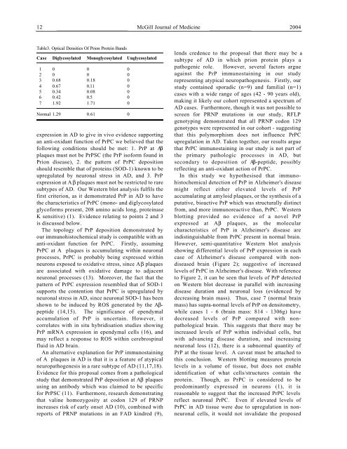
Table3. Optical Densities <strong>Of</strong> <strong>Prion</strong> <strong>Protein</strong> Bands<br />
C a s e Diglycosylated Monoglycosylated Unglycosylated<br />
1 0 0 0<br />
2 0 0 0<br />
3 0.68 0.18 0<br />
4 0.67 0.11 0<br />
5 0.34 0.08 0<br />
6 0.42 0.5 0<br />
7 1.92 1.71 0<br />
Normal 1.29 0.61 0<br />
expression in AD to give in vivo evidence supporting<br />
an anti-oxidant function of PrPC we believed that the<br />
following conditions should be met: 1. PrP at Aβ<br />
plaques must not be PrPSC (the PrP isoform found in<br />
<strong>Prion</strong> disease), 2. the pattern of PrPC deposition<br />
should resemble that of proteins (SOD-1) known to be<br />
upregulated by neuronal stress in AD, and 3. PrP<br />
expression at A β plaques must not be restricted to rare<br />
subtypes of AD. Our Western blot analysis fulfils the<br />
first criterion, as it demonstrated PrP in AD to have<br />
the characteristics of PrPC (mono- and diglycosylated<br />
glycoforms present, 208 amino acids long, proteinase<br />
K sensitive) (1). Evidence relating to points 2 and 3<br />
is discussed below.<br />
The topology of PrP deposition demonstrated by<br />
our immunohistochemical study is compatible with an<br />
anti-oxidant function for PrPC. Firstly, assuming<br />
PrPC at A plaques is accumulating within neuronal<br />
processes, PrPC is probably being expressed within<br />
neurons exposed to oxidative stress, since Aβ plaques<br />
are associated with oxidative damage to adjacent<br />
neuronal processes (13). Moreover, the fact that the<br />
pattern of PrPC expression resembled that of SOD-1<br />
supports the contention that PrPC is upregulated by<br />
neuronal stress in AD, since neuronal SOD-1 has been<br />
shown to be induced by ROS generated by the Aβpeptide<br />
(14,15). The significance of ependymal<br />
accumulation of PrP is uncertain. However, it<br />
correlates with in situ hybridisation studies showing<br />
PrP mRNA expression in ependymal cells (16), and<br />
may reflect a response to ROS within cerebrospinal<br />
fluid in AD brain.<br />
An alternative explanation for PrP immunostaining<br />
of A plaques in AD is that it is a feature of atypical<br />
neuropathogenesis in a rare subtype of AD (11,17,18).<br />
Evidence for this proposal comes from a pathological<br />
study that demonstrated PrP deposition at Aβ plaques<br />
using an antibody which was claimed to be specific<br />
for PrPSC (11). Furthermore, research demonstrating<br />
that valine homozygosity at codon 129 of PRNP<br />
increases risk of early onset AD (10), combined with<br />
reports of PRNP mutations in an FAD kindred (9),<br />
lends credence to the proposal that there may be a<br />
subtype of AD in which prion protein plays a<br />
pathogenic role. However, several factors argue<br />
against the PrP immunostaining in our study<br />
representing atypical neuropathogenesis. Firstly, our<br />
study contained sporadic (n=9) and familial (n=1)<br />
cases with a wide range of ages (42 - 90 years old),<br />
making it likely our cohort represented a spectrum of<br />
AD cases. Furthermore, though it was not possible to<br />
screen for PRNP mutations in our study, RFLP<br />
genotyping demonstrated that all PRNP codon 129<br />
genotypes were represented in our cohort - suggesting<br />
that this polymorphism does not influence PrPC<br />
upregulation in AD. Taken together, our results argue<br />
that PrPC immunostaining in our study is not part of<br />
the primary pathologic processes in AD, but<br />
secondary to deposition of Aβ-peptide, possibly<br />
reflecting an anti-oxidant action of PrPC.<br />
<strong>In</strong> this study we hypothesised that immunohistochemical<br />
detection of PrP in <strong>Alzheimer's</strong> disease<br />
might reflect either elevated levels of PrP<br />
accumulating at amyloid plaques, or the synthesis of a<br />
putative, bioactive PrP which was structurally distinct<br />
from, and more immunoreactive than, PrPC. Western<br />
blotting provided no evidence of a novel PrP<br />
expressed at Aβ plaques, as the molecular<br />
characteristics of PrP in <strong>Alzheimer's</strong> disease are<br />
indistinguishable from PrPC present in normal brain.<br />
However, semi-quantitative Western blot analysis<br />
showing differential levels of PrP expression in each<br />
case of <strong>Alzheimer's</strong> disease compared with nondiseased<br />
brain (Figure 2); suggestive of increased<br />
levels of PrPC in <strong>Alzheimer's</strong> disease. With reference<br />
to Figure 2, it can be seen that levels of PrP detected<br />
on Western blot decrease in parallel with increasing<br />
disease duration and neuronal loss (evidenced by<br />
decreasing brain mass). Thus, case 7 (normal brain<br />
mass) has supra-normal levels of PrP on densitometry,<br />
while cases 1 - 6 (brain mass: 814 - 1306g) have<br />
decreased levels of PrP compared with nonpathological<br />
brain. This suggests that there may be<br />
increased levels of PrP within individual cells, but<br />
with advancing disease duration, and increasing<br />
neuronal loss (12), there is a subnormal quantity of<br />
PrP at the tissue level. A caveat must be attached to<br />
this conclusion. Western blotting measures protein<br />
levels in a volume of tissue, but does not enable<br />
identification of what cells/structures contain the<br />
protein. Though, as PrPC is considered to be<br />
predominantly expressed in neurons (1), it is<br />
reasonable to suggest that the increased PrPC levels<br />
reflect neuronal PrPC. Even if elevated levels of<br />
PrPC in AD tissue were due to upregulation in nonneuronal<br />
cells, it would not invalidate the proposed