Fungal species isolated from Quercus castaneifolia in ... - Biodiversitas
Fungal species isolated from Quercus castaneifolia in ... - Biodiversitas
Fungal species isolated from Quercus castaneifolia in ... - Biodiversitas

You also want an ePaper? Increase the reach of your titles
YUMPU automatically turns print PDFs into web optimized ePapers that Google loves.
B I O D I V E R S IT A S ISSN: 1412-033X<br />
Volume 14, Number 2, October 2013 EISSN: 2085-4722<br />
Pages: 61-66<br />
<strong>Fungal</strong> <strong>species</strong> <strong>isolated</strong> <strong>from</strong> <strong>Quercus</strong> <strong>castaneifolia</strong> <strong>in</strong> Hyrcanian<br />
Forests, North of Iran<br />
MOHAMMAD REZA KAVOSI 1 , FERIDON FARIDI 1 , GOODARZ HAJIZADEH 2,<br />
1 Department of Forest Science, Faculty of Forest Ecology, Gorgan University of Agricultural Sciences and Natural Resources, Gorgan, Golestan, Iran.<br />
2 Department of Forestry, Faculty of Natural Resources, Sari University of Agricultural Sciences and Natural Resources, Sari, Mazandaran, Iran. Tel./fax.<br />
+98 151 3822715, email: goodarzhajizadeh@gmail.com<br />
Manuscript received: 6 May 2013, Revision accepted: 17 July 2013.<br />
ABSTRACT<br />
Kavosi MR, Feridon F, Hajizadeh G. 2013. <strong>Fungal</strong> <strong>species</strong> <strong>isolated</strong> <strong>from</strong> <strong>Quercus</strong> <strong>castaneifolia</strong> <strong>in</strong> Hyrcanian Forests, North of Iran.<br />
<strong>Biodiversitas</strong> 14: 61-66. In order to isolate and identify of fungi associated with <strong>Quercus</strong> <strong>castaneifolia</strong> seed, sampl<strong>in</strong>g carried out <strong>in</strong><br />
Shast-Kalate, Ghorogh, Loveh and Golestan forest. Collected seeds sterilized and then separated sections <strong>in</strong>clud<strong>in</strong>g: outer section of<br />
seed (crust) and <strong>in</strong>ner seed section (endosperm). Each section of seed tissue is cultured on potato dextrose agar media. After sub-culture<br />
and provid<strong>in</strong>g of the fungi pure cultures, various <strong>species</strong> <strong>isolated</strong> and identified by spores characteristics, their size and color, <strong>in</strong>clud<strong>in</strong>g:<br />
Aspergillus flavus, A. niger, Curvularia aff<strong>in</strong>is, Trichoderma harzianum, Trichothecium roseum, Eurotium rubrum, E. amstelodami,<br />
Penicillium implicatum, P. fellutanum, Diplodia sp. Nigrospora gossypi, Alternaria alternata , Fusarium oxysporum and Beltrania<br />
santapaui. The most frequency of fungus <strong>in</strong> Shast-Kalate forest was P. implicatum by 74% of frequency with<strong>in</strong> seed section, the most<br />
frequency <strong>in</strong> Ghorogh and Loveh Forest was P. fellutanum with 63 and 66% of frequency with<strong>in</strong> seed section respectively and the most<br />
frequency <strong>in</strong> Golestan forest was B. santapaui by 51% of frequency outer seed section. The result showed diversity of the fungi on the<br />
outer seed section is higher than with<strong>in</strong> seed section. The results also showed dur<strong>in</strong>g several isolation a saprophyte fungus always could<br />
be f<strong>in</strong>d<strong>in</strong>g on the acorn seeds. This is the comprehensive report on fungi associated with <strong>Quercus</strong> <strong>castaneifolia</strong> seed <strong>in</strong> Hyrcanian forest,<br />
North of Iran.<br />
Key words: crust, endosperm, fungal, Hyrcanian forest, <strong>Quercus</strong> <strong>castaneifolia</strong>, seed<br />
INTRODUCTION<br />
Forest tree seeds cont<strong>in</strong>uously are affected by physical<br />
and physiological disturbance which most of these diseases<br />
caused by fungi. Health and growth ability sapl<strong>in</strong>g<br />
considerably depend on seed quality (Mittal and Mathur<br />
1998). Most of fungi associate with seeds of forest trees are<br />
molds which expand on surface of seed and sometime they<br />
are <strong>in</strong>ner pollution factor (Huss 1956). Effect of mold on<br />
seeds is that they seem health apparently but they orig<strong>in</strong>ally<br />
have spoiled on basis of vitality considerable (Shea 1957).<br />
Recently known that all seeds conta<strong>in</strong> microscopic fungi<br />
spores whether on surface of seed or <strong>in</strong>side of seed (S<strong>in</strong>gh<br />
and Mathur 1993). Urosevic (1961) was specified that<br />
some of fungi spores have germ<strong>in</strong>ated and after grow<strong>in</strong>g<br />
and mycelium penetrat<strong>in</strong>g, it <strong>in</strong>fluences <strong>in</strong> to the cotyledon<br />
which through have nourished <strong>from</strong> germs.<br />
Fungus associate with seed can cause weakness of seed<br />
germ<strong>in</strong>ation directly and <strong>in</strong>directly and can dispose these<br />
seeds to earthborn pathogen fungi attack ( Gibson 1957).<br />
Healthy seed <strong>in</strong> forest for natural regeneration is important<br />
issues which future life forest depends on it. Disease and<br />
damaged seeds even under suitable environmental<br />
condition cannot have desired regeneration for forest<br />
survival or cannot cause specific <strong>species</strong>. The trees<br />
appeared <strong>from</strong> damaged and diseased seeds have slight<br />
growth and seeds produced by these trees will be had low<br />
vitality (Rai and Mamatha 2005).<br />
The purpose of this study was identification of <strong>in</strong>ner<br />
and outer fungi of Chestnut-leaved oak ( <strong>Quercus</strong><br />
<strong>castaneifolia</strong>) seed and specify<strong>in</strong>g their frequency <strong>in</strong><br />
Golestan prov<strong>in</strong>ce forests.<br />
MATERIALS AND METHODS<br />
Sampl<strong>in</strong>g site<br />
In this research four forest regions <strong>in</strong> Golestan<br />
prov<strong>in</strong>ce, north of Iran <strong>in</strong>clud<strong>in</strong>g (i) Shast-Kalate Research<br />
and Education Forest, (ii) Ghorogh Forest Park, (iii) Loveh<br />
Research Forest, and (iv) Golestan National Park were<br />
selected (Figure 1). In each region, four trees were chosen<br />
randomly, and 25 seeds of each tree were selected randomly.<br />
Collected seeds have been settled <strong>in</strong> new and sterile bags<br />
and after record<strong>in</strong>g region specification and collection date,<br />
they transferred to laboratory and settled <strong>in</strong> a place with<br />
suitable temperature and ventilation. In f<strong>in</strong>ally, four<br />
samples of 25 k<strong>in</strong>ds of seeds of each region were collected.<br />
Isolation and purification seed fungi<br />
For fungus isolation, seeds were divided less segments<br />
and also <strong>in</strong>ner (crust) and outer (endosperm) portion.<br />
Separation of <strong>in</strong>ner portion <strong>from</strong> outer portion of seed
62<br />
B I O D I V E R S IT A S 14 (2): 61-66, October 2013<br />
Golestan<br />
D<br />
C<br />
A<br />
B<br />
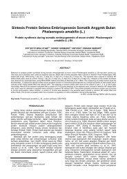
Figure 1. Location of the study site <strong>in</strong>side the Hyrcanian forests of Golestan Prov<strong>in</strong>ce, North of Iran, <strong>in</strong>clud<strong>in</strong>g: A. Shastkalate Research<br />
and Education Forest, B. Ghorogh Forest Park, C. Loveh Research Forest, and D. Golestan National Park.<br />
due to compar<strong>in</strong>g mentioned two portion fungi was<br />
conducted. After seed was divided less segment, its surface<br />
was dis<strong>in</strong>fected with 0.5% sodium hypochlorite and ethanol<br />
dur<strong>in</strong>g 1-2 m<strong>in</strong>utes and was washed three times with sterile<br />
Distilled water and settled <strong>in</strong> to sterile filter paper for<br />
desiccat<strong>in</strong>g. Then segments of <strong>in</strong>ner and outer portion of<br />
seed separately and with four repetitions on nutrition<br />
medium of potato-dextrose-agar (PDA) extract conta<strong>in</strong><br />
lactic acid and preserved <strong>in</strong> <strong>in</strong>cubator <strong>in</strong> 25±1°C. After<br />
three days, grown fungi were subculture on medium and<br />
hereby fungi become sterilization.<br />
Identification of fungi<br />
Identification of fungi genus after their grow<strong>in</strong>g on the<br />
seed segments was used valid reference of Barnett and<br />
Hunter (1998) and Ellis (1976) and their classification on<br />
basis of Eriksson (2006) and Alexopoulos et al. (1996). For<br />
identification of <strong>species</strong> was used various medium and<br />
valid reference. Further <strong>species</strong> were identified and purified<br />
on PDA medium and 25°C <strong>in</strong> absolute darkness. For some<br />
of <strong>species</strong> like Fusarium which do not know spore<br />
Carnation leaf-piece Agar (CLA) medium and optical<br />
period LD 12:12 <strong>in</strong> 25°C accord<strong>in</strong>g to Nelson et al. (1983)<br />
and Saremi (1998) method were used. For Alternaria, for<br />
denot<strong>in</strong>g spore number <strong>in</strong> spore cha<strong>in</strong>, LD 16:8 optical<br />
period <strong>in</strong> 20-23°C and Agar-Water (AW) medium <strong>in</strong><br />
addition to PDA were used accord<strong>in</strong>g to D<strong>in</strong>gra and<br />
S<strong>in</strong>clair (1995) method and on basis of Ellis (1971, 1976)<br />
cognition key. About Trichoderma, LD 12:12 optical<br />
period and 25°C accord<strong>in</strong>g to D<strong>in</strong>gra and S<strong>in</strong>clair (1995)<br />
method and, Kubicek and Harman (1998) cognition key<br />
were used. Pitt (1997, 2000) cognition keys was applied for<br />
identification of Eurotium, Aspergillus, Penicillium fungi.<br />
Litv<strong>in</strong>ov (1967) and Ellis (1976) description the<br />
identification of Trichothecium, Curvularia, Beltrania and<br />
Nigrospora fungi and Barnett & Hunter (1998) for<br />
identification Diplodia fungus.<br />
This identification was on basis of various criteria<br />
such as presence or absence of septum; shape and size of<br />
ascus; ascospore; conidia and phialid, k<strong>in</strong>d of ascospore;<br />
number of ascospore <strong>in</strong> each ascus; number of conidia laid<br />
on conidiophore or phialid; be<strong>in</strong>g one or more cellular of<br />
ascospore and conidia; <strong>in</strong> some <strong>species</strong>, presence or<br />
absence of metulae; diameter growth of colonies; colonies<br />
color and made.<br />
RESULTS AND DISCUSSION<br />
Species specification<br />
Results of this study showed that all seeds polluted<br />
with one or more <strong>species</strong> of separated fungi which most of<br />
them were imperfect fungi or<br />
Ascomycetes. After<br />
sterilization and specification of thallus and colonies, 12<br />
<strong>species</strong> <strong>in</strong>clud<strong>in</strong>g: Nigrospora gossypii, Aspergillus flavus,<br />
A. niger, Trichoderma harzianum, Alternaria alternate,<br />
Trichothecium roseum, Fusarium oxysporum, Beltrania<br />
santapaui, Penicillium implicatum, Eurotium rubrum,<br />
Curvularia aff<strong>in</strong>is and Diplodia sp. become isolation and<br />
identification on <strong>Quercus</strong> <strong>castaneifolia</strong> seeds that<br />
frequency and description of specification of each one <strong>in</strong><br />
detail is follow<strong>in</strong>g <strong>in</strong> Table 1. All identification fungi on Q.<br />
<strong>castaneifolia</strong> seeds were reported <strong>from</strong> Hyrcanian Forests,<br />
North of Iran for the first time.<br />
Alternaria alternata (Fr.) Keissl.<br />
Colonies usually was approximately olivaceous to black<br />
and sometimes grey with pubescent appearance on PDA<br />
medium and 25°C. Colonies diameter growth after three<br />
days was 3-3.5 cm (Figure 2A). Conidiophores were partly<br />
small with 7-10×43-50 μm dimensions, simple and<br />
branched, approximately brown and even surface. Conidia<br />
were formed on WA medium <strong>in</strong> 6 to 17 fold cha<strong>in</strong>s (Figure<br />
2C) and ovoid to obclavate or pear form and conta<strong>in</strong>
KAVOSI et al. – <strong>Fungal</strong> <strong>species</strong> <strong>isolated</strong> <strong>from</strong> <strong>Quercus</strong> castanifolia 63<br />
Table 1. Fungi frequency percent of <strong>in</strong>ner and outer portion of<br />
oak seed <strong>in</strong> four regions <strong>in</strong> Golestan prov<strong>in</strong>ce<br />
Regions<br />
Fungi<br />
Shastkalate<br />
park<br />
park<br />
Ghorogh<br />
Golestan<br />
Loveh<br />
I O I O I O I O<br />
Alternaria alternata 0 0 20 30 13 0 16 0<br />
Diplodia sp. 0 23 0 0 0 0 0 0<br />
Aspergillus flavus 27 46 0 8 0 0 5 3<br />
Aspergillus niger 11 16 16 14 0 0 0 0<br />
Curvularia aff<strong>in</strong>is 39 0 0 0 0 0 0 0<br />
Eurotium rubrum 0 35 0 34 0 0 0 0<br />
Nigrospora gossypii 0 0 0 17 0 15 0 0<br />
Penicillium implicatum 74 31 39 26 39 18 11 6<br />
Beltrania santapaui 0 0 0 0 0 56 0 51<br />
Fusarium oxysporum 0 0 0 0 0 35 0 19<br />
Trichothecium roseum 0 41 13 5 0 0 0 0<br />
Trichoderma harzianum 0 7 0 0 0 0 0 0<br />
Note: I = <strong>in</strong>ner, O = outer<br />
surface covered by t<strong>in</strong>y tubers. Conidia have 2 to 7<br />
transverse walls and 2 to 4 vertical walls and 12-34×6.5-<br />
12.5 μm dimensions and the end of the conidium nearest<br />
the conidiophore was round while it tapers towards the<br />
apex with 2.5-4 μm width (Figure 2B).<br />
Diplodia sp.<br />
Colonies was specified with whitish yellow on PDA<br />
medium and 25°C ( Figure 3A). Pycnidia were black,<br />
<strong>in</strong>dividual, spherical and stomatous (Figure 3B).<br />
Conidiophores were t<strong>in</strong>y and simple and conidia were dark,<br />
bicellular, 5-7 μm, elliptical or ovoid and <strong>in</strong> some of them<br />
there was curve (Figure 3C).<br />
Aspergillus flavus L<strong>in</strong>k<br />
Colonies on PDA medium and 25°C was olive to lime<br />
green with a cream reverse. Colonies has fast diameter<br />
growth, after three days it was about 5.2 cm and has woolly<br />
to cottony texture which conta<strong>in</strong> small granular ( Figure<br />
4A). Hyphae have light and septum. Conidia were settled<br />
on vesicle radially or perpendicular. Conidiophores were<br />
coarse and colourless and up to 800 μm length and 15-20<br />
μm width. Vesicles were spherical to semi spherical (20-40<br />
μm) and phialids (3-4×8-12 μm) covered approximately all<br />
surface of vesicle. Conidia were 3-6 μm, even, t<strong>in</strong>y and<br />
spherical to semi spherical (Figure 4B).<br />
Aspergillus niger Tiegh. nom. cons.<br />
Colonies at first was white but due to produc<strong>in</strong>g conidia<br />
become black and the reverse side seemed light yellow or<br />
pail on PDA medium and 25°C which dur<strong>in</strong>g the growth,<br />
they produced radial gaps on medium ( Figure 5A).<br />
Colonies diameter growth after three days was 4.3 cm.<br />
Hyphae had transparent and septum that conidia were<br />
settled on vesicle radially. This <strong>species</strong> produced metulae.<br />
Conidiophores were long (400 to 3000 μm), even and<br />
transparent that were dark <strong>in</strong> tip and end to bubble or cell<br />
of spherical vesicle (30 -75 μm). Metulaes and phialids<br />
cover all surface of vesicle. Conidia were brown to black,<br />
uneven and with tuber, spherical and 4-5 μm (Figure 5B).<br />
Curvularia aff<strong>in</strong>is Boedijn.<br />
Colonies was black to dark greenish black on PDA<br />
medium and 25°C which <strong>in</strong> white marg<strong>in</strong>s, colonies texture<br />
was cotton and its diameter growth after three days was 4.4<br />
cm ( Figure 6A). Conidiophores mostly were simple and<br />
had spores that formed with two sympodial geniculate.<br />
Conidia were 2 to 4 cells, 23-33×8-14 μm, fusiform and<br />
curve so that the central cell was typically darker and<br />
enlarged compared to the end cells <strong>in</strong> the conidium and the<br />
swell<strong>in</strong>g of the central cell usually gave the conidium a<br />
curved appearance (Figure 6B).<br />
Eurotium rubrum Jos. König etal Ba<strong>in</strong>ier & Sartory<br />
Colonies on PDA medium and 25°C was reddish<br />
orange that <strong>in</strong> white marg<strong>in</strong>, colonies texture was cotton<br />
and its diameter growth after three days was 3.5 cm (Figure<br />
7A). Cleistothecia were occurred spherical to nearly<br />
elliptical form and with yellow colour ( Figure 7B).<br />
Ascuses were almost egg form to elliptical, 12-13 μm and<br />
they had a t<strong>in</strong>y and unstable wall. Ascospores were<br />
unicellular, oblate (like a flattened sphere) and have<br />
equatorial ridges, thus resembl<strong>in</strong>g pulleys, 4.4-5×6.2-6.8<br />
μm, elliptical, yellow with even marg<strong>in</strong> and with eight fold<br />
form <strong>in</strong> to the Ascus (Figure 7C).<br />
Nigrospora gossypii Jacz.<br />
Colonies on PDA medium and 25°C was dark grey with<br />
small and large while po<strong>in</strong>t <strong>in</strong> its background which were<br />
basically cotton form that stick fungus mycelium to top of<br />
the conta<strong>in</strong>er. It’s colour was black grey and approximately<br />
dark-blue beh<strong>in</strong>d of conta<strong>in</strong>er and colonies diameter<br />
growth after 3 days was 7-7.5 cm ( Figure 8A).<br />
Conidiophores were simple, transparent and were settled<br />
vertically on mycelium which their length was 10-12.5 μm.<br />
Conidia were black, unicellular and semi spherical and<br />
partiy elliptical form with even surface and flat section that<br />
their size was 11-15 μm. This fungus also had middle<br />
chlamydospores (Figure 8B).<br />
Penicillium implicatum Biourge<br />
Colonies on PDA medium and 25°C at first was cotton<br />
white that f<strong>in</strong>ally will become powdery blue-green (Figure<br />
9A). Colonies diameter growth after 3 days was 2.6 cm.<br />
Conidiophores were out of growth mycelium <strong>in</strong>dividually<br />
and ended to phialids. Conidiophore height was 25-50 μm<br />
and phialids length was 8-10 μm. Conidia were spherical,<br />
dark green, 2-3.5 μm, unicellular, and were formed <strong>from</strong><br />
cha<strong>in</strong>s which youngest conidia settled <strong>in</strong> base of cha<strong>in</strong><br />
(Figure 9B).<br />
Beltrania santapaui Pirozynski & Patil<br />
Colonies on PDA medium and 25°C was grayish dark<br />
brown which beh<strong>in</strong>d of conta<strong>in</strong>er was light grey and<br />
colonies growth on PDA medium after three days was 3.5<br />
cm ( Figure 10A). Conidiophores were simple and had<br />
septum which at the end, they were branched and conidia<br />
were formed on each of these branches ( Figure 10B).<br />
Conidiophore length was 87.5 μm and conidia were seen
64<br />
B I O D I V E R S IT A S 14 (2): 61-66, October 2013<br />
2A 2B 2C<br />
A<br />
3A<br />
3B<br />
3C<br />
4A<br />
4B<br />
5A<br />
5B 6A 6B<br />
b 1<br />
7A 7B 7C 6C<br />
8A 8B<br />
9A 9B 10A 10B 10C<br />
11A 11B 11C
KAVOSI et al. – <strong>Fungal</strong> <strong>species</strong> <strong>isolated</strong> <strong>from</strong> <strong>Quercus</strong> castanifolia 65<br />
a<br />
12A 12B 13A 13B<br />
Figure 1. Alternaria alternata (a: colony on PDA medium, b: conidia, c: fold cha<strong>in</strong>s on WA medium). Bar = 300 µm<br />
Figure 3. Diplodia sp. (a: colony on PDA medium, b: pycnidia, c: conidia). Bar = 300 µm<br />
Figure 4. Aspergillus flavus (a: colony on PDA medium, b: conidiophore and conidia). Bar = 300 µm<br />
Figure 5. Aspergillus niger (a: colony on PDA medium, b: conidiophore and conidia). Bar = 20 µm<br />
Figure 6. Curvularia aff<strong>in</strong>is (a: colony on PDA medium, b: conidiophore and conidia). Bar = 20 µm<br />
Figure 7. Eurotium rubrum (a: colony on PDA medium, b: cleistothecia, bar = 150 µm, c: ascus and ascospore). Bar = 30 µm<br />
Figure 8. Nigrospora gossypii (a: colony on PDA medium, b: conidiophore and, b 1 : conidia). Bar = 20 µm<br />
Figure 9. Penicillium implicatum (a: colony on PDA medium, b: conidiophore and conidia). Bar = 20 µm<br />
Figure 10. Beltrania santapaui (a: colony on PDA medium, b: conidiophore and conidia, bar = 45 µm, c: conidia). Bar = 30 µm<br />
Figure 11. Fusarium oxysporum (a: colony on PDA medium, b: monophialide, bar = 75 µm, c: macroconidia and microconidia). Bar = 30 µm<br />
Figure 12. Trichothecium roseum (a: colony on PDA medium, b: conidiophore and conidia). Bar = 20 µm<br />
Figure 13. Trichoderma harzianum (a: colony on PDA medium, b: conidiophore and conidia). Bar = 20 µm<br />
bicellular and elliptical form with one appendage that was<br />
dark brawn. Conidia size without appendage was equal to<br />
5-7.5×15-21 μm and its length was 2.5-3.5 μm (Figure<br />
10C).<br />
Fusarium oxysporum Schltdl.<br />
Colonies diameter growth was measured 3.1-3.8 cm on<br />
PDA medium and 25°C, its color at first was light p<strong>in</strong>kish<br />
white and f<strong>in</strong>ally become violet that its center was lighter<br />
and its marg<strong>in</strong> was dark violet. Mycelium was cotton and<br />
scatter that were condensed by growth completion. Beh<strong>in</strong>d<br />
of conta<strong>in</strong>er <strong>in</strong> the marg<strong>in</strong> was dark violet and <strong>in</strong> the center<br />
was opaque orange (Figure 11A). Middle chlamydospores<br />
were formed frequently on mycelium. Macroconidia on<br />
abundant sporodochia that were sickle <strong>from</strong> and partly<br />
longitude, most of them had three t<strong>in</strong>y septum and their<br />
length was 3-5×24-30 μm (Figure 11C). Macroconidia and<br />
also Microconidia on short and <strong>in</strong>dividual phialides were<br />
formed which microconidia were false-heads on these<br />
phialides ( Figure 11B). Microconidia were most of time<br />
egg form or longitude elliptical unicellular or kidney form<br />
(Figure 11C).<br />
Trichothecium roseum (Pers.) L<strong>in</strong>k<br />
Colonies approximately grow up rapidly. Its diameter<br />
growth after three days on PDA medium and 25°C was 2.8<br />
cm and was whitish light p<strong>in</strong>k and partly powder form<br />
(Figure 12A). Until first conidia produce, conidiophores<br />
were not separation <strong>from</strong> growth section hyphae. They<br />
were vertical, without branch and most of time they had<br />
septum nearby base of conidiophore. Two conidia were<br />
formed alternatively and with overlap <strong>in</strong> tip of<br />
conidiophore. Conidia were bicellular, elliptical or pear<br />
form with jo<strong>in</strong>t place to curve, transparent, even to partly<br />
coarse and were 11-16×7-10 μm (Figure 12B).<br />
Trichoderma harzianum Rifai<br />
Colonies on PDA medium had rapid growth which at<br />
first was cotton white but after 2 days was approximately<br />
light green ( Figure 13A). hyphae had septum were<br />
branched and 2.5-5.5 μm diameter. Chlamydospores at the<br />
end or <strong>in</strong> the middle of hyphae were elliptical to fusiform<br />
with even wall and 8.5-10×5-7.5 μm diameter.<br />
Conidiophores were branched that through end of<br />
conidiophore, length of these branchs were smaller.<br />
Phialids were short, bar form, on the base they were<br />
narrower than middle area and on top of its conic, its<br />
dimensions were 5-7.5×2.5-3.5 μm. Conidia <strong>in</strong>dividually<br />
collected at the end of Phialids and were egg form to<br />
spherical with even wall and 2.5-3.4 μm dimensions<br />
(Figure 13B).<br />
Discussion<br />
In this study, specified that Penicillium fungus rather<br />
than identified fungi have more frequency which its <strong>species</strong><br />
had most frequency <strong>in</strong> all range site. This genus along with<br />
fungi such as Fusarium and Trichoderma caused for<br />
discolor of seeds (Dawn 2003).<br />
Fungi grow both on seed crust and on seed cotyledon<br />
but a variety of fungi of seed crust are more than cotyledon.<br />
In <strong>in</strong>ward section, Penicillium had further frequency <strong>in</strong> all<br />
regions whilst Trichoderma become isolation on seed crust.<br />
This is corresponded to W<strong>in</strong>ston (1956) and Dawn (2003)<br />
studies which <strong>isolated</strong> Penicillium, Fusarium and<br />
Trichoderma fungi on Red Oak (<strong>Quercus</strong> rubra) seeds.<br />
Dorsey et al. (1962) separated Penicillium on seeds of<br />
Q. velut<strong>in</strong>a and Q. rubra. Aspergillus and Penicillium are<br />
genuses that have generality <strong>in</strong> color change of cotyledon<br />
and even seed crust and f<strong>in</strong>ally causal lesion and crack on<br />
seed crust (Swiecki et al. 1991). In Swiecki studies,
66<br />
B I O D I V E R S IT A S 14 (2): 61-66, October 2013<br />
Fusarium oxysporum and Trichothecium sp. obta<strong>in</strong> on seed<br />
and seedl<strong>in</strong>g of Q. macrocarpa <strong>in</strong> northern California<br />
which similar to our study.<br />
In this study, <strong>isolated</strong> Fusarium that was <strong>isolated</strong> on Q.<br />
alba and Q. macrocarpa seeds by Vozzo (1984). Agbaba<br />
and Gradecki (2005) <strong>isolated</strong> ciboria batschiana,<br />
Phomopsis quercella, Fusarium sp. Ophiostoma sp.<br />
Penicillium sp. Trichothecium roseum, and Trichoderma<br />
veride <strong>from</strong> Q. pubescens seeds which Fusarium,<br />
Penicillium, Trichoderma genus and Trichothecium roseum<br />
<strong>species</strong> is corresponded to our study.<br />
Tiberi et al. (2002) <strong>isolated</strong> Fusarium solani, Fusarium<br />
eumartii, Verticillium dahliae, Diplodia mutila and<br />
Phomopsis querc<strong>in</strong> <strong>from</strong> oaks seed of Italy which Diplodia<br />
and Fusarium is observed <strong>in</strong> our study. Gallego et al.<br />
(1999) <strong>isolated</strong> Fusarium oxysporum seen <strong>in</strong> our study<br />
<strong>from</strong> Q. ilex seed for test<strong>in</strong>g of be<strong>in</strong>g pathogen the fungi.<br />
Santos et al. (2005) and Mer ouani et al. (2001) separated<br />
many fungi on Q. suber seed which among them can be<br />
referred to Penicilium implicatum, Trichoderma<br />
harzianum, Trichotecium roseum, Fusarium oxysporum,<br />
Diplodia mutila, Aspergillus niger, Aspergillus flavus and<br />
Alternaria alternata that whole of these <strong>species</strong> were seen<br />
<strong>in</strong> our study. Also some studies conducted on fungi along<br />
with major forest trees that fungus similar our study was<br />
<strong>in</strong>clud<strong>in</strong>gAlternaria, Fusarium, Aspergillus, Penicillium,<br />
Trichoderma, and Beltrania (Vladimir et al. 2005; Swapna<br />
and Nagaveni 2008).<br />
CONCLUSION<br />
The results show that all acorn seeds collected were<br />
<strong>in</strong>fected with one or more <strong>species</strong> of fungi have been<br />
<strong>isolated</strong> which are often classified to Ascomycetes fungi.<br />
S<strong>in</strong>ce the length of oak seed dormancy and physiological<br />
process is very long period. This could be due to<br />
opportunistic fungi such as contact with the surface of the<br />
seed coat and the seed easily reach and thereby is prevented<br />
<strong>from</strong> germ<strong>in</strong>at<strong>in</strong>g. This is the comprehensive report on<br />
fungi associated with <strong>Quercus</strong> <strong>castaneifolia</strong> seed <strong>in</strong><br />
Hyrcanian forest, North of Iran.<br />
REFERENCES<br />
Agbaba SN, Gradecki M. 2005. Health condition of common oak acorn<br />
(<strong>Quercus</strong> pubescens) and protection measures <strong>in</strong> Croatia. 5 th ISTA-<br />
SHC Seed Health Symposium. 10-13 May 2005, Angers France. 42-<br />
43.<br />
Alexopoulos CJ, Mims CW, Blackwell M. 1996. Introductory Mycology.<br />
4 th ed. John Wiley and Sons, New York.<br />
Barnett HL, Hunter BB. 1998. Illustrated Genera of Imperfect Fungi. 4 th<br />
ed. ASP Press, St. Paul, M<strong>in</strong>nesota, USA.<br />
Dh<strong>in</strong>gra OD, S<strong>in</strong>clair JB. 1985. Basic Plant Pathology Methods. CRC<br />
Press, Boca Raton, FL.<br />
Dorsey CK, Tryon EH, Carvell KL. 1962. Insect damage to acorns <strong>in</strong><br />
West Virg<strong>in</strong>ia and control studies us<strong>in</strong>g granular systematic<br />
<strong>in</strong>sectidies. Econ Entomol 55: 885-888.<br />
Ellis MB. 1971. Dematiaceous Hyphomycetes. C.A.B International<br />
Mycological Institute, Kew, UK.<br />
Ellis MB. 1976. More dematiaceous Hyphomycetes. C.A.B International<br />
Mycological Institute, Kew, UK.<br />
Eriksson OE. 2006. Outl<strong>in</strong>e of Ascomycota. Myconet. www.field<br />
museum. org/myconet/pr<strong>in</strong>ted_v12_a. asp: 1-82.<br />
Gallego FJ, de Algaba AP, Fernandez-Escobar R. 1999. Etiology of oak<br />
decl<strong>in</strong>e <strong>in</strong> Spa<strong>in</strong>. Eur J For Path 29: 17-27.<br />
Gibson IAS. 1957. Saprophytic fungi as destroyers of germ<strong>in</strong>at<strong>in</strong>g p<strong>in</strong>e<br />
seeds. E Afr Agric For J 22: 203-206.<br />
Huss E. 1956. Research <strong>in</strong>to damage to tree seeds by dew<strong>in</strong>g<strong>in</strong>g.<br />
Skogsforsk<strong>in</strong><strong>in</strong>gs-Institute, Stockholm.<br />
Kubicek CP, Harman GE. 1998: Trichoderma and Gliocladium. Vol. 1.<br />
Basic Biology, Taxonomy and Genetics. Taylor & Francis, London.<br />
Litv<strong>in</strong>ov AM. 1967. Identify Microscopic Soil-born Fungus. Len<strong>in</strong>grad<br />
Science Publisher, Len<strong>in</strong>grad.<br />
Merouani H, Branco C, Almeida MH, Pereira JS. 2001. Effect of acorn<br />
storage duration and parental tree on emergence and physiological<br />
status Cork oak (<strong>Quercus</strong> suber L.) seedl<strong>in</strong>gs. Ann For Sci 58: 534-<br />
554.<br />
Mittal RK, Mathur SB. 1998. Seed Pathology. Indian Council of<br />
Agricultural Research, New Delhi, India, and Danish Government<br />
Institute of Seed Pathology, Denmark.<br />
Nelson PE, Toussoun TA, Marasas WFO. 1983. Fusarium <strong>species</strong>: An<br />
illustrated manual for identification. Penn State University. University<br />
Park, Pennsylvania.<br />
Pitt JI, Hock<strong>in</strong>g AD. 1997. Fungi and food spoilage. 2 th ed. Blackie<br />
Academic & Professional, Chapman & Hall, London.<br />
Pitt JI. 2000. A Laboratory Guide to Common Penicillium Species. 3 th ed.<br />
N.S.W. Food Science Australia, North Ryde.<br />
Rai VR, Mamatha T. 2005. Seedl<strong>in</strong>g diseases of some important forest<br />
tree <strong>species</strong> and their management. In: Diseases and Insects <strong>in</strong> Forest<br />
Nurseries. Proceed<strong>in</strong>gs of the 5 th Meet<strong>in</strong>g of IUFRO Work<strong>in</strong>g Party<br />
S7.03.04, May 6-8 2003, at Peechi, Kerala, India.<br />
Saremi H. 1998. Ecology and Taxonomy of Fusarium Species. Ferdowsi<br />
University of Mashhad, Mashhad.<br />
Santos MN, Braganca MH, Casimiro PP. 2005. Cork oak associated<br />
microorganisms throughout cork manufacture process. EFN 13 (1):<br />
75-93.<br />
Shea KR. 1957. Problem analysis: Molds of forest tree seed.<br />
Weyerhaeuser Timber Company, Forestry Research Centre, [Place of<br />
publication unknown].<br />
S<strong>in</strong>gh P, Mathur SB. 1993. Disease problems of forest tree seeds:<br />
diagnosis and management. 309-324. In Proc. IUFRO Symp. On Tree<br />
Seed Problems, with special reference to Africa. Project Group P.<br />
2.04.00-Seed Problems, Ougadougou, Burk<strong>in</strong>a Faso, 23-28 Nov.<br />
Swapna PK, Nagaveni HC. 2008. Seed health problems and their impact<br />
on seedl<strong>in</strong>g production. National Sem<strong>in</strong>ar on Medic<strong>in</strong>al plants and<br />
herbal products. S.V University, Tirupati, A.P. on 7-9th March 2008.<br />
Swiecki TJ, Bernhardt EA, Arnnold RA. 1991. Insect and disease impacts<br />
on blue oak acorns and seedl<strong>in</strong>gs. Pages 149-155 <strong>in</strong> Standiford RB,<br />
technical coord<strong>in</strong>ator. Proceed<strong>in</strong>gs of the symposium on oak<br />
woodlands and hardwood rangeland management; October 31-<br />
November 2, 1990; Davis, California. General Technical Report<br />
PSW-GTR-126. USDA Forest Service, Pacific Southwest Research<br />
Station, Berkeley, California, USA.<br />
Tiberi R, Alessandro RA, Marianelli L, Peverieri S, Roversi PF. 2002.<br />
Insects and Fungi Involved <strong>in</strong> Oak Decl<strong>in</strong>e <strong>in</strong> Italy. IOBC/wprs<br />
Bullet<strong>in</strong>.<br />
Urosevic B. 1961. The <strong>in</strong>fluence of saprophytic and semi-parasitic fungi<br />
on the germ<strong>in</strong>ation of Norway spruce and Scots p<strong>in</strong>e seeds. Proc Int<br />
Seed Test Assoc 26 (3): 537-556.<br />
Vladimir L, Zlatan R, Bozica J. 2005: Mycoses of forest seed <strong>in</strong> object for<br />
production and warehouse. Bull Fac For Univ Banja Luka 4: 15-30.<br />
Vozzo JA. 1984. Insects and fungi associated with acorns of <strong>Quercus</strong> sp.<br />
Department of Agriculture, Forest Service, and Southeastern Forest<br />
Experiment Station. No. 6: 40-43.<br />
Wash<strong>in</strong>gton DM. 2003. Fungi associated with northern red oak (<strong>Quercus</strong><br />
rubra) acorns. [M.Sc. Thesis]. West Virg<strong>in</strong>ia University.<br />
Morgantown, WV.<br />
W<strong>in</strong>ston PW. 1956. The acorn microsphere, with special reference to<br />
arthropods. Ecology 37: 120-132.