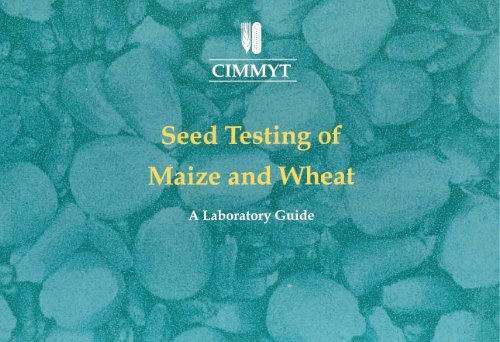
Seed Testing of Maize and Wheat A Laboratory Guide - Search ...
Seed Testing of Maize and Wheat A Laboratory Guide - Search ...
Seed Testing of Maize and Wheat A Laboratory Guide - Search ...

Create successful ePaper yourself
Turn your PDF publications into a flip-book with our unique Google optimized e-Paper software.
'I <br />
CIMMYT<br />
Sustainable <br />
<strong>Maize</strong> <strong>and</strong> <strong>Wheat</strong> <br />
Systems fol' the POOl' <br />
<strong>Seed</strong> <strong>Testing</strong> <strong>of</strong> <strong>Maize</strong> <strong>and</strong> <strong>Wheat</strong><br />
A <strong>Laboratory</strong> <strong>Guide</strong><br />
E.}. Warham - CIMMYT<br />
L.D. Butler - CIMMYT<br />
B.C. Sutton - IMI
Acknowledgements <br />
The authors gratefully<br />
acknowledge the Overseas<br />
Development Administration (UK)<br />
for providing the funds to cover the<br />
costs <strong>of</strong> producing this publication.<br />
This manual could not have been<br />
developed without their financial<br />
support, <strong>and</strong> their continued<br />
enthusiasm <strong>and</strong> support for this<br />
project is much appreciated.<br />
Special thanks go to Mr. Roger<br />
Smith for his encouragement <strong>and</strong><br />
support since the initial idea <strong>of</strong> the<br />
project.<br />
In addition, the help <strong>and</strong> advice<br />
from colleagues at CIMMYT,<br />
Mexico <strong>and</strong> the International<br />
Mycological Institute, UK, are<br />
greatly appreciated. Special thanks<br />
go to: Consuelo Rodriguez <strong>and</strong><br />
Clarissa Sanchez at CIMMYT for<br />
preparation <strong>of</strong> seed health tests<br />
used for the photographs; <strong>and</strong> Dr.<br />
Mark Holderness <strong>and</strong> Dr. Jim<br />
Waller at IMI for their constructive<br />
review <strong>and</strong> contribution to the<br />
contents <strong>and</strong> design <strong>of</strong> the manual.<br />
The authors are also grateful to<br />
CIMMYT's Information Services<br />
Unit, under the leadership <strong>of</strong> Mr.<br />
Tiffin Harris, for all the help <strong>and</strong><br />
support in the preparation <strong>of</strong> this<br />
manual. Special thanks go to<br />
Miguel Mellado, for his exceptional<br />
job <strong>of</strong> designing <strong>and</strong> layout, Eliot<br />
Sanchez for helping to prepare the<br />
manual for the printer, <strong>and</strong> to Alma<br />
McNab for her editorial support.<br />
Contents<br />
Foreword<br />
ii<br />
Introduction<br />
iii<br />
List <strong>of</strong> Organisms<br />
iii<br />
Key to Identification<br />
iv<br />
Descriptions <strong>of</strong> Organisms 1<br />
Index 65<br />
Annex A<br />
<strong>Seed</strong> Health Tests 66<br />
Annex B<br />
<strong>Seed</strong> Viability, Germination <strong>and</strong> Vigour Tests 75<br />
Photographs<br />
L. Butler, CIMMYT: 199, 200<br />
L. Gilchrist, CIMMYT: 225, 227, 229.<br />
D. Jeffers, CIMMYT: 191 , 202<br />
M. MacDonald & R. Chapman, PBI<br />
(ODAINRI Project X0225): 38 , 173.<br />
B. Ritchie, IMI: 83<br />
B.C. Sutton, IMI: 77, 78, 88, 89,112,<br />
192,203.<br />
E.J. Warham , CIMMYT: 1-37, 40-76, <br />
79-82, 84-87, 90-111, 113-166, 168<br />
172,174-179,184-190,206-209,213, <br />
215-218,220-224, 226,228,230. <br />
F. Zillinsky, CIMMYT: 39, 167, 182, <br />
204,205. <br />
CIMMYT: 180, 181, 183, 193-198,201.
Foreword <br />
CIMMYT aims to help the poor by<br />
increasing the productivity <strong>of</strong><br />
resources committed to maize <strong>and</strong><br />
wheat in developing countries,<br />
while protecting the environment,<br />
through agricultural research <strong>and</strong> in<br />
concert with national research<br />
systems.<br />
Germplasm improvement continues<br />
to be CIMMYT's main line <strong>of</strong> work,<br />
responding to a predicted increase<br />
in dem<strong>and</strong> for advanced germplasm<br />
products <strong>and</strong> for source populations<br />
containing special traits. For this<br />
reason, CIMMYT also serves as a<br />
world storage <strong>and</strong> trust facility for<br />
the genetic resources <strong>of</strong> maize <strong>and</strong><br />
wheat.<br />
CIMMYT's germplasm improvement<br />
programs rely heavily on the free<br />
international exchange <strong>of</strong> maize<br />
<strong>and</strong> wheat seed. All concerned<br />
institutions, cooperators, <strong>and</strong><br />
regulating authorities must have<br />
confidence in the safety <strong>of</strong> both<br />
imported <strong>and</strong> exported seed to<br />
facilitate such exchange.<br />
Accordingly, CIMMYT is fully<br />
committed to maintaining<br />
fundamental health st<strong>and</strong>ards in its<br />
worldwide operations to protect the<br />
agriculture <strong>of</strong> cooperators <strong>and</strong> host<br />
countries.<br />
The <strong>Seed</strong> Health Unit at CIMMYT<br />
carries out phytosanitary<br />
examination <strong>of</strong> maize <strong>and</strong> wheat<br />
seeds (the Center's two m<strong>and</strong>ated<br />
crops), prior to their export, <strong>and</strong><br />
examination <strong>of</strong> any imported seeds.<br />
These examinations are carried out<br />
in close collaboration with the plant<br />
quarantine authorities <strong>of</strong> the<br />
Government <strong>of</strong> Mexico.<br />
The unrestricted movement <strong>and</strong><br />
exchange <strong>of</strong> germplasm is vital to<br />
progress in crop improvement<br />
programmes, but this movement<br />
must not jeopardize crops by<br />
spreading pests or diseases.<br />
International exchange <strong>of</strong> diseasefree<br />
germ plasm is possible if<br />
safeguards are imposed during<br />
seed production <strong>and</strong> seed<br />
distribution. Quarantine stations<br />
receive seeds for certification from<br />
various sources including seed<br />
industries, growers, research<br />
stations, individual scientists,<br />
traders, etc. The risk <strong>of</strong> spreading<br />
seed-borne pathogens varies<br />
widely with seed source. <strong>Seed</strong> from<br />
scientists <strong>and</strong> well-equipped seed<br />
industries is usually monitored<br />
during its active growing season by<br />
a qualified pathologist, to reduce<br />
these risks.<br />
The authors, during the course <strong>of</strong><br />
their work in the <strong>Seed</strong> Health Unit<br />
at CIMMYT, have intercepted a<br />
nllmhar 1"\4 t:" .............. L.o. ____ ,rganisms, <br />
<strong>and</strong> recorded their characteristics<br />
for easy identification. This manual<br />
describes each organism recorded<br />
with a series <strong>of</strong> photographs <strong>and</strong><br />
gives a quick clue to facilitate rapid<br />
identification <strong>of</strong> the organism. In<br />
addition, details on distribution,<br />
significance, quarantine, detection<br />
techniques <strong>and</strong> references are<br />
given.<br />
It is hoped that this manual will<br />
provide useful information to<br />
agricultural scientists <strong>and</strong> seed<br />
quarantine agencies testing maize<br />
<strong>and</strong> wheat seeds for import <strong>and</strong><br />
export, <strong>and</strong> will help to minimize<br />
the risk <strong>of</strong> spreading diseases that<br />
could reduce crop yields <strong>and</strong> limit<br />
the amount <strong>of</strong> food produced by<br />
farmers.<br />
Timothy Reeves<br />
Director General,<br />
CIMMYT<br />
ii
Introduction <br />
<strong>Seed</strong>-borne organisms are either<br />
transm itted by, or transported with<br />
seed <strong>and</strong> survive as spores or<br />
resting structures within <strong>and</strong> on the<br />
seed. <strong>Seed</strong>-transmitted fungi <strong>of</strong>ten<br />
produce infected plants, while seedtransported<br />
fungi are considered <strong>of</strong><br />
less importance in disease<br />
dispersal. However, both can<br />
provide an avenue by which a<br />
pathogen can be introduced into an<br />
area from which it was originally<br />
absent, <strong>and</strong> are therefore important<br />
in plant quarantine <strong>and</strong> to plant<br />
pathologists. No distinction is made<br />
between seed-transmitted <strong>and</strong><br />
seed-transported fungi in this<br />
publication .<br />
This pictorial laboratory manual is<br />
designed to facilitate the<br />
identification <strong>of</strong> 64 seed-borne<br />
organisms <strong>of</strong> maize <strong>and</strong> wheat.<br />
Each organism is described with a<br />
series <strong>of</strong> photographs illustrating<br />
the type <strong>of</strong> colony on seed <strong>and</strong><br />
characteristics <strong>of</strong> the organism<br />
under a microscope. A quick clue is<br />
illustrated <strong>and</strong> described to facilitate<br />
rapid identification <strong>of</strong> the organism.<br />
In addition, details on distribution,<br />
significance, quarantine, detection<br />
technique <strong>and</strong> references are given.<br />
Quarantine details given are those<br />
identified in the FAO Global Plant<br />
Quarantine Information System<br />
(1994). Regulatory Plant Protection<br />
Organizations (RPPO's) are: EPPO<br />
(Europe), NEPPO (Near East),<br />
APPPC (Asia <strong>and</strong> Pacific), IAPSC<br />
(Africa), <strong>and</strong> COSAVE (South<br />
America). The list number is given<br />
for RPPO's only: A 1 =not present in<br />
the region; <strong>and</strong> A2 =present in part<br />
<strong>of</strong> the region <strong>and</strong> <strong>of</strong> quarantine<br />
importance elsewhere. Individual<br />
countries have been mentioned<br />
where known, but these are very<br />
incomplete. For Mexico, USA <strong>and</strong><br />
Canada it is important to keep the<br />
data for individual countries as<br />
NAPPO does not have any regional<br />
list <strong>of</strong> plant quarantine organisms.<br />
Additional details are also given<br />
where known from assignments<br />
within the CIMMYT <strong>Seed</strong> Health<br />
Unit.<br />
Differences in colony characters,<br />
morphology, <strong>and</strong> ornamentation <strong>of</strong><br />
fruiting bodies which can be seen<br />
under the stereoscopic microscope<br />
are used to differentiate among the<br />
various genera. Fungal characters<br />
visible under a compound<br />
microscope are used to identify<br />
species. If the presence <strong>of</strong> a seedborne<br />
pathogen <strong>of</strong> quarantine<br />
importance is suspected, it is<br />
advisable to confirm the identity <strong>of</strong><br />
the fungus with the help <strong>of</strong><br />
pr<strong>of</strong>essional mycologists, <strong>and</strong> to<br />
prove its pathogenicity.<br />
A synoptic key is provided at the<br />
beginning <strong>of</strong> the manual to facilitate<br />
the identification <strong>of</strong> the organism by<br />
distinguishing characteristics <strong>of</strong><br />
colour <strong>and</strong> texture <strong>of</strong> colony on<br />
seed, shape <strong>and</strong> size <strong>of</strong> spores,<br />
etc. Each organism has a key<br />
number for identification, which is<br />
included in each group <strong>of</strong><br />
characteristics positively displayed<br />
by the organism.<br />
International <strong>Seed</strong> Health <strong>Testing</strong><br />
Methods are used to detect the<br />
presence <strong>of</strong> seed-borne organisms<br />
on seeds. It is recommended that<br />
when testing seeds for quarantine<br />
<strong>and</strong> phytosanitary certification,<br />
these st<strong>and</strong>ardized procedures<br />
should be adopted, as this will help<br />
to eliminate situations where<br />
examinations in a receiving country<br />
reveal discrepancies with health<br />
certificates accompanying the<br />
seeds. The procedures for the<br />
principal St<strong>and</strong>ard International<br />
<strong>Seed</strong> Health <strong>Testing</strong> Methods are<br />
given in Annex A for both maize<br />
<strong>and</strong> wheat separately.<br />
High quality seed should not only<br />
be free from seed-borne diseases<br />
but should also have high<br />
germination capacity <strong>and</strong> vigour.<br />
Procedures for seed viability,<br />
germination <strong>and</strong> vigour tests<br />
routinely used in a seed testing<br />
laboratory are given in Annex B for<br />
both maize <strong>and</strong> wheat separately.
List <strong>of</strong> Organisms <br />
Hyphomycetes -<br />
19. Bipolaris zeicola<br />
Fusarium I Microdochium<br />
20. Curvularia spp.<br />
21 . Orechs/era avenacea<br />
1. Fusarium avenaceum<br />
22. Orechs/era demalioidea<br />
2. Fusarium crookwellense 23. Exserohilum rostra tum<br />
3. Fusarium culmorum<br />
24. Exserohilum turcicum<br />
4. Fusarium equiseti<br />
5. Fusarium graminearum Other Hyphomycetes<br />
6. Fusarium moniliforme<br />
7. Fusarium oxysporum<br />
25. Acremoniella spp.<br />
8. Fusarium poae<br />
26. Acremonium spp.<br />
9. Fusarium sambucinum<br />
27. Alternaria spp.<br />
10. Fusarium tricinctum<br />
28. Arthrinium spp.<br />
11. Microdochium dimerum<br />
29 . Aspergillus flavus / Aspergillus<br />
12. Microdochium nivale<br />
parasiticus<br />
30. Aspergillus niger<br />
Hyphomycetes - Bipolaris /<br />
31 . Botrytis spp.<br />
Drechslera / Exserohilum<br />
32. Cladosporium spp.<br />
33. Epicoccum spp.<br />
13. Bipolaris cynodonlis<br />
34. Gonatobotrys spp.<br />
14. Bipolaris hawaiiensis<br />
35. Monilia spp.<br />
15. Bipolaris maydis<br />
36 . Myrothecium spp.<br />
16. Bipolaris sorokiniana<br />
37. Nigrospora spp.<br />
17. Bipolaris spicifera<br />
38. Papulospora spp.<br />
18. Bipolaris victoriae<br />
39. Penicillium spp.<br />
40 . Rhinotrichum spp.<br />
41 . Stachybotrys spp.<br />
42 . Stemphylium spp.<br />
43. Torula spp.<br />
44. Trichoderma spp.<br />
45. Trichothecium roseum<br />
46 . Ulocladium spp.<br />
47. Verticillium spp.<br />
Coelomycetes<br />
48. Lasiodiplodia spp.<br />
49. Pestalotiopsis spp.<br />
50 . Phoma spp.<br />
51 . Septoria nodorum<br />
52. Stenocarpella macrospora<br />
53. Stenocarpella maydis<br />
Ascomycetes<br />
54. Chaetomium spp.<br />
55 . Claviceps spp.<br />
56 . Melanospora spp.<br />
57. Sordaria spp.<br />
Ustilaginales<br />
58. Sporisorium reilianum<br />
59. Tilletia caries / Tilletia laevis<br />
60. Tilletia controversa<br />
61 . Tillelia indica<br />
62. Ustilago maydis<br />
63 . Ustilago nuda<br />
Zygomycetes<br />
64. Rhizopus spp.<br />
iii
Key to Identification <strong>of</strong> Organisms <br />
The identification key is a synoptic<br />
one in which the characters used<br />
in distinguishing the organisms are<br />
grouped in a number <strong>of</strong> categories<br />
such as taxonomic position,<br />
colony, mycelium, spore <strong>and</strong> other<br />
characteristics. Within each <strong>of</strong><br />
these categories all the related<br />
characters are listed; for example,<br />
for conidium, all criteria <strong>of</strong> the<br />
conidium, such as pigmentation,<br />
shape, septation, size,<br />
ornamentation, formation etc. , are<br />
listed.<br />
Each organism included in the key<br />
has a unique number that<br />
corresponds to the number <strong>of</strong> the<br />
organism in the manual. The<br />
organism key number is listed in<br />
each group <strong>of</strong> characteristics<br />
where the feature is positively<br />
shown by that organism.<br />
The advantage <strong>of</strong> a synoptic key is<br />
that it can be entered at any point.<br />
It is quicker to enter at a pOint<br />
where there are only a few<br />
numbers among which one needs<br />
to choose. These numbers usually<br />
apply to unusual characteristics<br />
<strong>and</strong> organisms with such unusual<br />
or odd criteria are therefore easier<br />
to identify. For organisms which<br />
can be separated only by a<br />
combination <strong>of</strong> several characters,<br />
there is almost no substitute to the<br />
time-consuming procedure <strong>of</strong><br />
starting at the beginning <strong>of</strong> the key<br />
<strong>and</strong> working through it until the<br />
organism is identified. Identification<br />
can be speeded up if distinctive<br />
characters are used to reduce the<br />
number <strong>of</strong> initial possibilities. For<br />
example, if the conidia have<br />
appendages or hyaline end cells,<br />
then, because such features are<br />
only found in a comparatively small<br />
number <strong>of</strong> organisms in the key,<br />
the options are reduced <strong>and</strong><br />
identification is quicker.<br />
A working example <strong>of</strong> these two<br />
approaches is provided by<br />
attempting to key out Stenocarpella<br />
macrospora. One has the option <strong>of</strong><br />
starting with seed appearance <strong>and</strong><br />
working slowly through the key, or<br />
selecting a distinctive character <strong>and</strong><br />
trying a short cut.<br />
The longer option starts with seed<br />
appearance. The first criterion is<br />
part/whole <strong>of</strong> seed replaced by<br />
spores or seed intact. <strong>Seed</strong>s<br />
infected with Stenocarpella<br />
macrospora remain intact so there<br />
are 57 organisms to be considered:<br />
234 5 678 9 <br />
10 11 12 13 14 15 16 17 18 <br />
19 20 21 22 23 24 25 26 27 <br />
28 29 30 31 32 33 34 35 36 <br />
37 38 39 40 41 42 43 44 45 <br />
46 47 48 49 50 51 52 53 54 <br />
56 57 64 <br />
The second group <strong>of</strong> characters<br />
concerns the colony on seed, <strong>and</strong><br />
the first criterion is colour <strong>of</strong> colony.<br />
The colony colour <strong>of</strong> Stenocarpella<br />
macrospora is grey so a number <strong>of</strong><br />
organisms with different colours <strong>of</strong><br />
colonies may be eliminated:<br />
CD®@@@@0®®<br />
@@@ 13 @@@ 17 @<br />
19 20 21 @ @ ® @ 26 27 <br />
28 @@ 31 @@(§)@@<br />
@@)@@@@@@@<br />
@ 47 @ @ @ 51 52 53 ® <br />
@57@ <br />
Stenocarpella macrospora has a<br />
loose to spreading colony on seed<br />
<strong>and</strong> a few organisms not included<br />
here can be eliminated:<br />
13 @@ 20 21 @ 27 28 31 <br />
47 51 52 53 57
The development <strong>of</strong> mycelium in<br />
descriptions <strong>of</strong> 52 <strong>and</strong> 53 to decide<br />
<strong>and</strong> b) spores with transverse<br />
Thus, commencing with the<br />
colonies <strong>of</strong> Stenocarpella<br />
which best fits Stenocarpella<br />
cross walls (septa) so neither is<br />
pycnidia type <strong>of</strong> fruiting structure,<br />
macrospora is sparse to moderate<br />
macrospora. Alternatively, one can<br />
removed by these two categories.<br />
the possible organisms are:<br />
<strong>and</strong> some <strong>of</strong> the remaining<br />
organisms do not fall in this<br />
continue to look through the key at<br />
each characteristic until a character<br />
52 53<br />
48 50 51 52 53<br />
category, so can be eliminated:<br />
is found which eliminates either <strong>of</strong><br />
the two numbers: 52 or 53.<br />
The spores <strong>of</strong> Stenocarpella<br />
Turning now to the length <strong>of</strong><br />
13 20 21 27 @ 31 47 @) 52<br />
53 @<br />
Stenocarpella macrospora belongs<br />
to the taxonomic position <strong>of</strong><br />
macrospora have a variable<br />
number <strong>of</strong> septa between 1 - 3.<br />
Only organism 52 has this<br />
conidia, in Stenocarpella<br />
macrospora they are usually at<br />
least 50 ~m in length so would fall<br />
The spores <strong>of</strong> Stenocarpella<br />
macrospora appear wet in mass,<br />
so a few organisms with spores<br />
appearing dry in mass may be<br />
eliminated:<br />
coelomycetes, but since 52 <strong>and</strong> 53<br />
fall in this category neither can be<br />
eliminated here.<br />
52 53<br />
character <strong>and</strong> it proves to be<br />
Stenocarpella macrospora.<br />
52 ®<br />
The correct identity <strong>of</strong> the organism<br />
under the criterion length 51-80<br />
~m. In this criterion the majority <strong>of</strong><br />
organisms do not produce conidia<br />
within a pycnidia <strong>and</strong> are therefore<br />
eliminated:<br />
@@@@@ 47 52 53<br />
Stenocarpella macrospora spores<br />
are black in mass, <strong>and</strong> only 47 is<br />
not included here so it is<br />
eliminated:<br />
@ 52 53<br />
Fruiting structures <strong>of</strong> Stenocarpella<br />
macrospora are a) pynidia, b)<br />
spherical to flask-shaped pycnidia,<br />
<strong>and</strong> c) pycnidia without setae, but<br />
again both 52 <strong>and</strong> 53 fall in these<br />
three categories <strong>and</strong> neither can be<br />
eliminated here.<br />
52 53<br />
has been arrived at in 13 steps by<br />
using the synoptic key in this way.<br />
The shorter alternative is to select<br />
key characteristics <strong>and</strong> start with<br />
those, irrespective <strong>of</strong> where they<br />
are situated in the key. Two such<br />
characters for Stenocarpella<br />
macrospora are the presence <strong>of</strong><br />
CD0®0@@@@@<br />
@@@@52r@<br />
This leaves 52 as the correct<br />
identification <strong>and</strong> this has been<br />
arrived at in 2 steps, a simpler<br />
process than starting at the<br />
beginning <strong>of</strong> the synoptic key.<br />
This leaves 52 <strong>and</strong> 53. At this point<br />
the simplest option is to look up the<br />
Both 52 <strong>and</strong> 53, like Stenocarpella<br />
macrospora, have a) brown spores,<br />
pycnidia <strong>and</strong> relatively long conidia.<br />
iv
SYNOPTIC KEY<br />
1. Fusarium avenaceum<br />
2. Fusarium crookwellense<br />
3. Fusarium culmorum<br />
4. Fusarium equiseti<br />
5. Fusarium graminearum<br />
6. Fusarium moniliforme<br />
7. Fusarium oxysporum<br />
8. Fusarium poae<br />
9. Fusarium sambucinum<br />
10. Fusarium tricinctum<br />
11. Microdochium dime rum<br />
12. Microdochium nivale<br />
13. Bipolaris cynodontis<br />
14. Bipolaris hawaiiensis<br />
15. Bipolaris maydis<br />
16. Bipolaris sorokiniana<br />
17. Bipolaris spicifera<br />
18. Bipolaris victoriae<br />
19. Bipolaris zeicola<br />
20. Curvularia spp.<br />
21. Drechslera avenacea<br />
22. Drechslera dematioidea<br />
23. Exserohilum rostra tum<br />
24. Exserohilum turcicum<br />
25 . Acremoniella spp.<br />
26. Acremonium spp.<br />
27. Alternaria spp.<br />
28 . Arthrinium spp.<br />
29. Aspergillus f/avusl A. parasiticus<br />
30. Aspergillus niger<br />
31 . Botrytis spp.<br />
32 . Cladosporium spp.<br />
33. Epicoccum spp.<br />
34. Gonatobotrys spp.<br />
35. Monilia spp.<br />
36. Myrothecium spp.<br />
37. Nigrospora spp.<br />
38. Papulospora spp.<br />
39. Penicillium spp.<br />
40. Rhinotrichum spp.<br />
41 . Stachybotrys spp.<br />
42. Stemphylium spp.<br />
43. Torula spp.<br />
44. Trichoderma spp.<br />
45 . Trichothecium roseum<br />
46. Ulocladium spp.<br />
47. Verticillium spp.<br />
48 . Lasiodiplodia spp.<br />
49. Pestalotiopsis spp.<br />
50. Phoma spp.<br />
51. Septoria nodorum<br />
52 . Stenocarpella macrospora<br />
53. Stenocarpella maydis<br />
54. Chaetomium spp.<br />
55 . Claviceps spp.<br />
56. Melanospora spp.<br />
57. Sordaria spp.<br />
58 . Sporisorium reilianum<br />
59. Tilletia carieslT. laevis<br />
60 . Til/etia controversa<br />
61 . Tilletia indica<br />
62. Ustilago maydis<br />
63. Ustilago nuda<br />
64. Rhizopus spp.
SEED APPEARANCE<br />
Part or whole seed replaced by <br />
spores <br />
55,58,59,60,61,62,63 <br />
<strong>Seed</strong> intact <br />
1,2,3,4,5,6,7,8,9,10,11 ,12,13,14,15, <br />
16,17,18,19,20 ,21 ,22 ,23,24,25,26, <br />
27,28,29,30,31 ,32,33,34,35,36,37, <br />
38,39,40,41,42,43,44,45,46,47,48, <br />
49,50,51,52,53,54,56,57,64 <br />
CHARACTERISTICS OF<br />
COLONIES ON SEED<br />
Colony colour<br />
White<br />
10,26,34,40,47,49,50,51 ,54,56,57,64<br />
White to purple<br />
6,7<br />
White to tan/yellow/orangefbrown<br />
2,3,4,5,8,9,11 ,12,25<br />
White to peach/red<br />
1,3,5,7,9<br />
Cream to orange<br />
35,11<br />
Salmon pink<br />
45,51<br />
Yellow/orange/red<br />
33,38<br />
Blue/green<br />
39<br />
Green to olive brown<br />
27,29,32,36,43,44<br />
Grey<br />
13,17,19,20,21,26,27,28,31,47,51,<br />
52,53,57<br />
Brown<br />
14,15,16,17,18,19,20,23,24,27,38,<br />
42,56<br />
Black<br />
14,16,20,21,22,26,27,28,30,37,41,<br />
42,46,48<br />
Colony type<br />
Compact to cushion-like<br />
2,4,6,12,16,17,19,20,23,26,27,30,<br />
32,33,36,37,39,42,43,44,45,46,48,<br />
49,51 ,52,53,54<br />
Loose to spreading<br />
1,3,4,5,6,7,8,9,10,11,12,13,14,15,<br />
18,20,21 ,22,24,25,27,28,29,30,31 ,<br />
32,33,34,35,36,38,39,40,41,42,45,<br />
47,48,49,50,51,52,53,54,56,57,64<br />
Development <strong>of</strong> mycelium<br />
Sparse to moderate<br />
1,3,4,9,11,12,13,15,16,17,19,20.21 ,<br />
22,23,24,25,26,27,30,31,32,33,34,<br />
37 ,38,39,40,41,42,43,44,45,46,47,<br />
48,50,52,53,54<br />
Abundant<br />
2,4,5,6,7,8,10,14,18,20,27,28,29 ,32,<br />
33,35,36,45,47,48,49,51 ,56,57,64<br />
Appearance <strong>of</strong> spores in mass<br />
Dry<br />
6,8,12,13,14,15,16,17,18,19,20,21,<br />
22 ,23,24,25,27,28 ,29,30,31 ,32,33,<br />
34,35,37,38,39,40,42,43,44,45,46,<br />
54,55,56,57,58,59,60,61 ,62 ,63<br />
Wet<br />
1,2,3,4,5,6,7,8,9,10,11 ,26,36,41,47,<br />
48,49,50,51,52,53<br />
Colours <strong>of</strong> spores in mass<br />
White/Cream<br />
7,8,26,31,34,47,50,51,55<br />
Pink<br />
3,9,11,45<br />
Apricot<br />
35<br />
Orange/red<br />
1,2,3,4,6,10,11,12,38<br />
Green<br />
44<br />
Yellow Green<br />
29<br />
Blue/green<br />
39<br />
Grey<br />
13,31<br />
Brown<br />
4,5,13,14,15,17,18,20,21,22,23,24,<br />
25,29,38,48,58,62,64<br />
Purplish brown<br />
28,33<br />
Reddish brown<br />
59 ,60<br />
Olive brown<br />
19,29,32,43,63<br />
Greenish black<br />
36,41<br />
Black<br />
30,37,42,46,48,49,52,53,54,55,56,<br />
57,59,60 ,61<br />
TAXONOMIC<br />
POSITION<br />
Hyphomycetes<br />
1,2,3,4,5,6,7,8,9,10,11,12,13,14,15,<br />
16,17,18,19,20,21,22,23,24,25,26,<br />
27,28,29,30,31,32,33,34,35,36,37,<br />
38,39,40,41,42,43,44,45,46,47<br />
Coelomycetes<br />
48,49,50,51,52,53<br />
Helminthosporium <strong>and</strong> related<br />
genera<br />
13,14,15,16,17,18,19,20,21,22,23,24<br />
Fusarium <strong>and</strong> Microdochium<br />
1,2,3,4,5,6,7,8,9,10,11,12<br />
Ascomycetes<br />
42,51,54,55,56,57<br />
Ustilaginales (smuts)<br />
58,59,60,61,62,63<br />
Zygomycetes<br />
64<br />
FRUITING<br />
STRUCTURES<br />
Spores in relation to fungal<br />
structures<br />
Spores formed inside fungal<br />
structures<br />
42,48,49,50,51,52,53,54,56,57,64<br />
V
Spores formed outside fungal<br />
structures<br />
1,2,3,4,5 ,6,7,8,9,10,11 ,12,13,14,15,<br />
16,17,18,19,20,21 ,22,23,24,25,26,<br />
27,28,29,30,31 ,32,33,34 ,35 ,36,37,<br />
38,39,40,41,42,43,44,45,46,4 7,55,<br />
58 ,59,60,61 ,62,63<br />
Type <strong>of</strong> fruiting structure<br />
Pycnidia<br />
48 ,50,51 ,52 ,53<br />
Sporodochia (cushion-shaped<br />
cluster <strong>of</strong> conidiophores <strong>and</strong> conidia)<br />
1,2,3,4 ,5,6,7,8,9,10,11 ,12,33,36,49<br />
Hyphae<br />
1,2,3,4,5,6,7,8,9,10,11,12,13,14,15,<br />
16,17,18,19,20,21 ,22,23,24,25,26,<br />
27,28,29,30,31 ,32,34,35,37,38,39,<br />
40,41,42,43,44,45,46,47<br />
Ascocarps<br />
42,51 ,54 ,56,57<br />
Sori (mass <strong>of</strong> spores surrounded by<br />
a thin membrane in Ustilaginales)<br />
58,59,60,61,62,63<br />
Sporangia<br />
64<br />
Sclerotia (hard , dark <strong>and</strong> pigmented<br />
resting body)<br />
55<br />
Bulbils (compact irregular clusters <strong>of</strong><br />
small cells)<br />
38<br />
Shape<br />
Spherical to flask-shaped<br />
42,48,50,51 ,52,53,54 ,56,57<br />
Ornamentation<br />
With stiff or wavy hairs (setae)<br />
36,54,56<br />
Without setae<br />
1,2,3,4,5,6,7,8,9,10,11,12,13,14,15,<br />
16,17,18,19,20,21,22,23,24,25,26,<br />
27,28,29,30,31 ,32 ,33,34,35,37,38,<br />
39,40,41 ,42,43,44,45,46,47,48,49,<br />
50,51 ,52 ,53,55 ,57 ,58,59,60,61,62,<br />
63,64<br />
Setae brown<br />
54 ,56<br />
Setae colourless<br />
36<br />
SPORES<br />
Individual spore colour<br />
Hyaline<br />
1,2,3,4,5,6,7,8,9,10,11,12,26,29,31,<br />
34,35,40,44,45 ,47,50,51,55<br />
Yellow<br />
58,60,64<br />
Yellow-green<br />
29<br />
Olive green<br />
36,44<br />
Blue green<br />
39<br />
Brown<br />
13,14,15,16,17,18,19,20,21 ,22,23,<br />
24,25,27,28,30,32,33,38,41,42,43,<br />
46,48,49,51,52,53,54,56,59,61 ,64<br />
Yellowish brown<br />
63<br />
Olive brown<br />
59,62<br />
Reddish brown<br />
58,59,60<br />
Orange/Red<br />
38<br />
Black<br />
33,37,56,57,58,62<br />
Versicoloured (sections <strong>of</strong> spore<br />
varying in colour)<br />
20,23,49<br />
Cross walls (septa)<br />
Absent<br />
25,26,28,29,30,31,34,35 ,36,37,39 ,<br />
40,41,44,4 7,50,54 ,55,56,57,58,59,<br />
60 ,61 ,62 ,63,64<br />
Transverse only<br />
1,2,3,4,5,6,7,8,9,10,11,12,13,14,15,<br />
16,17,18,19,20,21,22,23,24,32,43,<br />
45 ,48,49,51 ,52 ,53,55<br />
Transverse, longitudinal <strong>and</strong> oblique<br />
27,33,38,42,46<br />
Number - consistently one<br />
45,48,53<br />
Number - consistently multi septate<br />
1,2,3,4,5,6,7,8,9,10,13,14,15,16,<br />
17,18,19,20,21,22,23,24,49,51 ,55<br />
Number - variable<br />
11,12,32,43,52<br />
Number - 1-3<br />
1,8,11,12,17,32,31 ,52<br />
Number - 3-7<br />
1,2,3,4,5,6,7,9,10,13,14,20,21 ,22,<br />
43,49,55<br />
Number - more than 7<br />
15,16,18,19,23,24<br />
Spore with one cell surrounded by<br />
outer wall (euseptate)<br />
1,2,3,4,5,6,7,8,9,10,11 ,12,20,27,32,<br />
33,38,42,43,45,46,48,49,51 ,52,53,55<br />
Spore with individual cells, each<br />
surrounded by a sac-like wall distinct<br />
from the outer wall (distoseptate)<br />
13,14,15,16,17,18,19,21,22,23,24
Ornamentation <br />
Smooth <br />
1,2,3,4,5,6,7,8,9,10,11,12,13,14,15, <br />
16,17,18,19,20,21 ,22,23,24,25,26,27, <br />
28,31,32,34,35,36,37,38,39,40,41,44, <br />
45,4 7,49,50 ,51,52,53 ,54 ,55,56,57,59 <br />
Roughened with tiny warts <br />
(verruculose) <br />
27,29 ,32 ,39,41,42,43,44,46,61 ,63 <br />
Spiny <br />
30,58,62,63 <br />
Roughened with large warts (warty) <br />
33 <br />
Marked with lines or grooves or <br />
ridges (striate) <br />
36,41,48,64 <br />
Polygonal network <br />
59 ,60 <br />
Pitted <br />
59 <br />
Mucilaginous (slimy) sheath <br />
57,61 <br />
Apical thread-like (filiform) <br />
appendages <br />
49 <br />
Shape <br />
Crescent-shaped (falcate) <br />
1,2,3,4,5,6,7,8,9,10,11,12,13,15,19 <br />
Boat-shaped (fusiform) <br />
1,2,3,4,5,6,7,8 ,9,10,11,12,13,15,16, <br />
18,19,23,24,35,41,49,52,53 <br />
Cylindrical<br />
14,17,21 ,23,24,32,43,51 ,52,53<br />
Lemon-shaped (Iimoniform)<br />
54,56,57<br />
Spherical<br />
28 ,29,30,33,37,38,39,44,58,59,60,<br />
61,62,63,64<br />
Lenticular in side view<br />
28<br />
Obovoid (egg-shaped with the apex<br />
broader)<br />
22,25,35,40,46<br />
Ovoid (egg-shaped with the base<br />
broader)<br />
27,42<br />
Oval (ellipsoid)<br />
26,27,31,34,36,39,41,45,4 7,48,50,<br />
55,64<br />
Slender <strong>and</strong> thread-like (filiform)<br />
51 ,55<br />
Unequally curved to one side<br />
20<br />
Size<br />
Width up to him<br />
1,2,3,4,5,6,7,8,9,10,11 ,12,26,28 ,29 ,<br />
30,31,32,35,36,39,40,43,44,4 7,50 ,<br />
51,53,55,64<br />
Width 7-15~m<br />
13,14,17,20,22,31,34,37,41,43,45,<br />
48,49,52,54,56,57,58,62,63<br />
Width more than 16~m<br />
15,16,18,19,21,23,24,25,27,33,38,<br />
42,46,59,60,61<br />
Length 1-1 O~m<br />
6,7,8,10,11 ,26,28,29,30,31 ,32,35,36,<br />
39,40,41,43,44,47,50,54 ,58,62,63<br />
Length 11-25~m<br />
1,11,12,14,17,25,31 ,32,33,34,37,42 ,<br />
43,45,46,48,53,56,57,58,59 ,60,62 ,64<br />
Length 26-50~m<br />
1,2,3,4,5,6,7,8,9,10,13,14,17,20,21 ,<br />
22,27,38,42,46,49,51,53,61<br />
Length 51-80~m<br />
1,4,6,7,13,16,18,19,21,23,24 ,27,<br />
38,52,55<br />
Length exceeding 80~m<br />
15,16,18,19,21 ,23,24,27,38 ,55<br />
Formation<br />
Solitary<br />
1,2,3,4,5,6,7,8,9,10,11 ,12,13,14,15,<br />
16,17,18,19,20 ,21,22,23 ,24,25,26<br />
In chains (sometimes breaking up<br />
easily)<br />
27,29,30,32,35,39,43,45<br />
Special features<br />
With a foot cell or heel<br />
1,2,3,4,5,6,7,8,9,10,11,12<br />
With a protruding basal scar<br />
23,24<br />
Formed on conspicuous brown<br />
conidiophores<br />
13,14,15,16,17,18,19,20,21 ,22,23,<br />
24 ,27,31 ,32,42,43,46<br />
Formed on conspicuous pale<br />
conidiophores<br />
26,29,30,34,35,39,40,41,44,45,47<br />
MICROCONIDIA<br />
Absent<br />
1,2,3,4,5,9,12,13,14,15,16,17,18,19,<br />
20,21 ,22,23,24,25,26,27,28,29,30,<br />
31 ,32,33,34 ,35,36,37,38,39,40,41 ,<br />
42,43,44,45,46,47,48,49,50,51,52,<br />
53,54,55,56,57 ,58,59,60,61 ,62,63,64<br />
Present<br />
6,7,8,10,11<br />
CHLAMYDOSPORES<br />
Absent<br />
6,12,13,14,15,16,17,18,19,20,21,<br />
22,23,24,25,26,27,28,29,30,31,32,<br />
33,34,35,36,37,38,39,40,41,42,43,<br />
44,45,46,47,48,49,50,51,52,53,54,<br />
55,56,57,58,59,60,61,62,63,64<br />
In conidia sometimes<br />
1,2,9<br />
In mycelium<br />
2,3,4,5,7,8,9,10,11,47,50<br />
SCLEROTIA<br />
Absent<br />
1,2,3,4,5,6,7,8,9,10,11,12,13,14,15,<br />
16,17,18,19,20,21,22,23,24,25,26,<br />
27,28,30,32,33,34,35,36,37,38,39,<br />
40,41,42,43,44,45,46,48,49,50,51,<br />
52,53,54,56,57,58,59,60,61,62,63,64<br />
Present<br />
29,31,47,55<br />
BULBILS<br />
Present<br />
38<br />
vi
Fusarium avenaceum (Fr.) Sacco<br />
Fusisporium avenaceum Fr.<br />
Disease<br />
Head blight or scab, crown rot <strong>and</strong> <br />
foot rot <strong>of</strong> wheat. <br />
Quick clue<br />
)<br />
Distribution<br />
Widespread in temperate zones <br />
but also found in parts <strong>of</strong> the <br />
humid tropics.<br />
I<br />
Significance<br />
Crop production: Of lesser <br />
virulence <strong>and</strong> importance than <br />
F graminearum <strong>and</strong> F Gulmorum, <br />
but may cause serious losses as a <br />
pre-emergence <strong>and</strong> seedling <br />
blight in cooler climates. <br />
Colony on wheat (x8)<br />
Quarantine: None known.<br />
Detection technique<br />
Freezing blotter method<br />
(See Annex A)<br />
Colony on wheat (x32)<br />
Conidia (x1000)
References<br />
Booth, C. 1977. Fusarium<br />
<strong>Laboratory</strong> <strong>Guide</strong> to the<br />
Identification <strong>of</strong> the Major<br />
Species. CMI, UK.<br />
CMI. 1964. Descriptions <strong>of</strong><br />
Pathogenic Fungi <strong>and</strong> Bacteria<br />
No. 25. Fusarium avenaceum.<br />
CAB, UK.<br />
Nath, R., Neergaard, P., <strong>and</strong><br />
Mathur, S.B. 1970. Identification<br />
<strong>of</strong> Fusarium species on seeds<br />
as they occur in blotter test.<br />
Proc. Int. <strong>Seed</strong> Test. Assoc. 35<br />
Colony on seed is generally white.<br />
Mycelium is white, very fine,<br />
cobweb type with tufts <strong>and</strong> tinged<br />
with peach. Blue or violet<br />
pigments are absent. Spore<br />
masses are bright orange to<br />
almost red, formed in big patches<br />
on aerial mycelium or sometimes<br />
formed in long rows on the seed.<br />
Note: F avenaceum is extremely<br />
variable in the appearance <strong>of</strong><br />
colonies.<br />
Microconidia are absent.<br />
Macroconidia formed from simple<br />
conidiophores in the aerial<br />
mycelium are hyaline, 1-3 septate,<br />
with a conspicuous foot cell, <strong>and</strong><br />
measure 8-50 x 3-5 11m.<br />
Macroconidia formed in clustered<br />
conidiophores are hyaline, long,<br />
narrow <strong>and</strong> curve more or less<br />
uniformly throughout their length<br />
with pointed tips, 4-7 septate, 40<br />
80 x 3-4 11m, <strong>and</strong> orange in mass.<br />
Chlamydospores absent in<br />
mycelium, are rarely present in<br />
conidia.<br />
Fusarium avenaceum is readily<br />
identified by its very long <strong>and</strong> very<br />
narrow, bow-shaped macroconidia<br />
with generally more than 3 septa.<br />
The shape <strong>of</strong> the macroconidia<br />
<strong>and</strong> the absence <strong>of</strong> both mycelial<br />
<strong>and</strong> conidial chlamydospores<br />
separate it from F equiseti <strong>and</strong><br />
F culmorum. F equiseti is the<br />
closest in appearance but has a<br />
more prominent foot cell.<br />
Note: F avenaceum is not as<br />
widespread as F culmorum<br />
<strong>and</strong> F equiseti.<br />
(1): 121-144.<br />
Perithecial state is not confirmed.<br />
Zillinsky, F.J. 1983. Common<br />
Diseases <strong>of</strong> Small Grain<br />
Cereals: A <strong>Guide</strong> to<br />
Identification. CIMMYT, Mexico.<br />
1
Fusarium crookwellense Burgess, Nelson & Toussoun<br />
Disease<br />
Stalk rot <strong>of</strong> maize. Component <strong>of</strong><br />
"scab" <strong>of</strong> wheat.<br />
Distribution<br />
Australia, China, Colombia,<br />
France, Mexico, South Africa,<br />
USA. More abundant in temperate<br />
i high rainfall or irrigated areas.<br />
Significance<br />
Crop production: As a member<br />
<strong>of</strong> "scab" group <strong>of</strong> some<br />
importance but less than that <strong>of</strong><br />
F graminearum, F culmorum, or<br />
F avenaceum.<br />
Identification 5 6 I Quick clue<br />
({)))<br />
Colony on wheat (x8)<br />
-;:CJ=<br />
Quarantine: None known.<br />
Detection technique<br />
Freezing blotter method <br />
(See Annex A) <br />
Conidia (x1000)
References<br />
Burgess, loW., Nelson, P.E ., <strong>and</strong><br />
Toussoun, T.A. 1982.<br />
Characterization , geographic<br />
distribution <strong>and</strong> ecology <strong>of</strong><br />
Fusarium crookwellense sp.<br />
nov. Trans. Brit. Mycol. Soc. 79<br />
(3): 497-505.<br />
Nelson , P.E., Toussoun, T.A., <strong>and</strong><br />
Marasas, W.F.O. 1983.<br />
Fusarium Species - An<br />
Illustrated Manual for<br />
Identification. The Pennsylvania<br />
State University Press, USA<br />
<strong>and</strong> London.<br />
Colony growth on seed is rapid ,<br />
with dense aerial mycelium,<br />
initially white in colour <strong>and</strong> then<br />
tan. Orange to red-brown spore<br />
masses generally appear early in<br />
the centre <strong>and</strong> later in other parts<br />
<strong>of</strong> the colony.<br />
Microconidia are absent.<br />
Macroconidia are hyaline, strongly<br />
septate, thick-walled, 3-7 septate<br />
<strong>and</strong> unequally curved, 34-54 x<br />
4-7 /-1m. The basal cell is distinctly<br />
foot-shaped. The apical cell is<br />
distinctly curved <strong>and</strong> tapers to a<br />
narrow tip.<br />
Chlamydospores may be present<br />
<strong>and</strong> are formed in the hyphae <strong>and</strong><br />
the macroconidia.<br />
Macroconidia are readily<br />
distinguished by the conspicuous<br />
foot at the end <strong>of</strong> the basal cell,<br />
<strong>and</strong> the distinctly curved apical cell<br />
that tapers to a narrow tip.<br />
Macroconidia may be confused with<br />
F. culmorum or F. graminearum or<br />
F. sambucinum. Macroconidia are<br />
longer but not as wide as those<br />
from F. culmorum or<br />
F. sambucinum; <strong>and</strong> shorter <strong>and</strong><br />
more curved than typical<br />
macroconidia <strong>of</strong> F. graminearum.<br />
The conspicuous foot at the end <strong>of</strong><br />
the basal cell is more obvious than<br />
that <strong>of</strong> F. culmorum or<br />
F. sambucinum. The absence <strong>of</strong> a<br />
small projection tip <strong>of</strong> the apical<br />
cell distinguishes F. crookwellense<br />
from F. sambucinum.<br />
Culture on PDA resembles that <strong>of</strong><br />
F. culmorum.<br />
2
Fusarium culmorum (W.G. Sm.) Sacco<br />
Fusisporium culmorum W .G. Smith<br />
Disease Identification 8 i 9 I Quick clue<br />
! <strong>Seed</strong>ling blight, foot <strong>and</strong> root rot,<br />
<strong>and</strong> head blight <strong>of</strong> wheat.<br />
Cob <strong>and</strong> stalk rot <strong>of</strong> maize.<br />
Distribution<br />
World-wide.<br />
Significance<br />
Crop production: Significant yield<br />
losses in humid areas. Survives <br />
greater extremes <strong>of</strong> drought <strong>and</strong><br />
freezing temperatures than F <br />
graminearum. Pathogenic to <br />
maize seeds. <br />
Colony on wheat (x8)<br />
Conidiophores <strong>and</strong> conidia (x400)<br />
10 <br />
Quarantine: Restrictions in Brazil.<br />
Note: Associated with mycotoxin <br />
(zearalenone) production in maize <br />
stalks. <br />
Conidiophore <strong>and</strong> conidia (x1 000)
Detection technique<br />
Freezing blotter method<br />
(See Annex A)<br />
References<br />
CMI. 1964. Descriptions <strong>of</strong><br />
Pathogenic Fungi <strong>and</strong> Bacteria<br />
No. 26. Fusarium culmorum.<br />
CAB, UK.<br />
McGee, D.C. 1988. <strong>Maize</strong><br />
Diseases: A Reference Source<br />
for <strong>Seed</strong> Technologists. APS<br />
Press, USA.<br />
Colony on seed has sparse, very<br />
loose mycelium, initially white,<br />
then becoming yellow <strong>and</strong> finally<br />
brown or red . There is an<br />
abundant production <strong>of</strong> dull<br />
orange to wh itish pink spore<br />
masses on the seed as well as in<br />
the mycelium . These spore<br />
masses vary in size <strong>and</strong> are<br />
irregular in shape <strong>and</strong> slimy in<br />
texture.<br />
Microconidia <strong>and</strong> perithecia are<br />
absent.<br />
Macroconidia are formed<br />
occasionally on conidiophores<br />
produced laterally on aerial<br />
mycelium but more frequently<br />
they are formed from loosely<br />
clustered conidiophores.<br />
Macroconidia are short, stout,<br />
hyaline, uniformly curved with one<br />
side straight <strong>and</strong> the other curved,<br />
with a pOinted apical cell <strong>and</strong> a<br />
distinctive foot cell. The conidia<br />
are generally 3-5 septate, <strong>and</strong><br />
measure 26-40 x 4-6 iJm.<br />
Distinguished from F avenaceum,<br />
F graminearum, F crookwelJense,<br />
<strong>and</strong> F sambucinum by the very<br />
uniform, short, distinctly septate,<br />
th ick-walled, stout macroconidia.<br />
Note: F culmorum is one <strong>of</strong> the<br />
most stable <strong>and</strong> uniform<br />
Fusarium sp. although mutants<br />
do occasionally occur <strong>and</strong><br />
these generally show a<br />
reduction in pigmentation.<br />
Nath , R., Neergaard, P., <strong>and</strong><br />
Mathur, S.B. 1970. Identification<br />
<strong>of</strong> Fusarium species on seeds<br />
as they occur in blotter test.<br />
Proc. Int. <strong>Seed</strong> Test. Assoc. 35<br />
(1): 121-144.<br />
Chlamydospores are oval to<br />
spherical, smooth to rough-walled,<br />
single, in chains, or in clumps, <strong>and</strong><br />
found at intervals along the<br />
hyphae.<br />
Nelson, P.E., Toussoun, T.A ., <strong>and</strong><br />
Marasas, W.F.O. 1983.<br />
Fusarium Species - An<br />
Illustrated Manual for<br />
Identification. The Pennsylvania<br />
State University Press, USA <strong>and</strong><br />
London.<br />
3
Fusarium equiseti (Corda) Sacco<br />
Seiellosporium equiseti Corda <br />
Fusarium scirpi Lamb. & Fautr. <br />
Teleomorph: Gibberella intricans Wollenw. <br />
Disease Identification 11 13 I Quick clue<br />
<strong>Seed</strong>ling blight <strong>and</strong> root rot <strong>of</strong><br />
wheat. Fusarium stalk <strong>and</strong> root rot<br />
((<br />
<strong>of</strong> maize.<br />
Distribution<br />
World·wide. Most common in<br />
tropical <strong>and</strong> subtropical areas but<br />
also occurs in temperate regions.<br />
/'<br />
Colony on wheat (x8)<br />
Conidia (x400)<br />
Significance<br />
Crop production: Not considered 12 I 14<br />
a serious cereal crop pathogen.<br />
Quarantine: None known.<br />
Note: Associated with mycotoxin<br />
(zearalenone) production in maize<br />
stalks.<br />
Detection technique<br />
Colony on maize (x8) Conidia (x1000)<br />
Freezing blotter method<br />
(See Annex A)
References<br />
Colony on seed is initially white<br />
with white tufted mycelium tinged<br />
Microconidia are absent.<br />
Diagnostic characteristics for<br />
F. equiseti macroconidia are the<br />
CM!. 1978. Descriptions <strong>of</strong><br />
Pathogenic Fungi <strong>and</strong> Bacteria<br />
No. 571 . Fusarium equiseti.<br />
CAB, UK.<br />
with peach but later changing to<br />
beige <strong>and</strong> finally deep olive light<br />
brownish yellow. Below or within<br />
the mycelium light to bright orange<br />
or sometimes brown spore<br />
Macroconidia are produced from<br />
simple or branched conodiophores.<br />
Macroconidia are variable in size,<br />
hyaline, sickle-shaped, distinctly<br />
curved, with a well-developed<br />
four to seven distinct septa, a very<br />
long elongated <strong>and</strong> strongly<br />
curved (whip-like) apical cell <strong>and</strong> a<br />
well-defined foot cell.<br />
Nath, R., Neergaard, P., <strong>and</strong><br />
Mathur, S.B. 1970. Identification<br />
<strong>of</strong> Fusarium species on seeds<br />
as they occur in blotter test.<br />
Proc. Int. <strong>Seed</strong> Test. Assoc. 35<br />
(1): 121-144.<br />
McGee, D.C. 1988. <strong>Maize</strong><br />
Diseases: A Reference Source<br />
for <strong>Seed</strong> Technologists. APS<br />
Press, USA.<br />
Zillinsky, F.J. 1983. Common<br />
Diseases <strong>of</strong> Small Grain<br />
Cereals: A <strong>Guide</strong> to<br />
Identification. CIMMYT, Mexico.<br />
masses <strong>of</strong> different size are<br />
present, which are 'dry' in tex1ure.<br />
In certain cases very little<br />
mycelium is seen on the seed <strong>and</strong><br />
spore masses arise from the seed<br />
surface in long, continuous rows<br />
with ridges <strong>and</strong> furrows from<br />
bottom to top. In other cases the<br />
mycelium is pale white or light<br />
orange, white, fluffy, quite<br />
compact, covering the whole<br />
seed, <strong>and</strong> spreading onto the<br />
blotter, with no evidence <strong>of</strong> spore<br />
masses when viewed with a<br />
stereoscopic microscope.<br />
However, in this case orange to<br />
brown spore masses can be seen<br />
distinct foot- shaped basal cell <strong>and</strong><br />
an elongated apical cell which<br />
curves inwards. Mature conidia<br />
have 4-7 thin but distinct septa <strong>and</strong><br />
measure 22-60 x 3-6 ~m .<br />
Chlamydospores are solitary,<br />
found at intervals along hyphae or<br />
in chains or knots, spherical,<br />
<strong>and</strong> 7-9 ~m diameter with thick<br />
roughened walls.<br />
Perithecia are rare <strong>and</strong> thinly<br />
scattered; they are oval with a<br />
rough outer wall, <strong>and</strong> 200-350 ~m<br />
high x 180-240 ~m diameter.<br />
Macroconidia are more or less<br />
intermediate in length <strong>and</strong> width<br />
between F. culmorum <strong>and</strong><br />
F. avenaceum, <strong>and</strong> differentiated<br />
by the characteristic apical <strong>and</strong><br />
basal cells.<br />
F. equiseti resembles<br />
F. semitectum in colony<br />
morphology <strong>and</strong> colour. However,<br />
the shape <strong>of</strong> the macroconidia<br />
produced in the aerial mycelium<br />
<strong>and</strong> spore masses are distinctive.<br />
on the seed surface by removing<br />
Asci are club-shaped, with 4-8<br />
some <strong>of</strong> the mycelium with a<br />
hyaline, 2-3 septate ascospores<br />
needle.<br />
which narrow towards the ends<br />
<strong>and</strong> measure 21-33 x 4-6 ~m .<br />
4
Fusarium graminearum Schwabe<br />
Fusarium roseum Link emend. Snyder & Hansen<br />
Teleomorph: Gibberella zeae (Schw.) retch<br />
Sphaeria zeae Schw.<br />
Disease<br />
Scab, root rot <strong>and</strong> crown rot <strong>of</strong><br />
wheat. <strong>Seed</strong>ling blight. stalk <strong>and</strong><br />
ear rots <strong>of</strong> maize.<br />
Identification 15 17 I Quick clue<br />
Distribution<br />
World-wide.<br />
Significance<br />
Crop production: Especially<br />
important in humid regions.<br />
Significant wheat yield losses from<br />
floret sterility <strong>and</strong> poor seed filling.<br />
Most destructive <strong>of</strong> maize stalk rots .<br />
Colony on wheat (x8) Conidia (x1 000)<br />
16<br />
Quarantine: Restrictions for Egypt.<br />
Note: Mycotoxins, formed by<br />
pathogen, cause economic<br />
losses <strong>and</strong> reduce germination<br />
in maize.<br />
Detection technique<br />
Freezing blotter method<br />
(See Annex A)<br />
Colony on maize (x8)
References<br />
Colony on seed with abundant,<br />
Microconidia are absent.<br />
Macroconidia are distinctly long,<br />
CMI. 1973. Descriptions <strong>of</strong><br />
Pathogenic Fungi <strong>and</strong> Bacteria<br />
No. 384. Gibberella zeae. CAB,<br />
UK.<br />
McGee, D.C. 1988. <strong>Maize</strong><br />
Diseases: A Reference Source<br />
for <strong>Seed</strong> Technologists. APS<br />
Press, USA.<br />
Sutton , J.C. 1982. Epidemiology<br />
<strong>of</strong> wheat head blight <strong>and</strong> maize<br />
ear rot caused by Fusarium<br />
graminearum. Can. J. Plant<br />
Path. 4:195-209.<br />
Zillinsky, F.J. 1983. Common<br />
Diseases <strong>of</strong> Small Grain<br />
Cereals: A <strong>Guide</strong> to<br />
Identification. CIMMYT, Mexico.<br />
fine , loose mycelium is initially<br />
white, but slowly becomes yellow<br />
<strong>and</strong> finally red. Below the<br />
mycelium or within it are present<br />
pale to pale brown spore masses<br />
<strong>of</strong> irregular size. Young spore<br />
masses are pale white, very slimy<br />
<strong>and</strong> irregular in shape <strong>and</strong> size.<br />
Note: Infected maize seeds are<br />
pink to reddish brown.<br />
Macroconidia are produced from<br />
simple or short multibranched<br />
conidiophores; or from clustered<br />
conidiophores in older cultures.<br />
Conidia are hyaline, straight or<br />
slightly cUNed, 3-7 septate, 25-50<br />
x 3-4 !-1m, with a well-developed<br />
foot-like basal cell, <strong>and</strong> apical cell<br />
cUNed <strong>and</strong> sharply pOinted.<br />
Chlamydospores if present are<br />
found at inteNals along hyphae.<br />
Chlamydospores are spherical,<br />
hyaline to pale brown, single, in<br />
chains or clumps; with a smooth<br />
or slightly roughened outer wall,<br />
<strong>and</strong> 10-12 !-1m in diameter.<br />
Oval bluish-black perithecia have<br />
a rough outer wall, <strong>and</strong> measure<br />
140-250 !-1m diameter.<br />
Asci are club-shaped, with a short<br />
foot-stalk (60-85 x 8-11 !-1m) , <strong>and</strong><br />
contain 8 hyaline to light brown,<br />
1-3 septate, cUNed, round-ended<br />
ascospores, measuring 19-24 x<br />
large, <strong>and</strong> thick-walled.<br />
Macroconidia are longer <strong>and</strong><br />
proportionately narrower than<br />
those <strong>of</strong> F. culm arum.<br />
In some F. graminearum colonies,<br />
conidia are not readily produced<br />
<strong>and</strong> identification during seed<br />
health tests depends on the<br />
recognition <strong>of</strong> its extremely rapidly<br />
growing colonies. Colonies may<br />
resemble those <strong>of</strong> Fusarium<br />
culmorum <strong>and</strong> Fusarium<br />
crookwellense but colonies <strong>of</strong><br />
these two species become deeply<br />
pigmented much more rapidly <strong>and</strong><br />
normally sporulate freely. The<br />
distinction becomes more evident<br />
as the colonies grow older. The<br />
occasional formation <strong>of</strong> spore<br />
masses <strong>and</strong> the formation <strong>of</strong><br />
perithecia help to distinguish<br />
F. graminearum from F. culm arum<br />
<strong>and</strong> F. crookwellense.<br />
3-4 !-1m.<br />
5
Fusarium moniliforme J. Sheld.<br />
Lisea fujikuroi Sawada<br />
Fusarium verticillioides (Sacc.) Nirenberg<br />
Teleomorph: Gibberella jujikuroi (Sawada) Ito<br />
Gibberella moniliforme Winel<strong>and</strong><br />
Disease<br />
<strong>Seed</strong>ling blights <strong>of</strong> maize <strong>and</strong>, very <br />
rarely, wheat. <br />
Ear <strong>and</strong> stalk rot <strong>of</strong> maize. <br />
Identification 18 20 I Quick clue<br />
Distribution<br />
World·wide. Widespread in both<br />
humid <strong>and</strong> subhumid temperate<br />
zones <strong>and</strong> subtropical <strong>and</strong> tropical<br />
zones.<br />
Significance<br />
Crop production: The most<br />
common maize ear-rot pathogen<br />
with significant losses worldwide<br />
due to poor crop establishment.<br />
Colony on wheat (x8)<br />
, Microconidia (x400)<br />
19 I 21<br />
Quarantine: Restrictions for Egypt.<br />
Note: Mycotoxin (moniliformin)<br />
formed by pathogen is toxic to<br />
humans <strong>and</strong> livestock when<br />
heavily infected grain is<br />
consumed.<br />
Colony on maize (x8)<br />
Microconidia <strong>and</strong><br />
macroconidia (x1000)
Detection technique<br />
Freezing blotter method<br />
(See Annex A)<br />
References<br />
Colony on seed grows rapidly with<br />
white aerial mycelium <strong>of</strong>ten<br />
becoming tinged with purple,<br />
particularly on the blotting paper.<br />
Mycelium has a powdery<br />
appearance due to the presence<br />
Microconidia, occurring in<br />
abundance, are hyaline, usually<br />
one-celled but occasionally twocelled,<br />
5-12 x 1-3 IJm, oval to ciubshaped<br />
<strong>and</strong> slightly flattened at<br />
each end.<br />
Abundant uniform microconidia<br />
are formed in long chains that can<br />
readily be observed using the<br />
scotch-tape method (see Annex A)<br />
under the microscope at low<br />
power (x100) .<br />
CMI. 1964. Descriptions <strong>of</strong><br />
Pathogenic Fungi <strong>and</strong> Bacteria<br />
No. 22, Gibberella fujikuroi.<br />
CAB, UK.<br />
<strong>of</strong> chains <strong>of</strong> microconidia. Tan to<br />
orange spore masses <strong>of</strong> irregular<br />
shape <strong>and</strong> size, are occasionally<br />
present.<br />
Macroconidia occur infrequently,<br />
are hyaline, delicate with thin walls<br />
<strong>and</strong> vary from curved to almost<br />
straight, 3-7 septate, 25-60 x 2-4<br />
Chlamydospores are never<br />
formed.<br />
Booth, C. 1971. The Genus<br />
Fusarium. CMI, UK.<br />
Note: Infected maize seeds <strong>of</strong>ten<br />
have white streaks or are<br />
rotten.<br />
j..Im, <strong>and</strong> have a foot-shaped<br />
basal cell.<br />
McGee, o..C. 1988. <strong>Maize</strong><br />
Diseases: A Reference Source<br />
Chlamydospores are never<br />
present in mycelium or conidia.<br />
for <strong>Seed</strong> Technologists. APS<br />
Press, USA.<br />
Perithecia, which occur rarely, are<br />
spherical, blue-black, <strong>and</strong> 250-350<br />
Shurtleff, M.C. 1980. Compendium<br />
j..Im high by 220-300 IJm diameter.<br />
<strong>of</strong> Corn Diseases. APS Press,<br />
USA.<br />
Asci are oval to club-shaped with<br />
4-8 ascospores.<br />
Ascospores are hyaline, straight,<br />
mostly one-septate, <strong>and</strong> measure<br />
4-7 x 12-17IJm.<br />
6
Fusal'ium oxysporum Schlecht.<br />
Disease Identification 22 24 I Quick clue<br />
None.<br />
Distribution<br />
World-wide,<br />
Significance<br />
Crop production: Negligible;<br />
occurring chiefly as a soil<br />
sap rophyte.<br />
Quarantine: None known .<br />
Colony on wheat (x8)<br />
~q i<br />
~<br />
Note: Fusarium oxysporum has<br />
been reported to be toxigenic.<br />
Detection technique<br />
Freezing blotter method<br />
(See Annex A)<br />
Colony on maize (x8)
References<br />
Colony on seed is moderately fast<br />
growing <strong>and</strong> produces a varying<br />
Microconidia borne on short,<br />
branched conidiophores, are<br />
The presence <strong>of</strong> chlamydospores,<br />
<strong>and</strong> microconidia borne on short,<br />
Booth, C. 1971 . The Genus<br />
Fusarium. CAB International,<br />
UK.<br />
Booth, C. 1977. Fusarium<br />
<strong>Laboratory</strong> <strong>Guide</strong> to the<br />
Identification <strong>of</strong> the Major<br />
Species. CMI , UK.<br />
amount <strong>of</strong> aerial mycelium, initially<br />
white, <strong>and</strong> changing to peach,<br />
salmon, wine grey to purple,<br />
violet.<br />
Mycelium becomes thickly matted<br />
<strong>and</strong> sometimes wrinkled in older<br />
colonies.<br />
generally abundant, hyaline,<br />
single-celled, variable, oval to<br />
kidney shaped, <strong>and</strong> measure 5-12<br />
x 2-4 ~m.<br />
Macroconidia, borne on more<br />
elaborately branched<br />
conidiophores, are infrequent in<br />
some strains.<br />
branched conidiophores are the<br />
most distinguishing features <strong>of</strong><br />
F oxysporum.<br />
The 3 septate macroconidia are<br />
most commonly found .<br />
The species may occasionally be<br />
confused with F moniliforme if<br />
CMI. 1970. Descriptions <strong>of</strong><br />
Pathogenic Fungi <strong>and</strong> Bacteria<br />
No. 211 . Fusarium oxysporum.<br />
CAB, UK.<br />
Spore masses are creamy white in<br />
colour.<br />
Macroconidia are hyaline, thinwailed,<br />
only slightly curved,<br />
pointed at both ends, 3-7 septate,<br />
with a somewhat hooked apex<br />
macroconidia <strong>and</strong><br />
chlamydospores are not readily<br />
evident. However, the presence <strong>of</strong><br />
variable microconidia should help<br />
to distinguish F oxysporum from<br />
Malone, J.P., <strong>and</strong> Muskett, A.E.<br />
<strong>and</strong> a foot-shaped basal cell; <strong>and</strong><br />
F moniliforme.<br />
1964. <strong>Seed</strong>-borne fungi <br />
description <strong>of</strong> 77 fungus<br />
species. Proc. Int. <strong>Seed</strong> Test.<br />
Assoc. 29 (2) : 179-384.<br />
measure 27-66 x 3-5 ~m .<br />
Chlamydospores are spherical,<br />
smooth or rough-walled, usually<br />
formed singly but occasionally in<br />
Note: F oxysporum is one <strong>of</strong> the<br />
most variable Fusarium<br />
species.<br />
Nelson, P.E., Toussoun, T.A., <strong>and</strong><br />
Marasas, w.F.O. 1983.<br />
Fusarium Species - An<br />
pairs or in chains, at intervals<br />
along hyphae, or on short lateral<br />
branches.<br />
I/Iustrated Manual for<br />
Identification. The Pennsylvania<br />
Perithecial state is not confirmed.<br />
State University Press, USA <strong>and</strong><br />
London.<br />
7
Fusarium poae (Peck) Wollenw.<br />
Sporotrichum poae Peck <br />
Fusarium poae (Peck) Wollenw. f. pallens Wollenw. <br />
Disease<br />
Silver top or white head <strong>of</strong> wheat.<br />
Head blight or white cob-rot <strong>of</strong><br />
maize.<br />
Identification<br />
Quick clue<br />
Distribution<br />
World-wide but predominantly in<br />
temperate regions.<br />
Significance<br />
Crop production: Negligible<br />
impact.<br />
Conidiophores <strong>and</strong><br />
microconidia (x400)<br />
26 28<br />
Quarantine: None known.<br />
Detection technique<br />
Freezing blotter method<br />
(See Annex A)<br />
Colony on maize (x8)<br />
Conidiophore <strong>and</strong><br />
microconidia (x1 000)
References<br />
Booth, C. 1971. The Genus<br />
Fusarium. Kew, Engl<strong>and</strong>: CAB<br />
International.<br />
CM I. 1971. Descriptions <strong>of</strong><br />
Pathogenic Fungi <strong>and</strong> Bacteria<br />
No. 308. Fusarium poae. CAB,<br />
UK.<br />
Malone, J.P., <strong>and</strong> Muskett, A.E.<br />
1964. <strong>Seed</strong>-borne fungi <br />
description <strong>of</strong> 77 fungus species.<br />
Proc. Int. <strong>Seed</strong> Test. Assoc.<br />
29 (2): 179-384.<br />
Nath, R., Neergaard, P., <strong>and</strong><br />
Mathur, S.B. 1970. Identification<br />
<strong>of</strong> Fusarium species on seeds as<br />
they occur in blotter test. Proc.<br />
Int. <strong>Seed</strong> Test. Assoc. 35 (1):<br />
121 -144.<br />
Colony on seed is cottony with<br />
fine , white, matted mycelium, <strong>and</strong><br />
assumes a powdery appearance<br />
with the formation <strong>of</strong> microconidia.<br />
Later the aerial mycelium turns<br />
reddish-brown .<br />
Conidial masses are generally<br />
quite small but variable in size.<br />
Any well-developed colony<br />
produces a sweet, very<br />
characteristic fruity smell.<br />
Microconidia forming slimy balls,<br />
are hyaline, spherical (7-10 jJm<br />
diameter), or pear-shaped (8-12 x<br />
7-10 jJm), <strong>and</strong> most <strong>of</strong>ten 1-celled<br />
but occasionally 2-celled (10-14 x<br />
6-7 jJm).<br />
Macroconidia are generally rare,<br />
hyaline, typically narrowed<br />
towards the ends, slightly wider<br />
above the middle septum, have a<br />
foot-shaped basal cell , <strong>and</strong> are 3<br />
septate when mature, measuring<br />
20-40 x 3-5 jJm.<br />
Chlamydospores occur<br />
infrequently <strong>and</strong> may be in clumps<br />
or chains.<br />
The most distinguishing<br />
characteristic <strong>of</strong> F paae is the<br />
abundant production <strong>of</strong> spherical<br />
to oval microconidia.<br />
However, it may be easily<br />
identified under the stereoscopic<br />
microscope, if present as a pure<br />
colony on the seed. In welldeveloped<br />
colonies there is<br />
abundant loose mycelium <strong>and</strong> the<br />
microconidial masses are<br />
irregularly arranged along the<br />
hyphae giving the colony a very<br />
rough appearance. Such welldeveloped<br />
colonies appear dull<br />
white or a little light pink.<br />
Nelson, P.E., Toussoun, T.A., <strong>and</strong><br />
Marasas, W.F.O. 1983. Fusarium<br />
Species - An Illustrated Manual<br />
far Identification. The<br />
Pennsylvania State University<br />
Press, USA <strong>and</strong> London.<br />
8
Fusarium sambucinum Fuckel<br />
Teleomorph: Gibberella pulicaris (Fr.) Sacco<br />
Disease<br />
Root <strong>and</strong> seedling rot <strong>of</strong> wheat.<br />
Identification 29 30 I Quick clue<br />
Distribution<br />
Common in north temperate <strong>and</strong><br />
Mediterranean regions; Asia,<br />
Europe, North Africa <strong>and</strong> North<br />
America.<br />
Significance<br />
Crop production: Relatively<br />
minor importance.<br />
Colony on wheat (x8)<br />
Conidia (x400)<br />
31<br />
Quarantine: None known.<br />
Detection technique<br />
Freezing blotter method<br />
(See Annex A)<br />
Conidiophore <strong>and</strong> conidia (x1 OOO)
References<br />
Colony on seed is peach to<br />
orange or light brownish yellow,<br />
Microconidia are generally absent.<br />
The most distinguishing<br />
characteristic <strong>of</strong> F. sambucinum is<br />
CMI. 1973. Descriptions <strong>of</strong><br />
<strong>and</strong> in some isolates wine<br />
Macroconidia are produced initially<br />
the shape <strong>of</strong> the macroconidia<br />
Pathogenic Fungi <strong>and</strong> Bacteria<br />
coloured to reddish brown.<br />
from conidiophores in the aerial<br />
with a distinguishing "bird's beak"<br />
No. 385. Gibberella pulicaris.<br />
CAB, UK.<br />
Aerial mycelium has white cottonlike<br />
groups or tufts tinged with<br />
mycelium <strong>and</strong> later from a<br />
cushion-shaped cluster <strong>of</strong><br />
conidiophores. Macroconidia are<br />
appearance at the apical cell.<br />
The macroconidia resemble those<br />
Booth, C. 1971. The Genus<br />
Fusarium. CMI, UK.<br />
rose.<br />
hyaline, curved, with a pointed<br />
apical cell <strong>and</strong> well-developed foot<br />
cell, 3-5 septate, <strong>and</strong> measure 35<br />
<strong>of</strong> F. culmorum in length but they<br />
are thinner <strong>and</strong> the constriction<br />
<strong>and</strong>/or curvature <strong>of</strong> the apical cell<br />
Booth, C. 1977. Fusarium<br />
55 x 4-6 j.Jm.<br />
is more pronounced.<br />
<strong>Laboratory</strong> <strong>Guide</strong> to the<br />
Jdentification <strong>of</strong> the Major<br />
Species. CMI, UK.<br />
Chlamydospores form infrequently<br />
as single spherical cells, 6-11 j.Jm<br />
in diameter, either at intervals<br />
The shorter length <strong>of</strong><br />
macroconidia <strong>and</strong> the presence <strong>of</strong><br />
a small projection tip <strong>of</strong> the apical<br />
Nelson, P.E., Toussoun, T.A. , <strong>and</strong><br />
Marasas, w.F.O. 1983.<br />
Fusarium Species - An<br />
Illustrated Manual for<br />
along hyphae, terminal on short<br />
lateral branches or in the cells <strong>of</strong><br />
the macroconidia, or later form in<br />
chains or clumps.<br />
cells distinguish the macroconidia<br />
from F. crookwellense.<br />
Identification. The Pennsylvania<br />
State University Press, USA<br />
<strong>and</strong> London.<br />
Perithecia are inverted pearshaped<br />
to spherical, <strong>and</strong> 180-200<br />
j.Jm in diameter. Asci are<br />
club-shaped, thin-walled with a<br />
slightly thickened apex (70-110 x<br />
11-16 j.Jm) <strong>and</strong> contain 6-8 spindlelike,<br />
curved ascospores with slight<br />
constriction at 3 septa <strong>and</strong><br />
measuring 20-28 x 6-9 j.Jm.<br />
9
Fusarium tricinctum (Corda) Sacco<br />
Selenosporium tricinctum Corda<br />
Fusarium citriforme Jamalainen<br />
Disease Identification 32 33 I Quick clue<br />
Fusarium stalk <strong>and</strong> root rot <strong>of</strong><br />
maize.<br />
Distribution<br />
World-wide but more common in <br />
temperate regions. <br />
Significance<br />
Crop production: Negligible.<br />
Colony on wheat (x8)<br />
Quarantine: None known.<br />
Note: Pathogen has been <br />
reported to be toxigenic. <br />
I<br />
Detection technique<br />
Freezing blotter method <br />
(See Annex A) <br />
Microconidia (x1000)
References<br />
Booth, C. 1971 . The Genus<br />
Fusarium. CMI, UK.<br />
Booth, C. 1977. Fusarium<br />
<strong>Laboratory</strong> <strong>Guide</strong> to the<br />
Identification <strong>of</strong> the Major<br />
Species. CMI, UK.<br />
Growth <strong>of</strong> colony on seed is rapid ,<br />
<strong>and</strong> the white aerial mycelium is<br />
light to dense, with orange spore<br />
masses appearing as the culture<br />
ages.<br />
Microconidia are formed initially<br />
from simple lateral conidiophores,<br />
<strong>and</strong> later from pr<strong>of</strong>usely branched<br />
conidiophores. Microconidia are<br />
hyaline, abundant, lemon- to pearshaped<br />
or spindle-shaped, 0-1<br />
septate, <strong>and</strong> <strong>of</strong>ten have a foot cell<br />
at the base, <strong>and</strong> measure:<br />
oseptate 7-11 x 4-8 IJm<br />
1 septate 10-16 x 4-6IJm.<br />
The most distinguishing<br />
characteristics are the abundant<br />
microconidia that are either<br />
lemon- to pear-shaped or broad in<br />
the middle <strong>and</strong> narrowing towards<br />
the ends. The shape <strong>of</strong> the<br />
microconidia distinguishes<br />
F. tricinctum from F. poae.<br />
McGee, D.C. 1988. <strong>Maize</strong><br />
Diseases: A Reference Source<br />
for <strong>Seed</strong> Technologists. APS<br />
Press, USA.<br />
Nelson, P.E., Toussoun, T.A ., <strong>and</strong><br />
Marasas, WF.O. 1983.<br />
Fusarium Species - An<br />
Illustrated Manual for<br />
Identification. The Pennsylvania<br />
State University Press, USA <strong>and</strong><br />
London.<br />
Macroconidia are abundant,<br />
hyaline, usually formed in pale to<br />
orange spore masses, sickleshaped,<br />
or more strongly curved,<br />
with a well-marked foot cell,<br />
3-5 septate, <strong>and</strong> measure<br />
26-53 x 3-5 IJm.<br />
Spherical chlamydospores (10<br />
12 11m) are present <strong>and</strong> formed at<br />
intervals along hyphae, singly or<br />
in chains.<br />
Perithecial state is unknown.<br />
10
Microdochium dimerum (Penz.) v. Arx<br />
Fusarium dime rum Penz.<br />
Fusarium episphaeria (Tode) Snyder & Hansen<br />
Fusarium aquaeductuum (Radlk. & Rabh.) Lagerh. var. dimerum (Penz.) Raillo<br />
Disease<br />
Foot rot <strong>of</strong> winter wheat.<br />
Distribution<br />
World-wide.<br />
Significance<br />
Crop production: Minor<br />
significance.<br />
(I '.<br />
~<br />
U [;<br />
r) ~<br />
I:<br />
(\ r)<br />
~ /) f)<br />
UD ~ \<br />
L e b<br />
::J:Cr::n;Y<br />
Quarantine: None known.<br />
Colony on wheat (x8)<br />
Detection technique<br />
Freezing blotter method<br />
(See Annex A)<br />
Conidiophores <strong>and</strong><br />
conidia (x1000)
References<br />
Colony on seed consists <strong>of</strong> small,<br />
isolated, oval to circular spore<br />
Conidia are crescent shaped,<br />
usually non-septate but<br />
M. dimerum has the following<br />
distinguishing characteristics:<br />
Booth, C. 1971 . The Genus<br />
masses on the seed surface. They<br />
occasionally have a central<br />
a) very small crescent shaped<br />
Fusarium. CAB, UK.<br />
range from dull orange to bright<br />
septum with the upper cell broader<br />
conidia which are usually non<br />
orange or sometimes light pink<br />
or have two septa. The apical cell<br />
septate.<br />
Booth, C. 1977. Fusarium<br />
<strong>and</strong> are generally smooth. When<br />
may be hooked <strong>and</strong> the basal cell<br />
b) spore masses coalescing to<br />
<strong>Laboratory</strong> <strong>Guide</strong> to the<br />
the infection on the seed is<br />
is blunt or slightly notched.<br />
form bigger slimy conidial<br />
Identification <strong>of</strong> the Major<br />
severe, they coalesce to form<br />
Conidia measure: 0 - septate, 6-11<br />
masses.<br />
Species. CMI , UK.<br />
bigger slimy spore masses<br />
x 2-3 j.Jm, 1-2 septate, 10-22 x 3-4<br />
c) colonies with very sparse<br />
covering the entire seed. In most<br />
j.Jm. Conidia are hyaline when<br />
mycelium.<br />
Nath, R., Neergaard, P., <strong>and</strong><br />
cases there is little mycelium<br />
dispersed but salmon pink in<br />
Mathur, S.B. 1970. Identification<br />
present, <strong>and</strong> only in the form <strong>of</strong> a<br />
mass.<br />
While the shape <strong>of</strong> the<br />
<strong>of</strong> Fusarium species on seeds<br />
few hyphal str<strong>and</strong>s.<br />
macroconidia may resemble those<br />
as they occur in blotter test.<br />
Proc. Int. <strong>Seed</strong> Test. Assoc. 35<br />
(1): 121-144.<br />
Chlamydospores are spherical or<br />
oval, smooth-walled, 8-12 j.Jm in<br />
diameter, <strong>and</strong> formed at intervals<br />
along hyphae, singly or in chains.<br />
<strong>of</strong> M. nivale, macroconidia <strong>of</strong><br />
M. dimerum are smaller. Further,<br />
the colony appearance is quite<br />
different <strong>and</strong> M. dimerum grows<br />
Nelson, P.E., Toussoun, TA, <strong>and</strong><br />
more slowly than M. nivale <strong>and</strong> is<br />
Marasas, W.F.O. 1983.<br />
Sclerotia are also formed.<br />
capable <strong>of</strong> producing<br />
Fusarium Species - An<br />
chlamydospores.<br />
Illustrated Manual for<br />
Perithecial state is unknown.<br />
Identification. The Pennsylvania<br />
State University Press, USA<br />
<strong>and</strong> London.<br />
11
Microdochium nivale (Fr.) Samuels & Hallett<br />
Fusarium nivale Ces. ex BerI. & YogI.<br />
Teleomorph: Monographella nivalis (Schaffn.) E. Muller<br />
Disease<br />
Pre-emergence blight, root rot <strong>and</strong><br />
occasionally head blight <strong>of</strong> wheat.<br />
Distribution<br />
Europe, USSR, Japan, Australia,<br />
New Zeal<strong>and</strong>, northeastern <strong>and</strong><br />
northwestern USA, Canada, India,<br />
<strong>and</strong> west Africa.<br />
Significance<br />
Crop production : A serious<br />
pathogen <strong>of</strong> wheat, especially in<br />
temperate regions where it may<br />
cause total loss <strong>of</strong> winter sown<br />
wheat.<br />
Identification 38 ...___.~ I ~uick clue<br />
Colony on rice (x8)<br />
Conidia (x400)<br />
~» »fi<br />
D~e J<br />
~~<br />
Quarantine: Restrictions in Brazil<br />
<strong>and</strong> other countries.<br />
Note: Reported to be toxigenic.
Detection technique<br />
Freezing blotter method<br />
(See Annex A)<br />
References<br />
CM!. 1971 . Descriptions <strong>of</strong><br />
Pathogenic Fungi <strong>and</strong> Bacteria<br />
No. 309. Micronectriella nivalis.<br />
CAB, UK.<br />
Nath, R., Neergaard, P. , <strong>and</strong><br />
Mathur, S.B. 1970. Identification<br />
<strong>of</strong> Fusarium species on seeds<br />
as they occur in blotter test.<br />
Proc. Int. <strong>Seed</strong> Test. Assoc. 35<br />
(1): 121-144.<br />
Wiese, M.V. 1977. Compendium<br />
<strong>of</strong> <strong>Wheat</strong> Diseases. APS Press,<br />
USA.<br />
Zillinsky, F.J. 1983. Common<br />
Diseases <strong>of</strong> Small Grain<br />
Cereals: A <strong>Guide</strong> to<br />
Identification. CIMMYT, Mexico.<br />
Colonies are white to pale peach<br />
to apricot with sparse or cottonlike<br />
tufts or felty mycelium. Colony<br />
on seed has very loose mycelium<br />
along with numerous orange<br />
spore masses which are irregular<br />
in shape <strong>and</strong> size. The mycelium<br />
appears a little pinkish due to the<br />
development <strong>of</strong> spore masses<br />
along the hyphae. The spore<br />
masses appear circular, smooth<br />
<strong>and</strong> rather 'dry'.<br />
Conidia are hyaline, short, curved ,<br />
with a pointed apex <strong>and</strong> flattened,<br />
wedge-shaped base, 1 -3 septate,<br />
but most frequently 1 septate, <strong>and</strong><br />
measure 10-30 x 2-5 IJm.<br />
Chlamydospores are not present.<br />
Perithecia are initially white, but<br />
become pink <strong>and</strong> finally<br />
greyish-black. They are oval to<br />
spherical, <strong>and</strong> measure 100-150 x<br />
120-180 IJm.<br />
Asci are hyaline, club-shaped, or<br />
occasionally cylindrical, thinwalled,<br />
6-9 x 60-70 IJm <strong>and</strong><br />
normally contain 6 to 8<br />
ascospores.<br />
Mature ascospores are hyaline, an<br />
oval curve, 2- or 4~celled <strong>and</strong><br />
measure 3-5 x 10-17IJm.<br />
M. nivale is readily identified by<br />
1-3 septate, short, curved conidia<br />
tapering towards the ends, <strong>and</strong><br />
with foot cells not well-marked.<br />
M. nivale is distinguished from<br />
M. dimerum, by the following :<br />
a) Generally M. nivale has more<br />
abundant mycelium than<br />
M. dimerum.<br />
b) M. nivale mycelium is pinkish<br />
due to the production <strong>of</strong> spore<br />
masses along the hyphae while<br />
M. dimerum mycelium is white .<br />
c) Spore masses in M. nivale are<br />
circular, smooth <strong>and</strong> rather<br />
'dry', while in M. dimerum they<br />
are flat, slimy <strong>and</strong> very irregular<br />
in shape.<br />
d) M. nivale conidia are longer<br />
<strong>and</strong> always septate.<br />
e) M. nivale does not produce<br />
chlamydospores.<br />
f) M. nivale grows <strong>and</strong> sporulates<br />
best at temperatures <strong>of</strong> 18 2 C or<br />
lower.<br />
12
Bipolaris cynodontis (Marig.) Shoem.<br />
Helminthosporium cynodontis Marignoni<br />
Drechslera cynodontis (Marig.) Subram. & Jain<br />
Teleomorph: Cochliobolus cynodontis Nelson<br />
Disease<br />
Leaf blight <strong>of</strong> maize.<br />
Distribution<br />
World-wide.<br />
Significance<br />
Crop production: Minor<br />
importance. Occasional<br />
significance only on maize.<br />
Causes dark brown lesions,<br />
withering <strong>and</strong> bleaching <strong>of</strong> leaves.<br />
Identification 40 42 I Quick clue<br />
Colony on wheat (x8)<br />
Conidiophores <strong>and</strong> conidia (x32)<br />
41 I 43<br />
~~ (& ~ .,~.<br />
.'<br />
e ,<br />
Quarantine: None known.<br />
Detection technique<br />
Freezing blotter method<br />
(See Annex A)<br />
Colony on maize (x8)<br />
Conidia (x400)
References<br />
Colony on seed grows fairly<br />
Conidiophores are single or in<br />
Conidia are straight or slightly<br />
rapidly <strong>and</strong> appears grey in colour;<br />
small groups, short, straight or<br />
curved, broader in the middle with<br />
CM!. 1990. Descriptions <strong>of</strong><br />
with little white aerial mycelium<br />
slightly bent, pale to mid brown,<br />
rounded ends, dark scar within the<br />
Pathogenic Fungi <strong>and</strong> Bacteria<br />
<strong>and</strong> a large number <strong>of</strong><br />
smooth, cylindrical, septate, up to<br />
basal cell <strong>and</strong> 3-9 septa.<br />
No. 1001. Cochliobolus<br />
cynodontis. CABI , UK.<br />
conidiophores arising from the<br />
seed surface or the blotter.<br />
170 ~m long, <strong>and</strong> 5-7 ~m thick.<br />
Conidia are mostly slightly curved,<br />
Note: Germination <strong>of</strong> conidia is<br />
from both ends, but the end<br />
Chidambaram, P., Mathur, S.B.,<br />
<strong>and</strong> Neergaard, P. 1973.<br />
Identification <strong>of</strong> seed-borne<br />
Drechslera species. Friesia 10:<br />
165-207.<br />
sometimes almost cylindrical,<br />
usually broadest in the middle<br />
tapering towards the rounded<br />
ends, pale to mid golden brown,<br />
smooth, dark scar within the basal<br />
cell, 3-9 (commonly 7-8) septate,<br />
cells sometimes swell to form a<br />
more or less spherical, thinwalled<br />
vesicle from which the<br />
germ tubes may originate.<br />
Nelson, R.R. 1964. The perfect<br />
<strong>and</strong> measure 30-75 x 1 0-16 ~m .<br />
stage <strong>of</strong> Helminthosporium<br />
cynodontis. Mycologia 56: 64-69.<br />
Pseudothecia are black, spherical<br />
or an oval curve, 280-460 x 230<br />
400 ~m with an inverted cone<br />
shaped beak (30-90 ~m long).<br />
Asci are cylindrical to clubshaped,<br />
short-stalked, 140-210 x<br />
16-28 ~m with 1-8 ascospores.<br />
Ascospores are slender <strong>and</strong><br />
thread-like, colourless, 3-9<br />
septate, 160-320 x 5-10 j.Jm, <strong>and</strong><br />
tightly coiled inside the ascus.<br />
13
, r<br />
Bipolaris hawaiiensis (M.B. Ellis) Uchida & Aragaki<br />
Helminthosporium hawaiiense Bugnicourt<br />
Drechslera hawaiiensis (Bugnicourt) Subram. & Jain<br />
Teleomorph: Cochliobolus hawaiiensis Alcorn<br />
,<br />
Distribution ~<br />
Disease 44 45 I Quick clue<br />
Leaf spot <strong>of</strong> maize.<br />
World-wide.<br />
Significance<br />
Crop production: Negligible<br />
impact. Minor disease detected in<br />
a sUNey in India.<br />
Colony on maize (x8)<br />
Conidiophores <strong>and</strong> conidia (x400)<br />
Quarantine: None known. 46<br />
Detection technique<br />
Freezing blotter method<br />
(See Annex A)<br />
~ ~<br />
I<br />
Conidia (x400)
References<br />
Colony on seed spreads loosely,<br />
<strong>and</strong> is black to grey dark brown in<br />
Conidiophores are solitary,<br />
alternately bent, septate, brown,<br />
Small slender, light to dark, cigarshaped,<br />
cylindric conidia with 4 or<br />
Chidambaram, P., Mathur, S.8.,<br />
<strong>and</strong> Neergaard, P. 1973.<br />
Identification <strong>of</strong> seed-borne<br />
Drechslera species. Friesia 10:<br />
165-207.<br />
colour.<br />
Conidiophores are short, with<br />
conidia borne in clusters towards<br />
the tip.<br />
up to 120 ~m long, <strong>and</strong> 2-7 ~m<br />
thick.<br />
Conidia are straight, oblong or<br />
cylindrical, rounded at the ends,<br />
pale to mid brown, 2-7 (mostly 5)<br />
more septa are borne in clusters<br />
towards the tip <strong>of</strong> the conidiophore<br />
pOinting out in different directions.<br />
Growth <strong>of</strong> colony on seed is<br />
similar to B. spiciferum, but with<br />
CMI. 1982. Descriptions <strong>of</strong><br />
septate, <strong>and</strong> 12-37 x 5-11 ~m .<br />
shorter conidiophores.<br />
Pathogenic Fungi <strong>and</strong> Bacteria<br />
No. 728. Cochliobolus<br />
hawaiiensis. CAB, UK.<br />
Pseudothecia are spherical, with<br />
a long cylindrical neck <strong>and</strong> 200<br />
450 ~m in diameter.<br />
Ellis, M.B. 1971 . Dematiaceous<br />
Hyphomycetes. CMI, UK.<br />
Asci are cylindrical to cylindrically<br />
club-shaped, 125-205 x 1 0-18 ~m ,<br />
McGee, D.C. 1988. <strong>Maize</strong><br />
<strong>and</strong> 1-8 spored.<br />
Diseases: A Reference Source<br />
for <strong>Seed</strong> Technologists. APS<br />
Press, USA.<br />
Ascospores are hyaline, slender<br />
<strong>and</strong> thread-like, tapering to a<br />
sharp point, loosely coiled in the<br />
Misra, A.P., <strong>and</strong> Singh, T.B. 1971.<br />
Two new leaf spot diseases <strong>of</strong><br />
maize in India. Indian<br />
ascus, 85-190 x 2-6 ~m, 4-15<br />
septate, <strong>and</strong> with a thin hyaline<br />
slimy sheath.<br />
Phytopathology 24:406-407.<br />
14
Bipolaris maydis (Nisikado & Miyake) Shoem.<br />
Heimintl1Osporium maydis Nisikado & Miyake<br />
Drechslera maydis (Nisikado & Miyake) Subram. & Jain<br />
Teleomorph: Cochliobolus heterostrophus (Drechsler) Drechsler<br />
Disease<br />
Southern leaf blight <strong>of</strong> maize.<br />
Identification 47 48 I Quick clue<br />
Distribution<br />
World·wide but predominantly in<br />
the tropics <strong>and</strong> subtropics.<br />
Significance<br />
Crop production: Epidemics <strong>of</strong><br />
disease during 1970 in USA.<br />
Quarantine: Many countries with<br />
restrictions including Malaysia.<br />
Germplasm with male sterile T<br />
cy10plasm is <strong>of</strong>ten restricted as<br />
well.<br />
Detection technique<br />
Freezing blotter method<br />
(See Annex A)<br />
Colony on maize (x8)<br />
Conidiophore <strong>and</strong> conidia (x400)<br />
Conidia (x400)<br />
49
References<br />
Colony on seed is pale to mid<br />
Conidiophores are mid to dark<br />
Conidia are light brown, slender,<br />
dark golden brown with some<br />
brown, medium to long, commonly<br />
typically curved, <strong>and</strong> tapering<br />
CMI. 1971 . Descriptions <strong>of</strong><br />
white aerial mycelium, <strong>and</strong><br />
long, slender, straight or curved,<br />
sharply towards both ends. The<br />
Pathogenic Fungi <strong>and</strong> Bacteria<br />
moderate in density.<br />
single or in groups <strong>of</strong> 2 or 3, pale<br />
curvature is more pronounced<br />
No. 301 . Cochliobolus<br />
near the apex, smooth, up to 700<br />
than in any other related species.<br />
heterostrophus. CAB, UK.<br />
Note: A black matted mould may<br />
IJm long, <strong>and</strong> 5-10 IJm thick, <strong>and</strong><br />
cover the affected maize<br />
bearing conidia at wide intervals.<br />
Conidiophores are usually long,<br />
McGee, D.C. 1988. <strong>Maize</strong><br />
Diseases: A Reference Source<br />
for <strong>Seed</strong> Technologists. APS,<br />
USA.<br />
kernels on the ear <strong>and</strong> can<br />
reduce seed germination.<br />
Conidia are distinctly curved,<br />
broad in the middle, sharply<br />
tapering towards rounded ends,<br />
pale to mid-dark golden brown,<br />
slender, alternately bent, <strong>and</strong><br />
bearing conidia at wide intervals.<br />
Singh, D.V., Mathur, S.B., <strong>and</strong><br />
smooth, 5-11 septate, mostly 70<br />
Neergaard, P. 1974. <strong>Seed</strong><br />
health testing <strong>of</strong> maize.<br />
Evaluation <strong>of</strong> testing<br />
techniques, with special<br />
160 IJm long, 15-20 IJm thick in<br />
the broadest part; <strong>and</strong> point <strong>of</strong><br />
attachment is dark, <strong>of</strong>ten flat, <strong>and</strong><br />
3-51Jm wide.<br />
reference to Drechslera maydis.<br />
<strong>Seed</strong> Sci. & Technol. 2: 349<br />
Pseudothecia rarely occur under<br />
365.<br />
natural conditions; <strong>and</strong> contain<br />
asci with four slender, thread-like,<br />
Shurtleff, M.C. 1980. Compendium<br />
5-9 septate ascospores (6-7 x<br />
<strong>of</strong> Corn Diseases. APS Press,<br />
130-340 IJm) arranged in parallel<br />
USA.<br />
coils.<br />
15
Bipolaris sorokiniana (Sacc.) Shoem.<br />
Helminthosporium sativum Pammel, King & Bakke<br />
Drechslera sorokiniana (Sacc.) Subram. & Jain<br />
Helminthosporium californicum Mackie & Paxton<br />
Helminthosporium soroki,tianum Sacco<br />
Helminthosporium acrothecioides Lindfors<br />
Teleomorph:<br />
Cochliobolus sativus (Ito & Kurib.) Drechsler ex Dastur<br />
Ophiobolus sativus Ito & Kurib.<br />
Disease Identification 50 52 I Quick clue<br />
Black point, seedling blight,<br />
common root rot, <strong>and</strong> spot blotch<br />
<strong>of</strong> temperate cereals.<br />
H. sativum root rot <strong>of</strong> maize.<br />
Distribution<br />
World-wide.<br />
Significance<br />
Crop production: Major disease<br />
<strong>of</strong> wheat <strong>and</strong> other temperate<br />
cereals. No economic importance<br />
on maize.<br />
Colony on wheat (x8)<br />
Quarantine: None known.<br />
Detection technique<br />
Freezing blotter method<br />
(See Annex A)
References<br />
Colony on seed is shiny dark<br />
brown to black, composed mainly<br />
Conidiophores are pale to dark<br />
brown, short, erect, in most cases<br />
Conidia appear black <strong>and</strong> shiny<br />
under low magnification, but under<br />
Chidambaram, P., Mathur, S.B.,<br />
<strong>of</strong> dense masses <strong>of</strong> conidiophores<br />
single, solitary or in small groups,<br />
higher magnification they are dark<br />
<strong>and</strong> Neergaard, P. 1973.<br />
Identification <strong>of</strong> seed-borne<br />
Drechslera species. Friesia 10:<br />
165-207.<br />
<strong>and</strong> conidia.<br />
straight or alternately bent, up to<br />
220 ~m long, 6-1 0 ~m thick <strong>and</strong><br />
bearing 1-6 conidia at short<br />
distances in the upper half.<br />
olive brown. Conidia are large,<br />
thick-walled, typically have five to<br />
nine cells, may be straight or<br />
slightly curved, <strong>and</strong> are<br />
characteristically somewhat<br />
CMI. 1981. Descriptions <strong>of</strong><br />
Pathogenic Fungi <strong>and</strong> Bacteria<br />
No. 701 . Cochliobolus sativus.<br />
CAB, UK.<br />
Conidia are curved to straight,<br />
dark olive-brown, smooth ,<br />
broadest in middle, ends rounded,<br />
scar clear within the basal cell,<br />
terminal part <strong>of</strong> end cells<br />
barrel-shaped.<br />
McGee, D.C. 1988. <strong>Maize</strong><br />
subhyaline, 3-12 (mostly 6-10)<br />
Diseases: A Reference Source<br />
septate <strong>and</strong> 40-120 x 17-28 ~m.<br />
for <strong>Seed</strong> Technologists. APS<br />
Press, USA.<br />
Pseudothecia are brown to black,<br />
flask-shaped, <strong>and</strong> up to 530 ~m<br />
Zillinsky, F.J. 1983. Common<br />
Diseases <strong>of</strong> Small Grain<br />
broad with a protruding beak 80<br />
110 ~m long.<br />
Cereals: A <strong>Guide</strong> to<br />
Identification. CIMMYT, Mexico.<br />
Asci are cylindrical to clubshaped,<br />
1-8 spored, <strong>and</strong> measure<br />
110-230 x 30-45 ~m.<br />
Ascospores are slender <strong>and</strong><br />
thread-like, hyaline to light brown,<br />
6-13 septate, <strong>and</strong> measure 160<br />
360 x 6-9 ~m .<br />
16
Bipolaris spicifera (Bainied Subram.<br />
Helminthosporium spiciferum (Bainier) Nicot<br />
Helminthosporium tetramera McKinney<br />
Curvularia spicifera (Bainier) Boedijn<br />
Teleomorph: Cochliobolus spicifer Nelson<br />
Disease<br />
Foot rot in winter wheat. <br />
Leaf spot <strong>of</strong> maize. <br />
Leaf blotch <strong>of</strong> wheat <strong>and</strong> barley. <br />
Distribution<br />
World-wide <strong>and</strong> very common in<br />
tropical or subtropical areas.<br />
Identification 54 55 I Quick clue<br />
~ ~<br />
@<br />
Q~ e<br />
Significance<br />
Crop production: Impact<br />
considered negligible.<br />
Colony on wheat (x8)<br />
Conidiophores <strong>and</strong> conidia (x400)<br />
56<br />
Quarantine: None known.<br />
Detection technique<br />
Freezing blotter method<br />
(See Annex A)<br />
Conidia (x400)
Bipolaris victoriae (Meehan & Murphy) Shoem.<br />
Helminthosporium victoriae Meehan & Murphy<br />
Drechslera victoriae (Meehan & Murphy) Subram. & Jain<br />
Teleomorph: Cochliobolus victoriae Nelson<br />
Disease<br />
Leaf spot <strong>of</strong> wheat.<br />
Victoria blight <strong>of</strong> oats.<br />
Distribution<br />
Australia, Europe, North America.<br />
,<br />
,~ k~ i<br />
Quick clue<br />
Significance<br />
Crop production: Negligible on<br />
wheat. None on maize.<br />
Quarantine: Restrictions by<br />
lAPSe (Africa) on list A 1 (not<br />
present in region) .<br />
Detection technique<br />
Freezing blotter method<br />
(See Annex A)<br />
Colony on blotter (x64)<br />
Conidia (x400)
References<br />
Colony on seed is shiny dark<br />
Conidiophores are solitary or in<br />
The growth characteristics on<br />
Chidambaram, P., Mathur, S.B.,<br />
<strong>and</strong> Neergaard, P. 1973.<br />
Identification <strong>of</strong> seed-borne<br />
Drechslera species. Friesia 10:<br />
165-207.<br />
brown, <strong>and</strong> composed mainly <strong>of</strong><br />
dense masses <strong>of</strong> conidiophores<br />
<strong>and</strong> conidia. Growth is fairly rapid,<br />
<strong>and</strong> spreads onto surrounding<br />
blotter paper.<br />
groups, straight or alternately<br />
bent, pale to mid-brown, up to<br />
250 ~m long, <strong>and</strong> 6-10 ~m thick.<br />
Conidia are slender, slightly<br />
curved, narrowed towards the<br />
ends or club-shaped, light golden<br />
seed are very similar to those <strong>of</strong><br />
H. sativum except the conidia <strong>of</strong><br />
B. victoriae are a little lighter in<br />
colour, slender <strong>and</strong> slightly curved.<br />
Ellis, M.B. 1971. Dematiaceous<br />
Hyphomycetes. CMI, UK.<br />
brown, smooth, 4-11 septate, <strong>and</strong><br />
measure 40-120 x 12-19 ~m.<br />
Ascomata are spherical to an oval<br />
curve, 225-430 x 210-370 ~m,<br />
with stiff hairs on the upper half,<br />
<strong>and</strong> an inverted cone-shaped neck<br />
30-170 ~m long with pore<br />
opening.<br />
Asci are cylindrical to club shaped,<br />
with short stalk, two walls, 1-8<br />
spores, <strong>and</strong> meastJre 98-207 x<br />
20-39 ~m.<br />
Ascospores are thread-like,<br />
hyaline, Slightly tapered at the<br />
ends, 5-9 septate, coiled in the<br />
ascus, <strong>and</strong> measure 147-302 x 6<br />
13 ~m.<br />
18
Bipola1'is zeicola (Stout) Shoem.<br />
Helminthosporium carbonum Ullstrup<br />
Drechslera zeicola (Stout) Subram. & Jain<br />
Teleomorph: Cochliobolus carbonum Nelson<br />
Disease I Identification 61 63 I Quick clue<br />
Black ear rot; carbonum leaf spot;<br />
northern leaf spot; <strong>and</strong><br />
Helminthosporium leaf spot <strong>of</strong><br />
maize.<br />
Distribution<br />
~ ~.<br />
World-wide.<br />
Significance<br />
Crop production: Minor disease<br />
although commonly found on<br />
stalks <strong>and</strong> leaves. Ear rots are only<br />
found occasionally. Moderate<br />
temperatures <strong>and</strong> high relative<br />
humidity favour the disease.<br />
fli<br />
Quarantine: Restrictions for<br />
Indonesia. Egypt. Chile. <strong>and</strong> by<br />
NEPPO (Near East) on list A 1 (not<br />
present in region); <strong>and</strong> by EPPO<br />
(Europe) <strong>and</strong> IAPSC (Africa) on list I Colony on maize (x64) <br />
A2 (present in part <strong>of</strong> the region, <strong>of</strong> <br />
quarantine importance elsewhere). <br />
Conidia (x400)
Detection technique<br />
Colony on seed is <strong>of</strong>ten deep<br />
Conidiophores arise singly or in<br />
Diagnostic features are dark thick<br />
Freezing blotter method<br />
seated <strong>and</strong> grey-brown, with<br />
small groups, are straight or<br />
walled hyphae <strong>and</strong> yellow to very<br />
(See Annex A)<br />
denser matted colonies greyish.<br />
curved, mid to dark brown or olive<br />
dark olive-brown, straight or<br />
References<br />
Mycelium has dark thick-walled<br />
hyphae.<br />
brown, up to 250 11m long, <strong>and</strong> 5<br />
811m thick.<br />
curved conidia with 6-12 (usually<br />
7-8) septa.<br />
CMI. 1972. Descriptions <strong>of</strong><br />
Pathogenic Fungi <strong>and</strong> Bacteria<br />
No. 349. Cochliobolus<br />
carbonum. CAB, UK.<br />
Note: Infected seeds on a maize<br />
ear are covered with very dark<br />
brown or black mycelium giving<br />
them a characteristic charcoal<br />
appearance.<br />
Conidia are curved or sometimes<br />
straight, occasionally almost<br />
cylindrical but usually broader in<br />
the middle <strong>and</strong> tapering towards<br />
the rounded ends, 30-100 x 12<br />
Ellis, M.B. 1971. Dematiaceous<br />
Hyphomycetes. CMI, UK.<br />
18 11m (mostly 60-80 x 14-16 11m) ,<br />
6-12 (usually 7-8) septate, golden<br />
yellow to very dark olive-brown,<br />
McGee, D.C. 1988. <strong>Maize</strong><br />
Diseases: A Reference Source<br />
for <strong>Seed</strong> Technologists. APS<br />
Press, USA.<br />
end cells sometimes paler than<br />
the middle ones; point <strong>of</strong><br />
attachment is not very<br />
conspicuous.<br />
Shurtleff, M.C. 1980. Compendium<br />
<strong>of</strong> Corn Diseases. APS Press,<br />
USA.<br />
Pseudothecia are spherical <strong>and</strong><br />
black, containing cylindrical or<br />
club-shaped, straight or slightly<br />
curved asci.<br />
Ascospores are slender, threadlike,<br />
hyaline, 5-9 septate, 6-10 x<br />
182-300 11m <strong>and</strong> coiled in the<br />
ascus.<br />
19
Curvularia Boedijn<br />
Disease Identification 65 67 I Quick clue<br />
Curvularia leaf spot <strong>of</strong> wheat <strong>and</strong><br />
maize (c. lunata; C. pa/lescens;<br />
C. inequalis; C. tuberculata;<br />
C. maculans; C. clavata).<br />
~<br />
Distribution<br />
f!<br />
World-wide - especially in the<br />
tropics.<br />
Significance<br />
Colony on wheat (x8)<br />
I Conidiophores <strong>and</strong> conidia (x400)<br />
Crop production: Frequently ~ I ~ <br />
encountered as a pathogen or <br />
saprophyte <strong>of</strong> maize <strong>and</strong> wheat. <br />
C. pallescens causes a late<br />
season disease <strong>of</strong> maize that can<br />
result in serious losses in tropical<br />
regions, but is a minor pathogen In<br />
temperate regions.<br />
Quarantine: None known.<br />
Colony on maize (x8)<br />
Conidiophore <strong>and</strong> conidia (x1000)
Detection technique<br />
Freezing blotter method<br />
(See Annex A)<br />
References<br />
Colony on seed is brown, grey or<br />
black, hairy, cottony or cushionlike,<br />
<strong>and</strong> spreads loosely.<br />
Hyphae are branched, septate,<br />
colourless or brown, smooth or<br />
with rough swellings. Stromata are<br />
<strong>of</strong>ten large, erect, black,<br />
cylindrical, <strong>and</strong> sometimes<br />
branched.<br />
The dark, slightly curved conidia<br />
with broader middle cells <strong>and</strong><br />
paler end cells, are typical <strong>of</strong> the<br />
common species <strong>of</strong> Curvularia.<br />
Species may be confused<br />
Barron, G.L. 1972. The Genera <strong>of</strong><br />
Hyphomycetes from Soil. R.E.<br />
Kreiger Publishing Co., USA.<br />
Conidiophores arise singly or in<br />
groups, terminally <strong>and</strong> laterally on<br />
the hyphae, also on stromata<br />
superficially with small-spored<br />
species <strong>of</strong> Drechslera <strong>and</strong><br />
Bipolaris.<br />
Benoit, M.A ., <strong>and</strong> Mathur, S.B.<br />
1970. Identification <strong>of</strong> species <strong>of</strong><br />
Curvularia on rice seed. Proc.<br />
when these are present; <strong>and</strong> are<br />
brown, septate, straight or curved,<br />
simple or branched.<br />
Int. <strong>Seed</strong> Test. Assoc. 35 (1):<br />
99-119.<br />
Conidia are borne at the apex or<br />
sides <strong>of</strong> the conidiophore. Conidia<br />
Ellis, M.B. 1966. Dematiaceous<br />
hyphomycetes. VII. Curvularia,<br />
Brachysporium, etc. CMI<br />
Mycological Papers, No. 106.<br />
are straight or curved, usually<br />
broad in the middle <strong>and</strong> narrow<br />
towards the ends, an oval, an<br />
inverted egg shape, club-shaped<br />
or pear-shaped, occasionally<br />
McGee, D.C. 1988. <strong>Maize</strong><br />
Diseases: A Reference Source<br />
for <strong>Seed</strong> Technologists. APS<br />
Press, USA.<br />
rounded at the base, or with a<br />
distinct point <strong>of</strong> attachment, 3 or<br />
more septate, smooth, or rough,<br />
<strong>and</strong> <strong>of</strong>ten with one or more middle<br />
cells larger <strong>and</strong> darker than the<br />
others.<br />
20
Drechslera avenacea (Curtis ex Cooke) Shoem.<br />
Helminthosporium avenae Eidam<br />
Helminthosporium avenaceum Curtis ex Cooke<br />
Teleomorph: Pyrenophora chaetomioides Speg.<br />
Disease Identification 69 70 I Quick clue<br />
Helminthosporium blight <strong>of</strong> oats.<br />
Distribution<br />
n ~<br />
World-wide.<br />
Significance<br />
Crop production: No significance<br />
on wheat or maize.<br />
Quarantine: None known.<br />
Detection technique<br />
Freezing blotter method<br />
(See Annex A)<br />
Colony on wheat (x1 0)<br />
Conidiophore <strong>and</strong> conidia (x400)<br />
71<br />
Conidia (x400)
References<br />
Chidambaram, P. , Mathur, S.B.,<br />
<strong>and</strong> Neergaard, P. 1973.<br />
Identification <strong>of</strong> seed-borne<br />
Colony on seed is blackish grey,<br />
with or without greyish white to<br />
white tufts, with little aerial<br />
mycelium <strong>and</strong> largely<br />
conidiophores bearing conidia .<br />
Conidiophores are single or in<br />
groups <strong>of</strong> 2 to 3, straight to<br />
alternately bent, infrequently<br />
branched, dark reddish brown, <strong>and</strong><br />
up to 1 mm long.<br />
Conidia are cylindrical with<br />
straight sides <strong>and</strong> semi-circular<br />
rounded ends, 2-6 septate <strong>and</strong> a<br />
conspicuous scar.<br />
Orechslera species. Friesia 10:<br />
165-207.<br />
Conidia are mostly cylindrical with<br />
semi-circular rounded ends, light<br />
Ellis, M.B. 1971. Dematiaceous<br />
Hyphomycetes. CMI, UK.<br />
Shoemaker, RA 1962.<br />
Orechslera Ito. Canadian<br />
Journal <strong>of</strong> Botany 40: 809-836.<br />
green to medium yellowish brown,<br />
lighter coloured basal cell, very<br />
conspicuous, nearly black, 4-9 ~m<br />
wide scar <strong>and</strong> average 25-140 x<br />
12-22 ~m with 2-6 septa.<br />
Ascomata are spherical to subspherical,<br />
with stiff dark brown<br />
hairs <strong>and</strong> measure 450-800 x 300<br />
600 ~m with a short cylindrical<br />
beak. Asci are cylindrical to clubshaped,<br />
2-8 spored, straight to<br />
slightly curved, with short stalk,<br />
<strong>and</strong> measure 180-400 x '32-60 ~m .<br />
Ascospores are hyaline to light<br />
yellow brown, oval to cylindrical,<br />
rounded at both ends, with 3-6<br />
transverse septa <strong>and</strong> 0-1<br />
longitudinal septa in all cells <strong>and</strong><br />
measure 35-75 x 17-30 ~m .<br />
21
Drechslera dematioidea (Bub. & Wr6b.) Subram. & Jain<br />
Helminthosporium dematioideum Bub. & Wr6b.<br />
Disease Identification 72 73 I Quick clue<br />
None.<br />
Distribution<br />
World-wide.<br />
Significance<br />
Crop production: Negligible<br />
impact; associated with<br />
inflorescence as well as withered<br />
or dead leaves <strong>of</strong> several grass<br />
Colony on wheat (x8)<br />
Conidiophores <strong>and</strong> conidia (x400)<br />
species. 74<br />
§ ,<br />
ijl<br />
(6) @<br />
Quarantine: None known.<br />
Detection technique<br />
Freezing blotter method<br />
(See Annex A)<br />
,<br />
,<br />
Conidia (x400)
References<br />
Colony on seed has a black<br />
appearance with sparse to<br />
Conidiophores are light brown,<br />
short, slender, <strong>and</strong> arise singly or<br />
Conidia are 3-5 septate, clubshaped,<br />
broader at the tip,<br />
Chidambaram, P., Mathur, S.8.,<br />
<strong>and</strong> Neergaard, P. 1973.<br />
Identification <strong>of</strong> seed-borne<br />
Drechs/era species. Friesia 10:<br />
165-207.<br />
moderate growth <strong>of</strong> mycelium.<br />
in pairs.<br />
Conidia are yellowish brown, with<br />
the basal cell lighter in colour,<br />
club-shaped, broader at the tip,<br />
tapering towards the base <strong>and</strong><br />
ending in a wide dark scar.<br />
Conidia average 24-40 x 9-15 tJm<br />
tapering towards the base with the<br />
basal cell lighter in colour <strong>and</strong> a<br />
wide dark basal scar.<br />
Conidia are formed on light brown<br />
conidiophores arising singly or in<br />
pairs.<br />
with 3-5 septa.<br />
22
Exserohilum rostratum (Drechsler) Leonard & Suggs<br />
Helminthosporium rostratum Drechsler<br />
Drechslera rostrata (Drechsler) Richardson & Fraser<br />
Bipolaris rostrata (Drechsler) Shoemaker<br />
Teleomorph: Setosphaeria rostrata Leonard<br />
Disease Identification 75 76 I Quick clue<br />
Leaf spot <strong>and</strong> foot rot <strong>of</strong> wheat.<br />
Stalk rot <strong>and</strong> ear rot <strong>of</strong> maize.<br />
Distribution<br />
Africa, Asia, Central America, <br />
Europe, North America.<br />
Significance<br />
Crop production: Minor disease<br />
<strong>of</strong> limited concern.<br />
Colony on maize (x8)<br />
Conidia (x400)<br />
0/) '<br />
? / /;<br />
It ,<br />
, <br />
t ; "<br />
,J<br />
Quarantine: None known.<br />
Detection technique<br />
Freezing blotter method<br />
(See Annex A)
References<br />
Colony on seed appears mid to<br />
dark brown or golden brown with<br />
Conidiophores are solitary or in<br />
groups, straight or bending, mid to<br />
Conidia have a distinctive shape<br />
<strong>and</strong> are straight or slightly curved,<br />
CM!. 1978. Descriptions <strong>of</strong><br />
Pathogenic Fungi <strong>and</strong> Bacteria<br />
No. 587. Setosphaeria rostrata.<br />
CAB, UK.<br />
very little white aerial mycelium.<br />
Conidiophores are formed<br />
together in a dense mat covering<br />
the seed.<br />
dark brown or olive-brown, up to<br />
200 ~m long <strong>and</strong> 8 ~m thick.<br />
Conidia are straight or slightly<br />
curved, tapering to both ends with<br />
with a pronounced beak, <strong>and</strong><br />
visible dark end septa.<br />
McGee, D.C. 1988. <strong>Maize</strong><br />
Diseases: A Reference Source<br />
for <strong>Seed</strong> Technologists. APS<br />
Press, USA.<br />
Note: Infected maize kernels<br />
show pink discolouration or are<br />
charcoal black when severely<br />
infected.<br />
one end typically wider, <strong>and</strong> the<br />
narrow end terminating in a<br />
pronounced beak. Conidia have<br />
6-16 transverse septa, hyaline or<br />
pale end cells with a thick dark<br />
Kucharek, T.A. 1973. Stalk rot <strong>of</strong><br />
corn caused by<br />
septum, golden brown<br />
intermediate cells; <strong>and</strong> measure<br />
Helminthosporium rostra tum.<br />
40-180 x 14-22 ~m.<br />
Phytopathology 63 (11): 1336<br />
1338.<br />
Ascocarps are spherical, black,<br />
340-600 x 330-580 ~m, with pore<br />
Whitehead, M.D., & Calvert, O.H.<br />
1959. Helminthosporium<br />
rostra tum inciting ear rot <strong>of</strong> corn<br />
opening <strong>and</strong> upper part<br />
surrounded with dark brown, blunt<br />
spine-like projections.<br />
<strong>and</strong> leaf spot <strong>of</strong> thirteen grass<br />
hosts. Phytopathology 49: 817<br />
820.<br />
Asci are short-stalked, clubshaped<br />
to cylindrical, 1-8 spored,<br />
<strong>and</strong> measure 105-260 x 26-42 ~m.<br />
Ascospores are hyaline to pale<br />
brown, straight to curved, 2-5<br />
septate, narrowed at septa, 29-85<br />
x 9-21 ~m, with a slimy sheath.<br />
23
Exserohilum tU l'cicum (Pass.) Leonard & Suggs<br />
Helminthosporium turcicum Pass.<br />
Drechslera turcica (Pa ss.) Subram. & Jain<br />
Helminthosporium illconspicuu/ll Cooke & Ellis<br />
Teleomorph: Setosphaeria turcica (Luttrell) Leonard & Suggs<br />
Disease Identification 77 I Quick clue<br />
Northern leaf blight <strong>of</strong> maize.<br />
Distribu t ion<br />
World-wide but predominantly in<br />
subtropical to temperate climates.<br />
Significance<br />
Crop production: Disease .~<br />
commonly occurs , but usually only<br />
causes min or losses. Severe<br />
Conidiophores <strong>and</strong> conidia (x400)<br />
outbreaks occasionally occur with 78<br />
susceptible varieties under cool<br />
temperatures, <strong>and</strong> heavy <strong>and</strong><br />
frequent dews.<br />
Quarantine: Restrictions for some<br />
countries.<br />
~ 9<br />
- '-.<br />
Conidia (x1000)
Detection technique<br />
Colony on seed is pale to mid<br />
Conidiophores are single or in<br />
Conidia arise from long<br />
Freezing blotter method<br />
(See Annex A)<br />
References<br />
dark brown with very little white<br />
aerial mycelium.<br />
groups <strong>of</strong> 2-6, straight or bent,<br />
light to dark olive-brown, medium<br />
to long, sometimes very long, <strong>and</strong><br />
measure 150-300 x 7-11 iJm.<br />
conidiophores <strong>and</strong> are large,<br />
yellowish brown, straight, or<br />
slightly curved, narrowing towards<br />
both ends (almost cigar shaped),<br />
with the basal cell bulging out at<br />
Chidambaram , P., Mathur, S.B.,<br />
<strong>and</strong> Neergaard, P. 1973.<br />
Identification <strong>of</strong> seed-borne<br />
Drechslera species. Friesia 10:<br />
165-207.<br />
Conidia are straight or slightly<br />
curved, club-shaped or widest<br />
near the middle, tapering towards<br />
the ends, with a rounded apex,<br />
<strong>and</strong> basal cell bulging out at the<br />
point <strong>of</strong> attachment. Conidia are<br />
the point <strong>of</strong> attachment.<br />
CM I. 1971. Descriptions <strong>of</strong><br />
Pathogenic Fungi <strong>and</strong> Bacteria<br />
No. 304. Trichometasphaeria<br />
turcica. CAB, UK.<br />
pale to mid straw cOloured or<br />
yellowish brown or olive grey in<br />
colour, 4-9 septate, <strong>and</strong> measure<br />
50-144 x 18-33 iJm.<br />
McGee, D.C. 1988. <strong>Maize</strong><br />
Perithecia are black <strong>and</strong> spherical.<br />
Diseases: A Reference Source<br />
for <strong>Seed</strong> Technologists. APS<br />
Press, USA.<br />
Asci are cylindrical with a short<br />
stalk, <strong>and</strong> contain 1-8 (usually 2-4)<br />
ascospores which are hyaline,<br />
Shurtleff, M.C. 1980. Compendium<br />
<strong>of</strong> Corn Diseases. APS Press,<br />
USA.<br />
straight or slightly curved, typically<br />
3-septate, <strong>and</strong> measure 13-17 x<br />
42-78 iJm.<br />
Note: Perithecia rarely occur in<br />
nature.<br />
24
Acremoniella Sacco<br />
Disease Identification 79 81 I Quick clue<br />
None.<br />
Distribution<br />
World-wide.<br />
Significance<br />
Crop production : Common<br />
saprophyte <strong>and</strong> secondary<br />
invader.<br />
Colony on wheat (x8)<br />
Conidiophores <strong>and</strong> conidia (x400)<br />
Quarantine: None known. 80<br />
Detection technique<br />
Freezing blotter method<br />
(See Annex A)<br />
Colony on blotter (x64)
References<br />
Barron, G.L. 1972. The Genera <strong>of</strong><br />
Hyphomycetes from Soil. R.E.<br />
Kreiger Publishing Co., New<br />
York, USA.<br />
Malone, J.P., <strong>and</strong> Musket!, A.E.<br />
1964. <strong>Seed</strong>-borne fungi <br />
description <strong>of</strong> 77 fungus<br />
species. Proc. Int. <strong>Seed</strong> Test.<br />
Assoc. 29 (2): 179-384.<br />
Colony on seed grows rapidly<br />
producing a thin cobwebby<br />
mycelium with numerous grapelike<br />
clusters <strong>of</strong> brown conidia.<br />
Note: Species grow more<br />
vigorously when in association<br />
with certain other fungi such as<br />
Alternaria tenuis, Fusarium<br />
moniliforme, <strong>and</strong> Acremonium<br />
strictum.<br />
Conidiophores are hyaline,<br />
septate, <strong>and</strong> either simple or<br />
branched several times (<strong>of</strong>ten at<br />
right angles). They are usually<br />
more than 10 IJm long, 4-71Jm<br />
wide, <strong>and</strong> sharply tapering<br />
towards the tip.<br />
Conidia are large, brown, solitary,<br />
continuous, egg shaped, smooth<br />
(A. atra) or rough (A. verrucosa) ,<br />
light brown in colour, <strong>and</strong> measure<br />
22-29 x 18-22 IJm.<br />
Little mycelium with clusters <strong>of</strong><br />
large, brown, egg shaped conidia<br />
borne singly on sharply pOinted<br />
conidiophores.<br />
25
Acremonium st7-ictum<br />
Cephalosporium acremonium Corda<br />
Acremonium lciliense Griitz<br />
Cephalosporium madurae Padhye, Sukapure & Thirumalachar<br />
Disease<br />
Black bundle disease <strong>of</strong> maize.<br />
Distribution<br />
World-wide - more frequent in the<br />
tropics.<br />
Significance<br />
Crop production: Black bundle<br />
disease <strong>of</strong> maize is a minor, late<br />
season disease <strong>of</strong> common<br />
occurrence in USA <strong>and</strong> other<br />
countries.<br />
Colony on wheat (x8)<br />
Conidiophores <strong>and</strong> conidia (x400)<br />
Quarantine: None known.<br />
Detection technique<br />
Freezing blotter method<br />
(See Annex A)
References<br />
Colony on seed is compact, slow<br />
Conidiophores arising directly <strong>and</strong><br />
The key characteristic <strong>of</strong><br />
Barron, G.L. 1972. The Genera <strong>of</strong><br />
Hyphomycetes from Soil.<br />
Robert E. Kreiger Publishing<br />
Co., USA.<br />
growing, white to pale grey <strong>and</strong><br />
becoming slate grey or black with<br />
age. Hyphae are hyaline, septate,<br />
simple or branched. The hyphae<br />
are <strong>of</strong>ten grouped together<br />
singly at right-angles from the<br />
vegetative hyphae, are hyaline,<br />
short, tapered towards the tip; <strong>and</strong><br />
measure 30-60 ~m long <strong>and</strong> 1.5<br />
11m at the base.<br />
Acremonium is the ball <strong>of</strong> spores<br />
produced on top <strong>of</strong> solitary,<br />
tapering conidiophores, usually<br />
borne at right angles to the<br />
hyphae.<br />
CMI. 1983. Descriptions <strong>of</strong><br />
Pathogenic Fungi <strong>and</strong> bacteria<br />
No. 741. Acremonium kiliense.<br />
CAB, UK.<br />
Gams, W. 1971. Cephalosporium <br />
artige Schimmelpilze<br />
(Hyphomycetes). Gustav<br />
Fischer Verlag, Stuttgart,<br />
Germany.<br />
forming threads <strong>and</strong> along the<br />
sides <strong>of</strong> the threads numerous<br />
solitary conidiophores are formed ,<br />
each with its globule <strong>of</strong> spores.<br />
Note: Infected maize seeds can<br />
show white streaks on the seed<br />
surface.<br />
Conidia are borne in a slimy<br />
matrix in balls or rarely in fragile<br />
chains at the apex <strong>of</strong> the<br />
conidiophore. Conidia are hyaline,<br />
cylindrical with rounded ends,<br />
sometimes curved, non-septate,<br />
<strong>and</strong> measure 3-10 x 1-2 ~m .<br />
Note: The genus can be readily<br />
confused with others such as<br />
Gliomastix, Verticillium, <strong>and</strong><br />
microconidial Fusarium or<br />
Cylindrocarpon. Nevertheless,<br />
it is perhaps one <strong>of</strong> the easiest<br />
fungi to identify to genus <strong>and</strong><br />
one <strong>of</strong> the most difficult in<br />
which to make species<br />
determinations.<br />
McGee, D.C. 1988. <strong>Maize</strong><br />
Diseases: A Reference Source<br />
for <strong>Seed</strong> Technologists. APS<br />
Press, USA.<br />
Zillinsky, F.J . 1983. Common<br />
Diseases <strong>of</strong> Small Grain<br />
Cereals: A <strong>Guide</strong> to<br />
Identification. CIMMYT, Mexico.<br />
26
Alternaria Nees<br />
Disease<br />
Alternaria leaf blight <strong>of</strong> wheat<br />
(A. triticina) <strong>and</strong> maize<br />
(A. alternata); <strong>and</strong> grey leaf spot<br />
<strong>of</strong> maize (A. maydis).<br />
Distribution<br />
World-wide.<br />
Identification<br />
it<br />
Yj<br />
Significance<br />
Crop production: Most species<br />
exist as saprophytes, but some,<br />
such as A. triticina, cause severe<br />
blight on leaves <strong>and</strong> spikes <strong>of</strong><br />
wheat <strong>and</strong> triticale, but do not<br />
infect barley or oats. A. alternata<br />
(A. tenuis) is <strong>of</strong> minor importance<br />
on maize.<br />
Colony on wheat (x8)<br />
" ·it<br />
I Conidia (x400)<br />
85 I 87<br />
Quarantine: Alternaria triticina is<br />
on many quarantine lists.<br />
Detection technique<br />
Freezing blotter method<br />
Colony on maize (x8)<br />
Conidiophore <strong>and</strong> conidia (x1000)
References<br />
Colony on seed is usually dark<br />
grey in colour, but may also be<br />
Conidiophores are dark to olive<br />
brown, arise singly or occasionally<br />
The distinctive pale brown,<br />
beaked, conidia with transverse<br />
white, olive green, brown, or<br />
in small groups, may be simple or<br />
<strong>and</strong> longitudinal septa are readily<br />
Barron, G.L. 1972. The Genera <strong>of</strong><br />
almost black.<br />
branched <strong>and</strong> usually measure<br />
identifiable in most cases.<br />
Hyphomycetes from Soil.<br />
Robert E. Kreiger Publishing<br />
Co., USA.<br />
3-6 IJm thick <strong>and</strong> 50 IJm long.<br />
Conidia <strong>of</strong> most <strong>of</strong> the saprophytic<br />
Alternaria speCies that occur on<br />
Note: Conidia <strong>of</strong> Alternaria<br />
species have unique<br />
morphological characteristics<br />
Ellis, M.B. 1971. Dematiaceous<br />
cereals develop in chains, are<br />
by which this genus can be<br />
Hyphomycetes. CMI, UK.<br />
egg-shaped or an oval, <strong>and</strong> <strong>of</strong>ten<br />
readily identified. However,<br />
taper to a beak at the apex. They<br />
similarities among species<br />
McGee, D.C. 1988. <strong>Maize</strong><br />
Diseases: A Reference Source<br />
for <strong>Seed</strong> Technologists. APS<br />
are medium to dark brown in<br />
colour, with smooth or slightly<br />
roughened walls, with several<br />
within the genus <strong>and</strong> the<br />
variability in shape, size <strong>and</strong><br />
septation <strong>of</strong> conidia within a<br />
Press, USA.<br />
transverse <strong>and</strong> longitudinal or<br />
species make species<br />
oblique septa, <strong>and</strong> measure 20-90<br />
identification very difficult.<br />
Simmons, E.G. 1967. Typification<br />
x 8-20 IJm.<br />
<strong>of</strong> Alternaria, Stemphylium<br />
<strong>and</strong> Ulocladium. Mycologia<br />
The species that cause leaf spots<br />
59:67-92.<br />
develop singly or in chains, have<br />
somewhat larger conidia, up to<br />
Shurtleff, M.C. 1980. Compendium<br />
<strong>of</strong> Corn Diseases. APS Press,<br />
100 IJm long <strong>and</strong> 30 IJm wide, but<br />
are very similar in other<br />
USA.<br />
characteristics.<br />
Zillinsky, F.J. 1983. Common<br />
Diseases <strong>of</strong> Small Grain<br />
Cereals: A <strong>Guide</strong> to<br />
Identification. CIMMYT, Mexico.<br />
27
Arthrinium Kunze<br />
Papularia Fr.<br />
Disease<br />
None.<br />
Identification<br />
Distribution<br />
World-wide.<br />
Significance<br />
Crop production: None.<br />
Common saprophyte <strong>and</strong><br />
secondary invader.<br />
Conidia (x160)<br />
Quarantine: None known .<br />
89<br />
Detection technique<br />
Freezing blotter method<br />
(See Annex A)<br />
Conidia (x400)
References<br />
Barron, G.L. 1972. The Genera <strong>of</strong><br />
Hyphomycetes from Soil. R.E.<br />
Kreiger Publishing Co., New<br />
York, USA.<br />
Ellis, M.B. 1971 . Dematiaceous<br />
Hyphomycetes. CMI, UK.<br />
IMI. 1991 . Descriptions <strong>of</strong><br />
Pathogenic Fungi <strong>and</strong> Bacteria<br />
No. 1052, Apiospora montagnei.<br />
CABI, UK.<br />
Malone, J.P., <strong>and</strong> Musket!, A.E.<br />
1964. <strong>Seed</strong>-borne fungi <br />
description <strong>of</strong> 77 fungus<br />
species. Proc. Int. <strong>Seed</strong> Test.<br />
Assoc. 29 (2): 179-384.<br />
Colony on seed initially has<br />
abundant white aerial mycelium,<br />
<strong>and</strong> becomes grey to black in<br />
colour with the production <strong>of</strong><br />
conidia in large masses.<br />
Conidiophores are hyaline except<br />
for the thick transverse septa<br />
which are <strong>of</strong>ten purplish brown to<br />
dark brown.<br />
Conidia are <strong>of</strong>ten formed in tight<br />
clusters along <strong>and</strong> at the ends <strong>of</strong><br />
very narrow conidiophores.<br />
Conidia are solitary, smooth, onecelled,<br />
dark, oval, <strong>of</strong>ten flattened<br />
in side view, or spherical in<br />
surface view, <strong>of</strong>ten with a hyaline<br />
b<strong>and</strong> around the margin <strong>and</strong><br />
attached by a short connection to<br />
the conidiophore, <strong>and</strong> measure<br />
5-6 x 3-4IJm.<br />
Sterile cells, when present, are in<br />
place <strong>of</strong> conidia, <strong>and</strong> are usually<br />
smaller, paler <strong>and</strong> not the same<br />
shape as conidia.<br />
Arthrinium is distinguished by the<br />
dark, one-celled, lentil shaped<br />
conidia with a pronounced hyaline<br />
(white) rim or germ slit.<br />
28
Aspergillus flavus Link IAspergillus parasiticus Spear<br />
Disease Identification 90 92 I Quick clue<br />
Storage rot <strong>of</strong> maize <strong>and</strong> wheat.<br />
Aspergillus ear rot <strong>of</strong> maize.<br />
Distribution<br />
World-wide.<br />
Significance<br />
Crop production: Major cause <strong>of</strong><br />
deterioration <strong>of</strong> maize stored<br />
above 15% moisture content. Colony on maize (x8) Conidiophore <strong>and</strong> conidia (x400)<br />
Drought <strong>and</strong> high temperatures<br />
favour the development <strong>of</strong><br />
aflatoxins which are toxic to<br />
humans <strong>and</strong> animals, <strong>and</strong> reduce<br />
grain palatability for feed or food.<br />
<strong>Seed</strong> infection can reduce seed<br />
germination.<br />
Quarantine: None known.<br />
91<br />
ill'<br />
~. \~\7<br />
~<br />
',' . 11 (i<br />
\ I<br />
o<br />
\i' \\~<br />
'\\!<br />
.""<br />
1))<br />
Note: The production <strong>of</strong> large<br />
numbers <strong>of</strong> air-disseminated<br />
spores can cause respiratory<br />
diseases in man <strong>and</strong> animals.<br />
Colony on maize (x32)<br />
l
Detection technique<br />
Freezing blotter method<br />
(See Annex A)<br />
References<br />
CMI. 1966, Descriptions <strong>of</strong><br />
Pathogenic Fungi <strong>and</strong> Bacteria<br />
No, 91 , Aspergillus flavus, CAB,<br />
UK.<br />
McGee, D,C, 1988, <strong>Maize</strong><br />
Diseases - A Reference Source<br />
for <strong>Seed</strong> Technologists, APS<br />
Colony on seed is usually<br />
spreading <strong>and</strong> very light yellow<br />
green, deep yellow green, olivebrown,<br />
or brown in colour.<br />
Conidiophores are swollen<br />
apically <strong>and</strong> bear numerous<br />
conidia bearing cells (phialides)<br />
with conidia in long dry chains.<br />
Conidial heads are typically<br />
spherical, splitting into several<br />
poorly defined columns, rarely<br />
exceeding 500-600 ~m diameter,<br />
Conidiophores are heavy walled,<br />
uncoloured, coarsely roughened,<br />
<strong>and</strong> usually less than 1 mm in<br />
length , with 10-20 ~m diameter<br />
just below the apex,<br />
Apices are elongated when<br />
young, becoming subspherical<br />
to spherical, <strong>and</strong> varying from 10<br />
65 ~m in diameter, but commonly<br />
25-45 ~m. There can be one or<br />
two series <strong>of</strong> conidia bearing cells<br />
(phialides <strong>and</strong> supporting cells)<br />
depending on the species.<br />
Aspergillus flavus!parasiticus<br />
species are recognised by their<br />
production <strong>of</strong> compact, spherical<br />
or columnar spore heads in some<br />
shade <strong>of</strong> very light yellow-green,<br />
deep yellow-green, olive-brown, or<br />
brown ,<br />
A parasiticus has deeper green<br />
colonies, only phialides, spherical<br />
heads, <strong>and</strong> conidia which tend to<br />
be smaller <strong>and</strong> more delicately<br />
spiny,<br />
Press, USA.<br />
but mostly 300-400 ~m ,<br />
Supporting cells are usually 6-10 x<br />
Malone, J.P" <strong>and</strong> Muskett, A.E.<br />
1964. <strong>Seed</strong>-borne fungi <br />
description <strong>of</strong> 77 fungus<br />
species, Proc. Int. <strong>Seed</strong> Test.<br />
Note: Severely infected wheat<br />
<strong>and</strong> maize seeds are<br />
discoloured <strong>and</strong> shrivelled.<br />
4-6 ~m but sometimes up to 15-16<br />
x 8-9 ~m in diameter.<br />
Phialides measure 6-10 x 3-5 ~m ,<br />
Assoc. 29 (2): 179-384.<br />
Conidia are typically spherical to<br />
Raper, K.B" <strong>and</strong> Fennell, 0,1.<br />
1973. The Genus Aspergillus,<br />
Robert E, Kreiger Publishing<br />
Co" USA.<br />
subspherical, conspicuously spiny,<br />
variable, 3-6 ~m diameter, <strong>and</strong><br />
sometimes oval or pear-shaped at<br />
first <strong>and</strong> occasionally remaining so,<br />
29
Aspergillus niger van Tieghem<br />
Disease<br />
~torage rot <strong>of</strong> maize <strong>and</strong> wheat.<br />
Black mould; aspergillus ear rot <strong>of</strong><br />
maize.<br />
Identification 93 95 I Quick clue<br />
i<br />
Distribution<br />
World-wide.<br />
Significance<br />
Crop production: Major cause <strong>of</strong><br />
deterioration <strong>of</strong> wheat <strong>and</strong> maize<br />
stored above 15% moisture<br />
content. <strong>Seed</strong> germination is<br />
reduced.<br />
Colony on wheat (x8)<br />
' .' ...<br />
. '....<br />
'.<br />
94<br />
Conidia (xlOOO)<br />
@<br />
o<br />
Quarantine: None known.<br />
Note: The production <strong>of</strong> large<br />
numbers <strong>of</strong> air-disseminated<br />
spores can cause respiratory<br />
diseases in man <strong>and</strong> animals.<br />
Colony on maize (x8)<br />
.. . -.<br />
- 4<br />
.. <br />
. .<br />
.I ..- -•<br />
..
Detection technique<br />
Colony on seed grows slowly,<br />
Conidiophores are smooth,<br />
Aspergillus niger is recognised by<br />
Freezing blotter method<br />
(See Annex A)<br />
References<br />
consisting <strong>of</strong> a compact to fairly<br />
loose white to faintly yellow basal<br />
mycelium, which bears abundant<br />
erect <strong>and</strong> usually crowded conidial<br />
structures, typically carbon black<br />
hyaline or faintly brownish near<br />
the apex <strong>and</strong> up to 3.0 mm in<br />
length <strong>and</strong> 15 - 20 IJm in diameter.<br />
Apices are spherical or nearly so,<br />
the production <strong>of</strong> compact,<br />
spherical or columnar spore heads<br />
in some shade <strong>of</strong> black - greenish<br />
black, brownish black, purplish<br />
black, or carbon black.<br />
CMI. 1966. Descriptions <strong>of</strong><br />
Pathogenic Fungi <strong>and</strong> Bacteria<br />
No. 94. Aspergillus niger. CAB,<br />
UK.<br />
but sometimes deep brown black,<br />
covering the entire colony except<br />
for a narrow growing margin.<br />
Conidial heads are typically large<br />
up to 75 IJm in diameter but <strong>of</strong>ten<br />
quite small.<br />
Two series <strong>of</strong> conidia bearing cells<br />
(supporting cells <strong>and</strong> phial ides)<br />
McGee, D.C. 1988. <strong>Maize</strong><br />
Diseases - A Reference Source<br />
for <strong>Seed</strong> Technologists. APS<br />
Press, USA.<br />
Raper, K.B., <strong>and</strong> Fennell, 0 .1.<br />
1973. The Genus Aspergillus.<br />
Robert E. Kreiger Publishing<br />
Co., New York.<br />
<strong>and</strong> black, compact at first,<br />
spherical, or split into two or more<br />
loose to reasonably well-defined<br />
columns, <strong>and</strong> commonly reach<br />
700-800 IJm in diameter.<br />
Note: Severely infected wheat<br />
<strong>and</strong> maize seeds are<br />
discoloured <strong>and</strong> shrivelled.<br />
are produced, but in some heads<br />
only phialides are present.<br />
Supporting cells are <strong>of</strong> varying<br />
lengths <strong>and</strong> sometimes septate,<br />
but when mature usually 20-30 IJm<br />
long.<br />
Phialides are more uniform in<br />
length, usually 7-10 x 2-3IJm.<br />
Conidia are typically spherical at<br />
maturity, <strong>of</strong>ten very rough or spiny,<br />
mostly 4-5 IJm diameter, <strong>and</strong> very<br />
dark in colour or with conspicuous<br />
longitudinal striations.<br />
30
Botrytis Pers.<br />
Disease<br />
Botrytis stalk rot <strong>of</strong> maize.<br />
98 I Quick clue<br />
Distribution<br />
World-wide.<br />
Significance<br />
Crop production: Pathogen is<br />
possibly a secondary invader <strong>of</strong><br />
maize stalks, <strong>and</strong> is <strong>of</strong> no<br />
economic importance.<br />
Quarantine: None known.<br />
Colony on maize (x8)<br />
97<br />
.-t ."<br />
Conidiophore <strong>and</strong> conidia (x160)<br />
Detection technique<br />
Freezing blotter method<br />
(See Annex A)<br />
Colony on maize (x50)
References<br />
Barnett, H.L., <strong>and</strong> Hunter, B.B.<br />
1972. Illustrated Genera <strong>of</strong><br />
Imperfect Fungi. Burgess<br />
Publishing Co., USA<br />
Barron, G.L. 1972. The Genera <strong>of</strong><br />
Colony on seed is white or grey<br />
or greyish-brown, <strong>and</strong> spreading<br />
for a short distance around the<br />
affected seed.<br />
Conidiophores are brown, tall,<br />
upright or nearly so, septate <strong>and</strong><br />
branched, up to 30 J,Jm wide <strong>and</strong><br />
2 mm long. The branches are<br />
constricted at their point <strong>of</strong> origin<br />
<strong>and</strong> quickly collapse when<br />
removed from a moist<br />
atmosphere.<br />
Botrytis is characterised by stout,<br />
brown, branched conidiophores<br />
supporting glistening grey heads<br />
<strong>of</strong> pale conidia, which can be<br />
observed under the low-power<br />
binocular microscope.<br />
Hyphomycetes from Soil. R.E.<br />
Kreiger Publishing Co., New<br />
York, USA.<br />
Ellis, M.B. 1971. Dematiaceous<br />
Hyphomycetes. CMI, UK.<br />
Malone, J.P., <strong>and</strong> Muskett, AE.<br />
1964. <strong>Seed</strong>-borne fungi <br />
description <strong>of</strong> 77 fungus<br />
species. Proc. Int. <strong>Seed</strong> Test.<br />
Assoc. 29 (2): 179-384.<br />
McGee, D.C. 1988. <strong>Maize</strong><br />
Diseases: A Reference Source<br />
for <strong>Seed</strong> Technologists. APS<br />
Press, USA<br />
Conidia occur in clusters at the<br />
swollen rounded apices <strong>and</strong> at<br />
intervals along the conidiophore<br />
on short blunt teeth . Conidia are<br />
oval or egg-shaped, <strong>of</strong>ten with a<br />
slightly projecting point <strong>of</strong><br />
attachment, colourless to<br />
pale brown, <strong>and</strong> measure 6-18 x<br />
4-11 J,Jm.<br />
Fairly large black irregular<br />
sclerotia can be produced, but not<br />
normally within the period <strong>of</strong> a<br />
seed health test. They are rather<br />
flat in appearance <strong>and</strong> measure<br />
5 x 2 x 2 J,Jm.<br />
31
Cladosporium Link<br />
Disease<br />
Cladosporium ear rot (C.<br />
herbarum, C. cladosporioides) <strong>of</strong><br />
maize. Black (sooty) head moulds<br />
(C. herbarum, C. macrosporum)<br />
<strong>of</strong> wheat.<br />
Distribution<br />
World-wide.<br />
Identification 99 101 I Quick clue<br />
@<br />
fe<br />
Significance<br />
Crop production: Many<br />
saprophytic species are commonly<br />
encountered on maize <strong>and</strong> wheat<br />
seeds. Cladosporium ear rot <strong>of</strong><br />
maize is <strong>of</strong> minor importance <strong>and</strong><br />
usually associated with frost<br />
damage <strong>and</strong> wet weather. Black<br />
head moulds <strong>of</strong> wheat are caused<br />
by saprophytic or weakly parasitic<br />
species <strong>and</strong> are usually associated<br />
with insect infestations, lodging,<br />
nutrient deficiencies, <strong>and</strong>/or wet<br />
weather at maturation <strong>and</strong> harvest.<br />
Colony on barley (x8)<br />
Colony on maize (x8)<br />
100<br />
Conidiophore <strong>and</strong> conidia (x1000)
Quarantine: None known.<br />
Detection technique<br />
Freezing blotter method<br />
Colony on seed spreading loosely<br />
or occasionally small point-like,<br />
cushion like, cotton-like groups or<br />
tufts, or hairy. Colour <strong>of</strong> colony is<br />
Conidiophores are erect, pale<br />
olive-brown to brown, branched<br />
irregularly at the apex, tree-like,<br />
up to 250 ~m long, 3-4 ~m wide at<br />
Cladosporium is characterised by<br />
erect, pigmented conidiophores<br />
with chains <strong>of</strong> conidia in tree-like<br />
heads. This genus can frequently<br />
(See Annex A)<br />
<strong>of</strong>ten olive green but also<br />
the base, <strong>and</strong> tapering slightly<br />
be identified by the distinctive<br />
References<br />
sometimes grey, light brownish<br />
yellow, brown or dark blackish<br />
brown. Colonies are relatively<br />
towards the tip. Conidia are<br />
produced in chains, on<br />
conidiophore branches.<br />
lemon-shaped conidia, which have<br />
well marked, dark attachment<br />
scars <strong>and</strong> show considerable<br />
Barron, G.L. 1972. The Genera <strong>of</strong><br />
Hyphomycetes from Soil. R.E.<br />
Kreiger Publishing Co., USA.<br />
slow growing <strong>and</strong> produce little<br />
aerial mycelium but normally<br />
sporulate freely. Conidiophores<br />
are produced in dense st<strong>and</strong>s<br />
Conidia are oval, cylindrical or<br />
oblong with rounded ends, hyaline<br />
or olive-brown to brown, smooth<br />
variation in size <strong>and</strong> septation<br />
within <strong>and</strong> between species.<br />
Tree-like heads <strong>of</strong> conidiophores<br />
Ellis, M.B. 1971. Dematiaceous<br />
Hyphomycetes. CMI, UK.<br />
McGee, D.C. 1988. <strong>Maize</strong><br />
Diseases: A Reference Source<br />
for <strong>Seed</strong> Technologists. APS<br />
Press, USA.<br />
from the seed.<br />
Note: Heavily infected maize<br />
seeds may have dark-green to<br />
black blotches, or streaks that<br />
extend from the kernel tips.<br />
or roughened, unicellular or 1-3<br />
septate, <strong>and</strong> measure 5-23 x<br />
3-8 ~m. Conidial chains are very<br />
fragile, breaking up readily;<br />
fragmentation at maturity<br />
frequently involving the branches,<br />
<strong>and</strong> leaving only naked stumps <strong>of</strong><br />
conidiophores entire.<br />
can be readily observed by using<br />
the scotCh-tape method (see<br />
Annex A) under the microscope at<br />
low power (x100).<br />
Wiese, M.V. 1977. Compendium<br />
<strong>of</strong> <strong>Wheat</strong> Diseases. APS Press,<br />
USA.<br />
32
Epicoccum nigrum Link<br />
Epicoccum purpurascens Ehrenb.<br />
Disease<br />
Red kernel disease <strong>of</strong> sweet corn .<br />
Identification 102 104 I Quick clue<br />
Distribution<br />
World-wide.<br />
Significance<br />
Crop production: Negligible<br />
impact; common saprophyte <strong>and</strong><br />
secondary invader.<br />
Colony on wheat (x8)<br />
..'<br />
Quarantine: None known .<br />
Detection technique<br />
Freezing blotter method<br />
(See Annex A)<br />
Colony on maize (x8)<br />
Conidia (x400)
References<br />
CM!. 1980. Descriptions <strong>of</strong><br />
Pathogenic Fungi <strong>and</strong> Bacteria<br />
No. 680, Epicoccum<br />
purpurascens. CAB, UK.<br />
McGee, D.C. 1988. <strong>Maize</strong><br />
Diseases: A Reference Source<br />
for <strong>Seed</strong> Technologists, APS<br />
Press, USA.<br />
Schol-Schwarz, M.B. 1959. The<br />
genus Epicoccum Link. Trans.<br />
Brit. Mycol. Soc. 42(2):149-173.<br />
Colony on seed grows rapidly,<br />
<strong>of</strong>ten producing a yellow, amber to<br />
orange, or red pigmentation within<br />
but particularly surrounding the<br />
white, compact mycelium.<br />
Epicoccum is occasionally<br />
confused with Fusarium spp. due<br />
to these features.<br />
Note: Infected maize seeds may<br />
be coloured red.<br />
Conidiophores are compact or<br />
occasionally branched, loose,<br />
dark, smooth, short, occurring in<br />
tight clusters from the hyphae <strong>and</strong><br />
produce a single terminal<br />
conidium.<br />
Mature conidia are dark brown to<br />
black, mostly spherical but also<br />
pear-shaped, irregularly septate,<br />
<strong>and</strong> may appear to be very<br />
coarsely marked like a net. The<br />
septa are <strong>of</strong>ten hidden by the<br />
thick, rough spore wall which<br />
appears to be covered by short,<br />
blunt projections. Conidia<br />
measure 15-25 ~m in diameter<br />
<strong>and</strong> most <strong>of</strong>ten occur in dark,<br />
cushion shaped spore masses <strong>of</strong><br />
variable size within <strong>and</strong> on the<br />
surface <strong>of</strong> the mycelium.<br />
Colonies <strong>of</strong> Epicoccum species<br />
are <strong>of</strong>ten variable in colour, <strong>and</strong> a<br />
number <strong>of</strong> shades may be present<br />
in any colony; those which occur<br />
most <strong>of</strong>ten are yellow, orange <strong>and</strong><br />
red, <strong>and</strong>, occasionally, brown <strong>and</strong><br />
green. If present, dark spore<br />
masses may look like black s<strong>and</strong><br />
sprinkled over the mycelium.<br />
Individual spores resemble dark,<br />
rough soccer balls, <strong>and</strong> may be<br />
confused with spores <strong>of</strong> smuts<br />
<strong>and</strong> bunts.<br />
33
Gonatobotrys Corda<br />
Gonatobotrys simplex Corda <br />
Gonatobotrys zeae Futrell & Bain (nomen nudum) <br />
Disease<br />
108 I Quick clue<br />
Gonatobotrys seed rot <strong>of</strong> maize<br />
u<br />
(G. zeae). None on wheat.<br />
o 0<br />
Cl 0<br />
Distribution<br />
CiO<br />
World-wide.<br />
o 0<br />
Cl<br />
O<br />
Significance<br />
o<br />
""'"<br />
Crop production: No definitive 1.)<br />
evidence that G. zeae causes a<br />
disease <strong>of</strong> maize. None on wheat.<br />
Quarantine: None known.<br />
Colony on maize with<br />
F. oxysporum (x8)<br />
Conidiophore <strong>and</strong> conidia (x400)<br />
107 109<br />
~<br />
Note: G. simplex is a parasite on<br />
Alternaria spp. <strong>and</strong> Cladosporium<br />
spp.<br />
Detection technique<br />
Freezing blotter method<br />
(See Annex A)<br />
Colony on wheat (x50)<br />
Conidiophore <strong>and</strong> conidia (x1000)
References<br />
Barnett, H.L., <strong>and</strong> Hunter, B.B.<br />
1972. Illustrated Genera <strong>of</strong> <br />
Imperfect Fungi. Burgess <br />
Publishing Co., USA. <br />
Colony on seed is white <strong>and</strong><br />
usually on the surface <strong>of</strong> another<br />
fungal species e.g. Alternaria,<br />
Cladosporium <strong>and</strong> Fusarium.<br />
Mycelium appears as a mass <strong>of</strong><br />
strings with clusters <strong>of</strong> 'flower like'<br />
bunches <strong>of</strong> conidia.<br />
Conidiophores are erect,<br />
sometimes tall, septate, simple or<br />
occasionally branched, with<br />
inflated cells covered with a<br />
series <strong>of</strong> blunt teeth bearing<br />
conidia, inserted at intervals <strong>and</strong><br />
terminally on the hyphae.<br />
Gonatobotrys is distinguished by<br />
the clusters <strong>of</strong> large, hyaline,<br />
conidia arising from "nodes" along<br />
the length <strong>of</strong> the conidiophores,<br />
with the appearance <strong>of</strong> a 'string <strong>of</strong><br />
beads'.<br />
Barron, G.L. 1972. The Genera <strong>of</strong><br />
Hyphomycetes from Soil. R.E.<br />
Kreiger Publishing Co., New<br />
York, USA.<br />
Conidia, borne singly on the blunt<br />
teeth, are 1-celled, hyaline, oval to<br />
subspherical, <strong>and</strong> measure 10-22<br />
x 6-12 ~m .<br />
Futrell, C. , <strong>and</strong> Bain, D.C. 1968.<br />
Gonatobotrys zeae sp. n., a<br />
new pathogen on corn.<br />
(Abstract). Phytopathology 58<br />
(6) : 728.<br />
McGee, D.C. 1988. <strong>Maize</strong><br />
Diseases: A Reference Source<br />
for <strong>Seed</strong> Technologists. APS<br />
Press, USA.<br />
Whaley, J.w. , <strong>and</strong> Barnett, H.L.<br />
1963. Parasitism <strong>and</strong> nutrition <strong>of</strong><br />
Gonatobotrys simplex.<br />
Mycologia 55 (2): 199-210.<br />
34
Monilia Pers.<br />
Disease<br />
None.<br />
Identification<br />
o 00<br />
Distribution<br />
World-wide. 0 0 ClOD 0<br />
o ~O<br />
Significance<br />
Crop production: Negligible<br />
R \)~<br />
impact; common saprophyte.<br />
Quarantine: None known.<br />
Note: Monilia sitophila, the red<br />
bread mould, has a very rapid<br />
growth rate <strong>and</strong> can be a<br />
nuisance as a contaminant <strong>of</strong><br />
seed health tests.<br />
Colony on maize (x8)<br />
110<br />
111<br />
Conidiophores <strong>and</strong> conidia (x400)<br />
Detection technique<br />
Freezing blotter method<br />
(See Annex A)<br />
Colony on maize (x20)
References<br />
Colony on seed is cream to<br />
apricot coloured, with loose, white<br />
Conidiophores are little<br />
differentiated from the white or<br />
Distinctive branched chains <strong>of</strong><br />
hyaline conidia with a bead-like<br />
Barnett, H.L., <strong>and</strong> Hunter, B.B.<br />
1972. JIIustrated Genera af<br />
Imperfect Fungi. Third Edition.<br />
Burgess Publishing Co., USA.<br />
or grey mycelium <strong>and</strong> rapid<br />
growth.<br />
grey vegetative hyphae. They are<br />
erect or straggling, simple or<br />
irregularly branched, hyaline, <strong>and</strong><br />
septate.<br />
appearance breaking up readily to<br />
form conidia <strong>of</strong> irregular shape<br />
<strong>and</strong> size.<br />
Barron, G.L. 1972. The Genera af<br />
Hyphamycetes fram Sail. R.E.<br />
Kreiger Publishing Co., New<br />
York, USA.<br />
von Arx, J.A. 1981. On Manilia<br />
sitaphila <strong>and</strong> some families <strong>of</strong><br />
ascomycetes. Sydawia 34:<br />
13-29.<br />
Conidia are produced in apical<br />
succession to give branched<br />
chains. Conidia are hyaline to subhyaline,<br />
continuous, spherical to<br />
egg shaped, <strong>and</strong> appear pink,<br />
grey or tan in mass.<br />
35
1<br />
Myrothecium Tode<br />
Disease Identification 113<br />
None.<br />
, ~<br />
Distribution /1 ./<br />
Ii q<br />
Widespread.<br />
1~<br />
11~ 4ii<br />
I // II,.<br />
Significance \~ t t ~. ()¥/<br />
Crop production: Negligible<br />
impact. Common saprophyte.<br />
Quarantine: None known.<br />
Detection technique<br />
Freezing blotter method<br />
(See Annex A)<br />
Colony on wheat (x8)<br />
Conidiophores <strong>and</strong><br />
conidia (x1000)<br />
Colony on wheat (x32)<br />
Conidia (x1000)
References<br />
Preston, N.C. 1943. Observations<br />
on the genus Myrothecium<br />
Tode. I. The three classic<br />
species. Trans. Brit. Mycol. Soc.<br />
26: 158-168.<br />
Preston, N.C. 1948. Observations<br />
on the genus Myrothecium II.<br />
Myrothecium gramineum Lib.<br />
<strong>and</strong> two new species. Trans.<br />
Brit. Mycol. Soc. 31 :271-276<br />
Colony on seed consists <strong>of</strong> rapidly<br />
growing white mycelium forming<br />
dark green spore masses on the<br />
seed surface <strong>and</strong> at the edge <strong>of</strong><br />
the colony on the blotter paper.<br />
Spore masses are formed from<br />
closely compacted conidiophores<br />
bearing a mass <strong>of</strong> slimy green to<br />
black conidia which becomes hard<br />
on drying.<br />
Conidiophores are hyaline or<br />
Olive-green or slightly darkened,<br />
irregularly <strong>and</strong> repeatedly<br />
branched forming several<br />
branches at each node, with the<br />
ultimate branches bearing the<br />
conidia in whorls .<br />
Dark olive green spore masses<br />
bearing a mass <strong>of</strong> slimy green to<br />
black unicellular, flattened or lentilshaped<br />
conidia.<br />
Tulloch , M. 1972. The genus<br />
Myrothecium Tode ex Fr. eMI<br />
Mycological Papers 130: 1-42.<br />
Conidia are unicellular, flattened<br />
or lentil-shaped, hyaline or dilute<br />
olive green, black in mass, <strong>and</strong><br />
slimy.<br />
36
Nigrospora Zimm.<br />
Disease I Identification 117 Quick clue<br />
Ear <strong>and</strong> stalk rot <strong>of</strong> maize.<br />
. --<br />
Distribution<br />
World-wide.<br />
Significance<br />
~<br />
Crop production: Common<br />
occurence but minor losses. Ear<br />
rot is <strong>of</strong> more importance than<br />
stalk rot.<br />
Quarantine: None known.<br />
Conidiophores <strong>and</strong> conidia (x400)<br />
120<br />
Note: Saprophyte on plant debris<br />
in warmer areas.<br />
Detection technique<br />
Freezing blotter method<br />
(See Annex A)<br />
Colony on wheat (x32)<br />
Conidiophores<br />
<strong>and</strong> conidia (x1000)
References<br />
Colony on seed is initially white<br />
<strong>and</strong> the shiny, black conidia<br />
Conodiophores are short, pale<br />
brown, inflated <strong>and</strong> borne at right<br />
Very dark conidia, which are<br />
slightly longer in the horizontal<br />
Barron, G.L. 1972. The Genera <strong>of</strong><br />
Hyphomycetes from Soil. R.E.<br />
Kreiger Publishing Co., New<br />
York, USA.<br />
st<strong>and</strong>ing out in sharp contrast<br />
giving the colonies a striking<br />
appearance under the binocular<br />
dissecting microscope. In older<br />
cultures the hyphae darken <strong>and</strong><br />
angles to hyphae; bearing conidia<br />
singly <strong>and</strong> terminally.<br />
Conidia are smoky brown or jet<br />
black, spherical or egg-shaped,<br />
axis <strong>and</strong> borne on very short pale<br />
brown conidiophores with a<br />
characteristic bulge.<br />
CM I. 1971 . Descriptions <strong>of</strong><br />
Pathogenic Fungi <strong>and</strong> Bacteria<br />
the colonies appear black, with<br />
pr<strong>of</strong>use conidia production.<br />
10-16 x 10-13 ~m, <strong>and</strong> commonly<br />
measure 12-14 ~m in diameter.<br />
No. 311 , Khuskia oryzae. CAB,<br />
UK.<br />
Hudson, H.J. 1963. The perfect<br />
state <strong>of</strong> Nigrospora oryzae.<br />
Trans. Brit. Mycol. Soc. 46 (3):<br />
Note: Infected seeds have white<br />
streaks with black spore<br />
masses near the tips.<br />
Perithecia, formed in clusters <strong>of</strong><br />
1-7 in series or irregular rows, up<br />
to 2 mm long, are spherical or<br />
oval, <strong>and</strong> up to 250 ~m in diameter<br />
with protruding pore openings.<br />
355-360.<br />
Asci are short-stalked, club<br />
McGee, D.C. 1988. <strong>Maize</strong><br />
Diseases: A Reference Source<br />
shaped, <strong>and</strong> measure 55-75 x<br />
8-12 ~m with 8 ascospores.<br />
for <strong>Seed</strong> Technologists. APS<br />
Press, USA.<br />
Ascospores are hyaline, granular,<br />
curved, 16-21 x 5-7 ~m , <strong>and</strong><br />
St<strong>and</strong>en, J.H. 1945. Nigrospora<br />
oryzae (B. <strong>and</strong> Br.) Petch on<br />
<strong>Maize</strong>. Phytopathology 35:<br />
552-564.<br />
tapering to the base with rounded<br />
ends. They are initially one-celled<br />
but after discharge from ascus<br />
may develop a single transverse<br />
septum dividing the spore<br />
unequally into two-cells.<br />
37
Papulaspol'a Preuss<br />
Disease<br />
None.<br />
Distribution<br />
World-wide.<br />
Significance<br />
Crop production: None.<br />
Common saprophyte <strong>and</strong><br />
secondary invader.<br />
Quarantine: None known.<br />
Detection technique<br />
Freezing blotter method<br />
(See Annex A)<br />
Colony on maize (x32)
References<br />
Colony on seed is initially <strong>of</strong><br />
dispersed, fine, cobwebby, white<br />
Bulbils are compact irregular<br />
clusters <strong>of</strong> small cells, formed by<br />
Papulaspora is distinguished by<br />
the characteristic 'bulbils'<br />
Barnett, H.L. , <strong>and</strong> Hunter, B.B.<br />
1972. "'ustrated Genera <strong>of</strong><br />
Imperfect Fungi. Third Edition.<br />
Burgess Publishing Co., USA.<br />
aerial mycelium, becoming brown<br />
or red in colour due to the<br />
formation <strong>of</strong> bulbils in the<br />
mycelium.<br />
the coiling <strong>of</strong> short lateral<br />
branches <strong>of</strong> the hyphae, the cells<br />
<strong>of</strong> which multiply <strong>and</strong> become<br />
enlarged. Bulbils are pale orange,<br />
red or brown <strong>and</strong> serve to<br />
(compact irregular clusters <strong>of</strong><br />
small cells) borne by the hyaline<br />
vegetative hyphae.<br />
Barron, G.L. 1972. The Genera <strong>of</strong><br />
Hyphomycetes from Soil. R.E.<br />
Kreiger Publishing Co. , New<br />
York, USA.<br />
reproduce the fungus. Some<br />
bulbils consist <strong>of</strong> a core <strong>of</strong> one or<br />
more darker cells surrounded by<br />
lighter coloured ones while others<br />
appear to be uniformly coloured.<br />
Malone, J.P., <strong>and</strong> Muskett, A.E.<br />
They are almost spherical or oval<br />
1964. <strong>Seed</strong>-borne fungi <br />
description <strong>of</strong> 77 fungus<br />
species. Proc. Int. <strong>Seed</strong> Test.<br />
in shape <strong>and</strong> measure 20-130 ~m<br />
in diameter depending on the<br />
species.<br />
Assoc. 29 (2): 179-384.<br />
Conidiophores <strong>and</strong> conidia are<br />
lacking. In this genus no true<br />
spores are formed.<br />
38
Penicillium Link<br />
Disease<br />
Ear rot <strong>of</strong> maize. <strong>Seed</strong> rot <strong>of</strong><br />
wheat. Storage mould <strong>of</strong> wheat<br />
<strong>and</strong> maize.<br />
Identification<br />
Distribution<br />
World-wide.<br />
i<br />
Significance<br />
Crop production: Minor losses<br />
from ear rot <strong>of</strong> maize <strong>and</strong> seed rot<br />
<strong>of</strong> wheat. However, rotting <strong>of</strong><br />
maize <strong>and</strong> wheat grain at elevated<br />
moisture <strong>and</strong> temperature regimes<br />
can be significant. Reduced<br />
germination <strong>and</strong> seedling blight,<br />
particularly <strong>of</strong> sweet corn seed,<br />
are important.<br />
Colony on wheat (x8)<br />
Quarantine: None known.<br />
Note: Grain mycotoxins are <strong>of</strong> <br />
minor concern. <br />
Colony on maize (x8)<br />
Conidiophore <strong>and</strong> conidia (x1000)
Detection technique<br />
Freezing blotter method<br />
(See Annex A)<br />
References<br />
Colonies on seed have a slow to<br />
moderate rate <strong>of</strong> growth.<br />
Mycelium is usually not very<br />
plentiful but sporulation takes<br />
place freely giving a colony with Ii!<br />
cushion-like appearance, which is<br />
Conidiophores are usually<br />
conspicuous, more or less erect,<br />
hyaline, rough or smooth, septate,<br />
with a series <strong>of</strong> branches giving a<br />
characteristic brush-like structure<br />
with typically flask-shaped, hyaline<br />
Long conidiophores that branch in<br />
a broom-like fashion with flaskshaped<br />
phialides bearing long<br />
chains <strong>of</strong> abundant conidia. The<br />
conidia are spherical to oval, <strong>and</strong><br />
under the microscope resemble<br />
Caldwell, R.w., Tuite, J., <strong>and</strong><br />
Carlton, W.W. 1981.<br />
Pathogenicity <strong>of</strong> Penicillia to<br />
Corn Ears. Phytopathology 71<br />
(2): 175-180.<br />
usually some shade <strong>of</strong> blue/green.<br />
Note: Infected seeds may have<br />
white streaks.<br />
phial ides producing long dry<br />
chains <strong>of</strong> conidia. The 'brush' may<br />
consist <strong>of</strong> a single whorl <strong>of</strong><br />
phial ides or a series <strong>of</strong> branches<br />
in whorls, each ending in a whorl<br />
<strong>of</strong> phialides.<br />
glass beads.<br />
Penicillium colonies may be<br />
confused with those <strong>of</strong> Aspergillus<br />
spp. However, the spore masses<br />
<strong>of</strong> Penicillium are usually blue/<br />
Malone, J.P., <strong>and</strong> Musket!, A.E.<br />
1964. <strong>Seed</strong>-borne fungi <br />
description <strong>of</strong> 77 fungus species.<br />
Proc. Int. <strong>Seed</strong> Test. Assoc. 29<br />
(2): 179-384.<br />
Conidia are one-celled, spherical<br />
or oval, smooth or roughened, <strong>and</strong><br />
brightly coloured - usually a shade<br />
<strong>of</strong> blue/green.<br />
green <strong>and</strong> conidia <strong>of</strong> AspergiIJus<br />
are borne on globe-like rather than<br />
broom-like structures.<br />
McGee, D.C. 1988. <strong>Maize</strong><br />
Diseases: A Reference Source<br />
Sclerotia are formed in some<br />
species.<br />
for <strong>Seed</strong> Technologists. APS<br />
Press, USA.<br />
Wiese, M.V. 1977. Compendium <strong>of</strong><br />
<strong>Wheat</strong> Diseases. APS Press,<br />
USA.<br />
39
Rhinotrichum Corda (Oidium Link)<br />
Disease<br />
None.<br />
Distribution<br />
Widespread.<br />
Significance<br />
Crop production: None.<br />
Common saprophyte <strong>and</strong><br />
secondary invader.<br />
Colony on maize (x8)<br />
Conidiophores <strong>and</strong> conidia (x400)<br />
Quick clue<br />
o<br />
il CJ 0<br />
o<br />
Quarantine: None known. 129<br />
Detection technique<br />
Freezing blotter method<br />
(See Annex A)<br />
Colony on maize (x64)
References<br />
Colony on seed is slow growing<br />
with sparse or dense white cotton<br />
Conidiophores are stout, more or<br />
less erect, septate, simple or<br />
Large, hyaline or subhyaline.<br />
spherical to oval conidia borne on<br />
Barnett, H.L., <strong>and</strong> Hunter, B.B.<br />
1972 . Illustrated Genera <strong>of</strong><br />
Imperfect Fungi. Third Edition .<br />
wool-like mycelium.<br />
branched with spore-bearing cells<br />
at the ends <strong>of</strong> branches <strong>and</strong> the<br />
main axis.<br />
pronounced blunt teeth at the<br />
apex <strong>of</strong> stout conidiophores are<br />
typical.<br />
Burgess Publishing Co ., USA.<br />
Spore-bearing cells are<br />
Barron, G.L. 1972. The Genera <strong>of</strong><br />
Hyphomycetes from Soil. RE.<br />
cylindrical, sometimes enlarged,<br />
with prominent blunt teeth.<br />
Kreiger Publishing Co., New<br />
York, USA.<br />
Conidia are 1-celled, spherical to<br />
oval , smooth, hyaline or slightly<br />
coloured, <strong>and</strong> produced singly<br />
with the growing point at the tip.<br />
40
Stachybotrys Corda<br />
Disease Identification 131 133 I Quick clue<br />
None.<br />
,
References<br />
Colony on seed has little aerial<br />
mycelium which is at first white,<br />
Conidiophores arise from the<br />
mycelium, without foot cells, <strong>and</strong><br />
The cluster <strong>of</strong> several swollen<br />
phial ides at the apex <strong>of</strong> a simple<br />
Barron, G.L. 1972. The Genera <strong>of</strong><br />
Hyphomycetes from Soil. R.E.<br />
then grey <strong>and</strong> finally becoming<br />
black with the production <strong>of</strong><br />
are rarely branched near the base,<br />
erect, septate, dark coloured, up to<br />
conidiophore is characteristic <strong>of</strong><br />
Stachybotrys.<br />
Kreiger Publishing Co., New<br />
York, USA.<br />
conidia.<br />
Conidiophores <strong>and</strong> conidia are<br />
100 ~m high, 3-5 ~m wide, with<br />
the upper portion <strong>of</strong>ten being<br />
roughened <strong>and</strong> darker in colour.<br />
In most <strong>of</strong> the common species <strong>of</strong><br />
Stachybotrys, the dark conidia<br />
Bisby. G.R. 1943. Stachybotrys.<br />
Trans. Brit. Mycol. Soc. 26:<br />
133-143.<br />
<strong>of</strong>ten formed on the blotter paper<br />
around the seed, giving the<br />
appearance <strong>of</strong> black or blackish<br />
green glistening patches.<br />
Phialides are dark, club-shaped,<br />
<strong>and</strong> approximately the same size<br />
as the conidia (1 0-13 ~m long <strong>and</strong><br />
slime down to form glistening<br />
heads.<br />
IMI. 1991. Descriptions <strong>of</strong><br />
Pathogenic Fungi <strong>and</strong> Bacteria<br />
No. 1060 . Stachybotrys atra.<br />
CABI, UK.<br />
4-6 ~m wide). Phialides arise in<br />
groups <strong>of</strong> 3-7 from the tip <strong>of</strong> each<br />
conidiophore, <strong>and</strong> remain attached<br />
to the conidiophore when the<br />
conidia are removed.<br />
Ellis, M.B. 1971. Oematiaceous<br />
Hyphomycetes. CMI, UK.<br />
Conidia occur in compact balls<br />
held together by mucilage. They<br />
Malone, J.P., <strong>and</strong> Muskett, A.E.<br />
1964. <strong>Seed</strong>-borne fungi <br />
description <strong>of</strong> 77 fungus<br />
species. Proc. Int. <strong>Seed</strong> Test.<br />
Assoc. 29 (2): 179-384.<br />
are oval to cylindrical with rounded<br />
ends, dark coloured, smoothwalled<br />
or minutely roughened,<br />
(especially when older), <strong>and</strong><br />
measure 8-11 ~m long, by 5-10<br />
~m wide.<br />
41
Stemphyliu11l Wallr.<br />
Teleoll1orph: Plcospom Rabenh. ex Ces. & de Not.<br />
Disease Identification 134<br />
Black (sooty) head mould <strong>of</strong><br />
wheat.<br />
Distribution<br />
World -wide.<br />
Significance<br />
Crop production: Negligible<br />
impact. Common saprophyte on<br />
dead plant material.<br />
Quick clue<br />
~@ ..<br />
n.<br />
~<br />
,.' , (': <br />
Q\ . ,....... '....<br />
t/ ~ .<br />
.,,' " ).'~ " .~ -,. .;. : <br />
~. . ".<br />
StemphyIJum<br />
Quarantine: None known.<br />
Detection technique<br />
Freezing blotter method<br />
(See Annex A)<br />
Pleospora colony Ascostromata <strong>and</strong> asci (x160) Pleospora<br />
on barley (x64)
References<br />
Barron, G.L. 1972. The Genera <strong>of</strong><br />
Hyphomycetes from Soil. R.E.<br />
Kreiger Publishing Co ., New<br />
York, USA.<br />
CM!. 1967. Descriptions <strong>of</strong><br />
Pathogenic Fungi <strong>and</strong> Bacteria<br />
No. 150, Pleospora herbarum.<br />
CAB, UK.<br />
Ellis, M.B. 1971. Dematiaceous<br />
Hyphomycetes. CMI , UK.<br />
Simmons, E.G. 1967. Typification<br />
<strong>of</strong> Alternaria, Stemphylium, <strong>and</strong><br />
Ulocladium. Mycologia 59:67 -92.<br />
Stemphylium colony on seed is<br />
grey, brown, olive brown or black<br />
in colour <strong>and</strong>, cushion-like or<br />
cottony in appearance. It grows<br />
fairly rapidly, consisting <strong>of</strong><br />
conidiophores <strong>and</strong> numerous<br />
brown/black conidia.<br />
Pleospora colony on seed has<br />
dispersed, thread-like, hyaline to<br />
brown aerial mycelium, with the<br />
production <strong>of</strong> relatively large black<br />
spherical or somewhat flattened<br />
ascostromata.<br />
Conidiophores are dark brown,<br />
simple or sometimes branched, 3<br />
to 6 Ilm in diameter, <strong>and</strong> develop<br />
dark successive terminal swellings<br />
as each new conidium is produced<br />
in succession.<br />
Conidia are olive to dark brown ,<br />
with horizontal, vertical <strong>and</strong><br />
oblique cross walls, rectangular to<br />
oval in shape; rounded at ends,<br />
smooth or roughened, <strong>and</strong> <strong>of</strong>ten<br />
narrowed at one or more <strong>of</strong> the<br />
septa. Older conidia are almost<br />
black, coarsely rough <strong>and</strong><br />
measure 15-20 x 18-35 Ilm.<br />
Slemphylium is identified by<br />
conidiophores with dark, terminal<br />
swellings at inteNals along the<br />
hyphae, bearing solitary,<br />
pigmented conidia with horizontal,<br />
vertical <strong>and</strong> oblique cross walls.<br />
Conidia lack the prominent beak<br />
that characterizes Alternaria<br />
species.<br />
Pleospora is identified by<br />
ascostromata which are large,<br />
black, spherical or somewhat<br />
flattened with club-shaped asci<br />
containing eight 7-septate<br />
ascospores with horizontal,<br />
Wiese, M.V. 1977. Compendium<br />
<strong>of</strong> <strong>Wheat</strong> Diseases. APS Press,<br />
USA.<br />
Ascostromata are spherical or<br />
somewhat flattened, black, <strong>and</strong><br />
100-500 Ilm in diameter.<br />
vertical <strong>and</strong> oblique cross walls.<br />
Zillinsky, F.J. 1983. Common<br />
Diseases <strong>of</strong> Small Grain<br />
Cereals. Mexico, CIMMYT.<br />
Asci are cylindrical to club-shaped,<br />
90-250 x 20-50 Ilm with eight<br />
ascospores ranked in two rows.<br />
Ascospores are light to dark yellow<br />
brown , club-shaped, 7-septate, 26<br />
50 x 10-20 Ilm , with horizontal,<br />
vertical <strong>and</strong> oblique cross walls.<br />
42
Torula Pers.<br />
Disease<br />
139 I Quick clue<br />
Distribution<br />
World-wide.<br />
Significance<br />
Crop production: None.<br />
Common saprophyte <strong>and</strong><br />
secondary invader.<br />
Colony on maize (x64)<br />
Sooty head mould <strong>of</strong> wheat.<br />
,<br />
Conidiophores <strong>and</strong> conidia (x400)<br />
I<br />
Quarantine: None known.<br />
Note: More predominant during<br />
wet harvests.<br />
Detection technique<br />
Freezing blotter method<br />
(See Annex A)
References<br />
Colony on seed forms small,<br />
compact, olive green mounds<br />
Conidiophores are short or<br />
lacking, <strong>and</strong> not readily<br />
Torula is characterized by simple<br />
or branched chains <strong>of</strong> dark conidia<br />
Barron, G.L. 1972. The Genera <strong>of</strong><br />
Hyphomycetes from Soil. R.E.<br />
Kreiger Publishing Co., New<br />
which may coalesce <strong>and</strong> when<br />
older tend to become brown.<br />
distinguished, with conidia arising<br />
more or less directly from the<br />
vegetative hyphae.<br />
which break up readily <strong>and</strong> which<br />
arise more or less directly from the<br />
vegetative hyphae.<br />
York, USA.<br />
Conidia develop in long chains,<br />
Malone, J.P., <strong>and</strong> Musket!, A.E.<br />
1964. <strong>Seed</strong>-borne fungi <br />
which break into segments <strong>of</strong> from<br />
one to many cells when mature.<br />
description <strong>of</strong> 77 fungus<br />
species. Proc. Int. <strong>Seed</strong> Test.<br />
Assoc. 29 (2): 179-384.<br />
Conidia are barrel shaped with the<br />
end cells rounded, about 6 ~m in<br />
diameter, smooth to minutely<br />
. Zillinsky, F.J. 1983. Common<br />
Diseases <strong>of</strong> Small Grain<br />
Cereals. Mexico, CIMMYT.<br />
rough surface <strong>and</strong> dark brown to<br />
black in colour. Branching <strong>of</strong> the<br />
chain <strong>of</strong>ten takes place at a<br />
spherical cell which is darker <strong>and</strong><br />
more distinctly spiny than the<br />
others.<br />
43
Trichoderma Pers.<br />
Disease Identification 140 142 I Quick clue<br />
Ear rot <strong>of</strong> maize.<br />
Distribution<br />
Widespread.<br />
Significance<br />
o<br />
o 0<br />
Crop production: Negligible G 0<br />
impact. Often secondary invader<br />
after severe leaf damage by other<br />
fungi or Bipolaris maydis ear<br />
Colony on wheat (x8)<br />
infection. 141<br />
Conidiophores<br />
<strong>and</strong> conidia (x1000)<br />
o 0 Q<br />
Quarantine: None known.<br />
Detection technique<br />
Freezing blotter method<br />
(See Annex A)<br />
Colony on maize (x8)
References<br />
Barron, G.L. 1972. The Genera <strong>of</strong><br />
Hyphomycetes from Soil. R.E.<br />
Kreiger Publishing Co., New<br />
York, USA.<br />
Malone, J.P., <strong>and</strong> Musket!, A.E.<br />
1964. <strong>Seed</strong>-borne fungi <br />
description <strong>of</strong> 77 fungus<br />
species. Proc. Int. <strong>Seed</strong> Test.<br />
Assoc. 29 (2): 179-384.<br />
Shurtleff, M.C. 1980. Compendium<br />
<strong>of</strong> Corn Diseases. APS Press,<br />
USA.<br />
Colony on seed forms small white<br />
mounds which coalesce <strong>and</strong> soon<br />
become green with spore masses.<br />
Note: Infected maize ears can<br />
have green woolly mycelia on<br />
<strong>and</strong> between the seeds.<br />
Conidiophores are hyaline,<br />
septate, erect or straggling, <strong>and</strong><br />
solitary or frequently branched<br />
more or less at right angles to the<br />
main axis.<br />
Conidia-bearing phialide cells are<br />
hyaline, oval to flask-shaped, <strong>and</strong><br />
measure 5-15 x 3-4 ).lm. Phial ides<br />
arise singly, in pairs, or in clusters<br />
<strong>and</strong> are frequently borne at right<br />
angles to the parent conidiophore<br />
or branch, with clusters in pairs or<br />
alternately.<br />
Conidia are hyaline or green,<br />
smooth, or rough, spherical to<br />
egg-shaped, 3-4 ).lm in diameter,<br />
<strong>and</strong> non-septate. Conidia gather in<br />
small spherical green balls<br />
consisting <strong>of</strong> 10 to 20 conidia ,<br />
each on the phialides.<br />
Very distinctive colony on seed<br />
with rapidly growing, cotton-like<br />
groups or tufts with cushions <strong>of</strong><br />
conidia, which are white, yellowgreen<br />
or bright green.<br />
Trichoderma <strong>of</strong>ten parasitizes <strong>and</strong><br />
overgrows other fungi present.<br />
44
Trichothecium roseum Link<br />
Disease<br />
None.<br />
Distribution<br />
Widespread.<br />
Significance<br />
Crop production: None.<br />
Common saprophyte <strong>and</strong><br />
secondary invader.<br />
Identification<br />
8<br />
Quarantine: None known.<br />
Detection technique<br />
Freezing blotter method<br />
(See Annex A)<br />
Colony on maize (x8)<br />
Conidiophores <strong>and</strong> conidia (x400)
References Colony on seed usually appears Conidiophores are erect or The short chains <strong>of</strong> two-celled<br />
as a salmon pink crust with the suberect, produced singly or in conidia at the apex <strong>of</strong> a hyaline,<br />
Barnett, H.L., <strong>and</strong> Hunter, B.B. production <strong>of</strong> numerous conidia. groups, simple or sparingly simple conidiophore are<br />
1972. Illustrated Genera <strong>of</strong> Colonies can be cushion-like or branched, long, slender, hyaline, diagnostic.<br />
Imperfect Fungi. Third Edition. powdery. <strong>and</strong> septate. Conidia are produced<br />
Burgess Publishing Co., USA. in short, fragile chains. Colony on seed superficially<br />
resembles the spore masses <strong>of</strong><br />
Barron, G.L. 1972. The Genera <strong>of</strong> Conidia are large (12-18 x Fusarium or Gliocladium species.<br />
Hyphomycetes from Soil. A.E .<br />
8-1 0 ~m), smooth, two-celled<br />
Kreiger Publishing Co. , New<br />
(slightly narrowed at the septum),<br />
York, USA.<br />
hyaline, more or less egg-shaped,<br />
with well marked attachment pOint<br />
Malone, J.P., <strong>and</strong> Musket!, A.E .<br />
<strong>and</strong> upper cell somewhat larger<br />
1964. <strong>Seed</strong>-borne fungi than the lower one.<br />
description <strong>of</strong> 77 fungus<br />
species. Proc. Int. <strong>Seed</strong> Test.<br />
Assoc. 29 (2): 179-384.<br />
Aifai, M.A., <strong>and</strong> Cooke, A.C . 1966.<br />
Studies on some didymosporous<br />
genera <strong>of</strong> nematode-trapping<br />
hyphomycetes. Trans. Br. Mycol.<br />
Soc. 49 (1) : 147-168.<br />
45
Ulocladium Preuss<br />
Disease<br />
None.<br />
Distribution<br />
Widespread.<br />
'0<br />
Significance<br />
Crop production: None.<br />
Common saprophyte <strong>and</strong><br />
secondary invader.<br />
Colony on wheat (x8)<br />
Conidia (x400)<br />
Quick clue<br />
, .<br />
Quarantine: None known. 148<br />
Detection technique<br />
Freezing blotter method<br />
(See Annex A)<br />
Colony on maize (x8)
References<br />
Colony on seed dark blackish<br />
brown to black, <strong>and</strong> cushion-like.<br />
Conidiophores arising as upright<br />
branches <strong>of</strong> mycelium are pale to<br />
Tips <strong>of</strong> conidiophores are not<br />
markedly swollen as in<br />
Barnett, H.L., <strong>and</strong> Hunter, B.B.<br />
1972. Illustrated Genera <strong>of</strong><br />
Imperfect Fungi. Third Edition.<br />
Burgess Publishing Co., USA.<br />
mid golden brown, mostly without<br />
branches, smooth, septate, up to<br />
100 11m long, <strong>and</strong> 3-5 11m thick.<br />
Conidia are golden brown,<br />
Stemphylium.<br />
Conidia are distinctly egg-shaped<br />
with the narrow end attached to<br />
the conidiophore; in contrast with<br />
Barron, G.L. 1972. The Genera <strong>of</strong><br />
Hyphomycetes from Soil. R.E.<br />
Kreiger Publishing Co., New<br />
York, USA.<br />
variable in shape, slightly rough,<br />
mostly egg-shaped, not beaked,<br />
with 1-3 horizontal septa <strong>and</strong> 1 or<br />
more vertical cross walls, <strong>and</strong><br />
usually without narrowing at major<br />
Alternaria sp. where similar<br />
shaped conidia are attached to the<br />
conidiophore at the wide base.<br />
Conidia also lack the prominent<br />
Ellis, M.B. 1971. Oematiaceous<br />
Hyphomycetes. CMI, UK.<br />
septum. They are borne singly or<br />
successively towards the apex<br />
with the youngest conidia at the<br />
beak that characterizes Alternaria<br />
species.<br />
Simmons, E.G. 1967. Typification<br />
<strong>of</strong> Alternaria, Stemphylium, <strong>and</strong><br />
apex, <strong>and</strong> measure 13-30 x 6<br />
19 11m.<br />
Ulocladium. Mycologia<br />
59:67-92.<br />
46
Verticillium Nees<br />
Disease Identification 150 I 152 I Quick clue<br />
None.<br />
I<br />
f'<br />
- :<br />
Distribution<br />
k1 u ..."\If<br />
.1\!.<br />
Widespread but predominantly in<br />
o p<br />
"\ '<br />
temperate regions. " I 0 v<br />
~ '" " J}"<br />
Significance<br />
f! '<br />
Crop production: None.<br />
•<br />
Common secondary invader.<br />
Colony on wheat (x8)<br />
Quarantine: None known. 151<br />
Detection technique<br />
Freezing blotter method<br />
(See Annex A)<br />
Conidiophores <strong>and</strong> conidia (x400)<br />
Colony on wheat (x64)
References<br />
Barnett, H.L., <strong>and</strong> Hunter, B.B.<br />
1972. Illustrated Genera <strong>of</strong><br />
Imperfect Fungi. Third Edition.<br />
Burgess Publishing Co., USA.<br />
Colony on seed is white to greyish<br />
with cotton-like mycelium in tufts.<br />
Conidiophores are abundant,<br />
more or less erect, hyaline,<br />
septate, variable in length, <strong>and</strong><br />
characteristically darkened at the<br />
base when growing on plant<br />
tissue. Conidia-bearing phialides<br />
are arranged in whorls (2-4<br />
Verticillium is characterised by fine<br />
white mycelium <strong>and</strong> distinctive<br />
conidiophores with whorls <strong>of</strong> two<br />
to four phialides at the tip <strong>and</strong> at<br />
intervals along the length, bearing<br />
conidia in globules at their tips.<br />
CMI. 1970. Descriptions <strong>of</strong><br />
Pathogenic Fungi <strong>and</strong> Bacteria<br />
No. 255 . Verticillium albo-atrum.<br />
CAB, UK.<br />
Malone, J.P., <strong>and</strong> Musket!, A.E.<br />
1964. <strong>Seed</strong>-borne fungi <br />
description <strong>of</strong> 77 fungus species.<br />
Proc. Int. <strong>Seed</strong> Test. Assoc. 29<br />
(2): 179-384.<br />
phial ides) at the tip <strong>and</strong> at<br />
intervals along the length, or<br />
singly or in groups laterally on the<br />
hyphae; <strong>and</strong> are sometimes<br />
secondarily branched.<br />
Phialides are hyaline, variable in<br />
size, mainly 20-30 x 1-3 ~m <strong>and</strong><br />
tapering towards the tip where the<br />
conidia are formed in a globule.<br />
Colonies may be confused with<br />
F. moniliforme <strong>and</strong> Acremonium<br />
spp. by colony appearance <strong>and</strong><br />
the abundance <strong>of</strong> small cylindrical<br />
conidia. However, they are<br />
differentiated by the dimensions <strong>of</strong><br />
conidia <strong>and</strong> conidiophores <strong>and</strong> the<br />
arrangement <strong>of</strong> the conidiabearing<br />
cells on the<br />
conidiophores.<br />
Conidia arising singly at the<br />
apices <strong>of</strong> the phialides, are<br />
hyaline, cylindrical with rounded<br />
ends, sometimes oval, mainly<br />
simple, occasionally 1-septate,<br />
<strong>and</strong> measure 3-11 x 2-4 ~m.<br />
47
Lasiodiplodia Ellis & Everh.<br />
Botryodiplodia Sacco<br />
Disease Identification 153 155 I Quick clue<br />
Black kernel rot <strong>of</strong> maize.<br />
Distribution ~,<br />
World-wide between 40 2 N <strong>and</strong><br />
40 2 S <strong>of</strong> equator.<br />
Significance<br />
Crop production: Disease<br />
causes extensive losses in India.<br />
Colony on maize (x8) I Pycnidia, conidiophores,<br />
Severely infected seeds are rotted<br />
<strong>and</strong> conidia (x100)<br />
<strong>and</strong> fail to germinate. 154 156<br />
Quarantine: None known.<br />
Detection technique<br />
Freezing blotter method<br />
(See Annex A)<br />
Colony on maize (x16)<br />
Conidiophores <strong>and</strong> conidia (x400)
References<br />
CM I. 1976. Descriptions <strong>of</strong><br />
Pathogenic Fungi <strong>and</strong> Bacteria<br />
No. 519. Botryodiplodia<br />
theobromae. CAB, UK.<br />
Colony on seed is initially white<br />
<strong>and</strong> gradually turns greyish-black<br />
<strong>and</strong> fluffy with abundant aerial<br />
mycelium. Black pycnidia occur<br />
singly or frequently in clumps<br />
surrounded by a dark mat <strong>of</strong><br />
mycelium.<br />
Black pycnidia are oval to oblong,<br />
simple, or compound, with a pore<br />
opening, <strong>and</strong> up to 5mm in<br />
diameter.<br />
Conidiophores are hyaline, simple<br />
or septate, short, cylindrical <strong>and</strong><br />
Lasiodiplodia species are<br />
characterised by black, oval<br />
pycnidia in clumps wit Ii! conidia<br />
exuding from the pore opening in<br />
a column. The column <strong>of</strong> conidia<br />
when yellowish in colour consists<br />
mostly <strong>of</strong> 1-celled conidia; but<br />
McGee, D.C. 1988. <strong>Maize</strong><br />
Diseases: A Reference Source<br />
for <strong>Seed</strong> Technologists. APS<br />
Press, USA.<br />
Note: Infected seeds are shinyblack<br />
or have grey-black<br />
streaks (with/without pycnidia).<br />
rarely branched.<br />
Conidia are oval, cinnamon to<br />
fawn brown, with longitudinal<br />
stripes, one-septate, <strong>and</strong> measure<br />
after 3-4 days it becomes black<br />
<strong>and</strong> consists mainly <strong>of</strong> 2-celled<br />
spores.<br />
Kumar, V., <strong>and</strong> Shetty, H.S. 1983.<br />
10-15 x 20-30 ~m .<br />
<strong>Seed</strong>-borne nature <strong>and</strong><br />
transmission <strong>of</strong> Botryodiplodia<br />
theobromae in maize (Zea<br />
mays). <strong>Seed</strong> Sci. <strong>and</strong> Technol.<br />
11:781-789.<br />
Shurtleff, M.C. 1980. Compendium<br />
<strong>of</strong> Corn Diseases. APS Press,<br />
USA.<br />
Sutton, B.C. 1980. The<br />
Coelomycetes - Fungi Imperfecti<br />
with Pycnidia, Acervuli <strong>and</strong><br />
Stromata. CMI, UK.<br />
48
Pestalotiopsis Stey. <br />
Disease Identification 157 159 I Quick clue<br />
None.<br />
Distribution<br />
World-wide.<br />
Significance<br />
Crop production: None.<br />
Common saprophyte <strong>and</strong><br />
secondary invader.<br />
Colony on wheat (x8)<br />
I Acervuli <strong>and</strong> conidia (x160)<br />
Quarantine: None known. 158 I 160<br />
Detection technique<br />
Freezing blotter method<br />
(See Annex A)<br />
1'({(/ '.<br />
" ~<br />
I. , "'!,,P<br />
-: i I r '<br />
i, ,.-.<br />
l' . .-<br />
~<br />
r tr',1 0 L}<br />
IVl,~ . ~<br />
)~l II i<br />
Colony on maize (x8)<br />
Conidia (x400)
References<br />
Colony on seed is white with<br />
occasional faint grey to yellowish<br />
Acervuli are dark, disc-shaped or<br />
cushion-shaped, <strong>and</strong> up to 300<br />
Pestalotiopsis is characterized by<br />
distinctive dark conidia, with<br />
Barnett, H.L. , <strong>and</strong> Hunter, B.B.<br />
1972. Illustrated Genera <strong>of</strong><br />
Imperfect Fungi. Third Edition.<br />
Burgess Publishing Co., USA.<br />
areas in the dense aerial<br />
mycelium.<br />
Acervuli develop from small<br />
clumps <strong>of</strong> hyphae <strong>and</strong> form<br />
11m in diameter.<br />
Conidiophores are hyaline,<br />
cylindrical, short, <strong>and</strong> branched.<br />
hyaline end cells, <strong>and</strong> the apex<br />
bearing two or more bristle-like<br />
appendages.<br />
Barron, G.L. 1972. The Genera <strong>of</strong><br />
Hyphomycetes from Soil. R.E.<br />
Kreiger Publishing Co., New<br />
York, USA.<br />
conspicuous greenish black slimy<br />
spore masses.<br />
Conidia emerge initially in black<br />
columns, <strong>and</strong> later become<br />
Conidia are narrowed towards the<br />
ends, straight or curved, 5-celled,<br />
<strong>and</strong> slightly narrowed at the septa.<br />
Conidia have hyaline (rarely<br />
dilutely coloured) , pOinted end<br />
CM!. 1980. Descriptions <strong>of</strong><br />
Pathogenic Fungi <strong>and</strong> Bacteria<br />
No. 675, Pestalotiopsis<br />
dichaeta. CAB, UK.<br />
loosely spread over the colony<br />
surface.<br />
cells, with two or more hyaline,<br />
apical appendages; intermediate<br />
cells equally or variably coloured,<br />
pale brown to almost black; <strong>and</strong> a<br />
hyaline, simple, rarely branched,<br />
Sutton, B.C. 1980. The<br />
basal appendage.<br />
Coelomycetes - Fungi Imperfecti<br />
with Pycnidia, Acervuli <strong>and</strong><br />
Stromata. CMI , UK.<br />
49
Phoma Westend.<br />
Disease Identification 163 I Quick clue<br />
Minor leaf disease <strong>of</strong> wheat.<br />
Stalk rot <strong>of</strong> maize.<br />
\) 00 0<br />
Distribution<br />
,..:<br />
00<br />
Q<br />
00<br />
World-wide.<br />
. .<br />
Significance<br />
Crop production: May occur as a<br />
pathogen aft er prolonged periods<br />
<strong>of</strong> hum id weather. Frequ ently<br />
Colony on wheat (x8)<br />
I Pycnidia on maize (x64)<br />
observed as a secondary invader. 162 I 164<br />
. :~: . @<br />
Quarantine: None known.<br />
Detection technique<br />
Freezing blotter method<br />
(See Annex A)<br />
Colony on maize (x8)<br />
Pycnidia <strong>and</strong> conidia (x160)
References<br />
Colony on seed has very little<br />
white/grey mycelium but produces<br />
Pycnidia are almost spherical,<br />
dark brown, thin-walled, <strong>and</strong><br />
Spherical, dark-brown pycnidia<br />
release unicellular, hyaline conidia<br />
Boerema, G.H. 1976. The Phoma<br />
large numbers <strong>of</strong> dark-brown or<br />
variable in size (100-300 j.lm<br />
through a pronounced pore<br />
species studied in culture by Dr.<br />
RW.G. Dennis. Trans. Brit.<br />
black pycnidia on the seed surface<br />
or the blotter paper.<br />
diameter), with one conspicuous<br />
protruding pore opening.<br />
opening in the form <strong>of</strong> a curved<br />
tendril.<br />
Mycol Soc. 67 (2): 289-319.<br />
McGee, D.C. 1988. <strong>Maize</strong><br />
Diseases: A Reference Source<br />
for <strong>Seed</strong> Technologists. APS<br />
Conidia are unicellular, oblong to<br />
oval, hyaline, <strong>and</strong> measure 5-8 x<br />
2-4j.lm.<br />
The pycnidia <strong>of</strong> Phoma species<br />
<strong>of</strong>ten develop in compact colonies<br />
<strong>and</strong> produce spores in pr<strong>of</strong>usion.<br />
Press, USA.<br />
Conidia are released from the<br />
Unicellular conidia distinguish<br />
Malone, J.P., <strong>and</strong> Musket!, AE.<br />
1964. <strong>Seed</strong>-borne fungi <br />
pycnidia in the form <strong>of</strong> a creamy<br />
coloured curved tendril.<br />
Phoma species from the pycnidial<br />
fungi <strong>of</strong> the Septoria complex.<br />
description <strong>of</strong> 77 fungus<br />
species. Proc. Int. <strong>Seed</strong> Test.<br />
Assoc. 29 (2): 179-384.<br />
Sutton, B.C. 1980. The<br />
Coelomycetes - Fungi Imperfecti<br />
with Pycnidia, Acervuli <strong>and</strong><br />
Stromata. CMI, UK.<br />
Wiese, M.V. 1977. Compendium<br />
<strong>of</strong> <strong>Wheat</strong> Diseases. APS Press,<br />
USA.<br />
Zillinsky, F.J . 1983. Common<br />
Diseases <strong>of</strong> Small Grain<br />
Cereals. Mexico, CIMMYT.<br />
50
Septoria nodorum (Berk.) Berk.<br />
Depazea l1odorum Berk. <br />
Hel1dersol1ia l1odoru11l (Berk.) Petrak <br />
Macrophoma he1111ebergii (Kuhn) Beri. & YogI. <br />
Teleomorph: Phaeosphaeria l1odorum (E. Muller) Hedjaroude <br />
Leptosphaeria l1odorum E. Muller<br />
Glume blotch <strong>of</strong> wheat.<br />
Distribution<br />
World-wide, <strong>and</strong> especially in<br />
milder climates.<br />
Significance<br />
Crop production: Disease may<br />
cause significant crop losses<br />
when epidemics develop before<br />
heading. 168<br />
Septoria nodorum<br />
Quarantine: Certain parts <strong>of</strong><br />
Europe may have restrictions.<br />
'<br />
Detection technique ,I<br />
Freezing blotter method<br />
I<br />
I<br />
I<br />
(See Annex A)<br />
Phaeosphaeria nodorum Ascocarp <strong>and</strong> asci (x160) Phaeosphaeria nodorum<br />
ascocarp on wheat seed glumes<br />
(x32)<br />
:I ~.i. . 1
Re:rerences<br />
Colony on seed is white, olive<br />
Pycnidia are spherical, honey<br />
Septoria nodorum is identified by<br />
grey or pink with a dense cotton<br />
brown, becoming darker, 140-200<br />
characteristic spherical pycnidia<br />
Baker, C.J. 1970. Influence <strong>of</strong><br />
wool appearance. Pycnidia are<br />
~m diameter, <strong>and</strong> rough-walled,<br />
with hyaline, straight or slightly<br />
environmental factors on<br />
found beneath mycelium on the<br />
with a pore opening which projects<br />
curved conidia having rounded<br />
development <strong>of</strong> symptoms on<br />
bottom side <strong>of</strong> seed or in the<br />
slightly, <strong>and</strong> measures up to 25<br />
ends <strong>and</strong> 3 conspicuous septa.<br />
INheat seedlings grown from<br />
mycelial mat on the blotter<br />
~m in diameter.<br />
seed infected with Leptos<br />
surface.<br />
Conidia are shorter, thicker <strong>and</strong><br />
phaeria nodorum. Trans. Br.<br />
Conidia are hyaline, cylindrical,<br />
straighter than any <strong>of</strong> the other<br />
Mycol. Soc. 55 (3): 443-447.<br />
Occasionally, mid brown to black,<br />
slender <strong>and</strong> thread-like, smooth,<br />
Septoria species, <strong>and</strong> have one to<br />
spherical ascocarps are found<br />
straight, sometimes irregularly<br />
three distinct septa when mature.<br />
CMi. 1966. Descriptions <strong>of</strong><br />
immersed in the seed glumes <strong>and</strong><br />
curved, mostly 3-septate, with<br />
Pathogenic Fungi <strong>and</strong> Bacteria<br />
iVo. 86. Leptosphaeria nodorum.<br />
CAB, UK.<br />
are seen if the seed is not properly<br />
cleaned.<br />
rounded ends, <strong>and</strong> measure 22-30<br />
x 2-3 ~m .<br />
pycnidia are less conspicuous<br />
than those <strong>of</strong> S. tritici.<br />
Note: Infected wheat seeds are<br />
Ascocarps are spherical, mid<br />
Conidia are released in<br />
Richardson, M.J. , <strong>and</strong> Noble, M.<br />
shrivelled.<br />
brown to black, 150-200 ~m<br />
gelatinuous white to pink droplets<br />
1970. Septoria species on<br />
diameter, with slightly projecting<br />
or columns from the pore opening<br />
cereals - a note to aid their<br />
pore opening <strong>of</strong> 15 ~m diameter.<br />
<strong>of</strong> the pycnidia when pycnidia are<br />
identification. Plant Pathology<br />
moistened.<br />
19: 159-163.<br />
Asci are club-shaped, cylindrical<br />
or curved, with a short stalk,<br />
Phaeosphaeria nodorum is<br />
Wiese, M.V. 1977. Compendium<br />
8-spored, <strong>and</strong> measure 47-65 x<br />
identified by mid brown to black,<br />
<strong>of</strong> <strong>Wheat</strong> Diseases. APS Press,<br />
8-10 ~m.<br />
spherical ascocarps with club<br />
USA.<br />
shaped asci comprising <strong>of</strong> 8<br />
Ascospores are narrow towards<br />
spindle-shaped, 3 septate<br />
Zillinsky, F.J. 1983. Common<br />
the ends, subhyaline to pale<br />
ascospores.<br />
Diseases <strong>of</strong> Small Grain<br />
brown, 3-septate, narrowed at the<br />
Cereals. Mexico, CIMMYT.<br />
septa, penultimate cell swollen,<br />
<strong>and</strong> 19- 23 x 4 ~m.<br />
51
Stenocarpella macrospora (Earle) Sutton<br />
Diplodia macrospora Earle<br />
Macrodiplodia macrospora (Earle) Hohnel<br />
Macro dip/odi(! zeae (Schw.) Petrak & Sydow var. macrospora (Earle) Petrak<br />
Stenocarpella zeae Sydow<br />
Disease<br />
Dry rot, diplodia ear rot, diplodia<br />
stalk rot, <strong>and</strong> diplodia macrospora<br />
leaf stripe <strong>of</strong> maize.<br />
Distribution<br />
World-wide.<br />
Significance<br />
Crop production: Minor disease<br />
in southeast USA but greater<br />
losses in Central America.<br />
Potentially higher incidence under<br />
minimum tillage.<br />
169 171 I Quick clue<br />
~ ~ ~<br />
j G<br />
Quarantine: Restrictions by<br />
EPPO (Europe) on list A2 (present<br />
in part <strong>of</strong> the region ; <strong>of</strong> quarantine<br />
importance elsewhere) <strong>and</strong><br />
NEPPO (Near East) on list A 1 (not<br />
present in region).<br />
Note: Mycotoxin (-diplodiol) , in<br />
1 grain, ~f minor im.e.::,rtan=<br />
Colony on maize (x64)<br />
Conidia (x400)
Detection technique<br />
Freezing blotter method<br />
(See Annex A)<br />
References<br />
CMI. 1966. Descriptions <strong>of</strong><br />
Pathogenic Fungi <strong>and</strong> Bacteria<br />
No. 83. Diplodia macrospora.<br />
CAB, UK.<br />
Latterell, F.M., <strong>and</strong> Rossi, A.E.<br />
1983. Stenocarpella<br />
macrospora (=Diplodia<br />
Colony on seed is grey with white<br />
tinge <strong>and</strong> slow growth rate. Dark<br />
brown to black pycnidia are<br />
interspersed in the mycelium <strong>and</strong><br />
immersed in the seed surface.<br />
Note: Infected maize seeds are<br />
grey with pycnidia on the sides<br />
<strong>and</strong> tip.<br />
Pycnidia are dark-brown to black,<br />
spherical or almost spherical <strong>and</strong><br />
200-300 11m in diameter, with a<br />
multicellular wall <strong>and</strong> darker round<br />
the circular pore opening which<br />
extends out, measuring 30-40 11m<br />
in diameter.<br />
Conidia are pale brown, smoothwalled,<br />
straight or curved, 0-3<br />
septate, <strong>and</strong> measure 44-82 x<br />
7-12 11m.<br />
Stenocarpella species are<br />
distinguished by pale brown twocelled,<br />
straight to slightly curved<br />
conidia.<br />
S. macrospora is readily<br />
distinguished from S. maydis<br />
which also occurs on maize by the<br />
larger, 0-3 septate conidia.<br />
S. macrospora appears to be<br />
more generally widespread in<br />
warm humid climates than<br />
S. maydis.<br />
macrospora) <strong>and</strong> S. maydis<br />
(=0. maydis) compared as<br />
pathogens <strong>of</strong> corn . Plant<br />
Disease 67 (7) : 725-729.<br />
c<br />
Shurtleff, M.C. 1980. Compendium<br />
<strong>of</strong> Corn Diseases. APS Press,<br />
USA.<br />
52
Stenocarpella maydis (Berk.) Sutton<br />
DiJJlollin I1It~ydis (Berk.) Sacco<br />
Splwcria zcac Schw.<br />
HClIdersoll ia zeae (Schw.) Haszl.<br />
Phacostagollosporopsis zeae (Schw.) Woronichin<br />
Sphaeria maydis Berk. <br />
DipLodia zeae (Schw.) Lev. <br />
MacrodipLodia zeae (Schw.) Petrak & Sydow <br />
Diplodia zeae-maydis Mechtij. <br />
Disease<br />
Diplodia stalk!o\, white ear rot,<br />
, dry rot, diplodia seedling blight,<br />
<strong>and</strong> diplodiosis <strong>of</strong> maize.<br />
Distribution<br />
World-wide.<br />
Significance<br />
Crop production: Minor losses in<br />
USA. Currently a serious disease <br />
in South Africa. Wet weather, late <br />
in season, favours disease. <br />
Colony on maize (x8)<br />
Pycnidia <strong>and</strong> conidia (x160)<br />
175<br />
Quick clue<br />
oQ ~ 11<br />
\J \j 0<br />
~ - ,:.<br />
®<br />
I I ."<br />
jI'\'<br />
~.'<br />
Quarantine: Restrictions for <br />
Egypt, Israel, <strong>and</strong> by EPPO <br />
(Europe) on list A2 (present in part <br />
<strong>of</strong> region; <strong>of</strong> quarantine <br />
importance elsewhere) <strong>and</strong> <br />
NEPPO (Near East) on list A 1 (not <br />
present in region). <br />
Conidia (x400)
Detection technique<br />
Freezing blotter method<br />
(See Annex A)<br />
References<br />
CM I. 1966. Descriptions <strong>of</strong><br />
Pathogenic Fungi <strong>and</strong> Bacteria<br />
No. 84 Diplodia maydis.<br />
CAB, UK.<br />
Latterell, F.M., <strong>and</strong> Rossi, A.E.<br />
1983. Stenocarpella<br />
Colony on seed is grey with white<br />
tinge <strong>and</strong> slow grow1h rate. Black<br />
pycnidia are interspersed in the<br />
mycelium <strong>and</strong> immersed in the<br />
seed surface.<br />
Pycnidia are spherical or flaskshaped,<br />
dark-brown to black, 150<br />
300 11m in diameter, with a<br />
multicellular wall <strong>and</strong> darker round<br />
the circular pore opening which<br />
extends outwards measuring<br />
40 11m in diameter.<br />
Conidia are olive-brown, straight<br />
to slightly curved, two-celled,<br />
smooth-walled <strong>and</strong> measure 5-8 x<br />
15-34 11m.<br />
Stenocarpella species are<br />
distinguished by pale brown<br />
two-celled, straight to slightly<br />
curved conidia.<br />
S. maydis is distinguished from<br />
S. macrospora which also occurs<br />
on maize, solely by the smaller<br />
conidia.<br />
S. maydis appears less<br />
widespread in warm humid<br />
climates than S. macrospora.<br />
macrospora (=Diplodia<br />
macrospora) <strong>and</strong> S. maydis<br />
(=0. maydis) compared as<br />
pathogens <strong>of</strong> corn. Plant<br />
Disease 67: 725-729.<br />
Shurtleff, M.C. 1980. Compendium<br />
<strong>of</strong> Corn Diseases. APS Press,<br />
USA.<br />
53
Cllaetomium Kunze<br />
Disease<br />
None.<br />
Quic k clue<br />
Distribution<br />
World-wide.<br />
Significance<br />
Crop production: None.<br />
Common saprophyte <strong>and</strong><br />
secondary invader.<br />
Quarantine: None known .<br />
Note: <strong>Seed</strong>s <strong>of</strong> low germinating<br />
capacity are sometimes found<br />
to be heavily contaminated with<br />
Chaetomium.<br />
Perithecia (x160)<br />
177 179 I<br />
Detection technique<br />
Freezing blotter method<br />
(See Annex A)<br />
Perithecia on maize (x64)<br />
Ascospores (x4 00j
References<br />
Colony on seed is white with the<br />
Perithecia are spherical or<br />
Colonies <strong>of</strong> Chaetomium species<br />
density <strong>of</strong> mycelium varying from<br />
elongate, with a pore opening, <strong>and</strong><br />
can be readily recognised by the<br />
Malone, J.P. , <strong>and</strong> Musket!, A.E.<br />
light to dense.<br />
a dark, membranous, cellular wall<br />
presence <strong>of</strong> perithecia with many<br />
1964. <strong>Seed</strong>-borne fungi<br />
which is covered with conspicuous<br />
stiff dark hairs.<br />
description <strong>of</strong> 77 fungus<br />
The perithecia are found on the<br />
hairs <strong>of</strong> various types.<br />
species. Proc. Int. <strong>Seed</strong> Test.<br />
Assoc. 29 (2): 179-384.<br />
seed surface beneath the aerial<br />
white mycelium.<br />
Asci are hyaline, usually clubshaped<br />
but in a few cases<br />
Skolko, A.J., <strong>and</strong> Groves, JW.<br />
cylindrical, <strong>and</strong> contain eight<br />
1953. Notes on seed-borne<br />
fungi VII. Chaetomium. Can. J.<br />
Bot. 31: 779-809.<br />
ascospores.<br />
Ascospores are one-celled <strong>and</strong> in<br />
most cases lemon-shaped; they<br />
Shurtleff, M.C. 1980. Compendium<br />
<strong>of</strong> Corn Diseases. APS Press,<br />
are extruded through the pore<br />
opening either as a mass amongst<br />
USA.<br />
the hairs or as a column<br />
depending on conditions.<br />
von Arx, J.A., Dreyfuss, M., <strong>and</strong><br />
Muller, E. 1984. A reevaluation<br />
<strong>of</strong> Chaetomium <strong>and</strong> the<br />
Chaetomiaceae. Persoonia 12<br />
(2): 169-179.<br />
54
Claviceps<br />
Claviceps purpurea (Fr.) Tu l.<br />
Claviceps gigantea Fuentes et al.<br />
Disease<br />
Ergot <strong>of</strong> wheat (C. purpurea) <strong>and</strong><br />
maize (C. gigantea).<br />
Distribution<br />
c. purpurea: World-wide, but more<br />
prevalent in cool temperate<br />
climates .<br />
C. gigantea: Mexico. Confined to<br />
high humid valleys <strong>of</strong> central<br />
Mexico<br />
Identification 180 Quick clue<br />
, f<br />
Stromata developing from<br />
sclerotia <strong>of</strong> C. purpurea<br />
~ .~ <<br />
~ i1~<br />
'~'-<br />
~ .,.<br />
f ·(<br />
'-I'<br />
Significance<br />
Crop production: Yield losses<br />
are less serious than losses from<br />
discounted grain quality. <strong>Maize</strong><br />
ergot is very rarely found.<br />
<strong>Wheat</strong> spike infected<br />
with C. purpurea<br />
181 183<br />
Quarantine: Restrictions by most<br />
countries for C. purpurea.<br />
Note: Alkaloid chemicals in<br />
C. purpurea sclerotia are toxic<br />
to humans <strong>and</strong> animals.<br />
.-....1 ..... ._-- ._<br />
<strong>Maize</strong> seeds infected<br />
Stromata developing from<br />
with C. gigantea sclerotia <strong>of</strong> C. giaantea .
Detection technique<br />
Visual examination<br />
References<br />
Fuentes, S.F., de Lourdes de la<br />
Isla, M., Ullstrup, AJ., <strong>and</strong><br />
Rodriguez, A E. 1964. Claviceps<br />
gigantea, a new pathogen <strong>of</strong><br />
maize in Mexico. Phytopathology<br />
54 (4): 379-381 .<br />
McGee, D.C. 1988. <strong>Maize</strong><br />
Diseases: A Reference Source<br />
for <strong>Seed</strong> Technologists. APS<br />
Press, USA.<br />
Ullstrup, AJ. 1973. <strong>Maize</strong> ergot: a<br />
disease with a restricted<br />
ecological niche. PANS 19 (3):<br />
389-391 .<br />
Wiese, M.V. 1977. Compendium<br />
<strong>of</strong> <strong>Wheat</strong> Diseases. APS Press,<br />
USA.<br />
C. purpurea sclerotia are purpleblack<br />
horn-like sclerotia (ergots)<br />
with white-grey contents, which<br />
replace one or more kernels in the<br />
spike <strong>and</strong> measure 2 to 20 mm.<br />
They stick out from the glumes as<br />
spikelets mature <strong>and</strong> are up to<br />
I four times larger than normal<br />
seeds.<br />
C. gigantea sclerotia are initially<br />
white to cream-coloured, s<strong>of</strong>t,<br />
sticky <strong>and</strong> hollow. Later they<br />
become hard <strong>and</strong> horny, white to<br />
brown, <strong>and</strong> <strong>of</strong>ten resemble a<br />
horse's tooth.<br />
Note: <strong>Seed</strong>s <strong>of</strong> maize ear<br />
adjacent to those replaced by<br />
C. gigantea sclerotia are<br />
shrivelled <strong>and</strong> become c<strong>of</strong>feecoloured.<br />
C. purpurea<br />
Sclerotia germinate to form one or<br />
more white stalked stromata which<br />
darken with age, <strong>and</strong> reach<br />
5-20mm in length, with flaskshaped<br />
perithecia embedded in the<br />
knob-like apex.<br />
Perithecia are 150 x 200 ~m , st<strong>and</strong><br />
out slightly from the stroma <strong>and</strong><br />
contain numerous hyaline clubshaped<br />
asci. Ascospores are<br />
thread-like, 3-7 septate, 8 per<br />
ascus <strong>and</strong> approximately 0.6 x<br />
60 ~ m.<br />
Conidia from honeydew stage are<br />
2-3 x 4-6 ~m , one-celled, hyaline,<br />
oval, <strong>and</strong> curved.<br />
C. gigantea<br />
Perithecia are arranged in<br />
stromatic heads. Ascospores are<br />
hyaline, non-septate <strong>and</strong> measure<br />
176-186 x 1-2 ~m.<br />
Claviceps can be readily identified<br />
by visual examination. <strong>Seed</strong>s are<br />
replaced by sclerotia which are up<br />
to four times the length <strong>of</strong> normal<br />
seeds <strong>and</strong> blue-black or white to<br />
cream in colour for wheat <strong>and</strong><br />
maize ergots respectively.<br />
Macroconidia are oval <strong>and</strong> 8-27 x<br />
4-6 ~m . Microconidia are oval <strong>and</strong><br />
4-7 x 2-4 ~m.<br />
55
Melanospora Corda<br />
Disease Identification 184 186 I Quick clue<br />
None.<br />
.<br />
'.<br />
•
T<br />
References<br />
Malone, J.P., <strong>and</strong> Muskett, A.E.<br />
Colony on seed comprises <strong>of</strong><br />
white aerial mycelium with golden<br />
to brown perithecia embedded in<br />
1964. <strong>Seed</strong>-borne fungi - seed surface <strong>and</strong> blotter paper. <br />
description <strong>of</strong> 77 fungus <br />
species. Proc. Int. <strong>Seed</strong> Test. <br />
Assoc. 29 (2): 179-384. <br />
Perithecia are golden yellow to<br />
brown , spherical with long necks;<br />
<strong>and</strong> the wall <strong>of</strong> the perithecium is<br />
cellular <strong>and</strong> covered with fine<br />
cottony hyphae. The necks are<br />
45-55 11m wide at the base,<br />
tapering slightly <strong>and</strong> terminating in<br />
a brush <strong>of</strong> blunt projections,<br />
approximately half the length <strong>of</strong><br />
the whole neck. The perithecia<br />
measure 125-230 11m in diameter,<br />
<strong>and</strong> the overall length including<br />
neck <strong>and</strong> projections is 358<br />
525 11m.<br />
Golden yellow brown perithecia<br />
with long necks terminating in a<br />
brush <strong>of</strong> blunt hairs; releasing<br />
columns or masses <strong>of</strong> lemonshaped,<br />
dark brown to black<br />
ascospores.<br />
Asci are club-shaped, measure 65<br />
x 35 11m <strong>and</strong> contain 8 spores<br />
arranged irregularly. The asci<br />
quickly disintegrate <strong>and</strong> are only<br />
seen in young perithecia.<br />
Ascospores are lemon-shaped,<br />
15-22 x 11-15 11m, dark brown to<br />
almost black. The free ascospores<br />
are forced up the neck <strong>of</strong> the<br />
perithecium <strong>and</strong> appear as a mass<br />
amongst the projections or as a<br />
column.<br />
56
Sordaria Ces. & de Not.<br />
Disease Identification 187 189 I Quick clue<br />
None.<br />
.' t~<br />
Distribution I~ \(/ .<br />
World-wide. ~ ;"_.-<br />
"'<br />
.<br />
~se~<br />
..<br />
•<br />
' ,,~ ,<br />
Significance / /:. .; ~., c,.<br />
Crop production: None.<br />
·0 ,<br />
Quarantine: None known,<br />
Colony on wheat (x8)<br />
,.<br />
'<br />
~;.::. . .<br />
I Perithecia, asci <strong>and</strong><br />
ascospores (xl00)<br />
Detection technique 188 190<br />
Freezing blotter method<br />
(See Annex A)<br />
"<br />
Ascocarps on wheat (x32) Asci <strong>and</strong> ascospores (xl 00)
References<br />
Malone, J.P., <strong>and</strong> Musket!, A.E.<br />
1964. <strong>Seed</strong>-borne fungi <br />
description <strong>of</strong> 77 fungus<br />
species. Proc. Int. <strong>Seed</strong> Test.<br />
Assoc. 29 (2): 179-384.<br />
Colony on seed with slowgrowing<br />
white/grey mycelium <strong>and</strong><br />
production <strong>of</strong> large, black<br />
spherical ascocarps exuding<br />
glistening black ascospores.<br />
Perithecia are large, black,<br />
spherical, 250 11m wide, with a<br />
cylindrical neck (110 11m long <strong>and</strong><br />
100 11m wide), <strong>and</strong> a few hyaline<br />
hairs.<br />
Asci are cylindrical, with a short<br />
stalk <strong>and</strong> a pore at the apex,<br />
measure 160-190 x 16-18 11m; <strong>and</strong><br />
contain 8 ascospores.<br />
Large, spherical ascocarps with<br />
glistening black lemon-shaped<br />
ascospores.<br />
Ascospores are glistening black,<br />
lemon-shaped, <strong>of</strong>ten more pOinted<br />
at one end, <strong>and</strong> measure 15-22 x<br />
11-12 11m.<br />
57
Sporisorium reilianum (Kuhn) Langdon Fullerton<br />
Sphacelotheca reiliana (Kuhn) Clint. <br />
Ustilago reil-iana Kuhn <br />
Sorospatium reilianum (Kuhn) McAlp. <br />
I<br />
Disease Identification 191 192 Quick clue<br />
Head smut <strong>of</strong> maize. -.:-....---....,-,~.-- .......... <br />
Distribution<br />
World-wide.<br />
-<br />
e<br />
generally minor, but yield losses <br />
Significance<br />
Crop production: Losses are <br />
<strong>of</strong> 30 to 50% have been reported<br />
in individual fields.<br />
Infected maize ear (x4)<br />
Teliospores (x400)<br />
Quarantine: Restrictions for <br />
Egypt <strong>and</strong> Chile . <br />
Detection technique<br />
<strong>Seed</strong> wash filtration <br />
(See Annex A)
References<br />
CM!. 1965. Descriptions <strong>of</strong><br />
Pathogenic Fungi <strong>and</strong> Bacteria<br />
No. 73. SphaceJotheca reiliana.<br />
CAB, UK.<br />
CM!. 1965. Descriptions <strong>of</strong><br />
Pathogenic Fungi <strong>and</strong> Bacteria<br />
No. 74. SphaceJotheca sorghi.<br />
CAB, UK.<br />
McGee, D.C. 1988. <strong>Maize</strong><br />
Diseases: A Reference Source<br />
for <strong>Seed</strong> Technologists. APS<br />
Press, USA.<br />
Sori destroying the inflorescences,<br />
are each at first covered by an<br />
outer coat <strong>of</strong> fungal tissue which<br />
soon disintegrates into spherical<br />
or subspherical, hyaline, or pale<br />
yellowish, 'sterile cells', 5-15 Ilm in<br />
diameter.<br />
Spore mass is powdery, dark<br />
brown, <strong>and</strong> quickly dispersed to<br />
expose a tangled mass <strong>of</strong><br />
vascular str<strong>and</strong>s <strong>of</strong> the host, or,<br />
rarely, a single central str<strong>and</strong>.<br />
Teliospores are spherical to<br />
subspherical, or somewhat<br />
angled, pale yellowish to dark<br />
reddish brown to black,<br />
abundantly spiny, <strong>and</strong> 9-14 Ilm in<br />
diameter.<br />
Teliospores germinate to form<br />
basidia <strong>and</strong> lateral sporidia that<br />
are small, hyaline, single-celled,<br />
<strong>and</strong> 7-15 Ilm in diameter.<br />
Teliospores <strong>of</strong> S. reilianum can be<br />
distinguished from teliospores <strong>of</strong><br />
S. cruenta <strong>and</strong> S. sorghi by their<br />
conspicuous spiny walls <strong>and</strong><br />
larger diameter.<br />
Shurtleff, M.C. 1980. Compendium<br />
<strong>of</strong> Corn Diseases. APS Press,<br />
USA.<br />
58
Tilletia CaneS (DC.) Tulasne<br />
Ureda caries de C<strong>and</strong>olle<br />
Tilletia tritici (Bjerk) Wolf<br />
Tilletia laevis Kiihn<br />
Erysibe foetida Wallr.<br />
Ustilago foetens Berk. & CurL<br />
Tilletia foetens (Berk. & CurL) Shroter<br />
Tilletia foetens (Berk. & CurL) Tere!'<br />
Tilletia foetida (Wallr.) Liro<br />
Disease Identification 193 194 I Quick clue<br />
Common bunt, hill bunt or stinking<br />
smut <strong>of</strong> wheat.<br />
Cia 0<br />
Distribution<br />
_., t!'A<br />
World-wide. Occurs in most<br />
countries where wheat is grown. ~..,.<br />
Significance<br />
Crop production: Reduction in<br />
<strong>Wheat</strong> seeds infected with<br />
T. caries teliospores (x400)<br />
yields <strong>and</strong> grain quality. Less<br />
T. caries <strong>and</strong> T. laevis (x4)<br />
frequent <strong>and</strong> usually less 195<br />
damaging on spring wheat than on<br />
winter wheat.<br />
Quarant ine: Restrictions for<br />
Burundi <strong>and</strong> Egypt <strong>and</strong> by IAPSC<br />
(Africa) on list A2 (present in part<br />
<strong>of</strong> region; <strong>of</strong> quarantine<br />
importance elsewhere) <strong>and</strong><br />
~i EPPO (Near East) on list A 1 (not<br />
present in region).<br />
T. laevis teliospores (x400)<br />
~~~@\
Detection technique<br />
<strong>Seed</strong> wash filtration<br />
Sori in the ovaries filling the grain<br />
T. caries teliospores are without a<br />
T. caries <strong>and</strong> T. laevis diHer<br />
(See Annex A)<br />
with teliospores, are partly<br />
sheath, spherical or subspherical<br />
slightly in shape <strong>of</strong> teliospores <strong>and</strong><br />
References<br />
concealed by the glumes, 4-8 mm<br />
long, <strong>and</strong> <strong>of</strong> similar diameter to<br />
uninfected grain.<br />
to oval, light brown to reddish<br />
brown, 14-23 11m in diameter, <strong>and</strong><br />
have a fine network <strong>of</strong> polygonal<br />
in the texture <strong>of</strong> teliospore walls.<br />
Tilletia caries produces spherical<br />
or kernel-shaped bunt balls with<br />
CMI. 1981. Descriptions <strong>of</strong><br />
Pathogenic Fungi <strong>and</strong> Bacteria<br />
No. 719 . Tilletia caries.<br />
CAB, UK.<br />
Spore mass is powdery, reddishbrown<br />
to blackish, <strong>and</strong> composed<br />
<strong>of</strong> teliospores <strong>and</strong> sterile cells.<br />
shapes on walls.<br />
T. laevis teliospores are without a<br />
sheath, spherical or subspherical<br />
to oval, olive brown, 13-25 11m in<br />
slightly net-like walls. T. laevis<br />
produces spherical to more oblong<br />
shaped or irregular bunt balls with<br />
smooth walls.<br />
CMI. 1981. Descriptions <strong>of</strong><br />
Pathogenic Fungi <strong>and</strong> Bacteria<br />
No. 720 . Tilletia foetida.<br />
CAB, UK.<br />
Bunt balls emit a strong fishy<br />
odour when crushed, <strong>and</strong> have a<br />
darkened appearance.<br />
Note T. caries <strong>and</strong> T. laevis are<br />
diameter, <strong>and</strong> have smooth to<br />
shallowly pit1ed walls, <strong>of</strong>ten with a<br />
short mycelial fragment attached.<br />
Sterile cells, intermixed with<br />
T. laevis teliospores are readily<br />
distinguished from other bunts or<br />
covered smuts on wheat by the<br />
apparently smooth-walled<br />
teliospores.<br />
Duran, R., <strong>and</strong> Fischer, G.w.<br />
1961. The Genus Tilletia.<br />
Washington State University,<br />
USA.<br />
Wiese, M.V. 1977. Compendium<br />
<strong>of</strong> <strong>Wheat</strong> Diseases. APS Press,<br />
USA.<br />
very similar in morphological<br />
features, life cycles, disease<br />
development, <strong>and</strong> in the<br />
occurrence <strong>of</strong> physiologic<br />
races.<br />
teliospores, are spherical, hyaline,<br />
thin-walled, smooth <strong>and</strong> 10-20 11m<br />
diameter.<br />
Teliospores <strong>of</strong> T. caries resemble<br />
those <strong>of</strong> T. controversa but have a<br />
finer network <strong>of</strong> polygonal shapes<br />
<strong>and</strong> are not surrounded by a<br />
sheath. T. caries teliospores are<br />
easily distinguished from the much<br />
larger, darker <strong>and</strong> coarsely<br />
roughened T. indica teliospores.<br />
Zillinsky, F.J. 1983. Common<br />
Diseases <strong>of</strong> Small Grain<br />
Cereals. CIMMYT, Mexico.<br />
59
Tilletia controversa Kuhn<br />
Tilletia brevifaciens Fischer<br />
Disease<br />
Dwarf bunt <strong>of</strong> winter wheat.<br />
Quick clue<br />
Distribution<br />
Europe (except Spain <strong>and</strong> UK);<br />
North Africa, West Asia, North<br />
America, Argentina <strong>and</strong> Uruguay.<br />
Significance<br />
Crop production: Yield losses are<br />
less serious than losses from<br />
discounted grain quality for export<br />
markets. Pathogen occurs where<br />
winter wheat is subject to<br />
prolonged snow cover on unfrozen<br />
ground.<br />
Infected wheat seeds (x4)<br />
Teliospores (x400)<br />
Quarantine: Restrictions by<br />
China, Turkey <strong>and</strong> Mexico; by<br />
NEPPO (Near East) on list A 1 (not<br />
present in region) <strong>and</strong> by EPPO<br />
(Europe) <strong>and</strong> IAPSC (Africa) on list<br />
A2 (present in part <strong>of</strong> region ; <strong>of</strong><br />
Quarantine importance elsewhere).
Detection technique<br />
Sori in the ovaries filling the grain<br />
Teliospores are spherical to<br />
Teliospores <strong>of</strong> T. controversa<br />
<strong>Seed</strong> wash filtration<br />
with spores, are on most hosts<br />
subspherical, yellowish to reddish<br />
resemble those <strong>of</strong> T. caries but are<br />
(See Annex A)<br />
little larger than uninfected grain<br />
brown , 16-25 J..lm in diameter, <strong>and</strong><br />
more deeply sculptured <strong>and</strong><br />
References<br />
<strong>and</strong> therefore partly hidden by the<br />
glumes.<br />
have walls with a strongly<br />
sculptured network <strong>of</strong> polygonal<br />
shapes - 1-3 J..lm high <strong>and</strong> 3-5 J..lm<br />
surrounded by a fragile sheath.<br />
The teliospores are also easily<br />
CM!. 1981. Descriptions <strong>of</strong><br />
Pathogenic Fungi <strong>and</strong> Bacteria<br />
No. 746 . Tilletia controversa.<br />
CAB, UK.<br />
Spore mass is powdery, pale to<br />
dark reddish to blackish brown,<br />
<strong>and</strong> composed <strong>of</strong> spores<br />
intermixed with sterile cells.<br />
across. A sheath is present, <strong>and</strong> is<br />
usually inconspicuous, extending<br />
little beyond the wall sculpturing,<br />
or rarely up to 5 J..lm thick.<br />
distinguished from the much<br />
larger, darker <strong>and</strong> coarsely<br />
roughened T. indica teliospores.<br />
Duran,A. , <strong>and</strong> Fischer, G.w. 1961 .<br />
The Genus Tilletia. Washington<br />
State University, USA.<br />
Sterile cells are spherical, hyaline,<br />
thin-walled, smooth, 10-18 Ilm<br />
diameter, <strong>and</strong> occasionally<br />
enclosed in a sheath.<br />
Trione, E.J. 1982. Dwarf bunt <strong>of</strong><br />
wheat <strong>and</strong> its importance in<br />
international wheat trade. Plant<br />
Disease 66 (11): 1083-1088.<br />
Wiese, M.V. 1977. Compendium<br />
<strong>of</strong> <strong>Wheat</strong> Diseases. APS Press,<br />
USA.<br />
60
Til/etia indica Mitra<br />
Neovossin il/diea (Mitra) Mundkur<br />
Disease<br />
Karnal bunt (partial bunt) <strong>of</strong> wheat.<br />
Quick clue<br />
Distribution<br />
India, Pakistan , Nepal ,<br />
Afghanistan, Iraq, <strong>and</strong> Mexico.<br />
~<br />
•<br />
Significance<br />
•<br />
~;<br />
Crop production: Crop yields in<br />
India <strong>and</strong> Mexico have not been<br />
significantly affected overall;<br />
however, occasionally high 201<br />
incidences <strong>of</strong> infected grains have<br />
caused significant reductions in<br />
quality.<br />
Quarantine: Stringent quarantine<br />
regulations have been applied by<br />
many countries. Restrictions by<br />
Cyprus, China, Canada, Mexico,<br />
USA, <strong>and</strong> by EPPO (Europe);<br />
COSAVE (South America); IAPSC<br />
(Africa), <strong>and</strong> NEPPO (Near East)<br />
all on list A 1 (not present in region).<br />
Infected seeds soaked in 0.2%<br />
NaOH (x8)<br />
Teliospores (x400)
Detection technique<br />
<strong>Seed</strong> wash filtration<br />
Infection always begins at the<br />
embryo end <strong>of</strong> the seed <strong>and</strong><br />
Mature teliospores <strong>of</strong> T. indica are<br />
comparatively large, more or less<br />
Partially infected grains with dark<br />
spore masses are present.<br />
(See Annex A)<br />
References<br />
proceeds along the seed crease.<br />
The disease is confined to the<br />
endosperm, which is eventually<br />
spherical, <strong>and</strong> very dark<br />
(immature ones are lighter in<br />
colour). The mean diameter <strong>of</strong><br />
mature spores, including the<br />
Infection always begins at the<br />
embryo end <strong>and</strong> proceeds along<br />
the crease within the endosperm.<br />
Warham, E.J. 1986. Karnal bunt<br />
disease <strong>of</strong> wheat: A literature<br />
review. Tropical Pest<br />
Management 32 (3): 229-242.<br />
CMI. 1983. Descriptions <strong>of</strong><br />
pathogenic fungi <strong>and</strong> bacteria<br />
No. 748, Tilletia indica.<br />
CAB, UK.<br />
Agarwal, V.K., <strong>and</strong> Verma, H.S.<br />
1982. A simple technique for the<br />
detection <strong>of</strong> Karnal bunt<br />
infection in wheat seed<br />
samples. <strong>Seed</strong> Res. 11: 100.<br />
converted to a mass <strong>of</strong> black<br />
teliospores. However, significant<br />
portions <strong>of</strong> the pericarp, <strong>and</strong><br />
usually <strong>of</strong> the endosperm as well,<br />
are generally left intact (hence, the<br />
name "partial bunt"). Infections<br />
occurring late in the development<br />
<strong>of</strong> the seed never progress much<br />
beyond the initial site <strong>of</strong> infection<br />
<strong>and</strong> are difficult to observe with<br />
the naked eye in dry seed<br />
samples.<br />
However, soaking these seeds in<br />
an aqueous 0.2% NaOH solution<br />
for 24 hours produces a mild<br />
sheath, is usually 33-40 ~m .<br />
Wetted spores can be seen easily<br />
under a dissecting microscope;<br />
however, examination with a<br />
compound microscope is needed<br />
to confirm identification.<br />
Large dark spores with sheaths<br />
are present. The sheath, which is<br />
generally present but may not be<br />
obvious in water alone, helps to<br />
distinguish T. indica spores from<br />
those <strong>of</strong> other fungal species (Le.<br />
Epicoccum) <strong>and</strong> from spherical<br />
inorganiC particles. Samples can<br />
be wetted with an aqueous 1-3%<br />
KOH solution to promote sheath<br />
expansion.<br />
bleaching <strong>of</strong> the endosperm that<br />
makes the blackened infection<br />
points st<strong>and</strong> out in stark contrast.<br />
61
Ustilago maydis (DC.) Corda<br />
Ureda maydis DC. <br />
Ureda zeae Schw. <br />
Ustilaga zeae-mays Magnus <br />
Ustilaga zeae (Schw.) Unger <br />
Disease<br />
Common smut <strong>of</strong> maize.<br />
Quick clue<br />
Distribution<br />
World-wide.<br />
Significance<br />
Crop production: Heavy losses<br />
can occur in susceptible varieties.<br />
Sweet corn is more susceptible<br />
than maize. Disease is favoured by<br />
dry conditions <strong>and</strong> temperatures <strong>of</strong><br />
26-342C.<br />
Infected maize ear<br />
Teliospores (x400)<br />
••• •<br />
••<br />
Quarantine: None known.<br />
Note: Teliospores, in addition to<br />
inducing allergic disorders in<br />
man, are toxic to both man <strong>and</strong><br />
animals.
Detection technique<br />
Sori in the inflorescences, leaves,<br />
Teliospores are olive-brown to<br />
Very small, black, spherical,<br />
<strong>Seed</strong> wash filtration<br />
<strong>and</strong> stems are irregular swellings<br />
black, spherical or subspherical,<br />
teliospores measuring 8-12 Ilm in<br />
(See Annex A)<br />
less than 1 cm to more than 10 cm<br />
with prominent blunt spines, <strong>and</strong><br />
diameter.<br />
References<br />
in length; <strong>and</strong> are at first limited by<br />
a white, cream or greenish<br />
membrane <strong>of</strong> fungus <strong>and</strong> host<br />
measure 8-12 Ilm in diameter.<br />
Teliospores germinate by<br />
CM!. 1965. Descriptions <strong>of</strong><br />
pathogenic fungi <strong>and</strong> bacteria<br />
No. 79, Ustilago maydis.<br />
CAB, UK.<br />
tissue which soon ruptures.<br />
Spore mass is powdery, <strong>and</strong> very<br />
dark brown in colour.<br />
formation <strong>of</strong> a promycelium with<br />
four or more hyaline, slender,<br />
thread-like sporidia.<br />
McGee, D.C. 1988. <strong>Maize</strong><br />
Diseases: A Reference Source<br />
for <strong>Seed</strong> Technologists. APS<br />
Press, USA.<br />
Shurtleff, M.C. 1980. Compendium<br />
<strong>of</strong> Corn Diseases. APS Press,<br />
USA.<br />
62
Ustilago nuda (]ensen) Rostrup<br />
Uredo carbo DC.<br />
Ustilago segetum (Pers.) Ditmar<br />
Ustilago tritici (Pers.) Rostrup<br />
Disease 204 205 I Quick clue<br />
Loose smut <strong>of</strong> wheat <strong>and</strong> barley,<br />
Distribution<br />
World-wide,<br />
Significance ~ (2)<br />
Crop production: Losses are<br />
normally less than 1 % <strong>of</strong> the ears<br />
<strong>of</strong> the crop but the introduction <strong>of</strong><br />
a susceptible cultivar as in the<br />
case <strong>of</strong> the British barley crop in<br />
1969 may increase infection as<br />
much as 20%, Spring crops are<br />
mainly affected.<br />
Infected barley spike<br />
Teliospores (x400)<br />
~<br />
Ifi:\ u .'...<br />
@<br />
~e<br />
Quarantine: Restrictions for<br />
Mexico <strong>and</strong> other countries.<br />
Detection technique<br />
Embryo staining technique
References<br />
CMI. 1970. Descriptions <strong>of</strong><br />
pathogenic fungi <strong>and</strong> bacteria<br />
No. 280, Ustilago nuda.<br />
CAB, UK.<br />
Malone, J.P. , <strong>and</strong> Muskett, A.E.<br />
1964. <strong>Seed</strong>-borne fungi <br />
description <strong>of</strong> 77 fungus<br />
species. Proc. In!. <strong>Seed</strong> Test.<br />
Assoc. 29 (2): 179-384.<br />
Sori in spikelets replace the<br />
ovaries, <strong>and</strong> are temporarily<br />
protected by a thin membrane but<br />
become dispersed, at maturity;<br />
leaving the bare rachis.<br />
Spore mass is powdery olive<br />
brown in colour.<br />
Note: Infected seed may be<br />
reduced in size <strong>and</strong> lighter than "<br />
healthy seed.<br />
Teliospores are spherical to<br />
subspherical or sometimes<br />
irregular, pale yellow brown, <strong>and</strong><br />
lighter in colour on one side <strong>and</strong><br />
measure 5-9 11m in diameter. They<br />
possess short spines on their<br />
surface which are more noticeable<br />
on the paler side.<br />
Germinating teliospores produce a<br />
four-celled promycelium, but no<br />
sporidia.<br />
Very small teliospores, measuring<br />
5-1 °11m in diameter.<br />
U. nuda is differentiated from the<br />
black loose smut pathogen<br />
U. avenae by the absence <strong>of</strong><br />
sporidia production on potato<br />
dextrose agar.<br />
The short spines on spore walls <strong>of</strong><br />
U. nuda a.nd U. avenae readily<br />
distinguish these species from<br />
U. horde; which has smooth walls.<br />
Zillinsky, F.J. 1983. Common<br />
Diseases <strong>of</strong> Small Grain<br />
Cereals. CIMMYT, Mexico.<br />
63
Rhizopus Ehrenb.<br />
Disease<br />
Rhizopus ear rot <strong>of</strong> maize.<br />
Distribution<br />
World-wide.<br />
Significance<br />
Crop production: Minor losses <strong>of</strong><br />
no significant importance.<br />
Can be important in storage rot<br />
complex under elevated moisture<br />
<strong>and</strong> temperature conditions.<br />
Identification 206 208 I Quick clue<br />
...<br />
•<br />
'.<br />
•<br />
Colony on wheat (x8)<br />
!<br />
• y<br />
.<br />
\<br />
. &<br />
~<br />
209<br />
"' r, 0 C.'"<br />
I" , . .• . .<br />
~1' ...<br />
Quarantine: None known.<br />
Note: Rhizopus growth, originating<br />
from seeds, commonly occurs in<br />
laboratory germination tests, but<br />
does not seem to affect results.<br />
Detection technique<br />
Freezing blotter method<br />
(See Annex A)<br />
Colony on maize (x8)<br />
Sporangiophores <strong>and</strong><br />
spores (x100)
References<br />
CMt. 1977. Descriptions <strong>of</strong><br />
Pathogenic Fungi <strong>and</strong> Bacteria<br />
No. 524, Rhizopus stolonifer.<br />
CAB, UK.<br />
Colony on the seed spreads<br />
rapidly by means <strong>of</strong> stolons with<br />
abundant loose grey mycelium.<br />
Stolons produce numerous brown<br />
sporangiophores, <strong>and</strong> rhizoids.<br />
Stolons are hyaline becoming<br />
brown towards nodes, near which<br />
a septum may occur.<br />
Rhizoids are short, brown <strong>and</strong><br />
sometimes absent.<br />
Dark, spherical sporangia can<br />
readily be seen under a low<br />
magnification( i.e. dissecting<br />
microscope), enabling<br />
identification <strong>of</strong> Rhizopus without<br />
removal <strong>of</strong> the lid.<br />
CMI. 1977. Descriptions <strong>of</strong><br />
Pathogenic Fungi <strong>and</strong> Bacteria<br />
No. 525, Rhizopus oryzae.<br />
CAB, UK.<br />
McGee, D.C. 1988. <strong>Maize</strong><br />
Diseases: A Reference Source<br />
for <strong>Seed</strong> Technologists. APS<br />
Press, USA.<br />
Malone, J.P., <strong>and</strong> Muskett, A.E.<br />
Note: The fungus is so common<br />
on maize seeds, that tests for<br />
other pathogens <strong>of</strong>ten employ<br />
precautionary measures to<br />
avoid growth <strong>of</strong> Rhizopus, for<br />
example by surface sterilisation<br />
<strong>of</strong> seeds with sodium<br />
hypochlorite.<br />
Sporangiophores arising singly or<br />
in small groups from nodes on the<br />
stolons, are brown, smooth or<br />
finely roughened, non-septate,<br />
1000-3500 Jlm long <strong>and</strong> up to 34<br />
Jlm wide.<br />
Sporangia are spherical, initially<br />
white but later black, <strong>and</strong> 100-350<br />
Jlm in diameter with numerous<br />
spores.<br />
Often referred to as pin mould as<br />
sporangia resemble black pin<br />
heads <strong>and</strong> are widely interspersed<br />
in cotton wool-like mycelium.<br />
1964. <strong>Seed</strong>-borne fungi <br />
description <strong>of</strong> 77 fungus<br />
species. Proc. Int. <strong>Seed</strong> Test.<br />
Assoc. 29 (2): 179-384.<br />
Columellae are light brown, subspherical,<br />
63-224 x 70-140 Jlm,<br />
<strong>and</strong> umbrella-shaped when<br />
dehisced.<br />
Sporangiospores are yellowish to<br />
dilute brown, spherical or oval,<br />
longitudinally striped, <strong>and</strong><br />
measure 5-8 x 20-26 Jlm.<br />
64
Index to Organisms <br />
Page<br />
Acremoniella<br />
Acremonium strictum<br />
Acremonium kiliense<br />
Alternaria<br />
Arthrinium<br />
Aspergillus flavus<br />
Aspergillus niger<br />
Aspergillus parasiticus<br />
Bipolaris cynodontis<br />
Bipolaris hawaiiensis<br />
Bipolaris maydis<br />
Bipolaris rostrata<br />
Bipolaris sorokiniana<br />
Bipolaris spicifera<br />
Bipolaris victoriae<br />
Bipolaris zeicola<br />
Botryodiplodia<br />
Botrytis<br />
Cephalosporium acremonium<br />
Cephalosporium madurae<br />
Chaetomium<br />
25<br />
26<br />
26<br />
27<br />
28<br />
29<br />
30<br />
29<br />
13<br />
14<br />
15<br />
23<br />
16<br />
17<br />
18<br />
19<br />
48<br />
31<br />
26<br />
26<br />
54<br />
Cladosporium<br />
Claviceps<br />
Claviceps purpurea<br />
Claviceps gigantea<br />
Cochliobolus carbonum<br />
Cochliobolus cynodontis<br />
Cochliobolus hawaiiensis<br />
Cochliobolus heterostrophus<br />
Cochliobolus sativus<br />
Cochliobolus spicifer<br />
Cochliobolus victoriae<br />
Curvularia<br />
Curvularia spicifera<br />
Depazea nodorum<br />
Diplodia macrospora<br />
Diplodia maydis<br />
Diplodia zeae<br />
Diplodia zeae-maydis<br />
Drechslera avenacea<br />
Drechslera cynodontis<br />
Drechslera dematioidea<br />
Drechslera hawaiiensis<br />
32 Drechslera maydis<br />
55 Drechslera rostrata<br />
55 Drechslera sorokiniana<br />
55 Drechslera turcica<br />
19 Drechslera victoriae<br />
13 Drechslera zeicola<br />
14<br />
15 Epicoccum nigrum<br />
16 Epicoccum purpurascens<br />
17 Erysibe foetida<br />
18 Exserohilum rostra tum<br />
20 Exserohilum turcicum<br />
17<br />
Gibberella fujikuroi<br />
51 Gibberella intricans<br />
52 Gibberella moniliforme<br />
53 Gibberella pulicaris<br />
53 Gibberella zeae<br />
53 Gonatobotrys<br />
21 Gonatobotrys simplex<br />
13 Gonatobotrys zeae<br />
22<br />
14<br />
15<br />
23<br />
16<br />
24<br />
18<br />
19<br />
33<br />
33<br />
59<br />
23<br />
24<br />
6<br />
4<br />
6<br />
9<br />
5<br />
34<br />
34<br />
34<br />
Fusarium aquaeductuum<br />
var. dimerum 11<br />
Fusarium avenaceum 1<br />
Fusarium citriforme 10<br />
Fusarium crookwellense 2<br />
Fusarium culmorum 3<br />
Fusarium dimerum 11<br />
Fusarium episphaeria 11<br />
Fusarium equiseti 4<br />
Fusarium graminearum 5<br />
Fusarium roseum 5<br />
Fusarium moniliforme 6<br />
Fusarium nivale 12<br />
Fusarium oxysporum 7<br />
Fusarium poae 8<br />
Fusarium sambucinum 9<br />
Fusarium scirpi 4<br />
Fusarium tricinctum 10<br />
Fusarium verticillioides 6<br />
Fusisporium avenaceum 1<br />
Fusisporium culmorum 3
Helminthosporium acrothecioides<br />
Helminthosporium avenae<br />
Helminthosporium avenaceum<br />
Helminthosporium califomicum<br />
Helminthosporium carbonum<br />
Helminthosporium cynodontis<br />
Helminthosporium dematioideum<br />
Helminthosporium hawaiiense<br />
Helminthosporium inconspicuum<br />
Helminthosporium maydis<br />
Helminthosporium rostra tum<br />
Helminthosporium sativum<br />
Helminthosporium sorokinianum<br />
Helminthosporium spiciferum<br />
Helminthosporium tetramera<br />
Helminthosporium turcicum<br />
Helminthosporium victoriae<br />
Hendersonia nodorum<br />
Hendersonia zeae<br />
16 <br />
21 <br />
21 <br />
16 <br />
19 <br />
13 <br />
22 <br />
14 <br />
24 <br />
15 <br />
23 <br />
16 <br />
16 <br />
17 <br />
17 <br />
24 <br />
18 <br />
51 <br />
53 <br />
Lasiodiplodia 48<br />
Leptosphaeria nodorum 51<br />
Lisea fujikuroi 6<br />
Macrodiplodia macrospora<br />
Macrodiplodia zeae<br />
var. macrospora<br />
Macrodiplodia zeae<br />
Macrophoma hennebergii<br />
Melanospora<br />
Microdochium dimerum<br />
Microdochium nivale<br />
Monilia<br />
Monographel/a nivalis<br />
Myrothecium<br />
Neovossia indica<br />
Nigrospora<br />
Ophiobolus sativus<br />
Papularia<br />
Papulaspora<br />
Penicillium<br />
Pestalotiopsis<br />
Phaeosphaeria nodorum<br />
Phaeostagonosporopsis zeae<br />
Phoma<br />
Pleospora<br />
Pyrenophora chaetomioides<br />
52 Rhinotrichum<br />
Rhizopus<br />
52<br />
53 Selenosporium tricinctum<br />
51 Selenosporium equiseti<br />
56 Septoria nodorum<br />
11 Setosphaeria turcica<br />
12 Setosphaeria rostrata<br />
35 Sordaria<br />
12 Sorosporium reilianum<br />
36 Sphacelotheca reiliana<br />
Sphaeria maydis<br />
61 Sphaeria zeae<br />
37 Sphaeria zeae<br />
Sporisorium reilianum<br />
16 Sporotrichum poae<br />
Stachybotrys<br />
28 Stemphylium<br />
38 Stenocarpella macrospora<br />
39 Stenocarpella maydis<br />
49 Stenocarpella zeae<br />
51<br />
53 Tilletia brevifaciens<br />
50 Tilletia caries<br />
42 Tilletia controversa<br />
21 Tilletia foetens<br />
40<br />
64<br />
10<br />
4<br />
51<br />
24<br />
23<br />
57<br />
58<br />
58<br />
53<br />
53<br />
5<br />
58<br />
8<br />
41<br />
42<br />
52<br />
53<br />
52<br />
60<br />
59<br />
60<br />
59<br />
Tilletia foetida 59<br />
Tilletia laevis 59<br />
Tilletia indica 61<br />
Tilletia tritici 59<br />
Torula 43<br />
Trichoderma 44<br />
Trichothecium roseum 45<br />
Ulocladium 46<br />
Uredo carbo 63<br />
Uredo caries 59<br />
Uredo maydis 62<br />
Uredo zeae 62<br />
Ustilago foe tens 59<br />
Ustilago maydis 62<br />
Ustilago riuda 63<br />
Ustilago reiliana 58<br />
Ustilago segetum 63<br />
Ustilago tritici 63<br />
Ustilago zeae-mays 62<br />
Ustilago zeae 62<br />
Verticil/ium 47<br />
65
Annex A<br />
<strong>Seed</strong> Health Tests<br />
<strong>Seed</strong>s carry a micr<strong>of</strong>lora which<br />
varies with the host species. This<br />
is especially true for the more<br />
deeply seated micr<strong>of</strong>loras, whilst<br />
on the surface many accidental<br />
guests may be carried as well. The<br />
seed-borne micr<strong>of</strong>lora can be<br />
identified through the use <strong>of</strong> seed<br />
health tests.<br />
<strong>Seed</strong> health tests include:<br />
1. Examination <strong>of</strong> seeds externally<br />
or internally, macro- or microscopically,<br />
for the presence <strong>of</strong><br />
pathogens (visual examination,<br />
filter wash test, embryo test).<br />
2. Incubating seeds on agar or<br />
moist blotter paper <strong>and</strong><br />
identifying the emergent<br />
organisms (agar plate test,<br />
blotter test, <strong>and</strong> freezing<br />
method).<br />
3. Germinating seeds <strong>and</strong> growing<br />
the seedlings in conditions<br />
known to encourage the<br />
production <strong>of</strong> diagnostic<br />
symptoms (seedling tests) .<br />
4. DNA probes for certain<br />
pathogens (e.g. Stewart's Wilt<br />
<strong>of</strong> maize).<br />
5. ELISA tests (bacteria <strong>and</strong><br />
viruses).<br />
The first three techniques are<br />
routinely used for seed-borne fungi<br />
<strong>and</strong> are described below.<br />
<strong>Seed</strong> Examination<br />
Some seed-borne pathogens are<br />
identified by visual examination <strong>of</strong><br />
the seed; for example, in wheat<br />
when no normal seed is produced<br />
<strong>and</strong> the seed is replaced by sori <strong>of</strong><br />
spores <strong>of</strong> bunts <strong>and</strong> smuts, ergot,<br />
sclerotia or nematode galls. These<br />
pathogens can then be confirmed<br />
by microscopic examination <strong>of</strong> the<br />
structures. Often spores <strong>of</strong> these<br />
pathogens are transported on the<br />
surface <strong>of</strong> other seeds <strong>and</strong> their<br />
presence can only be confirmed by<br />
the use <strong>of</strong> the filter wash test.<br />
If seeds are severely infected by<br />
some organisms they may be<br />
discoloured; for example, maize<br />
seeds infected with Nigrospora<br />
have white streaks with black<br />
spore masses near the tips, <strong>and</strong><br />
wheat seeds severely infected with<br />
Fusarium spp. are shrivelled <strong>and</strong><br />
have a pinkish colour.<br />
Loose smut <strong>of</strong> wheat <strong>and</strong> barley<br />
infect the embryo <strong>and</strong> can be<br />
detected by staining <strong>and</strong><br />
microscopic examination <strong>of</strong><br />
the embryo using the embryo test.<br />
Visual examination<br />
Whole seeds are carefully<br />
examined for the presence <strong>of</strong> sori,<br />
sclerotia, galls or severe<br />
discolouration. A low power<br />
illuminated magnifying glass is<br />
recommended.<br />
Filter wash test<br />
<strong>Seed</strong>s are agitated with water <strong>and</strong><br />
the wash water passed through<br />
filters. The filters are then soaked<br />
in dilute potassium hydroxide <strong>and</strong><br />
examined under a microscope,<br />
prinCipally for the presence <strong>of</strong><br />
teliospores or oospores, but spores<br />
<strong>of</strong> other pathogens as well.<br />
Embryo test<br />
<strong>Seed</strong>s are soakec in trypan blue<br />
<strong>and</strong> sodium hydroxide to stain the<br />
infected embryos <strong>and</strong> to separate<br />
them from the endosperm <strong>and</strong><br />
pericarp. After embryos are
separated from the chaff <strong>and</strong> other<br />
characters <strong>of</strong> mycelium, density <strong>of</strong><br />
The curve for mycelial growth may<br />
wheat <strong>and</strong> maize, the ISTA<br />
debris, they are mounted in<br />
colonies, etc.<br />
differ from the curves for<br />
prescribes 20°C <strong>and</strong> 20-30 o C,<br />
lactophenol <strong>and</strong> examined under a<br />
sporulation, germination <strong>and</strong><br />
respectively, which favours growth<br />
steroscopic microscope for the<br />
The major factors promoting<br />
infection. The temperature<br />
<strong>of</strong> a large range <strong>of</strong> pathogens.<br />
presence <strong>of</strong> infected embryos.<br />
Incubation tests<br />
growth, fructification <strong>and</strong><br />
development <strong>of</strong> symptoms <strong>of</strong><br />
pathogens are temperature,<br />
humidity, light <strong>and</strong> period <strong>of</strong><br />
response curve for development <strong>of</strong><br />
symptoms in one host may differ<br />
significantly from the curve for<br />
infection <strong>of</strong> another host by the<br />
Humidity<br />
In the blotter test, the main<br />
problem is to keep the blotters<br />
In the agar plate test, blotter test<br />
incubation. These are discussed<br />
same pathogen. Temperature<br />
adequately moist throughout the<br />
<strong>and</strong> freezing method, the seeds<br />
below with reference to<br />
affects the morphology <strong>of</strong> fungi <strong>and</strong><br />
period <strong>of</strong> incubation, preferably<br />
are incubated for a certain period<br />
st<strong>and</strong>ardization in the following<br />
conditions for normal typical<br />
under such conditions that further<br />
on, or in, a particular medium<br />
protocols as prescribed by the<br />
development are sometimes<br />
supply <strong>of</strong> water during this period<br />
under specific environmental<br />
International <strong>Seed</strong> <strong>Testing</strong><br />
confined to a sharply defined<br />
is not needed. An increase in the<br />
conditions, in order to allow the<br />
Association (ISTA).<br />
range. Therefore, the incubation<br />
amount <strong>of</strong> water <strong>and</strong> number <strong>of</strong><br />
pathogens on the seed to express<br />
their presence: this can be by the<br />
appearance either <strong>of</strong> symptoms <strong>of</strong><br />
the disease, or signs <strong>of</strong> growth <strong>of</strong><br />
the pathogen itself.<br />
The different fungi are identified by<br />
features such as form, length <strong>and</strong><br />
arrangement <strong>of</strong> conidiophores, the<br />
form, size, septation, colour, chain<br />
formation, etc., <strong>of</strong> conidia, <strong>and</strong> their<br />
arrangement on the conidiophores,<br />
appearance <strong>of</strong> spore masses,<br />
Temperature<br />
Incubation tests are greatly<br />
influenced by temperature, which<br />
affects germination, growth, <strong>and</strong><br />
reproduction <strong>of</strong> organisms. The<br />
temperature response curve,<br />
defining the minimum, optimum<br />
<strong>and</strong> maximum temperature, is<br />
specific for each <strong>of</strong> the different life<br />
processes <strong>of</strong> an organism,<br />
although some curves may more<br />
or less coincide.<br />
temperature for culture tests<br />
depends largely on the pathogen<br />
to be detected as well as the<br />
testing procedure to be applied. A<br />
test based on mycelial growth may<br />
require temperatures different from<br />
those for seedling growth <strong>and</strong><br />
development <strong>of</strong> symptoms.<br />
In general practice, however,<br />
seeds are incubated on substrates<br />
at a constant temperature. For<br />
layers <strong>of</strong> blotters per Petri dish<br />
increases the amount <strong>of</strong> water<br />
available, <strong>and</strong> a reduction <strong>of</strong> the<br />
number <strong>of</strong> seeds per container,<br />
especially <strong>of</strong> larger seeds, such as<br />
those <strong>of</strong> cereals, decreases total<br />
consumption <strong>of</strong> water for<br />
germination. It is important that<br />
blotters be adequately moist, but<br />
too much water is detrimental to<br />
the test because it favours<br />
bacterial growth.<br />
66
For the agar plate test, 2% nutrient<br />
agar is usually used. The water<br />
content <strong>of</strong> this medium is more<br />
than sufficient for the duration <strong>of</strong><br />
routine seed health testing. This<br />
wavelength induces sporulation <strong>of</strong><br />
a broad range <strong>of</strong> organisms<br />
without adverse effects.<br />
Length <strong>of</strong> incubation<br />
The length <strong>of</strong> the incubation period<br />
for any organism under test is<br />
prescribed by the rate <strong>of</strong> growth <strong>of</strong><br />
St<strong>and</strong>ard incubation<br />
procedure<br />
Therefore, the st<strong>and</strong>ard incubation<br />
procedure is:<br />
the test.<br />
Light<br />
Light, temperature <strong>and</strong> humidity<br />
are known to influence pr<strong>of</strong>oundly<br />
the reproduction <strong>of</strong> fungi . General<br />
growth characteristics, extent <strong>of</strong><br />
sporulation, spore morphology <strong>and</strong><br />
pigmentation are also markedly<br />
influenced by light.<br />
In general, cool white fluorescent<br />
lamps are used but the use <strong>of</strong><br />
fluorescent black light lamps which<br />
emit radiation from near ultraviolet<br />
light to visible light (320-420 nm)<br />
could be advantageous. They give<br />
a much higher proportion <strong>of</strong> near<br />
ultraviolet at 360 nm, which<br />
Some light-sensitive fungi<br />
sporulate when exposed<br />
continuously to near ultra-violet<br />
light, whilst others require a<br />
subsequent period <strong>of</strong> darkness to<br />
complete spore production. In the<br />
last-mentioned group, termed<br />
diurnal sporulators, sporulation is<br />
divided into two distinct phases <br />
an 'inductive phase', resulting in<br />
the formation <strong>of</strong> conidiophores,<br />
<strong>and</strong> a second 'terminal phase' or<br />
'conidial phase' which results in the<br />
formation <strong>of</strong> conidia. To induce<br />
sporulation <strong>of</strong> these diurnal<br />
sporulators, near ultraviolet lightdarkness<br />
cycles <strong>of</strong> 12/12 hare<br />
adopted.<br />
the organism. However, in most<br />
routine seed-testing, efforts are<br />
made to obtain easily readable<br />
results <strong>and</strong> maximum numbers.<br />
The length <strong>of</strong> incubation depends<br />
on other factors influencing the rate<br />
<strong>of</strong> development <strong>of</strong> the organisms<br />
involved. Temperature is<br />
particularly important. At incubation<br />
temperatures lower than the<br />
st<strong>and</strong>ard 20°C for wheat, in which<br />
10 days incubation is usually<br />
required for adequate detection <strong>of</strong><br />
most fungi, the length <strong>of</strong> incubation<br />
must be increased accordingly to<br />
obtain approximately the same<br />
development <strong>of</strong> colonies.<br />
- Number <strong>of</strong> seeds: 400<br />
Moisture: substrate moist<br />
enough to supply optimal<br />
moisture to the seeds thoughout<br />
the test<br />
- Substrate: filter/blotting paper or<br />
agar<br />
Temperature: maize 20-30 o C<br />
wheat 20°C<br />
Light: near ultraviolet lightdarkness<br />
cycles <strong>of</strong> 12/12 h<br />
Duration : maize 10 days<br />
wheat 10 days<br />
appears to be most suitable for
<strong>Seed</strong>ling tests<br />
<strong>Seed</strong>s are germinated according to<br />
the st<strong>and</strong>ard seed germination<br />
tests (given in Annex B) <strong>and</strong> the<br />
seedlings are examined for<br />
diagnostic symptoms; for example,<br />
wheat seedlings infected with<br />
Septoria nodorum show a wide<br />
range <strong>of</strong> symptoms <strong>and</strong> such<br />
seedlings, although not killed or<br />
severely damaged, are severely<br />
stunted or distorted <strong>and</strong> bear<br />
brown lesions. In some cases, the<br />
incubation temperature <strong>of</strong> the<br />
germination test may be altered to<br />
favour development <strong>of</strong> the<br />
pathogen; for example, infection <strong>of</strong><br />
seedlings with Septoria nodorum<br />
occurs more readily in the range <strong>of</strong><br />
10-20oC.<br />
Working sample<br />
Uses <strong>of</strong> seed health<br />
tests<br />
Routine seed health investigations<br />
<strong>of</strong> seeds may be used for several<br />
purposes:<br />
1. To assess the incidence <strong>of</strong> a<br />
seed-borne pathogen that may<br />
affect seed quality.<br />
2. To detect organisms <strong>of</strong><br />
quarantine concern.<br />
3. To determine seed quality in<br />
terms <strong>of</strong> germinability <strong>and</strong>/or<br />
vigour.<br />
4. To determine if pesticide<br />
treatment <strong>of</strong> the seed is<br />
necessary.<br />
PROTOCOLS FOR SEED HEALTH TESTS<br />
<strong>Wheat</strong>:<br />
- Embryo test<br />
- Filter wash test to detect smut <strong>and</strong> mildew spores<br />
- Blotter test<br />
- Freezing blotter test<br />
- Agar plate test<br />
- Semi-selective agar test for Septoria nodorum<br />
- Semi-selective agar test for Fusarium spp.<br />
<strong>Maize</strong>:<br />
- Filter wash test to detect smut <strong>and</strong> mildew spores<br />
- Blotter test<br />
- Freezing blotter test<br />
- Agar plate test<br />
- Stewart's wilt test<br />
- Downy mildew test<br />
Media:<br />
- Cornmeal agar <br />
- Oxgall agar <br />
-SNA <br />
Page<br />
68<br />
69<br />
69<br />
70<br />
70<br />
71<br />
71<br />
71<br />
72<br />
72<br />
73<br />
73<br />
73<br />
74<br />
74<br />
74<br />
The working sample for each <strong>of</strong> the<br />
seed health tests should be<br />
carefully drawn using an approved<br />
seed sample divider.<br />
Identification Procedures:<br />
- Scotch-tape method<br />
74<br />
67
<strong>Seed</strong> Health Test<br />
Embryo test to detect loose smut <strong>of</strong> wheat <strong>and</strong> barley<br />
Crop: <strong>Wheat</strong><br />
Sample: 100-120 g containing<br />
2000-4000 seeds<br />
210<br />
Collar<br />
Sieves to<br />
retain<br />
e~s<br />
~<br />
Fenwick can for embryo<br />
extraction<br />
water<br />
inlet<br />
211<br />
Funnel containing<br />
.------.,-- approx. equal parts <strong>of</strong><br />
lacto phenol <strong>and</strong> water<br />
Embryos float<br />
LrBeakerto <br />
d! retain embryos <br />
Separation <strong>of</strong> embryos <strong>and</strong> chaff<br />
Procedure:<br />
1) Soak seeds in 1 litre <strong>of</strong> a 5%<br />
solution <strong>of</strong> fresh , aqueous<br />
NaOH containing 0.2 g trypan<br />
blue, at approximately 20°C for<br />
22-24 hours. A weaker solution<br />
<strong>of</strong> NaOH or a lower temperature<br />
makes extraction difficult.<br />
212 9 cm petri<br />
dish lid<br />
Plastic<br />
plate<br />
3mm<br />
thick<br />
c- ) wide<br />
Embryo examination plate<br />
2) After soaking, wash seeds in<br />
warm water to separate the<br />
embryos which appear through<br />
the ruptured pericarps. A<br />
Fenwick can <strong>of</strong> the type used to<br />
wash eelworm cysts from soil<br />
samples can be used. A basal<br />
hot water inlet helps to<br />
separate the embryos <strong>and</strong><br />
carries them to the top <strong>of</strong> the<br />
can. If necessary, agitate further<br />
by stirring. Catch the detached<br />
embryos flowing over the lip <strong>of</strong><br />
the Fenwick can in a sieve <strong>of</strong><br />
1 mm mesh, <strong>and</strong> use additional<br />
sieves <strong>of</strong> larger mesh to collect<br />
pieces <strong>of</strong> endosperm <strong>and</strong> Chaff.<br />
3) Separate the embryos from the<br />
chaff <strong>and</strong> other debris in a<br />
funnel containing approximately<br />
equal parts <strong>of</strong> lactophenol (one<br />
third each <strong>of</strong> glycerol, phenol<br />
<strong>and</strong> lactic acid) <strong>and</strong> water.<br />
When the specific gravity has<br />
been correctly adjusted, the<br />
embryos float <strong>and</strong> the chaff<br />
sinks <strong>and</strong> can be run <strong>of</strong>f through<br />
a rubber tube with a stop clip.<br />
Repeat the process several<br />
times until a reasonably clean<br />
sample has been obtained.<br />
4) Clear the embryos in fresh<br />
water-free lactophenol<br />
maintained at boiling point for<br />
approximately 30 seconds.<br />
5) Totally immerse the embryos in<br />
fresh glycerol <strong>and</strong> arrange in<br />
rows for easier examination <strong>and</strong><br />
counting. A plate 3 mm thick<br />
with grooves, placed inside a<br />
plastic Petri dish lid, can be<br />
used.
Evaluation:<br />
Result:<br />
Notes:<br />
References<br />
Examine each embryo carefully<br />
The percentage <strong>of</strong> loose smut<br />
a) <strong>Wheat</strong> embryos are more easily<br />
Rennie, W.J., <strong>and</strong> Seaton, R.D.<br />
under a stereoscopic microscope<br />
infection is determined by the<br />
damaged during the extraction<br />
1975. Loose smut <strong>of</strong> barley. The<br />
x18-25, with substage illumination.<br />
number <strong>of</strong> infected embryos <strong>and</strong><br />
procedure than barley embryos,<br />
embryo test as a means <strong>of</strong><br />
Count the infected embryos <strong>and</strong><br />
the number <strong>of</strong> embryos examined<br />
<strong>and</strong> care must be taken to<br />
assessing loose smut infection<br />
the total number <strong>of</strong> embryos<br />
<strong>and</strong> not the number <strong>of</strong> seeds<br />
prevent excessive scutellum<br />
<strong>of</strong> seed stocks. <strong>Seed</strong> Science<br />
examined. Mycelium <strong>of</strong> Ustilago<br />
soaked.<br />
damage.<br />
<strong>and</strong> Technology 3: 697-709.<br />
nuda is approximately 3 \.1m thick,<br />
golden brown in colour <strong>and</strong> not<br />
b) The embryos are examined in<br />
Rennie, w.J. 1982. ISTA<br />
easily visible without the addition <strong>of</strong><br />
fresh glycerol to avoid the<br />
H<strong>and</strong>book on <strong>Seed</strong> Health<br />
a stain. When trypan blue is added<br />
unpleasant <strong>and</strong> potentially<br />
<strong>Testing</strong> - Working Sheet No. 25<br />
to the NaOH, mycelium appears<br />
hazardous fumes <strong>of</strong><br />
(2. Ed.) - Loose Smut <strong>of</strong> Barley.<br />
blue in infected embryos. Infection<br />
lactophenol.<br />
ISTA, Switzerl<strong>and</strong>.<br />
may vary from a few str<strong>and</strong>s <strong>of</strong><br />
short hyphae to complete invasion<br />
Rennie, w.J. 1982. ISTA<br />
<strong>of</strong> the scutellum tissues. Staining<br />
H<strong>and</strong>book on <strong>Seed</strong> Health<br />
can be variable with the mycelium<br />
<strong>Testing</strong> - Working Sheet No. 48<br />
in embryos that are difficult to<br />
- Loose Smut <strong>of</strong> <strong>Wheat</strong>. ISTA,<br />
extract only lightly stained.<br />
Switzerl<strong>and</strong>.<br />
Occasionally fungi other than<br />
Ustilago nuda occur in the<br />
scutellum but are usually darker in<br />
colour <strong>and</strong> quite distinct. When cell<br />
walls become discoloured, they<br />
may be confused with mycelium<br />
<strong>of</strong> U. nuda, but this can be<br />
checked by examination at x50,<br />
magnification or higher.<br />
68
<strong>Seed</strong> Health Test<br />
Filter wash test to detect smut <strong>and</strong> downy mildew spores<br />
Crop: <strong>Wheat</strong><br />
Sample: x replicates <strong>of</strong> 25-50<br />
seeds<br />
Procedure:<br />
1) Place seeds in 100 ml <strong>of</strong><br />
distilled water with one drop <strong>of</strong><br />
Tween 20 in a 250 ml flask.<br />
3) Rinse seed three times with<br />
distilled water <strong>and</strong> pass wash<br />
water through a Whatman #1<br />
filter paper in a Buchner funnel.<br />
4) Once all the wash water has<br />
been fi ltered, store filters in<br />
individual Petri dishes until<br />
evaluation.<br />
Evaluation:<br />
Filter papers are soaked with a 1 %<br />
solution <strong>of</strong> potassium hydroxide<br />
<strong>and</strong> examined under a microscope<br />
for the presence <strong>of</strong> teliospores or<br />
oospores.<br />
References<br />
Unpublished. Modifications <strong>of</strong> this<br />
technique are used by the<br />
CIMMYT <strong>Seed</strong> Health Unit.<br />
2) Agitate flasks on a shaker for 30<br />
minutes.
<strong>Seed</strong> Health Test<br />
Blotter test<br />
Crop: <strong>Wheat</strong><br />
Sample: 400 seeds (4 replicates <strong>of</strong><br />
100 seeds)<br />
Evaluation:<br />
Observe fungal colonies with<br />
dissecting <strong>and</strong> compound light<br />
Limitations:<br />
a) Fungi which produce little or no<br />
mycelium or do not sporulate<br />
References<br />
Neergaard, P. 1977. <strong>Seed</strong><br />
Pathology, Vol. I <strong>and</strong> II. John<br />
Pretreatment<br />
(optional) :<br />
Soak seeds for 3 minutes in a 10%<br />
solution <strong>of</strong> commercial sodium<br />
hypochlorite laundry bleach <strong>and</strong><br />
rinse (optional) with distilled water.<br />
microscopes.<br />
Applications:<br />
Provides excellent conditions for<br />
development <strong>of</strong> mycelium growth<br />
<strong>of</strong> many hyphomycetes, for<br />
conidial sporulation <strong>of</strong> many<br />
pr<strong>of</strong>usely under conditions <strong>of</strong><br />
the blotter test may readily be<br />
overgrown by more vigorous<br />
fungi.<br />
b) Pathogenic bacteria are very<br />
rarely observed in blotter tests.<br />
Wiley & Sons, New York.<br />
Procedure:<br />
1) Place seeds evenly spaced on<br />
2-4 layers <strong>of</strong> moist filter or<br />
blotter paper inside a<br />
transparent plastic box.<br />
imperfect fungi, <strong>and</strong> for<br />
development <strong>of</strong> signs <strong>of</strong> infection<br />
produced by pathogenic forms <strong>of</strong><br />
these fungi in seedlings<br />
germinating during incubation.<br />
c) Many important seed-borne<br />
obligate pathogens which do<br />
not produce vegetative<br />
structures under test conditions<br />
are not revealed in the blotter<br />
test (e.g. smuts).<br />
2) Seal box with parafilm.<br />
3) Incubate box at 20°C for 10<br />
days (with 12 h cool white or<br />
near UV light <strong>and</strong> 12 h dark per<br />
day) .<br />
69
<strong>Seed</strong> Health Test<br />
Freezing blotter test<br />
Crop: <strong>Wheat</strong><br />
Sample: 400 seeds (5 replicates<br />
<strong>of</strong> 80 seeds)<br />
Pre-treatment<br />
(optional):<br />
Soak seeds for 3 minutes in a 10%<br />
solution <strong>of</strong> commercial sodium<br />
hypochlorite laundry bleach <strong>and</strong><br />
rinse (optional) with distilled water.<br />
Procedure:<br />
1) Place seeds evenly spaced on<br />
2 - 4 layers <strong>of</strong> moist filter or<br />
blotter paper in a transparent<br />
plastic box.<br />
2) Seal box with parafilm.<br />
3) Incubate box at 20°C for 2 days<br />
(with 12 h cool white or near UV<br />
4) Transfer box to freezer at -15°C<br />
to -20°C for 1 day.<br />
5) Replace box in incubator at<br />
20°C for 11 days (with 12 h cool<br />
white or near UV light <strong>and</strong> 12 h<br />
dark per day).<br />
Evaluation:<br />
Observe fungal colonies with<br />
dissecting <strong>and</strong> compound light<br />
microscopes.<br />
Application:<br />
Used as a modification <strong>of</strong> the<br />
blotter test with the freezing period<br />
killing the seeds <strong>and</strong> thus providing<br />
a substrate for development <strong>of</strong><br />
fungi uninhibited by plant<br />
resistance.<br />
Advantages:<br />
a) Percentages <strong>of</strong> infection<br />
obtained are related to those<br />
obtained in agar plate tests; the<br />
dead seed provides a food base<br />
as an agar medium without host<br />
resistance operating as in the<br />
blotter test.<br />
b) Fewer materials <strong>and</strong> less labour<br />
needed than for the agar plate<br />
test.<br />
c) Sporulation <strong>of</strong> certain<br />
pathogens (e.g. Septoria spp.),<br />
<strong>of</strong>ten overgrown in agar plate<br />
tests, may be favoured by this<br />
procedure.<br />
d) Discrete colonies can be<br />
observed.<br />
Limitations:<br />
a) Unlike the st<strong>and</strong>ard blotter test,<br />
seedling infection cannot be<br />
observed.<br />
b) The freezing blotter test favours<br />
development <strong>of</strong> saprophytes<br />
(e.g. Alternaria tenuis,<br />
Penicillium expansum,<br />
Rhizopus spp. , Mucorspp. <strong>and</strong><br />
Aspergillus spp.), which may<br />
obscure recording <strong>of</strong><br />
pathogens. Such contamination<br />
may be controlled by pretreatment<br />
<strong>of</strong> seed.<br />
References<br />
Neergaard, P. 1977. <strong>Seed</strong><br />
Pathology, Vol I <strong>and</strong> II. John<br />
Wiley & Sons, New York.<br />
light <strong>and</strong> 12 h dark per day).
<strong>Seed</strong> Health Test<br />
Agar plate test<br />
Crop: <strong>Wheat</strong><br />
Sample: x replicates <strong>of</strong> 10 seeds<br />
Agar:<br />
The type <strong>of</strong> agar used will depend<br />
Procedure:<br />
1) Place 10 seeds evenly spaced<br />
in each agar plate.<br />
2) Seal Petri dishes with parafilm.<br />
Application:<br />
The agar plate test is applicable to<br />
those kinds <strong>of</strong> seeds in which<br />
saprophytic species will not inhibit<br />
the rapid identification <strong>of</strong> the<br />
Limitations:<br />
a) Slow growing fungi may be<br />
suppressed by vigorously<br />
growing fungi.<br />
on the species to be identified. The<br />
most common are malt extract <strong>and</strong><br />
potato dextrose agars.<br />
Pre-treatment<br />
(optional):<br />
Soak seeds for 3 minutes in a 10%<br />
solution <strong>of</strong> commercial sodium<br />
hypochlorite laundry bleach <strong>and</strong><br />
3) Incubate Petri dishes at 20°C<br />
for 5 - 8 days (with 12 h cool<br />
white or near UV light <strong>and</strong> 12 h<br />
dark per day).<br />
Evaluation:<br />
Observe fungal colonies under a<br />
binocular microscrope.<br />
pathogens. However, if present.<br />
this difficulty may be overcome by<br />
pre-treatment.<br />
Advantages:<br />
The use <strong>of</strong> selective or<br />
semi-selective media allows<br />
differentiation <strong>of</strong> organisms <strong>of</strong><br />
interest.<br />
b) Expensive test because <strong>of</strong><br />
materials required.<br />
References<br />
Neergaard, P. 1977. <strong>Seed</strong><br />
Pathology, Vol I <strong>and</strong> II . John<br />
Wiley & Sons, New York.<br />
rinse (optional) with distilled water.<br />
70
<strong>Seed</strong> Health Test<br />
Semi-selective agar test for Septoria nodorum<br />
Crop: <strong>Wheat</strong><br />
Sample: x replicates <strong>of</strong> 5 - 10<br />
seeds<br />
Pre-treatment<br />
(optional) : <br />
Soak seed for 3 minutes in 10% <br />
solution <strong>of</strong> commercial sodium <br />
hypochlorite laundry bleach <strong>and</strong> <br />
rinse (optional) with distilled water. <br />
Procedure:<br />
1) Place seeds evenly spaced on<br />
Oxgall media in Petri dishes<br />
(not exceeding 10 seeds/dish)<br />
<strong>and</strong> seal with parafilm.<br />
2) Incubate Petri dishes at 20°C<br />
for 6 - 8 days.<br />
Evaluation:<br />
1) After 2 days, S. nodorum<br />
colonies produce a metabolite<br />
which fluoresces under near<br />
ultraviolet light.<br />
<strong>Seed</strong> Health Test<br />
Semi-selective agar test for Fusarium spp.<br />
Crop: <strong>Wheat</strong><br />
Sample: x replicates <strong>of</strong> 5 - 10<br />
seeds<br />
Pre-treatment<br />
(optional) :<br />
Soak seed for 3 minutes in a 10%<br />
solution <strong>of</strong> commercial sodium<br />
hypochlorite laundry bleach <strong>and</strong><br />
rinse (optional) with distilled water.<br />
Procedure:<br />
1) Place seeds evenly spaced on<br />
SNA agar in Petri dishes (not<br />
exceeding 10 seeds/dish) <strong>and</strong><br />
seal with parafilm.<br />
2) Incubate Petri dishes at 17°C<br />
for 10 - 14 days (with 12 h cool<br />
white or near UV light <strong>and</strong> 12 h<br />
dark per day).<br />
Evaluation:<br />
Observation <strong>of</strong> conidiophores <strong>and</strong><br />
conidia with a compound light<br />
microscope.
<strong>Seed</strong> Health Test<br />
Filter wash test to detect smut <strong>and</strong> downy mildew spores<br />
Crop: <strong>Maize</strong><br />
Sample: x replicates <strong>of</strong> 40 seeds<br />
Procedure:<br />
1) Place seeds in 100 ml <strong>of</strong><br />
distilled water with one drop <strong>of</strong><br />
Tween 20 in a 250 ml flask.<br />
Evaluation:<br />
Filter papers are soaked in a 1 %<br />
solution <strong>of</strong> potassium hydroxide<br />
<strong>and</strong> examined under a microscope<br />
for the presence <strong>of</strong> teliospores or<br />
oospores.<br />
References<br />
Unpublished. Modifications <strong>of</strong> this<br />
technique are used by the<br />
CIMMYT <strong>Seed</strong> Health Unit.<br />
2) Agitate flasks on a shaker for 30<br />
minutes.<br />
3) Rinse seed three times with<br />
distilled water <strong>and</strong> pass the<br />
wash water through a Whatman<br />
#1 filter paper in a Buchner<br />
funnel.<br />
4) Once all the wash water has<br />
been filtered, store filters in<br />
individual plastiC Petri dishes<br />
until evaluation.<br />
71
<strong>Seed</strong> Health Test<br />
Blotter test<br />
Crop: <strong>Maize</strong><br />
Sample: 400 seeds (4 replicates <strong>of</strong><br />
100 seeds)<br />
Pretreatment<br />
(optional):<br />
Soak seeds for 3 minutes in a 10%<br />
Evaluation:<br />
Observe fungal colonies with<br />
dissecting <strong>and</strong> compound light<br />
microscopes.<br />
Applications:<br />
Provides excellent conditions for<br />
Limitations:<br />
a) Fungi which produce little or no<br />
mycelium or do not sporulate<br />
pr<strong>of</strong>usely under conditions <strong>of</strong><br />
the blotter test may readily be<br />
overgrown by more vigorous<br />
fungi.<br />
References<br />
Neergaard, P. 1977. <strong>Seed</strong><br />
Pathology, Vol. I <strong>and</strong> II. John<br />
Wiley & Sons, New York.<br />
solution <strong>of</strong> commercial sodium<br />
development <strong>of</strong> mycelium growth<br />
hypochlorite laundry bleach <strong>and</strong><br />
<strong>of</strong> many hyphomycetes, for<br />
b) Pathogenic bacteria are very<br />
rinse (optional) with distilled water.<br />
conidial sporulation <strong>of</strong> many<br />
rarely observed in blotter tests.<br />
Procedure:<br />
1) Place seeds evenly spaced on<br />
2-4 layers <strong>of</strong> moist filter or<br />
blotter paper inside a<br />
transparent plastic box.<br />
imperfect fungi, <strong>and</strong> for<br />
development <strong>of</strong> signs <strong>of</strong> infection<br />
produced by pathogenic forms <strong>of</strong><br />
these fungi in seedlings<br />
germinating during incubation.<br />
c) Many important seed-borne<br />
obligate pathogens which do<br />
not produce vegetative<br />
structures under test conditions<br />
are not revealed in the blotter<br />
test (e.g. smuts).<br />
2) Seal box with parafilm.<br />
3) Incubate box at 25°C for 10<br />
days with 12 h cool white or<br />
near UV light <strong>and</strong> 12 h dark per<br />
day.
<strong>Seed</strong> Health Test<br />
Freezing blotter test<br />
Crop: <strong>Maize</strong><br />
Sample: 400 seeds (8 replicates <strong>of</strong><br />
40 seeds)<br />
Pre-treatment<br />
(optional): <br />
Soak seeds for 3 minutes in a 10% <br />
solution <strong>of</strong> commercial sodium <br />
hypochlorite laundry bleach <strong>and</strong> <br />
rinse (optional) with distilled water. <br />
Procedure:<br />
1) Place seeds evenly spaced on<br />
2 - 4 layers <strong>of</strong> moist filter or<br />
blotter paper in a transparent<br />
plastiC box.<br />
2) Seal box with parafilm.<br />
3) Incubate box at 25°C for 2 days<br />
(with 12 h cool white or near UV<br />
light <strong>and</strong> 12 h dark per day).<br />
4) Transfer box to freezer at -15°C<br />
to -20°C for 1 day.<br />
5) Replace box in incubator at<br />
25°C for 11 days (with 12 h cool<br />
white or near UV light <strong>and</strong> 12 h<br />
dark per day) .<br />
Evaluation: <br />
Observe fungal colonies with <br />
dissecting <strong>and</strong> compound light <br />
microscopes. <br />
Application:<br />
Used as a modification <strong>of</strong> the<br />
blotter test with the freezing period<br />
killing the seeds <strong>and</strong> thus providing<br />
a substrate for development <strong>of</strong><br />
fungi uninhibited by plant<br />
resistance.<br />
Advantages:<br />
a) Percentages <strong>of</strong> infection<br />
obtained are related to those<br />
obtained in agar plate tests; the<br />
dead seed provides a food base<br />
as an agar medium without host<br />
resistance operating as in the<br />
blotter test.<br />
b) Fewer materials <strong>and</strong> less labour<br />
needed than for agar plate test.<br />
c) Sporulation <strong>of</strong> certain<br />
pathogens (e.g. Septoria spp.),<br />
<strong>of</strong>ten overgrown in the agar<br />
plate test, may be favoured by<br />
this procedure.<br />
d) Discrete colonies can be<br />
observed.<br />
Limitations:<br />
a) Unlike the st<strong>and</strong>ard blotter test,<br />
seedling infection cannot be<br />
observed.<br />
b) The freezing blotter test favours<br />
development <strong>of</strong> saprophytes<br />
(e.g . Alternaria tenuis,<br />
Penicillium expansum,<br />
Rhizopus spp., Mucorspp. <strong>and</strong><br />
Aspergillus spp.), which may<br />
obscure recording <strong>of</strong><br />
pathogens. Such contamination<br />
may be controlled by pretreatment<br />
<strong>of</strong> seed.<br />
References<br />
Neergaard, P. 1977. <strong>Seed</strong><br />
Pathology, Vol I <strong>and</strong> II. John<br />
Wiley & Sons, New York.<br />
72
<strong>Seed</strong> Health Test<br />
Agar plate test<br />
Crop: <strong>Maize</strong><br />
Sample: x replicates <strong>of</strong> 5 seeds<br />
Agar:<br />
The type <strong>of</strong> agar used will depend<br />
on the species to be identified. The<br />
most common are malt extract <strong>and</strong><br />
potato dextrose agars.<br />
Pre-treatment<br />
(optional) :<br />
Soak seeds for 3 minutes in a 10%<br />
solution <strong>of</strong> commercial sodium<br />
hypochlorite laundry bleach <strong>and</strong><br />
Procedure:<br />
1) Place 5 seeds evenly spaced in<br />
each agar plate.<br />
2) Seal Petri dishes with parafilm.<br />
3) Incubate Petri dishes at 25°C<br />
for 5 - 8 days (with 12 h cool<br />
white or near UV light <strong>and</strong> 12 h<br />
dark per day).<br />
Evaluation:<br />
Observe fungal colonies under a<br />
binocular microscrope.<br />
Application:<br />
The agar plate test is applicable to<br />
those kinds <strong>of</strong> seeds in which<br />
saprophytic species will not inhibit<br />
the rapid identification <strong>of</strong> the<br />
pathogens. However, if present,<br />
this difficulty may be overcome by<br />
pre-treatment.<br />
Advantages:<br />
The use <strong>of</strong> selective or<br />
semi-selective media allows<br />
differentiation <strong>of</strong> organisms <strong>of</strong><br />
interest.<br />
Limitations:<br />
a) Slow growing fungi may be<br />
suppressed by vigorously<br />
growing fungi.<br />
b) Expensive test because <strong>of</strong><br />
materials required.<br />
References<br />
Neergaard, P. 1977. <strong>Seed</strong><br />
Pathology, Vol I <strong>and</strong> II. John<br />
Wiley & Sons, New York.<br />
rinse (optional) with distilled water.
<strong>Seed</strong> Health Test<br />
Stewart's wilt test<br />
Crop: <strong>Maize</strong> Evaluation: References<br />
Sample: x replicates <strong>of</strong> 50 seeds Observe seedlings for McGee, D.C. 1988. <strong>Maize</strong><br />
water-soaked lesions with wavy Diseases: A Reference Source<br />
Procedure: margins. for <strong>Seed</strong> Technologists. APS<br />
1) Place seeds evenly spaced on Press, Minnesota, USA.<br />
2-4 layers <strong>of</strong> moist filter or<br />
blotter paper in a plastic tray<br />
<strong>and</strong> cover with 1 cm <strong>of</strong> unsterile<br />
soil.<br />
2) Incubate tray at 25°C for 10<br />
days.<br />
<strong>Seed</strong> Health Test<br />
Downy mildew test<br />
Crop: <strong>Maize</strong> Evaluation: References<br />
Sample: x replicates <strong>of</strong> 40 seeds Mash soaked seeds <strong>and</strong> examine McGee, D.C. 1988. <strong>Maize</strong><br />
pulp with binocular microscope for Diseases: A Reference Source<br />
Procedure: presence <strong>of</strong> blue stained oospores. for <strong>Seed</strong> Technologists. APS<br />
1) Soak seeds for 48 hours in 5% Press, Minnesota, USA.<br />
sodium hydroxide <strong>and</strong> 0.05%<br />
aniline blue solution.<br />
73
Media<br />
Cornmeal agar<br />
Ingredients:<br />
Cornmeal<br />
Agar<br />
Distilled water<br />
30 g<br />
20 g<br />
1 litre<br />
Procedure:<br />
If commercial cornmeal agar is not<br />
available, the medium may be<br />
prepared as follows:<br />
1) Place the cornmeal <strong>and</strong> water in<br />
a saucepan . (If meal is not<br />
available, break up 30 - 35 g <strong>of</strong><br />
maize grains <strong>and</strong> grind fairly<br />
fine or mill in a c<strong>of</strong>fee grinder.)<br />
2) Heat over a double saucepan<br />
(water bath) until boiling, stir<br />
<strong>and</strong> boil for 1 h.<br />
3) Filter through muslin, add agar<br />
<strong>and</strong> boil till dissolved.<br />
4) Autoclave at 120°C <strong>and</strong> 15 p.s.i.<br />
(1.05 kg/cm2) for 20 minutes.<br />
5) Allow agar to cool to 40-45 0 C.<br />
6) Pour agar into Petri dishes <strong>and</strong><br />
allow to cool completely prior to<br />
use <strong>and</strong>/or sealing with parafilm.<br />
Note:<br />
For Phytophthora <strong>and</strong> Pythium<br />
spp. <strong>and</strong> similar sensitive<br />
species, 0.5 g wheat germ oil<br />
may be added.<br />
Media<br />
Oxgall agar<br />
Ingredients:<br />
A) Base medium<br />
Distilled water 1000 ml<br />
Dextrose 10 g<br />
Peptone 10 g<br />
Oxgall<br />
15 g<br />
Agar<br />
20 g<br />
B) Non-autoclavable antibiotic<br />
components (optional)<br />
Streptomycin<br />
Chlorotetracyclin<br />
0.10 g<br />
0.05 g<br />
Procedure:<br />
1) Autoclave ingredients A) at<br />
120 C <strong>and</strong> 15 p.s.i.<br />
(1 .05 kg/cm2) for 20 minutes<br />
<strong>and</strong> allow to cool to 40-45 0 C.<br />
2) Add streptomycin (0.1 g/1000<br />
ml) <strong>and</strong> chlorotetracyclin<br />
(0.05 g/1000 ml) <strong>and</strong> gently<br />
agitate flask to mix antibiotics.<br />
3) Pour agar into Petri dishes <strong>and</strong><br />
allow to cool completely prior to<br />
use <strong>and</strong>/or sealing with<br />
parafilm.
Media<br />
SNA<br />
Ingredients:<br />
Distilled water<br />
KH 2 P0 4<br />
KN0 3<br />
MgS0 4 ·7HP<br />
KCL<br />
Glucose<br />
Sucrose<br />
Agar<br />
1000.0 ml<br />
1.0g<br />
1.0g<br />
0.5g<br />
0.5g<br />
0.2g<br />
0.2g<br />
15.0g<br />
Procedure:<br />
1) Autoclave above ingredients at<br />
120°C <strong>and</strong> 15 p.s.i. (1.05 kg/<br />
cm 2 ) for 20 minutes <strong>and</strong> allow<br />
to cool to 40-45 0 C.<br />
2) Pour agar into Petri dishes <strong>and</strong><br />
allow to cool completely prior to<br />
use <strong>and</strong>/or sealing with<br />
parafilm.<br />
References<br />
Nirenberg, H.I. 1981. A simplified<br />
method for identifying Fusarium<br />
spp. occurring on wheat.<br />
Canadian Journal <strong>of</strong> Botany 59:<br />
1599-1609.<br />
Identification Procedures<br />
Scotch-tape Method<br />
Procedure:<br />
1) Cut a small section <strong>of</strong> sellotape<br />
approximately 4 cm long.<br />
2) Gently hold the sellotape at<br />
each end between the thumb<br />
<strong>and</strong> forefinger with the sticky<br />
side pOinting downwards in a U<br />
shape <strong>and</strong> the least amount <strong>of</strong><br />
sellotape in contact with the<br />
fingers as possible.<br />
3) Gently place the bottom <strong>of</strong> the<br />
U onto the surface <strong>of</strong> a colony<br />
culture so that the sticky side<br />
picks up some mycelium <strong>and</strong><br />
conidia from the colony. Contact<br />
with the colony should be very<br />
light so as to only pick up a very<br />
small amount <strong>of</strong> mycelium.<br />
4) Place the piece <strong>of</strong> sellotape on<br />
top <strong>of</strong> a drop <strong>of</strong> water on a slide<br />
without touching the middle<br />
section <strong>of</strong> the sellotape.<br />
5) Place a coverslip on top <strong>of</strong> the<br />
sellotape.<br />
Evaluation:<br />
Observe slide under microscope.<br />
Application:<br />
Used to assist identification <strong>of</strong><br />
different organisms by preserving<br />
attachment <strong>of</strong> conidia to<br />
conidiophores. It is particularly<br />
useful for those organisms in which<br />
the conidia readily detach<br />
themselves from the conidiophore<br />
(e.g Cladosporium spp.) or those in<br />
which chains <strong>of</strong> conidia readily<br />
break up (e.g. Fusarium<br />
moniliforme) under normal<br />
procedures for slide preparation.<br />
74
Annex B<br />
<strong>Seed</strong> Viability, Germination <strong>and</strong> Vigour Tests<br />
<strong>Seed</strong>-borne diseases are one <strong>of</strong> a<br />
number <strong>of</strong> factors which affect<br />
deterioration <strong>of</strong> seed, including<br />
climatic conditions before harvest,<br />
seed maturity, mechanical<br />
damage, storage environment<br />
(temperature <strong>and</strong> humidity),<br />
insects <strong>and</strong> genetics. <strong>Seed</strong><br />
deterioration can be quantitatively<br />
measured by evaluating a sample<br />
<strong>of</strong> the seed lot using three different<br />
criteria:<br />
a) <strong>Seed</strong> viability - the capacity <strong>of</strong><br />
the seed to live, grow, <strong>and</strong><br />
develop;<br />
b) <strong>Seed</strong> germination - the<br />
capacity <strong>of</strong> seeds to develop<br />
normal seedlings in optimum<br />
conditions; <strong>and</strong><br />
c) <strong>Seed</strong> vigour - the properties <strong>of</strong><br />
the seed which determine the<br />
potential for uniform <strong>and</strong> rapid<br />
emergence <strong>and</strong> the<br />
development <strong>of</strong> normal<br />
seedlings in a wide range <strong>of</strong><br />
field conditions.<br />
<strong>Seed</strong> viability tests give results<br />
quickly, usually within two days,<br />
but can be quite labour intensive.<br />
The results <strong>of</strong>ten do not correlate<br />
with field emergence <strong>and</strong> are<br />
therefore not routinely used. <strong>Seed</strong><br />
germination tests remain the<br />
principal <strong>and</strong> internationally<br />
accepted criterion for measuring<br />
seed viability. A germination test<br />
result less than an acceptable<br />
st<strong>and</strong>ard (for example 90% for<br />
wheat) usually indicates seed<br />
deterioration, <strong>and</strong> therefore by<br />
itself is a strong indicator that seed<br />
lot performance is likely to be poor.<br />
However, even in high germinating<br />
seed lots, germination tests alone<br />
may not provide enough<br />
information as to potential field<br />
performance, <strong>and</strong> it is in these<br />
circumstances that the vigour<br />
status <strong>of</strong> the seed lot becomes<br />
important <strong>and</strong> vigour testing is<br />
necessary.<br />
When seeds are sown in optimum<br />
conditions (e.g. seed bed <strong>and</strong><br />
environmental factors), field<br />
emergence will correlate well with<br />
germination <strong>and</strong> seed lot vigour is<br />
not important. However, optimum<br />
field condiiions are rarely<br />
encountered in practice, <strong>and</strong><br />
environmental stresses (for<br />
example, soil-borne diseases, low<br />
or high soil temperature, excess or<br />
low soil moisture) will lead to<br />
varying field performance<br />
depending on the vigour status <strong>of</strong><br />
the seed lot. Differences in field<br />
performance include varying<br />
emergence rates , <strong>and</strong> uniformity <strong>of</strong><br />
crop growth. High vigour seed lots<br />
will perform better under<br />
environmentally stressed seed bed<br />
conditions than low vigour seed<br />
lots, even though the laboratory<br />
germination <strong>of</strong> the seed lots may<br />
not differ.<br />
The storage potential <strong>of</strong> seed lots<br />
is related to their vigour status on<br />
entering storage. High vigour seed<br />
lots will be better able to withst<strong>and</strong><br />
poor storage conditions. However,<br />
even with storage under controlled<br />
conditions (i.e. low temperature<br />
<strong>and</strong> low relative humidity), field<br />
performance can still depend on<br />
the vigour status <strong>of</strong> the seed lot.
<strong>Seed</strong> lots being transported within<br />
a country or exported to other<br />
countries may encounter<br />
environmental hazards (e.g. severe<br />
fluctuations in temperature <strong>and</strong><br />
relative humidity) while in transit.<br />
High vigour seed lots are more<br />
likely to be able to withst<strong>and</strong> these<br />
stresses than low vigour seed lots.<br />
The protocols for seed viability,<br />
seed germination, <strong>and</strong> seed vigour<br />
tests used for wheat <strong>and</strong> maize are<br />
described below (with<br />
st<strong>and</strong>ardization <strong>of</strong> incubation<br />
periods, etc., according to ISTA<br />
rules for seed testing).<br />
Tetrazolium seed<br />
viability test<br />
In living cells, dehydrogenase<br />
enzymes reduce colourless<br />
tetrazolium chloride salt to form the<br />
<strong>Seed</strong> germination<br />
tests<br />
<strong>Seed</strong> germination tests involve<br />
sowing seeds on a uniform<br />
substrate (rolled paper towels,<br />
sterilised s<strong>and</strong>, etc.) <strong>and</strong><br />
incubating the test at an optimum<br />
temperature for germination <strong>of</strong> the<br />
seeds. After the prescribed period<br />
<strong>of</strong> incubation, the numbers <strong>of</strong><br />
normal seedlings, abnormal<br />
seedlings, <strong>and</strong> ungerminated<br />
seeds are determined.<br />
The st<strong>and</strong>ard incubation<br />
procedures for seed germination<br />
tests for wheat <strong>and</strong> maize are as<br />
follows:<br />
- Number <strong>of</strong> seeds: 400<br />
- Moisture: substrate moist enough<br />
to supply optimal moisture to the<br />
seeds thoughout the test<br />
- Temperature: maize 20-30 o C (or<br />
alternating 30°C in<br />
light <strong>and</strong> 20°C in<br />
dark)<br />
wheat 20°C<br />
- Light: cool white fluorescent - at<br />
least 8 h/day<br />
- Duration: maize 7 days<br />
wheat 7 days<br />
Tolerances<br />
Tolerances are used to determine if<br />
the results for individual replicates<br />
within a test are consistent; <strong>and</strong> for<br />
comparing a test result with<br />
another test result. They are<br />
applicable to % germination with<br />
400 seeds for:<br />
maximum tolerated ranges <br />
between replicates <br />
compatibility <strong>of</strong> tests <br />
<strong>Seed</strong> vigour tests<br />
Cold test-simulates early spring<br />
field conditions by providing high<br />
soil moistures <strong>and</strong> low soil<br />
temperature.<br />
Accelerated aging test - seeds<br />
are stressed in temperatures <strong>of</strong><br />
40-45 0 C <strong>and</strong> near 100% relative<br />
humidity for varying lengths <strong>of</strong> time<br />
prior to a germination test.<br />
Complex stress vigour test <br />
seeds are soaked in sodium<br />
hypochlorite at a low temperature<br />
prior to a germination test.<br />
<strong>Seed</strong>lings are then measured <strong>and</strong><br />
classified according to length.<br />
<strong>Seed</strong>ling vigour classification is<br />
similar to the st<strong>and</strong>ard germination<br />
test but normal seedlings are<br />
further classified as 'strong' <strong>and</strong><br />
'weak'.<br />
water insoluble red compound<br />
- Substrate: paper, s<strong>and</strong> or soil<br />
formazan. The red-coloured living<br />
<strong>Seed</strong> vigour tests are based on the<br />
parts <strong>of</strong> the seeds can then be<br />
simulation <strong>of</strong> environmental stress<br />
distinguished from the colourless<br />
conditions encountered during<br />
dead parts.<br />
storage or during field emergence.<br />
75
Uses <strong>of</strong> seed<br />
germination/vigour<br />
tests<br />
Routine seed germination/vigour<br />
investigations <strong>of</strong> seeds may be<br />
used for several purposes:<br />
Quality control programmes<br />
Indication <strong>of</strong> storage life<br />
- Evaluation <strong>of</strong> effect <strong>of</strong> seed<br />
treatments <strong>and</strong> other critical<br />
operations<br />
Anticipation <strong>of</strong> potential<br />
problems<br />
Identification <strong>of</strong> good quality<br />
seed lots<br />
- Consumer dem<strong>and</strong><br />
Labelling<br />
Protocols for seed viability, germination<br />
<strong>and</strong> vigour tests<br />
<strong>Wheat</strong>:<br />
- Tetrazolium seed viability test<br />
- Rolled paper towel seed germination test<br />
- S<strong>and</strong>/soil seed germination test<br />
- Accelerated ageing vigour test<br />
- Complex stress vigour test<br />
<strong>Maize</strong>:<br />
- Tetrazolium seed viability test<br />
- Rolled paper towel seed germination test<br />
- S<strong>and</strong>/soil seed germination test<br />
- Cold vigour test - rolled paper towels<br />
- Cold vigour test - soil<br />
- Accelerated ageing vigour test<br />
- Complex stress vigour test<br />
<strong>Seed</strong> germination evaluation:<br />
- Normal seedlings<br />
- Abnormal seedlings<br />
- Ungerminated seeds<br />
- Maximum tolerated ranges between replicates<br />
Page<br />
76<br />
77<br />
77<br />
78<br />
78<br />
79<br />
79<br />
80<br />
80<br />
81<br />
81<br />
82<br />
82<br />
83<br />
84<br />
84
<strong>Seed</strong> Viability Test<br />
Tetrazolium seed viability test<br />
Crop: <strong>Wheat</strong><br />
Sample: 400 seeds (4 replicates <strong>of</strong><br />
100 seeds)<br />
213<br />
Viable <strong>and</strong> non-viable wheat seeds<br />
stained with tetrazolium<br />
214<br />
Classification <strong>of</strong> viable <strong>and</strong><br />
non-viable wheat seeds by<br />
distribution <strong>of</strong> tetrazolium staining<br />
Procedure:<br />
1) Tetrazolium salt is unstable in<br />
light, especially at higher<br />
temperatures so care should be<br />
taken in preparing the solution<br />
(1 %). Heat water to no more<br />
than 60°C <strong>and</strong> place in an<br />
amber glass bottle or aluminium<br />
foil covered flask with the solid<br />
salt (4 g solid tetrazolium<br />
chloride to 400 ml distilled<br />
water). Agitate the solution until<br />
the solid dissolves. Allow to cool<br />
to ambient temperatures prior to<br />
use.<br />
2) Soak seeds overnight in water<br />
(6-18 h).<br />
3) Drain seeds <strong>and</strong> sub-section<br />
longitudinally through the<br />
embryo.<br />
4) Immerse one half <strong>of</strong> each seed<br />
in a beaker containing the<br />
tetrazolium solution <strong>and</strong><br />
incubate in the dark at 30°C for<br />
2 to 6 hours; discard the<br />
remaining halves. Cover<br />
beakers with aluminium foil to<br />
keep out light.<br />
5) Rinse seeds thoroughly after<br />
treatment <strong>and</strong> examine<br />
immediately, taking care not to<br />
expose seed to light before<br />
evaluation . Approximately 12<br />
hours after incubation, the<br />
starch begins to hydrolyse <strong>and</strong><br />
the residual chemical is<br />
reduced, causing deposition <strong>of</strong><br />
the dye on all parts <strong>of</strong> the<br />
embryo, <strong>and</strong> masking the<br />
original staining.<br />
Evaluation:<br />
Classification is carried out<br />
according to the parts <strong>of</strong> the<br />
embryo stained based on the<br />
International <strong>Seed</strong> <strong>Testing</strong><br />
Association (ISTA) rules. The<br />
maximum area <strong>of</strong> unstained tissue<br />
permitted includes the root area<br />
except for 2 root initials, <strong>and</strong> a third<br />
<strong>of</strong> the tips <strong>of</strong> the scutellum . The<br />
seed in Figure I is completely<br />
stained, <strong>and</strong> therefore, is viable.<br />
Figures II, III, <strong>and</strong> IV show the<br />
maximum area <strong>of</strong> unstained tissue<br />
allowed in viable seeds. Figure V<br />
illustrates a non-viable seed; with<br />
the unstained (necrotic) tissue at<br />
the centre <strong>of</strong> the scutellum<br />
indicating heat damage.<br />
Result:<br />
<strong>Seed</strong> viability test = % <strong>of</strong> viable<br />
seeds.<br />
References<br />
International <strong>Seed</strong> <strong>Testing</strong><br />
Association. 1985. International<br />
Rules for <strong>Seed</strong> <strong>Testing</strong> 1985.<br />
<strong>Seed</strong> Science <strong>and</strong> Technology<br />
13 (2): 299-520.<br />
76
<strong>Seed</strong> Germination Test<br />
Rolled paper towel seed germination test<br />
Crop: <strong>Wheat</strong><br />
Sample: 400 seeds (4 replicates <strong>of</strong><br />
Procedure:<br />
1) Space 4 replicates <strong>of</strong> 100 seeds<br />
100 seeds) each uniformly on the upper<br />
Rolled paper towel germination<br />
test <strong>of</strong> wheat<br />
215 halves <strong>of</strong> moist filter or blotter<br />
paper towels (50 x 25 cm).<br />
2) Fold towels in the middle <strong>and</strong><br />
cover seeds completely with the<br />
..;;\<br />
:. ~.~<br />
3) Roll towels <strong>and</strong> place 4<br />
lower half folded upwards.<br />
replicates in a plastic bag open<br />
at the top.<br />
4) Place the plastic bag upright in<br />
an incubator at 20°C for 8 days.<br />
Evaluation:<br />
Determine numbers <strong>of</strong> normal<br />
seedlings, abnormal seedlings <strong>and</strong><br />
ungerminated seeds for each<br />
replicate <strong>of</strong> 100 seeds.<br />
Result:<br />
The germination percentage is the<br />
mean number <strong>of</strong> normal seedlings<br />
in the four replicates, provided that<br />
the four replicates <strong>of</strong> a test are<br />
within the maximum tolerated<br />
range (see note on tolerances at<br />
the end <strong>of</strong> Annex). The mean<br />
percentage is rounded to the<br />
nearest whole number.<br />
References<br />
International <strong>Seed</strong> <strong>Testing</strong><br />
Association 1985. International<br />
Rules for <strong>Seed</strong> <strong>Testing</strong> 1985.<br />
<strong>Seed</strong> Science <strong>and</strong> Technology<br />
13 (2): 299-520.
<strong>Seed</strong>Germination Test<br />
S<strong>and</strong>/soil seed germination test<br />
Crop: <strong>Wheat</strong><br />
Sample: 400 seeds (4 replicates <strong>of</strong><br />
100 seeds)<br />
216<br />
Soil germination test <strong>of</strong> wheat<br />
Procedure:<br />
1) Fill 4 trays with 4 cm <strong>of</strong> s<strong>and</strong> or<br />
soil.<br />
2) Place 100 seeds at uniform<br />
intervals in each tray.<br />
3) Place another 4 cm <strong>of</strong> s<strong>and</strong> or<br />
soil on top <strong>of</strong> the seeds.<br />
4) Incubate seed trays at 20 0 C<br />
<strong>and</strong> evaluate after 8 days.<br />
Evaluation:<br />
Determine numbers <strong>of</strong> normal<br />
seedlings, abnormal seedlings <strong>and</strong><br />
ungerminated seeds for each<br />
replicate <strong>of</strong> 100 seeds.<br />
Result:<br />
The germination percentage is the<br />
mean number <strong>of</strong> normal seedlings<br />
in the four replicates , provided that<br />
the four replicates <strong>of</strong> a test are<br />
within the maximum tolerated<br />
range (see note on tolerances at<br />
the end <strong>of</strong> Annex) . The mean<br />
percentage is rounded to the<br />
nearest whole number.<br />
References<br />
International <strong>Seed</strong> <strong>Testing</strong><br />
Association 1985. International<br />
Rules for <strong>Seed</strong> <strong>Testing</strong> 1985.<br />
<strong>Seed</strong> Science <strong>and</strong> Technology<br />
13 (2): 299-520.<br />
Note:<br />
This test is recommended as a<br />
confirmation test for seed treated<br />
with fungicide <strong>and</strong>/or insecticide<br />
if paper towel tests indicate an<br />
unexpected high degree <strong>of</strong><br />
toxicity. Some chemical products<br />
wili be more toxic in rolled paper<br />
towel tests than in s<strong>and</strong> or soil.<br />
77
<strong>Seed</strong> Vigour Test<br />
Accelerated ageing test<br />
Crop: <strong>Wheat</strong><br />
Sample: 400 seeds (4 replicates <strong>of</strong><br />
100 seeds)<br />
217<br />
Procedure:<br />
1) Place sufficient distilled water in<br />
plastic or glass containers to<br />
cover the bottom surface <strong>and</strong><br />
4) Space 4 replicates <strong>of</strong> 100 seeds<br />
uniformly on the upper halves <strong>of</strong><br />
moist fiiter or blotter paper<br />
towels.<br />
Result:<br />
Normal seedlings are considered<br />
to have been produced from seeds<br />
<strong>of</strong> acceptable vigour. The<br />
maintain relative humidity near<br />
100% . Place one layer <strong>of</strong> seeds<br />
in galvanised wire-mesh trays<br />
supported by a galvanized<br />
5) Fold towels in the middle <strong>and</strong><br />
cover seeds completely with the<br />
lower half folded upwards.<br />
percentage <strong>of</strong> vigorous seeds is<br />
the mean number <strong>of</strong> normal<br />
seedlings in the four replicates.<br />
The mean percentage is rounded<br />
wire-mesh st<strong>and</strong> in each<br />
container. The number <strong>of</strong> seeds<br />
per container will depend on the<br />
size <strong>of</strong> the container.<br />
6) Roll towels <strong>and</strong> place the 4<br />
replicates in a plastic bag open<br />
at the top.<br />
to the nearest whole number.<br />
References<br />
International <strong>Seed</strong> <strong>Testing</strong><br />
Accelerated ageing vigour<br />
test <strong>of</strong> wheat<br />
2) Cover the containers with lids<br />
<strong>and</strong> completely immerse in a<br />
water bath at 45°C for 48 hours.<br />
7) Place the plastic bag upright in<br />
an incubator at 20°C for 8 days.<br />
Evaluation:<br />
Association. 1987. H<strong>and</strong>book <strong>of</strong><br />
Vigor <strong>Testing</strong> Methods. ISTA,<br />
Switzerl<strong>and</strong>.<br />
3) Remove samples from<br />
Determine numbers <strong>of</strong> normal<br />
containers after the ageing<br />
seedlings, abnormal seedlings,<br />
period, <strong>and</strong> sow seeds on paper<br />
<strong>and</strong> ungerminated seeds for each<br />
towels (50 x 25 cm).<br />
replicate <strong>of</strong> 100 seeds.
<strong>Seed</strong> Vigour Test<br />
Complex stress vigour test<br />
Crop: <strong>Wheat</strong><br />
Sample: 400 seeds (4 replicates <strong>of</strong><br />
Procedure:<br />
1) Soak seeds in 0.009-0.15%<br />
Evaluation:<br />
1) Measure length <strong>of</strong> each <strong>of</strong> the<br />
References<br />
Barla-szabo, G. & Dolinka, B.<br />
100 seeds; with 5 sub-replicates <strong>of</strong><br />
sodium hypochlorite solution for<br />
ten longest seedlings for each<br />
(1988). Complex stressing vigor<br />
20 seeds each)<br />
2 days @ 20 o C.<br />
replicate <strong>of</strong> 100 seeds <strong>and</strong><br />
test: a new method for wheat<br />
calculate the mean length.<br />
<strong>and</strong> maize seeds. <strong>Seed</strong> Science<br />
2) Soak seeds in the same solution<br />
<strong>and</strong> Technology 16: 63-73.<br />
for a further 2 days @ 5 0 C.<br />
3) Place seeds in a straight line on<br />
the upper halves <strong>of</strong> moist filter<br />
or blotter paper towels (50 x 25<br />
cm), 25 seeds per towel. Place<br />
seeds with the radicle end<br />
pointed toward the bottom <strong>of</strong> the<br />
2) Count the numbers <strong>of</strong> seeds, for<br />
each replicate <strong>of</strong> 100 seeds, in<br />
each <strong>of</strong> the following categories:<br />
a) length greater than 2/3 mean<br />
length <strong>of</strong> 10 longest,<br />
b) length between 1/3 <strong>and</strong> 2/3,<br />
c) length less than 113,<br />
d) ungerminated seeds.<br />
Note:<br />
The procedure given here is<br />
the modified version <strong>of</strong> the<br />
published technique as<br />
followed by the National<br />
Institute <strong>of</strong> Agricultural Botany,<br />
Cambridge, UK.<br />
towel <strong>and</strong> the embryo side<br />
facing up.<br />
4) Fold towels in the middle <strong>and</strong><br />
cover seeds completely with the<br />
lower halves folded upwards.<br />
Result:<br />
Vigour score is presented as a<br />
value from 1-10 based on the<br />
number <strong>of</strong> seeds in the first<br />
category.<br />
5) Roll towels <strong>and</strong> place the 16<br />
sub-replicates in 4 plastic bags<br />
open at the top.<br />
6) Place bags upright in an<br />
incubator at 20 0 C for 8 days.<br />
78
<strong>Seed</strong> Viability Test<br />
Tetrazolium seed viability test<br />
Crop: <strong>Maize</strong><br />
Sample: 400 seeds (4 replicates <strong>of</strong><br />
100 seeds)<br />
Non-viable maize seed stained<br />
with tetrazolium<br />
218<br />
219<br />
GOO<br />
use.<br />
(18 h).<br />
""~ <br />
nn <br />
3) Drain seeds <strong>and</strong> subsection<br />
\1\1 <br />
Classification <strong>of</strong> viable <strong>and</strong><br />
non-viable maize seeds by<br />
distribution <strong>of</strong> tetrazolium<br />
staining<br />
Procedure:<br />
1) Tetrazolium salt is unstable in<br />
light, especially at higher<br />
temperatures so care should be<br />
taken in preparing the solution<br />
(1 %). Heat water to no more<br />
than 60°C <strong>and</strong> place in an<br />
amber glass bottle or aluminium<br />
foil covered flask with the solid<br />
salt (4 g solid tetrazolium<br />
chloride to 400 ml distilled<br />
water). Agitate the solution until<br />
the solid dissolves. Allow to cool<br />
to ambient temperatures prior to<br />
2) Soak seeds overnight in water<br />
longitudinally through the<br />
embryo.<br />
4) Immerse one half <strong>of</strong> each seed<br />
in a beaker containing the<br />
tetrazolium solution <strong>and</strong><br />
incubate in the dark at 30 C for<br />
2 to 6 hours; discard the<br />
remaining halves. Cover<br />
beakers with aluminium foil to<br />
keep out light.<br />
5) Rinse seeds thoroughly after<br />
treatment <strong>and</strong> examine<br />
immediately, taking care not to<br />
expose seed to light before<br />
evaluation. Approximately 12<br />
hours after incubation, the<br />
starch begins to hydrolyse <strong>and</strong><br />
the residual chemical is<br />
reduced , causing deposition <strong>of</strong><br />
the dye on all parts <strong>of</strong> the<br />
embryo, <strong>and</strong> masking the<br />
original staining.<br />
Evaluation:<br />
Classification is carried out<br />
according to the parts <strong>of</strong> the<br />
embryo stained based on the<br />
International <strong>Seed</strong> <strong>Testing</strong><br />
Association (ISTA) rules . The<br />
maximum area <strong>of</strong> unstained tissue<br />
permitted includes the primary<br />
root, <strong>and</strong> a third <strong>of</strong> the tips <strong>of</strong> the<br />
scutellum. The seed in Figure I is<br />
completely stained, <strong>and</strong> therefore,<br />
is viable. Figures II, III, <strong>and</strong> IV<br />
show the maximum area <strong>of</strong><br />
unstained tissue allowed in viable<br />
seeds. Figure V illustrates a<br />
non-viable seed with the unstained<br />
tissue at the centre <strong>of</strong> the<br />
scutellum indicating heat damage.<br />
Result:<br />
<strong>Seed</strong> viability test =% <strong>of</strong> viab le<br />
seeds.<br />
References<br />
International <strong>Seed</strong> <strong>Testing</strong><br />
Association . 1985. International<br />
Rules for <strong>Seed</strong> <strong>Testing</strong> 1985.<br />
<strong>Seed</strong> Science <strong>and</strong> Technology<br />
13 (2): 299-520.
<strong>Seed</strong> Germination Test<br />
Rolled paper towel seed germination test<br />
Crop: <strong>Maize</strong><br />
Sample: 400 seeds (4 replicates <strong>of</strong><br />
100 seeds with 2 sub-replicates <strong>of</strong><br />
50 seeds each)<br />
220<br />
,I•• <br />
Rolled paper towel germination<br />
test <strong>of</strong> maize<br />
Procedure:<br />
1) Space 8 sub-replicates <strong>of</strong> 50<br />
seeds each uniformly on the<br />
upper halves <strong>of</strong> moist filter or<br />
blotter paper towels (50 x 25<br />
cm) . Place seeds with the<br />
radicle end pointed towards the<br />
bottom <strong>of</strong> the towel <strong>and</strong> the<br />
embryo end side up.<br />
2) Fold towels in the middle <strong>and</strong><br />
cover seeds completely with the<br />
lower halves folded upwards.<br />
3) Roll towels <strong>and</strong> place the 8<br />
sub-replicates in two plastic<br />
bags open at the top.<br />
Evaluation:<br />
Determine numbers <strong>of</strong> normal<br />
seedlings, abnormal seedlings <strong>and</strong><br />
ungerminated seeds for each<br />
replicate <strong>of</strong> 100 seeds.<br />
Result:<br />
The germination percentage is the<br />
mean number <strong>of</strong> normal seedlings<br />
in the four replicates, provided that<br />
the four replicates <strong>of</strong> a test are<br />
within the maximum tolerated<br />
range (see note on tolerances at<br />
the end <strong>of</strong> Annex). The mean<br />
percentage is rounded to the<br />
nearest whole number.<br />
References<br />
International <strong>Seed</strong> <strong>Testing</strong><br />
Association 1985. International<br />
Rules for <strong>Seed</strong> <strong>Testing</strong> 1985.<br />
<strong>Seed</strong> Science <strong>and</strong> Technology<br />
13 (2): 299-520.<br />
4) Place the plastic bags upright in<br />
an incubator at 25°e (or<br />
alternating 30 0 e in light <strong>and</strong><br />
20 0 e in dark) for 7 days.<br />
79
<strong>Seed</strong> Germination Test<br />
S<strong>and</strong>/soil seed germination test<br />
Crop: <strong>Maize</strong><br />
Sample: 400 seeds (4 replicates <strong>of</strong><br />
100 seeds with 2 sub-replicates <strong>of</strong><br />
50 seeds each)<br />
221<br />
Procedure:<br />
1) Fill 8 trays with 4 cm <strong>of</strong> s<strong>and</strong> or<br />
soil.<br />
2) Place 50 seeds at uniform<br />
intervals in each tray.<br />
Result:<br />
The germination percentage is the<br />
mean number <strong>of</strong> normal seedlings<br />
in the four replicates, provided that<br />
the four replicates <strong>of</strong> a test are<br />
within the maximum tolerated<br />
References<br />
International <strong>Seed</strong> <strong>Testing</strong><br />
Association 1985. International<br />
Rules for <strong>Seed</strong> <strong>Testing</strong> 1985.<br />
<strong>Seed</strong> Science <strong>and</strong> Technology<br />
13 (2): 299-520.<br />
Soil germination test with maize<br />
3) Place another 4 cm <strong>of</strong> s<strong>and</strong> or<br />
soil on top <strong>of</strong> the seeds.<br />
4) Incubate seed trays at 25 0 e (or<br />
alternating 30 0 e in light <strong>and</strong><br />
20 0 e in dark) <strong>and</strong> evaluate after<br />
7 days.<br />
range (see note on tolerances at<br />
the end <strong>of</strong> Annex). The mean<br />
percentage is rounded to the<br />
nearest whole number.<br />
Note:<br />
This test is recommended as a<br />
confirmation test for seed<br />
treated with fungicide <strong>and</strong>/or<br />
insecticide if paper towel tests<br />
indicate an unexpected high<br />
degree <strong>of</strong> toxicity. Some<br />
Evaluation:<br />
Determine numbers <strong>of</strong> normal<br />
chemical products will be more<br />
toxic in rolled paper towel tests<br />
than in s<strong>and</strong> or soil.<br />
seedlings, abnormal seedlings <strong>and</strong><br />
ungerminated seeds for each<br />
replicate <strong>of</strong> 100 seeds.
<strong>Seed</strong> Vigour Test<br />
Cold test - rolled paper towels - incubator<br />
Crop: <strong>Maize</strong><br />
Sample: 400 seeds (4 replicates <strong>of</strong><br />
100 seeds; with 2 sub-replicates <strong>of</strong><br />
50 seeds each)<br />
Rolled paper towel cold test <strong>of</strong><br />
maize<br />
222<br />
Procedure:<br />
1) Soak blotter paper towels (50 x<br />
25 cm) in cold water or soak<br />
<strong>and</strong> chill to 1QoC. Place 50<br />
seeds uniformly on the upper<br />
halves <strong>of</strong> the moist filter or<br />
blotter paper towels with the<br />
radicle end pointed towards the<br />
bottom <strong>of</strong> the towel <strong>and</strong> the<br />
embryo side facing up.<br />
2) Cover seeds with a thin layer <strong>of</strong><br />
a s<strong>and</strong>-soil mixture (1 part s<strong>and</strong><br />
to 1 part field soil). The soil<br />
should be screened through a<br />
5 mm sieve before mixing with<br />
the s<strong>and</strong>.<br />
3) Fold towels in the middle <strong>and</strong><br />
cover the seeds completely with<br />
the lower half folded upwards.<br />
4) Roll towels <strong>and</strong> place the 8<br />
sub-replicates in two plastic<br />
bags open at the top.<br />
5) Place the plastic bags upright in<br />
an incubator at 1QoC.<br />
6) After 7 days, transfer the bags<br />
to an incubator at 25°C (or<br />
alternating 30°C in light <strong>and</strong><br />
20°C in dark), <strong>and</strong> evaluate the<br />
test after 4 days.<br />
Evaluation:<br />
Determine numbers <strong>of</strong> normal<br />
seedlings, abnormal seedlings <strong>and</strong><br />
ungerminated seeds for each<br />
replicate <strong>of</strong> 100 seeds.<br />
Result:<br />
Normal seedlings are considered<br />
to have been produced from seeds<br />
<strong>of</strong> acceptable vigour. The<br />
percentage <strong>of</strong> vigorous seeds is<br />
the mean number <strong>of</strong> normal<br />
seedlings in the four replicates.<br />
The mean percentage is rounded<br />
to the nearest whole number.<br />
References<br />
International <strong>Seed</strong> <strong>Testing</strong><br />
Association. 1987. H<strong>and</strong>book <strong>of</strong><br />
Vigor <strong>Testing</strong> Methods. ISTA,<br />
Switzerl<strong>and</strong>.<br />
80
<strong>Seed</strong> Vigour Test<br />
Cold test - soil - greenhouse<br />
Crop: <strong>Maize</strong><br />
Sample: 400 seeds (4 replicates <strong>of</strong><br />
100 seeds with 2 sub-replicates<br />
<strong>of</strong> 50 seeds each)<br />
... .---------------------~....<br />
223<br />
Procedure:<br />
1) Fill 4 trays with 4 cm <strong>of</strong> a<br />
s<strong>and</strong>-soil mixture (1 part s<strong>and</strong> to<br />
1 part SOil) .<br />
2) Place 50 seeds uniformly in<br />
each seed tray.<br />
6) After 7 days, transfer the trays<br />
to an incubator at 25°C (or<br />
alternating 30°C in light <strong>and</strong><br />
20°C in dark).<br />
7) Evaluate seedlings after a<br />
further 4 days.<br />
References<br />
International <strong>Seed</strong> <strong>Testing</strong><br />
Association. 1987. H<strong>and</strong>book <strong>of</strong><br />
Vigor <strong>Testing</strong> Methods. ISTA,<br />
Switzerl<strong>and</strong> .<br />
Soil cold test <strong>of</strong> maize on left, with<br />
normal soil germination test on<br />
right<br />
3) Cover seeds with another 4 cm<br />
<strong>of</strong> s<strong>and</strong> <strong>and</strong> soil mixture.<br />
4) Water trays with cold water<br />
(10°C) until s<strong>and</strong>-soil mixture is<br />
at approximately 70% <strong>of</strong> its<br />
water holding capacity.<br />
5) Place trays in an incubator at<br />
10°C.<br />
Evaluation:<br />
Determine numbers <strong>of</strong> normal<br />
seedlings, abnormal seedlings <strong>and</strong><br />
ungerminated seeds for each<br />
replicate <strong>of</strong> 100 seeds.<br />
Result:<br />
Normal seedlings are considered<br />
to have been produced from seeds<br />
<strong>of</strong> acceptable vigour. The<br />
percentage <strong>of</strong> vigorous seeds is<br />
the mean number <strong>of</strong> normal<br />
seedlings in the four replicates.<br />
The mean percentage is rounded<br />
to the nearest whole number.
<strong>Seed</strong> Vigour Test<br />
Accelerated ageing test<br />
- -<br />
Crop: <strong>Maize</strong><br />
Sample: 400 seeds (4 replicates <strong>of</strong><br />
100 seeds)<br />
224<br />
Procedure:<br />
1) Place sufficient distilled water in<br />
a plastic or glass recipient to<br />
cover the bottom surface <strong>and</strong><br />
maintain relative humidity near<br />
100%. Place one layer <strong>of</strong> seeds<br />
on top <strong>of</strong> a galvanised wire<br />
4) Space 8 sub-replicates <strong>of</strong> 50<br />
seeds uniformly on the upper<br />
halves <strong>of</strong> moist filter or blotter<br />
towels. Place seeds with the<br />
radicle end towards the bottom<br />
<strong>of</strong> the towel <strong>and</strong> the embryo<br />
side facing upwards.<br />
Result:<br />
Normal seedlings are considered<br />
to have been produced from seeds<br />
<strong>of</strong> acceptable vigour. The<br />
percentage <strong>of</strong> vigorous seeds is<br />
the mean number <strong>of</strong> normal<br />
seedlings in the four replicates.<br />
mesh tray supported by a<br />
The mean percentage is rounded<br />
galvanized wire-mesh st<strong>and</strong> in<br />
each container. The number <strong>of</strong><br />
seeds per container will depend<br />
5) Fold towels in the middle <strong>and</strong><br />
cover seeds completely with the<br />
lower half folded upwards.<br />
to the nearest whole number.<br />
References<br />
on the size <strong>of</strong> the container.<br />
2) Cover the containers with lids<br />
<strong>and</strong> completely immerse in a<br />
6) Roll towels <strong>and</strong> place the 8<br />
sub-replicates in two plastic<br />
bags open at the top.<br />
International <strong>Seed</strong> <strong>Testing</strong><br />
Association. 1987. H<strong>and</strong>book <strong>of</strong><br />
Vigor <strong>Testing</strong> Methods. ISTA,<br />
Switzerl<strong>and</strong>.<br />
Accelerated ageing vigour<br />
test <strong>of</strong> maize<br />
water bath at 92°C for 96 hours.<br />
3) Remove samples from<br />
containers after the ageing<br />
period, <strong>and</strong> sow seeds on paper<br />
7) Place the plastic bags upright in<br />
an incubator at 25°C (or<br />
alternating 30°C in light <strong>and</strong><br />
20°C in dark) for 7 days.<br />
towels (50 x 25 cm).<br />
Evaluation:<br />
Determine numbers <strong>of</strong> normal<br />
seedlings, abnormal seedlings,<br />
<strong>and</strong> ungerminated seeds for each<br />
replicate <strong>of</strong> 100 seeds.<br />
81
<strong>Seed</strong> Vigour Test<br />
Complex stress vigour test<br />
Crop: <strong>Maize</strong><br />
Sample: 400 seeds (4 replicates <strong>of</strong><br />
100 seeds; with 5 sub-replicates <strong>of</strong><br />
20 seeds each)<br />
Procedure:<br />
1) Soak seeds in 0.009-0.15%<br />
sodium hypochlorite solution for<br />
2 days @ 25°C.<br />
6) Place bags upright in an<br />
incubator at 25°C (or alternating<br />
30 D C in light <strong>and</strong> 20 D C in dark)<br />
for 7 days.<br />
References<br />
Barla-szabo, G. & Dolinka, B.<br />
(1988). Complex stressing vigor<br />
test: a new method for wheat<br />
2) Soak seeds in the same<br />
solution for a further 2 days @<br />
5°C.<br />
Evaluation:<br />
1) Measure length <strong>of</strong> each <strong>of</strong> the<br />
ten longest seedlings for each<br />
<strong>and</strong> maize seeds. <strong>Seed</strong> Science<br />
<strong>and</strong> Technology 16: 63-73.<br />
Note:<br />
3) Place seeds in a straight line on<br />
the upper halves <strong>of</strong> moist filter<br />
or blotter paper towels (50 x 25<br />
cm), 20 seeds per towel. Place<br />
seeds with the radicle end<br />
pointed toward the bottom <strong>of</strong><br />
the towel <strong>and</strong> the embryo side<br />
facing up.<br />
replicate <strong>of</strong> 100 seeds <strong>and</strong><br />
calculate the mean length.<br />
2) Count the numbers <strong>of</strong> seeds, for<br />
each replicate <strong>of</strong> 100 seeds, in<br />
each <strong>of</strong> the following categories:<br />
a) length greater than 2/3 mean<br />
length <strong>of</strong> 10 longest,<br />
b) length between 1/3 <strong>and</strong> 2/3,<br />
The procedure given here is<br />
the modified version <strong>of</strong> the<br />
published technique as<br />
followed by the CIMMYT <strong>Seed</strong><br />
Health Unit.<br />
4) Fold towels in the middle <strong>and</strong><br />
cover seeds completely with the<br />
c) length less than 1/3,<br />
d) ungerminated seeds.<br />
lower halves folded upwards.<br />
Result:<br />
5) Roll towels <strong>and</strong> place the 20<br />
sub-replicates in 5 plastic bags<br />
open at the top.<br />
Vigour score is presented as a<br />
value from 1-10 based on number<br />
<strong>of</strong> seeds in the first category.
<strong>Seed</strong> Germination Evaluation<br />
225<br />
Normal seedlings • <strong>Seed</strong>lings with slight defects<br />
- seedlings showing certain<br />
<strong>Seed</strong>lings showing potential for slight defects <strong>of</strong> their essential<br />
continued developmenl inlo<br />
structures, provided they show<br />
satisfactory plants when grown in an otherwise satisfactory <strong>and</strong><br />
good quality soil under optimum balanced development<br />
conditions. To be classified as<br />
comparable to that <strong>of</strong> intact<br />
normal, a seedling must conform seedlings <strong>of</strong> the same test.<br />
with one <strong>of</strong> the following<br />
categories: • <strong>Seed</strong>lings with secondary<br />
infection seedlings which<br />
Normal seedlings <strong>of</strong> wheat • Intact seedlings seedlings would have conformed with 1 or<br />
with all the ir essential structures 2 above, but which have been<br />
well developed, complete, in affected by fungi or bacteria<br />
proportion <strong>and</strong> healthy.<br />
from sources other than the<br />
parent seed.<br />
Normal seedlings <strong>of</strong> maize<br />
82
<strong>Seed</strong> Germination Evaluation<br />
227<br />
Abnormal seedlings<br />
<strong>Seed</strong>lings that do not show the<br />
,'; J" \(", . . 1 ~/. /'<br />
potential to develop into a normal<br />
.<br />
\:' ..<br />
, )<br />
Abnormal seedlings <strong>of</strong> wheat<br />
\<br />
\<br />
228<br />
plant when grown in good quality<br />
soil <strong>and</strong> under favou rable<br />
conditions <strong>of</strong> moisture,<br />
temperature <strong>and</strong> light. The<br />
following seedlings are classified<br />
as abnormal:<br />
• Damaged seedlings <br />
seedlings with any <strong>of</strong> the<br />
essential structures missing or<br />
so badly or irreparably<br />
damaged that balanced<br />
development cannot be<br />
expected.<br />
• Deformed or unbalanced<br />
seedlings - seedlings with<br />
weak deve lopm ent or<br />
physiological disturbance or in<br />
which essential structures are<br />
deformed or out <strong>of</strong> proportion.<br />
• Decayed seedlingsseedlings<br />
with any <strong>of</strong> their<br />
essential structures so diseased<br />
or decayed as a result <strong>of</strong><br />
primary infection (i.e. from the<br />
parent seed) that normal<br />
development is prevented.<br />
Abnormal seedlings <strong>of</strong> maize
One or a combination <strong>of</strong> the<br />
following defects in maize or wheat<br />
renders it abnormal:<br />
Radicle/seminal roots:<br />
stunted<br />
missing<br />
constricted<br />
with negative geotropism<br />
short <strong>and</strong> thick<br />
broken<br />
thin <strong>and</strong> weak<br />
glassy<br />
slow development<br />
split from the tip<br />
trapped in seed coat<br />
decayed as a result <strong>of</strong> primary<br />
infection<br />
Seminal roots:<br />
only one or none<br />
Coleoptile:<br />
deformed<br />
with the tip damaged or missing<br />
tightly twisted<br />
split for more than 1/3 <strong>of</strong> the<br />
length from the tip<br />
damaged<br />
strongly bent over<br />
thin <strong>and</strong> weak<br />
decayed as a result <strong>of</strong> primary<br />
infection <br />
missing <br />
forming a loop or spiral <br />
split at the base <br />
First leaf:<br />
extending less than half-way up<br />
the coleoptile <br />
missing <br />
shredded or otherwise <br />
deformed<br />
Whole seedling:<br />
deformed<br />
coleoptile emerging before the<br />
radicle<br />
thin <strong>and</strong> weak<br />
decayed as a result <strong>of</strong> primary<br />
infection <br />
fractured <br />
yellow or white <br />
glassy <br />
Note: seminal roots showing one<br />
or more <strong>of</strong> the above defects<br />
are abnormal; <strong>and</strong> at least two<br />
seminal roots must be present<br />
in Triticum spp.<br />
83
<strong>Seed</strong> Germination Evaluation<br />
229<br />
Ungerminated seeds Reference<br />
International <strong>Seed</strong> <strong>Testing</strong><br />
<strong>Seed</strong>s which have not germinated Association. 1985. International<br />
\~ 1) '11<br />
by the end <strong>of</strong> the test period for a Rules for <strong>Seed</strong> <strong>Testing</strong> 1985.<br />
~\ In<br />
Ungerminated seeds <strong>of</strong> wheat<br />
st<strong>and</strong>ard germination test.<br />
<strong>Seed</strong> Science <strong>and</strong> Technology<br />
13 (2): 299-520.<br />
230<br />
Ungerminated seeds <strong>of</strong> maize
Maximum tolerated<br />
ranges between<br />
replicates<br />
This table indicates the maximum<br />
range (i .e. difference between<br />
highest <strong>and</strong> lowest) in germination<br />
percentage tolerable between<br />
replicates, allowing for r<strong>and</strong>om<br />
sampling variation <strong>of</strong> 2.5 %. To find<br />
the maximum tolerated range,<br />
calculate the average percentage,<br />
to the nearest whole number, <strong>of</strong><br />
the four replicates. If necessary ,<br />
form 100-seed replicates by<br />
combining the sub-replicates <strong>of</strong> 50<br />
or 25 seeds which were closest<br />
together in the germinator. Locate<br />
the average in column 1 or 2 <strong>of</strong> the<br />
table <strong>and</strong> read the maximum<br />
tolerated range in column 3.<br />
Average percentage Maximum Average percentage Maximum<br />
germination range (%) germination range (%)<br />
2 3 2 3<br />
99 2 5 87 to 88 13 to 14 13<br />
98 3 6 84 to 86 15 to 17 14<br />
97 4 7 81 to 83 18 to 20 15<br />
96 5 8 78 to 80 21 to 23 16<br />
95 6 9 73 to 77 24 to 28 17<br />
93 to 94 7 to 8 10 67 to 72 29 to 34 18<br />
91 to 92 9 to 10 11 56 to 66 35 to 45 19<br />
89 to 90 11 to 12 12 51 to 55 46 to 50 20<br />
These tolerances are applicable only in the conditions defined. They are not, for example, suitable for comparing<br />
two test results from the same sample.<br />
84
AGROVOC descriptors: Zea mays; maize; Triticum; wheats:<br />
seed testing; seed borne organisms:<br />
identification; laboratory diagnosis:<br />
disease control: quarantine<br />
AGRIS category codes : F03<br />
Dewey decimal<br />
classification: 631 .521072<br />
ISBN: 968-6923-70-5<br />
{{I).)<br />
~ItNA'f\Q~<br />
CAB International<br />
Wallingford , Oxon OX1 0 SDE. UK<br />
u<br />
CIMMYT<br />
International <strong>Maize</strong> <strong>and</strong> <strong>Wheat</strong> Improvement Center<br />
Lisboa 27 . Apartado Postat 6-641. 06600 Mexico. D.F. Mexico<br />
http://www.cimmyt.mx or: http://www.cgiar.org