VOLUME - I II ISSUE - XXX NOV / DEC 2008 - The Tulip Group, India
VOLUME - I II ISSUE - XXX NOV / DEC 2008 - The Tulip Group, India
VOLUME - I II ISSUE - XXX NOV / DEC 2008 - The Tulip Group, India
You also want an ePaper? Increase the reach of your titles
YUMPU automatically turns print PDFs into web optimized ePapers that Google loves.
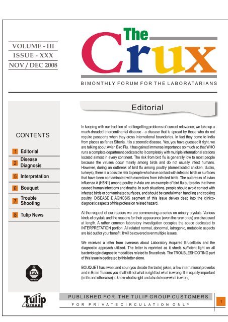
<strong>VOLUME</strong> - I <strong>II</strong><br />
<strong>ISSUE</strong> - <strong>XXX</strong><br />
<strong>NOV</strong> / <strong>DEC</strong> <strong>2008</strong><br />
1<br />
2<br />
5<br />
6<br />
7<br />
8<br />
Editorial<br />
Disease<br />
Diagnosis<br />
Interpretation<br />
Bouquet<br />
Trouble<br />
Shooting<br />
<strong>Tulip</strong> News<br />
In keeping with our tradition of not forgetting problems of current relevance, we take up a<br />
much-dreaded intercontinental disease - a disease that is spread by those who do not<br />
require passports when they cross international boundaries. In fact they come to <strong>India</strong><br />
from places as far as Siberia. It is a zoonotic disease. Yes, you have guessed it right, we<br />
are talking about Avian Bird Flu. It has gained immense importance so much so that WHO<br />
runs a complete department dedicated to it completely with multiple international stations<br />
located almost in every continent. <strong>The</strong> risk from bird flu is generally low to most people<br />
because the viruses occur mainly among birds and do not usually infect humans.<br />
However, during an outbreak of bird flu among poultry (domesticated chicken, ducks,<br />
turkeys), there is a possible risk to people who have contact with infected birds or surfaces<br />
that have been contaminated with excretions from infected birds. <strong>The</strong> outbreaks of avian<br />
influenza A (H5N1) among poultry in Asia are an example of bird flu outbreaks that have<br />
caused human infections and deaths. In such situations, people should avoid contact with<br />
infected birds or contaminated surfaces, and should be careful when handling and cooking<br />
poultry. DISEASE DIAGNOSIS segment of this issue delves deep into the clinicodiagnostic<br />
aspects of this profession related hazard.<br />
At the request of our readers we are commencing a series on urinary crystals. Various<br />
kinds of crystals and the reasons for their appearance (even the rarer ones) are discussed<br />
at length. A rather common laboratory investigation occupies the space dedicated to<br />
INTERPRETATION portion. All related normal, abnormal, iatrogenic, metabolic aspects<br />
are laid out for your benefit. It will be covered over multiple issues.<br />
We received a letter from overseas about Laboratory Acquired Brucellosis and the<br />
diagnostic approach utilized. <strong>The</strong> letter is reprinted as it sheds sufficient light on all<br />
bacteriologic diagnostic modalities related to Brucellosis. <strong>The</strong> TROUBLESHOOTING part<br />
of this issue is dedicated to this letter alone.<br />
BOUQUET has sweet and sour (you decide the taste) jokes, a few international proverbs<br />
and in Brain Teasers you shall tell not what is right but what is wrong. It is equally important<br />
(in life and otherwise) to know what is right and also to know what is wrong!<br />
1
<strong>NOV</strong>/<strong>DEC</strong><br />
AVIAN (BIRD) FLU<br />
DISEASE DIAGNOSIS<br />
Avian influenza in birds: Avian influenza is an infection caused by avian (bird)<br />
influenza (flu) viruses. <strong>The</strong>se influenza viruses occur naturally among birds. Wild<br />
birds worldwide carry the viruses in their intestines, but usually do not get sick from<br />
them. However, avian influenza is very contagious among birds and can make<br />
some domesticated birds, including chickens, ducks, and turkeys, very sick and kill<br />
them. Infected birds shed influenza virus in their saliva, nasal secretions, and<br />
feces. Susceptible birds become infected when they have contact with<br />
contaminated secretions or excretions or with surfaces that are contaminated with<br />
secretions or excretions from infected birds. Domesticated birds may become<br />
infected with avian influenza virus through direct contact with infected waterfowl or<br />
other infected poultry, or through contact with surfaces (such as dirt or cages) or<br />
materials (such as water or feed) that have been contaminated with the virus.<br />
Infection with avian influenza viruses in domestic poultry causes two main forms of<br />
disease that are distinguished by low and high extremes of virulence. <strong>The</strong> “low<br />
pathogenic” form may go undetected and usually causes only mild symptoms<br />
(such as ruffled feathers and a drop in egg production). However, the highly<br />
pathogenic form spreads more rapidly through flocks of poultry. This form may<br />
cause disease that affects multiple internal organs and has a mortality rate that can<br />
reach 90-100% often within 48 hours.<br />
Human infection with avian influenza viruses: <strong>The</strong>re are many different<br />
subtypes of type A influenza viruses. <strong>The</strong>se subtypes differ because of changes in<br />
certain proteins on the surface of the influenza A virus (hemagglutinin [HA] and<br />
neuraminidase [NA] proteins). <strong>The</strong>re are 16 known HA subtypes and 9 known NA<br />
subtypes of influenza A viruses. Many different combinations of HA and NA<br />
proteins are possible. Each combination represents a different subtype. All known<br />
subtypes of influenza A viruses can be found in birds. Usually, “avian influenza<br />
virus” refers to influenza A viruses found chiefly in birds, but infections with these<br />
viruses can occur in humans. <strong>The</strong> risk from avian influenza is generally low to most<br />
people, because the viruses do not usually infect humans. However, confirmed<br />
cases of human infection from several subtypes of avian influenza infection have<br />
been reported since 1997. Most cases of avian influenza infection in humans have<br />
resulted from contact with infected poultry (e.g., domesticated chicken, ducks, and<br />
turkeys) or surfaces contaminated with secretion/excretions from infected birds.<br />
<strong>The</strong> spread of avian influenza viruses from one ill person to another has been<br />
reported very rarely, and transmission has not been observed to continue beyond<br />
one person. “Human influenza virus” usually refers to those subtypes that spread<br />
widely among humans. <strong>The</strong>re are only three known A subtypes of influenza viruses<br />
(H1N1, H1N2, and H3N2) currently circulating among humans. It is likely that<br />
some genetic parts of current human influenza A viruses came from birds<br />
originally. Influenza A viruses are constantly changing, and they might adapt over<br />
time to infect and spread among humans. During an outbreak of avian influenza<br />
among poultry, there is a possible risk to people who have contact with infected<br />
birds or surfaces that have been contaminated with secretions or excretions from<br />
infected birds. Symptoms of avian influenza in humans have ranged from typical<br />
human influenza-like symptoms (e.g., fever, cough, sore throat, and muscle<br />
aches) to eye infections, pneumonia, severe respiratory diseases (such as acute<br />
respiratory distress), and other severe and life-threatening complications. <strong>The</strong><br />
symptoms of avian influenza may depend on which virus caused the infection.<br />
Studies done in laboratories suggest that some of the prescription medicines used<br />
worldwide for human influenza viruses should work in treating avian influenza<br />
infection in humans. However, influenza viruses can become resistant to these<br />
drugs, so these medications may not always work. Additional studies are needed<br />
to demonstrate the effectiveness of these medicines.<br />
Avian influenza A (H5N1) in Asia and Europe: Influenza A (H5N1) virus also<br />
called “H5N1 virus” is an influenza A virus subtype that occurs mainly in birds, is<br />
highly contagious among birds, and can be deadly to them. Outbreaks of avian<br />
influenza H5N1 occurred among poultry in eight countries in Asia (Cambodia,<br />
China, Indonesia, Japan, Laos, South Korea, Thailand, and Vietnam) during late<br />
2003 and early 2004. At that time, more than 100 million birds in the affected<br />
countries either died from the disease or were killed in order to try to control the<br />
outbreaks. By March 2004, the outbreak was reported to be under control. Since<br />
late June 2004, however, new outbreaks of influenza H5N1 among poultry were<br />
reported by several countries in Asia (Cambodia, China [Tibet], Indonesia,<br />
Kazakhstan, Malaysia, Mongolia, Russia [Siberia], Thailand, and Vietnam). It is<br />
believed that these outbreaks are ongoing. Influenza H5N1 infection also has<br />
been reported among poultry in Turkey Romania, and Ukraine. Outbreaks of<br />
influenza H5N1 have been reported among wild migratory birds in China, Croatia,<br />
Mongolia, and Romania. As of January 7, 2006, human cases of influenza A<br />
(H5N1) infection have been reported in Cambodia, China, Indonesia, Thailand,<br />
Vietnam, and most recently, several cases in Turkey. For the most current<br />
information about avian influenza and cumulative case numbers, see the World<br />
Health Organization (WHO) website.<br />
Human health risks during the H5N1 outbreak: H5N1 virus does not usually<br />
infect people, but more than 140 human cases have been reported by the World<br />
Health Organization since January 2004. Most of these cases have occurred as a<br />
result of people having direct or close contact with infected poultry or contaminated<br />
surfaces. So far, the spread of H5N1 virus from person-to-person has been rare<br />
and has not continued beyond one person. Of the few avian influenza viruses that<br />
have crossed the species barrier to infect humans, H5N1 has caused the largest<br />
number of detected cases of severe disease and death in humans. In the current<br />
outbreaks in Asia and Europe, more than half of those infected with the virus have<br />
died. Most cases have occurred in previously healthy children and young adults.<br />
However, it is possible that the only cases currently being reported are those in the<br />
most severely ill people, and that the full range of illness caused by the H5N1 virus<br />
has not yet been defined. So far, the spread of H5N1 virus from person to person<br />
has been limited and has not continued beyond one person. Nonetheless,<br />
because all influenza viruses have the ability to change, scientists are concerned<br />
that H5N1 virus one day could be able to infect humans and spread easily from one<br />
person to another. Because these viruses do not commonly infect humans, there is<br />
little or no immune protection against them in the human population. If H5N1 virus<br />
were to gain the capacity to spread easily from person to person, an influenza<br />
pandemic (worldwide outbreak of disease) could begin. No one can predict when a<br />
pandemic might occur. However, experts from around the world are watching the<br />
H5N1 situation in Asia and Europe very closely and are preparing for the possibility<br />
that the virus may begin to spread more easily and widely from person to person.<br />
Treatment and vaccination for H5N1 virus in humans: <strong>The</strong> H5N1 virus that has<br />
caused human illness and death in Asia is resistant to amantadine and<br />
rimantadine, two antiviral medications commonly used for influenza. Two other<br />
antiviral medications, oseltamavir and zanamavir, would probably work to treat<br />
influenza caused by H5N1 virus, but additional studies still need to be done to<br />
demonstrate their effectiveness. <strong>The</strong>re currently is no commercially available<br />
vaccine to protect humans against H5N1 virus that is being seen in Asia and<br />
Europe. However, vaccine development efforts are taking place. Research studies<br />
to test a vaccine to protect humans against H5N1 virus began in April 2005, and a<br />
series of clinical trials is under way.<br />
Notable Epidemiologic Findings:<br />
H7N7, United Kingdom, 1996: One adult developed conjunctivitis after a piece of<br />
straw contacted her eye while cleaning a duck house. Low pathogenic avian<br />
influenza A (H7N7) virus was isolated from a conjunctiva specimen. <strong>The</strong> person<br />
was not hospitalized and recovered. H5N1, Hong Kong, Special Administrative<br />
Region, 1997: Highly pathogenic avian influenza A (H5N1) infections occurred in<br />
both poultry and humans. This was the first time an avian influenza A virus<br />
transmission directly from birds to humans had been found to cause respiratory<br />
illness. During this outbreak, 18 people were hospitalized and six of them died. To<br />
control the outbreak, authorities killed about 1.5 million chickens to remove the<br />
source of the virus. <strong>The</strong> most significant risk factor for human H5N1 illness was<br />
visiting a live poultry market in the week before illness onset. H9N2, China and<br />
Hong Kong, Special Administrative Region, 1999: Low pathogenic avian<br />
influenza A (H9N2) virus infection was confirmed in two hospitalized children and<br />
resulted in uncomplicated influenza-like illness. Both patients recovered, and no<br />
additional cases were confirmed. <strong>The</strong> source is unknown. Several additional<br />
human H9N2 virus infections were reported from China in 1998-99. H7N2,<br />
Virginia, 2002: Following an outbreak of low pathogenic avian influenza A (H7N2)<br />
among poultry in the Shenandoah Valley poultry production area, one person<br />
developed uncomplicated influenza-like illness and had serologic evidence of<br />
infection with H7N2 virus. H5N1, China and Hong Kong, Special Administrative<br />
Region, 2003: Two cases of highly pathogenic avian influenza A (H5N1) virus<br />
infection occurred among members of a Hong Kong family that had traveled to<br />
China. One person recovered, the other died. How or where these two family<br />
members were infected was not determined. Another family member died of a<br />
2<br />
<strong>NOV</strong>/<strong>DEC</strong>
<strong>NOV</strong>/<strong>DEC</strong><br />
respiratory illness in China, but no testing was done. H7N7, Netherlands, 2003:<br />
<strong>The</strong> Netherlands reported outbreaks of highly pathogenic avian influenza A<br />
(H7N7) virus among poultry on multiple farms. Overall, 89 people were confirmed<br />
to have H7N7 virus infections associated with poultry outbreaks. Most human<br />
cases occurred among poultry workers. H7N7-associated illness was generally<br />
mild and included 78 cases of conjunctivitis (eye infections); five cases of<br />
conjunctivitis and influenza-like illness with fever, cough, and muscle aches; two<br />
cases of influenza-like illness; and four cases that were classified as “other.” One<br />
death occurred in a veterinarian who visited one of the affected farms and<br />
developed complications from H7N7 infection, including acute respiratory distress<br />
syndrome. <strong>The</strong> majority of H7N7 cases occurred through direct contact with<br />
infected poultry. However, Dutch authorities reported three possible instances of<br />
human-to-human H7N7 virus transmission from poultry workers to family<br />
members. H9N2, Hong Kong, Special Administrative Region, 2003: Low<br />
pathogenic avian influenza A (H9N2) virus infection was confirmed in a child in<br />
Hong Kong. <strong>The</strong> child was hospitalized with influenza-like illness and recovered.<br />
H7N2, New York, 2003: In November 2003, a patient with serious underlying<br />
medical conditions was admitted to a hospital in New York with respiratory<br />
symptoms. <strong>The</strong> patient recovered and went home after a few weeks. Testing<br />
revealed that the patient had been infected with a low pathogenic avian influenza A<br />
(H7N2) virus. H7N3, Canada, 2004: In March 2004, two poultry workers who were<br />
assisting in culling operations during a large influenza A (H7N3) poultry outbreak<br />
had culture-confirmed conjunctivitis, one of whom also had coryza. Both poultry<br />
workers recovered. One worker was infected with low pathogenic H7N3 and the<br />
other with high pathogenic H7N3. H5N1, Thailand and Vietnam, 2004: In late<br />
2003 and early 2004, severe and fatal human infections with highly pathogenic<br />
avian influenza A (H5N1) viruses were associated with widespread poultry<br />
outbreaks. Most cases had pneumonia and many had respiratory failure.<br />
Additional human H5N1 cases were reported during mid-2004, and late 2004.<br />
Most cases appeared to be associated with direct contact with sick or dead poultry.<br />
One instance of probable, limited, human-to-human spread of H5N1 virus is<br />
believed to have occurred in Thailand. Overall, 50 human H5N1 cases with 36<br />
deaths were reported from three countries. H5N1, Cambodia, China, Indonesia,<br />
Thailand and Vietnam, 2005: Severe and fatal human infections with highly<br />
pathogenic avian influenza A (H5N1) viruses were associated with the ongoing<br />
H5N1 epizootic among poultry in the region. Overall, 98 human H5N1 cases with<br />
43 deaths were reported from five countries. H5N1, Azerbaijan, Cambodia,<br />
China, Djibouti, Egypt, Indonesia, Iraq, Thailand, Turkey, 2006: Severe and<br />
fatal human infections with highly pathogenic avian influenza A (H5N1) viruses<br />
occurred in association with the ongoing and expanding epizootic. While most of<br />
these cases occurred as a result of contact with infected poultry, in Azerbaijan, the<br />
most plausible cause of exposure to H5N1 in several instances of human infection<br />
is thought to be contact with infected dead wild birds (swans). <strong>The</strong> largest family<br />
cluster of H5N1 cases to date occurred in North Sumatra, Indonesia during May<br />
2006, with seven confirmed H5N1 cases and one probable H5N1 case, including<br />
seven deaths. Overall, 115 human H5N1 cases with 79 deaths were reported in<br />
nine countries. H5N1, Cambodia, China, Egypt, Indonesia, Laos, Nigeria,<br />
Vietnam, 2007: Severe and fatal human infections with highly pathogenic avian<br />
influenza A (H5N1) viruses occurred in association with poultry outbreaks. In<br />
addition, during 2007, Nigeria (January) and Laos (February) confirmed their first<br />
human infections with H5N1. H7N2, United Kingdom, 2007: Human infection with<br />
low pathogenic avian influenza A (H7N2) virus resulting in influenza-like illness<br />
and conjunctivitis were identified in four hospitalized cases. <strong>The</strong> cases were<br />
associated with an H7N2 poultry outbreak in Wales. H9N2, China, 2007: In March<br />
2007,Hong Kong, Special Administrative Region, confirmed low pathogenic avian<br />
influenza A (H9N2) virus infection in a 9-month-old girl with mild signs of disease.<br />
Symptoms of Avian Influenza in Humans<br />
Symptoms: Although the exact incubation period for bird flu in humans isn't clear,<br />
illness seems to develop within one to five days of exposure to the virus.<br />
Common signs and symptoms: Most often, signs and symptoms of bird flu<br />
resemble those of conventional influenza, including: Cough, Fever, Sore throat,<br />
Muscle aches. A relatively mild eye infection (conjunctivitis) is sometimes the only<br />
indication of the disease.<br />
Severe signs and symptoms: People with bird flu also may develop lifethreatening<br />
complications, particularly: Viral pneumonia. Acute respiratory<br />
distress the most common cause of bird flu-related death.<br />
LABORATORY DIAGNOSIS<br />
Specimen collection: Standard and appropriate barrier precautions should<br />
3<br />
always be taken when collecting specimens<br />
from patients. Specimens should be<br />
obtained as soon as possible after the onset<br />
of symptoms. In general, a nasopharyngeal<br />
swab or aspirate is considered the preferred<br />
specimen for seasonal influenza testing;<br />
however, recent data suggest that<br />
oropharyngeal and lower respiratory tract<br />
specimens (i.e., sputa and bronchoalveolar<br />
lavage fluid) are superior to nasopharyngeal<br />
specimens for the detection of avian<br />
influenza A (H5N1) infection in humans.<br />
Specimens from multiple sites may yield the<br />
H5N1 virus, electron<br />
microscopy - photomicrograph.<br />
best results. In cases of atypical presentations, such as gastroenteritis and<br />
encephalopathy, stool and cerebrospinal fluid specimens, respectively, are<br />
advised.<br />
Specimen storage and transportation: Specimens should be collected in the<br />
appropriate viral transport medium and shipped immediately to the testing<br />
laboratory (on ice, if possible) in accordancewith regulations of the Transportation<br />
of Dangerous Goods Act. Specimens may be refrigerated at 4°C (± 2°C) for up to<br />
48-72 hours; after that, specimens should befrozen at -70°C. Swabs and transport<br />
media intended for bacteriologic testing are not suitable for influenza testing. In<br />
addition, swabs with calcium alginate or cotton tips and wooden shafts are not<br />
recommended. Swabs for influenza testing should have a dacron tip and an<br />
aluminum or plastic shaft.<br />
STANDARD DIAGNOSTIC RECOMMENDATIONS<br />
During epidemics, a presumptive diagnosis can be made on the basis of the<br />
clinical symptoms. However, influenza A and B can co-circulate, and mixed<br />
infections of influenza and other viruses have been reported. Isolated cases of<br />
suspected influenza should be investigated for these may represent the first cases<br />
of an impending epidemic. Virus Isolation: Throat swabs, NPA and nasal<br />
washings may be used for virus isolation. It is reported that nasal washings are the<br />
best specimens for virus isolation. <strong>The</strong> specimen may be inoculated in<br />
embryonated eggs or tissue culture. 10-12 day embryonated eggs are used for<br />
virus isolation. <strong>The</strong> specimen is inoculated into the amniotic cavity. <strong>The</strong> virus<br />
replicates in the cells of the amniotic membrane and large quantities are released<br />
back into the amniotic fluid. After 2-3 days incubation, virus in the amniotic fluid can<br />
be detected by adding aliquots of harvested amniotic fluid to chick, guinea pig, or<br />
human erythrocytes. Pathological specimens can be inoculated on to tissue<br />
cultures of kidney, chicks or a variety of other species. Rhesus monkey cells are<br />
the most sensitive. Although no CPE (Cytopathic effect) is produced, newly<br />
produced virus can be recognized by haemadsorption using the cells in the tissue<br />
culture, and haemagglutination using the culture medium which contains free virus<br />
particles. Influenza B virus and occasionally influenza A will produce a CPE in<br />
MDCK cells. Influenza viruses isolated from embryonated eggs or tissue culture<br />
can be identified by serological or molecular methods. Influenza viruses can be<br />
recognized as A, B, or C types by the use of complement fixation tests against the<br />
soluble antigen. (A soluble antigen is found for all influenza A, B or C type virus but<br />
antibody against one type does not cross react with the soluble antigen of the<br />
other. <strong>The</strong> further classification of influenza isolates into subtypes and strains is a<br />
highly specialized responsibility of the WHO reference laboratories. <strong>The</strong> HA type is<br />
identified by HAI tests, the NA type is also identified. Rapid Diagnosis by<br />
Immunofluorescence and other immunologic techniques: cells from<br />
pathological specimens may be examined for the presence of influenza A and B<br />
antigens by indirect immunofluorescence. Although many workers are convinced<br />
of the value of this technique, others have been disappointed with the specificity of<br />
the antisera and the level of background fluorescence that makes the test difficult<br />
to interpret. EIA tests for the detection of influenza A viral antigens are available<br />
that are easier to interpret than immunofluorescence. PCR assays for the<br />
detection of influenza RNA have also been developed but there usefulness in a<br />
clinical setting is highly questionable. <strong>The</strong> test uses technology known as Reverse<br />
Transcription--Polymerase Chain Reaction (RT-PCR) to amplify and detect<br />
genetic material from the influenza A/H5 from the Asian lineage, the same virus<br />
that has been associated with bird flu outbreaks in animals and humans in east<br />
Asia, Turkey, and Iraq. Serology: Virus cannot be isolated from all cases of<br />
suspected infection. More commonly, the diagnosis is made retrospectively by the<br />
demonstration of a rise in serum antibody to the infecting virus. CFT is the most<br />
common method used using the type specific soluble antigen. However, the CF<br />
<strong>NOV</strong>/<strong>DEC</strong>
<strong>NOV</strong>/<strong>DEC</strong><br />
test is thought to have a low specificity. A more specific test is the HAI test. Infection<br />
by influenza viruses results in a rise in serum antibody titre, but the requirement for<br />
a 4-fold or greater rise in titre of HI of CF antibody reflects the inaccuracy of these<br />
tests for detecting smaller increases in antibody. A more precise method for<br />
measuring antibody is by SRH. SRH is more sensitive than CF or HAI tests and has<br />
a greater degree of precision. A 50% increase in zone area represents a rise in<br />
antibody and is evidence of recent infection. Sera do not have to be pretreated to<br />
remove non-specific inhibitors which plaque the HAI test. SRH may well replace<br />
CF and HAI tests in diagnostic laboratory in future.<br />
WHO GUIDELINES<br />
Specimen testing: Rapid antigen testing is not currently recommended for the<br />
detection of avian influenza A (H5N1): a negative result does not exclude avian<br />
influenza, and a positive result of an antigen test (including immunofluorescence<br />
methods) does not differentiate between seasonal and avian influenza A viruses.<br />
Confirmatory testing and subtyping must be performed by molecular methods<br />
(e.g., reverse transcriptase polymerase chain reaction), virus culture or both.<br />
Culture of this high-risk pathogen is restricted to certified containment level 3<br />
facilities. Allspecimens that test positive for influenza A (H5N1) must beconfirmed<br />
by the National Microbiology Laboratory or its designate. Given the increased<br />
likelihood of seasonal influenza infections, these guidelines for H5N1 testing in<br />
humans should be applied only to patients who have a history of travel, or contact<br />
with a traveller, to areas affected by outbreaks of avian influenza and a significant<br />
clinical and exposure history. <strong>The</strong> need exists for increased vigilance for the<br />
surveillance, recognition, reporting and prompt investigation of patients with<br />
severe influenza-likeillness or severe respiratory illness.<br />
Recommended laboratory tests to identify avian influenza A virus in<br />
specimens from humans ( All kits/ devices are available from WHO offices/<br />
outlets)<br />
General information: Highly pathogenic avian influenza (HPAI) caused by certain<br />
subtypes of influenza A virus in animal populations, particularly chickens, poses a<br />
continuing global human public health risk. Direct human infection by an avian<br />
influenza A(H5N1) virus was first recognized during the 1997 outbreak in Hong<br />
Kong Special Administrative Region of China. Subsequently, human infections<br />
with avian strains of the H9 and H7 subtypes have been further documented. <strong>The</strong><br />
current outbreak in humans of avian A (H5N1) and the apparent endemicity of this<br />
subtype in the poultry in southeast Asia require increased attention to the need for<br />
rapid diagnostic capacity for non-typical influenza infections. Laboratory<br />
identification of human influenza A virus infections is commonly carried out by<br />
direct antigen detection, isolation in cell culture, or detection of influenza-specific<br />
RNA by reverse transcriptasepolymerase chain reaction. <strong>The</strong>se<br />
recommendations are intended for laboratories receiving requests to test<br />
specimens from patients with an influenza-like illness, in cases where there is<br />
clinical or epidemiological evidence of influenza A infection. <strong>The</strong> optimal specimen<br />
for influenza A virus detection is a nasopharyngeal aspirate obtained within 3 days<br />
of the onset of symptoms, although nasopharyngeal swabs and other specimens<br />
can also be used. All manipulation of specimens and diagnostic testing should be<br />
carried out following standard biosafety guidelines. <strong>The</strong> strategy for initial<br />
laboratory testing of each specimen should be to diagnose influenza A virus<br />
infection rapidly and exclude other common viral respiratory infections. Results<br />
should ideally be available within 24 hours.<br />
Procedures for influenza diagnosis<br />
Assays available for the diagnosis of influenza A virus infections include:<br />
Rapid antigen detection: Results can be obtained in 15-30 minutes. Near-patient<br />
tests for influenza. <strong>The</strong>se tests are commercially available Immunofluorescence<br />
assay. A widely used, sensitive method for diagnosis of influenza A and B virus<br />
infections and five other clinically important respiratory viruses Enzyme<br />
immunoassay. For influenza A nucleoprotein (NP). Virus culture: Provides results<br />
in 2-10 days. Both shell-vial and standard cell-culture methods may be used to<br />
detect clinically important respiratory viruses. Positive influenza cultures may or<br />
may not exhibit cytopathic effects but virus identification by immunofluorescence<br />
of cell cultures or haemagglutination-inhibition (HI) assay of cell culture medium<br />
(supernatant) is required. Polymerase chain reaction and Real-time PCR<br />
assays: Primer sets specific for the haemagglutinin (HA) gene of currently<br />
circulating influenza A/H1, A/H3 and B viruses are becoming more widely used.<br />
Results can be available within a few hours from either clinical swabs or infected<br />
cell cultures. Additionally several WHO Collaborating Centres are developing PCR<br />
and RT-PCR reagents for non-typical avian/human influenza strains. Any<br />
specimen with a positive result using the above approaches for influenza A virus<br />
and suspected of avian influenza infection should be further tested and verified by<br />
a designated WHO H5 Reference Laboratory ii. Laboratories that lack the capacity<br />
to perform specific influenza A subtype identification procedures are requested to:<br />
(1) Forward specimens or virus isolates to National Institute of Virology (NIV),<br />
National Institute of Immunology (N<strong>II</strong>) or to a WHO H5 Reference Laboratory for<br />
further identification or characterization. (2) Inform the WHO Office in the country<br />
or WHO Regional Office or WHO HQ Global Influenza Programme that specimens<br />
or virus isolates are being forwarded to other laboratories for further identification<br />
or further characterization.<br />
Identification of avian influenza A subtypes<br />
Immunofluorescence assay: Immunofluorescence assay (IFA) can be used for<br />
the detection of virus in either clinical specimens or cell cultures. Clinical<br />
specimens, obtained as soon as possible after the onset of symptoms, are<br />
preferable as the number of infected cells present decreases during the course of<br />
infection. Performing IFA on Inoculated cell cultures is preferable as it allows for<br />
the amplification of any virus present. Virus culture: Virus isolation is a sensitive<br />
technique with the advantage that virus is available both for identification and for<br />
further antigenic and genetic characterization, drug susceptibility testing, and<br />
vaccine preparation. MDCK cells are the preferred cell line for culturing influenza<br />
viruses. Identification of an unknown influenza virus can be carried out by IFA<br />
using specific monoclonal antibodies (see above) or, alternatively, by<br />
haemagglutination (HA) and antigenic analysis (subtyping) by<br />
haemagglutinationinhibition (HAI) using selected reference antisera. Unlike other<br />
influenza A strains, influenza A/H5 will also grow in other common cell lines such as<br />
Hep-2 and RD cells. Standard biosafety precautions should be taken when<br />
handling specimens and cell cultures suspected of containing highly pathogenic<br />
avian influenza A.<br />
GOLDEN RULE. Clinical specimens from humans and from swine or birds<br />
should never be processed in the same laboratory.<br />
Polymerase chain reaction: Polymerase chain reaction (PCR) is a powerful<br />
technique for the identification of influenza virus genomes. <strong>The</strong> influenza virus<br />
genome is single-stranded RNA, and a DNA copy (cDNA) must be synthesised<br />
first using a reverse transcriptase (RT) polymerase. <strong>The</strong> procedure for amplifying<br />
the RNA genome (RTPCR) requires a pair of oligonucleotide primers. <strong>The</strong>se<br />
primer pairs are designed on the basis of the known HA sequence of influenza A<br />
subtypes and of N1 and will specifically amplify RNA of only one subtype. DNAs<br />
generated by using subtype-specific primers can be further analysed by molecular<br />
genetic techniques such as sequencing. Laboratory confirmation: All laboratory<br />
results for influenza A/H5, H7 or H9 during Interpandemic and Pandemic Alert<br />
periods of the WHO Global Influenza Preparedness Plan should be confirmed by a<br />
WHO H5 Reference Laboratory ii or by a WHO recommended laboratory.<br />
Influenza A/H5, H7 or H9 -positive materials, including human specimens, RNA<br />
extracts from human specimens, and influenza A/H5, H7 or H9 virus in cell-culture<br />
fluid or egg allantoic fluid, should be forwarded to a WHO H5 Reference Laboratory<br />
ii or a WHO recommended laboratory. Communication and publication of analysis<br />
results should be according to the WHO Guidance for the timely sharing of<br />
influenza viruses/specimens with potential to cause human influenza pandemics.<br />
Serological identification of influenza A/H5 infection: Serological tests<br />
available for the measurement of influenza A-specific antibody include the<br />
haemagglutination inhibition test, the enzyme immunoassay, and the virus<br />
neutralization tests. <strong>The</strong> microneutralization assay is the recommended test for<br />
the measurement of highly pathogenic avian influenza A specific antibody.<br />
Because this test requires the use of live virus, its use for the detection of highly<br />
pathogenic avian influenza A specific antibody is restricted to those laboratories<br />
with Biosafety Level 3 containment facilities. Antiviral Agents for Avian<br />
Influenza A Virus Infections of Humans: CDC and WHO recommend<br />
oseltamivir, a prescription antiviral medication, for treatment and<br />
chemoprophylaxis of human infection with avian influenza A viruses. Analyses of<br />
available H5N1 viruses circulating worldwide suggest that most viruses are<br />
susceptible to oseltamivir. However, some evidence of resistance to oseltamivir<br />
has been reported in H5N1 viruses isolated from some human H5N1 cases.<br />
Ongoing monitoring for antiviral resistance among avian influenza A viruses is<br />
critical. Prevention of Avian Influenza A Virus Infections of Humans: Persons<br />
exposed to avian influenza A-infected or potentially infected poultry are<br />
recommended to follow good infection control practices including careful attention<br />
to hand hygiene, and to use personal protective equipment. In addition, they<br />
should be vaccinated against seasonal influenza and should take influenza<br />
antiviral agents for prophylaxis. Exposed persons should be carefully monitored<br />
for symptoms that develop during and in the week after exposure to infected<br />
poultry or to potentially avian influenza-contaminated environments.<br />
4 <strong>NOV</strong>/<strong>DEC</strong>
<strong>NOV</strong>/<strong>DEC</strong><br />
INTERPRETATION<br />
URINARY CRYSTALS<br />
Crystals precipitate in urine due to high concentrations of solutes. <strong>The</strong>re are four<br />
factors that enter into crystal formation: [1] Solute concentration: factors include<br />
dehydration, dietary excesses, and medications. [2] pH: Solubility is pH<br />
dependent. Crystals that precipitate in neutral or alkaline urine are less soluble<br />
than the crystals that precipitate in acidic urine. As a rule, inorganic salts<br />
(calcium, phosphate, ammonium, and magnesium) precipitate in alkaline urine.<br />
Organic solutes (uric acid, cystine, bilirubin, and x-ray dye) tend to precipitate out<br />
in acidic urine. [3] <strong>The</strong> rate of flow through the tubules affect crystal formation. A<br />
slow rate of flow produces a concentrated urine and promotes crystal formation.<br />
A rapid flow rate produced a more dilute urine and decreased crystal formation.<br />
[4] Temperature: If warm, the solutes remain in solution better and crystallization<br />
is retarded or does not occur. If cold, then solutes become less soluble and<br />
crystallization occurs readily. Normal, healthy urine seldom contains crystals.<br />
<strong>The</strong> presence of crystals in urine is called crystalluria. If crystals are present in<br />
the urine at the time of voiding, it is possible that this may be clinically significant.<br />
Most crystals observed in urine precipitate out after sitting (especially if the urine<br />
is refrigerated before testing), because the solute concentration is high, a super<br />
saturated solution, and the solubility threshold is exceeded as the urine cools.<br />
AMORPHOUS SEDIMENT IN URINE AND ITS CLINICAL SIGNIFICANCE.<br />
<strong>The</strong>re are three types of amorphous sediments. Each has the following details:<br />
[1] precipitated salts, [2] not clinically significant, [3] are coarse granular in<br />
appearance. If the amount of amorphous sediment is abundant, it can make the<br />
microscopic evaluation difficult, hence they become a nuisance and hindrance.<br />
<strong>The</strong> term "amorphous" literally means "without any form". <strong>The</strong>se crystals are<br />
shapeless and formless, resembling sawdust or sand. Amorphous sediment is<br />
the most commonly encountered types of crystals in urine. If urine is tested within<br />
the first hour after collecting (without refrigeration), amorphous formation is<br />
minimized. In neutral to alkaline urine, there are two types of amorphous<br />
crystals. <strong>The</strong> most common type is "amorphous phosphates". As you observe<br />
urine specimens in lab, a urine with a moderate amount (or larger) of amorphous<br />
phosphates present, the macroscopic, cloudy appearance will be white. <strong>The</strong> are<br />
O<br />
soluble in dilute acids and will not dissolve when heated to 60 C. <strong>The</strong>se are<br />
made up of magnesium and calcium phosphates. <strong>The</strong> other type of formless<br />
crystal is amorphous carbonate. This type is found infrequently in urine and<br />
tends to be composed of rod-shaped calcium carbonate. Because of their small<br />
shape, they have been confused with bacteria. If large calcium carbonate<br />
crystals are present, they will take on a dumbbell-like shape. If a dilute acid is<br />
added, effervescence will be observed as CO 2 is given off. Both of these crystals<br />
appear colourless when viewed with the microscope. In neutral to acid urine,<br />
amorphous urates are observed. Macroscopically, these crystals will appear as a<br />
pink, brick-like dust. Because of their chemical nature, they readily absorb the<br />
urinary pigments. It is uroerythrin that imparts the reddish colour. In the<br />
microscope, they will appear colourless or sometimes a brownish colouration.<br />
<strong>The</strong>se crystals are uric acid salts of sodium, potassium, magnesium, or calcium.<br />
O<br />
If these crystals are warmed to body temperature or to 60 C, they will dissolve.<br />
<strong>The</strong>y will also dissolve in dilute alkali. If you add a strong mineral acid like HCl or<br />
glacial acetic acid to the sediment, allow the mixture to "sit" for a period of time.<br />
uric acid will crystallize out.<br />
NORMAL URINARY CRYSTALS AND THE pH AT WHICH THEY ARE FOUND.<br />
Amorphous urates (pH: acid to neutral), Calcium oxalate (pH: acid to neutral,<br />
sometimes can be observed in a slightly alkaline pH), Uric acid (pH: acid to<br />
neutral, sometimes can be observed in a slightly alkaline pH, Monosodium urate<br />
(pH: acid to neutral), Calcium oxalate, both di- and monohydrate forms, (pH: acid<br />
to neutral, sometimes can be observed in slightly alkaline pH), Amorphous<br />
phosphates (pH; neutral to alkaline), Triple phosphate (pH: neutral to alkaline),<br />
Dicalcium phosphate (pH: neutral to alkaline), Calcium phosphate (pH; neutral to<br />
alkaline), Calcium carbonate (pH: neutral to alkaline), Ammonium biurate<br />
(neutral to alkaline), Calcium sulfate (pH; acidic).<br />
CALCIUM OXALATE CRYSTALS AND THEIR SIGNIFICANCE IN URINE.<br />
Also called envelope crystals. <strong>The</strong>y are colourless and do not absorb pigments<br />
from the urine. <strong>The</strong> most common shape is the octahedral form which appears as<br />
two pyramids joined at their bases. When focusing on this crystal, there is the<br />
appearance of a refractile cross or star in the center of a cube. If there is an<br />
increased number of these crystals, then it is called "oxaluria". <strong>The</strong> persistent<br />
finding of numerous calcium oxalate crystals could be an indicator of small bowel<br />
disease, urinary calculi, renal failure disease, diabetes mellitus, high milk intake,<br />
bone fractures, CNS injuries, ethylene glycol poisoning, or acetazolamide<br />
therapy. <strong>The</strong>se crystals are formed from the calcium salts of oxalic acid and other<br />
oxalates. Foods high in oxalic acid and oxalates are: oranges, cabbage, rhubarb,<br />
asparagus, brussel sprouts, tomatoes, spinach, broccoli, and berries. <strong>The</strong>re are<br />
two basic types of calcium oxalate crystals; dihydrate and monohydrate forms.<br />
<strong>The</strong> dihydrate form tends to form squares and rectangles, whereas the<br />
monohydrate form tends to form oval and dumbbell shapes. Biconcave disk<br />
forms have been reported. Caution: Monohydrate may form long ovals and<br />
closely resemble acetaminophen crystals. Because there is a variety of forms,<br />
these crystals may be described as being pleomorphic. Regardless of the type,<br />
either form is generally considered to be clinically insignificant. Calcium oxalate<br />
crystals are soluble in dilute hydrochloric acid but not dilute acetic acid.<br />
Dihydrate form of<br />
calcium oxylate<br />
Monohydrate form of<br />
calcium oxylate<br />
URIC ACID CRYSTALS AND THEIR SIGNIFICANCE.<br />
This is the most pleomorphic of the crystals found in urine. Forms/patterns<br />
include rhombic (diamond), cubes, rosettes (when multiple crystals cluster and<br />
fuse), needles, wedge, dumbbells, hexagons, and irregular plates/shapes. Uric<br />
acid crystals, when first formed are colourless, but because of their chemical<br />
properties, will adsorb pigments from the urine. Uric acid crystals will appear in<br />
varying shades of yellow or yellow-brown, dependent upon the amount of<br />
pigment in the urine. This colouration is a key to their identification. Uric acid<br />
crystals are quite variable in size. <strong>The</strong>se<br />
crystals are soluble in alkali but are<br />
insoluble in acids or alcohol. Generally,<br />
the presence of uric acid crystals are<br />
considered to be clinically insignificant.<br />
In any condition (examples: gout,<br />
leukemia, lymphoma) in which there is<br />
an increase in the cellular turnover rate,<br />
uric acid crystals will be increased. If a patient is on cytotoxin therapy, there will<br />
be an increase in cell destruction. This means that purine metabolism will be<br />
increased and uric acid crystal formation will occur.<br />
SODIUM URATE CRYSTALS AND THEIR CLINICAL SIGNIFICANCE.<br />
Sodium urates are a variant of uric acid crystals. <strong>The</strong>se are colourless (most<br />
often observed) to slightly yellow rods or slender prisms. Some laboratorians call<br />
these uric acid spears. <strong>The</strong>y are found singly or<br />
o<br />
in clusters. <strong>The</strong>y will dissolved at 60 C. <strong>The</strong>y<br />
are clinically insignificant and may be reported<br />
as "urate crystals". In the presence of<br />
hydrochloric acid, they will change to the uric<br />
acid form.<br />
TRIPLE PHOSPHATE CRYSTALS AND THEIR CLINICAL SIGNIFICANCE.<br />
Also called ammonium, magnesium phosphate crystals, "coffin lids", and "hiproof"<br />
crystals, they are colourless and exhibit much variation in size. <strong>The</strong>y often<br />
5 <strong>NOV</strong>/<strong>DEC</strong>
<strong>NOV</strong>/<strong>DEC</strong><br />
appear as three- or six-sided prisms,<br />
colourless, and very refractile. Other<br />
pattern variations are "feathery starshapes"<br />
and fern-like forms, but these<br />
forms are uncommon. <strong>The</strong>re presence is<br />
generally non-significant, however they<br />
are observed with chronic UTI, obstructive<br />
uropathy, and urinary calculi. <strong>The</strong>se<br />
crystals are characterized by imperfections (not perfectly formed). <strong>The</strong>re are<br />
very variable in size and are soluble in 10% acetic acid.<br />
DICALCIUM PHOSPHATE CRYSTALS AND THEIR SIGNIFICANCE.<br />
This is a uncommon variation of calcium<br />
phosphate and may be found in slightly acidic<br />
urine. <strong>The</strong> correct designation is dicalcium<br />
hydrogen phosphate. <strong>The</strong>y tend to be long<br />
slender prisms, with one end pointed. <strong>The</strong>y<br />
are often found in clusters and for this reason<br />
may be called "stellar phosphates". <strong>The</strong>y are<br />
colourless, soluble in dilute acetic acid, and<br />
are clinically insignificant.<br />
CALCIUM PHOSPHATE CRYSTALS AND THEIR SIGNIFICANCE.<br />
<strong>The</strong>se crystals are usually observed as large, colourless, irregular, thin plates<br />
that are granular in appearance. <strong>The</strong>y float<br />
on top of the urine and resemble a type of<br />
"scum". <strong>The</strong>y have been reported as wedge<br />
shaped prisms. <strong>The</strong>se are soluble in 10%<br />
acetic acid. CAUTION: Small plates may<br />
resemble a degenerate squamous<br />
epithelial cell.<br />
CALCIUM CARBONATE CRYSTALS AND THEIR SIGNIFICANCE.<br />
<strong>The</strong>se crystals appear most often in an<br />
amorphous form. On occasions, they will<br />
appear in crystalline form and then they are<br />
dumbbell in shape. It has been suggested that<br />
this shape is due to clumping and fusing of the<br />
amorphous crystals. <strong>The</strong>y are soluble in dilute<br />
acetic acid and will effervesce.<br />
AMMONIUM BIURATE CRYSTALS AND THEIR CLINICAL SIGNIFICANCE.<br />
Synonyms are: thorn apple<br />
crystal and starfish crystal.<br />
<strong>The</strong>ir shape is peculiar for it<br />
fused spheres, tortuous<br />
shape, and the presence or<br />
absence of spiny projections.<br />
<strong>The</strong>y are yellow brown in<br />
colour, rarely occurring in fresh<br />
urine. If alkaline urine is<br />
allowed to stand, then these<br />
crystals may precipitate out.<br />
<strong>The</strong>y are not clinically<br />
significant and will dissolve at [with and without spicules (thorns)]<br />
0<br />
60 C, in acetic acid, or sodium<br />
hydroxide. <strong>The</strong>y have been confused with yeast cells and leucine crystals. If<br />
concentrated HCl is added, these crystals can reform to uric acid crystals.<br />
CALCIUM SULPHATE CRYSTALS AND THEIR CLINICAL SIGNIFICANCE.<br />
This is a very rare crystal that occurs as long, thin needles and prisms. It is found<br />
only in acidic urine.<br />
To be continued...<br />
In Lighter Vein<br />
BOUQUET<br />
A woman wanted a pet so she went to the local pet shop. She looked at the dogs<br />
and the cats but finally settled on a parrot that was perched in the back of the<br />
store for $50.00.<br />
She asked the shopkeeper why the parrot was so cheap, to which he replied,<br />
"Well, I have to tell you, the birds last owner was a madam at a whorehouse and<br />
he occasionally makes off colour remarks that may offend some people."<br />
Thinking that the price was right and she could handle anything he might say, she<br />
took him. When she got home she set the bird down on the table. He looked<br />
around and said, "New house, new madam".<br />
"That's not so bad," she thought.<br />
A little while later, her daughters got home from school, and the parrot spoke<br />
again, "New house, new madam, new whores."<br />
Even though she felt a little insulted, she thought that wasn't so bad either.<br />
Later that evening, her husband Ray came home.<br />
<strong>The</strong> parrot again spoke out...<br />
This time it said, "Hi Ray!"<br />
<strong>The</strong> woman met with a divorce attorney the next day.<br />
Two women that are dog owners are arguing about which dog is smarter....<br />
First Woman : "My dog is so smart, every morning he waits for the paper boy to<br />
come around and then he takes the newspaper and brings it to me.<br />
Second Woman : "I know..."<br />
First Woman : "How?"<br />
Second Woman : "My dog told me."<br />
6<br />
Wisdom Whispers<br />
<br />
<br />
<br />
<br />
<br />
<br />
<br />
<br />
<br />
"<strong>The</strong> road to happiness lies in two simple principles: find what interests you and that<br />
you can do well, and put your whole soul into it - ever bit of energy and ambition and<br />
natural ability you have."<br />
"To me, fair friend, you never can be old For as you were when first your eye I eyed,<br />
Such seems your beauty still."<br />
To put off repentance is dangerous."<br />
"<strong>The</strong> more the universe seems comprehensible, the more it also seems pointless."<br />
"To beg of the miser is to dig a trench in the sea."<br />
"Nothing falls into the mouth of a sleeping fox."<br />
Cut your coat to suit your cloth."<br />
"<strong>The</strong> dead open the eyes of the living."<br />
He keeps watch over a good castle who has guarded his own constitution."<br />
Brain Teasers<br />
WITH EACH HEADING (KEYWORD) ARE GIVEN FOUR SUPPLEMENTARY STATEMENTS,<br />
IDENTIFY THE INCRRECT ONE/ STATEMENT THAT IS LEAST OR NOT RELATED TO THE<br />
HEADING.<br />
1. Local signs of acute inflammation<br />
A. Pallor B. Dolor C. Tumor D. Calor<br />
2. Lymphocytosis<br />
A. Visceral larva migrans B. Whoopping cough<br />
C. German measles D. Infectious monocytosis<br />
3. Indications for doingg a liver biopsy<br />
A. Granulomatous hepatitis B. Hydatid cyst of liver<br />
C. Post-necrotic cirrhosis D. Hepatocellular carcinoma<br />
4. Low ESR<br />
A. Sickle cell anaemia B. Multiple myeloma<br />
C. Severe dehydration D. Polycythaemia vera<br />
5. Mixed tumours<br />
A. Teratoma B. Haemangioendothelioma<br />
C. Fibroadenoma D. Pleomorphic adenoma<br />
6. Special forms of macrophages<br />
A. Siderophges B. Lipophages C. Epithelioid cell D. Neutrophil<br />
Answers. 1. A., 2. A., 3. B., 4. C., 5. B., 6. D.<br />
<strong>NOV</strong>/<strong>DEC</strong>
<strong>NOV</strong>/<strong>DEC</strong><br />
( Letter to the Editor from Turkey)<br />
Laboratory-acquired Brucellosis<br />
Dear Editor,<br />
TROUBLESHOOTING<br />
Brucellosis is a serious disease seen worldwide and has been historically<br />
known as undulant fever, Bang's disease, Gibraltar fever, Mediterranean<br />
fever, and Malta fever. Brucellosis has a limited geographic distribution but<br />
remains a major problem in Mediterranean and Middle Eastern countries.<br />
In 2001, a total of 15,510 brucellosis cases were reported in Turkey.<br />
In our centre there are approximately 20 to 25 new cases each year. Brucella<br />
is most commonly transmitted by the consumption of contaminated raw or<br />
unpasteurised milk and cheese. Laboratory-acquired brucellosis has also<br />
been documented and is considered the most important laboratory-acquired<br />
bacterial infection. All Brucella spp. have been implicated in laboratoryassociated<br />
infections, and they may account for up to 2% of all laboratoryassociated<br />
infections.<br />
In this paper, we report 3 laboratory-acquired brucellosis.<br />
Case 1 was a 26-year-old female laboratory worker, who works as<br />
microbiology technologist. She presented with joint pain and fever of 1<br />
week's duration. <strong>The</strong>re was no history of trauma. <strong>The</strong> patient had given birth<br />
2 weeks prior. Physical examination and haematological parameters were<br />
normal. However, the levels of aminotranferases were elevated [aspartate<br />
transferase (AST), 45 U/L; alanine transferase (ALT), 55 U/L].<br />
<strong>The</strong> past history of the patient did not reveal any raw cheese consumption.<br />
She had been working on the determination of subgroups of Brucella<br />
positive cultures. She was tested for brucella using the Rose Bengal<br />
microagglutination test (<strong>Tulip</strong> Diagnostics Ltd. Goa, <strong>India</strong>) as well as<br />
serological titre of anti-Brucella abortus antibodies were evaluated by using<br />
a standard tube agglutination test (Seromed Laboratory Products, Turkey).<br />
<strong>The</strong> Rose Bengal microagglutination test was positive. <strong>The</strong> Brucella serum<br />
agglutination test was reactive (1/640). Two sets of blood samples were<br />
obtained for culture. <strong>The</strong> blood cultures showed bacterial growth (Bactec<br />
9050, Becton Dickinson, USA) following 72 hours of incubation. Bacteria<br />
were isolated in 5% sheep blood agar. Grams' stain revealed small gramnegative<br />
coccobacilli. <strong>The</strong> organism was confirmed to be B. melitensis by<br />
standard biochemical reactions (production of urease, catalase - positive,<br />
oxidase-positive, H2S and indole negative, the dyes basic fuchsin, thionine,<br />
thionine blue are positive).<br />
In addition, biochemical identification using an API 20 NE (BioMerieux,<br />
France) was done. Treatment was initiated with a combination of 2 g of<br />
ceftriaxone plus 600 mg of rifampin every day for 6 weeks. <strong>The</strong> patient had a<br />
full recovery without any coexisting problem.<br />
Case 2 was a 28-year-old female laboratory worker who presented with nonspecific<br />
symptoms of malaise, vomiting and fever. She had been working in<br />
the same microbiology laboratory as Case 1 and Case 3. She had worked<br />
with the same Brucella samples as the patient in Case 1 two weeks prior.<br />
She did not have any risk factors for brucellosis exposure such as ingestion<br />
of unpasteurised milk products.<br />
<strong>The</strong> results of her physical examination and haematological and<br />
biochemical studies were normal. Brucella serology was positive at 1/160.<br />
<strong>The</strong> blood culture revealed B.melitensis growth after 3 days. She received<br />
doxycycline at a dosage of 100 mg po twice daily and rifampin at a dosage of<br />
600 mg po qd for 6 weeks. She recovered completely.<br />
Case 3 was a 24-year-old female microbiology technologist who was<br />
suffering from fever and pain of the lower extremities. Her physical<br />
examination and routine laboratory tests were normal. Brucella serology<br />
was positive at 1/640. B.melitensis was isolated from the blood culture. She<br />
received the same treatment as the second case and recovered fully. Like<br />
the preceeding 2 patients, she denied other exposure to brucellosis.<br />
Brucellosis has been considered the most important laboratory-acquired<br />
bacterial infection. Aerosol transmission generated accidentally or during<br />
microbiologic techniques from contaminated materials are the proposed<br />
routes of transmission. Our present report includes 3 patients with an<br />
exposure history of working Brucella bacteria in a microbiology laboratory.<br />
No accident occurred in the laboratory during the time they were exposed. All<br />
3 cases were found to be working on the specimen of the index case patient.<br />
<strong>The</strong> index case was a 45-year-old man who had a Brucella serology of 1/640<br />
and blood culture that grew B.melitensis. Only 3 of the microbiology staff<br />
work on the specimen.<br />
<strong>The</strong> other personnel in the laboratory who did not work with the specimen<br />
were also examined with Rose- Bengal test, brucella antibody and brucellarelated<br />
symptoms. <strong>The</strong> results were negative for brucellosis. Brucellosis is<br />
an endemic disease in our country.<br />
<strong>The</strong> 3 patients presented did not have any suspicious history of<br />
unpasteurised milk consumption or animal contact. <strong>The</strong> absence of this kind<br />
of contamination led us to believe that the transmission to our patients was<br />
through the laboratory route. <strong>The</strong> patients were thought to have been<br />
infected during subculturing for collecting bacteria.<br />
<strong>The</strong>se laboratory workers were unaware of the hazards of aerosol<br />
transmission of Brucella spp. <strong>The</strong>y handled the biosafety cabinet, and used<br />
gloves and masks. Sniffing culture plates is another risk factor, and is a<br />
common practice in bacteriology laboratories in Turkey, as in other<br />
countries. Brucella spp. are highly infectious because the infectious dose by<br />
an aerosol is only 10 to 100 organisms. Laboratory-acquired brucella<br />
infections are very important in developing countries and in countries with<br />
endemic disease, such as Turkey.<br />
In our country, biosafety cabinets do not exist in most hospital laboratories.<br />
<strong>The</strong>refore, using gloves, masks and goggles and also continuous education<br />
on biosafety where brucellosis is endemic is very important. <strong>The</strong><br />
transmission in our cases was probably due to aerosol contamination<br />
because of the current practice of sniffing culture plates. Additionally,<br />
catalase test used for bacteria identification might have produced aerosol.<br />
In 1986, the World Health Organization recommended the use of<br />
doxycycline in combination with rifampin for 6 weeks as the preferred<br />
treatment for adult acute brucellosis.<br />
<strong>The</strong> second and third cases received doxycycline and rifampin for 6 weeks.<br />
However, the first case received rifampin + ceftriaxone for 6 weeks. It is<br />
known that tetracyclines accumulate in fetal bones and teeth, and<br />
furthermore, pass into breast milk.1 For this reason, the combination of<br />
rifampin + ceftriaxone was preferred instead of the combination of rifampin +<br />
doxycycline for Case 1, who had given birth 2 weeks prior and was<br />
breastfeeding. Laboratories should be aware of laboratory-associated<br />
hazards and take adequate safety precautions even after the use of<br />
biosafety cabinet, gloves and masks.<br />
In addition, all laboratory workers should be educated periodically for<br />
occupational risks in the laboratory. In our hospital, all microbiology<br />
laboratory staff were educated concerning laboratory-acquired bacterial<br />
infections. <strong>The</strong> laboratory staff are discouraged from smelling the culture<br />
plaques. In order to prevent future infections, close collaboration between<br />
clinicians and laboratory staff was initiated.<br />
Biosafety level 3 has to be advocated and used whenworking with microorganisms<br />
such as Brucella spp. Clinicians should alert laboratory<br />
personnel if they suspect Brucellosis in patients so they can take extra<br />
precautions.<br />
7<br />
<strong>NOV</strong>/<strong>DEC</strong>
<strong>NOV</strong>/<strong>DEC</strong><br />
<strong>Tulip</strong> sets its foot<br />
into ELISA automation<br />
ulip’s Instrumentation Division was introduced<br />
in the year 2000 Since then, a range of<br />
Tinstruments (both semiautomatic and fully<br />
automated) for various IVD testing have been<br />
successfully placed in the <strong>India</strong>n market. Each of these<br />
instruments were introduced selectively into the market<br />
based on market requirements and their suitability to<br />
<strong>India</strong>n work conditions. Apart from these, instruments<br />
launched by <strong>Tulip</strong> <strong>Group</strong> are backed by an excellent after<br />
sales service support system.<br />
Now <strong>Tulip</strong> partners with Dynex Technologies Inc., in<br />
introducing the most appropriate instrument for <strong>India</strong>n<br />
IVD market<br />
DYNEX DS2, Fully Automated<br />
2-Plate ELISA Processor<br />
Step Up to Automation…<br />
With DS2 modules.<br />
YNEX Technologies, Inc. has a long and distinguished track record of bringing<br />
break through products to market and the company has been a leading innovator<br />
specifically in microplate analysis. DYNEX introduced the first manual microplate<br />
DELISA reader and molded immunoassay microplates. In 1990s, DYNEX entered the market<br />
with the first contiguous ELISA processing system consisting of reader, incubator, multireagent<br />
dispenser, and washer.<br />
TULIP<br />
P R E S E N T S<br />
BRUCEL - RB<br />
New<br />
Improved<br />
SENSITIVITY<br />
25 lU/mL<br />
Slide screening test for Brucella antibodies<br />
<strong>The</strong> improved sensitivity will help in diagnosis of patients in early<br />
seroconversion phase (low level of IgM antibodies) as well as diagnosis<br />
of chronic individuals who present with high IgG antibodies,<br />
undetected by the usual Wright tube test method.<br />
Pack<br />
Size<br />
5 ml<br />
TM<br />
BioShields<br />
<strong>NOV</strong>/<strong>DEC</strong>


