MRI and Positron Emission Tomography Findings in Heidenhain ...
MRI and Positron Emission Tomography Findings in Heidenhain ...
MRI and Positron Emission Tomography Findings in Heidenhain ...

You also want an ePaper? Increase the reach of your titles
YUMPU automatically turns print PDFs into web optimized ePapers that Google loves.
Copyright © North American Neuro-Ophthalmology Society. Unauthorized reproduction of this article is prohibited.<br />
Photo Essay<br />
Section Editor: Timothy J. McCulley, MD<br />
<strong>MRI</strong> <strong>and</strong> <strong>Positron</strong> <strong>Emission</strong> <strong>Tomography</strong> <strong>F<strong>in</strong>d<strong>in</strong>gs</strong> <strong>in</strong><br />
Heidenha<strong>in</strong> Variant Creutzfeldt-Jakob Disease<br />
Sashank Prasad, MD, Edward B. Lee, MD, PhD, John H. Woo, MD,<br />
Abass Alavi, MD, Steven L. Galetta, MD<br />
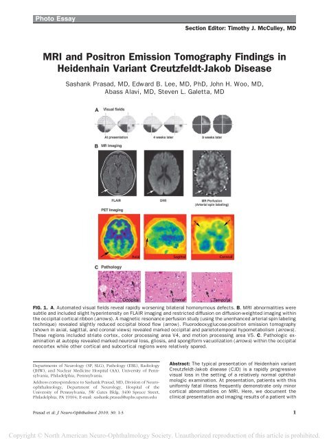
FIG. 1. A. Automated visual fields reveal rapidly worsen<strong>in</strong>g bilateral homonymous defects. B. <strong>MRI</strong> abnormalities were<br />
subtle <strong>and</strong> <strong>in</strong>cluded slight hyper<strong>in</strong>tensity on FLAIR imag<strong>in</strong>g <strong>and</strong> restricted diffusion on diffusion-weighted imag<strong>in</strong>g with<strong>in</strong><br />
the occipital cortical ribbon (arrows). A magnetic resonance perfusion study (us<strong>in</strong>g the unenhanced arterial sp<strong>in</strong> label<strong>in</strong>g<br />
technique) revealed slightly reduced occipital blood flow (arrow). Fluorodeoxyglucose-positron emission tomography<br />
(shown <strong>in</strong> axial, sagittal, <strong>and</strong> coronal views) revealed marked occipital <strong>and</strong> parietotemporal hypometabolism (arrows).<br />
These regions <strong>in</strong>cluded striate cortex, color process<strong>in</strong>g area V4, <strong>and</strong> motion process<strong>in</strong>g area V5. C. Pathologic exam<strong>in</strong>ation<br />
at autopsy revealed marked neuronal loss, gliosis, <strong>and</strong> spongiform vacuolization (arrows) with<strong>in</strong> the occipital<br />
neocortex while other cortical <strong>and</strong> subcortical regions were relatively spared.<br />
Departments of Neurology (SP, SLG), Pathology (EBL), Radiology<br />
(JHW), <strong>and</strong> Nuclear Medic<strong>in</strong>e Hospital (AA), University of Pennsylvania,<br />
Philadelphia, Pennsylvania.<br />
Address correspondence to Sashank Prasad, MD, Division of Neuroophthalmology,<br />
Department of Neurology, Hospital of the<br />
University of Pennsylvania, 3W Gates Bldg, 3400 Spruce Street,<br />
Philadelphia, PA 19104; E-mail: sashank.prasad@uphs.upenn.edu<br />
Abstract: The typical presentation of Heidenha<strong>in</strong> variant<br />
Creutzfeldt-Jakob disease (CJD) is a rapidly progressive<br />
visual loss <strong>in</strong> the sett<strong>in</strong>g of a relatively normal ophthalmologic<br />
exam<strong>in</strong>ation. At presentation, patients with this<br />
uniformly fatal illness frequently demonstrate only m<strong>in</strong>or<br />
cortical abnormalities on <strong>MRI</strong>. Here, we document the<br />
cl<strong>in</strong>ical presentation <strong>and</strong> imag<strong>in</strong>g results of a patient with<br />
Prasad et al: J Neuro-Ophthalmol 2010; 30: 1-3 1
Copyright © North American Neuro-Ophthalmology Society. Unauthorized reproduction of this article is prohibited.<br />
Photo Essay<br />
Heidenha<strong>in</strong> variant CJD <strong>in</strong> whom abnormalities on positron<br />
emission tomographic imag<strong>in</strong>g were more evident than<br />
changes on <strong>MRI</strong>. These changes were present <strong>in</strong> striate<br />
cortex <strong>and</strong> visual association areas, provid<strong>in</strong>g cl<strong>in</strong>icalanatomical<br />
correlation with our patient’s visual deficits.<br />
Nuclear imag<strong>in</strong>g provides a considerably more sensitive<br />
measure of neural dysfunction early <strong>in</strong> the course of this<br />
disease.<br />
Journal of Neuro-Ophthalmology 2010;30:1–3<br />
doi: 10.1097/WNO.0b013e3181e2aef7<br />
Ó 2010 by North American Neuro-Ophthalmology Society<br />
A66-year-old woman noticed slowly progressive blurred<br />
vision <strong>in</strong> the left <strong>in</strong>ferior visual field. There were no<br />
headaches or other accompany<strong>in</strong>g symptoms. An ophthalmologic<br />
exam<strong>in</strong>ation revealed a homonymous left <strong>in</strong>ferior<br />
field cut <strong>and</strong> no other abnormalities. Bra<strong>in</strong> <strong>MRI</strong> was<br />
normal.<br />
Over several weeks, visual loss gradually extended <strong>in</strong>to<br />
the right <strong>in</strong>ferior visual field (Fig. 1). She was referred for<br />
neuro-ophthalmic consultation. The patient described visual<br />
halluc<strong>in</strong>ations <strong>in</strong> the form of shimmer<strong>in</strong>g orange lights<br />
<strong>in</strong> her peripheral vision along with pal<strong>in</strong>opsia. Knitt<strong>in</strong>g<br />
had become difficult because of impaired depth perception.<br />
She had developed mild gait unstead<strong>in</strong>ess. There were no<br />
deficits of memory, language, or behavior, nor was there<br />
weakness or numbness.<br />
On exam<strong>in</strong>ation, she was alert <strong>and</strong> fully oriented. She<br />
named 28 of 30 items on the Boston nam<strong>in</strong>g task <strong>and</strong><br />
comprehended complex verbal comm<strong>and</strong>s. She was fluent,<br />
<strong>and</strong> she could repeat normally. On memory test<strong>in</strong>g the<br />
patient recalled 10 of 10 elements <strong>in</strong> a story after a 5-m<strong>in</strong>ute<br />
delay. Visuospatial test<strong>in</strong>g revealed difficulty copy<strong>in</strong>g a<br />
cube, although she drew a clock face correctly. On test<strong>in</strong>g of<br />
executive functions, she named 18 words beg<strong>in</strong>n<strong>in</strong>g with<br />
the letter F <strong>in</strong> 1 m<strong>in</strong>ute. She completed oral trials successfully,<br />
performed simple calculations, <strong>and</strong> demonstrated<br />
normal praxis.<br />
Corrected visual acuity was 20/25 <strong>in</strong> each eye. The<br />
patient correctly named colors but reported desaturation of<br />
blue <strong>and</strong> yellow. She identified the control Ishihara color<br />
plate but none of the test plates <strong>and</strong> made numerous errors<br />
arrang<strong>in</strong>g the desaturated L’anthony D-15 color panel. She<br />
was unable to perceive a stereoscopic image with the Titmus<br />
stereotest (3,000 arcsec ret<strong>in</strong>al disparity). Confrontation<br />
visual fields demonstrated a dense left <strong>in</strong>ferior quadrant<br />
scotoma <strong>and</strong> a partial right <strong>in</strong>ferior quadrant scotoma.<br />
With<strong>in</strong> the bl<strong>in</strong>d field, however, she correctly discrim<strong>in</strong>ated<br />
motion cues (the Riddoch phenomenon). The patient reported<br />
persistence of visual images a few moments after shift<strong>in</strong>g<br />
gaze but correctly described all elements of both the ‘‘cookiethief’’<br />
picture <strong>and</strong> a Navon figure. Pupillary responses, ocular<br />
motility, <strong>and</strong> fundus exam<strong>in</strong>ations were normal.<br />
Strength was normal. There was no myoclonus,<br />
numbness, or dysmetria. She reached for objects accurately,<br />
without past-po<strong>in</strong>t<strong>in</strong>g or tremor. T<strong>and</strong>em gait was mildly<br />
impaired. Reflexes were normal, <strong>and</strong> symmetric <strong>and</strong> plantar<br />
responses were flexor.<br />
A repeat bra<strong>in</strong> <strong>MRI</strong> revealed slight abnormalities of the<br />
occipital cortical ribbon, <strong>in</strong>clud<strong>in</strong>g hyper<strong>in</strong>tensity on<br />
FLAIR imag<strong>in</strong>g <strong>and</strong> diffusion-weighted imag<strong>in</strong>g that was<br />
more prom<strong>in</strong>ent on the right (Fig. 1). In addition, there<br />
were nonspecific white matter hyper<strong>in</strong>tensities, consistent<br />
with small vessel ischemia. The basal ganglia <strong>and</strong> thalami<br />
were normal. An <strong>MRI</strong> perfusion study (us<strong>in</strong>g the<br />
unenhanced arterial sp<strong>in</strong> label<strong>in</strong>g technique) revealed<br />
slightly decreased occipital blood flow. Fluorodeoxyglucosepositron<br />
emission tomography, <strong>in</strong> contrast to the <strong>MRI</strong><br />
studies, revealed strik<strong>in</strong>g abnormalities, with severe<br />
hypometabolism of the bilateral occipital <strong>and</strong> parietotemporal<br />
cortices (right greater than left) (Fig. 1).<br />
Cerebrosp<strong>in</strong>al fluid analysis showed no cells, prote<strong>in</strong> 52<br />
mg/dL, glucose 56 mg/dL, <strong>and</strong> normal cytology. The 14-3-<br />
3 immunoassay revealed only weak immunoreactivity <strong>and</strong><br />
was considered an ambiguous result. CT of the chest <strong>and</strong><br />
abdomen were normal. Test<strong>in</strong>g for paraneoplastic antibodies<br />
was negative. An electroencephalogram (EEG)<br />
revealed a normal posterior dom<strong>in</strong>ant rhythm, without<br />
focal slow<strong>in</strong>g or paroxysmal sharp waves. Visual evoked<br />
responses were normal (P100 latency, 103 milliseconds <strong>in</strong><br />
the right eye, 101 milliseconds <strong>in</strong> the left eye).<br />
The patient went completely bl<strong>in</strong>d over a period of 8<br />
weeks. She died 12 weeks from the onset of symptoms <strong>and</strong><br />
term<strong>in</strong>ally she had myoclonus <strong>and</strong> impaired arousal <strong>and</strong><br />
orientation. At autopsy, there was severe neuronal loss <strong>and</strong><br />
gliosis with spongiform vacuolization that predom<strong>in</strong>antly<br />
affected the occipital lobes (Fig. 1). Western blot analysis<br />
demonstrated accumulation of prote<strong>in</strong>ase-resistant PrP sc<br />
(type 1), <strong>and</strong> genetic sequenc<strong>in</strong>g revealed the homozygous<br />
meth<strong>in</strong>on<strong>in</strong>e polymorphism at codon 129 of the PrP gene.<br />
The Heidenha<strong>in</strong> variant of sporadic Creutzfeldt-Jakob<br />
disease (CJD) describes a rare rapidly progress<strong>in</strong>g dementia<br />
<strong>in</strong> which prom<strong>in</strong>ent visual changes constitute the <strong>in</strong>itial<br />
symptoms. In 1929, Heidenha<strong>in</strong> first described this entity,<br />
report<strong>in</strong>g 3 cases shar<strong>in</strong>g this strik<strong>in</strong>g cl<strong>in</strong>ical presentation <strong>in</strong><br />
whom histopathological analysis revealed severe abnormalities<br />
<strong>in</strong>clud<strong>in</strong>g neuronal loss, gliosis, <strong>and</strong> vacuolization<br />
that were most prom<strong>in</strong>ent <strong>in</strong> the occipital lobes (1). Publication<br />
of cases with similar cl<strong>in</strong>ical <strong>and</strong> pathological features<br />
led to the proposal that this entity be named the<br />
Heidenha<strong>in</strong> variant (2). After several decades without<br />
<strong>in</strong>sight <strong>in</strong>to the pathogenesis of these disorders, Stanley<br />
Prus<strong>in</strong>er (3) advanced the prion hypothesis, which<br />
implicates the misfold<strong>in</strong>g of the normal PrP prote<strong>in</strong> <strong>in</strong>to the<br />
protease-resistant PrP sc isoform. Almost all cases of<br />
Heidenhe<strong>in</strong> variant CJD (<strong>in</strong>clud<strong>in</strong>g our patient) are<br />
homozygous for methion<strong>in</strong>e at codon 129 of the PrP gene,<br />
but the significance of this association rema<strong>in</strong>s unclear (4).<br />
The cl<strong>in</strong>ical diagnostic features of Heidenha<strong>in</strong> variant<br />
CJD have been well characterized (5–15). In comparison to<br />
2 Prasad et al: J Neuro-Ophthalmol 2010; 30: 1-3
Copyright © North American Neuro-Ophthalmology Society. Unauthorized reproduction of this article is prohibited.<br />
Photo Essay<br />
patients with ataxia-predom<strong>in</strong>ant CJD, Heidenha<strong>in</strong><br />
patients have a similar age at onset, although they have<br />
a more rapid deterioration (mean disease duration, 5.7 vs<br />
7.5 months) (7). Heidenha<strong>in</strong> patients commonly report<br />
a variety of visual symptoms, <strong>in</strong>clud<strong>in</strong>g blurr<strong>in</strong>g, field<br />
constriction, metamorphopsia, visual halluc<strong>in</strong>ations, or<br />
visual neglect. In our patient, widespread posterior metabolic<br />
abnormalities <strong>in</strong> striate cortex <strong>and</strong> visual association<br />
areas (<strong>in</strong>clud<strong>in</strong>g color process<strong>in</strong>g area V4 <strong>and</strong> motion<br />
process<strong>in</strong>g area V5) accounted for the patient’s bilateral<br />
homonymous visual field defects, impaired color process<strong>in</strong>g,<br />
<strong>and</strong> pal<strong>in</strong>opsia.<br />
It is common for the bra<strong>in</strong> <strong>MRI</strong> <strong>in</strong> Heidenha<strong>in</strong> variant<br />
CJD to be normal or show only m<strong>in</strong>imal changes,<br />
particularly early <strong>in</strong> the disease course (8). As our case<br />
demonstrates, severe progressive cortical visual loss may<br />
occur with only m<strong>in</strong>imal structural changes identified by<br />
<strong>MRI</strong>. On the other h<strong>and</strong>, several recent reports of<br />
Heidenha<strong>in</strong> variant CJD have demonstrated that nuclear<br />
imag<strong>in</strong>g studies may reveal conspicuously abnormal areas<br />
of hypometabolism (9–12). Reduced occipital blood flow<br />
has also been reported us<strong>in</strong>g nuclear imag<strong>in</strong>g techniques,<br />
<strong>in</strong>clud<strong>in</strong>g Xe-133 SPECT (5), 99mTc-SPECT (13), <strong>and</strong><br />
[ 15 O]H 2 O PET (11). In many of these cases, however,<br />
Heidenha<strong>in</strong> variant CJD was suspected without pathological<br />
confirmation (9–12).<br />
The case illustrated here demonstrates pathologically<br />
confirmed Heidenha<strong>in</strong> variant CJD with prom<strong>in</strong>ent focal<br />
hypometabolism observed on bra<strong>in</strong> PET scan. In contrast,<br />
other ancillary tests (<strong>in</strong>clud<strong>in</strong>g st<strong>and</strong>ard <strong>MRI</strong> sequences,<br />
magnetic resonance perfusion, CSF 14-3-3, <strong>and</strong> EEG)<br />
demonstrated only mild abnormalities dur<strong>in</strong>g the disease<br />
course <strong>and</strong> provided only limited cl<strong>in</strong>ical-anatomical correlation<br />
with our patient’s visual compla<strong>in</strong>ts. Nuclear imag<strong>in</strong>g<br />
is a particularly sensitive <strong>in</strong>dicator of the extent of<br />
neural dysfunction early <strong>in</strong> the course of Heidenha<strong>in</strong> variant<br />
CJD, demonstrat<strong>in</strong>g severe posterior hypometabolism <strong>in</strong><br />
cortical regions that correlate with the visually predom<strong>in</strong>ant<br />
cl<strong>in</strong>ical deficits <strong>in</strong> these patients.<br />
REFERENCES<br />
1. Heidenha<strong>in</strong> A. Kl<strong>in</strong>ische und anatomische Untersuchungen<br />
uber e<strong>in</strong>e eigenartige organische Erkrankung des<br />
Zentralnervensystems im Praesenium. Z Ges Neurol<br />
Psychiatr. 1929;118:49–114.<br />
2. Meyer A, Leigh D, Bagg CE. A rare presenile dementia<br />
associated with cortical bl<strong>in</strong>dness (Heidenha<strong>in</strong>’s<br />
syndrome). J Neurol Neurosurg Psychiatry. 1954;17:<br />
129–133.<br />
3. Prus<strong>in</strong>er SB. Novel prote<strong>in</strong>aceous <strong>in</strong>fectious particles cause<br />
scrapie. Science. 1982;216:136–144.<br />
4. Parchi P, Castellani R, Capellari S, Ghetti B, Young K, Chen SG,<br />
Farlow M, Dickson DW, Sima AA, Trojanowski JQ, Petersen RB,<br />
Gambetti P. Molecular basis of phenotypic variability <strong>in</strong><br />
sporadic Creutzfeldt-Jakob disease. Ann Neurol. 1996;39:<br />
767–778.<br />
5. Mathews D, Unw<strong>in</strong> DH. Quantitative cerebral blood flow<br />
imag<strong>in</strong>g <strong>in</strong> a patient with the Heidenha<strong>in</strong> variant of<br />
Creutzfeldt-Jakob disease. Cl<strong>in</strong> Nucl Med. 2001;26:<br />
770–773.<br />
6. Cooper SA, Murray KL, Heath CA, Will RG, Knight RS.<br />
Isolated visual symptoms at onset <strong>in</strong> sporadic Creutzfeldt-<br />
Jakob disease: the cl<strong>in</strong>ical phenotype of the ‘‘Heidenha<strong>in</strong><br />
variant.’’ Br J Ophthalmol. 2005;89:1341–1342.<br />
7. Kropp S, Schulz-Schaeffer WJ, F<strong>in</strong>kenstaedt M,<br />
Riedemann C, W<strong>in</strong>dl O, Ste<strong>in</strong>hoff BJ, Zerr I, Kretzschmar<br />
HA, Poser S. The Heidenha<strong>in</strong> variant of Creutzfeldt-Jakob<br />
disease. Arch Neurol. 1999;56:55–61.<br />
8. Jacobs DA, Lesser RL, Mourelatos Z, Galetta SL, Balcer LJ.<br />
The Heidenha<strong>in</strong> variant of Creutzfeldt-Jakob disease:<br />
cl<strong>in</strong>ical, pathologic, <strong>and</strong> neuroimag<strong>in</strong>g f<strong>in</strong>d<strong>in</strong>gs. J<br />
Neuroophthalmol. 2001;21:99–102.<br />
9. Clarencxon F, Gutman F, Giannes<strong>in</strong>i C, Pénicaud A,<br />
Galanaud D, Kerrou K, Marro B, Talbot JN. <strong>MRI</strong> <strong>and</strong> FDG<br />
PET/CT f<strong>in</strong>d<strong>in</strong>gs <strong>in</strong> a case of probable Heidenha<strong>in</strong> variant<br />
Creutzfeldt-Jakob disease. J Neuroradiol. 2008;35:<br />
240–243.<br />
10. Grunwald F, Pohl C, Bender H, Hartmann A, Menzel C,<br />
Ruhlmann J, Keller E, Biersack HJ. 18F-fluorodeoxyglucose-<br />
PET <strong>and</strong> 99mTc-bicisate-SPECT <strong>in</strong> Creutzfeldt-Jakob<br />
disease. Ann Nucl Med. 1996;10:131–134.<br />
11. Thomas A, Kle<strong>in</strong> JC, Galldiks N, Hilker R, Grond M, Jacobs AH.<br />
Multitracer PET imag<strong>in</strong>g <strong>in</strong> Heidenha<strong>in</strong> variant of Creutzfeldt-<br />
Jakob disease. J Neurol. 2006;253:258–260.<br />
12. Tsuji Y, Kanamori H, Murakami G, Yokode M, Mezaki T,<br />
Doh-ura K, Taniguchi K, Matsubayashi K, Fukuyama H,<br />
Kita T, Tanaka M. Heidenha<strong>in</strong> variant of Creutzfeldt-Jakob<br />
disease: diffusion-weighted <strong>MRI</strong> <strong>and</strong> PET characteristics.<br />
J Neuroimag<strong>in</strong>g. 2004;14:63–66.<br />
13. Pichler R, Ciovica I, Rach<strong>in</strong>ger J, Weiss S, Aichner FT.<br />
Multitracer study <strong>in</strong> Heidenha<strong>in</strong> variant of Creutzfeldt-Jakob<br />
disease: mismatch pattern of cerebral hypometabolism<br />
<strong>and</strong> perfusion imag<strong>in</strong>g. Neuro Endocr<strong>in</strong>ol Lett. 2008;29:<br />
67–68.<br />
14. Hopf HC, Althaus HH, Sabuncu S. [Type Heidenha<strong>in</strong> of<br />
the subacute spongious encephalopathy (SSE)<br />
Creutzfeldt-Jakob (author’s transl)]. Z Neurol. 1974;206:<br />
149–156.<br />
15. Bosch PL, Vass K. [Subacute spongious encephalopathy<br />
of the Heidenha<strong>in</strong> type]. Nervenarzt. 1975;46:160–162.<br />
Prasad et al: J Neuro-Ophthalmol 2010; 30: 1-3 3