MRI and Positron Emission Tomography Findings in Heidenhain ...
MRI and Positron Emission Tomography Findings in Heidenhain ...
MRI and Positron Emission Tomography Findings in Heidenhain ...
You also want an ePaper? Increase the reach of your titles
YUMPU automatically turns print PDFs into web optimized ePapers that Google loves.
Copyright © North American Neuro-Ophthalmology Society. Unauthorized reproduction of this article is prohibited.<br />
Photo Essay<br />
patients with ataxia-predom<strong>in</strong>ant CJD, Heidenha<strong>in</strong><br />
patients have a similar age at onset, although they have<br />
a more rapid deterioration (mean disease duration, 5.7 vs<br />
7.5 months) (7). Heidenha<strong>in</strong> patients commonly report<br />
a variety of visual symptoms, <strong>in</strong>clud<strong>in</strong>g blurr<strong>in</strong>g, field<br />
constriction, metamorphopsia, visual halluc<strong>in</strong>ations, or<br />
visual neglect. In our patient, widespread posterior metabolic<br />
abnormalities <strong>in</strong> striate cortex <strong>and</strong> visual association<br />
areas (<strong>in</strong>clud<strong>in</strong>g color process<strong>in</strong>g area V4 <strong>and</strong> motion<br />
process<strong>in</strong>g area V5) accounted for the patient’s bilateral<br />
homonymous visual field defects, impaired color process<strong>in</strong>g,<br />
<strong>and</strong> pal<strong>in</strong>opsia.<br />
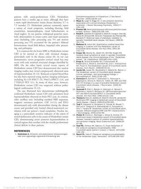
It is common for the bra<strong>in</strong> <strong>MRI</strong> <strong>in</strong> Heidenha<strong>in</strong> variant<br />
CJD to be normal or show only m<strong>in</strong>imal changes,<br />
particularly early <strong>in</strong> the disease course (8). As our case<br />
demonstrates, severe progressive cortical visual loss may<br />
occur with only m<strong>in</strong>imal structural changes identified by<br />
<strong>MRI</strong>. On the other h<strong>and</strong>, several recent reports of<br />
Heidenha<strong>in</strong> variant CJD have demonstrated that nuclear<br />
imag<strong>in</strong>g studies may reveal conspicuously abnormal areas<br />
of hypometabolism (9–12). Reduced occipital blood flow<br />
has also been reported us<strong>in</strong>g nuclear imag<strong>in</strong>g techniques,<br />
<strong>in</strong>clud<strong>in</strong>g Xe-133 SPECT (5), 99mTc-SPECT (13), <strong>and</strong><br />
[ 15 O]H 2 O PET (11). In many of these cases, however,<br />
Heidenha<strong>in</strong> variant CJD was suspected without pathological<br />
confirmation (9–12).<br />
The case illustrated here demonstrates pathologically<br />
confirmed Heidenha<strong>in</strong> variant CJD with prom<strong>in</strong>ent focal<br />
hypometabolism observed on bra<strong>in</strong> PET scan. In contrast,<br />
other ancillary tests (<strong>in</strong>clud<strong>in</strong>g st<strong>and</strong>ard <strong>MRI</strong> sequences,<br />
magnetic resonance perfusion, CSF 14-3-3, <strong>and</strong> EEG)<br />
demonstrated only mild abnormalities dur<strong>in</strong>g the disease<br />
course <strong>and</strong> provided only limited cl<strong>in</strong>ical-anatomical correlation<br />
with our patient’s visual compla<strong>in</strong>ts. Nuclear imag<strong>in</strong>g<br />
is a particularly sensitive <strong>in</strong>dicator of the extent of<br />
neural dysfunction early <strong>in</strong> the course of Heidenha<strong>in</strong> variant<br />
CJD, demonstrat<strong>in</strong>g severe posterior hypometabolism <strong>in</strong><br />
cortical regions that correlate with the visually predom<strong>in</strong>ant<br />
cl<strong>in</strong>ical deficits <strong>in</strong> these patients.<br />
REFERENCES<br />
1. Heidenha<strong>in</strong> A. Kl<strong>in</strong>ische und anatomische Untersuchungen<br />
uber e<strong>in</strong>e eigenartige organische Erkrankung des<br />
Zentralnervensystems im Praesenium. Z Ges Neurol<br />
Psychiatr. 1929;118:49–114.<br />
2. Meyer A, Leigh D, Bagg CE. A rare presenile dementia<br />
associated with cortical bl<strong>in</strong>dness (Heidenha<strong>in</strong>’s<br />
syndrome). J Neurol Neurosurg Psychiatry. 1954;17:<br />
129–133.<br />
3. Prus<strong>in</strong>er SB. Novel prote<strong>in</strong>aceous <strong>in</strong>fectious particles cause<br />
scrapie. Science. 1982;216:136–144.<br />
4. Parchi P, Castellani R, Capellari S, Ghetti B, Young K, Chen SG,<br />
Farlow M, Dickson DW, Sima AA, Trojanowski JQ, Petersen RB,<br />
Gambetti P. Molecular basis of phenotypic variability <strong>in</strong><br />
sporadic Creutzfeldt-Jakob disease. Ann Neurol. 1996;39:<br />
767–778.<br />
5. Mathews D, Unw<strong>in</strong> DH. Quantitative cerebral blood flow<br />
imag<strong>in</strong>g <strong>in</strong> a patient with the Heidenha<strong>in</strong> variant of<br />
Creutzfeldt-Jakob disease. Cl<strong>in</strong> Nucl Med. 2001;26:<br />
770–773.<br />
6. Cooper SA, Murray KL, Heath CA, Will RG, Knight RS.<br />
Isolated visual symptoms at onset <strong>in</strong> sporadic Creutzfeldt-<br />
Jakob disease: the cl<strong>in</strong>ical phenotype of the ‘‘Heidenha<strong>in</strong><br />
variant.’’ Br J Ophthalmol. 2005;89:1341–1342.<br />
7. Kropp S, Schulz-Schaeffer WJ, F<strong>in</strong>kenstaedt M,<br />
Riedemann C, W<strong>in</strong>dl O, Ste<strong>in</strong>hoff BJ, Zerr I, Kretzschmar<br />
HA, Poser S. The Heidenha<strong>in</strong> variant of Creutzfeldt-Jakob<br />
disease. Arch Neurol. 1999;56:55–61.<br />
8. Jacobs DA, Lesser RL, Mourelatos Z, Galetta SL, Balcer LJ.<br />
The Heidenha<strong>in</strong> variant of Creutzfeldt-Jakob disease:<br />
cl<strong>in</strong>ical, pathologic, <strong>and</strong> neuroimag<strong>in</strong>g f<strong>in</strong>d<strong>in</strong>gs. J<br />
Neuroophthalmol. 2001;21:99–102.<br />
9. Clarencxon F, Gutman F, Giannes<strong>in</strong>i C, Pénicaud A,<br />
Galanaud D, Kerrou K, Marro B, Talbot JN. <strong>MRI</strong> <strong>and</strong> FDG<br />
PET/CT f<strong>in</strong>d<strong>in</strong>gs <strong>in</strong> a case of probable Heidenha<strong>in</strong> variant<br />
Creutzfeldt-Jakob disease. J Neuroradiol. 2008;35:<br />
240–243.<br />
10. Grunwald F, Pohl C, Bender H, Hartmann A, Menzel C,<br />
Ruhlmann J, Keller E, Biersack HJ. 18F-fluorodeoxyglucose-<br />
PET <strong>and</strong> 99mTc-bicisate-SPECT <strong>in</strong> Creutzfeldt-Jakob<br />
disease. Ann Nucl Med. 1996;10:131–134.<br />
11. Thomas A, Kle<strong>in</strong> JC, Galldiks N, Hilker R, Grond M, Jacobs AH.<br />
Multitracer PET imag<strong>in</strong>g <strong>in</strong> Heidenha<strong>in</strong> variant of Creutzfeldt-<br />
Jakob disease. J Neurol. 2006;253:258–260.<br />
12. Tsuji Y, Kanamori H, Murakami G, Yokode M, Mezaki T,<br />
Doh-ura K, Taniguchi K, Matsubayashi K, Fukuyama H,<br />
Kita T, Tanaka M. Heidenha<strong>in</strong> variant of Creutzfeldt-Jakob<br />
disease: diffusion-weighted <strong>MRI</strong> <strong>and</strong> PET characteristics.<br />
J Neuroimag<strong>in</strong>g. 2004;14:63–66.<br />
13. Pichler R, Ciovica I, Rach<strong>in</strong>ger J, Weiss S, Aichner FT.<br />
Multitracer study <strong>in</strong> Heidenha<strong>in</strong> variant of Creutzfeldt-Jakob<br />
disease: mismatch pattern of cerebral hypometabolism<br />
<strong>and</strong> perfusion imag<strong>in</strong>g. Neuro Endocr<strong>in</strong>ol Lett. 2008;29:<br />
67–68.<br />
14. Hopf HC, Althaus HH, Sabuncu S. [Type Heidenha<strong>in</strong> of<br />
the subacute spongious encephalopathy (SSE)<br />
Creutzfeldt-Jakob (author’s transl)]. Z Neurol. 1974;206:<br />
149–156.<br />
15. Bosch PL, Vass K. [Subacute spongious encephalopathy<br />
of the Heidenha<strong>in</strong> type]. Nervenarzt. 1975;46:160–162.<br />
Prasad et al: J Neuro-Ophthalmol 2010; 30: 1-3 3


