Balanitis Xerotica Obliterans and Its Differential Diagnosis
Balanitis Xerotica Obliterans and Its Differential Diagnosis
Balanitis Xerotica Obliterans and Its Differential Diagnosis

You also want an ePaper? Increase the reach of your titles
YUMPU automatically turns print PDFs into web optimized ePapers that Google loves.
<strong>Balanitis</strong> <strong>Xerotica</strong> <strong>Obliterans</strong> <strong>and</strong> <strong>Its</strong> <strong>Differential</strong><br />
<strong>Diagnosis</strong><br />
Isaac M. Neuhaus <strong>and</strong> Robert A. Skidmore, MD<br />
Background: <strong>Balanitis</strong> xerotica obliterans is a subcategory of lichen sclerosus et atrophicus limited to<br />
the male genitalia <strong>and</strong> is associated with destructive inOammation, phimosis, urethral stenosis, <strong>and</strong><br />
squamous cell carcinoma.<br />
Methods: The medical literature was searched from 1983-1998 using key words balanitis, lichen,<br />
<strong>and</strong> sclerosis using the MEDLINE system.<br />
Results <strong>and</strong> Conclusions: <strong>Balanitis</strong> xerotica obliterans can be distinguished from other genital dermatoses<br />
with similar characteristics through patient history, clinical findings, <strong>and</strong> laboratory evaluation<br />
.. Tzanck smear <strong>and</strong> cutaneous biopsy, along with a rapid protein reagin test, will provide a definitive<br />
diagnosis. Treatment with high-dose topical corticosteroids relieves symptoms, <strong>and</strong> therapy focuses<br />
on prevention of disease progression (J Am Board Fam Pract 1999;12:473-6.)<br />
<strong>Balanitis</strong> xerotica obliterans is a subcategory of lichen<br />
sclerosus et atrophicus limited to the male<br />
genitalia. 1 It is associated with destructive inflammation,<br />
phimosis, urethral stenosis, <strong>and</strong> squamous<br />
cell carcinoma. <strong>Balanitis</strong> xerotica obliterans will often<br />
be a diagnostic challenge because of the broad<br />
differential diagnosis of genital dermatoses with<br />
similar characteristics.<br />
Methods<br />
We describe a case of balanitis xerotica obliterans<br />
in a 26-year-old man. The clinical findings of balanitis<br />
xerotica obliterans can provide for a differential<br />
diagnosis to include infectious, rheumatic,<br />
reactive <strong>and</strong> neoplastic etiologies. A discussion of<br />
the history, symptoms, physical examination, <strong>and</strong><br />
laboratory elements that aid in diagnosis is presented.<br />
Treatment options <strong>and</strong> preventive measures<br />
are addressed. MEDLINE was searched from<br />
1983-1998 using key words balanitis, lichen, <strong>and</strong><br />
sclerosis.<br />
Case Report<br />
A 26-year-old man was referred to the dermatology<br />
clinic for evaluation of penile lesions. He stated<br />
that areas of lighter skin on the glans <strong>and</strong> foreskin<br />
had been increasing in size during the last 2 to 3<br />
Suhmitted, revised, 12 February 1999.<br />
From the College of Medicine (IMN), <strong>and</strong> the Department<br />
of Medicine (Dermatology) (RS), University of Florida,<br />
Gainesville. Address reprint requests to Robert A. Skidmore,<br />
MD, PO Box 100277, Gainesville, FL 32610.<br />
years <strong>and</strong> recounted a several year history of pruritus.<br />
Erections were associated with fissuring of<br />
the glans <strong>and</strong> shallow erosions along the foreskin.<br />
The patient was taking no medications, including<br />
laxatives or anti-inflammatory agents. The patient's<br />
medical history was noncontributory. His sexual<br />
history was negative for sexually transmitted diseases<br />
<strong>and</strong> anal receptive intercourse. He was currently<br />
involved in a heterosexual, monogamous relationship<br />
<strong>and</strong> did not use condoms. He had a<br />
lifetime history of fewer than four sexual partners.<br />
A review of symptoms was pertinent for a lack of<br />
headache, malaise, myalgias, arthralgias, or eye<br />
complaints. The patient's initial reason for seeking<br />
care was prompted by concerns of a possible herpes<br />
infection <strong>and</strong> the start of a new relationship.<br />
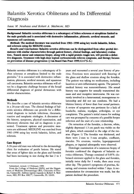
On retraction of the foreskin, a discrete, white,<br />
slightly depressed 1.3-cm patch was visible on the<br />
left glans, which extended to the edge of the meatus.<br />
(Figure 1) The foreskin was thickened, <strong>and</strong><br />
there were 2-mm to 3-mm atrophic, confluent,<br />
macules. No erosions, vesicles, hyperpigmented<br />
plaques, or inguinal adenopathy were observed.<br />
Histologic examination of a cutaneous biopsy of<br />
foreskin confirmed the diagnosis of balanitis xerotica<br />
obliterans. Therapy consisted of 0.05% clobetasol<br />
ointment applied to the glans <strong>and</strong> foreskin,<br />
initially twice daily for 3 weeks, then once every<br />
other day. The patient's symptoms of pruritus, fissuring,<br />
<strong>and</strong> erosions resolved within 7 days. A recommendation<br />
for circumcision was made, but the<br />
patient declined the procedure.<br />
<strong>Balanitis</strong> <strong>Xerotica</strong> <strong>Obliterans</strong> 473
Figure 1. <strong>Balanitis</strong> xerotica obUterans. WelJ-defined,<br />
hypopigmented patch on the distal glans. Sclerotic<br />
process extends to urethral meatus with mild erythema<br />
of urethral os. Multiple hypopigmented macules,<br />
coalescing into plaques, are present on retracted<br />
foreskin.<br />
Discussion<br />
<strong>Balanitis</strong> xerotica obliterans is a chronic, sclerotic<br />
dermatitis involving the genital skin of men. When<br />
it occurs at sites other than the penis, it is called<br />
lichen sclerosus et atrophicus. Clinically, patients<br />
with balanitis xerotica obliterans develop discrete,<br />
angular, white, atrophic macules <strong>and</strong> patches on the<br />
glans, prepuce, <strong>and</strong> foreskin of the penis, with only<br />
rare involvement of the shaft. The prepuce is often<br />
thickened, <strong>and</strong> fissures <strong>and</strong> erosions appear over the<br />
glans. Because the atropruc sclerotic lesions can be<br />
quite ensitive to trauma, bullae, shallow erosions,<br />
<strong>and</strong> bleeding are sometimes observed. Phimosis,<br />
the inability to retract the foreskin over the corona<br />
of the penis, <strong>and</strong> paraphimosis, the inability to<br />
extend the foreskin over the glans, are potential<br />
sequelae. <strong>Balanitis</strong> xerotica obliterans is found most<br />
commonly in patients aged between 15 to 50 years<br />
who are uncircumcised or who had circumcisions<br />
later in life. It tends to progress slowly <strong>and</strong> insidiously<br />
for an extended period <strong>and</strong> can be accompanied<br />
by pruritus, tenderness, painful erections, dysesthesia,<br />
dysuria, <strong>and</strong> a reduction in the force of<br />
urine flow.<br />
These symptoms can be quite troubling for a<br />
patient <strong>and</strong> often prompt his visit for medical attention.<br />
Despite considerable clinical transformation<br />
<strong>and</strong> damage, however, some patients can remain<br />
asymptomatic.<br />
Although the cause of balanitis xerotica obliterans<br />
is unknown, autoimmune, hormonal, keratin,<br />
collagenase, elastase, <strong>and</strong> genetic factors are all implicated.<br />
<strong>Balanitis</strong> xerotica obliterans is diagnosed<br />
definitively by histologic evaluation of a cutaneous<br />
biopsy. Affected tissues display variable hyperkeratosis,<br />
parakeratosis, epidermal atrophy, liquefaction,<br />
degeneration of the basal layer, flattening of<br />
the rete pegs, homogenization of collagen in the dermis,<br />
<strong>and</strong> a sparse dermal mononuclear cell infiltrate.<br />
Given the clinical appearance of balanitis xerotica<br />
obliterans, the differential diagnosis includes<br />
genital herpes, syphilis, fixed dmg eruption, vitiligo,<br />
Reiter syndrome, squamous cell carcinoma,<br />
<strong>and</strong> erythroplasia of Queyrat. (Table 1)<br />
Genital herpes simplex vims (HSV) infection is a<br />
sexually transmitted disease classically described as<br />
grouped vesicles on an erythematous base associated<br />
with HSV type 2.2 Vesicles generally range<br />
from 2 to 4 mm <strong>and</strong> are distributed over the glans,<br />
prepuce, <strong>and</strong> shaft. Because the vesicles are fragile,<br />
patients infrequently come to their primaly care<br />
provider with intact lesions; more commonly, the<br />
lesions consist of shallow erosions. Primary genital<br />
herpes is associated with a severe cutaneous eruption<br />
<strong>and</strong> fever <strong>and</strong> symptoms of headache, malaise,<br />
myalgias, <strong>and</strong> arthralgias. Recurrent genital herpes<br />
infections are generally less severe <strong>and</strong> might not<br />
produce systemic synlptoms. A suspected diagnosis<br />
of genital herpes infection can be confirmed by the<br />
evaluation of a tissue scraping from the base of an<br />
erosion, a positive Tzanck smear with multinucleated<br />
giant cells, or a positive rapid fluorescent antibody<br />
test specific for herpes simplex.<br />
Syphilis is a sexually transmitted disease caused<br />
by the spirochete, Treponema pallidum. Primary<br />
syphilis usually appears at the corona of tlle penis as<br />
a painless chancre consisting of a central ulcer with<br />
raised edges. These early lesions generally regress<br />
after several weeks. The resolving primary chancre<br />
looks like an atrophic macule or patch, mimicking<br />
balanitis xerotica obliterans. Syphilis is diagnosed<br />
by identifying the spirochete with dark-field microscopy<br />
or by serologic studies. Nonspecific serologic<br />
tests that use a nontreponemal antigen are the<br />
rapid protein reagin test <strong>and</strong> the VDRL test. As<br />
false-positive results occur (most commonly in collagen<br />
vascular disease), a specific treponemal antigen<br />
test (FTA-Abs) will confirm the diagnosis of<br />
syphilis.<br />
A fixed drug eruption is a recurrent allergic<br />
reaction to an ingested substance. Both skin <strong>and</strong><br />
mucous membranes can be involved. In order of<br />
474 JABFP ovember-December 1999 Vol. 12 o. 6
Table 1. Characteristics of the Dift'erential <strong>Diagnosis</strong> of <strong>Balanitis</strong> <strong>Xerotica</strong> <strong>Obliterans</strong>.<br />
Disease Systemic Symptoms Extragenital Cause <strong>Diagnosis</strong><br />
<strong>Balanitis</strong> xerotica No Occasionally Unknown Biopsy<br />
obliterans<br />
Herpes simplex virus Fever, myalgia, Yes HSV type 2, HSV type 1 (less Tzanck smear,<br />
(HSV) headache, malaise frequently) immunofluorescence, viral<br />
culture, biopsy<br />
Syphilis Splenomegaly, fever, Secondary: palms, Trepolle71Ul pallMum Dark-field microscopy, VDRL<br />
lymphadenopathy soles, trunk, neck or rapid protein reagin<br />
followed by ITA Abs<br />
Fixed drug eruption No Oral mucosa can be Drugs, phenolphthalein, History <strong>and</strong> physical<br />
positive of sulfonamides, tetracycline, examination<br />
negative<br />
ampicillin, nonsteroidal antiinflammatory<br />
medications<br />
Reiter syndrome Arthritis, Palms <strong>and</strong> soles Unknown Clinical findings<br />
conjunctivitis,<br />
Association with HLA B27<br />
fever<br />
Vitiligo No Often generalized Unknown Biopsy<br />
Erythroplasia of No No Possibly human papilloma Biopsy<br />
Queyrat<br />
virus<br />
ITA Abs-treponemal antigen test specific for Trep01le71Ul pallMum. HLA-human leukocyte antigen.<br />
frequency, the genitalia, face, <strong>and</strong> torso are most<br />
commonly involved. A bulla on an erythematous<br />
base or a painful erosion can be found during physical<br />
examination of active lesions.<br />
As the acute phase of the fixed drug eruption<br />
resolves, discrete postinflammatory hyperpigmented<br />
macules are typically observed. Fixed drug eruption<br />
of the male genitalia is usually confined to the<br />
glans. <strong>Diagnosis</strong> is based on a careful history, in<br />
which the lesion reappears in the same location <strong>and</strong><br />
is associated with ingestion of a drug. The more<br />
common medications implicated in fixed drug<br />
eruption are phenolphthalein (a laxative agent that<br />
has been removed from the US market), barbiturates,<br />
sulfonamides, tetracycline, ampicillin, <strong>and</strong><br />
nonsteroidal anti-inflammatory medications. 3,4<br />
Vitiligo is depigmentation of the skin resulting<br />
from the loss of melanocytes. Genital lesions tend<br />
to be located on the penile shaft <strong>and</strong> are not sclerotic,<br />
atrophic, or symptomatic. <strong>Diagnosis</strong> is often<br />
made on the basis of finding other hypopigmented<br />
lesions. <strong>Balanitis</strong> xerotica obliterans can also have<br />
extragenital involvement, however, <strong>and</strong> a complete<br />
history <strong>and</strong> physical examination are essential to<br />
find all areas of involvement. For patients with<br />
lighter skin tones, a Wood lamp (ultraviolet light)<br />
can be used to augment depigmented patches. Additionally,<br />
biopsy shows the absence of melanocytes<br />
without dermal sclerosis or loss of the rete pegs.<br />
Reiter disease, found almost exclusively in men,<br />
is classically associated with the triad of arthritis,<br />
urethritis, <strong>and</strong> conjunctivitis. Cutaneous manifestations<br />
include circinate balanitis (sharply marginated<br />
erosions that can involve the glans, foreskin, shaft,<br />
<strong>and</strong> scrotum) <strong>and</strong> keratoderma blennorrhagicum<br />
(psoriatic-like pustular plaques often noted on the<br />
palms <strong>and</strong> soles). Systemic findings, such as fever<br />
<strong>and</strong> malaise, are common. <strong>Diagnosis</strong> of Reiter disease<br />
is predicated on a seronegative asymmetric<br />
arthropathy lasting more than 1 month <strong>and</strong> one or<br />
more of the following: urethritis, cervicitis, dysentery,<br />
inflammatory eye disease, or mucocutaneous<br />
disease. A complete history <strong>and</strong> physical examination<br />
are of paramount importance, as biopsy findings<br />
of Reiter disease are often indistinguishable<br />
from psoriasis. The development of Reiter disease<br />
is strongly associated with urogenital infections<br />
caused by a variety of agents in patients who are<br />
positive for human leukocyte antigen B27.5<br />
Squamous cell carcinoma of the penis <strong>and</strong> erythroplasia<br />
of Queyrat (squamous cell carcinoma in<br />
situ of the glans or prepuce) both must be kept in<br />
the forefront of any differential diagnosis of penile<br />
lesions because of the serious nature of the disease.<br />
Characteristically, squamous cell carcinoma appears<br />
as a velvety, red, sharply demarcated plaque.<br />
The clinical signs can be quite variable, however,<br />
<strong>and</strong> range from white macules to brown verrucous<br />
plaques.<br />
Human papillomavirus has been implicated in<br />
invasive squamous cell carcinoma <strong>and</strong>, to an even<br />
greater degree, in squamous cell carcinoma in-<br />
<strong>Balanitis</strong> <strong>Xerotica</strong> <strong>Obliterans</strong> 475
situ. 6 Specifically, Papillomavirus type 16 is most<br />
closely associated with malignant transformation,<br />
<strong>and</strong> research suggests a role for human papillomavirus<br />
in the pathogenesis of squamous cell carcinoma.<br />
Definitive diagnosis is made by histologic<br />
evaluation of a cutaneous biopsy.<br />
Treatment<br />
Currently there is no curative therapy for balanitis<br />
xerotica obliterans. There are, however, many therapeutic<br />
options available to limit involvement <strong>and</strong><br />
relieve symptoms. High-potency topical corticosteroids<br />
(ie, clobetasol 0.05% ointment) are shown to<br />
be effective in women with lichen sclerosus et atrophicus<br />
<strong>and</strong> are currently the treatment of choice for<br />
balanitis xerotica obliterans?·8 Alternatively, intralesional<br />
corticosteroid injections have been proposed.<br />
In cases of urethral involvement <strong>and</strong> stenosis,<br />
meatotomy or urethral dilation might be<br />
required. Circumcision can relieve symptoms of<br />
phimosis <strong>and</strong> often results in remission. Alternatively,<br />
carbon dioxide laser therapy might be an<br />
option should a patient not respond to more conservative<br />
treatments. 9 - 11 Despite the limitations of<br />
the current therapeutic options, reports have<br />
shown that balanitis xerotica obliterans is usually<br />
restricted to those men who had circumcisions later<br />
in life, whereas neonatal circumcision appears to<br />
prevent the development of this disorder. 12 In addition<br />
to symptomatic treatment <strong>and</strong> preventative<br />
measures, patients need regular follow-up visits<br />
(every 12 months), he cause squamous cell carcinoma<br />
arising from the lesions of balanitis xerotica<br />
obliterans has been reported. lJ ,14<br />
Conclusion<br />
<strong>Balanitis</strong> xerotica obliterans can be distinguished<br />
from most other genital dermatoses that mimic it<br />
by a careful history, focused review of symptoms,<br />
<strong>and</strong> physical examination. A Tzanck smear <strong>and</strong><br />
cutaneous biopsy are office-based procedures that,<br />
along with a rapid protein reagin test, will provide<br />
a definitive diagnosis. Treatment with high-dose<br />
topical corticosteroids is aimed at reducing the inflammatory<br />
process. <strong>Balanitis</strong> xerotica obliterans<br />
has been associated with additional genital abnormalities,<br />
including phimosis <strong>and</strong> squamous cell carcinoma.<br />
Neonatal circumcision reduces its occurrence.<br />
References<br />
1. Stuhmer A. <strong>Balanitis</strong> <strong>Xerotica</strong> <strong>Obliterans</strong> (post-operationem)<br />
und ihre Beziehungem zur Kraurosis<br />
gl<strong>and</strong>is et praeputii penis. Arch Dermatol Syphilogie<br />
1928;156:613.<br />
2. Brugha R, Keersmaekers K, Renton A, Meheus A.<br />
Genital herpes infection: a review. Int] Epidemiol<br />
1997;26:698-709.<br />
3. Pasricha]S. Drugs causing fixed eruptions. Br] Dermatol<br />
1979;100:183-5.<br />
4. Sharma VK, Dhar S, Gill AN. Drug-related involvement<br />
of specific sites in fixed eruptions: a statistical<br />
evaluation.] DermatoI1996;23:530-4.<br />
5. Hughes RA, Keat AC. Reiter's syndrome <strong>and</strong> reactive<br />
arthritis: a current view. Semin Arthritis Rheum<br />
1994;24: 190-21 O.<br />
6. Cupp MR, Malek RS, Goellner ]R, Smith TF, Espy<br />
M]. The detection of human papillomavirus deoxyribonucleic<br />
acid in intraepithelial, in situ, verrucous<br />
<strong>and</strong> invasive carcinoma of the penis.] UroI1995;154:<br />
1024-9.<br />
7. Bornstein ], Heifetz S, Kellner Y, Stolar Z,<br />
Abramovici H. Clobetasol dipropionate 0.05% versus<br />
testosterone propionate 2% topical application<br />
for severe vulvar lichen sclerosus. Am] Obstet GynecoI1998;178(1<br />
Pt 1):80-4.<br />
8. Jorgensen ET, Svensson A. The treatment of phimosis<br />
in boys, with a potent topical steroid (c1obetasol<br />
propionate 0.05%) cream. Acta Derm Venereol<br />
1993;73:55-6.<br />
9. Hrebinko RL. Circumferential laser vaporization for<br />
severe meatal stenosis secondary to balanitis xerotica<br />
obliterans. ] Urol 1996; 156: 1735-6.<br />
10. Kartamaa M, Reitamo S. Treatment of lichen sclerosus<br />
with carbon dioxide laser vaporization. Br ]<br />
Dermatol 1997;136:356-9.<br />
11. Windahl T, Hellsten S. Carbon dioxide laser treatment<br />
of lichen sclerosus et atrophicus. ] Urol 1993;<br />
150:868-70.<br />
12. Ledwig PA, Weig<strong>and</strong> DA. Late circumcision <strong>and</strong><br />
lichen sclerosus et atrophicus of the penis.] Am Acad<br />
Dermatol 1989;20(2 Pt 1):211-4.<br />
13. Pride HB, Miller OF 3rd, Tyler WE. Penile squamous<br />
cell carcinoma arising from balanitis xerotica<br />
obliterans. ] Am Acad Dermatol 1993;29:469-73.<br />
14. Campus GV, Alia F, Bosincu L. Squamous cell carcinoma<br />
<strong>and</strong> lichen sclerosus et atrophicus of the<br />
prepuce. Plast Reconstr Surg 1992;89:962-4.<br />
Additional Reading<br />
Meffert ]], Davis BM, Grimwood RE. Lichen sclerosus.<br />
] Am Acad Dermatol 1995;32:393-416.<br />
Lynch P], Edwards L. Genital dermatology. New York:<br />
Churchill Livingstone, 1994.<br />
476 JABFP November-December 1999 Vol. 12 No.6