tylosis palmaris et plantaris familiaris associated - SAMJ Archive ...
tylosis palmaris et plantaris familiaris associated - SAMJ Archive ...
tylosis palmaris et plantaris familiaris associated - SAMJ Archive ...

You also want an ePaper? Increase the reach of your titles
YUMPU automatically turns print PDFs into web optimized ePapers that Google loves.
170 S.A. MEDICAL JOURNAL 4 March 1961<br />
finns and very soon the new drug is advertised under<br />
many different propri<strong>et</strong>ary names. Alternatively, a series<br />
of compounds may be introduced with very similar<br />
therapeutic actions, but differing only lightly in their<br />
chemical constitution. These all become advertised under<br />
different names. The result is confusion in the nomenclature<br />
of new drugs. After an interval a new drug may be<br />
accepted for the phannacopoeia, and is given an official<br />
name. For obvious reasons it becomes difficult to establish<br />
the popular use of the official name.<br />
Practitioners are well aware of the confusing nomenclature,<br />
and pharmacists are al 0 perplexed by the multiplicity<br />
of similar-sounding names. There are many<br />
examples that could be quoted but a few will suffice.<br />
Among the tranquillizers there are triflupromazine and<br />
trifluoperazine, perphenazine and prochlorperazine. There<br />
are digoxin and digitoxin; raudixin and redoxon; sigmagen<br />
and sigmamycin; romical and romilar; librium and<br />
libratar; and m'<strong>et</strong>r<strong>et</strong>on and m<strong>et</strong>icorten. There must be even<br />
b<strong>et</strong>ter examples of drugs with different actions and indications<br />
whose names are very similar. It is surprising that<br />
accidents do not occur frequently from the use of the<br />
wrong drug. It is to the credit of doctors, nurses, and<br />
phannacists that mistakes occur very infrequently.<br />
It would be a blessing to both prescribers and dispensers<br />
of drugs if the manufacturers would relegate their brand<br />
names to second place and mark<strong>et</strong> their products as<br />
penicillin (Smith) or penicillin (Jones). Unfortunately this<br />
m<strong>et</strong>hod of nomenclature is unacceptable to commercial<br />
finns, although it is applied to insulin preparations. .<br />
As the output of new preparations increases, the search<br />
for new names becomes a big problem. One firm in the<br />
USA has 'commissioned a machine to produce a dictionary<br />
of forty-two thousand nonsense words of an appropriate<br />
scientific look and sound'. An official said 'Thinking up<br />
names has been driving us cuckoo ... A good trade<br />
name carries a lot of weight with doctors ... there are<br />
enough names in the new dictionary to keep us going<br />
for years ... We don't y<strong>et</strong> know what proportion of<br />
names is unpronounceable ... how many are obscene ...<br />
how many objectionable on other grounds of good taste.<br />
"Godamycin" would be a mild example1.'<br />
It is therefore clear that a difficult situation confronts<br />
the practising physician. Perhaps the doctors have themselves<br />
to blame for this. In many countries attempts have<br />
been made and progress achieved in obtaining uniformity<br />
on a national level. The World Health Organization has<br />
attempted to coordinate these efforts at an international<br />
level, and has published lists of proposed international<br />
non-propri<strong>et</strong>ary names for drugs.<br />
It is obvious that there is much to be done, and a<br />
difficult task lies ahead to avoid the confusion which<br />
exists in the multiplicity of names for the same preparations<br />
and the similarity of names for different preparations.<br />
1. New Yorker. 14 Juh' 1956.<br />
TYLOSIS PALMARIS ET PLANTARIS FAMILIARIS ASSOCIATED<br />
~TH CLINODACTYLY<br />
-<br />
GORDO. K. KLINIWORTH, B.sc., M.B., B.ar. (RAND) and INGRAM F. ANDERSON, M.B., B.CH. (RAND)<br />
Department of Medicine, Johannesburg General Hospital<br />
Tylosis <strong>palmaris</strong> <strong>et</strong> <strong>plantaris</strong> <strong>familiaris</strong> (hereafter referred<br />
to as <strong>tylosis</strong>) is a familial ectodermal anomaly of the<br />
palms and soles producing marked hyperkeratosis. It is<br />
to Thost 25 and Unna 27 that credit must be given for the<br />
original description of the disease. Since then numerous<br />
reports have appeared in the literature. The most comprehensive<br />
review of the whole subject, however, is that<br />
of Cockayne. 3 Several workers have noted the rare association<br />
b<strong>et</strong>ween <strong>tylosis</strong> and other conditions. Among them<br />
are Howel-Evans <strong>et</strong> aP who discussed the association of<br />
thi disease with carcinoma of the oesophagus. Modern<br />
gen<strong>et</strong>ic studies have focussed attention on the co-existence<br />
of familial conditions. In view of this it was felt necessary<br />
to study, in more d<strong>et</strong>ail, the family of a patient who<br />
presented with a myocardial infarction and a family history<br />
of <strong>tylosis</strong>.<br />
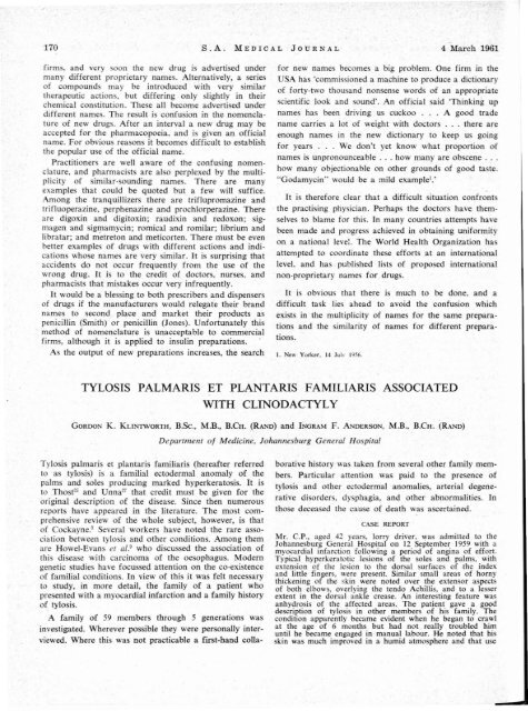
A family of 59 members through 5 generations was<br />
investigated. wherever possible they were personally interviewed.<br />
Where this was not practicable a first-hand collaborative<br />
history was taken from several other family members.<br />
Particular attention was paid to the presence of<br />
<strong>tylosis</strong> and other ectodermal anomalies, arterial degenerative<br />
disorders, dysphagia, and other abnormalities. In<br />
those deceased the cause of death was ascertained.<br />
CASE REPORT<br />
Mr. C.P., aged 42 years, lorry driver, ""as admitted to the<br />
Johannesburg General Hospital on 12 September 1959 with a<br />
myocardial infarction following a period of angina of effort.<br />
Typical hyperkeratotic lesions of the soles and palms, with<br />
extension of the lesion to the dorsal surfaces of the index<br />
and little fingers, were present. Similar small areas of horny<br />
thickening of the skin were noted over the extensor aspects<br />
of both elbows, overlying the tendo Achillis, and to a lesser<br />
extent in the dorsal ankle crease. An interesting feature was<br />
anhydrosis of the affected areas. The patient gave a good<br />
description of <strong>tylosis</strong> in other members of his family. The<br />
condition apparently became evident when he began to crawl<br />
at the age of 6 months but had not really troubled him<br />
until he became engaged in manual labour. He noted that his<br />
skin was much improved in a humid atmosphere and that use
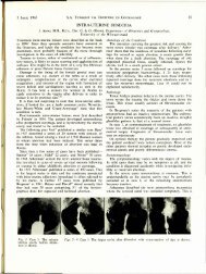
4 Maart 1961 S.A. TYDSKRIF VIR GENEESKUNDE 171<br />
ill<br />
IT I<br />
••<br />
UAMW£O<br />
[J <strong>et</strong><br />
0 0<br />
NOT<br />
TYlOSlS AND CUHOOACTYU ,<br />
rn<br />
OA1IW«O<br />
CD<br />
[XAIIIIfWEO NO HtSTOflV OF f)TH(A CONOITlC»i<br />
IJ ~<br />
"<br />
,.<br />
1<br />
IJ ()<br />
(llAMlN(O fYlOSlS OJrf\.y<br />
NO T'f't..OSIS _0 CUHOO ...cn ,<br />
6000 ~1()lt' or mosrs Iro() loIrSTOIll' 01' Cl.JNOOACTTlY<br />
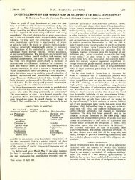
Fig. 1. Family tree of patient, showing 4 generations (1, 11, ill and IV) md one member in fifth generation. Arrow 'points to patient (Ill 12).<br />
TABLE I. SUBJECTS WHO WERE PERSONALLY EXAMINED OR HAD HISTORY OF TYLOSIS<br />
"!-<br />
Distribution and severity oflesions<br />
.. ~<br />
-c ...<br />
Ol<br />
::<br />
<br />
'c-<br />
'o-~<br />
"".::;<br />
~ 0""<br />
E I<br />
~ "'"13 .. .. - too-<br />
0 .. .::;<br />
~ .~<br />
'"<br />
§ .;:: ~<br />
~ too'"' ~.::; -'-<br />
~ :=~ .so c::-§<br />
-~<br />
... '" '"' ~6 ~:::: "13~ ~<br />
)( g:, .~ a ~ ..::: ~- ~~<br />
:::~<br />
..<br />
~..s:<br />
O-c 00<br />
.. c:: ,. -<br />
-~<br />
t':l ~ ~ ~~ ~~ ~ ~ l::l"Ol ~~ l::l~ ~~ v)..:;::<br />
~<br />
~<br />
~~<br />
I 4 F 95 NE i> Yes<br />
IT 2 F 50 E L Yes ++ ++ + Increased<br />
IT 3 F 51 NE D Yes<br />
IT 5 F 64 E L Yes +++ +++ + + Decreased<br />
ill 4 M 50 E L No Yes ormdl<br />
ill 5 F 11 E L No No Normal<br />
ill 6 F 9 E L No 0 Normal<br />
ill 7 M 7 E L No No Normal<br />
ill 8 F 38 E L Yes ++ +++ + + Yes Decreased<br />
ill 10 F 33 E L No 0 Normal<br />
ill 12 M 42 E L Yes +++ +++ + + No Decreased<br />
ill 13 M 35 E L Yes ++ +++ + No Decreased<br />
ill 14 F 24 E L No Yes Normal<br />
ill 15 M 41 E L No No Normal<br />
ill 16 M 18 E L No No Normal<br />
ill 17 M 42 E L No No Normal<br />
IV 5 F 17 NE L Yes<br />
IV 7 F 14 E L Yes ++ ++ + + Yes Decreased<br />
IV 8 M 13 E L No 0 ormal<br />
IV 9 F 7 E L No 0 ormal<br />
IV 10 F 14 E L No 0 ormal<br />
IV 11 M 12 E L 0 0 ormal<br />
IV 12 F 10 E L 0 Yes ormal<br />
IV 16 M 5 E L Yes ' '..J...<br />
+++ TT, + + Yes Decreased<br />
IV 17 F 12 E L Yes ++ ++ Yes ormal<br />
IV 18 M 10 E L Yes ++ ++ + + + Yes Decreased<br />
IV 19 M 7 E L Yes ++ ++ + + ormaJ<br />
IV 20 F 3 E L Yes ++ ++ 0 ormal<br />
IV 21 M 1 E L 0 0 ormal<br />
IV 23 M 9 E L 0 0 ormal<br />
IV 24 F 5 E L 0 Yes ormal<br />
IV 25 M 10 E L 0 0 ormal<br />
IV 26 F 6 E L 0 0 ormaJ<br />
IV 27 M 8 E L 0 0 ormal<br />
IV 28 M 2 E L 0 0 ormal<br />
F=female, M=male, NE=not elGUIlined, E=examined, D=deceased, L=living, + = slight, ++= more pronounced, and +++-severe.<br />
t For numbers refer to Fig. 1.
172 S.A. MEDICAL JOURNAL 4 March 1961<br />
of the hands, especially in cold dry weather, resulted in gm s<br />
thickening, cracking and even bleeding of the palmar skin.<br />
Except for the presence of <strong>tylosis</strong> and evidence of acute myocardial<br />
infarction,<br />
essentially normal.<br />
general examination of the patient was<br />
Family<br />
[n the 59 member of the family there were at least 14 with<br />
<strong>tylosis</strong> (9 female, 5 male). Of the 32 member per onally<br />
examined, 11 were affected; a good de cription was obtained<br />
regarding 3 other (Fig. 1 and Table D. In all affected ca es<br />
the age of on <strong>et</strong> was in the fir t year of life. In 10 of the<br />
ubjects clinodactyly was noted. In this condition the little<br />
finger is curved inwards towards the other fingers. No other<br />
abnormality of skin or skel<strong>et</strong>on was noted nor were there<br />
any abnormalitie of hair or te<strong>et</strong>h. Besides the patient, 2 (1I5,<br />
llIJ I) of the 33 living member bad sustained myocardial<br />
infarctions. 0 other members gave a history of cardiovascular<br />
disease. There wa no history of dysphagia or buccal leukoplakia<br />
and as far as could be ascertained no one had died of<br />
oe ophageal carcinoma. Of the 7 in whom the cause of death<br />
was known (14, Ill, 113, 1I4, ITI9, III18, and IIJI9), 3 were<br />
due to cardiac di ease (14, Ill, and 113). one of the 7 bad<br />
died of malignant disea e.<br />
DISCUSSION<br />
Tylosis<br />
Much confusion exists in the literature regarding the<br />
terminology of the group of hyperkeratoses. Tylosis <strong>palmaris</strong><br />
<strong>et</strong> <strong>plantaris</strong> has the following synonyms: '3 keratosis<br />
<strong>palmaris</strong> <strong>et</strong> <strong>plantaris</strong>, hyperkeratosis <strong>palmaris</strong> <strong>et</strong> <strong>plantaris</strong>,<br />
ichthyosis <strong>palmaris</strong> <strong>et</strong> <strong>plantaris</strong>, keratoderma palmare <strong>et</strong><br />
plantare, and symm<strong>et</strong>rical keratoderma.<br />
The condition has been described as rare.:!:! In Nprthern<br />
Ireland the incidence was calculated to be I in 40,000. 2 '<br />
Histopathologically,IO the skin usually shows considerable<br />
hypertrophy of all its layers, more espe ially the stratum<br />
corneum, which is grossly thickened. The tratum granulosum<br />
is normal in appearance and there is no change in<br />
the stratum spinosum. Occasionally there i flattening of<br />
the papillary body. These papillae may be increa ed fivefold<br />
in depth. The dermis is unaffected, except outside the<br />
area of horny thickening or, where fissures are present,<br />
when mild inflammatory changes may be noted. The sweat<br />
glands and their ducts may be hypertrophied. The histopathological<br />
section in the present case i shown (Fig. 2)<br />
compared with a normal section (Fig. 3).<br />
Clinically <strong>tylosis</strong> is rarely manifest at birth and is usually<br />
not recognized until the third or fourth month. In exceptional<br />
case the ons<strong>et</strong> may be delayed until the age of<br />
6 years. I9 . 23 The lesions are bilateral, symm<strong>et</strong>rical, and<br />
situated almost exclusively on the palms and soles (Figs.<br />
4 and 5) either of which may be predominantly involved.<br />
Occasionally the hyperkeratosis is noted on the dorsum<br />
of the hands, fe<strong>et</strong> and phalanges. In some cases it is present<br />
on ·the extensor surfaces of the elbows, over the knees<br />
and about the ankles. I5 . I9 The nails may be involved and<br />
become thickened and opaque and are raised up from<br />
the nail bed by a horny accumulation beneath them. This<br />
complication may result in more severe symptoms than<br />
those produced by the skin lesions.<br />
The distribution is d<strong>et</strong>ermined to a large extent by<br />
physical factors such as pressure and friction. Thus, in<br />
infants, the lesions will be on the knees; on beginning<br />
to walk the fe<strong>et</strong> are most involved in the weight-bearing<br />
areas of the soles; the manual labourer exhibits gross<br />
Fig. 2. Low-power section of hyperkeratotic palmar skin<br />
(x 45).<br />
Fig. 3. Low-power section of normal palmar skin (x 45).
4 Maart 1961 S.A. TYDSKRIF VIR GENEESKUNDE 173<br />
Fig. 4. This shows the bilateral lesions of <strong>tylosis</strong> on the<br />
palms.<br />
lesions of the palms, and the office worker may have a<br />
predominant affection of the elbows.<br />
The condition varies not only in intensity in different<br />
individuals of the same family but also at different times<br />
in an affected individual. The condition is aggravated<br />
during very warm or very cold weather and also during<br />
manual labour, especially if the work involves exposure<br />
of the hands to moisture and cold." The influence of<br />
climatic and occupational factors was also well illustrated<br />
in members of the family we studied.<br />
Many subjects may be so slightly affected that they do<br />
not realize that they have the condition until their attention<br />
i drawn to it. On the other hand, marked thick~ning<br />
of the palmar skin may reduce tactile sensibility and<br />
Fig. 5. This shows the bilateral lesions of <strong>tylosis</strong> OD the<br />
soles. Note symm<strong>et</strong>ry of the lesions.<br />
interfere \ it.h the finer finger mo emenl . P in i nOl<br />
u ually a marked fearure of the h nd I ion. although<br />
Ander on l reponed a ca e with e tremely painful I ion<br />
of the hand. The ole tend to be more painful than the<br />
palm and thi may interfere with normal walking. ffe ted<br />
area are predi po ed to fi uring b au e of a la k of<br />
normal ela ticity.2:!·~ The normal fi ure are ex gger ted<br />
and produce a mo aic-like appearance. lnvol emenl of the<br />
dermis by the fi ures cau e pain and om<strong>et</strong>ime h e<br />
morrhage.<br />
Hyperhydro i i pre ent in the majorit of ca reponed<br />
in the literature. Familie are on rerord in whi h<br />
sweating was dimini hed. 3 In the famil under on ideration<br />
both states obtained. There appeared to be an in erse<br />
relationship b<strong>et</strong>ween the degree of hyperkerato i and the<br />
amount of sweating, and we ugge t that the hypohydro i<br />
in severely affected cases may be due to ob tru tion of<br />
the sweat-gland duct by the thickened and hyperkeratotic<br />
skin. Sweat-gland hypertrophy i not an uncommon feature<br />
and may account for the increa ed weating noted in<br />
milder cases.<br />
Tylosis should be differentiated from the acquired type<br />
of hyperkeratosis, notably lichen simplex (neurodermatili ),<br />
contact dermatitis, psoriasis, tertiary yphili, fungal infeclions<br />
(particularly TrichophYTOn rubrum), volar verrucae.<br />
calluses, and the now rarely- een ar enical keratoderma.<br />
19 • 23 • 2S<br />
Certain familial skin conditions resembling tylo i mu t<br />
be differentiated. Hereditary di eminate keratoderma<br />
<strong>palmaris</strong> <strong>et</strong> <strong>plantaris</strong> consists of multiple, ymm<strong>et</strong>rical.<br />
discr<strong>et</strong>e plaques on the palms and soles. The lesions do<br />
not coalesce and the ons<strong>et</strong> usually occurs at adole ence<br />
but is som<strong>et</strong>imes delayed until adulthood. 21<br />
Mal de Meleda 3 is a rare type of palmar and plantar<br />
hyperkeratosis described only on the i land of 1eleda off<br />
the Dalmatian coa t. Mo t of the i-nhabitan are onsanguinously<br />
related; this favour tran mi ion of lhe<br />
di ease by what icon idered to be .a rece ive gene. In<br />
addition to the usual site the hyperkeratosi involve<br />
the dorsum of the hands and fe<strong>et</strong>, and may extend up<br />
the legs and forearms to the elbow and knees. Hyperhydrosis<br />
is present.<br />
There is no known cure for <strong>tylosis</strong>. Amelioration may<br />
occur with change of occupation or during humid weather.<br />
Treatment is purely palliative and numerou therapeutic<br />
mea ures have been employed with variable temporary<br />
benefit. Keratolytics uch a alicylic-acid preparation<br />
are employed to soften and remove the homy layer.<br />
Superficial X-ray therapy ha been advo ated by ome<br />
but has not m<strong>et</strong> with general approval, mainly becau e<br />
of the risk of causing carcinoma and becau e effective<br />
do age may lead to cicatricial atrophy with teJangiecta i .10<br />
Various hormone, notably thyroid extract and oe trogen ,<br />
have been tried. The u e of large dose of vitamin A ha<br />
been reported to control the gro s manife tation of the<br />
disease. 16 Its use is purely empirical and no ati factory<br />
explanation of the mechanism is forthcoming. andpapering<br />
and mechanical abra ion of the affecled area ha been<br />
used. 10 severe ca e benefit ha been claimed by compl<strong>et</strong>e<br />
exci ion and grafting.'·13·l:l·29<br />
Tylosis and Associated AbnormaliTies<br />
Many abnormalitie as ociated with tylo i have been
174 S.A. MEDICAL JOURNAL 4 March 1961<br />
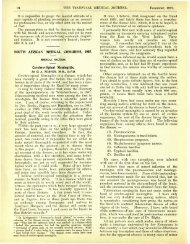
Fig. 6. Tylosis and clinodactyly. Note radial deflection of both little fingers.<br />
Fig. 7. Clinodactyly. X-ray showing sloping of the distal surface of the middle<br />
shortening of the radial surface of the phalanx.<br />
fingers. The rest of the phalanges are normal. The incidence<br />
has been calculated at 1 in 1,000 in Northern Ohio. s Hersh<br />
<strong>et</strong> af.S fed that the term clinodactyly should be limited to<br />
those cases which are due to incompl<strong>et</strong>e ossification and<br />
should not be confused with other causes of familial<br />
crooked little fingers such as those due to abnormal tendons5-7<br />
and to fused ossification,u<br />
Gen<strong>et</strong>ic Transmission<br />
Tylosis is inherited as a Mendelian dominant with high<br />
pen<strong>et</strong>rance. It appears to be controlled by a single autosomal<br />
gene. Cockayne 3 reviewed 47 families in the literature<br />
and the proportion of affected to normal members<br />
was 594: 483. The sex ratio was 318 males to 284 females.<br />
He reported 2 small families in whom the lesion only<br />
occurred in the females and not in the males. Subsequent<br />
studies, however, indicated that the two sexes are equally<br />
affected and no race is immune. 20 • 23<br />
Lawler and RenwickJ..i are of the opinion that there are<br />
at least 2 types of inherited <strong>tylosis</strong> which apparently run<br />
true in families and appear to be due to different genes.<br />
The 2 forms have been differentiated on clinical grounds.<br />
Type A has a variable age of ons<strong>et</strong> ranging from 5 to 15<br />
years, whereas Type B is recognizable during the first year<br />
of life. The latter is clinically distinguishable from Type A<br />
by the uniform thickness of the keratosis, by the sharply<br />
delimited edges of the lesion, and by the rare incidence<br />
of painful fissuring. Both types are inherited as a<br />
Mendelian dominant. This clear-cut distinction was not a<br />
feature of our cases, since fissuring was not an uncommon<br />
finding in lesions which had been noted in the first<br />
year of life with relatively sharply demarcated edges and<br />
in which keratosis was of uniform thickness.<br />
Tylosis is due to a mutant gene and the extent, distribution<br />
and severity are independent aspects which are<br />
largely d<strong>et</strong>ermined by composite gen<strong>et</strong>ic and environmental<br />
factors. Thus the same mutant gene may cause several<br />
distinct clinicopathological pictures and this difference in<br />
expressivity may be evoked to explain the large and varied<br />
nomenclature applied to the hyperkeratoses.<br />
Clinodactyly has not previously been described.' asso-<br />
described in isolated instances. Howel-Evans <strong>et</strong> al. 9 reported<br />
the association of carcinoma of the oesophagus<br />
and <strong>tylosis</strong> in 2 families. There was unequivocal evidence<br />
that carcinoma of the oesophagus was <strong>associated</strong> with<br />
<strong>tylosis</strong> in 17 of their cases, and in only 1 case in which<br />
the neoplasm was found was it impossible to establish<br />
that <strong>tylosis</strong> was present. Members of the family unaffected<br />
by <strong>tylosis</strong> were unaffected by carcinoma. In the cases<br />
of carcinoma of the oesophagus in which the oesophageal<br />
epithelium adjacent to the tumour was examined histologically,<br />
no evidence of hyperkeratosis was found. The<br />
association b<strong>et</strong>ween palmar and plantar hyperkeratosis and<br />
leukoplakia of the mouth has been recorded. 12 • 23 • 26<br />
Malignant degeneration in areas of hyperkeratosis has been<br />
considered likely by some dermatologists,IS and Ingram and<br />
Brain lO mentioned a case in which squamous carcinoma<br />
developed in <strong>tylosis</strong>.<br />
Hanhart (quoted by Cockayne 3 ) described a Swiss family<br />
in which the affected members developed <strong>tylosis</strong> and<br />
multiple lipomata during late adolescence. Cockayne 3 commented<br />
that the late age of appearance of <strong>tylosis</strong> made<br />
it doubtful wh<strong>et</strong>her it was the ordinary form of the<br />
disease.<br />
Tylosis has been reported to occur occasionally with<br />
extensive ichthyosis hystrix or with epidermolysis bullosa. IO<br />
It has been found <strong>associated</strong> with other ectodermal abnormalities<br />
3 IO • ,23 or with hypogenitalism, oxycephaly, clubbing<br />
of the fingers, or mental r<strong>et</strong>ardation. 2 • 3<br />
Clinodactyly, as described in our cases, is a shortening<br />
of the middle phalanx of the fifth finger, mainly on its<br />
radial aspect, which results in a radial deflection of the<br />
terminal phalanx of from 15° to 30° (Fig. 6). The remaining<br />
digits are normal and there is no flexion curvature. s On<br />
X-ray (Fig. 7), the middle phalanx of the little finger is<br />
slightly shorter on the radial side, thus forming an appreciable<br />
slope on its distal surface. X-ray observations indicate<br />
that this seems to be caused by a slowing-down process<br />
of ossification, specifically at the upper radial part of the<br />
middle phalanx of the fifth finger. Due to this slanting,<br />
the distal phalanx is inclined inwards towards the other<br />
phalanx of the little finger due to
4 Maart 1961 S.A. TYDSKRIF VIR GENEESKUNDE 175<br />
ciated with <strong>tylosis</strong>. The only reported association of clinodactyly<br />
is that with webbing of the toes. s In the family<br />
we studied there were 10 members with clinodactyly<br />
6 female and 4 male. In 8 cases the curving of the little<br />
finger was bilateral but in 2 this curving was only present<br />
in the left hand. Of those with clinodactyly, 6 were<br />
affected with <strong>tylosis</strong> as well. Clinodactyly is transmitted<br />
as an autosomal dominant. Lack of compl<strong>et</strong>e pen<strong>et</strong>rance.<br />
was shown by the fact that in 6 cases the parents did not<br />
have the condition. In 1 case (N24) neither the parents<br />
nor the grandparents had clinodactyly. These features of<br />
dominant transmission with incompl<strong>et</strong>e pen<strong>et</strong>rance have<br />
been reported by others. s<br />
Is there a gen<strong>et</strong>ic linkage b<strong>et</strong>ween clinodactyly and<br />
<strong>tylosis</strong>, or is the association purely one of chance? We<br />
were unable to obtain statistically significant results in<br />
favour of gen<strong>et</strong>ic linkage, using the sibling pair m<strong>et</strong>hod<br />
of PenroseY·lS However, assuming that the incidence of<br />
<strong>tylosis</strong> and clinodactyly in South Africa is 1 in 40,()()()24<br />
and 1 in I,OOOS respectively, the probability of these 2<br />
co~Oitions occurring tog<strong>et</strong>her is in the region of 1 in 40<br />
miJ1fon.<br />
SUMMARY<br />
In a family affected with <strong>tylosis</strong> <strong>palmaris</strong> <strong>et</strong> <strong>plantaris</strong> 59<br />
members were studied. The family tree and the mode' of<br />
gen<strong>et</strong>ic transmission are outlined. The incidence, histopathology,<br />
clinical features, differential diagnosis and treatment<br />
of <strong>tylosis</strong> are discussed. The association of <strong>tylosis</strong><br />
and clinodactyly is noted. In addition some of the reported<br />
abnormalities occurring with <strong>tylosis</strong> are revi"ewed.<br />
We wish to thank Profs. G. A. Elliott, B. J. P. Becker, and<br />
P. V. Tobias for encouragement and a i lance in the preparation<br />
of this article; the Photographic Unit of the Department<br />
of Medicine and Mr. S. Dry of the Anatomy Department,<br />
Univer ity of the Witwater rand, for reproduction of<br />
the figures; and Dr. K. F. Mills, Superintendent, Johanneburg<br />
General Hospital, for perrni sion to publish.<br />
REFERE CES<br />
1. Anderson, J. (1951): Brit. J. Deem., 63, 127.<br />
2. Bureau, G., Horeau, M., Barriere, H. and DuFage, C. (1958): Bull.<br />
Soc. rran9. Derm. Syph., 65, 32 .<br />
3. Cockayne, E. A. (1933): The Inherited Abnormaliries of the Skin and<br />
itS Appendages. London: Oxford University Press.<br />
4. Dencer, D. (1953): Brit. Plast. Surg., 6, 130.<br />
5. Heffner, R. A. (1924): J. Hered., IS, 323.<br />
6. Idem (1929): Ibid., 20, 395.<br />
7. Idem (1941): Ibid., 32, 37.<br />
8. Hersh, A. H., De Marinis, F. snd Stecher, R. M. (1953): Amer. J.<br />
Hum. Gen<strong>et</strong>., 5, 257.<br />
9. Howel-Evans, W., McConnell, R. B., Clarke, C. A. and Sheppard, P. M.<br />
(1958): Quart. J. Med., 27, 413.<br />
10. Ingram, J. T. and Brain, R. T. (1957): Sequeira's Diseases of rhe Skin,<br />
6th ed. London: Churchill.<br />
11. Knenner, D. M. (1934): J. Hered.. 25. 329.<br />
J2. Kumer, L. and Loos, H. O. (1935): Wien. kJin. Wschr., 48, 174.<br />
13. Landazuri, H. F. (195 ): Plast. Reconstr. Susg., 22, 557.<br />
14. Lawler, S. D. and Renwick, J. H. (1957): Personal communication to<br />
Howel·Evans <strong>et</strong> al. loco cil.9<br />
15. Ormshy, O. J. and Montgomery, H. (1948): Diseases of rhe Skin,<br />
7th ed. Philadelphia: Lea and Febiger.<br />
16. Porter, A. D. (1951): Brit. J. Derm., 63, 123.<br />
17. Penrose, L. S. (1935): Ann. Eugen., 6, 133.<br />
18. Idem (1938): Ibid., 8, 233.<br />
19. Pupa, J. A. and Farina, R. (1953): Plast. Reconstr. Surg., 12, 446.<br />
20. SeOlt, O. L. S. In Sorsby, A. ed. (1953): Clinical Gen<strong>et</strong>ics. u.ndon:<br />
Buttesworth.<br />
21. SeOlt, M. J., Costello, M. J. and Simuangco, S. (1951): Arch. Derm.<br />
Syph., 64, 301.<br />
22. Semon, H. C. G. (1953): An Atlas of rhe Commoner Skin Diseases,<br />
4th ed. Baltimore: Williams and Wilkin.