32 Molecular Hardware of Copper Homeostasis in Enterococcus hirae
32 Molecular Hardware of Copper Homeostasis in Enterococcus hirae
32 Molecular Hardware of Copper Homeostasis in Enterococcus hirae

Create successful ePaper yourself
Turn your PDF publications into a flip-book with our unique Google optimized e-Paper software.
0<br />
1<br />
2<br />
3<br />
4<br />
5<br />
6<br />
7<br />
8<br />
9<br />
10<br />
11<br />
12<br />
13<br />
14<br />
15<br />
16<br />
17<br />
18<br />
19<br />
20<br />
21<br />
22<br />
23<br />
24<br />
25<br />
26<br />
27<br />
28<br />
29<br />
30<br />
31<br />
<strong>32</strong><br />
33<br />
34<br />
35<br />
36<br />
37<br />
38<br />
39<br />
40<br />
41<br />
42<br />
43<br />
44<br />
45<br />
46<br />
47<br />
48<br />
49<br />
50<br />
51<br />
52<br />
53<br />
54<br />
55<br />
56<br />
Job: 943-9<br />
Operator: Nettype<br />
Chapter: <strong>32</strong>-Solioz Date: March 1, 2002<br />
Pub Date: 2002<br />
Revision: First Pro<strong>of</strong><br />
Template: Massar0(943-9)7X10 temp<br />
<strong>Molecular</strong> <strong>Hardware</strong> <strong>of</strong> Cu <strong>Homeostasis</strong> 527<br />
1. INTRODUCTION<br />
<strong>32</strong><br />
<strong>Molecular</strong> <strong>Hardware</strong> <strong>of</strong> <strong>Copper</strong><br />
<strong>Homeostasis</strong> <strong>in</strong> <strong>Enterococcus</strong> <strong>hirae</strong><br />
Re<strong>in</strong>hard Wimmer, Charles T. Dameron, and Marc Solioz<br />
In the past, copper transport <strong>in</strong> bacteria has been considered only <strong>in</strong> terms <strong>of</strong> resistance mechanisms<br />
to permit survival <strong>of</strong> the cells <strong>in</strong> adverse copper-rich environments. Indeed, most <strong>of</strong> the systems<br />
characterized early on are plasmid encoded, as is typical for defense mechanisms <strong>of</strong> prokaryotes.<br />
Recently, similar copper “resistance” systems were also identified <strong>in</strong> chromosomal locations, giv<strong>in</strong>g<br />
rise to the concept that bacteria may need systems for copper homeostasis not only <strong>in</strong> extreme environments<br />
but also under normal growth conditions.<br />
The system for copper homeostasis <strong>in</strong> <strong>Enterococcus</strong> <strong>hirae</strong> is currently the best understood example<br />
<strong>of</strong> heavy-metal homeostasis. Because E. <strong>hirae</strong> can easily be genetically manipulated, it represents an<br />
ideal system for the study <strong>of</strong> fundamental aspects <strong>of</strong> the regulation <strong>of</strong> cytoplasmic copper. It appears<br />
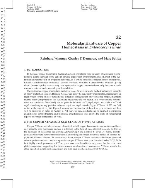
that the major components <strong>of</strong> this system are encoded by the cop operon. It is located on the chromosome<br />
and consists <strong>of</strong> four closely spaced genes <strong>in</strong> the order copY, copZ, copA, and copB. CopY and<br />
copZ encode regulatory prote<strong>in</strong>s, whereas copA and copB encode P-type ATPases <strong>of</strong> 727 and 745<br />
am<strong>in</strong>o acids, respectively (1). Figure 1 summarizes the function <strong>of</strong> these four gene products and they<br />
will be discussed <strong>in</strong> detail <strong>in</strong> Section 2. All four cop gene products can be purified <strong>in</strong> milligram<br />
quantities for direct structural and functional <strong>in</strong>vestigations. This allows the study <strong>of</strong> fundamental<br />
aspects <strong>of</strong> copper homeostasis <strong>in</strong> vitro.<br />
2. THE COPPER ATPASES: A NEW CLASS OF P-TYPE ATPASES<br />
<strong>Copper</strong> ATPases are a key element <strong>of</strong> most, if not all, copper homeostatic mechanisms and have<br />
only recently been discovered and are a milestone <strong>in</strong> the field <strong>of</strong> trace element research. Follow<strong>in</strong>g<br />
the discovery <strong>of</strong> the copper-transport<strong>in</strong>g ATPases CopA and CopB <strong>in</strong> E. <strong>hirae</strong> (2), highly homologous<br />
ATPases were reported from humans as underly<strong>in</strong>g the copper metabolic defects <strong>of</strong> Menkes 4)”<br />
(3,4) and Wilson’s disease (5), respectively. Later, copper ATPases were described from yeast and<br />
other organisms and over two dozen putative copper ATPases have been described today 7)” (6,7). In<br />
fact, highly homologous copper ATPase genes have been found <strong>in</strong> every genome that has been completely<br />
sequenced, suggest<strong>in</strong>g that these enzymes are ubiquitous. Homologous ATPases specific for<br />
other transition metals such as cadmium and z<strong>in</strong>c have also been discovered 9)” (8,9).<br />
From: Handbook <strong>of</strong> <strong>Copper</strong> Pharmacology and Toxicology<br />
Edited by: E. J. Massaro © Humana Press Inc., Totowa, NJ<br />
527
0<br />
1<br />
2<br />
3<br />
4<br />
5<br />
6<br />
7<br />
8<br />
9<br />
10<br />
11<br />
12<br />
13<br />
14<br />
15<br />
16<br />
17<br />
18<br />
19<br />
20<br />
21<br />
22<br />
23<br />
24<br />
25<br />
26<br />
27<br />
28<br />
29<br />
30<br />
31<br />
<strong>32</strong><br />
33<br />
34<br />
35<br />
36<br />
37<br />
38<br />
39<br />
40<br />
41<br />
42<br />
43<br />
44<br />
45<br />
46<br />
47<br />
48<br />
49<br />
50<br />
51<br />
52<br />
53<br />
54<br />
55<br />
56<br />
Job: 943-9<br />
Operator: Nettype<br />
Chapter: <strong>32</strong>-Solioz Date: March 1, 2002<br />
Pub Date: 2002<br />
Revision: First Pro<strong>of</strong><br />
Template: Massar0(943-9)7X10 temp<br />
528 Wimmer, Dameron, and Solioz<br />
Fig. 1. Schematic draw<strong>in</strong>g <strong>of</strong> the cop operon and model <strong>of</strong> copper homeostasis <strong>in</strong> E. <strong>hirae</strong>. <strong>Copper</strong>(I) is<br />
taken up by CopA under copper-limit<strong>in</strong>g conditions. Inside the cell, CopZ complexes copper(I) to safely deliver<br />
it to the CopY repressor, which regulates expression <strong>of</strong> the cop operon. If <strong>in</strong>tracellular copper is excessive,<br />
CopZ delivers copper to CopB for secretion.<br />
2.1. CPx-Type ATPases<br />
The structure and function <strong>of</strong> copper ATPases has become one <strong>of</strong> the focal po<strong>in</strong>ts <strong>of</strong> research on<br />
copper homeostasis. <strong>Copper</strong> ATPases differ significantly <strong>in</strong> their primary structure, membrane topology,<br />
and evolutionary relationship from the previously known P-type ATPases, such as the Ca 2+ -<br />
ATPases or the Na + K + -ATPases (Fig. 2). They thus form a dist<strong>in</strong>ct subclass that has been called<br />
P 1 -type ATPases (6) or CPx-type ATPases based on the conserved <strong>in</strong>tramembranous motif CPC or<br />
CPH (7). Bacterial cadmium ATPases, silver ATPases, and the Escherichia coli z<strong>in</strong>c ATPase are<br />
close relatives <strong>of</strong> the copper ATPases and these heavy-metal ATPases are also members <strong>of</strong> the CPxtype<br />
ATPase subclass (10–12). CPx-type ATPases are highly conserved from bacteria to man and<br />
must have arisen very early <strong>in</strong> evolution, probably before the division <strong>of</strong> prokaryotes and eukaryotes<br />
(13). Figure 3 shows a phylogenetic tree <strong>of</strong> representative members <strong>of</strong> the CPx-type heavy metal and<br />
the P-type non-heavy-metal ATPase families.<br />
2.1.1. The E. <strong>hirae</strong> CopA ATPase<br />
CopA <strong>of</strong> E. <strong>hirae</strong> exhibits 43% sequence identity with the human Menkes and Wilson ATPases; <strong>in</strong><br />
the transduction doma<strong>in</strong>, sequence identity between these enzymes is even 92% (15). This suggests<br />
that CopA is a representative model <strong>of</strong> a copper ATPase. Based on <strong>in</strong>direct evidence, CopA appears<br />
to function <strong>in</strong> copper uptake. Cells disrupted <strong>in</strong> copA cease to grow <strong>in</strong> media <strong>in</strong> which the copper has<br />
been complexed with 8-hydroxyqu<strong>in</strong>ol<strong>in</strong>e or o-phenanthrol<strong>in</strong>e. This growth <strong>in</strong>hibition could be overcome<br />
by add<strong>in</strong>g copper to the growth media. Interest<strong>in</strong>gly, null mutants <strong>in</strong> copA could grow <strong>in</strong> the<br />
presence <strong>of</strong> 5 µM AgNO 3 , conditions that fully <strong>in</strong>hibit the growth <strong>of</strong> wild-type cells. Thus, the CopA<br />
ATPase provides a route for the entry <strong>of</strong> copper, but also silver <strong>in</strong>to the cell (1). Silver transport by<br />
CopA is probably fortuitous, as silver has no known biological role. The transport <strong>of</strong> Ag(I) by CopA<br />
would <strong>in</strong>dicate that Cu(I) rather than Cu(II) is transported by CopA.<br />
CopA could be expressed <strong>in</strong> E. coli and purified to homogeneity by Ni–NTA aff<strong>in</strong>ity chromatography<br />
by means <strong>of</strong> an added histid<strong>in</strong>e tag. Figure 4 shows the s<strong>in</strong>gle-step purification <strong>of</strong> CopA from<br />
E. coli extracts on a Ni–NTA agarose column, eluted with an imidazole gradient. Purified CopA was<br />
active and had a pH optimum <strong>of</strong> 6.3 and a K m for ATP <strong>of</strong> 0.2 mM. The enzyme formed an<br />
acylphosphate <strong>in</strong>termediate, which is a hallmark <strong>of</strong> P- and CPx-type ATPases (16). This purified
0<br />
1<br />
2<br />
3<br />
4<br />
5<br />
6<br />
7<br />
8<br />
9<br />
10<br />
11<br />
12<br />
13<br />
14<br />
15<br />
16<br />
17<br />
18<br />
19<br />
20<br />
21<br />
22<br />
23<br />
24<br />
25<br />
26<br />
27<br />
28<br />
29<br />
30<br />
31<br />
<strong>32</strong><br />
33<br />
34<br />
35<br />
36<br />
37<br />
38<br />
39<br />
40<br />
41<br />
42<br />
43<br />
44<br />
45<br />
46<br />
47<br />
48<br />
49<br />
50<br />
51<br />
52<br />
53<br />
54<br />
55<br />
56<br />
Job: 943-9<br />
Operator: Nettype<br />
Chapter: <strong>32</strong>-Solioz Date: March 1, 2002<br />
Pub Date: 2002<br />
Revision: First Pro<strong>of</strong><br />
Template: Massar0(943-9)7X10 temp<br />
<strong>Molecular</strong> <strong>Hardware</strong> <strong>of</strong> Cu <strong>Homeostasis</strong> 529<br />
Fig. 2. Comparison <strong>of</strong> the membrane topology <strong>of</strong> a CPx-type ATPase and a P-type ATPase. Shown are<br />
CopB (A) <strong>of</strong> E. <strong>hirae</strong> and the Ca 2+ -ATPase <strong>of</strong> sarcoplasmic reticulum (B). Helices common to both type <strong>of</strong><br />
ATPases are <strong>in</strong> black and helices unique to one type <strong>of</strong> ATPase are <strong>in</strong> gray. Key sequence motifs are <strong>in</strong>dicated<br />
<strong>in</strong> the one-letter am<strong>in</strong>o acid code and the numbers denote the position <strong>of</strong> residues <strong>in</strong> the sequence. In the center<br />
<strong>of</strong> the figure, the approximate locations <strong>of</strong> the three cytoplasmic doma<strong>in</strong>s A, P, and N are <strong>in</strong>dicated. MBD,<br />
metal-b<strong>in</strong>d<strong>in</strong>g doma<strong>in</strong>-conta<strong>in</strong><strong>in</strong>g repeat metal-b<strong>in</strong>d<strong>in</strong>g sites; TGE, conserved site <strong>in</strong> transduction doma<strong>in</strong> (A);<br />
CPx, putative copper-b<strong>in</strong>d<strong>in</strong>g site; DKTGT, phosphorylation site <strong>in</strong> doma<strong>in</strong> P; HP, motif <strong>of</strong> unknown function,<br />
probably <strong>in</strong> doma<strong>in</strong> N; GDG, nucleotide-b<strong>in</strong>d<strong>in</strong>g site residues <strong>in</strong> doma<strong>in</strong> N.<br />
Fig. 3. Phylogram <strong>of</strong> the CPX-type ATPases with a selected sample <strong>of</strong> P-type ATPases. Divergence was<br />
scored by the Jukes–Cantor method (50). Relationships between distant branches are not reliable.
0<br />
1<br />
2<br />
3<br />
4<br />
5<br />
6<br />
7<br />
8<br />
9<br />
10<br />
11<br />
12<br />
13<br />
14<br />
15<br />
16<br />
17<br />
18<br />
19<br />
20<br />
21<br />
22<br />
23<br />
24<br />
25<br />
26<br />
27<br />
28<br />
29<br />
30<br />
31<br />
<strong>32</strong><br />
33<br />
34<br />
35<br />
36<br />
37<br />
38<br />
39<br />
40<br />
41<br />
42<br />
43<br />
44<br />
45<br />
46<br />
47<br />
48<br />
49<br />
50<br />
51<br />
52<br />
53<br />
54<br />
55<br />
56<br />
Job: 943-9<br />
Operator: Nettype<br />
Chapter: <strong>32</strong>-Solioz Date: March 1, 2002<br />
Pub Date: 2002<br />
Revision: First Pro<strong>of</strong><br />
Template: Massar0(943-9)7X10 temp<br />
530 Wimmer, Dameron, and Solioz<br />
Fig. 4. Purification <strong>of</strong> CopA. Extract from E. coli express<strong>in</strong>g CopA endowed with a 6xhis tag was bound to<br />
a Ni–NTA agarose column and eluted with an imidazole gradient. Fractions were analyzed on a 10% polyacrylamide<br />
gel, sta<strong>in</strong>ed with Coomassie blue. The arrowhead shows the position <strong>of</strong> purified CopA.<br />
preparation <strong>of</strong> CopA can now serve to analyze mechanistic aspects <strong>of</strong> copper transport and to characterize<br />
structure–function relationships.<br />
2.1.2. The E. <strong>hirae</strong> CopB ATPase<br />
CopB differs from CopA and Menkes/Wilson-type copper ATPases ma<strong>in</strong>ly at the N-term<strong>in</strong>us.<br />
Instead <strong>of</strong> a CxxC metal-b<strong>in</strong>d<strong>in</strong>g motif, CopB features a histid<strong>in</strong>e-rich N-term<strong>in</strong>us similar to the one<br />
observed <strong>in</strong> the z<strong>in</strong>c ATPase <strong>of</strong> E. coli (9). Wild-type cells <strong>of</strong> E. <strong>hirae</strong> can grow <strong>in</strong> the presence <strong>of</strong> up<br />
to 6 mM CuSO 4. The CopB ATPase was found to be required for this copper-resistant growth. Null<br />
mutation <strong>of</strong> copB rendered the cells sensitive to copper, whereas null mutation <strong>of</strong> copA had no significant<br />
effect on the copper tolerance. This suggested that the CopB ATPase is a copper-export<br />
ATPase, extrud<strong>in</strong>g excess copper from the cytoplasm and thus conferr<strong>in</strong>g copper resistance. Us<strong>in</strong>g<br />
64 Cu + and 110m Ag + , CopB was shown to catalyze ATP-driven accumulation <strong>of</strong> copper(I) and silver(I)<br />
<strong>in</strong> native membrane vesicles. Uptake <strong>of</strong> copper by these vesicles would correspond to copper extrusion<br />
<strong>in</strong> whole cells. Use <strong>of</strong> null mutants <strong>in</strong> either copA, copB, or copA and copB allowed one to<br />
clearly assign the observed transport to the activity <strong>of</strong> the CopB ATPase. <strong>Copper</strong> transport exhibited<br />
an apparent K m for Cu + <strong>of</strong> 1 µM and a V max <strong>of</strong> 0.07 nmol/m<strong>in</strong>/mg <strong>of</strong> membrane prote<strong>in</strong>. 110m Ag + was<br />
transported with similar aff<strong>in</strong>ity and rate (17). However, because Cu + and Ag + were not free <strong>in</strong> solution<br />
but complexed to Tris-buffer and dithiothreitol under the experimental conditions, the K m values<br />
must be considered as relative only. The results with membrane vesicles were further supported by<br />
110m Ag + extrusion from whole cells loaded with this isotope. Aga<strong>in</strong>, transport depended on the presence<br />
<strong>of</strong> functional CopB (18). Vanadate showed an <strong>in</strong>terest<strong>in</strong>g biphasic <strong>in</strong>hibition <strong>of</strong> ATP-driven<br />
copper and silver transport: Maximal <strong>in</strong>hibition <strong>of</strong> Cu + transport was observed at 40 µM VO<br />
3– 4 and <strong>of</strong>
0<br />
1<br />
2<br />
3<br />
4<br />
5<br />
6<br />
7<br />
8<br />
9<br />
10<br />
11<br />
12<br />
13<br />
14<br />
15<br />
16<br />
17<br />
18<br />
19<br />
20<br />
21<br />
22<br />
23<br />
24<br />
25<br />
26<br />
27<br />
28<br />
29<br />
30<br />
31<br />
<strong>32</strong><br />
33<br />
34<br />
35<br />
36<br />
37<br />
38<br />
39<br />
40<br />
41<br />
42<br />
43<br />
44<br />
45<br />
46<br />
47<br />
48<br />
49<br />
50<br />
51<br />
52<br />
53<br />
54<br />
55<br />
56<br />
Job: 943-9<br />
Operator: Nettype<br />
Chapter: <strong>32</strong>-Solioz Date: March 1, 2002<br />
Pub Date: 2002<br />
Revision: First Pro<strong>of</strong><br />
Template: Massar0(943-9)7X10 temp<br />
<strong>Molecular</strong> <strong>Hardware</strong> <strong>of</strong> Cu <strong>Homeostasis</strong> 531<br />
Ag + transport at 60 µM VO 4 3– . Higher concentrations relieved the <strong>in</strong>hibition <strong>of</strong> transport. This behavior<br />
is unexpla<strong>in</strong>ed at present, but may relate to the complex chemistry <strong>of</strong> vanadate <strong>in</strong>volv<strong>in</strong>g many<br />
oxidation states (19).<br />
For the purification <strong>of</strong> CopB for further functional analysis and crystallization attempts, a stra<strong>in</strong><br />
for the overproduction <strong>of</strong> the prote<strong>in</strong> was eng<strong>in</strong>eered. Y1 is a repressor-deficient stra<strong>in</strong> (see Section<br />
4.) that overexpresses CopA and CopB about 50-fold (20). CopB could be extracted from Y1 membranes<br />
with dodecyl-β-D-maltoside and purified by Ni–NTA chromatography by means <strong>of</strong> the endogenous<br />
metal-b<strong>in</strong>d<strong>in</strong>g capacity <strong>of</strong> CopB. This s<strong>in</strong>gle-step purification removed the majority <strong>of</strong> all<br />
contam<strong>in</strong>at<strong>in</strong>g prote<strong>in</strong>s. F<strong>in</strong>al purification was achieved by anion-exchange chromatography on Mono<br />
Q Sepharose 21)” (21,22).<br />
When CopB was reconstituted <strong>in</strong>to Asolect<strong>in</strong> proteoliposomes by the method <strong>of</strong> Apell et al. (23),<br />
a threefold <strong>in</strong>crease <strong>in</strong> ATPase activity was observed. Fifty to eighty percent <strong>of</strong> this activity could be<br />
<strong>in</strong>hibited by vanadate. Reconstituted CopB was shown to form an acylphosphate <strong>in</strong>termediate, and<br />
thus to exhibit active turnover (22). Us<strong>in</strong>g this model system, it was possible to analyze the significance<br />
<strong>of</strong> <strong>in</strong>dividual am<strong>in</strong>o acid residues for their functional <strong>in</strong>volvement <strong>in</strong> catalysis. The Menkes<br />
disease mutation C1000R, which changes the conserved CPC motif <strong>in</strong> membrane helix 6, was mimicked<br />
<strong>in</strong> CopB with the C396S mutation. This mutant CopB ATPase was unable to restore copper<br />
resistance <strong>in</strong> a CopB knockout stra<strong>in</strong> <strong>in</strong> vivo. The purified C396S ATPase still formed an enzyme–<br />
phosphate <strong>in</strong>termediate, but had no detectable ATPase activity. The Wilson’s disease mutation<br />
H1069Q, which is the s<strong>in</strong>gle most frequent mutation <strong>in</strong> Europe, was <strong>in</strong>troduced <strong>in</strong>to CopB as H480Q.<br />
This mutant CopB similarly failed to restore copper resistance <strong>in</strong> a CopB knockout stra<strong>in</strong>. Purified<br />
H480Q CopB formed an acylphosphate <strong>in</strong>termediate and reta<strong>in</strong>ed significant ATPase activity (24).<br />
These f<strong>in</strong>d<strong>in</strong>gs show that S396 and H480 <strong>of</strong> CopB are key residues for ATPase function, which<br />
suggests similar roles for S1000 and H1069 <strong>in</strong> the Menkes and Wilson ATPase, respectively. The<br />
results also suggested that these mutations do not directly affect the site <strong>of</strong> ATP b<strong>in</strong>d<strong>in</strong>g and phosphorylation.<br />
3. THE COPY REPRESSOR<br />
The two copper ATPases <strong>of</strong> E. <strong>hirae</strong> are <strong>in</strong>duced by ambient copper. Induction <strong>of</strong> the genes is<br />
lowest <strong>in</strong> standard growth media (copper content = 10 µM). If the media copper is <strong>in</strong>creased, an up to<br />
50-fold <strong>in</strong>duction is observed at 2 mM extracellular copper. Full <strong>in</strong>duction is also obta<strong>in</strong>ed by 5 µM<br />
Ag + or 5 µM Cd 2+ . The <strong>in</strong>duction by silver and cadmium is, <strong>in</strong> all likelihood, fortuitous, because it<br />
does not confer resistance to these highly toxic metal ions. Because CopA serves <strong>in</strong> copper uptake<br />
and CopB <strong>in</strong> its extrusion, this co<strong>in</strong>duction <strong>of</strong> CopA and CopB by high and low copper seems puzzl<strong>in</strong>g<br />
at first. However, it may be a safety precaution: If the cells would express, under copperlimit<strong>in</strong>g<br />
conditions, only the import ATPase, they would become highly vulnerable to copper<br />
poison<strong>in</strong>g <strong>in</strong> case <strong>of</strong> a sudden <strong>in</strong>crease <strong>in</strong> ambient copper.<br />
The regulatory gene, copY, upstream <strong>of</strong> the genes encod<strong>in</strong>g the CopA and CopB ATPases, was<br />
cloned by chromosome walk<strong>in</strong>g. CopY encodes a repressor prote<strong>in</strong> <strong>of</strong> 145 am<strong>in</strong>o acids (20). As shown<br />
<strong>in</strong> Fig. 5A, the N-term<strong>in</strong>al half <strong>of</strong> CopY exhibits around 30% sequence identity to the bacterial<br />
repressors <strong>of</strong> β-lactamases, MecI, PenI, and BlaI (25–27). In the best studied <strong>of</strong> these, PenI, this N-<br />
term<strong>in</strong>al portion appears to be the doma<strong>in</strong> that recognizes the operator (28). At position 31, there is a<br />
diglutam<strong>in</strong>e motif <strong>in</strong> CopY. This motif is also found <strong>in</strong> the phage 434 and lambda Cro repressors at a<br />
similar position. By X-ray crystallography, it could be shown that the diglutam<strong>in</strong>e motif <strong>of</strong> these<br />
phage repressors <strong>in</strong>teract with the ACA motif <strong>in</strong> the DNA with an extraord<strong>in</strong>arily tight fit. Because<br />
the CopY DNA-b<strong>in</strong>d<strong>in</strong>g sites feature ACA motifs, it appeared likely that they <strong>in</strong>teract with the QQ<br />
motif <strong>in</strong> CopY (Fig. 5B). Although there is no significant sequence homology between the phage<br />
repressors and CopY, they appeared to be a good model.<br />
Because both, the 434 and the Cro repressor are dimeric, the aggregation state <strong>of</strong> CopY <strong>in</strong> solution<br />
was <strong>in</strong>vestigated. It could be shown by crossl<strong>in</strong>k<strong>in</strong>g as well as by size-exclusion chromatography that<br />
Author: OK?
0<br />
1<br />
2<br />
3<br />
4<br />
5<br />
6<br />
7<br />
8<br />
9<br />
10<br />
11<br />
12<br />
13<br />
14<br />
15<br />
16<br />
17<br />
18<br />
19<br />
20<br />
21<br />
22<br />
23<br />
24<br />
25<br />
26<br />
27<br />
28<br />
29<br />
30<br />
31<br />
<strong>32</strong><br />
33<br />
34<br />
35<br />
36<br />
37<br />
38<br />
39<br />
40<br />
41<br />
42<br />
43<br />
44<br />
45<br />
46<br />
47<br />
48<br />
49<br />
50<br />
51<br />
52<br />
53<br />
54<br />
55<br />
56<br />
Job: 943-9<br />
Operator: Nettype<br />
Chapter: <strong>32</strong>-Solioz Date: March 1, 2002<br />
Pub Date: 2002<br />
Revision: First Pro<strong>of</strong><br />
Template: Massar0(943-9)7X10 temp<br />
5<strong>32</strong> Wimmer, Dameron, and Solioz<br />
Fig. 5. Structural features <strong>of</strong> the CopY repressor. (A) The N-term<strong>in</strong>al part <strong>of</strong> CopY exhibits sequence homology<br />
to the β-lactamase repressors <strong>of</strong> MecI, PenI, and BlaI, whereas the C-term<strong>in</strong>al portion features cyste<strong>in</strong>e<br />
residues that are probably <strong>in</strong>volved <strong>in</strong> copper b<strong>in</strong>d<strong>in</strong>g. (B) Schematic draw<strong>in</strong>g <strong>of</strong> the putative <strong>in</strong>teraction <strong>of</strong> the<br />
QQ motif <strong>of</strong> CopY with the ACA triplet <strong>in</strong> the promoter.<br />
Fig. 6. Seiz<strong>in</strong>g <strong>of</strong> CopY by gel permeation chromatography. R f values <strong>of</strong> the <strong>in</strong>dicated standard prote<strong>in</strong>s and<br />
<strong>of</strong> purified CopY were determ<strong>in</strong>ed on a TSK-100 column.<br />
CopY is a dimer <strong>in</strong> solution (Fig. 6). By DNaseI f<strong>in</strong>gerpr<strong>in</strong>t<strong>in</strong>g and by band-shift assays, it was<br />
shown that CopY <strong>in</strong>teracts at two discrete sites on the promoter, featur<strong>in</strong>g an <strong>in</strong>verted repeat (29).<br />
Presumably, each one CopY dimer bound to each <strong>of</strong> the sites. The two CopY b<strong>in</strong>d<strong>in</strong>g sites also<br />
featured two ACA triplets each, suggest<strong>in</strong>g that each CopY monomer <strong>in</strong>teracts with an ACA<br />
sequence. A possible <strong>in</strong>teraction <strong>of</strong> CopY with ACA was <strong>in</strong>vestigated by site-directed mutagenesis<br />
<strong>of</strong> the promoter. It could be shown that the aff<strong>in</strong>ity <strong>of</strong> CopY for b<strong>in</strong>d<strong>in</strong>g sites mutated from ACA to
0<br />
1<br />
2<br />
3<br />
4<br />
5<br />
6<br />
7<br />
8<br />
9<br />
10<br />
11<br />
12<br />
13<br />
14<br />
15<br />
16<br />
17<br />
18<br />
19<br />
20<br />
21<br />
22<br />
23<br />
24<br />
25<br />
26<br />
27<br />
28<br />
29<br />
30<br />
31<br />
<strong>32</strong><br />
33<br />
34<br />
35<br />
36<br />
37<br />
38<br />
39<br />
40<br />
41<br />
42<br />
43<br />
44<br />
45<br />
46<br />
47<br />
48<br />
49<br />
50<br />
51<br />
52<br />
53<br />
54<br />
55<br />
56<br />
Job: 943-9<br />
Operator: Nettype<br />
Chapter: <strong>32</strong>-Solioz Date: March 1, 2002<br />
Pub Date: 2002<br />
Revision: First Pro<strong>of</strong><br />
Template: Massar0(943-9)7X10 temp<br />
<strong>Molecular</strong> <strong>Hardware</strong> <strong>of</strong> Cu <strong>Homeostasis</strong> 533<br />
TCA was strongly reduced. When both CopY b<strong>in</strong>d<strong>in</strong>g sites <strong>of</strong> the <strong>in</strong>verted repeat were mutated ACA<br />
to TCA, the operator became hyper<strong>in</strong>ducible by low copper concentrations (30).<br />
In the C-term<strong>in</strong>al half <strong>of</strong> CopY, there are multiple cyste<strong>in</strong>e residues, arranged as CXCX 4 CXC.<br />
The consensus motif CXCX 4-5 CXC is also found <strong>in</strong> the three yeast-copper-responsive transcriptional<br />
activators, ACE1, AMT1, and MAC1 (31-33) and appears to be the copper-b<strong>in</strong>d<strong>in</strong>g site <strong>of</strong> the repressor.<br />
Disruption <strong>of</strong> the E. <strong>hirae</strong> copY gene results <strong>in</strong> constitutive overexpression <strong>of</strong> the cop operon <strong>in</strong><br />
vivo (20). B<strong>in</strong>d<strong>in</strong>g <strong>of</strong> CopY to an <strong>in</strong>verted repeat sequence upstream <strong>of</strong> the copY gene has been<br />
demonstrated <strong>in</strong> vitro . Thus, CopY appears to be a copper-responsive repressor prote<strong>in</strong> with an N-<br />
term<strong>in</strong>al DNA-b<strong>in</strong>d<strong>in</strong>g doma<strong>in</strong> and a C-term<strong>in</strong>al copper-b<strong>in</strong>d<strong>in</strong>g doma<strong>in</strong>.<br />
CopY was overexpressed <strong>in</strong> E. coli and purified to near homogeneity. The <strong>in</strong>teraction <strong>of</strong> the purified<br />
repressor with the promoter region was shown <strong>in</strong> band-shift assays as follows: DNA fragments<br />
<strong>of</strong> 530 base pairs encompass<strong>in</strong>g the putative promoter region were <strong>in</strong>cubated with purified repressor<br />
prote<strong>in</strong>. The formation <strong>of</strong> DNA–prote<strong>in</strong> complexes was visualized by the change <strong>in</strong> electrophoretic<br />
mobility <strong>of</strong> the radioactively labeled DNA band on polyacrylamide gels. Increas<strong>in</strong>g concentrations <strong>of</strong><br />
CopY lead to a shift <strong>of</strong> the DNA band. This shift occurred <strong>in</strong> two steps, suggest<strong>in</strong>g that two monomers<br />
or two multimers <strong>of</strong> the repressor <strong>in</strong>teract with the promoter sequence. Competition experiments<br />
with either cold promoter DNA or DNA carry<strong>in</strong>g the promoter <strong>of</strong> the Na + H + -antiporter gene <strong>of</strong><br />
E. <strong>hirae</strong> clearly demonstrate that CopY b<strong>in</strong>d<strong>in</strong>g to the cop promoter is specific (29).<br />
Thus, the comb<strong>in</strong>ed evidence <strong>of</strong> CopY b<strong>in</strong>d<strong>in</strong>g to promoter DNA <strong>in</strong> vitro and the observed<br />
hyper<strong>in</strong>ducibility <strong>of</strong> promoter mutations <strong>in</strong> the ACA triplets <strong>in</strong> vivo po<strong>in</strong>ts to the follow<strong>in</strong>g mechanism<br />
<strong>of</strong> regulation: If <strong>in</strong>tracellular copper is <strong>in</strong> the physiological range, CopY is bound to the promoter<br />
and transcription <strong>of</strong> the cop operon is turned <strong>of</strong>f. If cytoplasmic copper is <strong>in</strong>creased, CopY is<br />
released from the promoter and the expression <strong>of</strong> the cop genes is turned on. But how does CopY<br />
sense the cytoplasmic copper level? The answer to this question came from the study <strong>of</strong> the CopZ<br />
chaperone, discussed next.<br />
4. THE COPZ COPPER CHAPERONE<br />
It has recently been discovered that the <strong>in</strong>tracellular delivery <strong>of</strong> copper to copper-utiliz<strong>in</strong>g enzymes<br />
requires the action <strong>of</strong> specialized prote<strong>in</strong>s, the so-called chaperones (34). In E. <strong>hirae</strong>, the 69-am<strong>in</strong>oacid<br />
prote<strong>in</strong> CopZ fulfills this role. CopZ-like copper chaperones have also been described <strong>in</strong> humans<br />
(HAH1), C. elegans (CUC-1), and yeast (ATX1) (35-37). The conserved doma<strong>in</strong>s feature a universal<br />
CxxC metal-b<strong>in</strong>d<strong>in</strong>g motif and exhibit sequence similarity over a region <strong>of</strong> 50–60 am<strong>in</strong>o acids (Fig.<br />
7). Interest<strong>in</strong>gly, the N-term<strong>in</strong>i <strong>of</strong> heavy-metal-b<strong>in</strong>d<strong>in</strong>g prote<strong>in</strong>s, such as copper ATPases, cadmium<br />
ATPases, and mercuric reductases also conta<strong>in</strong> one to six copies <strong>of</strong> the conserved copper chaperone<br />
sequence 37, 38)” (7,38,39). Figure 8 schematically shows the occurrence <strong>of</strong> CopZ-like build<strong>in</strong>g<br />
blocks <strong>in</strong> a number <strong>of</strong> enzymes <strong>in</strong>volved <strong>in</strong> heavy-metal metabolism. Clearly, there has been the<br />
evolution <strong>of</strong> a heavy-metal-b<strong>in</strong>d<strong>in</strong>g motif that can function either as an isolated unit as <strong>in</strong> CopZ or as<br />
a component <strong>of</strong> a larger heavy-metal-b<strong>in</strong>d<strong>in</strong>g prote<strong>in</strong>. These CopZ-like build<strong>in</strong>g blocks <strong>in</strong> the copper<br />
ATPases have been shown to b<strong>in</strong>d copper ions (40–42). However, whether these copper-b<strong>in</strong>d<strong>in</strong>g sites<br />
function as an <strong>in</strong>tegral part <strong>of</strong> enzyme catalysis or whether they fulfill a more accessory role <strong>in</strong><br />
scaveng<strong>in</strong>g metal ions or regulat<strong>in</strong>g enzyme activity rema<strong>in</strong>s to be shown.<br />
4.1. Intracellular <strong>Copper</strong> Rout<strong>in</strong>g<br />
CopZ is so far the only chaperone for which copper transfer has been shown directly <strong>in</strong> vitro.<br />
Purified Zn(II)CopY b<strong>in</strong>ds to a synthetic cop promoter fragment <strong>in</strong> vitro (Fig. 9). CopZ was shown to<br />
specifically deliver copper(I) to Zn(II)CopY, thereby releas<strong>in</strong>g CopY from the DNA. It could also<br />
been shown by lum<strong>in</strong>escence spectroscopy that two copper(I) were thereby quantitatively transferred<br />
from Cu(I)CopZ to Zn(II)CopY, with displacement <strong>of</strong> the z<strong>in</strong>c(II) and transfer <strong>of</strong> copper from a<br />
nonlum<strong>in</strong>escent exposed b<strong>in</strong>d<strong>in</strong>g site <strong>in</strong> CopZ to a lum<strong>in</strong>escent solvent-shielded b<strong>in</strong>d<strong>in</strong>g site <strong>in</strong>
0<br />
1<br />
2<br />
3<br />
4<br />
5<br />
6<br />
7<br />
8<br />
9<br />
10<br />
11<br />
12<br />
13<br />
14<br />
15<br />
16<br />
17<br />
18<br />
19<br />
20<br />
21<br />
22<br />
23<br />
24<br />
25<br />
26<br />
27<br />
28<br />
29<br />
30<br />
31<br />
<strong>32</strong><br />
33<br />
34<br />
35<br />
36<br />
37<br />
38<br />
39<br />
40<br />
41<br />
42<br />
43<br />
44<br />
45<br />
46<br />
47<br />
48<br />
49<br />
50<br />
51<br />
52<br />
53<br />
54<br />
55<br />
56<br />
Job: 943-9<br />
Operator: Nettype<br />
Chapter: <strong>32</strong>-Solioz Date: March 1, 2002<br />
Pub Date: 2002<br />
Revision: First Pro<strong>of</strong><br />
Template: Massar0(943-9)7X10 temp<br />
534 Wimmer, Dameron, and Solioz<br />
Fig. 7. Alignment <strong>of</strong> the conserved doma<strong>in</strong> <strong>of</strong> CopZ with related metal b<strong>in</strong>d<strong>in</strong>g motifs. EMBL/GenBank<br />
accession numbers are given <strong>in</strong> parentheses. CopZ, copper chaperone <strong>of</strong> E. <strong>hirae</strong> (Z46807); HAH1, human<br />
copper chaperone (U70660); ATX1, yeast copper chaperone (L35270); CUC-1, C. elegans copper chaperone<br />
(AB017201); Menkes, copper-b<strong>in</strong>d<strong>in</strong>g motif <strong>of</strong> human Menkes ATPase (L06133); MerP, periplasmic mercuryb<strong>in</strong>d<strong>in</strong>g<br />
prote<strong>in</strong> (P04129); CCC2, copper-b<strong>in</strong>d<strong>in</strong>g motif <strong>of</strong> yeast CCC2 copper ATPase (L36317); CopA, copper-b<strong>in</strong>d<strong>in</strong>g<br />
motif <strong>of</strong> E. <strong>hirae</strong> CopA copper ATPase (L1<strong>32</strong>92); CadA, cadmium-b<strong>in</strong>d<strong>in</strong>g motif <strong>of</strong> Staphylococcus<br />
aureus cadmium ATPase (J04551); MerA, mercury-b<strong>in</strong>d<strong>in</strong>g motif <strong>of</strong> mercuric reductase (A00406). The asterisks<br />
denote the universally conserved cyste<strong>in</strong>e residues.<br />
Fig. 8. Schematic representation <strong>of</strong> the occurrence <strong>of</strong> CopZ-like motifs <strong>in</strong> various prote<strong>in</strong>s. The polypeptide<br />
cha<strong>in</strong>s are drawn to scale as boxes. Transmembranous helices are <strong>in</strong>dicated by open rectangles and CopZ-like<br />
build<strong>in</strong>g blocks by filled rectangles.
0<br />
1<br />
2<br />
3<br />
4<br />
5<br />
6<br />
7<br />
8<br />
9<br />
10<br />
11<br />
12<br />
13<br />
14<br />
15<br />
16<br />
17<br />
18<br />
19<br />
20<br />
21<br />
22<br />
23<br />
24<br />
25<br />
26<br />
27<br />
28<br />
29<br />
30<br />
31<br />
<strong>32</strong><br />
33<br />
34<br />
35<br />
36<br />
37<br />
38<br />
39<br />
40<br />
41<br />
42<br />
43<br />
44<br />
45<br />
46<br />
47<br />
48<br />
49<br />
50<br />
51<br />
52<br />
53<br />
54<br />
55<br />
56<br />
Job: 943-9<br />
Operator: Nettype<br />
Chapter: <strong>32</strong>-Solioz Date: March 1, 2002<br />
Pub Date: 2002<br />
Revision: First Pro<strong>of</strong><br />
Template: Massar0(943-9)7X10 temp<br />
<strong>Molecular</strong> <strong>Hardware</strong> <strong>of</strong> Cu <strong>Homeostasis</strong> 535<br />
Fig. 9. Effect <strong>of</strong> Cu(I)CopZ on the CopY–DNA <strong>in</strong>teraction. Native CopY retarded a <strong>32</strong> P-labeled oligonucleotide<br />
promoter fragment <strong>in</strong> the band-shift assay. Cu(I)CopZ abolished b<strong>in</strong>d<strong>in</strong>g <strong>of</strong> CopY to the promoter, but not<br />
Cu(I)MNKr4. The artificial copper(I) complex Cu(I)acetonitrile could also donate copper to CopY.<br />
CopY (43). These f<strong>in</strong>d<strong>in</strong>gs were further supported by quantitative gel filtration chromatography,<br />
paired with metal analysis (see Chapter 9 for a more extensive discussion <strong>of</strong> copper chaperon<strong>in</strong>g by<br />
CopZ).<br />
Author: Chapter 9 correct<br />
here?<br />
4.2. Solution Structure <strong>of</strong> CopZ<br />
To further study the chaperon<strong>in</strong>g function by CopZ, its three-dimensional structure was determ<strong>in</strong>ed.<br />
Universally 15 N-labeled CopZ was overexpressed <strong>in</strong> E. coli and purified to homogeneity. By<br />
nuclear magnetic resonance (NMR) spectroscopy, the solution structures <strong>of</strong> apo-CopZ and Cu(I)CopZ<br />
were derived (44). The structure <strong>of</strong> apo-CopZ was very well def<strong>in</strong>ed: the r.m.s.d. <strong>of</strong> the backbone Author: Please spell out.<br />
heavy atoms with<strong>in</strong> the secondary structure elements was 0.<strong>32</strong> Å. The bundle <strong>of</strong> the 20 best conformers<br />
is shown <strong>in</strong> Fig. 10. The am<strong>in</strong>o acid cha<strong>in</strong> <strong>in</strong> CopZ adopts a βαββαβ fold. The β-strands form an<br />
antiparallel β-sheet that is strongly twisted. The two α-helices are packed aga<strong>in</strong>st the β-sheet. They<br />
enclose an angle <strong>of</strong> about 45°. Figure 11 shows a ribbon draw<strong>in</strong>g <strong>of</strong> the CopZ molecule. The two<br />
copper-b<strong>in</strong>d<strong>in</strong>g residues Cys-11 and Cys-14 are located <strong>in</strong> the loop that connects the first β-strand<br />
with the first α-helix.<br />
The charged side cha<strong>in</strong>s on the surface <strong>of</strong> the prote<strong>in</strong> are distributed very unevenly, so that large<br />
negatively and positively charged patches exist on the prote<strong>in</strong> surface. The global fold is essentially<br />
identical to that <strong>of</strong> the mercury-b<strong>in</strong>d<strong>in</strong>g prote<strong>in</strong> MerP (45), mbd4, the fourth metal-b<strong>in</strong>d<strong>in</strong>g doma<strong>in</strong> <strong>of</strong><br />
the Menkes copper-transport<strong>in</strong>g ATPase (39), Atx1, the yeast analogon to CopZ (46), and Hah1, the<br />
human analogon to both CopZ and Atx-1 (47). A detailed comparison <strong>of</strong> the structure <strong>of</strong> CopZ with<br />
those <strong>of</strong> mbd4 and MerP shows that the structures are nearly identical except for the metal-b<strong>in</strong>d<strong>in</strong>g<br />
loop, where the CxxC motif is located. The relative conformations <strong>of</strong> Cys-11 and Cys-14 <strong>in</strong> CopZ are<br />
such that metal b<strong>in</strong>d<strong>in</strong>g by both <strong>of</strong> them requires structural rearrangement (see Fig. 12). This is clearly<br />
not the case <strong>in</strong> mbd4, which can accommodate Ag(I) apparently without any changes <strong>in</strong> structure. In<br />
MerP, only the loop between β 1 and α 1 is rearrang<strong>in</strong>g upon Hg(II)-b<strong>in</strong>d<strong>in</strong>g whereas <strong>in</strong> CopZ, it seems<br />
that the first α-helix is tak<strong>in</strong>g part <strong>in</strong> the required rearrangement. This difference <strong>in</strong> behavior might be<br />
the result <strong>of</strong> the presence <strong>of</strong> two prol<strong>in</strong>es flank<strong>in</strong>g the metal-b<strong>in</strong>d<strong>in</strong>g loop <strong>in</strong> MerP and may be prevent<strong>in</strong>g
0<br />
1<br />
2<br />
3<br />
4<br />
5<br />
6<br />
7<br />
8<br />
9<br />
10<br />
11<br />
12<br />
13<br />
14<br />
15<br />
16<br />
17<br />
18<br />
19<br />
20<br />
21<br />
22<br />
23<br />
24<br />
25<br />
26<br />
27<br />
28<br />
29<br />
30<br />
31<br />
<strong>32</strong><br />
33<br />
34<br />
35<br />
36<br />
37<br />
38<br />
39<br />
40<br />
41<br />
42<br />
43<br />
44<br />
45<br />
46<br />
47<br />
48<br />
49<br />
50<br />
51<br />
52<br />
53<br />
54<br />
55<br />
56<br />
Job: 943-9<br />
Operator: Nettype<br />
Chapter: <strong>32</strong>-Solioz Date: March 1, 2002<br />
Pub Date: 2002<br />
Revision: First Pro<strong>of</strong><br />
Template: Massar0(943-9)7X10 temp<br />
536 Wimmer, Dameron, and Solioz<br />
Fig. 10. apo-CopZ conformers. Bundle <strong>of</strong> 20 conformers with the lowest residual target function. The orientation<br />
<strong>of</strong> the molecule is the same as <strong>in</strong> Fig. 11.<br />
Fig. 11. Ribbon diagram <strong>of</strong> apo-CopZ. apo-CopZ with the lowest residual target function is shown, illustrat<strong>in</strong>g<br />
the secondary structure elements: α 1 (14–24), α 2 (51–59), β 1 (2–7), β 2 (28–34), β 3 (39–44), and β 4 (64–67).
0<br />
1<br />
2<br />
3<br />
4<br />
5<br />
6<br />
7<br />
8<br />
9<br />
10<br />
11<br />
12<br />
13<br />
14<br />
15<br />
16<br />
17<br />
18<br />
19<br />
20<br />
21<br />
22<br />
23<br />
24<br />
25<br />
26<br />
27<br />
28<br />
29<br />
30<br />
31<br />
<strong>32</strong><br />
33<br />
34<br />
35<br />
36<br />
37<br />
38<br />
39<br />
40<br />
41<br />
42<br />
43<br />
44<br />
45<br />
46<br />
47<br />
48<br />
49<br />
50<br />
51<br />
52<br />
53<br />
54<br />
55<br />
56<br />
Job: 943-9<br />
Operator: Nettype<br />
Chapter: <strong>32</strong>-Solioz Date: March 1, 2002<br />
Pub Date: 2002<br />
Revision: First Pro<strong>of</strong><br />
Template: Massar0(943-9)7X10 temp<br />
<strong>Molecular</strong> <strong>Hardware</strong> <strong>of</strong> Cu <strong>Homeostasis</strong> 537<br />
Fig. 12. <strong>Copper</strong>-b<strong>in</strong>d<strong>in</strong>g residues <strong>of</strong> CopZ. The six best conformers <strong>of</strong> CopZ are represented by the mean <strong>of</strong><br />
the backbone coord<strong>in</strong>ates and by a superposition <strong>of</strong> the two copper-b<strong>in</strong>d<strong>in</strong>g cyste<strong>in</strong>e side-cha<strong>in</strong> arrangements <strong>in</strong><br />
the six conformers.<br />
structural changes <strong>of</strong> a larger part <strong>of</strong> the prote<strong>in</strong>, whereas no prol<strong>in</strong>es are present <strong>in</strong> CopZ. A detailed<br />
structural comparison between Atx1 and MerP and mbd4, respectively, can be found <strong>in</strong> ref. 47.<br />
4.3. Structural Changes <strong>of</strong> CopZ upon Interaction with Cu(I)<br />
CopZ undergoes significant changes upon <strong>in</strong>teraction with copper(I). Whereas <strong>in</strong> apo-CopZ, all<br />
but a few 1 H-NMR signals were observable, the signals <strong>of</strong> residues 11–21 were miss<strong>in</strong>g <strong>in</strong> the NMR<br />
spectra <strong>of</strong> Cu(I)CopZ. Some weak signals were visible, but because <strong>of</strong> miss<strong>in</strong>g NOEs, they could not<br />
be assigned unambiguously. Paramagnetic ions [e.g., Cu(II)], could cause a disappearance <strong>of</strong> the<br />
NMR signals <strong>in</strong> their vic<strong>in</strong>ity, but electron paramagnetic resonance (EPR) measurements showed<br />
that no Cu(II) was conta<strong>in</strong>ed <strong>in</strong> the sample. Hence, the disappearance <strong>of</strong> the signals was ascribed to<br />
conformational exchange between two or—presumably—more conformations. This co<strong>in</strong>cides with<br />
the f<strong>in</strong>d<strong>in</strong>gs made with Cu(I)Atx1 (47) <strong>in</strong> the crystal form. Studies <strong>of</strong> Cu(I)Atx1 <strong>in</strong> solution revealed<br />
a similar behavior only when the prote<strong>in</strong> concentration exceeded 2 mM; otherwise, a well-def<strong>in</strong>ed<br />
b<strong>in</strong>d<strong>in</strong>g site could be observed (48).<br />
A comparison <strong>of</strong> the backbone 15 N, H N , and H α chemical shifts revealed that apart from the metalb<strong>in</strong>d<strong>in</strong>g<br />
loop and the first helix, no structural changes occurred upon Cu(I) b<strong>in</strong>d<strong>in</strong>g (see Fig. 13). This<br />
was corroborated by careful exam<strong>in</strong>ation <strong>of</strong> the NOESY spectra <strong>of</strong> Cu(I)CopZ and compar<strong>in</strong>g them<br />
to the NOESY spectra <strong>of</strong> apo-CopZ. Essentially the same NOEs could be found <strong>in</strong> both spectra and a<br />
structure calculation on Cu(I)CopZ yielded an identical structure to that <strong>of</strong> CopZ—except for the part<br />
<strong>of</strong> the prote<strong>in</strong>, where no signals could be observed. X-ray absorption studies suggested a mixture <strong>of</strong><br />
75% diagonally coord<strong>in</strong>ated/25% triagonally coord<strong>in</strong>ated copper for Cu(I)-CopZ, with all ligands<br />
be<strong>in</strong>g sulfur atoms (49). The orig<strong>in</strong> <strong>of</strong> the third contribut<strong>in</strong>g ligand <strong>in</strong> addition to Cys-11 and Cys-14<br />
rema<strong>in</strong>ed unknown. NMR data showed, clearly, that neither <strong>of</strong> the additional sulfur atoms <strong>in</strong> CopZ
0<br />
1<br />
2<br />
3<br />
4<br />
5<br />
6<br />
7<br />
8<br />
9<br />
10<br />
11<br />
12<br />
13<br />
14<br />
15<br />
16<br />
17<br />
18<br />
19<br />
20<br />
21<br />
22<br />
23<br />
24<br />
25<br />
26<br />
27<br />
28<br />
29<br />
30<br />
31<br />
<strong>32</strong><br />
33<br />
34<br />
35<br />
36<br />
37<br />
38<br />
39<br />
40<br />
41<br />
42<br />
43<br />
44<br />
45<br />
46<br />
47<br />
48<br />
49<br />
50<br />
51<br />
52<br />
53<br />
54<br />
55<br />
56<br />
Job: 943-9<br />
Operator: Nettype<br />
Chapter: <strong>32</strong>-Solioz Date: March 1, 2002<br />
Pub Date: 2002<br />
Revision: First Pro<strong>of</strong><br />
Template: Massar0(943-9)7X10 temp<br />
538 Wimmer, Dameron, and Solioz<br />
Fig. 13. Structural differences between apo- and Cu-CopZ. Differences between correspond<strong>in</strong>g backbone<br />
chemical shifts <strong>in</strong> apo-CopZ and Cu(I)-CopZ plotted versus the sequence. Amide protons ∆δ(H N ) (a), amide<br />
nitrogens ∆δ 15 N) (b), α-protons ∆δ(H α ) (c) <strong>in</strong> the case <strong>of</strong> glyc<strong>in</strong>es the pair <strong>of</strong> H α l<strong>in</strong>es with the greatest difference<br />
was chosen, where ∆δ=δ(apo-CopZ) – δ(Cu(I)-CopZ). (Repr<strong>in</strong>ted with permission.)<br />
takes part <strong>in</strong> copper b<strong>in</strong>d<strong>in</strong>g. The SH γ <strong>of</strong> Cys-55 can be observed <strong>in</strong> the NMR spectra <strong>of</strong> both apo- and<br />
Cu(I)CopZ. The H œ and C œ <strong>of</strong> Met-9 does not show a significant difference <strong>in</strong> chemical shift between<br />
apo- and Cu(I)CopZ.<br />
Determ<strong>in</strong>ation <strong>of</strong> NMR relaxation times f<strong>in</strong>ally expla<strong>in</strong>ed the orig<strong>in</strong> <strong>of</strong> the third ligand. As can be<br />
seen from Fig. 14, the longitud<strong>in</strong>al relaxation time T 1 <strong>in</strong>creased upon copper b<strong>in</strong>d<strong>in</strong>g whereas the<br />
transverse relaxation time T 2 decreased. This is <strong>in</strong>dicative <strong>of</strong> a decreased molecular tumbl<strong>in</strong>g, which<br />
<strong>in</strong> the experimental setup used can only be expla<strong>in</strong>ed by aggregation—presumably dimerization.<br />
These f<strong>in</strong>d<strong>in</strong>gs were corroborated by light-scatter<strong>in</strong>g measurements on a different set <strong>of</strong> samples.<br />
Thus, the third ligand for copper b<strong>in</strong>d<strong>in</strong>g comes most likely from a different CopZ molecule. It is,<br />
however, possible, that under biological conditions, a small thiol-conta<strong>in</strong><strong>in</strong>g molecule like glutathione<br />
or cyste<strong>in</strong>e plays the role as a third ligand.<br />
4.4. Comparison <strong>of</strong> the CopZ with Other Metal Chaperones<br />
The structures <strong>of</strong> metal chaperones homologous to CopZ have also been <strong>in</strong>vestigated with bound<br />
metal ligands. Although the structures and functions <strong>of</strong> these prote<strong>in</strong>s seem to be very similar, a<br />
different behavior upon ligand b<strong>in</strong>d<strong>in</strong>g was observed. Table 1 gives an overview over the data reported<br />
so far. In general, copper is bound by three sulfur ligands, one <strong>of</strong> which has a longer distance to the<br />
copper ion than the other two. The stoichiometry <strong>of</strong> copper:prote<strong>in</strong> has been reported to be 1:1 for<br />
CopZ (43) and for Hah1 (47), but 0.6–0.8 for Atx1 (50). A well-def<strong>in</strong>ed structure <strong>of</strong> the metalb<strong>in</strong>d<strong>in</strong>g<br />
site was obta<strong>in</strong>ed only for Hah1 (47) and Atx1 <strong>in</strong> solution at concentrations below 2 mM<br />
(48). CopZ (44) and Atx 1 <strong>in</strong> the crystal (47) exhibited disordered metal-b<strong>in</strong>d<strong>in</strong>g sites.<br />
The structure <strong>of</strong> Cu(I)Hah1, however, would suggest a stoichiometry copper:prote<strong>in</strong> 1:2 rather<br />
than 1:1 found experimentally. CopZ, too, showed aggregation—presumably dimerization—upon<br />
<strong>in</strong>teraction with copper. Unlike <strong>in</strong> X-ray crystallography, the Cu ion cannot be observed directly by<br />
Author: OK?<br />
Author: Reference<br />
number please (source).
0<br />
1<br />
2<br />
3<br />
4<br />
5<br />
6<br />
7<br />
8<br />
9<br />
10<br />
11<br />
12<br />
13<br />
14<br />
15<br />
16<br />
17<br />
18<br />
19<br />
20<br />
21<br />
22<br />
23<br />
24<br />
25<br />
26<br />
27<br />
28<br />
29<br />
30<br />
31<br />
<strong>32</strong><br />
33<br />
34<br />
35<br />
36<br />
37<br />
38<br />
39<br />
40<br />
41<br />
42<br />
43<br />
44<br />
45<br />
46<br />
47<br />
48<br />
49<br />
50<br />
51<br />
52<br />
53<br />
54<br />
55<br />
56<br />
Job: 943-9<br />
Operator: Nettype<br />
Chapter: <strong>32</strong>-Solioz Date: March 1, 2002<br />
Pub Date: 2002<br />
Revision: First Pro<strong>of</strong><br />
Template: Massar0(943-9)7X10 temp<br />
<strong>Molecular</strong> <strong>Hardware</strong> <strong>of</strong> Cu <strong>Homeostasis</strong> 539<br />
Fig. 14. NMR relaxation times for apo- and Cu-CopZ. Relaxation times and steady-state 15 N{ 1 H} NOEs<br />
measured for the backbone amide nitrogen atoms <strong>of</strong> apo-CopZ (_) and Cu(I)-CopZ (_). (a) T 1 /T 2 recorded at a Author: Please complete.<br />
15 N frequency <strong>of</strong> 50.7 MHz; (b) longitud<strong>in</strong>al relaxation time T 1 ; (c) transverse relaxation time T 2 ; (d) 15 N{ 1 H}-<br />
NOEs recorded at a 15 N frequency <strong>of</strong> 60.8 MHz. For Cu(I)-CopZ no measurements were obta<strong>in</strong>ed for the<br />
residues 11–20 (see text). (Repr<strong>in</strong>ted with permission.)<br />
solution NMR. Therefore, noth<strong>in</strong>g def<strong>in</strong>ite about the number <strong>of</strong> copper ions <strong>in</strong> the CopZ dimers can<br />
be said. Cu–Cu scatter peaks <strong>in</strong> Cu(I)mbd2, however, suggest a copper-b<strong>in</strong>d<strong>in</strong>g site with at least two<br />
copper ions for this prote<strong>in</strong> doma<strong>in</strong>, which would fit a dimeric state with a 1:1 stoichiometry, but no<br />
structural <strong>in</strong>formation for mbd2 is available.<br />
Solutions <strong>of</strong> Cu(I)Atx1 <strong>in</strong> concentrations higher than 2 mM exhibited a loss <strong>of</strong> NMR signals from<br />
the metal-b<strong>in</strong>d<strong>in</strong>g loop and a part <strong>of</strong> the follow<strong>in</strong>g helix (47) similar to what was found for Cu(I)CopZ.<br />
The formation <strong>of</strong> precipitate was reported under these conditions, but the aggregation state <strong>of</strong> the<br />
prote<strong>in</strong> rema<strong>in</strong>ed uncharacterized. In the case <strong>of</strong> CopZ, the NMR signals <strong>of</strong> the metal-b<strong>in</strong>d<strong>in</strong>g loop<br />
and the first two turns <strong>of</strong> the follow<strong>in</strong>g helix disappeared; <strong>in</strong> Cu(I)Atx1 at high concentrations, only<br />
the NMR signals <strong>of</strong> the metal-b<strong>in</strong>d<strong>in</strong>g loop and two residues <strong>of</strong> the first helix turn vanish.<br />
For both the Cu(I)Hahl crystals and the Cu(I)CopZ solution, it has been stated that the observed<br />
aggregation/dimerization could be an artifact <strong>in</strong>troduced by the non-physiological sample conditions.
0<br />
1<br />
2<br />
3<br />
4<br />
5<br />
6<br />
7<br />
8<br />
9<br />
10<br />
11<br />
12<br />
13<br />
14<br />
15<br />
16<br />
17<br />
18<br />
19<br />
20<br />
21<br />
22<br />
23<br />
24<br />
25<br />
26<br />
27<br />
28<br />
29<br />
30<br />
31<br />
<strong>32</strong><br />
33<br />
34<br />
35<br />
36<br />
37<br />
38<br />
39<br />
40<br />
41<br />
42<br />
43<br />
44<br />
45<br />
46<br />
47<br />
48<br />
49<br />
50<br />
51<br />
52<br />
53<br />
54<br />
55<br />
56<br />
Job: 943-9<br />
Operator: Nettype<br />
Chapter: <strong>32</strong>-Solioz Date: March 1, 2002<br />
Pub Date: 2002<br />
Revision: First Pro<strong>of</strong><br />
Template: Massar0(943-9)7X10 temp<br />
540 Wimmer, Dameron, and Solioz<br />
Table 1<br />
Overview <strong>of</strong> Structural Work Reported on CopZ and Similar Metal Chaperones<br />
Prote<strong>in</strong>/ Natural No. <strong>of</strong> Def<strong>in</strong>ed Aggregation /<br />
organism ligand Ligand a ligands b<strong>in</strong>d<strong>in</strong>g site b dimerization Ref.<br />
CopZ Cu(I) apo Yes No 44<br />
E. <strong>hirae</strong> Cu(I) 3 No Yes 44<br />
Ag(I) 2 No Yes Wimmer and<br />
Solioz, unpublished<br />
Hah1 human Cu(I) Cu(I) 3 Yes Yes 47<br />
Hg(II) 3 Yes Yes 47<br />
Cd(II) 4 Yes Yes 47<br />
Atx1 Cu(I) apo Yes No 1<br />
S. cerevisiae apo ox. Yes No 46<br />
Cu(I) 3 No c No c 50<br />
2 Yes d No d 1<br />
Hg(II) 2 Yes No 46<br />
mbd4 e human Cu(I) apo Yes No 39<br />
Ag(I) 2 Yes No 39<br />
mbd2 e,f human Cu(I) Ag(I) 2 51<br />
Cu(I) 3 52<br />
MerP Hg(II) apo Yes No 45<br />
Shigella flex<strong>in</strong>eri apo ox. Yes No 53<br />
Hg(II) 2 Yes No 45<br />
a<br />
apo denotes the reduced form <strong>of</strong> the prote<strong>in</strong> without ligand, apo ox. denotes the oxidized form <strong>of</strong> the prote<strong>in</strong> without<br />
ligand (i.e., the two b<strong>in</strong>d<strong>in</strong>g cyste<strong>in</strong>es form a disulfide bridge).<br />
b<br />
“Def<strong>in</strong>ed” denotes that the metal-b<strong>in</strong>d<strong>in</strong>g site is not completely disordered, no classification <strong>of</strong> r.m.s.d or resolution<br />
is <strong>in</strong>tended.<br />
c<br />
The authors suggest that no well-ordered metal-b<strong>in</strong>d<strong>in</strong>g site was observed because <strong>of</strong> partial copper-ion loss from<br />
the crystal.<br />
d<br />
The authors mention, however, that at prote<strong>in</strong> concentrations higher than 2 mM, the b<strong>in</strong>d<strong>in</strong>g site can no longer be<br />
def<strong>in</strong>ed because <strong>of</strong> the loss <strong>of</strong> NMR signals <strong>in</strong> that region and aggregation <strong>in</strong> the form <strong>of</strong> a precipitate.<br />
e<br />
mbd2 and mbd4 are the second and fourth, respectively, metal-b<strong>in</strong>d<strong>in</strong>g doma<strong>in</strong>s from the Menkes copper-transport<strong>in</strong>g<br />
ATPase. They are thus not occurr<strong>in</strong>g as <strong>in</strong>dependent prote<strong>in</strong> molecules.<br />
f<br />
No structure was determ<strong>in</strong>ed for mbd2, but the ligand b<strong>in</strong>d<strong>in</strong>g <strong>in</strong>teractions were <strong>in</strong>vestigated by EXAFS.<br />
Author: Please spell out.<br />
5. CONCLUSION<br />
Nature has chosen copper as a c<strong>of</strong>actor for more than 30 enzymes because <strong>of</strong> its apparently ideal<br />
redox properties. On the other hand, free-copper ions are toxic to cells by <strong>in</strong>itiat<strong>in</strong>g the formation <strong>of</strong><br />
radicals, which can oxidize prote<strong>in</strong>s, nucleic acids, and lipids. Thus, the control <strong>of</strong> cellular copper<br />
levels is crucial for proper cell function. Only <strong>in</strong> recent years have genes directly <strong>in</strong>volved <strong>in</strong> copper<br />
homeostasis been cloned. This led to the identification <strong>of</strong> novel copper pumps and carriers <strong>in</strong> bacteria,<br />
yeast, and humans. Today, copper homeostasis is a research area <strong>of</strong> <strong>in</strong>tense <strong>in</strong>terest and work <strong>in</strong><br />
this field has recently uncovered several surpris<strong>in</strong>g new concepts—and more are likely to emerge.<br />
Study <strong>of</strong> the E. <strong>hirae</strong> model system has significantly contributed to the current understand<strong>in</strong>g <strong>of</strong><br />
copper homeostasis.<br />
ACKNOWLEDGMENTS<br />
We thank Kurt Wüthrich for valuable discussions on the CopZ structure. Some <strong>of</strong> the work described<br />
here was supported by grant no. <strong>32</strong>-56716.99 <strong>of</strong> the Swiss National Foundation and by a<br />
grant from the Novartis Foundation to M.S., grant no. J1559-GEN from the Austrian Science Fund<br />
Author: No 1a.<br />
Author: Please spell out.
0<br />
1<br />
2<br />
3<br />
4<br />
5<br />
6<br />
7<br />
8<br />
9<br />
10<br />
11<br />
12<br />
13<br />
14<br />
15<br />
16<br />
17<br />
18<br />
19<br />
20<br />
21<br />
22<br />
23<br />
24<br />
25<br />
26<br />
27<br />
28<br />
29<br />
30<br />
31<br />
<strong>32</strong><br />
33<br />
34<br />
35<br />
36<br />
37<br />
38<br />
39<br />
40<br />
41<br />
42<br />
43<br />
44<br />
45<br />
46<br />
47<br />
48<br />
49<br />
50<br />
51<br />
52<br />
53<br />
54<br />
55<br />
56<br />
Job: 943-9<br />
Operator: Nettype<br />
Chapter: <strong>32</strong>-Solioz Date: March 1, 2002<br />
Pub Date: 2002<br />
Revision: First Pro<strong>of</strong><br />
Template: Massar0(943-9)7X10 temp<br />
<strong>Molecular</strong> <strong>Hardware</strong> <strong>of</strong> Cu <strong>Homeostasis</strong> 541<br />
(FWF) and grant no. 3392.1 from the Commission for Technological Innovation (KTI) to R.W., and<br />
grants from the Australian Research Council to C.T.D.<br />
REFERENCES<br />
1. Odermatt, A., Suter, H., Krapf, R., and Solioz, M. (1993) Primary structure <strong>of</strong> two P-type ATPases <strong>in</strong>volved <strong>in</strong> copper<br />
homeostasis <strong>in</strong> <strong>Enterococcus</strong> <strong>hirae</strong>. J. Biol. Chem. 268, 12,775–12,779.<br />
2. Odermatt, A., Suter, H., Krapf, R., and Solioz, M. (1992) An ATPase operon <strong>in</strong>volved <strong>in</strong> copper resistance by <strong>Enterococcus</strong><br />
<strong>hirae</strong>. Ann. NY Acad. Sci. 671, 484–486.<br />
3. Vulpe, C., Lev<strong>in</strong>son, B., Whitney, S., Packman, S., and Gitschier, J. (1993) Isolation <strong>of</strong> a candidate gene for Menkes<br />
disease and evidence that it encodes a copper-transport<strong>in</strong>g ATPase. Nature Genet. 3, 7–13.<br />
4. Mercer, J. F. B., Liv<strong>in</strong>gston, J., Hall, B., Paynter, J. A., Begy, C., Chandrasekharappa, S., et al. (1993) Isolation <strong>of</strong> a<br />
partial candidate gene for Menkes disease by positional clon<strong>in</strong>g. Nature Genet. 3, 20–25.<br />
5. Bull, P. C., Thomas, G. R., Rommens, J. M., Forbes, J. R., and Cox, D. W. (1993) The Wilson disease gene is a putative<br />
copper transport<strong>in</strong>g P-type ATPase similar to the Menkes gene. Nature Genet. 5, <strong>32</strong>7–337.<br />
6. Lutsenko, S. and Kaplan, J. H. (1995) Organization <strong>of</strong> P-type ATPases: significance <strong>of</strong> structural diversity. Biochemistry<br />
34, 15,607–15,613.<br />
7. Solioz, M. and Vulpe, C. (1996) CPx-type ATPases: a class <strong>of</strong> P-type ATPases that pump heavy metals. Trends Biochem.<br />
Sci. 21, 237–241.<br />
8. Silver, S. and Ji, G. (1994) Newer systems for bacterial resistances to toxic heavy metals. Environ. Health Perspect.<br />
102(Suppl. 3), 107–113.<br />
9. Rens<strong>in</strong>g, C., Mitra, B., and Rosen, B. P. (1997) The zntA gene <strong>of</strong> Escherichia coli encodes a Zn(II)-translocat<strong>in</strong>g P-type<br />
ATPase. Proc. Natl. Acad. Sci. USA 94, 14,<strong>32</strong>6–14,331.<br />
10. Nucifora, G., Chu, L., Misra, T. K., and Silver, S. (1989) Cadmium resistance from Staphylococcus aureus plasmid<br />
pI258 cadA gene results from a cadmium-efflux ATPase. Proc. Natl. Acad. Sci. USA 86, 3544–3548.<br />
11. Gupta, A., Matsui, K., Lo, J. F., and Silver, S. (1999) <strong>Molecular</strong> basis for resistance to silver cations <strong>in</strong> Salmonella. Nat.<br />
Med. 5, 183–188.<br />
12. Beard, S. J., Hashim, R., Membrillo-Hernandez, J., Hughes, M. N., and Poole, R. K. (1997) Z<strong>in</strong>c(II) tolerance <strong>in</strong><br />
Escherichia coli K-12: evidence that the zntA gene (o7<strong>32</strong>) encodes a cation transport ATPase. Mol. Microbiol. 25, 883–<br />
891.<br />
13. Solioz, M. (2001) Bacterial copper transport, <strong>in</strong> Microbial Transport Systems (W<strong>in</strong>kelmann, G., ed.), Wiley, We<strong>in</strong>heim,<br />
<strong>in</strong> press.<br />
14. Sw<strong>of</strong>ford and Olson (1990) Phylogeny reconstruction, <strong>in</strong> <strong>Molecular</strong> Systematics (Hillis, D. M. and Moritz, C., eds.),<br />
S<strong>in</strong>auer Associates, pp. 21–1<strong>32</strong>.<br />
15. Solioz, M., Odermatt, A., and Krapf, R. (1994) <strong>Copper</strong> pump<strong>in</strong>g ATPases: common concepts <strong>in</strong> bacteria and man.<br />
FEBS Lett. 346, 44–47.<br />
16. Wunderli-Ye, H. and Solioz, M. (2001) Purification and functional analysis <strong>of</strong> the copper ATPase CopA <strong>of</strong> <strong>Enterococcus</strong><br />
<strong>hirae</strong>. Biochem. Biophys. Res. Commun. 280, 713–719.<br />
17. Solioz, M. and Odermatt, A. (1995) <strong>Copper</strong> and silver transport by CopB-ATPase <strong>in</strong> membrane vesicles <strong>of</strong> <strong>Enterococcus</strong><br />
<strong>hirae</strong>. J. Biol. Chem. 270, 9217–9221.<br />
18. Odermatt, A., Krapf, R., and Solioz, M. (1994) Induction <strong>of</strong> the putative copper ATPases, CopA and CopB, <strong>of</strong> <strong>Enterococcus</strong><br />
<strong>hirae</strong> by Ag + and Cu 2+ , and Ag + extrusion by CopB. Biochem. Biophys. Res. Commun. 202, 44–48.<br />
19. Pope, M. T. and Dale, B. W. (1968) Isopoly-vanadates, -niobates, and -tantalates. Rev. Chem. Soc. 22, 527–545.<br />
20. Odermatt, A. and Solioz, M. (1995) Two trans-act<strong>in</strong>g metalloregulatory prote<strong>in</strong>s controll<strong>in</strong>g expression <strong>of</strong> the copper-<br />
ATPases <strong>of</strong> <strong>Enterococcus</strong> <strong>hirae</strong>. J. Biol. Chem. 270, 4349–4354.<br />
21. Duda, P., Strausak, D., and Solioz, M. (1996) The <strong>Enterococcus</strong> <strong>hirae</strong> copper ATPases: structure, function and regulation,<br />
<strong>in</strong> Therapeutic Uses <strong>of</strong> Trace Elements (Nève, J., Chappuis, P., and Lamand, M., eds.), Plenum, New York, pp.<br />
421–428.<br />
22. Wyler-Duda, P. and Solioz, M. (1996) Phosphoenzyme formation by purified, reconstituted copper ATPase <strong>of</strong> <strong>Enterococcus</strong><br />
<strong>hirae</strong>. FEBS Lett. 399, 143–146.<br />
23. Apell, H.-J. and Solioz, M. (1990) Electrogenic transport by the <strong>Enterococcus</strong> <strong>hirae</strong> ATPase. Biochim. Biophys. Acta<br />
1017, 221–228.<br />
24. Bissig, K.-D., Wunderli-Ye, H., Duda, P., and Solioz, M. (2001) Structure–function analysis <strong>of</strong> purified <strong>Enterococcus</strong><br />
<strong>hirae</strong> CopB copper ATPase: effect <strong>of</strong> Menkes/Wilson disease mutation homologues. Biochem. J., <strong>in</strong> press.<br />
25. Suzuki, E., Kuwahara Arai, K., Richardson, J. F., and Hiramatsu, K. (1993) Distribution <strong>of</strong> mec regulator genes <strong>in</strong><br />
methicill<strong>in</strong>-resistant Staphylococcus cl<strong>in</strong>ical stra<strong>in</strong>s. Antimicrob. Agents Chemother. 37, 1219–1226.<br />
26. Himeno, T., Imanaka, T., and Aiba, S. (1986) Nucleotide sequence <strong>of</strong> the penicill<strong>in</strong>ase repressor gene penI <strong>of</strong> Bacillus<br />
licheniformis and regulation <strong>of</strong> penP and penI by the repressor. J. Bacteriol. 168, 1128–11<strong>32</strong>.<br />
27. Hackbarth, C. J. and Chambers, H. F. (1993) blaI and blaR1 regulate β-lactamase and PBP 2a production <strong>in</strong> methicill<strong>in</strong>-resistant<br />
Staphylococcus aureus. Antimicrob. Agents Chemother. 37, 1144–1149.<br />
28. Wittman, V. and Wong, H. C. (1988) Regulation <strong>of</strong> the penicill<strong>in</strong>ase genes <strong>of</strong> Bacillus licheniformis: <strong>in</strong>teraction <strong>of</strong> the<br />
pen repressor with its operators. J. Bacteriol. 170, <strong>32</strong>06–<strong>32</strong>12.<br />
29. Strausak, D. and Solioz, M. (1997) CopY is a copper-<strong>in</strong>ducible repressor <strong>of</strong> the <strong>Enterococcus</strong> <strong>hirae</strong> copper ATPases. J.<br />
Biol. Chem. 272, 89<strong>32</strong>-8936.<br />
Author: Please update.<br />
Author: First <strong>in</strong>itials and<br />
location please.<br />
Author: OK?<br />
Author: Please update.
0<br />
1<br />
2<br />
3<br />
4<br />
5<br />
6<br />
7<br />
8<br />
9<br />
10<br />
11<br />
12<br />
13<br />
14<br />
15<br />
16<br />
17<br />
18<br />
19<br />
20<br />
21<br />
22<br />
23<br />
24<br />
25<br />
26<br />
27<br />
28<br />
29<br />
30<br />
31<br />
<strong>32</strong><br />
33<br />
34<br />
35<br />
36<br />
37<br />
38<br />
39<br />
40<br />
41<br />
42<br />
43<br />
44<br />
45<br />
46<br />
47<br />
48<br />
49<br />
50<br />
51<br />
52<br />
53<br />
54<br />
55<br />
56<br />
Job: 943-9<br />
Operator: Nettype<br />
Chapter: <strong>32</strong>-Solioz Date: March 1, 2002<br />
Pub Date: 2002<br />
Revision: First Pro<strong>of</strong><br />
Template: Massar0(943-9)7X10 temp<br />
542 Wimmer, Dameron, and Solioz<br />
30. Wunderli-Ye, H. and Solioz, M. (1999) Effects <strong>of</strong> promoter mutations on the <strong>in</strong> vivo regulation <strong>of</strong> the cop operon <strong>of</strong><br />
<strong>Enterococcus</strong> <strong>hirae</strong> by copper(I) and copper(II). Biochem. Biophys. Res. Commun. 259, 443–449.<br />
31. Dobi, A., Dameron, C. T., Hu, S., Hamer, D., and W<strong>in</strong>ge, D. R. (1995) Dist<strong>in</strong>ct regions <strong>of</strong> Cu(I)•ACE1 contact two<br />
spatially resolved DNA major groove sites. J. Biol. Chem. 270, 10,171–10,178.<br />
<strong>32</strong>. Zhou, P. B. and Thiele, D. J. (1991) Isolation <strong>of</strong> a metal-activated transcription factor gene from Candida glabrata by<br />
complementation <strong>in</strong> Saccharomyces cerevisiae. Proc. Natl. Acad. Sci. USA 88, 6112–6116.<br />
33. Jungmann, J., Re<strong>in</strong>s, H. A., Lee, J. W., Romeo, A., Hassett, R., Kosman, D., et al. (1993) MAC1, a nuclear regulatory<br />
prote<strong>in</strong> related to Cu-dependent transcription factors is <strong>in</strong>volved <strong>in</strong> Cu/Fe utilization and stress resistance <strong>in</strong> yeast.<br />
EMBO J. 12, 5051–5056.<br />
34. Harrison, M. D., Jones, C. E., Solioz, M., and Dameron, C. T. (2000) Intracellular copper rout<strong>in</strong>g: the role <strong>of</strong> copper<br />
chaperones. Trends Biochem. Sci. 25, 29–<strong>32</strong>.<br />
35. Klomp, L. W., L<strong>in</strong>, S. J., Yuan, D. S., Klausner, R. D., Culotta, V. C., and Gitl<strong>in</strong>, J. D. (1997) Identification and<br />
functional expression <strong>of</strong> HAH1, a novel human gene <strong>in</strong>volved <strong>in</strong> copper homeostasis. J. Biol. Chem. 272, 9221–9226.<br />
36. Wakabayashi, T., Nakamura, N., Sambongi, Y., Wada, Y., Oka, T., and Futai, M. (1998) Identification <strong>of</strong> the copper<br />
chaperone, CUC-1, <strong>in</strong> Caenorhabditis elegans: tissue specific coexpression with the copper transport<strong>in</strong>g ATPase, CUA-<br />
1. FEBS Lett. 440, 141–146.<br />
37. L<strong>in</strong>, S. J. and Culotta, V. C. (1995) The ATX1 gene <strong>of</strong> Saccharomyces cerevisiae encodes a small metal homeostasis<br />
factor that protects cells aga<strong>in</strong>st reactive oxygen toxicity. Proc. Natl. Acad. Sci. USA 92, 3784–3788.<br />
38. Bull, P. C. and Cox, D. W. (1994) Wilson disease and Menkes disease: new handles on heavy-metal transport. Trends<br />
Genet. 10, 246–252.<br />
39. Gitschier, J., M<strong>of</strong>fat, B., Reilly, D., Wood, W. I., and Fairbrother, W. J. (1998) Solution structure <strong>of</strong> the fourth metalb<strong>in</strong>d<strong>in</strong>g<br />
doma<strong>in</strong> from the Menkes copper-transport<strong>in</strong>g ATPase. Nat. Struct. Biol. 5, 47–54.<br />
40. Lutsenko, S., Petrukh<strong>in</strong>, K., Cooper, M. J., Gilliam, C. T., and Kaplan, J. H. (1997) N-term<strong>in</strong>al doma<strong>in</strong>s <strong>of</strong> human<br />
copper-transport<strong>in</strong>g adenos<strong>in</strong>e triphosphatases (the Wilson’s and Menkes disease prote<strong>in</strong>s) b<strong>in</strong>d copper selectively <strong>in</strong><br />
vivo and <strong>in</strong> vitro with stoichiometry <strong>of</strong> one copper per metal-b<strong>in</strong>d<strong>in</strong>g repeat. J. Biol. Chem. 272, 18,939-18,944.<br />
41. Forbes, J. R., Hsi, G., and Cox, D. W. (1999) Role <strong>of</strong> the copper-b<strong>in</strong>d<strong>in</strong>g doma<strong>in</strong> <strong>in</strong> the copper transport function <strong>of</strong><br />
ATP7B, the P-type ATPase defective <strong>in</strong> wilson disease. J. Biol. Chem. 274, 12,408–12,413.<br />
42. Voskobo<strong>in</strong>ik, I., Strausak, D., Greenough, M., Brooks, H., Petris, M. J., Smith, S., et al. (1999) Functional analysis <strong>of</strong><br />
the N-term<strong>in</strong>al CXXC metal-b<strong>in</strong>d<strong>in</strong>g motifs <strong>in</strong> the human menkes copper-transport<strong>in</strong>g P-type ATPase expressed <strong>in</strong><br />
cultured mammalian cells. J. Biol. Chem. 274, 22,008–22,012.<br />
43. Cob<strong>in</strong>e, P., Wickramas<strong>in</strong>ghe, W. A., Harrison, M. D., Weber, T., Solioz, M., and Dameron, C. T. (1999) The <strong>Enterococcus</strong><br />
<strong>hirae</strong> copper chaperone CopZ delivers copper(I) to the CopY repressor. FEBS Lett. 445, 27–30.<br />
44. Wimmer, R., Herrmann, T., Solioz, M., and Wüthrich, K. (1999) NMR structure and metal <strong>in</strong>teractions <strong>of</strong> the CopZ<br />
copper chaperone. J. Biol. Chem. 274, 22,597–22,603.<br />
45. Steele, R. A. and Opella, S. J. (1997) Structures <strong>of</strong> the reduced and mercury-bound forms <strong>of</strong> MerP, the periplasmic<br />
prote<strong>in</strong> from the bacterial mercury detoxification system. Biochemistry 36, 6885–6895.<br />
46. Rosenzweig, A. C., Huffman, D. L., Hou, M. Y., Wernimont, A. K., Pufahl, R. A., and O’Halloran, T. V. (1999) Crystal<br />
structure <strong>of</strong> the Atx1 metallochaperone prote<strong>in</strong> at 1.02 Å resolution. Structure 7, 605–617.<br />
47. Wernimont, A. K., Huffman, D. L., Lamb, A. L., O’Halloran, T. V., and Rosenzweig, A. C. (2000) Structural basis for<br />
copper transfer by the metallochaperone for the Menkes/Wilson disease prote<strong>in</strong>s. Nat. Struct. Biol. 7, 766–771.<br />
48. Arnesano, F., Banci, L., Bert<strong>in</strong>i, I., Huffman, D. L., and O’Halloran, T. V. (2001) Solution structure <strong>of</strong> the Cu(I) and<br />
apo forms <strong>of</strong> the yeast metallochaperone, Atx1. Biochemistry 40, 1528–1539.<br />
49. Cob<strong>in</strong>e, P., George, G. N., Wickramas<strong>in</strong>ghe, W. A., Solioz, M., and Dameron, C. T. (2001) Coord<strong>in</strong>ation environments<br />
<strong>in</strong> the CopZ copper chaperone and its target, the CopY repressor. Biochemistry, submitted.<br />
50. Pufahl, R. A., S<strong>in</strong>ger, C. P., Peariso, K. L., L<strong>in</strong>, S., Schmidt, P. J., Fahrni, C. J., et al. (1997) Metal ion chaperone<br />
function <strong>of</strong> the soluble Cu(I) receptor Atx1. Science 278, 853–856.<br />
51. Harrison, M. D., Meier, S., and Dameron, C. T. (1999) Characterisation <strong>of</strong> copper-b<strong>in</strong>d<strong>in</strong>g to the second sub-doma<strong>in</strong> <strong>of</strong><br />
the Menkes prote<strong>in</strong> ATPase (MNKr2). Biochim. Biophys. Acta 1453, 254–260.<br />
52. Cob<strong>in</strong>e, P., Harrison, M. D., and Dameron, C. T. (1999) <strong>Copper</strong>-b<strong>in</strong>d<strong>in</strong>g properties <strong>of</strong> the N-term<strong>in</strong>us <strong>of</strong> the Menkes<br />
prote<strong>in</strong>. Adv. Exp. Med. Biol. 448, 153–164.<br />
53. Qian, H., Sahlman, L., Eriksson, P. O., Hambraeus, C., Edlund, U., and Sethson, I. (1998) NMR solution structure <strong>of</strong><br />
the oxidized form <strong>of</strong> MerP, a mercuric ion b<strong>in</strong>d<strong>in</strong>g prote<strong>in</strong> <strong>in</strong>volved <strong>in</strong> bacterial mercuric ion resistance. Biochemistry<br />
37, 9316–9<strong>32</strong>2.<br />
Author: Please update.