PDF(2064K) - Wiley Online Library
PDF(2064K) - Wiley Online Library
PDF(2064K) - Wiley Online Library

You also want an ePaper? Increase the reach of your titles
YUMPU automatically turns print PDFs into web optimized ePapers that Google loves.
Gabriele Rosano<br />
Silvio Taschieri<br />
Jean-François Gaudy<br />
Tommaso Weinstein<br />
Massimo Del Fabbro<br />
Maxillary sinus vascular anatomy and its<br />
relation to sinus lift surgery<br />
Authors’ affiliations:<br />
Gabriele Rosano, Silvio Taschieri, Tommaso Weinstein,<br />
Massimo Del Fabbro, Dental Clinic, Department of<br />
Health Technologies, Galeazzi Orthopaedic Institute,<br />
University of Milan, Milan, Italy<br />
Jean-François Gaudy, Department of Cranial<br />
Cervicofacial Anatomy, Faculty of Medicine,<br />
University René Descartes–Paris5,Paris,France<br />
Corresponding author:<br />
Dr Gabriele Rosano<br />
Università degli Studi di Milano<br />
Dipartimento di Tecnologie per la Salute<br />
IRCCS Istituto Ortopedico Galeazzi<br />
Via R. Galeazzi, 4<br />
20161 – Milano<br />
Italy<br />
Tel.: þ 39 02 50319950<br />
Fax: þ 39 02 50319960<br />
e-mail: gabriele.rosano@unimi.it<br />
Key words: haemorrhage risk, maxillary sinus vascularisation, sinus lift surgery<br />
Abstract<br />
Objectives: To investigate the prevalence, location, size and course of the anastomosis between the<br />
dental branch of the posterior superior alveolar artery (PSAA), known as alveolar antral artery (AAA),<br />
and the infraorbital artery (IOA).<br />
Material and methods: The first part of the study was performed on 30 maxillary sinuses deriving from<br />
15 human cadaver heads. In order to visualize such anastomosis, the vascular network afferent to the<br />
sinus was injected with liquid latex mixed with green India ink through the external carotid artery. The<br />
second part of the study consisted of 100 CT scans from patients scheduled for sinus lift surgery.<br />
Results: An anastomosis between the AAA and the IOA was found by dissection in the context of the<br />
sinus anterolateral wall in 100% of cases, while a well-defined bony canal was detected<br />
radiographically in 94 out of 200 sinuses (47% of cases).<br />
The mean vertical distance from the lowest point of this bony canal to the alveolar crest was<br />
11.25 2.99 mm (SD) in maxillae examined by CT. The canal diameter was o1 mm in 55.3% of cases,<br />
1–2 mm in 40.4% of cases and 2–3 mm in 4.3% of cases.<br />
In 100% of cases, the AAA was found to be partially intra-osseous, that is between the Schneiderian<br />
membrane and the lateral bony wall of the sinus, in the area selected for sinus antrostomy.<br />
Conclusions: A sound knowledge of the maxillary sinus vascular anatomy and its careful analysis by CT<br />
scan is essential to prevent complications during surgical interventions involving this region.<br />
Date:<br />
Accepted 7 July 2010<br />
To cite this article:<br />
Rosano G, Taschieri S, Gaudy J-F, Weinstein T, Del Fabbro<br />
M. Maxillary sinus vascular anatomy and its relation to sinus<br />
lift surgery.<br />
Clin. Oral Impl. Res. 22, 2011; 711–715.<br />
doi: 10.1111/j.1600-0501.2010.02045.x<br />
Sinus augmentation using autogenous bone or<br />
bone substitutes is a safe procedure with high<br />
predictability (Wallace & Froum 2003; Aghaloo<br />
& Moy 2007; Del Fabbro et al. 2008; Pjetursson<br />
et al. 2008) for the rehabilitation of severely<br />
atrophic posterior maxillae.<br />
However, given the extensiveness of the maxillary<br />
vascular network, it is not infrequent to run<br />
into vascular complications that may compromise<br />
the outcome of surgery. For example, severe<br />
haemorrhage may occur as a result of arterial<br />
injury (Chanavaz 1996).<br />
A sound knowledge of the arterial supply of the<br />
maxillary sinus is mandatory for surgical procedures<br />
involving this area, such as sinus floor<br />
elevation and implantation of grafting materials.<br />
The vascularization of the antero-lateral wall of<br />
the sinus, which is involved in sinus lift surgery<br />
when the lateral approach is carried out, is<br />
characterized by the presence of an intra-osseous<br />
anastomosis between the dental branch of the<br />
posterior superior alveolar artery (PSAA), also<br />
known as alveolar antral artery (AAA) (Gaudy<br />
2003; Rosano et al. 2009), and the infraorbital<br />
artery (IOA).<br />
Such anastomosis, although radiographically<br />
evident in almost 50% of cases (Elian et al.<br />
2005; Mardinger et al. 2007), courses intra-osseously<br />
halfway up the lateral sinus wall and is<br />
reported in the width of the cortical bone of the<br />
lateral wall of the maxillary sinus in 100% of<br />
cases (Solar et al. 1999; Traxler et al. 1999;<br />
Rosano et al. 2009).<br />
The AAA, whose reported diameter is up to<br />
2.5–3 mm (Mardinger et al. 2007; Ella et al.<br />
2008), supplies the sinus membrane and the<br />
antero-lateral wall of the sinus, and as a<br />
consequence, has the potential to cause bleeding<br />
complications during lateral window osteotomies.<br />
Even if the transection of such artery is not life<br />
threatening because its haemorrhage mostly resolves<br />
itself owing to a reactive contraction<br />
(Rosano et al. 2009), impairment in visualization<br />
of the Schneiderian membrane may occur,<br />
especially when the AAA diameter is relevant,<br />
c 2010 John <strong>Wiley</strong> & Sons A/S 711
Rosano et al Haemorrhage risk during sinus surgery<br />
making its elevation far more difficult and interfering<br />
with placement of the graft material.<br />
In such a context, the purpose of this cadaveric<br />
and CT scan study was to investigate the prevalence,<br />
location, size and course of the AAA<br />
located on the anterior lateral wall of the maxillary<br />
sinus, so as to provide indications for<br />
improving the safety of sinus floor elevation<br />
procedure, especially in cases of extreme atrophy<br />
of the alveolar process.<br />
Material and methods<br />
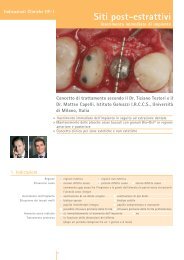
Fig. 1. View of the anterolateral wall of the maxillary sinus by transillumination: the alveolar antral artery dissection is carried<br />
out in the area selected for sinus antrostomy as far as the infraorbital artery (a) and the posterior superior alveolar artery (b) are<br />
visible respectively at its medial and distal extremity. The bony vessel is strictly stick to the Schneiderian membrane.<br />
Fig. 2. Computed tomography scan 3D view of the lateral wall of the maxillary sinus which shows the point of emergence of<br />
the infraorbital artery (IOA) (1), the point of anastomosis between the IOA and the alveolar antral artery (AAA) (2) as well as<br />
the route of the AAA (3) forming a small concavity (white arrow).<br />
Thefirstpartofthestudywasperformedon30<br />
maxillary sinuses, deriving from 15 human<br />
cadaver heads. The specimens belonged to subjects<br />
with an age range of 59–90 years (mean age<br />
76 years) and equal sex distribution, who had<br />
donated their body for research purpose. The<br />
study obtained ethical approval from the Department<br />
of Anatomy at the Faculty of Medicine<br />
René Descartes of Paris 5 (Paris 5 University,<br />
Paris). Direct visualization of the AAA was<br />
obtained by fenestrating the anterior lateral<br />
wall of the sinus cavity and its dissection was<br />
carried out as far as the IOA and the PSAA were<br />
visible at its extremities (Fig. 1), in order to<br />
determine its course with respect to both the<br />
Schneiderian membrane and the buccal antral<br />
wall.<br />
To detect such an artery, the vascular network<br />
afferent to the sinus was injected with liquid latex<br />
mixed with green India ink through the external<br />
carotid artery.<br />
The second part of the study consisted of 100<br />
CT scans from 100 patients scheduled for sinus<br />
lift surgery at the Dental Clinic of the IRCCS<br />
Istituto Ortopedico Galeazzi, Università degli<br />
Studi di Milano. The age range was 29–78<br />
(mean: 53.5) years.<br />
The CT scans were performed using a 2000<br />
SOMATOM Volume Zoom 4 slice CT scanner<br />
(Siemens AG, Medical Solutions, Forchheim,<br />
Germany) with slices of 0.5 mm thickness. CT<br />
images were investigated for the presence of a<br />
bony canal, housing the AAA, in the context of<br />
the sinus anterolateral wall.<br />
Coronal, axial and sagittal views of the maxillary<br />
sinus were obtained by means of a software<br />
for 3D reconstruction (OneScan 3D, 3D-MED<br />
s.r.l., Brescia, Italy), offering a photorealistic<br />
rendering quality and able to import Dicom<br />
formatted CT images.<br />
The route of the AAA was assessed with<br />
respect to the Schneiderian membrane and to<br />
the bony wall, as well as its diameter and the<br />
distance from its lowest point to the alveolar<br />
crest, with a precision of 0.1 mm. The corresponding<br />
ridge height was measured. The correlation<br />
between the residual ridge height and the<br />
distance between the AAA and the crest was also<br />
analysed.<br />
Only edentulous or partially edentulous maxillae<br />
displaying Class V or VI resorption of<br />
the alveolar process, according to Cawood &<br />
Howell’s classification (1988), were taken into<br />
consideration.<br />
Results<br />
The anatomical dissection confirmed that the<br />
PSAA divides into two branches along its course:<br />
an external (gingival) branch is directed towards<br />
the superior buccal fornix and the maxillary<br />
tuberosity; the other branch is internal (dental)<br />
and, after coursing below the zygomatic process,<br />
was found to point towards the inside of the<br />
orbit making a circular anastomosis with the<br />
IOA.<br />
An intra-osseous anastomosis between the<br />
AAA and the IOA was found by dissection in<br />
100% of the anatomical cases (30/30 sinuses),<br />
while a well-defined bony canal, located in the<br />
context of the sinus anterolateral wall, was detected<br />
radiographically in 94 out of 200 sinuses<br />
examined (47% of cases).<br />
The diameter of such bony canal was o1mm<br />
in 52 sinuses (55.3% of 94 cases), 1–o2mm in<br />
38 sinuses (40.4%) and 2mminfoursinuses<br />
(4.3%).<br />
The AAA displayed three different courses: (1)<br />
within the buccal antral wall cortex; (2) between<br />
the Schneiderian membrane and the lateral bony<br />
wall of the sinus, in which a small concavity was<br />
often visible (Figs 2 and 3); and (3) under the<br />
periosteum of the sinus lateral wall.<br />
In particular, the AAA course was found to be<br />
(1) completely intra-osseous at its extremities in<br />
100% of cases (Fig. 4); (2) partially intra-osseous<br />
in the area usually involved with sinus antrostomy<br />
(from second premolar to second molar) in<br />
100% of cases (Fig. 4). In such an area, the AAA<br />
was strictly close to the Schneiderian membrane<br />
and partially encased in the lateral sinus wall<br />
in all specimens. No bony layer interposed<br />
between the AAA and the sinus membrane<br />
could be identified by dissection (Figs 1 and 5);<br />
and (3) variable (either intra-osseous or intra-<br />
712 | Clin. Oral Impl. Res. 22, 2011 / 711–715 c 2010 John <strong>Wiley</strong> & Sons A/S
Rosano et al Haemorrhage risk during sinus surgery<br />
sinusal or sub-periosteal) in the maxillary tuberosity<br />
area.<br />
The vertical distance from the lowest point of<br />
the vessel, corresponding to the first molar area,<br />
to the alveolar crest averaged 11.25 2.99 (SD)<br />
mm (range between 7.2 and 17.7 mm).<br />
The residual ridge height ranged from 0.7 to<br />
5.1 mm (mean height 3.60 1.28 mm). A slight<br />
positive correlation between such a distance and<br />
the ridge height was observed (r ¼ 0.38). When<br />
considering a threshold of 3 mm for the residual<br />
ridge height, the AAA-to-alveolar crest distance<br />
averaged 9.33 2.41 (n ¼ 39) and 12.45 2.71<br />
(n ¼ 55) for cases with ridge height o3mm and<br />
3 mm, respectively.<br />
Discussion<br />
The anastomosis between PSAA and IOA provides<br />
blood supply to the sinus membrane, to the<br />
periosteal tissues, and especially, to the anterolateral<br />
wall of the sinus (Solar et al. 1999; Rosano<br />
et al. 2009).<br />
The scientific literature reports that this vessel<br />
is located at an average distance of 19 mm (Solar<br />
et al. 1999; Traxler et al. 1999), 16.4 mm (Elian<br />
et al. 2005) and 16.9 mm (Mardinger et al. 2007)<br />
from the alveolar crest of the posterior maxilla.<br />
Nevertheless, such data can be misleading<br />
because the height of the residual bony ridge,<br />
the maxillary atrophy class and the presence of<br />
Fig. 3. Internal view of the maxillary sinus: the arrow A shows the alveolar antral artery, the endosseous branch of the<br />
posterior superior alveolar artery (PSAA), partially encased in the lateral sinus wall, while the arrow B shows the infraorbital<br />
artery deriving from the maxillary artery and forming a vascular arcade with the PSAA.<br />
teeth play a relevant role in determining the<br />
location of the vessel.<br />
In the present study, the average distance of the<br />
AAA from the alveolar ridge in atrophic maxillae<br />
of Cawood & Howell class V and VI was<br />
11.25 mm. For the most atrophic cases, in which<br />
the ridge height is inferior to 3 mm, such a<br />
distance was significantly lower with respect to<br />
lesser atrophic cases. This would confirm that<br />
the more resorbed the bone crest, the higher the<br />
risk of violation of such a vessel during sinus<br />
augmentation procedure.<br />
These results are substantially in agreement<br />
with the study by Mardinger et al. (2007), which<br />
found that this vessel was located at a mean<br />
distance of 10.9 mm from the crest in classes<br />
D, E (Lekholm & Zarb 1985) and at a distance<br />
greaterthan15mminclassesA,BandC.<br />
Differences concerning the mean distance from<br />
the vessel to the crest, with the studies by Solar<br />
et al. (1999), Traxler et al. (1999) and Elian et al.<br />
(2005) are probably due to the more strict inclusion<br />
criteria considered in the present study,<br />
where only highly atrophic ridges have been<br />
examined.<br />
Moreover, because a well-distinguished bony<br />
wall between the intra-osseous maxillary anastomosis<br />
and the maxillary sinus has never been<br />
found by anatomic dissection (Fig. 1), it could be<br />
speculated that the lowest border of such a vessel<br />
Fig. 4. Computed tomography scan transversal views of the anterior lateral wall of a sinus where it is possible to evidence the course of the alveolar antral artery from the infraorbital artery (1)<br />
to the posterior superior alveolar artery (2): completely intra-osseous at its extremities, between the Schneiderian membrane and the bony wall in the sinus antrostomy area, sub-periosteal in<br />
the maxillary tuberosity area.<br />
c 2010 John <strong>Wiley</strong> & Sons A/S 713 | Clin. Oral Impl. Res. 22, 2011 / 711–715
Rosano et al Haemorrhage risk during sinus surgery<br />
Fig. 5. Macro-anatomical dissection of the sinus lateral wall: the alveolar antral artery is close to the Schneiderian membrane<br />
and no bony layer between such vessel and the membrane is visible after antrostomy.<br />
could often be completely adherent to the sinus<br />
membrane (that means not radiographically visible)<br />
instead of being located inside the buccal wall<br />
cortex.<br />
This would justify the contradiction between a<br />
100% prevalence of this artery found by dissection<br />
and an only 47% prevalence detected by CT<br />
scan in the present study.<br />
The authors’ opinion is that such contradiction<br />
may not depend on the AAA small diameter,<br />
which makes it radiographically undetectable in<br />
some cases, as suggested by Elian et al. (2005)<br />
and Mardinger et al. (2007), but on an entirely<br />
intra-sinusal location of the vessel.<br />
The course of the AAA, as identified in this<br />
study, is in agreement with the CT study by Ella<br />
et al. (2008) where intra-osseous, intra-sinusal<br />
and sub-periosteal courses of this artery were<br />
detected.<br />
As stated by Ella et al. (2008), a ‘‘superficial’’<br />
location of such anastomosis, which is under<br />
the periosteum of the sinus lateral wall<br />
should also be considered. In the present study,<br />
such sub-periosteal course was identified by<br />
means of both CT scan analysis and anatomical<br />
dissection in the maxillary tuberosity area but<br />
never in the area usually selected for sinus<br />
antrostomy.<br />
When carrying out sinus lift surgery, the bony<br />
window height should be almost 13 mm from the<br />
ridge if the purpose is to place 11–13 mm dental<br />
implants. Thus, in patients with severely<br />
atrophic posterior maxillae (classes V, VI), the<br />
possibility of lacerating the AAA must be considered,<br />
especially when the residual ridge is<br />
o3mmhigh.<br />
The diameter of the anastomosis was<br />
2 mm in a very few cases (3.3% by dissection<br />
and 2% by CT scan); anyway, this eventuality,<br />
even if infrequent, is worthy to be taken into<br />
serious consideration.<br />
As a matter of fact, if the damage of a bony<br />
vessel o2 mm can be barely relevant under a<br />
clinical point of view, the transection of an AAA<br />
with a diameter over 2 mm is likely to produce<br />
bleeding and impairment of vision, which may<br />
lead to a potential membrane perforation, thus<br />
prolonging the overall operation time, interfering<br />
with the placement of bone graft and constituting<br />
atruesurgicalcomplication.<br />
In addition, the haemorrage from this artery (a)<br />
may displace the grafting material due to a<br />
‘‘washing’’ effect caused by the blood pressure,<br />
thus reducing or compromising the filling of the<br />
space below the Schneiderian membrane after<br />
sinus floor elevation, and (b) may produce relevant<br />
haematomas of the cheek area causing<br />
discomfort to patients and creating an ideal ‘‘pabulum’’<br />
for bacteria growth and consequent infection.<br />
It is the authors’ opinion that the excision of a<br />
large diameter AAA in combination with the<br />
inadvertent tearing of the sinus membrane has<br />
the potential to induce sinus mucosa swelling,<br />
extrusion of blood into the sinus cavity as<br />
well as a postoperative sinusitis as a major<br />
drawback.<br />
In fact, if the maxillary sinus is, even partly,<br />
filled up by mucosal oedema, haematoma or<br />
seroma, a delay of maxillary sinus clearance<br />
may occur because of the reduction of maxillary<br />
ostium patency, and maxillary sinusitis may<br />
develop as well, compromising the success of<br />
the grafting procedure (Timmenga et al. 2003).<br />
Thepreservationofsuchanastomosisisimportant<br />
not only to avoid bleeding complications<br />
but also to support bone graft neoangiogenesis<br />
(Taschieri & Rosano 2010); in this perspective,<br />
its concomitant reflection with the Schneiderian<br />
membrane during sinus augmentation procedures,<br />
if possible and especially when its diameter<br />
is consistent, should be seriously<br />
considered.<br />
In conclusion, the authors recommend to rely<br />
upon CT scan imaging, which has been proved to<br />
be the most appropriate radiographic method for<br />
detecting any anatomical variation within the<br />
sinus (Schwarz et al. 1987; Quirynen et al.<br />
1990; Alder et al. 1995; Dula et al. 2001), before<br />
sinus lift surgery is performed, in order to presurgically<br />
evaluate the location, size and thus the<br />
clinical relevance of this anastomotic vessel.<br />
Extreme caution should be taken when the residual<br />
ridge height is o3mm.<br />
References<br />
Aghaloo, T.L. & Moy, P.K. (2007) Which hard tissue<br />
augmentation techniques are the most successful in<br />
furnishing bony support for implant placement? The<br />
International Journal of Oral & Maxillofacial Implants<br />
22 (Suppl.): 49–70.<br />
Alder, M.E., Deahl, S.T. & Matteson, S.R. (1995)<br />
Clinical usefulness of two dimensional reformatted<br />
and three dimensionally rendered computerized<br />
tomographic images: literature review and<br />
a survey of surgeons’opinions. Journal of Oral and<br />
Maxillofacial Surgery 53: 375–386.<br />
Cawood, J.I. & Howell, R.A. (1988) A classification of<br />
the edentulous jaws. The International Journal of<br />
Oral and Maxillofacial Surgery 17: 232–236.<br />
Chanavaz, M. (1996) Sinus grafting related to implantology.<br />
Statistical analysis of 15 years of surgical<br />
experience (1979–1994). Journal of Oral Implantology<br />
22: 119–130.<br />
Del Fabbro, M., Rosano, G. & Taschieri, S. (2008)<br />
Implant survival rates after maxillary sinus augmentation.<br />
European Journal of Oral Sciences 116:<br />
497–506.<br />
Dula, K., Mini, R., Van der Stelt, P.F. & Buser, D.<br />
(2001) The radiographic assessment of implant patients:<br />
decision making criteria. The International<br />
Journal of Oral & Maxillofacial Implants 16: 80–89.<br />
Elian, N., Wallace, S., Cho, S.C., Jalbout, Z.N. &<br />
Froum, S. (2005) Distribution of the maxillary artery<br />
as it relates to sinus floor augmentation. The International<br />
Journal of Oral & Maxillofacial Implants<br />
20: 784–787.<br />
Ella, B., Sédarat, C., Da Costa Noble, R., Normand, E.,<br />
Lauverjat, Y., Siberchicot, F., Caix, P. & Zwetyenga,<br />
714 | Clin. Oral Impl. Res. 22, 2011 / 711–715 c 2010 John <strong>Wiley</strong> & Sons A/S
Rosano et al Haemorrhage risk during sinus surgery<br />
N. (2008) Vascular connections of the lateral wall of<br />
the sinus: surgical effect in sinus augmentation. The<br />
International Journal of Oral & Maxillofacial Implants<br />
23: 1047–1052.<br />
Gaudy, J.-F. (2003) Anatomie Clinique. Rueil-Malmaison<br />
Cedex: Groupe Liaisons, Editions CdP, 11pp.<br />
Lekholm, U. & Zarb, G.A. (1985) Patient selection.<br />
In: Brånemark, P.-I., Zarb, G.A. & Albrektsson, T.,<br />
eds. Tissue Integrated Prosthesis. Osseointegration<br />
in Clinical Dentistry, 199–209. Chicago: Quintessence.<br />
Mardinger, O., Abba, M., Hirshberg, A. & Schwartz-<br />
Arad, D. (2007) Prevalence, diameter and course of<br />
the maxillary intraosseous vascular canal with relation<br />
to sinus augmentation procedure: a radiographic<br />
study. The International Journal of Oral and Maxillofacial<br />
Surgery 36: 735–738.<br />
Pjetursson, B.E., Tan, W.C., Zwahlen, M. & Lang, N.P.<br />
(2008) A systematic review of the success of sinus<br />
floor elevation and survival of implants inserted in<br />
combination with sinus floor elevation. Journal of<br />
Clinical Periodontology 35: 216–240.<br />
Quirynen, M., Lamoral, Y., Dekeyser, C., Peene, P.,<br />
van Steenberghe, D., Bonte, J. & Baert, AL. (1990)<br />
The CT scan standard reconstruction technique for<br />
reliable jaw bone volume determination. The International<br />
Journal of Oral & Maxillofacial Implants 5:<br />
384–389.<br />
Rosano, G., Taschieri, S., Gaudy, J.-F. & Del Fabbro, M.<br />
(2009) Maxillary sinus vascularization: a cadaveric<br />
study. Journal of Craniofacial Surgery 20:<br />
940–943.<br />
Schwarz, M.S., Rothman, S.L.G., Rhodes, M.L. &<br />
Chafetz, N. (1987) Computed tomography: part II.<br />
Preoperative assessment of the maxilla for endosseous<br />
implant surgery. Journal of Oral & Maxillofacial<br />
Implants 2: 143–148.<br />
Solar, P., Geyerhofer, U., Traxler, H., Windisch, A.,<br />
Ulm, C. & Watzek, G. (1999) Blood supply to the<br />
maxillary sinus relevant to sinus floor elevation<br />
procedures. Clinical Oral Implants Research 10:<br />
34–44.<br />
Taschieri, S. & Rosano, G. (2010) Management of the<br />
alveolar antral artery during sinus floor augmentation<br />
procedures. The International Journal of Oral and<br />
Maxillofacial Surgery 68: 230.<br />
Timmenga, N.M., Raghoebar, G.M., Liem, R.S.B., van<br />
Weissenbruch, R., Manson, W.L. & Vissink, A.<br />
(2003) Effects of maxillary sinus floor elevation surgery<br />
on maxillary sinus physiology. European Journal<br />
of Oral Sciences 111: 189–197.<br />
Traxler, H., Windisch, A., Geyerhofer, U., Surd, R.,<br />
Solar, P. & Firbas, W. (1999) Arterial blood supply<br />
of the maxillary sinus. Clinical Anatomy 12: 417–<br />
421.<br />
Wallace, S.S. & Froum, S.J. (2003) Effect of maxillary<br />
sinus augmentation on the survival of endosseous<br />
dental implants. A systematic review. Annals of<br />
Periodontology 8: 328–343.<br />
c 2010 John <strong>Wiley</strong> & Sons A/S 715 | Clin. Oral Impl. Res. 22, 2011 / 711–715