Skin Scrape Test: An Inexpensive and Painless ... - Bentham Science
Skin Scrape Test: An Inexpensive and Painless ... - Bentham Science
Skin Scrape Test: An Inexpensive and Painless ... - Bentham Science
Create successful ePaper yourself
Turn your PDF publications into a flip-book with our unique Google optimized e-Paper software.
Send Orders of Reprints at reprints@benthamscience.net<br />
The Open Allergy Journal, 2013, 6, 9-17 9<br />
Open Access<br />
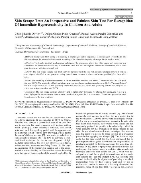
<strong>Skin</strong> <strong>Scrape</strong> <strong>Test</strong>: <strong>An</strong> <strong>Inexpensive</strong> <strong>and</strong> <strong>Painless</strong> <strong>Skin</strong> <strong>Test</strong> For Recognition<br />
Of Immediate Hypersensitivity In Children <strong>An</strong>d Adults<br />
Celso Eduardo Olivier 1,2,* , Daiana Guedes Pinto Argentão 2 , Raquel Acácia Pereira Gonçalves dos<br />
Santos 2 , Mariana Dias da Silva 2 , Regiane Patussi Santos Lima 2 <strong>and</strong> Ricardo de Lima Zollner 1<br />
1 Discipline <strong>and</strong> Laboratory of Clinical Immunology, Department of Internal Medicine, Faculty of Medical <strong>Science</strong>s,<br />
University of Campinas, São Paulo, Brazil<br />
2 Instituto Alergoimuno de Americana - São Paulo - Brazil<br />
Abstract: Background: <strong>Skin</strong> testing is a mainstay in allergology, <strong>and</strong> its importance is increasing in several fields. The<br />
ability to choose the most suitable technique according to the clinical setting is an advantage for the medical team.<br />
Objectives: To describe in detail an alternative technique of the coetaneous allergy test (skin scrape test) conceived as a<br />
variation of the former skin scratch test; to evaluate its value as a tool for diagnosis of immune sensitization; <strong>and</strong> to compare<br />
its accuracy with the skin prick test.<br />
Methods: The skin scrape test <strong>and</strong> skin prick test were performed side by side with the same allergen extracts in 162 human<br />
subjects classified in two groups according to the known presence or absence of serum specific-IgE to these allergens.<br />
Results: The sensitivity of the skin scrape test to detect immediate reactions was 85.0%. The sensitivity of the skin prick<br />
test was 86.5%. The sensitivity of both techniques analyzed together as a unique procedure was 94.2%. The specificity of<br />
the skin scrape test was 90.1%.The specificity of the skin prick test was 72.9%.The specificity of both tests analyzed together<br />
as a unique procedure was 70.5%.<br />
Conclusions: The skin scrape test is an alternative <strong>and</strong> complementary technique for allergic skin testing, <strong>and</strong> it is able to<br />
detect IgE-specific immune sensitization without the disadvantages of the skin scratch test. The skin scrape test has similar<br />
outcomes to the skin prick test.<br />
Keywords: Immediate Hypersensitivity (Medline ID D006969), Diagnosis (Medline ID D003933), <strong>Skin</strong> <strong>Test</strong> (Medline ID<br />
D012882), Dermatophagoides <strong>An</strong>tigens (Medline ID D039741), Child (Medline ID D002648), Atopic Dermatitis (Medline ID<br />
D003876), Rhinitis (Medline ID D012220), Asthma (Medline ID D001249).<br />
INTRODUCTION<br />
The skin scratch test was the first test described as a tool<br />
for allergy diagnosis. It was reported in 1873 by Charles<br />
Blackley, who abraded a quarter-inch area of his own forearm<br />
to correlate his nasal symptoms with Lolium italicum<br />
pollen grains [1]. After Blackley’s initiative, skin scratch<br />
tests were used during a long period until the appearance of<br />
the skin prick test(SPT) in the early 1950s [2], which, despite<br />
the use of different devices [3], was easier to submit to a<br />
st<strong>and</strong>ardization process [4]. The SPT is currently a wellestablished<br />
technique for the in vivo diagnosis of IgEmediated<br />
sensitization, but it is not always a reproducible<br />
technique due to numerous interfering factors [5]. The major<br />
inconvenience of the former skin scratch test is the associated<br />
skin trauma leading to false-positive results depending<br />
upon the type of device used <strong>and</strong> the strength applied by the<br />
*Address correspondence to this author at the Discipline <strong>and</strong> Laboratory of<br />
Clinical Immunology, Department of Internal Medicine, Faculty of Medical<br />
<strong>Science</strong>s, University of Campinas, São Paulo, Brazil; Tel: 55 19 34635941;<br />
Fax: 55 19 34555726; E-mail: celso@docsystems.med.br<br />
health care professional to scarify the skin [6]. One of most<br />
commonly used devices to perform the skin scratch test is<br />
the blood lancet [7]. Blood lancets were not designed to scarify<br />
skin <strong>and</strong> were used upside-down to scratch the skin with<br />
their blunt base. Usually, the bottoms of the lancets are not<br />
adequately polished to provide a burr-free edge, which is<br />
what accounts for the production of actual trauma to the<br />
skin. In the chamber-scarification technique, the authors<br />
used the point of a needle to break the skin in a crisscross<br />
pattern before applying the allergen [8]. The literature suggests<br />
that the skin scratch test has been ab<strong>and</strong>oned due to<br />
poor reproducibility, great discomfort <strong>and</strong> the possibility of<br />
residual pigmented or depigmented areas for some time afterwards<br />
[9, 10]. Here, we propose an alternative method for<br />
performing skin testing using a modified skin scratch test<br />
technique called the skin scrape test (SST), which was developed<br />
to avoid the original inconveniences of the early<br />
skin scratch test <strong>and</strong> the current SPT. We describe this<br />
method in detail with the objective of providing a st<strong>and</strong>ard<br />
methodology both for daily clinical work <strong>and</strong> for use in<br />
multi-center studies in order to effectively determine the<br />
predictive value of skin tests [11]. To evaluate the accuracy<br />
1874-8384/13 2013 <strong>Bentham</strong> Open
10 The Open Allergy Journal, 2013, Volume 6 Olivier et al.<br />
of the SST, we compared it with the SPT. We performed the<br />
tests side by side <strong>and</strong> employed st<strong>and</strong>ardized allergens that<br />
patients had previously been diagnosed, by the quantification<br />
of specific IgE (s-IgE), as being sensitized to or not sensitized<br />
to. This accuracy study, which was performed as recommended<br />
by the GRADE approach [12] (Grades of Recommendation,<br />
Assessment, Development <strong>and</strong> Evaluation),<br />
allowed us to determine the sensitivity <strong>and</strong> specificity of the<br />
SST in this specific selected population using the s-IgE as a<br />
proxy of sensitization [13-15]. Patients were analyzed according<br />
to the presence or absence of medically diagnosed<br />
atopic diseases such as persistent rhinitis, asthma <strong>and</strong>/or<br />
atopic dermatitis. These conditions were chosen due to their<br />
particular association with IgE-mediated immune reactivity<br />
[16].<br />
METHODS<br />
Study Design <strong>and</strong> Subjects<br />
The study was approved by the ethical review board of<br />
the University of Sorocaba registered at the Plataforma Brasil<br />
(CAAE 07453312.2.0000.5500), <strong>and</strong> it was conducted<br />
according to the principles of the Declaration of Helsinki.<br />
We examined 74 male <strong>and</strong> 88 female subjects (1 to 68 years,<br />
mean 26.1 +/- 18.3 years). To evaluate the utility of the SST,<br />
we compared it with the SPT performed at the same time<br />
with the same allergen solution in patients previously diagnosed<br />
by serum s-IgE (CAP Systems Pharmica; results expressed<br />
as kU/L) as being sensitive or not to these allergens<br />
[17]. A group of atopic patients was used to study the sensitivity<br />
of the skin tests. This group was composed of 125 patients<br />
with s-IgE> 1.0 kU/L to at least one of three house<br />
dust mite (HDM) antigens (Dermatophagoides pteronyssinus,<br />
Dermatophagoides farinae <strong>and</strong> Blomia tropicalis).<br />
Some patients had increased s-IgE to more than one mite <strong>and</strong><br />
were included in two or three analyses, totaling 208 pairs of<br />
tests. The control group consisted of asymptomatic subjects<br />
to study the specificity of the skin tests <strong>and</strong> was composed of<br />
37 subjects with s-IgE< 0.35 kU/mL to at least one of the<br />
following five allergens: the three HDM mentioned above<br />
plus Aspergillus fumigatus <strong>and</strong> Hevea brasiliensis. In this<br />
group, 122 pairs of tests were performed.<br />
<strong>Skin</strong> <strong>Test</strong>ing Methodology<br />
Glycerinated saline diluent was used as a negative control.<br />
Histamine sulfate 1 mg/mL was used as a positive control.<br />
The SPT <strong>and</strong> the SST were performed by trained <strong>and</strong><br />
supervised health care professionals on the volar aspect of<br />
the forearm in adults <strong>and</strong> children <strong>and</strong> on the back of toddlers<br />
<strong>and</strong> babies after withholding antihistamines for at least<br />
10 days. Babies were tested on a parent’s lap. Toddlers were<br />
tested while they were seated over the table edge in front of<br />
their mother. The SPT was performed with a disposable sterile<br />
acrylic pricker with a 1-mm lancet over a blunt basis<br />
(Punctor ® purchased from Alko do Brasil). The pricker was<br />
introduced at a 90° angle perpendicular to the skin, through<br />
allergen solution, retrieved after 5 seconds <strong>and</strong> then discarded.<br />
To scrape the skin, we used the bevel of a sterilized<br />
<strong>and</strong> disposable 18-G 1½ (1.20 x 40) hypodermic needle<br />
(st<strong>and</strong>ard bevel). The skin was not scraped with the needle’s<br />
point. Its hub (allowing some flexibility in touching the skin)<br />
gently held the needle at a 10° to 25°angle. The health care<br />
professional scraped the skin with the help of the bevel’s<br />
edge in only one direction. The bevel did not scrape the skin<br />
backwards, <strong>and</strong> its movement resembled a broom cleaning<br />
the floor. The objective was to wipe the most superficial<br />
epidermal layer instead of traumatizing or scarifying the<br />
skin. This movement was repeated approximately3 to 6<br />
times (depending on the skin thickness), until the health care<br />
professional observed a thin homogeneous desquamation or<br />
a slight hyperemia (see Video 1). If scraping occurred beyond<br />
this depth, a skin scratch test would be performed instead<br />
of an SST. The extension of the scraped area was irrelevant<br />
because the tested area depends on the diameter of<br />
the drop applied. The width of the scraped area measured at<br />
least 3 mm wide. The health care professional scraped all of<br />
the areas to be tested in a single step (a single needle was<br />
used to perform all of the tests). During the first scrape, the<br />
health care professional counted the number of movements<br />
necessary to produce desquamation <strong>and</strong> repeated the same<br />
procedure on other are as to perform a homogeneous test.<br />
Sometimes, the scraped area was hardly visible, <strong>and</strong> it was<br />
helpful to mark the areas with a dermographic pen prior to<br />
scraping. Good illumination in the procedure room was essential<br />
for the skin tests. The allergens solutions are applied<br />
after scraping. Optionally, after scraping <strong>and</strong> before applying<br />
the allergens, the health care professional can wait 15 minutes<br />
to observe an occasional dermographism that could invalidate<br />
the test. Disrupting the stratum corneum barrier by<br />
the SST makes the skin permeable to soluble allergens; however,<br />
they do not gain immediate access to reactive layers.<br />
Thus, unlike the SPT (in which allergen solution is directly<br />
introduced via a 1-mm lancet into the skin, thus allowing the<br />
immediate cleaning of the allergen solution), when performing<br />
the SST, the allergen solution remained on the skin to<br />
allow absorption until the final reading was made at 15 minutes.<br />
At this time, the wheal’s longest diameter (WLD) was<br />
assessed. We perform a negative control to draw the cut-offs<br />
for positive <strong>and</strong> negative tests. A wheal reaction was defined<br />
as positive if the WLD was ≥ 3 mm 15 minutes after the application<br />
of the allergen extracts <strong>and</strong> after the subtraction of<br />
each patient’s reaction to the negative control [10, 18].<br />
The SST is a more convenient technique for use in small<br />
children because it is painless <strong>and</strong> promotes fewer struggles<br />
than the SPT. Children usually struggle more during pricking<br />
than during scraping sessions. Most children do not struggle<br />
during dermographic marking or allergen application. Struggles<br />
are less inconvenient during scraping because allergens<br />
have not yet been applied. One can apply allergens during a<br />
calmer second step some minutes after scraping. Struggles<br />
during pricking (when the allergens are already on the skin)<br />
make the procedure more difficult due to the increased possibility<br />
of dislodgements of solutions <strong>and</strong> the mixing of allergens.<br />
In this case, it is more convenient to extend the<br />
steps, apply one allergen, prick <strong>and</strong> dry the solution immediately<br />
after the prick before the next allergen application. The<br />
possibility of recognizing dermographism before applying<br />
the allergen solution is a clear advantage of the SST over the<br />
SPT because in the SPT, the trauma associated with puncture<br />
may produce an immediate reaction that sometimes may be<br />
indistinguishable from the reaction produced by the allergen.
The <strong>Skin</strong> <strong>Scrape</strong> <strong>Test</strong> Original Description The Open Allergy Journal, 2013, Volume 6 11<br />
Fig. (1). Dropping of antigen extract solution onto the plastic antigen dispenser being applied to the scraped area. On the table are the container<br />
to keep the unused disposable dispensers (without cover) <strong>and</strong> the container to dispose the used dispensers (with a hole in the cover).<br />
Disposable Allergen Dispenser<br />
We used a disposable allergen dispenser to intermediate<br />
the allergen solution between the vial <strong>and</strong> the scraped skin.<br />
The allergen dispenser is a plastic spatula with an enlarged<br />
end where the allergen solution was dropped before being<br />
applied to the scraped area. Immediately after allergen application,<br />
this plastic dispenser was discarded. This procedure<br />
was also adopted for the performance of the SPT in order to<br />
st<strong>and</strong>ardize allergen application. The disposable allergen<br />
dispenser allows better visual control of the drop’s size <strong>and</strong><br />
avoids the waste of allergen solutions. To discard the dispenser;<br />
we used a steel container topped with a cover with a<br />
narrow hole to allow the health care professional to dispose<br />
of the dispenser but not to (unadvisedly) retrieve it. <strong>An</strong>other<br />
container (without a cover) holding unused dispensers was<br />
also available on the examination table (See Fig. 1).<br />
Statistical <strong>An</strong>alyses<br />
Group comparisons of the skin test outcomes were performed<br />
with paired <strong>and</strong> unpaired t tests. The data are reported<br />
as the arithmetic mean with 95% confidence intervals<br />
(CI). Box column graphs with 95% CI whiskers were plotted<br />
between the means of the SPT WLD <strong>and</strong> the SST WLD.<br />
Paired correlation charts between the SPT WLD <strong>and</strong> the SST<br />
WLD were plotted, <strong>and</strong> the Spearman rank correlation was<br />
used to analyze the results.<br />
Prospective contingency tables analyzed by Fisher’s exact<br />
tests were used to compare the categorical diagnostic<br />
performance between tests [19].The sensitivity of the skin<br />
tests was calculated by grouping WLD results of ≥ 3mm into<br />
a single categorical value. The specificity of the skin tests<br />
was calculated by grouping WLD results of
12 The Open Allergy Journal, 2013, Volume 6 Olivier et al.<br />
Table 1. Mean Wheal’s Longest Diameter for the <strong>Skin</strong> <strong>Scrape</strong> <strong>Test</strong> (SST) <strong>and</strong> the <strong>Skin</strong> Prick <strong>Test</strong> (SPT) with Histamine Sulphate<br />
1mg/mL (Positive Control) <strong>and</strong> the Corresponding Allergen in Patients with s-IgE > 1.0 kU/L to Dermatophagoides pteronyssinus,<br />
Dermatophagoides farinae <strong>and</strong> Blomia Tropicalis<br />
Mean Wheal’s Longest Diameter<br />
SPT<br />
SST<br />
D. pteronyssinus 6.1 mm 8.2 mm<br />
D. farinae 6.5 mm 7.9 mm<br />
Blomia tropicalis 5.3 mm 4.8 mm<br />
Average mean 6.0 mm 7.6 mm<br />
Positive control 6.5 mm 8.3 mm<br />
Table 2. Accuracy (Number of Positive <strong>Test</strong>s) Followed by the Percentage from Total (Sensitivity) <strong>and</strong> the False Negative Proportion<br />
of the <strong>Skin</strong> <strong>Scrape</strong> <strong>Test</strong> (SST), the <strong>Skin</strong> Prick <strong>Test</strong> (SPT) <strong>and</strong> Both Simultaneous <strong>Test</strong>s in Patients with s-IgE > 1.0 kU/L to<br />
Dermatophagoides pteronyssinus (D1), Dermatophagoides farinae (D2) <strong>and</strong> Blomia Tropicalis (RD201)<br />
SST<br />
Accuracy<br />
(Sensitivity)<br />
SST False<br />
Negative<br />
(Proportion)<br />
SPT Accuracy<br />
(Sensitivity)<br />
SPT False<br />
Negative<br />
(Proportion)<br />
SST+SPT<br />
Accuracy<br />
(Sensitivity)<br />
SST+SPT False<br />
Negative<br />
(Proportion)<br />
D1<br />
83<br />
9<br />
81<br />
11<br />
88<br />
4<br />
(92 tests)<br />
(90.2%)<br />
(9.7%)<br />
(88.0%)<br />
(11.6%)<br />
(95.5%)<br />
(4.3%)<br />
D2<br />
67<br />
10<br />
67<br />
10<br />
73<br />
4<br />
(77 tests)<br />
(87.0%)<br />
(12.9%)<br />
(87.0%)<br />
(12.9%)<br />
(94.8%)<br />
(5.2%)<br />
RD201<br />
27<br />
12<br />
32<br />
7<br />
35<br />
4<br />
(39 tests)<br />
(69.2%)<br />
(30.7%)<br />
(82.0%)<br />
(17.9%)<br />
(89.4%)<br />
(10.2%)<br />
Total<br />
177<br />
31<br />
180<br />
28<br />
196<br />
12<br />
(208 tests)<br />
(85.0%)<br />
(14.0%)<br />
(86.5%)<br />
(13.4%)<br />
(94.2%)<br />
(5.7%)<br />
Table 3. The <strong>Skin</strong> Prick <strong>Test</strong> (SPT) <strong>and</strong> the <strong>Skin</strong> <strong>Scrape</strong> <strong>Test</strong> (SST) with the Corresponding <strong>An</strong>tigens According to Medical Diagnosis<br />
in Patients with s-IgE > 1.0 kU/L to Dermatophagoides Pteronyssinus, Dermatophagoides farinae <strong>and</strong>/or Blomia tropicalis<br />
Medical Diagnosis<br />
Mean Age<br />
(Years)<br />
Mean<br />
s-IgE<br />
(kU/L)<br />
Positive to the<br />
SPT (Sensitivity)<br />
Positive to<br />
the SST<br />
(Sensitivity)<br />
Positive to Both<br />
SST <strong>and</strong>/or SSP<br />
(Sensitivity)<br />
Atopic dermatitis<br />
(79 tests)<br />
Asthma<br />
(55 tests)<br />
Rhinitis<br />
(171 tests)<br />
19.9 47.0 68 (86.0%) 74 (93.6%) 77 (97.4%)<br />
18.6 40.3 46 (83.6%) 48 (87.2%) 51 (92.7%)<br />
20.3 35.1 152 (88.8%) 144 (84.2%) 162 (94.7%)<br />
RESULTS<br />
Information on current rhinitis, asthma <strong>and</strong> atopic dermatitis<br />
was available for all subjects. The mean SPT positive<br />
control WLD was 6.5mm (SD 2.7). The mean SST positive<br />
control WLD was 8.3mm (SD 4.6). The mean SPT <strong>and</strong> SST<br />
allergens WLD in patients with s-IgE> 1.0kU/mL to HDM<br />
are shown in Table 1. In this group, the sensitivity of the<br />
SST to detect HDM sensitization was 85.0% <strong>and</strong> the sensitivity<br />
of the SPT was 86.5% (p>0.05 Fisher’s exact test - see<br />
Table 2). Some patients did not react to the SPT but reacted<br />
to the SST, <strong>and</strong> others reacted to the SPT but did not react to<br />
the SST. Considering both tests analyzed together as a<br />
unique procedure, the sensitivity increased to 94.2%. Table 3<br />
presents the sensitivity of the SPT <strong>and</strong> the SST according to<br />
clinical diagnosis. Among the three conditions studied, a<br />
topic dermatitis showed better predictability by the SST<br />
alone (sensitivity of 93.6%) <strong>and</strong> in association with the SPT<br />
(sensitivity of 97.4%), but the differences with the other<br />
conditions were not significant using Fisher’s exact test (p ><br />
0.05). In the group of patients with s-IgE< 0.35 kU/mL, the<br />
specificity of the SST was 90.1% <strong>and</strong> the specificity of the<br />
SPT was 72.9% (p> 0.05 Fisher’s exact test). The specificity
The <strong>Skin</strong> <strong>Scrape</strong> <strong>Test</strong> Original Description The Open Allergy Journal, 2013, Volume 6 13<br />
Table 4. Accuracy (Number of Non Reagent <strong>Test</strong>s) Followed by the Percentage from Total (Specificity) <strong>and</strong> False Positive Proportion<br />
of the <strong>Skin</strong> <strong>Scrape</strong> <strong>Test</strong> (SST), the <strong>Skin</strong> Prick <strong>Test</strong> (SPT) <strong>and</strong> both Simultaneous <strong>Test</strong>s in Patients with s-IgE < 1.0 kU/L to<br />
Dermatophagoides Pteronyssinus (D1), Dermatophagoides Farinae (D2), Blomia tropicalis (RD201), Aspergillus fumigatus<br />
(M3) <strong>and</strong> Hevea Brasiliensis (K82)<br />
Non-Reactive<br />
to the SST<br />
(Specificity)<br />
SST False<br />
Positive<br />
(Proportion)<br />
Non-Reactive to<br />
the SPT<br />
(Specificity)<br />
SPT False<br />
Negative (Proportion)<br />
Non-reactive to SST + SPT<br />
(Specificity)<br />
SST + SPT<br />
False Positive (Proportion)<br />
D1<br />
26<br />
2<br />
19<br />
9<br />
19<br />
9<br />
(28 tests)<br />
(92.9%)<br />
(7.1%)<br />
(67.9%)<br />
(32.1%)<br />
(67.9%)<br />
(32.1%)<br />
D2<br />
22<br />
3<br />
16<br />
9<br />
16<br />
9<br />
(25 tests)<br />
(88%)<br />
(12%)<br />
(64%)<br />
(36%)<br />
(64%)<br />
(36%)<br />
RD201<br />
21<br />
2<br />
15<br />
8<br />
15<br />
8<br />
(23 tests)<br />
(91.3%)<br />
(8.7%)<br />
(65.2%)<br />
(34.8%)<br />
(65.2%)<br />
(34.8%)<br />
M3<br />
24<br />
2<br />
20<br />
6<br />
20<br />
6<br />
(26 tests)<br />
(92.3%)<br />
(7.7%)<br />
77%<br />
23%<br />
77%<br />
23%<br />
K82<br />
(20 tests)<br />
17<br />
(85%)<br />
3<br />
(15%)<br />
19<br />
95%<br />
1<br />
5%<br />
16<br />
(80%)<br />
4<br />
(20%)<br />
Total<br />
(122 tests)<br />
110<br />
(90.1%)<br />
12<br />
(10.9%)<br />
89<br />
(72.9%)<br />
33<br />
(27.1%)<br />
86<br />
(70.5%)<br />
36<br />
(29.5%)<br />
Fig. (2). Dispersion chart with 95% CI for the linear regression with 208 XY pairs between the skin scrape test (SST) wheal’s longest diameters<br />
plotted against the skin prick test (SPT) wheal’s longest diameters for airborne allergens in patients with s-IgE> 1.0 kU/L to corresponding<br />
allergens. Spearman r = 0.40 (0.27 to 0.51;p< 0.001).<br />
of both tests analyzed together as a unique procedure was<br />
70.5% (Table 4). Fig. (2) presents the dispersion chart with<br />
95% CI of the linear regression with 208 XY pairs between<br />
the allergens SST WLD plotted against the allergens SPT
14 The Open Allergy Journal, 2013, Volume 6 Olivier et al.<br />
Fig. (3). Dispersion chart with 95% CI for the linear regression with 122 XY pairs between the skin scrape test (SST) wheal’s longest diameters<br />
plotted against the skin prick test (SPT) wheal’s longest diameters to airborne allergens in patients with s-IgE< 0.35 kU/L to corresponding<br />
allergens. Spearman r = 0.37 (0.20 to .51; p < 0.001).<br />
WLD in patients with s-IgE> 1.0kU/mL to HDM (Spearman<br />
r = 0.40; 0.27 to 0.51; p< 0.001). Fig. (3) presents the dispersion<br />
chart with 95% CI of the linear regression with 122 XY<br />
pairs between the allergens SST WLD plotted against the<br />
allergens SPT WLD in patients with s-IgE< 0.35 kU/mL<br />
(Spearman r = 0.37; 0.20 to 0.51; p< 0.001). The difference<br />
of the allergens mean WLD between the SPT (6.0 mm) <strong>and</strong><br />
the SST (7.6 mm) in patients with s-IgE> 1.0kU/L was 1.6<br />
mm (0.88 to 2.21 mm; p< 0.001 by paired t test). The difference<br />
of the allergens mean WLD between the SPT (1.10<br />
mm) <strong>and</strong> the SST (0.54 mm) in patients with s-IgE< 0.35<br />
kU/L was 0.56 mm (0.19 to 0.93 mm; p = 0.003 by paired t<br />
test). Differences between the allergens mean WLDSST between<br />
the group of patients with s-IgE>1.0kU/mL <strong>and</strong> the<br />
group with s-IgE< 0.35 kU/mL was 7.07 ± 0.44 mm (6.20 to<br />
7.95 mm; p< 0.001 unpaired t test). Differences between the<br />
allergens mean WLDSPT between the group of patients with<br />
s-IgE>1.0kU/mL <strong>and</strong> the group with s-IgE< 0.35 kU/mL was<br />
4.91 ± 0.41 mm (4.15 to 5.76 mm; p< 0.001 by unpaired t<br />
test - see Fig. 4).<br />
DISCUSSION<br />
There is no perfect exam that can identify every single allergen<br />
responsible for every single symptom in every single<br />
allergic patient. Even the s-IgE measurement has issues concerning<br />
differences in accuracy between distinct techniques,<br />
<strong>and</strong> these are somehow resolved by means of skin testing<br />
[20]. Therefore, a careful clinical history performed by an<br />
experienced physician remains the best way to establish a<br />
reasonable link between the results of skin or blood tests <strong>and</strong><br />
allergic disease [21, 22]. A reactive skin test is a lead to be<br />
pursued by means of challenge tests to establish a diagnosis<br />
<strong>and</strong> to offer a proper therapy, or to remove the sources of the<br />
allergen [23]. In performing the SST, the skin is not scarified<br />
or broken. A needle’s bevel is properly polished during its<br />
production in order to not present a reminiscent burr (unlike<br />
the primitive scarifies). The SST was perform to remove<br />
only the stratum corneum by sweeping the outermost skin<br />
layer that is largely responsible for the hydrophobic barrier<br />
function of the skin [24]. The stratum corneum has been described<br />
as a “brick-mortar wall” in which the terminally differentiated<br />
corneocytes (bricks) are embedded in the continuous<br />
matrix of specialized lipids (mortar). These lipids<br />
provide the essential element for the water barrier, while the<br />
corneocytes protect against physical injury [25]. Before the<br />
mid-1970s, the stratum corneum was thought to be biologically<br />
inert, but it is now appreciated to be both metabolically<br />
active <strong>and</strong> interactive with the underlying cell layers [26].<br />
The simple removal of the impermeable barrier is enough to<br />
allow allergen penetration into adjacent skin layers without<br />
the need for deeper scarification. In order to remove the stra-
The <strong>Skin</strong> <strong>Scrape</strong> <strong>Test</strong> Original Description The Open Allergy Journal, 2013, Volume 6 15<br />
Fig. (4). Box column graphs with 95% CI whiskers plotted with the mean of the skin prick test (SPT) <strong>and</strong> the skin scrape test (SST) positive<br />
control (PC) mean wheal's longest diameter; SPT <strong>and</strong> SST allergens (Ag) mean wheal's longest diameter in patients with s-IgE> 1.0 kU/L;<br />
<strong>and</strong> the SPT <strong>and</strong> the SST allergens (Ag) mean wheal's longest diameter in patients with s-IgE< 0.35 kU/L.<br />
Table 5. Comparative Features of <strong>Skin</strong> <strong>Scrape</strong> <strong>Test</strong> <strong>and</strong> <strong>Skin</strong> Prick <strong>Test</strong><br />
<strong>Skin</strong> Prick <strong>Test</strong><br />
<strong>Skin</strong> <strong>Scrape</strong> <strong>Test</strong><br />
Number of skin devices One for each tested allergen One for all tested allergens<br />
Discomfort Pain full for toddlers <strong>and</strong> babies Minimal tickles<br />
Dermographism May be recognized only in negative control May be recognized on each test before applying the allergens<br />
Sensitivity 86.5% 85%<br />
Specificity 72.9% 90.1%<br />
tum corneum as a preparation for patch testing for type IV<br />
allergy some centers routinely used tape stripping procedure<br />
with normal adhesive tape that is affixed <strong>and</strong> removed from<br />
the skin between one <strong>and</strong> ten times depending on the<br />
prob<strong>and</strong>’s skin type. We tried to perform this methodology,<br />
but it did not result in appreciable immediate skin reactions.<br />
We think that the residual glue left by the tape may diminishes<br />
skin permeability <strong>and</strong> be an interferent to the test.<br />
The SSP was effective in demonstrating skin reactivity in<br />
some patients with the presence of s-IgE <strong>and</strong> who had a nonreactive<br />
SPT to HDM. On some occasions, we observed an<br />
inverse pattern with patients reacting to the SPT but not to<br />
the SST. Individual differences in the affinity threshold for<br />
IgE-allergen binding [27] may explain this phenomenon, as<br />
well the selective competition between immunoglobulins,<br />
which promote variable results to binding allergen affinity<br />
<strong>and</strong> skin tests [28]. For these reasons, some authors recommend<br />
performing an intradermic test to confirm a nonreactive<br />
SPT in a patient with a strong suggestive history of<br />
hypersensitivity to a particular allergen [29]. As the SST <strong>and</strong><br />
the SPT are mutually complementary, the use of the other<br />
technique when the first does not react may spare the indication<br />
of the more traumatic <strong>and</strong> risky intradermic test. This is<br />
especially valuable when testing for food allergies because<br />
intradermic tests are usually not recommended for food allergens<br />
due to their association with an unacceptable rate of<br />
false-positive reactions [30]. The possibility of another “percutaneous”<br />
[31] technique that may be performed before the<br />
“intracutaneous” technique is very convenient because one<br />
can use the same allergen solution for the SPT <strong>and</strong> for the<br />
SST, while the intradermic test requires a non-glycerinated,<br />
more diluted <strong>and</strong> rigorously sterilized solution [9].<br />
<strong>An</strong>other advantage of the SST is that it is less expensive<br />
than the SPT, as the health care professional may apply all<br />
antigens using a single hypodermic 18-G 1½ needle available<br />
in most medical units (Table 5). To perform the SPT,<br />
one needs a disposable pricker for each tested antigen. In<br />
addition, the plastic dispenser saves the allergen solutions.<br />
With practice, the support provided by the plastic dispenser<br />
allows the health care professional to decrease the volume of<br />
the antigen solution applied to the skin. A st<strong>and</strong>ard drop applied<br />
directly with the dropper over the skin weighs an aver-
16 The Open Allergy Journal, 2013, Volume 6 Olivier et al.<br />
age of 0.035 g. The drop applied with the help of the dispenser<br />
weighs an average of 0.002 g. Despite the lower volume<br />
applied to the skin, there was no significant reduction in<br />
the wheal reading of the SPT or the SST. The wheals produced<br />
by the SST were approximately 2 mm longer than the<br />
wheals produced by the SPT (Table 1). In fact, most of the<br />
solution applied to the skin during the SPT <strong>and</strong> the SST was<br />
discarding after the test because only a tiny fraction of the<br />
solution reaches the mast cells. Squire estimated that during<br />
a SPT, only 0.000003 mL was introduced into the skin [32].<br />
This reduction in costs may be a great advantage in public<br />
health facilities where allergy testing is limited by lack of<br />
resources [33].<br />
CONCLUSIONS<br />
The skin scrape test performed with the antigen dispenser<br />
is an inexpensive, painless <strong>and</strong> suitable technique to demonstrate<br />
IgE-specific sensitization with similar outcomes to the<br />
skin prick test. It may be used as an alternative duplicate test<br />
to the skin prick test or may even be used as a primary triage<br />
test, though it is subject to further confirmation by the skin<br />
prick test, the intradermic test, specific-IgE measurements<br />
<strong>and</strong>/or challenge tests.<br />
CLINICAL IMPLICATIONS<br />
The skin scrape test is a modified skin scratch test, without<br />
the pitfalls of the original technique, able to diagnose allergic<br />
sensitization with accuracy similar to the skin prick test.<br />
CAPSULE SUMMARY<br />
The skin scrape test is described in details. Sensitivity is<br />
defined in patients with IgE-mediated a topic conditions <strong>and</strong><br />
specificity is defined with help of subjects with undetectable<br />
s-IgE to the studied antigens. The technique is compared<br />
with skin prick test.<br />
SUPPLEMENTARY MATERIAL<br />
Supplementary material is available on the publisher’s<br />
web site along with the published article.<br />
CONFLICT OF INTEREST<br />
The authors confirm that this article content has no conflicts<br />
of interest.<br />
ACKNOWLEDGEMENT<br />
Declared none.<br />
REFERENCES<br />
[1] Blackley CH. Experimental researches on the causes <strong>and</strong> nature of<br />
catarrhus æstivus - Historical Digital ed. [Acessed July 2010]<br />
Available at: http://www.archive.org/stream/experimentalres00blacgoog<br />
Oxford: Oxford University 1873.<br />
[2] Oppenheimer JJ. Devices for epicutaneous skin testing. Immunol<br />
Allergy Clin North Am 2001; 21(2): 263-72.<br />
[3] Cohen SG, King JR. <strong>Skin</strong> tests a historic trail. Immunol Allergy<br />
Clin North Am 2001; 21(2): 191-249.<br />
[4] Basomba A, Sastre A, Peláez A, Romar A, Campos A, García-<br />
Villalmanzo A. St<strong>and</strong>ardization of the prick test. Allergy 1985;<br />
40(6): 395-9.<br />
[5] Nelson HS. Variables in allergy skin testing. Immunol Allergy Clin<br />
North Am 2001; 21(2): 281-90.<br />
[6] Dreborg S, Backman A, Basomba A, Bousquet J, Dieges P,<br />
Malling HJ. Methods for skin testing. Allergy 1989; 44(s10): 22-<br />
30.<br />
[7] Indrajana T, Spieksma FT, Voorhorst R. Comparative study of the<br />
intracutaneous, scratch <strong>and</strong> prick tests in allergy. <strong>An</strong>n Allergy<br />
1971; 29(12): 639-50.<br />
[8] Frosch PJ, Kligman AM. The chamber-scarification test for<br />
irritancy. Contact Derm 1976; 2(6): 314-24.<br />
[9] Oppenheimer JJ, Nelson HS. <strong>Skin</strong> testing. <strong>An</strong>n Allergy Asthma<br />
Immunol 2006; 96(2): S6-12.<br />
[10] Bernstein IL, Li JT, Bernstein DI, et al. Allergy diagnostic testing:<br />
an updated practice parameter. <strong>An</strong>n Allergy Asthma Immunol<br />
2008; 100(3 Suppl 3): S1-148.<br />
[11] Clough GF, Lucas JSA. Reinventing the weal? Clin Exp Allergy<br />
2007; 37(1): 1-3.<br />
[12] Brozek JL, Akl EA, Jaeschke R, et al. Grading quality of evidence<br />
<strong>and</strong> strength of recommendations in clinical practice guidelines:<br />
Part 2 of 3. The GRADE approach to grading quality of evidence<br />
about diagnostic tests <strong>and</strong> strategies. Allergy 2009; 64(8): 1109-16.<br />
[13] Pastorello EA, Incorvaia C, Ortolani C, et al. Studies on the<br />
relationship between the level of specific IgE antibodies <strong>and</strong> the<br />
clinical expression of allergy: I. Definition of levels distinguishing<br />
patients with symptomatic from patients with asymptomatic allergy<br />
to common aeroallergens. J Allergy Clin Immunol 1995; 96(5):<br />
580-7.<br />
[14] Williams PB, Barnes JH, Szeinbach SL, Sullivan TJ. <strong>An</strong>alytic<br />
precision <strong>and</strong> accuracy of commercial immunoassays for specific<br />
IgE: establishing a st<strong>and</strong>ard. J Allergy Clin Immunol 2000; 105(6):<br />
1221-30.<br />
[15] Wang J, Godbold JH, Sampson HA. correlation of serum allergy<br />
(IgE) tests performed by different assay systems. J Allergy Clin<br />
Immunol 2008; 121(5): 1219-24.<br />
[16] Illi S, Mutius E, Lau S, et al. The natural course of atopic<br />
dermatitis from birth to age 7 years <strong>and</strong> the association with<br />
asthma. J Allergy Clin Immunol 2004; 113(5): 925-31.<br />
[17] Bousquet J, Chanez P, Chanal I, Michel FB. comparison between<br />
RAST <strong>and</strong> Pharmacia CAP system. A new automated specific IgE<br />
assay. J Allergy Clin Immunol 1990; 85(6): 1039-43.<br />
[18] Pepys J. skin tests for immediate, type I, allergic reactions. Proc R<br />
Soc Med 1972; 65(3): 271-2.<br />
[19] Simel DL, Samsa GP, Matchar DB. Likelihood ratios with<br />
confidence: sample size estimation for diagnostic test studies. J<br />
Clin Epidemiol 1991; 44(8): 763-70.<br />
[20] Ollert M, Weissenbacher S, Rakoski J, Ring J. Allergen-specific<br />
ige measured by a continuous r<strong>and</strong>om-access immunoanalyzer:<br />
Interassay comparison <strong>and</strong> agreement with skin testing. Clin Chem<br />
2005; 51(7):1241-9.<br />
[21] Cox L, Williams B, Sicherer S, et al. Pearls <strong>and</strong> pitfalls of allergy<br />
diagnostic testing: report from the American College of Allergy,<br />
Asthma <strong>and</strong> Immunology/American Academy of Allergy, Asthma<br />
<strong>and</strong> Immunology Specific IgE <strong>Test</strong> Task Force. <strong>An</strong>n Allergy<br />
Asthma Immunol 2008; 101(6): 580-92.<br />
[22] Hagy GW, Settipane GA. Prognosis of positive allergy skin tests in<br />
an asymptomatic population. A three year follow-up of college<br />
students. J Allergy Clin Immunol 1971; 48(4): 200-11.<br />
[23] Dreborg S, Lee TH, Kay AB, Durham SR. Immunotherapy is<br />
allergen-specific: a double-blind trial of mite or timothy extract in<br />
mite <strong>and</strong> grass dual-allergic patients. Int Arch Allergy Immunol<br />
2012; 158(1): 63-70.<br />
[24] Elias PM. Epidermal lipids, barrier function, <strong>and</strong> desquamation. J<br />
Invest Dermatol 1983; 80 Suppl: 44s-9.<br />
[25] Harding CR. The stratum corneum: structure <strong>and</strong> function in health<br />
<strong>and</strong> disease. Dermatol Ther 2004; 17(s1): 6-15.<br />
[26] Elias PM, Wood LC, Feingold KR. Epidermal pathogenesis of<br />
inflammatory dermatoses. Am J Contact Dermatitis 1999; 10(3):<br />
119-26.<br />
[27] Pierson-Mullany LK, Jackola DR, Blumenthal MN, Rosenberg A.<br />
Evidence of an affinity threshold for IgE-allergen binding in the<br />
percutaneous skin test reaction. Clin Exp Allergy 2002; 32(1): 107-<br />
16.<br />
[28] Jackola DR, Pierson-Mullany LK, Liebeler CL, Blumenthal MN,<br />
Rosenberg A. Variable binding affinities for allergen suggest a<br />
'selective competition' among immunoglobulins in atopic <strong>and</strong> nonatopic<br />
humans. Mol Immunol 2002; 39(5-6): 367-77.
The <strong>Skin</strong> <strong>Scrape</strong> <strong>Test</strong> Original Description The Open Allergy Journal, 2013, Volume 6 17<br />
[29] Nelson HS, Kolehmainen C, Lahr J, Murphy J, Buchmeier A. A<br />
comparison of multiheaded devices for allergy skin testing. J<br />
Allergy Clin Immunol 2004; 113(6): 1218-9.<br />
[30] Chapman JA, Bernstein L, Lee RE, Oppenheimer JJ. Food allergy a<br />
practice parameter. <strong>An</strong>n Allergy Asthma Immunol 2006; 96(3<br />
Suppl 2): S1-68.<br />
[31] Turkeltaub PC. Percutaneous <strong>and</strong> intracutaneous diagnostic tests of<br />
IgE-mediated diseases (immediate hypersensitivity). Clin Allergy<br />
Immunol 2000; 15: 53-87.<br />
[32] Stenius B. <strong>Skin</strong> <strong>and</strong> provocation tests with Dermatophagoides<br />
pteronyssinus in allergic rhinitis. Comparisson of prick <strong>and</strong><br />
intracutaneous skin test methods <strong>and</strong> correlation with specific IgE.<br />
Allergy 1973; 28(2): 81-100.<br />
[33] Stingone JA, Claudio L. Disparities in allergy testing <strong>and</strong> health<br />
outcomes among urban children with asthma. J Allergy Clin<br />
Immunol 2008; 122(4): 748-53.<br />
Received: February 10, 2013 Revised: March 02, 2013 Accepted: March 13, 2013<br />
© Olivier et al.; Licensee <strong>Bentham</strong> Open.<br />
This is an open access article licensed under the terms of the Creative Commons Attribution Non-Commercial License<br />
(http://creativecommons.org/licenses/by-nc/3.0/) which permits unrestricted, non-commercial use, distribution <strong>and</strong> reproduction in any medium, provided the<br />
work is properly cited.


