View/Open - American Museum of Natural History
View/Open - American Museum of Natural History
View/Open - American Museum of Natural History
Create successful ePaper yourself
Turn your PDF publications into a flip-book with our unique Google optimized e-Paper software.
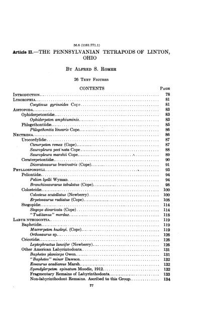
56.6 (1161:771.1)<br />
Aticle II.-THE PENNSYLVANIAN TETRAPODS OF LINTON,<br />
OHIO<br />
BY ALFRED S.<br />
26 TEXT FIGURES<br />
ROMER<br />
CONTENTS<br />
PAGE<br />
INTRODUCTION......................................................... 78<br />
LYBOROPHIA........................................................... 81<br />
Cocytinus gyrinoides Cope .. 81<br />
AIsTOPODA........................................................... 83<br />
Ophiderpetontida .................................................. 83<br />
Ophiderpeton amphiuminis .. 83<br />
Phlegethontiidae.........................8.....................<br />
I'll,<br />
85<br />
Phlegethontia linearis Cope......................................... 86<br />
NECTRIDIA........................................................... 86<br />
Urocordylidae..................................................... 87<br />
Ctenerpeton remex (Cope)......................................... 87<br />
Sauropleura pect nata Cope ....................................... 88<br />
Sauropleura marshii Cope ...................... 89<br />
Ceraterpetontide.................................................. 90<br />
Diceratosaurus brevirostris (Cope).................................. 91<br />
PHYLLOSPONDYLI............................... .................. 93<br />
Peliontida ........................................................ 94<br />
Pelion lyelli Wyman ........................................... 94<br />
Branchiosauravus tabulatus (Cope) .. 98<br />
Colosteide.......................................................... 100<br />
Colosteus scutellatus (Newberry) .................... 100<br />
Erpetosaurus radiatus (Cope) ..................... 108<br />
Stegopida ........................................................... 114<br />
Stegops divaricata (Cope) .................. ..................... 114<br />
" Tuditanus" mordax ........................ 118<br />
LABYR-NTHODONTIA ............................. 119<br />
Baphetidae........................................................... 119<br />
Macrerpeton huxleyi. (Cope) ...................... 119<br />
Orthosaurus sp ........................... 126<br />
Cricotide........................................................... 126<br />
Leptophractus lancifer (Newberry) ................... 126<br />
Other <strong>American</strong> Labyrintodonts ..................... 131<br />
Baphetes planiceps Owen ........................ 131<br />
"Baphetes" minor Dawson ...................... 132<br />
Eosaurus acadianus Marsh ...................... 132<br />
Spondylerpeton spinatum Moodie, 1912........................... 132<br />
Fragmentary Remains <strong>of</strong> Labyrinthodonts........................... 133<br />
Non-labyrinthodont Remains. Ascribed to this Group .......... 134<br />
77
78 Bulletin <strong>American</strong> <strong>Museum</strong> <strong>of</strong> <strong>Natural</strong> <strong>History</strong><br />
[Vol. LIX<br />
REPTILIA.... : ........................... 134<br />
Tuditanus punctulatus.134<br />
Eusauropleura digitata (Cope).135<br />
FISH REMAINS PREVIOUSLY DESCRIBED AS THOSE OF AMPHIBIANS.137<br />
Cercariomorphis parvisquamis Cope.137<br />
Eurythorax sublaeis Cope ....................................... 138<br />
Peplorhina exanthematicaCope. 138<br />
Sauropleura longipes Cope.138<br />
DisCUSSION.139<br />
Summary <strong>of</strong> the Fauna.139<br />
Relative Abundance . 140<br />
Amphibian Vertebae.141<br />
Environment <strong>of</strong> the Linton Amphibians.142<br />
Comparison with Other Faunas.142<br />
LITERATURE CITED .................................................... 144<br />
INTRODUCTION<br />
For the early history <strong>of</strong> land vertebrates the material from the<br />
Coal Measures is <strong>of</strong> paramount importance. Permo-Carboniferous forms<br />
are numerous, but they show us a stage in which many early amphibian<br />
groups were highly specialized, and some were apparently already extinct,<br />
while the deployment <strong>of</strong> the reptiles was well under way. On the<br />
other hand, pre-Pennsylvanian remains are extremely rare, although<br />
tetrapod development must have begun in the Devonian.<br />
In Europe, a true Coal Measures fauna was early described by Huxley<br />
from Kilkenny, while Atthey, Huxley and others gave accounts <strong>of</strong> the<br />
forms (mainly labyrinthodonts) from English and Scotch localities,<br />
especially Newcastle. Watson has, in various papers, reinterpreted<br />
much <strong>of</strong> this material (pointing out especially the importance <strong>of</strong> the<br />
Embolomeri), while the work <strong>of</strong> Fritsch, Credner and others on forms<br />
from central Europe has shed considerable light on Coal Measures fossils,<br />
although much <strong>of</strong> their material dates from. a somewhat higher series <strong>of</strong><br />
horizons, comparable with the Permo-Carboniferous <strong>of</strong> North America.<br />
In America a superficial survey <strong>of</strong> the state <strong>of</strong> our knowledge <strong>of</strong><br />
true Pennsylvanian forms (in which category late Pennsylvanian deposits<br />
containing the " Red Beds " fauna are to be excluded) seems to show that<br />
it is in a satisfactory condition. More than 80 species <strong>of</strong> tetrapods have<br />
been described from deposits <strong>of</strong> Westphalian age, mainly from three<br />
localities, the Linton cannel discussed below, the erect trees <strong>of</strong> the<br />
Joggins, Nova Scotia, and the nodule beds <strong>of</strong> Mazon Creek, Ill. The<br />
Linton material was described mainly by Cope, that from the Joggins<br />
by Dawson, while Moodie has described additional forms, mainly from
19301<br />
Romer, The Pennsylvanian Tetrapods <strong>of</strong> Linton, Ohio<br />
Mazon Creek and Linton. The latter has summarized our knowledge<br />
<strong>of</strong> the <strong>American</strong> Pennsylvanian amphibians in his useful " Coal Measures<br />
Amphibia <strong>of</strong> North America" (1916).<br />
The prospect is at first pleasing. From this wealth <strong>of</strong> species it<br />
would at first sight seem possible to obtain an adequate conception <strong>of</strong> the<br />
life <strong>of</strong> the Pennsylvanian <strong>of</strong> North America. Closer inspection,<br />
however, is disappointing in its results. In almost no instance is there<br />
sufficient material for us to either be able to assign a form to its proper<br />
position in the classification, or to obtain an idea <strong>of</strong> its general<br />
morphology. One species is known only from an element <strong>of</strong> the shoulder<br />
girdle; a second from a jaw fragment; a third from the top <strong>of</strong> the skull;<br />
and so on. This in itself gives rise to the suspicion that if these various<br />
elements could be correlated, the number <strong>of</strong> supposed forms might be<br />
considerably reduced.<br />
Stronger support for the idea that this is really the case is suggested<br />
by a consideration <strong>of</strong> the Linton fauna. From this single locality have<br />
come about two-thirds <strong>of</strong> described <strong>American</strong> forms. Nearly 60 species<br />
from this locality are remains <strong>of</strong> tetrapods or have been thought to be<br />
such. And yet all these specimens have come from an area not much<br />
more than 100 yards in diameter, in a layer <strong>of</strong> cannel coal four to six<br />
inches in thickness, the formation <strong>of</strong> which can have taken but a very<br />
short time, geologically speaking (Newberry, 1878, pp. 736-737). Is it<br />
reasonable to assume that more than half a hundred species <strong>of</strong> amphibians<br />
inhabited this pool almost simultaneously?<br />
The present 'paper is an attempt to restudy the Linton tetrapods.<br />
A few forms from Cannelton, about 15 miles to the northeast, and somewhat<br />
lower in the section, have been included.<br />
I have attempted to effect a systematic revision <strong>of</strong> the fauna, with<br />
the frank purpose <strong>of</strong> obtaining a reduction in the number <strong>of</strong> species, so<br />
that some idea <strong>of</strong> the morphology and appearance and habits <strong>of</strong> these<br />
forms may be obtained. It is quite probable that in this "lumping" <strong>of</strong><br />
species I may have proceeded too far, and that further material may<br />
prove that in several instances forms regarded here as synonyms may<br />
really be separate species. However, the results are, perhaps, somewhat<br />
nearer the truth than the present situation.<br />
Since many <strong>of</strong> these amphibians undoubtedly passed their entire<br />
lives in this type <strong>of</strong> habitat, immature forms are to be expected among<br />
the finds, and size can thus not be used as a criterion <strong>of</strong> specific or generic<br />
difference.<br />
The specimens are all preserved, in a crushed condition, as a car-<br />
79
80 Bulletin <strong>American</strong> Muweum <strong>of</strong> <strong>Natural</strong> <strong>History</strong><br />
[Vol. LIX<br />
bonaceous material in slabs <strong>of</strong> cannel coal. Usually the blocks are split<br />
through skulls and other elements, leaving half <strong>of</strong> the "bones" on one<br />
side, half on the other. Such specimens <strong>of</strong>ten show little or nothing <strong>of</strong><br />
surface structure. In such cases, as pointed out to me by Pr<strong>of</strong>essor<br />
D. M. S. Watson, the "organic " material can be removed by the use <strong>of</strong><br />
hydrochloric acid, enabling both surfaces <strong>of</strong> the bone to be recovered in<br />
squeezes. Unfortunately, many <strong>of</strong> the specimens are represented by one<br />
side only, and since many are types this method <strong>of</strong> treatment could not<br />
always obtain.<br />
This material is normally difficult <strong>of</strong> interpretation. In addition,<br />
pyritization and consequent decay have usually occurred to some extent,<br />
rendering what were probably good specimens when first collected very<br />
poor at the present time. I have not hesitated to criticise former interpretations<br />
<strong>of</strong> structures and sutures, and I need not add that my own<br />
are likewise open to criticism.<br />
Many <strong>of</strong> the specimens discussed were figured by Cope in 1875,<br />
and by Moodie in 1916; the illustrations in these two works should, in<br />
most cases, be consulted in connection with the descriptions given below.<br />
The largest collection, containing most <strong>of</strong> the types, is that in the<br />
<strong>American</strong> <strong>Museum</strong> <strong>of</strong> <strong>Natural</strong> <strong>History</strong>, New York. This includes most<br />
<strong>of</strong> the specimens collected by Newberry, while a few <strong>of</strong> his finds are to be<br />
found at Columbia University. A considerable number <strong>of</strong> slabs from<br />
the Lacoe Collection are in the U. S. National <strong>Museum</strong>, Washington,<br />
and a few specimens are in Walker <strong>Museum</strong>, University <strong>of</strong> Chicago.<br />
These specimens were obtained while the Linton " Diamond " Mine<br />
was in active operation, roughly from about 1855 to 1880. In 1920, the<br />
mine was reopened for a short time, and Dr. J. E. Hyde <strong>of</strong> Western<br />
Reserve University obtained a small but interesting collection. The<br />
ro<strong>of</strong> <strong>of</strong> the mine has now fallen in, and it is improbable that additional<br />
material will ever be obtained from this locality.<br />
For rendering the above material available to me for study, I wish<br />
to thank Curators Barnum Brown, Walter Granger, W. K. Gregory, and<br />
G. G. Simpson <strong>of</strong> the <strong>American</strong> <strong>Museum</strong>, Curator C. W. Gilmore <strong>of</strong> the<br />
National <strong>Museum</strong>, and Pr<strong>of</strong>essor J. E. Hyde <strong>of</strong> Western Reserve University.<br />
Specimens mentioned below without further designation are those<br />
in the <strong>American</strong> <strong>Museum</strong> <strong>of</strong> <strong>Natural</strong> <strong>History</strong>; those prefixed by NM are<br />
in the National <strong>Museum</strong>; those with WR in Western Reserve University;<br />
with WM in Walker <strong>Museum</strong>, University <strong>of</strong> Chicago.<br />
Abroad, there are a number <strong>of</strong> specimens from Linton in the
19301 Romer, The Pennsylvanian Tetrap9ds <strong>of</strong> Lintrn, Ohio<br />
81<br />
British <strong>Museum</strong>, others in Berlin (some <strong>of</strong> which have been described by<br />
Jaekel and Schwarz), and it is not impossible that there are a few<br />
specimens in other institutions.<br />
As this study was approaching completion, Pr<strong>of</strong>essor Watson informed<br />
me that the British <strong>Museum</strong> specimens were being prepared<br />
and studied by him and Miss M. Steen at University College, London.<br />
A comparison <strong>of</strong> our results shows that we are in essential agreement in<br />
most respects. Certain <strong>of</strong> the forms are not represented, or poorly<br />
shown in the British <strong>Museum</strong> material, but in several cases the material<br />
is better, and two forms represented by single specimens in this collection<br />
are, as far as I know, not found in the <strong>American</strong> material.<br />
LYSOROPHIA<br />
Lysorophus is a well-known form from the Lower Permian <strong>of</strong> North<br />
America, with characteristic vertebrae, an elongate body with very<br />
small limbs, and, especially, a very peculiar type <strong>of</strong> skull and branchial<br />
arch structure, which has most recently been investigated by Sollas<br />
(1920). Its position has long been a matter <strong>of</strong> debate; Broili believed<br />
it to be an ancestor <strong>of</strong> the reptiles, but the more general opinion has been<br />
that it is a very "advanced " amphibian, and Williston and others have<br />
suggested its possible relationship to the urodeles. There are no pre-<br />
Permian forms which have been assigned to this group previously.<br />
However, a member <strong>of</strong> this group (although without such great specialization<br />
<strong>of</strong> the cranial ro<strong>of</strong>) is present in the Lower Carboniferous <strong>of</strong> Scotland,<br />
and is described in a paper by Watson now in press (Palzeontographica<br />
Hungarica). It appears probable that the form from Linton generally<br />
known as Cocytinus gyrinoides is a close relative <strong>of</strong> Lysorophus, a conclusion<br />
which Pr<strong>of</strong>essor Watson informs me he has also reached.<br />
Cocytinus gyrinoides Cope<br />
C. gyrinoides COPE, 1871a, p. 177; 1874, p. 278; 1875, pp. 360-365, Fig. 5, P1.<br />
xxxix, fig. 4; MOODIE, 1916, pp. 67-69, Figs. 16, 16a.<br />
Molgophis wheatleyi COPE, 1874, p. 263; 1875, pp. 369-370, P1. XLV, fig. 1;<br />
MOODIE, 1916, pp. 149-150.<br />
? =Brachydectes newberryi COPE, 1868, p. 214; 1869, pp. 14-15; 1875, p. 388,<br />
P1. xxVII, fig. 2; MOODIE, 1916, pp. 175-176.<br />
TYPES.-C. gyrinoides, 6925; M. wheatleyi, 6897; B. newberryi, 6941.<br />
This form is<br />
best represented by the type, figured and described by<br />
Cope. The specimen shows the under side <strong>of</strong> a skull, well developed<br />
branchial arches, and the anterior portion <strong>of</strong> the vertebral column. I<br />
have figured the cranial region in comparison with a modification <strong>of</strong>
82 Bulletin <strong>American</strong> <strong>Museum</strong> <strong>of</strong> <strong>Natural</strong> <strong>History</strong><br />
[Vol. LIX<br />
Sollas' ventral view <strong>of</strong> Lysorophus. The comparison is quite close.<br />
The width <strong>of</strong> the skull<br />
'~~<br />
<strong>of</strong> the present form appears to be somewhat<br />
greater, but much <strong>of</strong> this may be due to crushing. The slight ridge<br />
visible behind the left jaw may be an indication <strong>of</strong> the squamosal<br />
region leading up from the quadrate. Extending far back <strong>of</strong> the jaws<br />
there is seen what appears to be a broad plate representing the parasphenoid<br />
region, behind which appears the first vertebra. The configuration<br />
here is remarkably similar to that <strong>of</strong> Lysorophus.<br />
Fig. 1. Right, Cocytinus gyrinoides, ventral view <strong>of</strong> type, X 152. Left, Lysorophus,<br />
modified from Sollas, for comparison.<br />
The branchial arches are quite well preserved and are similar to<br />
those <strong>of</strong> Lysorophus. The hypohyals and ceratohyals are large. On<br />
the left side I cannot distinguish without some doubt the first ceratobranchial<br />
from the adjacent ceratohyal. On the right side, a fourth<br />
branchial appears to be present.<br />
The type <strong>of</strong> M. wheatleyi is undoubtedly a specimen <strong>of</strong> this form,<br />
although the displacement <strong>of</strong> the ribs gives it a somewhat different<br />
appearance at first sight. The skull is not now visible. The description<br />
<strong>of</strong> it by Cope, however, is almost word for word that <strong>of</strong> an imperfectly<br />
preserved cranium <strong>of</strong> Lysorophus.<br />
Cope has described a pair <strong>of</strong> small lower ja*s as Brachydectes<br />
newberryi. The teeth are rather large, few in number and confined to the
1930] Romer, The Pennmylvanian Tetrapods <strong>of</strong> Linion, Ohio<br />
83<br />
anterior half <strong>of</strong> the jaw, which is short, rather shallow in front and very<br />
deep posteriorly. There is no Linton form other than Cocytinus with<br />
which these jaws can be correlated (unless, quite improbably, Ctenerpeton,<br />
in which the skull is unknown). The short tooth row rules out all<br />
others except the diceratosaurids, and the teeth are much too large for<br />
that genus.<br />
The lower jaws <strong>of</strong> the type <strong>of</strong> Cocytinus are seen only in ventral<br />
view, and a direct comparison is thus out <strong>of</strong> the question. But a comparison<br />
with the jaw <strong>of</strong> Lysorophus (as seen, for example, in the figures <strong>of</strong><br />
Sollas, 1920) shows that there is close agreement in the proportions,<br />
tooth row, etc., and it is highly probable that the "Brachydectes" jaw<br />
is that <strong>of</strong> Cocytinus.<br />
Moodie assigns Cocytinus to the urodeles on account <strong>of</strong> its well<br />
developed branchial arches. This is hardly an adequate reason. It is<br />
quite possible that the lysorophids are related to the modern caudates;<br />
but for the present it seems advisable to separate these forms from the<br />
modern ones and from other Palaeozoic types not closely related as well.<br />
The great specialization already attained by this Pennsylvanian form<br />
suggests that the lysorophids were a very early <strong>of</strong>fshoot <strong>of</strong> the primitive<br />
amphibian stock and are perhaps best regarded as constituting a separate<br />
order.<br />
AISTOPODA<br />
This group was erected by Miall for Ophiderpeton and Dolichosoma.<br />
The characters then listed were the possession <strong>of</strong> elongate vertebral<br />
centra and the absence <strong>of</strong> limbs. The body is elongate. The ribs are<br />
short and straight, sometimes with an "uncinate process" projecting<br />
from them.<br />
The aistopods are already highly specialized in the Pennsylvanian<br />
and apparently had become extinct by Permian times. It seems certain<br />
that they had had a long history as an independent group.<br />
Two <strong>American</strong> forms, corresponding closely to the genera named<br />
above, are to be placed here.<br />
OPHIDERPZTONTIDZ<br />
The less "degenerate" members <strong>of</strong> the group, with well developed<br />
(although short) ribs <strong>of</strong> a complicated structure, and a good ventral<br />
armor.<br />
Ophiderpeton amphiuiminis (Cope)<br />
Oestocephalus amphiuminis COPE, 1868, pp. 218-220; 1871, p. 41.<br />
Oestocephalus remex (partim) COPE, 1869, pp. 17-19; 1871, p. 46; 1871b, p. 53;
84A<br />
Bulletin <strong>American</strong> <strong>Museum</strong> <strong>of</strong> <strong>Natural</strong> <strong>History</strong><br />
[Vol. LIX<br />
1875, pp. 380-386, Pis. XXVII, fig. 5; P1. xxxi, fig. 1, P1. xxxiii, fig. 2; P1. xxxv, fig.<br />
5; P1. XLIV, fig. 3; MOODIE, 1909a, p. 27; 1916, pp. 143-145.<br />
Thyrsidium fasciculare COPE, 1875, pp. 365-866, P1. xxxi, fig. 2; P1. XLII,<br />
fig. 3; SCHWARZ, 1908, pp. 70-73, Figs. 5-9; MOODIE, 1909a, p. 27; 1916, pp. 145-<br />
146, Fig. 8.<br />
Ichthyerpeton squamosum MOODIE, 1909, pp. 69-72; 1909a, p. 24; 1916, pp. 135-<br />
137.<br />
TYPES.-O. amphiuminis, 6857 (a cotype) (Cope's P1. xxxIII, fig. 2) and probably<br />
T. fasciculare, 6900 (Cope's P1. xxxi, fig. 2), I. 8squamosum, NM 4459, 4476.<br />
Our knowledge <strong>of</strong> the genus has hitherto been obtained mainly<br />
from the material from Kilkenny and NyMan, described by Huxley and<br />
Fritsch. Huxley (1867, pp. 364-366, P1. Ii)<br />
figured an extremely elongate amphibian, with<br />
upwards <strong>of</strong> sixty holospondylous vertebrae<br />
present in his tomplete specimen. On the abdominal<br />
surface is a ventral armor <strong>of</strong> thin and<br />
much elongated scales; no traces <strong>of</strong> limbs or<br />
i+G)\l\ /1 : girdles are to be seen. The skull, unfortu-<br />
( /S unately, is not well preserved, but apparently<br />
was not <strong>of</strong> great length. The Nyfan material<br />
has added considerably to our information, as<br />
(K<br />
.'W27 described by Fritsch (1883, pp. 119-124, Pls.<br />
XVII, xix, xxi, xxiv) and Schwarz (1908). The<br />
structure <strong>of</strong> the vertebraw, including the complicated<br />
transverse processes, is well illustrated<br />
by these authors, as well as the peculiar ribs.<br />
Unfortunately the head has not been well preserved<br />
in this material.<br />
Fig. 2. Ophiderpeton<br />
amphiuminis, vertebra Remais <strong>of</strong> this type have been recognized<br />
and ribs, X 2. From a previously in <strong>American</strong> material only by<br />
specimen in Walker Schwarz, in Thyrsidium. However, it is not<br />
<strong>Museum</strong>. uncommon at Linton, but its identification<br />
had been prevented by a mistake on the part<br />
<strong>of</strong> Cope. Cope described Oestocephalus amphiuminis from two skulls<br />
and anterior portions <strong>of</strong> the body. The skulls were elongated, the<br />
teeth small, in a single series, and somewhat recurved. The body<br />
showed, as may be seen from his figures, an abdominal armor <strong>of</strong> long<br />
needle-shaped scutes, very similar to those <strong>of</strong> Ophiderpeton. Cope<br />
later, in error, associated these specimens with the Urocordylus-like<br />
tails which he had described as Sauropleura remex, and this hybrid<br />
animal has since been known as Oestocephalus remex. However, a check
19301<br />
Romer, The Pennsylvanian Tetrapods <strong>of</strong> Linton, Ohio<br />
85<br />
<strong>of</strong> the material available shows that there is no evidence for such an<br />
association, whereas, as noted elsewhere, the 0. remex caudals are very<br />
definitely to be associated with another type <strong>of</strong> body.<br />
Meanwhile, Thyrsidium fasciculare had been described by him from<br />
specimens similar to those <strong>of</strong> 0. amphiuminis, but showing complicated<br />
transverse processes. Schwarz (1908) described vertebrae and ribs <strong>of</strong> a<br />
specimen which he identified as Thyrsidium, and noted its resemblance<br />
to Ophiderpeton. I have developed specimens <strong>of</strong> 0. amphiuminis,<br />
some so identified by Cope, revealing structures comparable to those <strong>of</strong><br />
Schwarz's figure, and so similar to those <strong>of</strong> Urocordylus that generic<br />
identity seems highly probable. There are minor differences in the shape<br />
<strong>of</strong> the ribs and transverse processes, but these may well be regional in<br />
their nature. Even the notch in the prezygapophyses noted by Fritsch<br />
(1883, p. 120, Fig. 65) in Ophiderpeton is repeated in this material, It<br />
seems certain that 0. amphiuminis and T. fasciculare are synonymous,<br />
and that the form is a member <strong>of</strong> the genus Ophiderpeton.<br />
While the ventral squamation consists <strong>of</strong> thin elongate elements,<br />
the more lateral scutes are shorter and rounded in shape. Such scutes<br />
appear to have been well up the sides <strong>of</strong> the body, if not forming a<br />
complete covering. This is in agreement with Fritsch's findings.<br />
Moodie has described (1909b) two specimens in the National <strong>Museum</strong><br />
as representing a new species, I. squamosum, belonging to the genus<br />
Ichthyerpeton, found by Huxley (1867, pp. 367-368, P1. xxiii) in the<br />
Jarrow material. Moodie characterized it as a completely scaled amphibian,<br />
with elongate scutes ventrally and rounded ones dorsally. Upon<br />
examination these specimens clearly pertain to the present species,<br />
whereas Huxley's Ichthyerpeton is an embolomerous labyrinthodont<br />
and, incidentally, is stated by him not to be completely scaled.<br />
Of the forms included in the above synonymy, two are associated<br />
by Moodie in a common family with the elongate nectridian types, and a<br />
third placed in a family <strong>of</strong> labyrinthodonts, both <strong>of</strong> which associations<br />
are obviously incorrect.<br />
PHLEGETHONTIDA<br />
This is best known from Dolichosoma from Jarrow (Huxley, 1867,<br />
pp.. 366-367, P1. xxxi, fig. 3) and Nyran. It is an extremely elongate<br />
form, with vertebra essentially similar to those <strong>of</strong> Ophiderpeton, but<br />
with reduced ribs, as described especially by Fritsch (1883, pp. 108-109,<br />
Pls. xvii, XVIII, XXII, xxiii) and Schwarz (1908, pp. 76-79, Figs. 14-16).<br />
The skull is elongate; a general idea <strong>of</strong> its superficial features may be<br />
had from Fritsch's description.
86 BulUetin <strong>American</strong> <strong>Museum</strong> <strong>of</strong> <strong>Natural</strong> <strong>History</strong><br />
[Vol. LIX<br />
Phlegethontia linearis Cope<br />
Molgophis macrurus (partim, in errore) COPE, 1868, pp. 220-221.<br />
P. linearis COPE, 1871a, p. 177; 1874, p. 262; 1875, pp. 366-367, P1. XLIII, figs.<br />
1, 2; SCHWARZ, 1908, pp. 74-76, Figs. 12, 13; MOODIE, 1916, p. 154, Fig. 8.<br />
P. serpens COPE, 1874, p. 263; 1875, p. 367, P1. xxxii, fig. 1; MOODIE, 1916, p.<br />
154.<br />
TYPES.-P. linearis, 6886; P. serpens, 6899.<br />
The general characters <strong>of</strong> this form may be made out from Cope's<br />
figures; it appears to be very similar to Dolichosoma. Phlegethontia<br />
possessed a very elongate body (with more than 100 vertebrae in one<br />
specimen) and an elongate head. Cope states that ribs are absent, but<br />
in the larger specimen very thin rib-like structures are seen. There is no<br />
trace <strong>of</strong> them in the smaller specimens described as P. linearis, but as<br />
Fritsch (1883, p. 107) recognized, this seems due to the fact that these<br />
are immature forms in which ossification <strong>of</strong> these slender structures<br />
would obviously not yet have taken place. The vertebrae have been<br />
described by Schwarz (1908).<br />
Hay and Moodie both place the genus in the Molgophidae. There<br />
does not appear to be any reason for this association other than the<br />
supposed limbless condition in both cases. The structure <strong>of</strong> the vertebrae<br />
seems to be quite different. The vertebrae are somewhat similar to<br />
those <strong>of</strong> Dolichosoma; the differences are perhaps sufficient to retain the<br />
generic separation, but there can be no doubt <strong>of</strong> the close relationship<br />
<strong>of</strong> the two forms.<br />
NECTRIDIA<br />
This group was established by Miall to include especially Ceraterpeton<br />
and Urocordylus, described by Huxley (1867) from Jarrow. While<br />
these appear to differ widely in their skulls, they have a common type <strong>of</strong><br />
vertebral structure, the caudals being quite unlike those found in any<br />
other described forms. The chevrons attach to the under side <strong>of</strong> the<br />
single centrum and expand into a fan-shaped structure which is usually<br />
pectinated distally; the -neural spine is similar in general appearance.<br />
It seems probable, because <strong>of</strong> the chevron attachment, that the "centrum"<br />
is really the intercentrum, with which the haemal arches usually<br />
articulate in land forms. The dorsals likewise have anteroposteriorly<br />
elongated neural arches, <strong>of</strong>ten with a pectination present. Zygosphenezyantrum<br />
articulations are present. Watson (1913) has pointed out<br />
that in Batrachiderpeton the skull structure may be interpreted as a<br />
modification <strong>of</strong> the embolomerous plan, and that even in forms with a<br />
peculiarly modified skull pattern, the palatal structure is still quite<br />
primitive.
19301<br />
Romer, The Pennsylvanian Tetrapods <strong>of</strong> Linton, Ohio<br />
Two main types may be distinguished, both <strong>of</strong> which are represented<br />
at Linton; one with an elongate body with a well developed ventral<br />
armor, and with a skull which perhaps is rather "normal" except for<br />
elongation; the other with little or no ventral armor, but with the tabular<br />
region <strong>of</strong> the skull produced into long horn-like structures. Both groups<br />
are common in the Pennsylvanian, and both are represented in the <strong>American</strong><br />
Permo-Carboniferous by end forms. In addition, there are a number<br />
<strong>of</strong> forms <strong>of</strong> rather uncertain position, such as Scincosaurus <strong>of</strong> Bohemia,<br />
with Ctenerpeton-like armor, Sauravus <strong>of</strong> the French Carboniferous,<br />
MLepterpeton from Jarrow and probably others.<br />
UROCORDYLIDZ<br />
This family includes several carboniferous genera.<br />
It is represented<br />
by Urocordylus <strong>of</strong> Huxley (1867) from Ireland and Bohemia, and selveral<br />
<strong>American</strong> forms. All have elongate bodies, with the vertebrae characteristic<br />
<strong>of</strong> the Nectridia, an elongate tail, limbs <strong>of</strong> moderate to small size, a<br />
somewhat elongate head, and a well-developed ventral armor. The<br />
family is typically Pennsylvanian, although there is no doubt that<br />
Crossotelos, <strong>of</strong> the Wichita, was a late survivor <strong>of</strong> the group.<br />
There appear to be three members <strong>of</strong> this family in the Linton material:<br />
(1) a form best represented by Ctenerpeton alveolatum (most <strong>of</strong> the<br />
remains <strong>of</strong> which are usually incorrectly placed in Oestocephalus); (2) and<br />
(3) two smaller forms with shorter neurals and hsemals commonly<br />
described as Ptyonius, more correctly as Sauropleura.<br />
Ctenerpeton remex (Cope)<br />
Sauropleura remex COPE, 1868, pp. 217-218; 1871, p. 53.<br />
Oestocephalus remex (partim) COPE, 1869, p. 17, Fig. 2; 1871, p. 41; 1871b, p. 53;<br />
1875, pp. 381-386, Figs. 9 ("3"), Pls. xxxii, Fig. 2, xxxiv, Fig. 4; SCHWARZ, 1908,<br />
pp. 86-87, Figs. 29-30.; MOODIE, 1909a, p. 27; 1916, pp. 143-145, Figs. 3, 8.<br />
Ctenerpeton alveolatum COPE, 1897, pp. 83-84, P1. iii, fig. 1; MOODIE, 1909a,<br />
p. 24, P1. x; 1916, pp. 166-167, Pls. xix, xxiii, fig. 2.<br />
TYPES.-S. remex, 6903 (Cope, 1875, Fig. 3), C. alveolatum, NM 4475.<br />
Urocordylus was first described by Huxley (1867, pp. 359-362, P1.<br />
xx) from Kilkenny; a good idea <strong>of</strong> much <strong>of</strong> its general appearance may<br />
be gained from his plate. Girdles and both anterior and posterior limbs<br />
are present; the ventral armor consisted <strong>of</strong> rather broad thin scales<br />
arranged in chevron. The caudals are especially characteristic, with<br />
very tall and fluted hemals and neurals. An <strong>American</strong> form which is<br />
very closely related is that included in the above synonymy. Its general<br />
characteristics, as far as known, may be seen in Moodie's plate <strong>of</strong><br />
87
88 Bulletin <strong>American</strong> <strong>Museum</strong> <strong>of</strong> <strong>Natural</strong> <strong>History</strong><br />
[Vol. LIX<br />
Ctenerpeton. I have no certain knowledge <strong>of</strong> its skull, and the limbs are<br />
also incompletely known. The belly was covered by a series <strong>of</strong> broad<br />
dermal scutes 'well illustrated in Moodie's figure. They are punctate<br />
along their posterior edges, much as in Fritsch's Scincosaurus. The<br />
lateral members <strong>of</strong> each series are robust, and the series terminates<br />
laterally with a comb-shaped edge. As far as can be seen from Huxley's<br />
figures the ventral armor <strong>of</strong> Urocordylus wandesfordii was comparable.<br />
The tail vertebrae are remarkably similar to those <strong>of</strong> that type.<br />
This form is usually called Oestocephalus. Sauropleura remex was<br />
first described by Cope frQm caudal vertebrae <strong>of</strong> the urocordylid type;<br />
Oestocephalus amphiuminis from remains which included the head and<br />
anterior portion <strong>of</strong> the body. Cope later associated the two as Oestocephalus<br />
remex, but a check <strong>of</strong> his material shows that there is not the<br />
slightest evidence for this combination. As noted previously, the<br />
cranial and thoracic material is that <strong>of</strong> an aistopod. On the other hand,<br />
the association <strong>of</strong> "Sauropleura" remex and Ctenerpeton is shown in the<br />
type <strong>of</strong> the last-named genus, in a specimen figured by Cope (1875, Pl.<br />
xxxiv, fig. 4; No. 6909) and in an excellent specimen (5453) in Western<br />
Reserve University. The name Oestocephalus is not available for this form,<br />
since the type <strong>of</strong> that genus is an aistopod.<br />
0. rectidens is probably a synonym <strong>of</strong> Erpetosaurus radiatus.<br />
Sauropleura pectinata<br />
Cope<br />
S. pectinata COPE, 1868, pp. 216-217.<br />
Oestocephalus pectinats CoPE, 1869, p. 20; 1874, p. 266.<br />
Ptyonius pectinatus COPE, 1875, pp. 377-378, Pls. xxviii, figs. 2 (pt.), 4, 6;<br />
xxix, fig. 2; xxx, fig. 3; xxxv, figs. 1, 3, 4?; XLIV, fig. 3?; SCHWARZ, 1908, pp. 82-86,<br />
figs. 23, 24, 26; MOODIE, 1909a, pp. 24-25, P1. VIII, fig. 3; 1916, pp. 142-143, Figs. 8,<br />
30,P1. x, fig. 2.<br />
Oestocephalus serrulus CoPE, 1871a, p. 177.<br />
Ptyonius serrulus COPE, 1875, pp. 379-380, Pls. xxviii, fig. 5, xxx, fig. 1;<br />
MOODIE, 1916, pp. 142-143.<br />
Oestocephalus vinchellianus COPE, 1871a, p. 177.<br />
Ptyonius vinchellianus COPE, 1874, p. 266; 1875, pp. 376-377, P1. xxviii, fig. 1;<br />
SCHWARZ, 1908, Fig. 27; MOODIE, 1916, p. 141.<br />
TYPES. S. pectinata, 6876, 6882, 6896, S. serrula, 6895, 0. vinchellianus, 6872.<br />
This and the following species include the amphibians commonly<br />
The general characters <strong>of</strong> the body are well seen in<br />
known as Ptyonius.<br />
Cope's figures. The form is elongate, with a very long tail, with short,<br />
pectinated fan-shaped neurals and haemals, in contrast with the more<br />
slender elements <strong>of</strong> Ctenerpeton. The dorsal surface <strong>of</strong> the skull has been<br />
figured by Jaekel (1909). There are large clavicles and inteclavicle, and
19301<br />
Romer, The Pennsylvanian Tetrapods <strong>of</strong> Linton, Ohio<br />
89<br />
the ventral armor is well developed. The limb material is poor; I have<br />
not studied the evidence with' care, but while some <strong>of</strong> the specimens<br />
which, in Cope's figures, appear to show limbs are doubtful, and others<br />
are now imperfect, it seems certain that small limbs were present.<br />
This is perhaps the most abundant form in the Linton cannel. A<br />
considerable number <strong>of</strong> species <strong>of</strong> "Ptyonius" have been described, but<br />
I have been able to distinguish only two with any assurance. S. pectinata<br />
seems to be characterised by ventral scales much more slender<br />
than those <strong>of</strong> S. marshii (the contrast is well seen in Cope, 1875, P1.<br />
xxVIII, fig. 2), and a more elongate head. (Compare his P1. xxxv, fig. 1,<br />
and P1. XLI, fig. 2.)<br />
The only character given to distinguish vinchellianus from pectinatus<br />
by Cope is the shape <strong>of</strong> the interclavicle, and the difference here,<br />
as Moodie notes, is slight. S. serrulus was founded on a small specimen,<br />
which as Moodie states cannot be differentiated from S. pectinatus. (The<br />
contrast appears marked if comparison be made with the form shown on<br />
the same plate <strong>of</strong> Cope's 1875 paper. This specimen, however, appears to<br />
belong to the following species.)<br />
Cope proposed the name Ptyonius for the forms here called Sauropleura,<br />
and stated (1897, p. 86) that S. digitata was the type <strong>of</strong> this last<br />
genus. However, he himself had previously designated S. pectinata as<br />
the type <strong>of</strong> Sauropleura (1868, p. 217); S. pectinata has been designated<br />
by Moodie (1916) as the type <strong>of</strong> Ptyonius; hence this last name must<br />
unfortunately be relegated to the synonymy.<br />
In Europe this genus has been recognized only at Ny'an by Fritsch.<br />
Sauropleura marshii Cope<br />
Colosteus marshii COPE, 1869, p. 24.<br />
Oestocephalus marshii COPE, 1871a, p. 177.<br />
Ptyonius marshii COPE, 1875, pp. 375-376, Pis. xxvii, figs. 6, 7; xxviii, figs. 2, 3;<br />
MOODIE, 1916, pp. 141-142.<br />
Ptyonius nummifer COPE, 1875, pp. 374-375, P1. XLI, figs. 2, 3; MOODIE, 1909b,<br />
p. 356, P1. LxIII, fig. 3,; 1916, p. 142.<br />
Hyphasma l1svis COPE, 1875a, p. 16; 1875, pp. 387-388, P1. xxxvii, fig. 4;<br />
MOODIE, 1909b, pp. 356-357, P1. Lxiii, fig. 3; 1916, p. 71.<br />
TYPES.-S. marshii, 6862, S. nummifer, 6891, H. lxevis, 6877.<br />
The form discussed here has been differentiated above from S.<br />
pectinata, as far as our present knowledge <strong>of</strong> it goes. Specimens which are<br />
definitely assigned to it are few, but in default <strong>of</strong> certain distinctions in<br />
the caudals there are a number <strong>of</strong> specimens which may pertain to this<br />
form rather than S. pectinata. The best example is the type <strong>of</strong> nummifer,<br />
as illustrated by CcIpe. S. marshii is differentiated from this by Cope
90 BuUetin <strong>American</strong> <strong>Museum</strong> <strong>of</strong> <strong>Natural</strong> <strong>History</strong><br />
[Vol. LIX<br />
because <strong>of</strong> the more elongate interclavicle <strong>of</strong> the former; but it seems<br />
probable that the anterior end <strong>of</strong> this element has been obscured in the<br />
type <strong>of</strong> nummifer. Hyphasma levis was characterised by Cope as possessing<br />
both thin and thick scutellae, in a double layer. It seems, however,<br />
that this specimen is one <strong>of</strong> S. marshii with the ventral armor in a<br />
crumpled condition, and the thin scutes are the normal ones <strong>of</strong> the species<br />
turned somewhat sideways.<br />
CZRATERPETONTIDZ<br />
This family is represented by a number <strong>of</strong> Coal Measures forms, <strong>of</strong><br />
which Ceraterpeton <strong>of</strong> Huxley (1867) is the oldest and in some respects<br />
the best known (Watson, 1913, has pointed out that the material may<br />
represent two allied genera). The elongate tail is <strong>of</strong> nectridian type,<br />
with characteristic arches; the body is elongate, the limbs fairly well<br />
developed. There is some evidence <strong>of</strong> the presence <strong>of</strong> five toes in the<br />
manus, as suggested by Smith Woodward's figure (1897) <strong>of</strong> this genus,<br />
and confirmatory evidence in Diceratosaurus (Jaekel, 1903) and Diplocaulus<br />
(Douthitt, 1917).<br />
The most remarkable feature, however, is the presence <strong>of</strong> the<br />
peculiar horn-like projections from the tabular region.<br />
Batrachiderpeton, from Newsham, the skull <strong>of</strong> which has been well<br />
described by Watson (1913), is obviously a relative. Scincosaurus <strong>of</strong><br />
Ny-an may belong here, despite some apparent discrepancies, in Fritsch's<br />
description. Diceratosaurus, discussed below, is the only known <strong>American</strong><br />
representative, this form having appeared under several names in<br />
the existing literature.<br />
The structure <strong>of</strong> the vertebre and skull in these amphibians is such<br />
that they cannot be regarded as related to the ancestral reptiles. But<br />
there are several curious points <strong>of</strong> similarity in other parts <strong>of</strong> the skeleton:<br />
(1) Diplocaulus, as described by Douthitt (1917) possesses a<br />
separate coracoid, a structure not known in other Palaeozoic Amphibia;<br />
(2) this same form has an entepicondylar foramen in the humerus, not<br />
found in any other known amphibian; (3) as noted above, it is possible<br />
that these forms may have had 5 digits in the hand. The third may be<br />
an error, the other two may be due either to parallel development, or,<br />
more probably, to the retention <strong>of</strong> primitive characters lost<br />
by most<br />
amphibians. But it is not impossible that the group <strong>of</strong> presumable<br />
Embolomeri from which these forms are derived may have been close<br />
to the ancestors <strong>of</strong> the reptiles.<br />
Diplocautus is obviously derived from the Cer&terpeton group; but
19301<br />
Romer, The Pennsylvanian 7'etrapods <strong>of</strong> Linton, Ohio<br />
not merely its greater emphasis <strong>of</strong> the peculiar "horns" but also, as<br />
Watson has noted, its palatal modifications, seem to render it necessary<br />
to place it in a separate family.<br />
Diceratosaurus brevirostris (Cope)<br />
Tuditanus brevirostris COPE, 1874, p. 272; 1875, pp. 393-394, P1. xxvi, fig. 3;<br />
MOODIE, 1909b, P1. LXIV; 1916, pp. 88-89.<br />
Ceraterpeton punctolineatum COPE, 1875a, p. 16; 1875, p. 372, P1. XLII, fig. 4.<br />
Diceratosaurus punctolineatus JAEKEL, 1903, pp. 109-134, Pis. II-v; MOODIE,<br />
1909b, p. 356. P1. Lxv; 1916, pp. 115-119, Pls. xv, xvi, fig. 5, P1. xiv, fig. 4.<br />
Ceraterpeton tenuicorne COPE, 1875, p. 372, P1. XLII, fig. 2; 1897, pp. 85-88,<br />
P1. Iii, fig. 2.<br />
Eoserpeton tenuicorne MOODIE, 1909, pp. 76-79, Fig. 20; 1909a, p. 355, P1.<br />
LXIII, fig. 1; 1916, pp. 123-125, Figs. 11, 25; 1875, p. 388, P1. xxvii, fig. 2; MOODIE,<br />
1916, pp. 175-176.<br />
TYPES.-T. brevirostris 6933, D. punctolineatum 6866, E. tenuicorne 6836.<br />
The <strong>American</strong> horned forms are all apparently to be included in<br />
one species, Diceratosaurus brevirostris. Jaekel has given figures <strong>of</strong> much<br />
<strong>of</strong> the skeleton, showing the upper surface <strong>of</strong> the skull, palate, vertebrae,<br />
shoulder girdle and anterior limb in great part. On the dorsal surface<br />
91<br />
Fig. 3. Diceratosaurusbrevirostris. Left, type <strong>of</strong> "Tuditanus" brevirostris, X 1.<br />
Right, type <strong>of</strong> "Eoserpeton tenuicorne," X 14.<br />
<strong>of</strong> the skull, as described by him, the "perisquamosal," occupying the<br />
usual position <strong>of</strong> tabular, squamosal and supratemporal, is a unique<br />
feature. The palate has moderate interpterygoid vacuities, intermediate<br />
in size between those <strong>of</strong> Batrachiderpeton and Diplocaulus but with the<br />
pterygoids structurally united with the parasphenoid; the marginal<br />
teeth are confined to the anterior end. The sculpturing which Jaekel
92 Bulletin <strong>American</strong> <strong>Museum</strong> <strong>of</strong> <strong>Natural</strong> <strong>History</strong><br />
[Vol. LIX<br />
shows to have been present on the dorsal surface <strong>of</strong> the neural arches is a<br />
feature not reported in other genera. The dermal shoulder girdle has<br />
expanded clavicles and interclavicle and, according to Jaekel, small<br />
sculptured cleithra. Five fingers are reported in the manus.<br />
With his description the <strong>American</strong> material may be easily brought<br />
into line in many regards. The type skull <strong>of</strong> D. punctolineatus has been<br />
figured by both Cope and Moodie. This shows a portion <strong>of</strong> the anterior<br />
part <strong>of</strong> the body. The characteristic "horn." is well exhibited, and the<br />
sculpturing is identical with that described by Jaekel. To this is attached<br />
a portion <strong>of</strong> the skull, covering approximately the "perisquamosal"<br />
region. Clavicle and interclavicle resemble those described by Jaekel.<br />
Moodie says there is no cleithrum, but as Cope's figure shows, there is a<br />
small element just behind the "horn" which corresponds well with the<br />
bone described by Jaekel. To the left <strong>of</strong> the column is a peculiar element<br />
variously called by Moodie coracoid or scapula. It is obviously the<br />
same element as that described by Smith Woodward (1897) as a scapula,<br />
and would appear to be the element described by Watson (1913) as a<br />
cleithrum in Batrachiderpeton. The surface exhibited does not show<br />
sculpturing. It is possible that it is the internal surface <strong>of</strong> the left<br />
cleithrum, but it is also possible that it is a peculiar scapula.<br />
Jaekel says there are 12 presacrals, a sacral and over 100 caudals.<br />
This may be questioned, as the type appears to show 13 without a sacral.<br />
Tuditanus brevirostris was described by Cope from an individual<br />
which exhibits a skull and the anterior portion <strong>of</strong> the body. The specimen,<br />
as may be seen from his figure, was covered by matrix at the right<br />
posterior corner. Excavation here reveals the fact that a typical " horn"<br />
is present, and traces <strong>of</strong> the other can be made out lying close in to the<br />
backbonre. The skull bones are represented mainly by impressions <strong>of</strong> the<br />
inner surface <strong>of</strong> the ro<strong>of</strong>. Such sutures as I can determine are shown in<br />
the figure. They agree with those shown by Jaekel in Diceratosaurus in<br />
general, but there appears to be a suture separating the " perisquamosal"<br />
into tabular and squamosal (see below).<br />
The contour <strong>of</strong> the horns is somewhat different from that given by<br />
Jaekel in Diceratosaurus. In his figure there is no constriction posteriorly,<br />
the curve <strong>of</strong> the side <strong>of</strong> the skull passing smoothly into the horn, while<br />
there is a marked constriction in this specimen. This apparent difference<br />
appears to be due to the varied effects <strong>of</strong> crushing.<br />
Posterior to the skull, Cope noted that there seemed to be some sort<br />
<strong>of</strong> continuous notochordal sheath enveloping the region <strong>of</strong> the vertebral<br />
column. This appearance, upon examination, seems to be due to the
19301 Romer, The Pennsylvanian Tetrapods <strong>of</strong> Linton, Ohio<br />
93<br />
fact that the vertebrae are preserved with their original articulation, and<br />
since the neural arches extend the full length <strong>of</strong> the spine, they form a<br />
continuous mass in which it is difficult to detect breaks. The sculpturing<br />
covering the arches can be seen in the specimen. Some <strong>of</strong> the ribs appear<br />
to be distinctly double-headed, despite Jaekel's statement that the<br />
apparent tuberculum is not such a structure. A well developed transverse<br />
process is visible is some cases. Generally, however, details are<br />
not clear in the specimen.<br />
The type skull <strong>of</strong> Eoserpeton tenuicorne has been restudied, and I can<br />
find no grounds for a separation from the present species. My interpretation<br />
<strong>of</strong> the elements <strong>of</strong> the ro<strong>of</strong> differs in a number <strong>of</strong> respects from that <strong>of</strong><br />
Moodie, certain <strong>of</strong> his sutures being, apparently, cracks in the ventral<br />
aspect <strong>of</strong> the then unprepared ro<strong>of</strong>ing elements. His attempted subdivision<br />
<strong>of</strong> the " perisquamosal " is certainly incorrect, and I was at first<br />
inclined to believe in the unity <strong>of</strong> this element. However, there is,<br />
apparently, a line <strong>of</strong> demarcation running back on either side from the<br />
parietal to the outer side <strong>of</strong> the "horn," quite obviously separating the<br />
tabular from the squamosal (but with no trace <strong>of</strong> a distinct supratemporal).<br />
The sculpture patterns <strong>of</strong> the two elements have adjacent<br />
centers near this suture, and consequently the superficial appearance is<br />
that <strong>of</strong> a single element. Further, it is not improbable that the suture<br />
might be obliterated during life, as appears to have been the case in<br />
Jaekel's individual.<br />
" Diceratosaurus " robustus and " Diceratosaurus " k.evis are not<br />
members <strong>of</strong> this genus, but belong to quite another group, as does<br />
"Tuditanus " mordax.<br />
PHYLLOSPONDYLI<br />
Branchiosaurs have not been previously reported in the Linton<br />
fauna; the typical members <strong>of</strong> the group are <strong>of</strong> Permo-Carboniferous<br />
age.' Moodie, however, has assigned several forms from the Pennsylvanian<br />
<strong>of</strong> Mazon Creek to this group, and other forms from this<br />
locality (including Amphibamus) are also branchiosaurs; while Watson<br />
(1921) has described a primitive branchiosaur (Eogyrinus) from the<br />
English Coal Measures.<br />
Two forms (Family Peliontidae) from the Linton deposit are certainly<br />
primitive branchiosaurs, morphologically close to the ancestry<br />
<strong>of</strong> the Permian forms. Two others (Family Colosteidae) are aberrant in<br />
the shape and ro<strong>of</strong>ing elements <strong>of</strong> their skulls, but possess palates suggesting<br />
the branchiosaurs and are perhaps to*be associated with vertebral<br />
'Bulman and Whittard (1926) have recently given excellent redescriptions <strong>of</strong> many <strong>of</strong> these forms
94 BuUetin <strong>American</strong> <strong>Museum</strong> <strong>of</strong> <strong>Natural</strong> <strong>History</strong><br />
[Vol. LIX<br />
material apparently <strong>of</strong> a primitive branchiosaur type. Still another<br />
branchiosaur group is that represented by the specialized Stegops and<br />
its allies.<br />
PELIONTIDZ<br />
This family may be retained for the inclusion <strong>of</strong> primitive branchiosaurs,<br />
differing from the more typical forms in the presence <strong>of</strong> an intertemporal.<br />
The palate is primitive, including such features as a movable<br />
articulation <strong>of</strong> the pterygoid with the basipterygoid process, the retention<br />
<strong>of</strong> the ectopterygoid, etc. The postcranial skeleton, as far as known<br />
in the forms discussed below, is essentially branchiosaurian in character.<br />
Fig. 4. Pelion Iyelli. Type <strong>of</strong> "Saurerpeton latithorax," X .<br />
Pelion lyelli Wyman<br />
Raniceps IyeUi WYMAN, 1858, P. 160; COPE, 1869, P. 9.<br />
Pelion Iyelli WYM&AN, in Cope, 1875, PP. 380-391, PI. XXVI, fig. 1 ; MOODIE,<br />
1916, PP. 72-74, Fig. 17; PI. xxriv, fig. 1.<br />
Dendrerpleton obtusum COPER, 1868, P. 213; 1869, PP. 12-13, Fig. 1.<br />
Tuditanus obtusus COPE, 1875, PP. 396-397, Fig. 11; 1885, P. 407.<br />
Erpetosaurus obtusus MOODIE, 1909b, Pl. LX, fig. 2; 1916, PP. 98-100.<br />
Sauropleura latithorax COPE, 1897, PP. 86-88, PI. III, fig. 4.<br />
Saurerpeton latithorax MOODrE, 1909, P. 80, Fig. 23; 1909a, PP. 23-24; PI. IX,<br />
1916, PP. 162-165; PI. xvii; Figure 35.<br />
TYPES.-P. IyeUi, 6841, D. obtusum, 6928, S. latithorax, NM4471.<br />
This form is known mainly from the types <strong>of</strong> the three species<br />
named above. Except for the labyrinthodonts and Colosteus scutellatu8,<br />
the presumably adult specimens are the largest amphibians in the Linton<br />
fauna<br />
Ṫhe type <strong>of</strong> S. latithorax gives the best idea <strong>of</strong> the general appearance<br />
<strong>of</strong> the form, well shown in Moodie's plate. The specimen includes a
1930] Romer, The Pennsylvanian Tetrapods <strong>of</strong> Linton, Ohio<br />
95<br />
nearly complete skull seen in dorsal view and considerable postcranial<br />
material. The type <strong>of</strong> D. obtusum is a somewhat imperfect skull,<br />
originally showing mainly the impression <strong>of</strong> the inner surface <strong>of</strong> the<br />
ro<strong>of</strong>ing bones. This has been prepared to show the palate.<br />
As may be seen from the figures, the two specimens are nearly<br />
identical in size and in the arrangement <strong>of</strong> the cranial elements, orbits,<br />
etc. That they are cospecific is also evidenced by the fact that a small<br />
bit <strong>of</strong> sculptured bone originally present on D. obtusum corresponded<br />
exactly with the sculpturing on the better preserved specimen.<br />
The type <strong>of</strong> P. lyelli is a somewhat smaller individual, <strong>of</strong> which the<br />
ventral aspect <strong>of</strong> the skull and jaws and anterior portion <strong>of</strong> the body is<br />
shown. As noted below, there can be little doubt that it is identical<br />
with the skulls just mentioned, despite the differences in size.<br />
The dorsal surface <strong>of</strong> the skull is well shown in the S. latithorax<br />
skull; much <strong>of</strong> the pattern can be confirmed by the findings on the other<br />
specimen. Moodie has attempted an interpretation <strong>of</strong> the cranial<br />
elements; I find myself in disagreement with his results in a number <strong>of</strong><br />
respects.<br />
In neither specimen are the sutures in the internarial region visible,<br />
and in both the posterior end <strong>of</strong> the table is incomplete.<br />
The skull is broad in general contour, with a height, on a somewhat<br />
unreliable estimate, <strong>of</strong> not more than one-third the width. The orbits<br />
are placed far forward. The table <strong>of</strong> the skull was broad, the otic notches<br />
shallow.<br />
In general appearance and in the disposition <strong>of</strong> many <strong>of</strong> the elements<br />
the cranial ro<strong>of</strong> is similar to that <strong>of</strong> Leptorophus <strong>of</strong> the Permian. One<br />
feature.<strong>of</strong> interest, however, is the presence <strong>of</strong> an intertemporal. This is<br />
roughly diamond-shaped, and has the apparently primitive relations<br />
with neighboring bones. The sutures bounding this element may be<br />
clearly seen on the left side in Moodie's figure <strong>of</strong> S. latithorax, where the<br />
ventral impression <strong>of</strong> the bone is visible. On the right side, bone is<br />
present on the anterior portion <strong>of</strong> the intertemporal; although the surface<br />
is not perfect, it seems certain that the ridges and enclosed pits<br />
radiate from the supposed center <strong>of</strong> this element. The presence <strong>of</strong> the<br />
intertemporal appears to be confirmed on one side <strong>of</strong> the T. obtusus<br />
specimen; the sutures can be seen in Moodie's figure (1909b).<br />
The type <strong>of</strong> D. obtusum has been developed so as to exhibit the palate. The<br />
marginal teeth are small and numerous; almost universally there is a regular alternation<br />
<strong>of</strong> tooth and vacant pit. The maxillary extends back past the posterior boundary<br />
<strong>of</strong> the ectopterygoid. The premaxilla appears to have been excluded from the<br />
internal naris by the prevomer; medially, the two premaxilhe appear to extend back-
96 Bulletin <strong>American</strong> <strong>Museum</strong> <strong>of</strong> <strong>Natural</strong> <strong>History</strong><br />
[Vol. LIX<br />
ward in a narrow process (on which one or two teeth are present on each side) to<br />
effect a contact with the anteromedial extremities <strong>of</strong> the prevomers. Lateral to this<br />
process are circular palatal vacuities, surrounded by these two elements and presumably<br />
for the accommodation <strong>of</strong> large mandibular teeth. The prevomer is a bone<br />
<strong>of</strong> complicated shape. The two prevomers have little or no contact with each other;<br />
their medial boundaries overlap the parasphenoid ventrally, and a process extends<br />
back a short distance over the lateral margin <strong>of</strong> that element. There is a long contact<br />
with the premaxillae lateral to the palatal vacuity, just internal to which lie a large<br />
thenar tooth and pit. There is a short contact with the palatine between the internal<br />
naris and the interpterygoid vacuity. A foramen, presumably for a blood vessel, is<br />
present internal to the narial margin. The internal nares are rather large and subcircular<br />
in outline, and are almost entirely surrounded by the prevomers and palatines,<br />
although the maxilla enters into the lateral boundary.<br />
The palatine is a Y-shaped element, the longer anterior arm extending inwardly<br />
as a narrow bar between internal naris and interpterygoid vacuity to the vomerine<br />
Fig. 5. Pelion Iyelli. Ventral and dorsal views <strong>of</strong> the type <strong>of</strong> "Dendrerpeton<br />
obtu8uvm," X Y/4.<br />
contact, the lateral arm. extending a short distance along the outer side <strong>of</strong> the internal<br />
naris, and bearing a very large tooth. On neither side is there any evidence <strong>of</strong> a pit<br />
accompanying this tooth. The structure <strong>of</strong> the posterior portion <strong>of</strong> the element is not<br />
altogether certain, partially owing to the fact that on the right side <strong>of</strong> the skull the<br />
lateral ro<strong>of</strong>ing elements are broken and buckled under the palate, breaking the bones<br />
in this region and obscuring their relations. It appears, however, that the posterior<br />
branch is thin and elongate, has long lateral contacts with the maxilla laterally and<br />
the pterygoid medially, and is in contact with the ectopterygoid posteriorly. This<br />
last element is a small bone, bearing a thenar tooth and pit much smaller than those<br />
<strong>of</strong> the more anterior elements. It has a long contact laterally with the maxilla and<br />
medially with the palatine; there is little if any contact with the pterygoid. Posteriorly<br />
the jugal appears to send a strong inward extension directed somewhat<br />
ventrally for the support <strong>of</strong> the pterygoid.
19301<br />
Romer, The Pennsylvanicn Tetrapods <strong>of</strong> Linton, Ohio<br />
The pterygoid has a long palatal ramus, which nearly reaches the prevomer<br />
anteriorly, and posteriorly appears to bear a flange (although not a strong one) for<br />
the lower jaw. That portion <strong>of</strong> the ramus which lies external to the palatine and<br />
ectopterygoid is thickly studded with smail teeth. The pterygoid is freely movable<br />
on the braincase, having a strong V-shaped inwardly directed process the posterior<br />
edge <strong>of</strong> which articulates with the anterior edge <strong>of</strong> the basipterygoid process. The<br />
quadrate ramus is short, and, although crushed in the specimen, appears to have been<br />
moderately deep. In the angle between the articular process and the quadrate ramus,<br />
a paraotic process, imperfectly preserved on either side, extends dorsally, inwardly,<br />
and somewhat posteriorly.<br />
The parasphenoid extends forward as a broad bar, slightly ridged in the midline,<br />
between the two interpterygoid vacuities. It expands anteriorly, and appears to<br />
continue far forward, being covered laterally in ventral view by the prevomers. The<br />
basicranium is imperfect posteriorly. There are well-developed thickened basipterygoid<br />
processes. The braincase is broad, partially due to crushing.<br />
The postcranial skeleton <strong>of</strong> " Saurerpeton " shows the squamation to<br />
have been somewhat " cycloid " in character, as in typical branchiosaurs.<br />
The vertebrae are not well preserved. The ribs are moderately short and<br />
but slightly curved. The clavicles and interclavicle are broadly ex-<br />
97<br />
Fig. 6. Pelion lyelli. Restoration <strong>of</strong> skull, dorsal and ventral views, XA,<br />
the size <strong>of</strong> presumably adult specimens.<br />
panded on the ventral surface, much as in typical branchiosaurs, and<br />
suggesting a bottomliving habit. The interclavicle is ornamented with<br />
small pits in the central portion, and light radiating lines towards the<br />
margins. Most <strong>of</strong> the elements <strong>of</strong> the anterior limbs are present; there<br />
appear to have been four toes in the manus.<br />
Pelion lyelli, one <strong>of</strong> the earliest amphibians described from the<br />
Linton deposit, shows (covered with an obscuring film) the ventral<br />
surface <strong>of</strong> a rather small skull, with jaws in place, rather large anterior<br />
limbs and part <strong>of</strong> the vertebral column. Ribs, and dermal scutes, are
98 Bulletin <strong>American</strong> <strong>Museum</strong> <strong>of</strong> <strong>Natural</strong> <strong>History</strong><br />
[Vol. LIX<br />
absent, presumably due to post mortem separation. The skull is short<br />
and wide, and obviously <strong>of</strong> the general type found in this species and the<br />
following one. It is <strong>of</strong> the size <strong>of</strong> P. tabulatus, and I believed at one time<br />
that it represented that form. However, the limbs in that species are<br />
much smaller in proportion to the skull than in the P. lyelli type, while<br />
the more robust limbs <strong>of</strong> the Saurerpeton specimen are <strong>of</strong> the same<br />
proportions.<br />
That this form is a primitive branchiosaur is certain. The dorsal<br />
surface <strong>of</strong> the skull, as noted above, is very similar to that <strong>of</strong> certain <strong>of</strong><br />
the Permian forms, except for the presence <strong>of</strong> the intertemporal, a primitive<br />
feature. The postcranial skeleton, as far as preserved, is in agreement<br />
with this conclusion.<br />
The palate is in most respects that which might have been expected<br />
in an ancestor <strong>of</strong> the branchiosaurs. The absence <strong>of</strong> the anterior palatal<br />
vacuities in typical branchiosaurs is undoubtedly related to the reduction<br />
<strong>of</strong> the larger internal mandibular teeth normally present in the<br />
Embolomeri. The thenar teeth <strong>of</strong> the present form are essentially on the<br />
embolomerous plan; they are reduced or absent in later branchiosaurs.<br />
The shape <strong>of</strong> the palatine is essentially that <strong>of</strong> this element in the Permian<br />
forms. The presence <strong>of</strong> an ectopterygoid is a very primitive feature, not<br />
found in forms <strong>of</strong> any later stage. This element is here <strong>of</strong> comparatively<br />
small size, with the thenar teeth somewhat reduced. Its loss would<br />
convert the lateral region <strong>of</strong> the palate into the type found in typical<br />
members <strong>of</strong> this group. The pterygoid is <strong>of</strong> the primitive type noted by<br />
Watson in Eogyrinus. The articulation <strong>of</strong> this element is with the anterior<br />
margin <strong>of</strong> the basisphenoid region, much as in the Embolomeri,<br />
whereas in later branchiosaurs it attached more posteriorly and dorsally,<br />
and was presumably fixed.<br />
The palate <strong>of</strong> Pelion tends strongly to confirm Watson's thesis<br />
that the branchiosaurs are descended from primitive labyrinthodonts, or,<br />
at least, from forms with a skull structure <strong>of</strong> the type found in primitive<br />
members <strong>of</strong> that group.<br />
Branchiosauravus tabulatus (Cope)<br />
Tuditanus tabulatus COPE, 1877, p.577.<br />
Erpetosaurus tabulatus MOODIE, 1909, pp. 52-56, Figs. 8, 9; 1909b, pp. 347-351,<br />
P1. LIX, fig. 2, P1. LXII, fig. 2; 1916, pp. 100-104, Fig. 22, P1. xxv, fig. 2.<br />
Tuditanus sculptilis MOODIE, 1909, pp. 61-63, Figs. 11, 12; 1909a, pp. 23-23;<br />
1916, pp. 105-107, P1. xviii, fig. 1.<br />
Tuditanus walcotti MOODIE, 1909a, pp. 16-19, PF. vi, fig. 1; PF. vii, fig. 1.<br />
Erpetosaurus walcotti MOODIE, 1916, pp. 93-96, Fig. 21.
19301<br />
Romer, The Pennsylvanian Tetrapods <strong>of</strong> Linton, Ohio<br />
99<br />
Erpetosaurus minutus MOODIE, 1909a, pp. 21-23, P.1. VIII, fig. 1; 1916, pp. 104-<br />
105, Fig. 22, P1. xx, fig. 1.<br />
TYPES.-T. tabulatus, 6837, T. sculptilis, WM 12315, T. walcotti, NM 4474,<br />
E. minutus, NM 4545.<br />
tially <strong>of</strong> a system <strong>of</strong> radiating and somewhat<br />
branching ridges, with some pitting near the<br />
NV- w\\' \ / center <strong>of</strong> the elements.<br />
&\'\ The anterior end <strong>of</strong> the skull is missing;<br />
--7W1' the outlines <strong>of</strong> the elements preserved are for<br />
Fig. 7. Branchiosaura us<br />
the most part very similar to those described<br />
tabulatus. Dorsal surface <strong>of</strong> by Watson in Eogyrinus from the English<br />
skull <strong>of</strong> type, X 1. Coal Measures. In the English form the<br />
postfrontal is described as an elongated<br />
This small and primitive branchiosaur is best represented by the<br />
type skull and by the greater part <strong>of</strong> the skeleton in a specimen in<br />
Western Reserve University (No. 5446).<br />
The skull is <strong>of</strong> small size, short and<br />
, N broad (especially the table), and with large<br />
orbits situated somewhat anteriorly; the<br />
t1'.'E ,otic notches are shallow, and the general<br />
appearance is suggestive <strong>of</strong> branchiosaur<br />
4i\\\vi;rjX1 \'j'f
10l Bulletin <strong>American</strong> <strong>Museum</strong> <strong>of</strong> <strong>Natural</strong> <strong>History</strong> [Vol. LIX<br />
appears to have been similar to that <strong>of</strong> Amphibamus, from Mazon Creek,<br />
which is a rather primitive branchiosaur.<br />
E. sculptilis has for its type a small skull in Walker <strong>Museum</strong>, from<br />
the Cannelton locality. It is quite imperfect, and the sculpturing is<br />
poorly preserved, and most <strong>of</strong> the sutures are doubtful. I cannot confirm<br />
Moodie's interpretation <strong>of</strong> the cranial elements. As far as I can<br />
make them out, they appear to agree with those <strong>of</strong> P. tabulatus. The<br />
type <strong>of</strong> T. walcotti appears to be an immature specimen <strong>of</strong> the present<br />
species, apparently identical in the sculpturing <strong>of</strong> the interclavicle, and<br />
in the ribs and limb elements as far as. preserved. The skull has been<br />
crushed, making it appear narrower than was really the case. E. minutus<br />
appears to be the skull <strong>of</strong> an immature individual <strong>of</strong> this type; the<br />
sutures are difficult to determine, but are seemingly similar to those <strong>of</strong> P.<br />
tabulatus.<br />
It is unfortunate that among the many generic names included in<br />
the synonymy <strong>of</strong> the Linton material, there is none which may be<br />
applied to the present form. It is possible that it is generically identical<br />
with some <strong>of</strong> the branchiosaurs from other Pennsylvanian localities, but<br />
provisionally it may be called Branchiosauravus tabulatus.<br />
COLOSTEIDA<br />
The two forms placed in this family are probably to be regarded as<br />
branchiosaurs, but branchiosaurs <strong>of</strong> a peculiar type, and with many<br />
primitive characters reminiscent <strong>of</strong> the labyrinthodonts. Especially<br />
characteristic is the anterior position <strong>of</strong> the orbit and the consequent<br />
elongation <strong>of</strong> the elements posterior to it.<br />
Colosteus scutellatus (Newberry)<br />
Pygopterus scutellatus NEWBERRY, 1856, p. 98.<br />
Colosteus scutellatus COPE, 1869, pp. 22-24; 1873, p. 418; 1874, p. 275; 1875,<br />
pp. 407-408, P1. xxix, fig. 3, P1. xxxiii, fig. 1; 1877, p. 578.<br />
Sauropleura scutellata COPE, 1897, p. 88; MOODIE, 1916, pp. 156-157, P1. xxi,<br />
fig. 5<br />
Ṙhizodus angustus NEWBERRY, 1856, p. 99; 1868, p. 145; 1873, p. 342, P1.xxxix,<br />
fig. 6; SMITH WOODWARD, 1891, p. 348.<br />
Colosteus crassiscutatus COPE, 1869, pp. 22-23.<br />
Sauropleura newberryi COPE, 1875, pp. 404-405, P1. xxxvii, Fig. 2(?), P1. XLI,<br />
fig. 5; MOODIE, 1916, p. 158.<br />
AnisodeXis enchodus COPE, 1885, p. 406; 1897, p. 88.<br />
Sauropleura enchodus MOODIE, 1916, p. 162, P1. xvi, fig. 4.<br />
Leptophractus lineolatus COPE, 1877, p. 576; MOODIE, 1916, pp. 169-170, P1.<br />
XXII, fig. 1.
19301 Romer, The Pennsylvanian Tetrapods <strong>of</strong> Linton, Ohio<br />
101<br />
Sauropleura longidentata MOODIE, 1909, pp. 74-76, Figs. 18, 19; 1916, pp. 160-161,<br />
P1. XVI, figs. 2, 3.<br />
Macrerpeton deani MOODIE, 1916, pp. 184-185, Fig. 40, P1. xxi, figs. 1, 2.<br />
Diceratosaurus robustus MOODIE, 1909, p. 67, Fig. 15; 1909b, p. 355, P1. Lxiii,<br />
fig. 2; 1916, pp. 122-123, Fig. 24b.<br />
=?Molgophis macrurus COPE, 1868, p. 220; 1869, p. 20; 1874, p. 263; 1875,<br />
p. 368, P1. XLIII, fig. 1; MOODIE, 1916, pp. 147-148.<br />
Molgophis brevicostatus COPE, 1875, p. 369, P1. XLIV, fig. 1; SCHWARZ, 1908, pp.<br />
73-74, Figs. 10, 11; MOODIE, 1909a, p. 27; 1916, pp. 148-149, Fig. 32.<br />
Pleuroptyx clavatus COPE, 1875, pp. 370-371, P1. XLII, fig. 1; P1. XLIV, fig. 2;<br />
1875a, p. 16; MOODIE, 1909a, p. 27; 1916, pp. 151-153, Fig. 33.<br />
TYPEs.-Pygopterus scutellatus 6916, R. angustus, unknown, C. crassiscutatus,<br />
?6916, S. newberryi, 6932, L. lineolatus, 6828, S. longidentata, 6945, and reverse in<br />
British <strong>Museum</strong>, S. enchodus, 2558, M. deani, 2934, D. robustus, 6929, M. macrurus,<br />
specimen in Columbia University, M. brevicostatus, 6840, P. clavatus, 6838.<br />
Fig. 8. Colosteus 8cutellatus. Left, right and left sides <strong>of</strong> type skull, X Y4.<br />
Right, above, table <strong>of</strong> skull <strong>of</strong> No.- 6918, X 2, below, type <strong>of</strong> "Sauropleura newberryi,"<br />
X3/4/<br />
The species described below is one <strong>of</strong> the larger Linton forms,<br />
rivaling the labyrinthodonts in size and having many labyrinthodont<br />
characters; in all probability, however, it is an aberrant although
102 Bulletin <strong>American</strong> <strong>Museum</strong> <strong>of</strong> <strong>Natural</strong> <strong>History</strong><br />
[Vol. LIX<br />
primitive branchiosaur. It is known from numerous specimens showing<br />
the remains <strong>of</strong> the skull, while several individuals exhibit the dermal<br />
armor <strong>of</strong> the body and the dermal shoulder girdle. In addition, it is<br />
probable that considerable postcranial material <strong>of</strong> the Molgophis-<br />
Pleuroptyx type is to be associated with this form.<br />
It is certainly related to Erpetosaurus radiatus (next to be described)<br />
in many features, including the anterior position <strong>of</strong> the orbits and the<br />
consequent peculiar character <strong>of</strong> the postorbital, but differs from it in<br />
the well-defined lateral line canals, the smaller orbits, and in other<br />
features mentioned below.<br />
It is necessary to discuss briefly the somewhat checkered history<br />
<strong>of</strong> the type species. It was first described by Newberry as a fish, Pygopterus<br />
scutellatus. Cope later described an amphibian, Colosteus crassiscu-<br />
Fig. 9. Colosteus scutellatus. Left, No. 6915, X /. Right, above, type <strong>of</strong><br />
"Sauropleura longidentata," X 32, below, part <strong>of</strong> type <strong>of</strong> "Macrerpeton deani," X 8.<br />
tatus, and apparently used the type specimen <strong>of</strong> Newberry's species as his<br />
own. Later he recognized the identity, but since the form was obviously<br />
amphibian, the new generic name was retained (Pygopterus being a good<br />
fish genus). In 1897, however, he suggested its relationship to the form<br />
known as Sauropleura digitata, transferred the present species to that<br />
genus, and abandoned Colosteus. But in this he was doubly in error, for
19301<br />
Romer, The Pennsylvanian Tetrapods <strong>of</strong> Linton, Ohio<br />
not only is it improbable that Colosteus is related to "Sauropleura"<br />
digitata (discussed later), but also the type <strong>of</strong> the genus Sauropleura, as<br />
has been seen, is a nectridian amphibian quite unrelated to either <strong>of</strong> these<br />
forms. Colosteus scutellatus is hence the proper name <strong>of</strong> this animal.<br />
Our knowledge <strong>of</strong> the skull is derived from a considerable number <strong>of</strong><br />
specimens, between which practically every detail <strong>of</strong> the dorsal surface<br />
can be determined. These include, among others:<br />
103<br />
No. 6916, the type <strong>of</strong> the genus and species, a skull (poorly figured by Cope)<br />
showing the external surface <strong>of</strong> both sides, the lower jaws, the interclavicle and a<br />
considerable portion <strong>of</strong> the dermal armor.<br />
No. 6932, the type <strong>of</strong> S. newberryi, much <strong>of</strong> the right side <strong>of</strong> a skull and jaw.<br />
No. 2826, the type <strong>of</strong> M. deani, part <strong>of</strong> the skull top and jaw <strong>of</strong> a large specimen.<br />
No. 6945, the type <strong>of</strong> S. dentatus, the right side <strong>of</strong> a skull and anterior half <strong>of</strong> a<br />
jaw.<br />
No. 6918, the posterior portion <strong>of</strong> a skull and a large part <strong>of</strong> the ventral armor,<br />
described by Cope in 1877.<br />
No. 6915, a "macerated" skull showing part <strong>of</strong> the palate as well as ro<strong>of</strong>ing<br />
elements.<br />
There is considerable variation in size in these specimens, ranging<br />
from an estimated skull length <strong>of</strong> 59 mm. in No. 6932, to about 215 mm.<br />
in No. 2826, the last specimen rivaling the contemporary labyrinthodonts<br />
in size.<br />
The skull is <strong>of</strong> the shape found in the more "normal" labyrinthodonts,<br />
except that the orbits are situated somewhat in front <strong>of</strong> the middle<br />
<strong>of</strong> the length. While it is impossible to be sure <strong>of</strong> the exact proportions,<br />
the.fact that a majority <strong>of</strong> the skulls have been crushed sideways suggests<br />
that the skull was, in life, comparatively high and narrow. The<br />
otic notch is shallow.<br />
The sculpture <strong>of</strong> the ro<strong>of</strong>ing elements consists <strong>of</strong> a number <strong>of</strong> pits in the center<br />
<strong>of</strong> each element, from which ridges, bifurcating, radiate toward the periphery. The<br />
enclosed valleys deepen centrally, where a pore is present. The pattern is similar to<br />
that <strong>of</strong> the next species. Lateral line canals are evident in a number <strong>of</strong> places. The<br />
lyran are well developed on the postfrontals, frontals, prefrontals, nasals and premaxillae.<br />
A postorbital canal extends down across the postorbital and jugal. Near the<br />
ventral edge <strong>of</strong> the latter element it meets a canal which extends posteriorly and upward<br />
onto the squamosal and then turns down over the posterior edge <strong>of</strong> the quadratojugal<br />
to continue forward along the ventral edge <strong>of</strong> the lower jaw. The anterior prolongation<br />
on the upper jaw <strong>of</strong> these canals presumably passes forward along the<br />
maxilla, curves upward onto the lacrymal, and recurves abruptly downward to the<br />
edge <strong>of</strong> the jaw a short distance behind the nares.<br />
The tabulars are small, not extending nearly as far forward as the dermal supraoccipitals.<br />
The table <strong>of</strong> the skull has a curved posterior border, and there are no<br />
tabular horns. The supratemporals are elongate, their anterior ends pushing forward<br />
between parietal and postorbital. The last element is very elongate, but narrows
104 Bulletin <strong>American</strong> <strong>Museum</strong> <strong>of</strong> <strong>Natural</strong> <strong>History</strong><br />
[Vol. LIX<br />
anteriorly, and appears not to reach the orbit, being crowded out by the postfrontal<br />
and jugal, a feature undoubtedly related to the forward position <strong>of</strong> that opening.<br />
The frontals are long and narrow. The prefrontals are narrow, but extend forward<br />
nearly to the nares lateral to the nasals, which elements have a posterior wedge-shaped<br />
extension along the outer sides <strong>of</strong> the frontals. The premaxillae carry an anterior<br />
prolongation <strong>of</strong> the supraorbital lateral line canals. The lacrymals were wedged in<br />
between maxilla and prefrontals, and I cannot be certain whether they reach the nares.<br />
Posteriorly they effect a contact with the elongate jugal below the orbits. The maxille<br />
are very long, reaching back to about the anterior end <strong>of</strong> the quadrate-jugals. The<br />
marginal teeth are rather thinly spaced.<br />
Curiously enough, I have been unable to obtain a satisfactory view<br />
<strong>of</strong> the palate in any one <strong>of</strong> the rather numerous specimens. Our knowledge<br />
<strong>of</strong> it is gained mainly from specimens laterally crushed which exhibit<br />
portions <strong>of</strong> the palate between the jaw margins. Such evidence<br />
as we have tends to show that it was similar, in most respects, to that <strong>of</strong><br />
Erpetosaurus radiatus.<br />
The details <strong>of</strong> the braincase are not known. The parasphenoid was<br />
toothed for much <strong>of</strong> its extent. The pterygoids articulated movably<br />
with the basisphenoid region; they extended far forward, to about the<br />
level <strong>of</strong> the palatine, with interpterygoid vacuities probably <strong>of</strong> moderate<br />
size. There is some evidence that the palatal ramus bore a series <strong>of</strong> large<br />
teeth-a very unusual circumstance-as well as a series <strong>of</strong> smaller<br />
" denticles. " Three large sets <strong>of</strong> palatal teeth are present, presumably on<br />
the ectopterygoid, palatine and prevomer. Those on the ectopterygoid<br />
Fig. 10.. Colosteus scutellatus. Type <strong>of</strong> "Diceratosaurus robustus," X Y.<br />
are the smallest; the others, in several instances, show both teeth <strong>of</strong> a<br />
pair functioning at the same time. The contacts between the palatine<br />
and ectopterygoid, seen in one instance, seem to be comparable to those<br />
in 'Erpetosaurus.<br />
The lower jaw is present in several cases but only its outer surface is<br />
seen in its entirety. The dentary extends back nearly to the posterior<br />
end, in agreement with the posterior reach <strong>of</strong> the maxillary above. It<br />
bears a series <strong>of</strong> rather stout but thinly spaced marginal teeth, and a
1930]<br />
Romer, The Pennsylvanian Tetrapods <strong>of</strong> Linton, Ohio<br />
larger internal series, one near the anterior end being especially large<br />
and predicting an opening in the prevomerine region <strong>of</strong> the palate for its<br />
reception. There is a large notch on the outer side <strong>of</strong> the jaw which<br />
allows the passage <strong>of</strong> the prevomerine thenars, which must have been<br />
situated very close to the maxilla. Apparently both splenials were<br />
present, as well as the angular and a rather large surangular.<br />
105<br />
Fig. 11. Colosteubs scutellatus. Restoration <strong>of</strong> skull in dorsal and lateral views<br />
About 3 the size <strong>of</strong> the largest known skull.<br />
Some postcranial skeletal material is associated not only with the<br />
type but also with several other specimens. The clavicles and interclavicle<br />
are considerably expanded ventrally, with a more subdued<br />
sculpture than that found on the head. The ventral surface <strong>of</strong> the body<br />
was covered by a regularly arranged series <strong>of</strong> rhombic scutes <strong>of</strong> the type<br />
usually associated with the labyrinthodonts. These have obscured most
106 BuUetin <strong>American</strong> <strong>Museum</strong> <strong>of</strong> <strong>Natural</strong> <strong>History</strong><br />
[Vol. LIX<br />
<strong>of</strong> the remains <strong>of</strong> the axial skeleton. I have found no describable remains<br />
<strong>of</strong> the vertebrae in these specimens. A few anterior ribs were probably<br />
nearly straight, the more posterior ones had about the length and curvature<br />
found in those <strong>of</strong> " Molgophis " (see below).<br />
With regard to the positon <strong>of</strong> the forms represented by cranial<br />
material and regarded as synonyms, it is at once obvious from the figures<br />
that the types <strong>of</strong> S. newberryi, S. longidentata, and M. deani are the same<br />
as the type <strong>of</strong> the present species. The jaw fragment which is the type <strong>of</strong><br />
Anisodexis enchodus appears to be quite comparable to that <strong>of</strong> S. longidentata.<br />
The fragment <strong>of</strong> jaw which Newberry described as Rhizodus<br />
angustus would appear from his figure to be <strong>of</strong> this type as well. "Leptophractus"<br />
lineolatus is a portion <strong>of</strong> the jaws <strong>of</strong> a large specimen. Diceratosaurus<br />
robustus <strong>of</strong> Moodie is a fragment which shows the characteristic<br />
postorbital <strong>of</strong> this form (it is not a diceratosaurid).<br />
This species grew to a size which would normally warrant us in<br />
assigning it to the Labyrinthodontia, and there are many characters suggestive<br />
<strong>of</strong> that group. But the palate, as far as known, appears to differ<br />
radically from that <strong>of</strong> the early members <strong>of</strong> this order (as discussed<br />
under Erpetosaurus). Also there are other features such as (for example)<br />
the long dentaries, the absence <strong>of</strong> tabular horns, the small size <strong>of</strong> the<br />
tabulars, the absence <strong>of</strong> an intertemporal, etc., which suggest a relationship<br />
with the branchiosaurs. Except for the characteristic postorbital,<br />
the general features <strong>of</strong> the skull are not dissirmilar to those <strong>of</strong> Leptorophus.<br />
The vertebrae and ribs variously known as Molgophis and Pleuroptyx<br />
are perhaps to be assigned to this form. M. macrurus was described from<br />
a specimen showing a dorsal view <strong>of</strong> vertebrae and ribs, M. brevicostatus,<br />
a lateral view; Pleuroptyx clavatus from a lateral view showing similar<br />
vertebrae, but with expanded ribs.' As Cope, Schwarz and Moodie<br />
have all suggested, it is highly probable that these forms are the same,<br />
the vertebrae being quite similar, and the difference in the ribs is probably<br />
a regional one (although I have seen as many as twenty vertebrae without<br />
much indication <strong>of</strong> such a shift). There is considerable difference in<br />
size, varying from forms considerably smaller than the types figured by<br />
Cope up to one specimen in which the height to the top <strong>of</strong> the arch is 25<br />
mm. It is not improbable that two or more species may be concealed in<br />
the fairly abundant material to which these names are given; the types,<br />
however, are all among the larger specimens.<br />
There is a single notochordal centrum, with a ventral flattened ridge, asfiguredby<br />
Schwarz. At about the middle <strong>of</strong> the height there is a well marked ridge extending<br />
'As noted elsewhere, Molgophis whealeyi does not belong here.
1930]<br />
Romer, The Pennsylvanian Tetrapods <strong>of</strong> Linton, Ohio<br />
107<br />
horizontally the entire length <strong>of</strong> the centrum. The neural arch is broad, with a<br />
comparatively narrow anterior-posterior attachment to the centrum. There is a large<br />
transverse process on the side <strong>of</strong> the arch somewhat back <strong>of</strong> the middle; I cannot<br />
determine its exact shape as seen in lateral view. There is no neural spine, properly<br />
speaking, the dorsal margin <strong>of</strong> the arch merely rising slightly anterior to the posterior<br />
zygapophtses and then curving smoothly downward and forward to the anterior<br />
zygaphophyses. The ribs are not elongate, and are comparatively little curved.<br />
In some specimens they are simple, in others the "Pleuroptyx" type <strong>of</strong> expansion is<br />
visible.<br />
These types are usually placed in the aistopoda, solely on the<br />
grounds that they are limbless. In at least one specimen, however<br />
(NM4477), there are a number <strong>of</strong> elements which appear to belong to a<br />
rather large limb. There are at present, as far as I can see, no grounds<br />
for placing these vertebrae in the aistopods.<br />
On the other hand, the vertebrae resemble considerably those <strong>of</strong><br />
Amphibamus, a small and primitive branchiosaur from Mazon Creek,<br />
especially in the construction <strong>of</strong> the neural arches. This suggests that<br />
they pertain to primitive branchiosaurs <strong>of</strong> some type. The ribs, it is true,<br />
are somewhat too long and curved for those <strong>of</strong> a typical branchiosaur<br />
(although Branchiosauravus tabulatus approaches their condition),<br />
but obviously the primitive branchiosaurs must have passed through a<br />
stage comparable to that found here.<br />
Fig. 12. "Pleuroptyx." At right,<br />
vertebrse and ribs, from NM 4471.<br />
Left, limb bones, from the same Fig. 13. Erpetosaurus radiatus,<br />
specimen, X 1. type, X %.<br />
This suggests a possible connection with Colosteus. In that form we<br />
have an animal <strong>of</strong> comparatively large size represented by numerous<br />
specimens showing the skull, but with the vertebral column and ribs<br />
practically unknown. In the Molgophis-Pleuroptyx group we have<br />
vertebrae <strong>of</strong> a form <strong>of</strong> similar size, represented by numerous specimens
Buletin <strong>American</strong> <strong>Museum</strong> <strong>of</strong> <strong>Natural</strong> <strong>History</strong><br />
108 [Vol. LIX<br />
showing the backbone and ribs, but without any cranial material. Except<br />
for the Molgophis-Pleuroptyx forms, there is no type in the Linton<br />
material whose vertebrae could be associated with the Colosteus skulls;<br />
except for Colosteus, there is no type in the material whose skull could be<br />
associated with the Molgophis vertebrae. It is highly probable that these<br />
specimens represent the postcranial axial skeleton <strong>of</strong> Colosteus.<br />
If these vertebrae are really branchiosaurian, they afford support to<br />
the suggestion that the Salientia are derived from the branchiosaur stock.<br />
The origin <strong>of</strong> the transverse process from the side <strong>of</strong> the arch in the frogs<br />
is in agreement with the condition in these vertebrae, although in contrast<br />
to Credner's diagrammatic figure <strong>of</strong> the branchiosaur vertebra.<br />
Erpetosaurus radiatus (Cope)<br />
Tuditanus radiatus COPE, 1874, p. 273; 1875, pp. 394-395, P1. xxvii, fig. 1,<br />
P1. xxxiv, fig. 3, text fig. 10.<br />
Erpetosaurus radiatus MOODIE, 1909b, p. 384, P1. LXII, fig. 1; 1916, pp. 97-98,<br />
Fig. 22f, P1. xxv, fig. 1.<br />
Oestocephalus rectidens COPE, 1874, p. 268; 1875, pp. 386-387, P1. xxvii, fig. 3;<br />
MOODIE, 1916, p. 145.<br />
E. acutirostris MOODIE, 1909b, pp. 349-351, P1. LXI, fi6. 1; 1916, pp. 107-109,<br />
P1. xxv, fig. 1, fig. 22d.<br />
Diceratosaurus lavis MOODIE, 1909, p. 63, Figs. 13, 14; 1916, pp. 120-122, Fig.<br />
24a.<br />
Odonterpeton triangularis MOODIE, 1990a, p. 19, P1. vi, fig. 3; 1916, pp. 110-111,<br />
Fig. 22e.<br />
Tuditanus minimus MOODIE, 1909, p. 56, Fig. 10; 1909a, p. 23, P1. vIiE, Fig. 2;<br />
1916, pp. 91-93.<br />
? = Colosteus foveata COPE, 1869, p. 24.<br />
Sauropleurafoveata COPE, 1875, p. 406, P1. xxxvi, fig. 1; MOODIE, 1916, p. 161.<br />
TYPES.-T. radiatus, 6922, 0. rectidens, not identified, E. acutirostris. 6924, D.<br />
tlvis, 6923, 0. triangularis, NM 4465, T. minimus, NM 4555, C. foveata, 6919.<br />
This form appears to be a close relative <strong>of</strong> the last, as is suggested<br />
by the great similarity in the general shape <strong>of</strong> the skull and the arrangement<br />
<strong>of</strong> the ro<strong>of</strong>ing elements. It differs in its considerably smaller size,<br />
its larger and still more anteriorly placed orbits, the almost entire<br />
absence <strong>of</strong> indications <strong>of</strong> lateral line canals, and the comparatively<br />
smaller size and closer crowding <strong>of</strong> the marginal teeth.<br />
The cranium is known chiefly from the following specimens:<br />
No. 6922, the type <strong>of</strong> E. radiatus, a skull seen in dorsal view. Despite the incomplete<br />
nature <strong>of</strong> the specimen, most <strong>of</strong> the postorbital sutures can be made out. The<br />
orbits appear to be somewhat smaller than in other specimens assigned to this form,<br />
an effect probably due to the crushing <strong>of</strong> the muzzle.
19301 Romer, The Pennsylvanian Tetrapods <strong>of</strong> Linton, Ohio<br />
109<br />
No. 6923, the type <strong>of</strong> Moodie's Diceratosaurus levis. Developed to show both<br />
dorsal and ventral surfaces, both nearly completely. Moodie's interpretation <strong>of</strong> the<br />
sutures is partially incorrect, and the animal is not a diceratosaurid, the "horns" being<br />
the normal quadrate region. It was correctly identified by Cope as belonging to T.<br />
radiatus.<br />
No. 6927, a specimen showing most <strong>of</strong> the dorsal surface and part <strong>of</strong> the palate<br />
and lower jaw. This was figured by Moodie as a "palate <strong>of</strong> Erpetosaurus sp." but<br />
the figure given by him represents a ventral view <strong>of</strong> the ro<strong>of</strong>ing bones <strong>of</strong> the skull.<br />
No. 6924, the type <strong>of</strong> Moodie's E. acutirostris, showing the dorsal surface <strong>of</strong> a<br />
nearly complete skull. This was identified by Cope as pertaining to the present<br />
species. I cannot agree with Moodie's interpretation <strong>of</strong> the skull elements.<br />
WR, 5440, the anterior portion <strong>of</strong> a palate and lower jaw.<br />
There is considerable variation in size in these specimens, ranging from an estimated<br />
skull length <strong>of</strong> 82 mm. in the type, down to 39 mm. in No. 6927, while two<br />
partial skeletons probably to be assigned here, are apparently larval forms with skulls<br />
<strong>of</strong> much smaller size.<br />
The general appearance <strong>of</strong> the skull, in dorsal view, presents a number <strong>of</strong> features<br />
<strong>of</strong> similarity to Colosteus, which render it highly probable that the two are closely<br />
related. In both, the orbits are situated well forward, although they are considerably<br />
I,~~~~~~~~~<br />
Fig. 14. Erpetosaurus radiatus. Type <strong>of</strong> "Dicerato8aurus 1evis," dorsal and<br />
ventral views, X 1.<br />
larger and more anteriorly placed in the present form. In both cases the skulls<br />
appear to have been high in proportion to their width. The relations <strong>of</strong> the elements<br />
in the region <strong>of</strong> the table are similar in the two species. In both the postorbitals<br />
are very elongate and nearly entirely excluded from the margin <strong>of</strong> the orbit,<br />
this being apparently due to the forward movement <strong>of</strong> that opening. The prefrontals,<br />
postfrontals and frontals are much narrower in this form than in Colosteus, obviously<br />
in relation to the increase in size <strong>of</strong> the orbits, while the more anterior position <strong>of</strong> these<br />
openings has resulted in a shortening <strong>of</strong> the elements in the region <strong>of</strong> the muzzle.
110 Bulletin <strong>American</strong> <strong>Museum</strong> <strong>of</strong> <strong>Natural</strong> <strong>History</strong><br />
[Vol. LIX<br />
Only two specimens give us any information concerning the preorbital region, and<br />
while there has been considerable crushing and dislocation, the apparent conditions<br />
in the two seem to corroborate each other. The maxilla, narrow posteriorly, increases<br />
in depth about opposite the anterior end <strong>of</strong> the orbit, the jugal meeting the lacrymal<br />
here in a rather narrow suture. The lacrymal is a short, broad bone, apparently<br />
reaching the external naris anteriorly. The prefrontal is small, the short but broad<br />
nasal extending down in front <strong>of</strong> it to border the lacrymal for some distance.<br />
The sculpturing consists <strong>of</strong> subcircular pits in the center <strong>of</strong> the elements,<br />
bounded by sharp ridges which laterally bifurcate to enclose deep valleys; each valley<br />
contains a small pore at its inner end. There are practically no traces <strong>of</strong> the lateral<br />
line system; in a few instances there are faint indications <strong>of</strong> a canal on the jugals<br />
and postorbitals.<br />
The marginal teeth <strong>of</strong> both upper and lower jaws are, in contrast with those <strong>of</strong><br />
Colosteus, very small, numerous and closely spaced. The specimen made the type <strong>of</strong><br />
Oestocephalus rectidens by Cope bears teeth appropriate to the present species.<br />
Portions <strong>of</strong> the palate are shown in three specimens, from which it has been<br />
possible to make the reconstruction figured, although there are possibilities <strong>of</strong> error<br />
in several respects. The description given below is mainly from the type <strong>of</strong> "D.<br />
levis." The interpterygoid vacuities are large, the degree <strong>of</strong> development being<br />
about that <strong>of</strong> the more advanced Rachitomi. In one example much <strong>of</strong> the space<br />
between the pterygoids is seen to have been filled by a large number <strong>of</strong> small thin<br />
bones, bearing small teeth similar to those found on the pterygoids. The cultriform<br />
process <strong>of</strong> the parasphenoid extends far forward, its expanded anterior end lying<br />
beneath and between the prevomers. Most <strong>of</strong> the braincase is preserved, although<br />
it is crushed dorso-ventrally, thus accentuating its breadth (which may have been<br />
considerable originally). Lying somewhat dorsal to the plane <strong>of</strong> the bottom <strong>of</strong> the<br />
braincase are large basipterygoid processes with which I he pterygoids were movably<br />
articulated. Carotid foramina are present, one example showing a well developed<br />
groove leading to the opening. A pair <strong>of</strong> V-shaped depressions, bounded anteriorly<br />
and internally by ridges, represent the tubera basisphenoidales. Lateral to the parasphenoid<br />
region there are remains <strong>of</strong> the otic elements, which, however, are crushed<br />
and partially obscured by the pterygoids. Near the posterior margin a transverse<br />
suture appears to mark the termination <strong>of</strong> the thin lamina <strong>of</strong> the parasphenoid which<br />
covers much <strong>of</strong> the ventral surface <strong>of</strong> the braincase. Posteriorly across this suture<br />
runs a ridge which forms a continuation <strong>of</strong> the medial boundaries <strong>of</strong> the tubera.<br />
This, apparently at about the anterior edge <strong>of</strong> the basioccipital, bears a deep pit (or<br />
foramen?) lying in the mid-line <strong>of</strong> the skull. The basisphenoid appears to have formed<br />
the greater portion <strong>of</strong> the single occipital condyle, which apparently was deeply<br />
concave. On the right side <strong>of</strong> the type <strong>of</strong> "D. lzets," a small fragment <strong>of</strong> bone lying<br />
on the upper surface <strong>of</strong> the condyle appears to be part <strong>of</strong> the exoccipital.<br />
The quadrates are preserved on both sides <strong>of</strong> this specimen. The non-articular<br />
portionappears to have been fairly large and to have extended some distance upward<br />
and inward between pterygoid and squamosal. The last element has a well developed<br />
flange extending ventrally to meet the quadrate ramus <strong>of</strong> the pterygoid, which is very<br />
deep and appears to have extended ventrally much beyond the ventral surface <strong>of</strong> the<br />
braincase. Just behind the basipterygoid processes it extends far dorsally and inwardly<br />
in a pronounced paraotic process. A fan-shaped bone, complete on the left
19301 Romer, The Pennsylvanian Tetrapods <strong>of</strong> Linton, Ohio<br />
ill<br />
side <strong>of</strong> "D. lawis" and partially visible on the left, appears to be the disarticulated<br />
epipterygoid. At about the division between quadrate and palatal rami the pterygoid<br />
is excavated ventrally in a semicircular area. The articular process commences<br />
near the lateral margin <strong>of</strong> the palate and extends inward on the anterior edge <strong>of</strong> the<br />
palatal ramus at a somewhat higher level than the main body <strong>of</strong> this structure.<br />
The palatal ramus (much <strong>of</strong> which is studded with small teeth) appears to have run<br />
forward along the inner side <strong>of</strong> the ectopterygoid to the posterior end <strong>of</strong> the palatine.<br />
Fig. 15. Erpetosaurus radiatus. Above No. 6927, X 1. Below, left, type <strong>of</strong><br />
"Erpetosaurus acutirostris," X 4, right WR 5440, X 1.<br />
The ectopterygoid is an elongate element, probably sending a slender extension posteriorly<br />
internal to the maxilla; it bears a tooth and pit near its anterior termination.<br />
The palatine is considerably larger, and <strong>of</strong> irregular shape, bearing a tooth and pit<br />
near its short external contact with the maxilla. Just anterior to this is a deep pit for<br />
the accommodation <strong>of</strong> a long tooth in the lower jaw; this pit is apparently entirely in<br />
the palatine process <strong>of</strong> the maxilla, the anterior border <strong>of</strong> the palatine passing inward<br />
posterior to'it and forming most <strong>of</strong> the posterior and lateral boundaries <strong>of</strong> the internal<br />
naris. Behind this opening the bone has a short contact with the prevomer, and<br />
sends a long process back internal to the ectopterygoid, articulating with the pterygoid.
Bulletin <strong>American</strong> <strong>Museum</strong> <strong>of</strong> <strong>Natural</strong> <strong>History</strong><br />
112 [Vol. LIX<br />
The prevomer is <strong>of</strong> irregular shape, articulating with the palatine by a lateral<br />
process behind the naris, and forming much <strong>of</strong> the medial margin <strong>of</strong> that opening.<br />
Its anterior portion is not preserved, but it appears to have borne a large tooth, while a<br />
smaller tooth and pit lie internal to the naris. The two prevomers appear not to<br />
have had much contact with each other, the parasphenoid lying between them, and<br />
also extending dorsal to them as well.<br />
The lower jaw is not well preserved. All the primitive elements <strong>of</strong> the outer<br />
surface appear to have been present, but the dentary was remarkably large and<br />
reached far back, while the surangular, beneath its posterior end, extended much<br />
farther ventrally at the expense <strong>of</strong> the angular than was usually the case. A notch<br />
near the anterior end, as in Colosteus, allowed the passage <strong>of</strong> the prevomerine thenar.<br />
At least one large internal tooth was present in the lower jaw (not far back <strong>of</strong> the<br />
notch) and presumably others were present.<br />
Fig. 16. Erpetosaurus radiatus. Restoration <strong>of</strong> skull, about % the size <strong>of</strong> the<br />
type.<br />
No postcranial material is associated with any <strong>of</strong> the specimens<br />
used in the above description. However, at least two small skeletons<br />
which were made the types <strong>of</strong> new species by Moodie are probably larval<br />
forms <strong>of</strong> this species. The type <strong>of</strong> T. minimus is a small skull and partial
1930] Romer, The Pennmylvanian Tetrapods <strong>of</strong> Linton, Ohio<br />
113<br />
skeleton, which has been figured by Moodie. Such sutures as can be<br />
observed on the skull (mainly in the region <strong>of</strong> the table) are in agreement<br />
with those in the present species. It will be noted that the remains<br />
suggest a body <strong>of</strong> about the proportions <strong>of</strong> the Permian branchiosaurs,<br />
for example, with the length <strong>of</strong> the trunk about two and one-half times<br />
that <strong>of</strong> the skull, and rather well developed limbs.' The dermal shoulder<br />
girdle shows a considerable ventral expansion <strong>of</strong> clavicles and interclavicle.<br />
The type <strong>of</strong> Odonterpeton is a still smaller skull and partial skeleton.<br />
Little can be made <strong>of</strong> the cranial sutures, but the general characters <strong>of</strong><br />
the skull suggest that it is a larva <strong>of</strong> E. radiatus.<br />
The interclavicle, which is the type <strong>of</strong> Colosteusfoveata, is possibly to<br />
be assigned to this form. If compared with the interclavicle <strong>of</strong> Colosteus,<br />
it will be found to differ from it mainly in the somewhat greater emphasis<br />
<strong>of</strong> the pits and ridges <strong>of</strong> the sculpture, a difference which is also true, to<br />
some extent, as regards the sculpture <strong>of</strong> the cranial elements.<br />
The. problem <strong>of</strong> the Molgophis-Pleuroptyx material has been<br />
previously discussed.<br />
Under Colosteus the relationships <strong>of</strong> these two forms have been<br />
discussed in general. The palate, not well known in that type, remains<br />
for consideration here.<br />
That it possesses a large number <strong>of</strong> primitive features, in which it<br />
resembles the Embolomeri, is obvious without detailed consideration.<br />
The palate is comparable to the palate <strong>of</strong> the Rachitomi in certain<br />
respects. But that Erpetosaurus can be considered as a form ancestral<br />
to that group is highly improbable. For example, while the interpterygoid<br />
vacuities are much larger than those <strong>of</strong> Eryops, the free articulation<br />
<strong>of</strong> the pterygoids with the braincase, ordinarily lost in the Rachitomi, is<br />
still preserved in this type.<br />
A comparison is suggested with the palate <strong>of</strong> a primitive branchiosaur,<br />
such as Pelion <strong>of</strong> the same locality. There is a considerable difference<br />
in skull proportions which may be responsible for the lack <strong>of</strong> a long<br />
contact between pterygoid and palatine in the present form, Pelion here<br />
being more primitive. The two are similar in the development <strong>of</strong> large<br />
interpterygoid vacuities but with the retention <strong>of</strong> freely articulated<br />
pterygoids; in the great anterior prolongation <strong>of</strong> the interpterygoid<br />
vacuities, which leaves but a small bar between these openings and the<br />
nares; in the consequent narrow articulation <strong>of</strong> palatine and prevomer;<br />
in the Y-shaped palatines; in the general structure <strong>of</strong> the prevomers<br />
'Five metatarsals are present.
114 Bulletin <strong>American</strong> <strong>Museum</strong> <strong>of</strong> <strong>Natural</strong> <strong>History</strong><br />
[Vol. LIX<br />
(as far as known in the present form); in the broad anterior end <strong>of</strong> the<br />
parasphenoid, which has a large ventral exposure between the prevomers<br />
and lies above much <strong>of</strong> the prevomers in both cases. Many <strong>of</strong> these<br />
resemblances in the anterior portion <strong>of</strong> the palate are features which<br />
are carried over into such a Permian branchiosaur as Leptorophus, and<br />
suggest that Colosteus and Erpetosaurus are related to the branchiosaurs,<br />
and are to be considered members <strong>of</strong> that group in a broad sense.<br />
STIGOPIDA<br />
This family has been erected primarily for the reception <strong>of</strong> Stegops,<br />
a peculiar " horned " type, which, as noted below, is probably a branchiosaur.<br />
In it may also be included other forms which resemble Stegops<br />
in the general construction <strong>of</strong> the palate, etc., but differ in degree and<br />
type <strong>of</strong> specialization.<br />
Fig. 17. Stegops divaricata. Dorsal views <strong>of</strong> the type skull and that <strong>of</strong> "Erpetosaurus<br />
tuberculatus," X 1.<br />
Stegops divaricata (Cope)<br />
Ceraterpeton divaricatum COPE, 1885, pp. 406-407.<br />
Stegops divaricata MOODIE, 1909, p. 79, Figs. 21, 22; 1916, pp. 112-114, Fig. 23,<br />
P1. xxv, fig. 3.<br />
Erpetosaurus tuberculatus MOODIE, 1909b, pp. 348-349, P1. LVIII; 1916, pp. 109-<br />
110, P1. xxvi, fig. 1.<br />
TYPES.-C. divaricatum, 2559 and WM 12311 (same specimen), E. tuberculatus,<br />
6952.
1930] Romer, The Pennsylvanian Tetrapods <strong>of</strong> Linton, Ohio<br />
115<br />
Stegops is known almost entirely from the two skulls which are the<br />
types <strong>of</strong> the species given above. That <strong>of</strong> Stegops divaricata is practically<br />
complete, and exhibits a nearly perfect skull ro<strong>of</strong>, together with rpuch <strong>of</strong><br />
the palate, although the under surface is partially obscured by the jaws<br />
which lay along its lateral margins. The type <strong>of</strong> "E. tuberculatus"<br />
showed little in its original condition, but upon development reveals the<br />
greater part <strong>of</strong> both suifaces <strong>of</strong> a similar skull, with some postcranial<br />
material.<br />
As seen from above, the skull is comparatively short and broad, with the orbits<br />
about in the middle <strong>of</strong> the length and placed far apart. The most striking feature is<br />
the presence <strong>of</strong> "horns" in the tabular region, while other projections are found more<br />
medially. The ornamentation consists <strong>of</strong> a group <strong>of</strong> rounded tubercles in the center<br />
<strong>of</strong> each element, and radiating from them a series <strong>of</strong> nearly straight ridges, the most<br />
prominent <strong>of</strong> which are usually in groups <strong>of</strong> three or more, running parallel to one<br />
another towards the centers <strong>of</strong> ossification <strong>of</strong> the adjacent elements.<br />
What appear to be the lateral line canals appear as a set <strong>of</strong> rounded much raised<br />
ridges, which, as far as I have seen them, run forward from the "horns" along the<br />
outer side <strong>of</strong> the tabulars and reach the posterior margin <strong>of</strong> the orbits on the postorbital.<br />
From here they form a ring entirely surrounding the orbits. Anteriorly two<br />
ridges extend the system forward, one traversing the prefrontal to pass forward along<br />
the length <strong>of</strong> the nasal and loop backward on the premaxilla anterior to the external<br />
naris; the second ridge turns down over the lacrymal and presumably turned forward<br />
on the maxilla to join the ridge just described. There are apparently fine pores on the<br />
surface <strong>of</strong> these ridges, while the ventral surface <strong>of</strong> the bone is also elevated, suggesting<br />
the presence <strong>of</strong> a tubular "canal" beneath the ro<strong>of</strong>ing bones.<br />
The elements <strong>of</strong> the median row <strong>of</strong> ro<strong>of</strong>ing bones are all remarkable for their<br />
breadth; especially noticeable is the large size <strong>of</strong> the nasals, and the shortness <strong>of</strong> the<br />
parietals. The lacrymal is short and is excluded from the naris by a contact between<br />
maxilla and nasal. The postfrontal is large, but both the postorbital and jugal are<br />
comparatively small, although the former has a long posterior process carrying one<br />
<strong>of</strong> the lateral line ridges. There is but one large element, in which I can detect no<br />
suture in the usual position <strong>of</strong> the tabular and supratemporal.<br />
The sides <strong>of</strong> the skulls are imperfect in the temporal region, but the squamosal<br />
seems to have been rather small, and the quadratojugal comparatively large, with<br />
some evidence <strong>of</strong> spinescence on this last element.<br />
The teeth in both jaws are numerous, and rather stout and bluntly conical.<br />
On the palatal surface, one is immediately struck with the very large size <strong>of</strong> the<br />
interpterygoid vacuities and the large prevomers. A chagreen <strong>of</strong> small teeth covers<br />
practically all the exposed surface <strong>of</strong> the prevomers, vomers and ectopterygoids<br />
and pterygoids and most <strong>of</strong> the exposed surface <strong>of</strong> the parasphenoid and the ventral<br />
surface <strong>of</strong> the braincase as well. As in Erpetosaurus, there are indications <strong>of</strong> small<br />
ossifications bearing these small teeth which lay in the ro<strong>of</strong> <strong>of</strong> the mouth in the interpterygoid<br />
vacuities. The thenar teeth on the prevomers and pterygoids are in their<br />
usual position, but are <strong>of</strong> very small size ( I have not seen those which may have<br />
been present on the ectopterygoids). The prevomers, as noted above, are extremely<br />
large, have a broad contact anteriorly with the premaxille, and form most <strong>of</strong> the
116 Bulletin <strong>American</strong> <strong>Museum</strong> <strong>of</strong> <strong>Natural</strong> <strong>History</strong><br />
[Vol. LIX<br />
internal margins <strong>of</strong> the large internal nares. Medially a process extends back from<br />
them some way along the anterior end <strong>of</strong> the parasphenoid. The palatines are very<br />
broad anteriorly, forming the posterior margin <strong>of</strong> the nares, and have a broad contact<br />
with the prevomers medially; posteriorly they appear to send back a long process<br />
between the pterygoids and ectopterygoids. These last elements are not well seen,<br />
but were apparently small. The palatal ramus <strong>of</strong> the pterygoid extends well forward,<br />
but apparently does not reach the prevomers. More posteriorly, where it turns<br />
medially towards the braincase, its surface is very broa'd. There is a well developed<br />
articular process, with an anterior portion turned upward and at a somewhat higher<br />
level; this portion <strong>of</strong> the process extends farther medially than the more posterior<br />
portion, which is on the level <strong>of</strong> the palatal ramus. The quadrate ramus is not well<br />
preserved, but appears to have been short; it is covered with small teeth for some<br />
distance.<br />
The anterior end <strong>of</strong> the parasphenoid broadens out and appears to have extended<br />
for some distance dorsal to the prevomers, as well as appearing between them for<br />
Fig. 18. Stegops divaricata. Ventral views <strong>of</strong> the type skull and that <strong>of</strong> "Erpetosaurus<br />
tuberculatus, " X 1.<br />
some distance. The braincase shows few details. It appears to bave been very broad,<br />
with large basipterygoid processes. The carotid canals and the grooves leading to<br />
them are seen in one specimen. Much <strong>of</strong> the occipital complex is present in both<br />
specimens. The condyle appears to have been far advanced towards the double<br />
condition; the foramen magnum was small.<br />
The lower jaw was remarkably slender anteriorly. More posteriorly there seems<br />
to have been a flange on the ventral surface, the posterior border <strong>of</strong> which is uncertain;<br />
still more posteriorly there is a very long outgrowth from the lower margin.<br />
This "spinescence " is in keeping with the simiilar features <strong>of</strong> the skull ro<strong>of</strong>. -
19301 Romer, The Pennsylvtanian Tetrapods <strong>of</strong> Linton, Ohio<br />
117<br />
A small amount <strong>of</strong> skeletal material is present in the E. tuberculatus type. A<br />
clavicle preserved is slim, little expanded ventrally, and ornamented with straight<br />
lines. The humerus is a fairly large element. There are crumpled masses <strong>of</strong> rather<br />
thin scutes which appear to resemble somewhat those <strong>of</strong> Eusauropleura. There are<br />
no traces <strong>of</strong> vertebrae preserved.<br />
I know <strong>of</strong> no Pennsylvanian forms (other than those mentioned<br />
below as belonging to this family) which are at all closely related,<br />
although Platystegos <strong>of</strong> Dawson (1895) may be <strong>of</strong> this nature. It<br />
is obvious, however, that Stegops is closely related to several<br />
Permian forms, including Acanthostoma <strong>of</strong> the Rothliegende, Dasyceps<br />
<strong>of</strong> the English Permian, and Zatrachys and Platyhistrix <strong>of</strong> the <strong>American</strong><br />
Permo-Carboniferous, the skulls <strong>of</strong> which are best figured by Jaekel<br />
(1911), von Huene (1910), Broom (1913) and Williston (1911) respectively.<br />
All these forms differ from Stegops in the development <strong>of</strong> an "internasal<br />
vacuity," in the greater development <strong>of</strong> the "muzzle" as a<br />
whole (not so much expanded, however, in Acanthostoma) and in the<br />
lesser development <strong>of</strong> the tabular "horns."<br />
The similarities, however, are extremely numerous: the general<br />
"spinescence" <strong>of</strong> the posterior portion <strong>of</strong> the skull, with the elongation<br />
<strong>of</strong> the tabulars; the similarity in the sculpturing <strong>of</strong> the ro<strong>of</strong>ing elements<br />
(that <strong>of</strong> Platyhistrix, the only form available to me, is almost identical<br />
with that <strong>of</strong> Stegops); the development <strong>of</strong> raised ridges for the lateral<br />
line (seen, for example, in the preorbital region in Zatrachys); the<br />
apparent absence <strong>of</strong> the supratemporal (except perhaps in Zatrachys),<br />
the tabular extending far forward; the short, broad parietals, frontals<br />
and (especially) the nasals; the large prevomers studded with small teeth<br />
(Acanthostoma, Platyhistrix); the slender lower jaw (Acanthostoma,<br />
Platyhistrix); the reduced thenar teeth (Acanthostoma).<br />
Acanthostoma appears to be intermediate in structure between<br />
Stegops and the larger Permian forms.<br />
Of these forms, Zatrachys, Dasyceps and Platyhistrix are usually<br />
regarded as members <strong>of</strong> an aberrant family <strong>of</strong> rachitomous labyrinthodonts;<br />
Acanthostoma is thought to be an aberrant branchiosaur.<br />
The assignment to the Rachitomi <strong>of</strong> the three forms named is<br />
apparently based upon the assumption that any large Palaeozoic amphibian<br />
must be a labyrinthodont. There is little association <strong>of</strong> postcranial<br />
skeletal material with these forms, and certainly no sure association with<br />
a rachitomous type <strong>of</strong> column. Aspidosaurid vertebrae have been tentatively<br />
associated with Zatrachys, but there is no evidence for this, and
Bulletin <strong>American</strong> <strong>Museum</strong> <strong>of</strong> <strong>Natural</strong> <strong>History</strong><br />
118 [Vol. LIX<br />
these vertebrae are known to belong to a very different type <strong>of</strong> skull. In<br />
Platyhistrix, association with elongate neural spines (not at all aspidosaurid<br />
in character) is certain. In one instance, Williston (1911) found<br />
the greater part <strong>of</strong> a centrUlm, which he then stated to be certainly associated<br />
with a spine, although crumbling had obscured the actual contact.<br />
This centrum is obviously " holospondylous "; it is much too large to be<br />
a rachitomous intercentrum. Williston later (1915) figured this spine<br />
again but omitted the centrum; he had then associated Platyhistrix<br />
with the Rachitomi and, presumably, this structure was embarrassing.<br />
In Acantho.toma there appear to have been no such elongate neurals.<br />
Geinitz and Deichmuller (1882) have figured several vertebral columns<br />
associated with skulls. From their figures it would appear that the<br />
vertebrae and ribs are unmistakably branchiosaurian in character.<br />
The skulls <strong>of</strong> these forms show no certain indications <strong>of</strong> relationship<br />
to the Rachitomi. There are many characters reminiscent <strong>of</strong> the Labyrinthodontia,<br />
but these are featPires which might have been inherited<br />
from a common primitive ancestor. On the other hand, the palate,<br />
known in Acanthostoma and Stegops, is quite similar to that <strong>of</strong> typical<br />
branchiosaurs. It is reasonably certain that all these forms are specialized<br />
phyllospondyls.<br />
It is quite possible that other large Permian forms, generally considered<br />
as Rachitomi, but without positive evidence in the way <strong>of</strong> postcranial<br />
material, may prove to be members <strong>of</strong> other groups.<br />
"Tuditanus" mordax<br />
T. mordax COPE, 1874, p. 274; 1875, p. 395; MOODIE, 1909b, P1. LXIV, fig. 2;<br />
1916, P. 119.<br />
Tuditanus mordax was founded by Cope on a poor specimen exhibiting<br />
sculptured elements. He later suggested that it should be assigned<br />
to the form now known as Diceratosaurus. Moodie has regarded it as a<br />
synonym <strong>of</strong> D. punctolineatus, and believed it to represent the dermal<br />
shoulder girdle. Unfortunately for this interpretation, the specimen is<br />
an imperfect skull, with teeth extending far back along the margins,<br />
thus rendering its assignment to Diceratosaurus out <strong>of</strong> the question,<br />
although the sculpturing is not dissimilar. The skull is very short and<br />
broad, with large orbits. I have been able to make little <strong>of</strong> it. An excellent<br />
specimen, however, is to be described by Pr<strong>of</strong>essor Watson and<br />
Miss Steen. It is a member <strong>of</strong> this group.<br />
The British <strong>Museum</strong> collection also contains a single specimen <strong>of</strong> a<br />
form with a palatal construction similar to that found in this group, but
19301 Romer, The Pennsylvanian Tetrapods <strong>of</strong> Linton, Ohio<br />
119<br />
with a very elongate skull. This type, to be described by Pr<strong>of</strong>essor<br />
Watson and Miss Steen, is, as far as I know, entirely new.<br />
LABYRINTHODONTIA<br />
The structure and relationships <strong>of</strong> the Carboniferous labyrinthodonts1<br />
were poorly understood until Watson, in 1925, described the skulls<br />
<strong>of</strong> several European genera and attempted a restoration <strong>of</strong> the postcranial<br />
skeleton. He pointed out that these early forms, as far as known, belonged<br />
to the Embolomeri, a group which appears to include the<br />
ancestors <strong>of</strong> the later labyrinthodonts and which exhibits many <strong>of</strong> the<br />
characters to be expected in the ancestors <strong>of</strong> all land vertebrates.<br />
Three members <strong>of</strong> this group are present at Linton. I have, in<br />
addition, discussed briefly below labyrinthodont remains from the<br />
Pennsylvanian (proper) <strong>of</strong> other North <strong>American</strong> localities.<br />
BAPHITIDZ<br />
Apparently an early side branch <strong>of</strong> the labyrinthodont stock, this<br />
group is abundant throughout the Ciarboniferous, and is represented by<br />
such forms as Baphetes, Loxomma and Orthosaurus, in all <strong>of</strong> which are<br />
to be found the elongated orbits which are a characteristic feature. This<br />
type has not previously been reported from Linton.<br />
Macrerpeton husleyi (Cope)<br />
Tuditanus huxleyi COPE, 1874, p. 274; 1875, pp. 397-398, P1. xxxiv, fig. 2.<br />
Macrerpeton huxleyi MOODIE, 1909, pp. 72-73, Fig. 17; 1909b, p. 354, P1. LIX,<br />
fig. 1; 1916, pp. 182-184, P1. xxvi, fig. 2.<br />
Sauropleura pauciradiata COPE, 1874, p. 275; 1875, p. 408, P1. XL, figS. 1-2;<br />
MOODIE, 1916, pp. 158-160, Fig. 34.<br />
Sauropleura newberryi (partim, in errore) COPE, 1875, p. 404, P1. xxxvii, fig. 3;<br />
MOODIE, 1916, p. 158.<br />
Colosteus scutellata (partim, in errore) COPE, 1875, P1. xxxvi, fig. 2.<br />
Sauropleura scutellata (partim, in errore) MOODIE, 1916, P1. xiv, fig. 3.<br />
Leptophractus dentatus MOODIE, 1916, p. 169, Fig. 36.<br />
=?Ichthyacanthus ohioensis COPE, 1877, pp. 573-574; 1885, p. 9; MOODIE,<br />
1916, pp. 171-172.<br />
TYPEs.-Macrerpeton huxleyi 6834, Sauropleura pauciradiata, 6920, Leptophractus<br />
dentatus, 6946, I. ohioensis, lost.<br />
This baphetid is known from at least two specimens showing the<br />
ventral armor and dermal shoulder girdle, several isolated dermal girdle<br />
elements, and, more especially, two skulls. Our knowledge <strong>of</strong> the skull<br />
'I use this term in the broad sense, to include Embolomeri and Rachitomi, as well as the Stereospondyli<br />
(labyrinthodonts proper).
120 Bulletin <strong>American</strong> <strong>Museum</strong> <strong>of</strong> <strong>Natural</strong> <strong>History</strong><br />
[Vol LIX<br />
<strong>of</strong> this form rests mainly on two specimens. One, a small individual<br />
(No. 2933), was incorrectly associated by Cope with the type <strong>of</strong> Sauropleura<br />
newberryi. It was figured by Cope, but he was able to make little<br />
<strong>of</strong> it; upon development it proves to consist <strong>of</strong> a complete skull and jaws.<br />
The second is the type <strong>of</strong> M. huxleyi, a partial skull <strong>of</strong> larger size. This<br />
was incorrectly interpreted by Cope and Moodie. The orbit visible in<br />
their figures appears to be circular in outline. Upon development,<br />
Fig. 19. Macrerpeton huxleyi. Dorsal and ventral views <strong>of</strong> the type specimen,<br />
X34.<br />
however, the other orbit shows a typical loxommid shape. Moodie in<br />
1909 attempted an interpretation <strong>of</strong> the skull elements which is incorrect,<br />
since he has mistaken the lateral for the median side. Much <strong>of</strong> the<br />
palate is shown on the reverse.<br />
The dorsal surface <strong>of</strong> the skull is best shown in the smaller specimen.<br />
The sculpturing consists, in the region <strong>of</strong> the table, <strong>of</strong> large pits showing little radial<br />
arrangement; elsewhere in the skull a few small pits are to be found in the center <strong>of</strong><br />
the elements, and from these radiate numerous fine lines. The quadrate region is<br />
missing on both sides. The posterior portion <strong>of</strong> the table is in good condition, showing<br />
rather small tabulars and long supratemporals, which run forward to the postfrontals.<br />
On both sides <strong>of</strong> this specimen there is some suggestion that the anterior portion <strong>of</strong><br />
these elements is to be separated as an intertemporal. This, however, is certainly<br />
not the case in the larger specimen, and it is possible that this appearance is merely<br />
due to the general broken condition <strong>of</strong> the median portion <strong>of</strong> the skull in the orbital<br />
region. The dermal-supraoccipitals are moderately long. Only the posterior portions<br />
<strong>of</strong> the parietals are preserved; anterior to this, the dorso-ventral crushing to which
19301<br />
Romer, The Pennsylvanian Tetrapods <strong>of</strong> Linton, Ohio<br />
121<br />
the skull has been subjected has resulted in one <strong>of</strong> the pterygoids being pushed through<br />
the skull ro<strong>of</strong>, with a consequent destruction <strong>of</strong> elements in this region. Only the<br />
anterior portions <strong>of</strong> the frontals are visible.<br />
The prefrontals are elongate elements bounding the upper anterior margins <strong>of</strong><br />
the elongate orbits; the nasals are elongate and very broad anteriorly. The premaxillae<br />
are misising. It is possible that internasals may have been present, as in<br />
Orthosaurus and possibly Baphetes, but if so they must have been small, and a<br />
premaxillary <strong>of</strong> rather normal size would have filled this space satisfactorily. A small,<br />
T-shaped septomaxilla is visible on the right side in the position <strong>of</strong> the external<br />
naris<br />
Ṫhe lacrymal extends the full distance between naris and orbit, and extends<br />
some distance back below the "orbit" to a contact with the jugal. This latter element<br />
is very elongate. On it are traces <strong>of</strong> lateral line canals, leading up anteriorly from<br />
the maxillary suture and extending onto the postorbital, with the beginning <strong>of</strong> a<br />
branch backward towards the squamosal from a point low down on the jugal. The<br />
postorbital is rather small in this specimen, with but a short contact with the prefrontal<br />
above. The squamosal appears to extend upward to a line running forward<br />
from the lateral margin <strong>of</strong> the table, somewhat in contrast with Orthosaurus;<br />
there<br />
are traces <strong>of</strong> a lateral line canal on this element. It and the quadrate-jugal are<br />
truncated posteriorly by the margin <strong>of</strong> the slab, and the quadrate region is absent.<br />
The other specimen, the type <strong>of</strong> M. huxleyi, presents some conspicuous<br />
differences from the last one. Both are obviously baphetids, and<br />
both have the same type <strong>of</strong> sculpturing, but in proportions and in sutures<br />
they differ in many respects. However, similar very marked differences<br />
are found in the various specimens <strong>of</strong> Orthosaurus (Compare Embleton<br />
and Atthey's specimen, 1874, and that figured by Watson, 1925), and<br />
I have ventured to consider that the differences here are due likewise to a<br />
difference in growth stages and individual variations. The most obvious<br />
apparent contrast is in the width <strong>of</strong> the interorbital region. This is real,<br />
to some extent; however, it is much exaggerated by the crushing and<br />
compression <strong>of</strong> the smaller specimen. A real difference is to be found in.<br />
the mutual relations <strong>of</strong> squamosal, postorbital, postfrontal and supratemporal.<br />
In the large specimen, the contact between postorbital and<br />
postfrontal is much larger than in the small specimen, and the postorbital-supratemporal<br />
contact correspondingly smaller, although the<br />
postorbital extends farther posteriorly. The postfrontal extends farther<br />
back in the large specimen, and the line <strong>of</strong> contacts between postfrontal<br />
and supratemporal and the elements medial and lateral to them are<br />
straighter. These differences are, curiously enough, almost exactly the<br />
differences between Watson's restoration and Embleton and Atthey's<br />
skull<br />
Ṫhe interpretation <strong>of</strong> the fronto-nasal region in this specimens is<br />
perplexing.<br />
What appears to be the center <strong>of</strong> ossification <strong>of</strong> the frontals
122 Bulletin <strong>American</strong> <strong>Museum</strong> <strong>of</strong> <strong>Natural</strong> <strong>History</strong><br />
[Vol. LIX<br />
seems clear from the arrangement <strong>of</strong> the lines <strong>of</strong> the sculpturing. There<br />
is considerable evidence for a suture separating these apparent elements<br />
from more anterior ones with perfectly distinct centers which might at<br />
first glance be considered as nasals. But if so, these bones extend much<br />
farther back than they do in the smaller specimen, meeting the postfrontals.<br />
On the left side an apparent suture anteriorly might be considered<br />
as separating the nasal from an internasal, while on the opposite<br />
Fig. 20. Macrerpeton huxleyi. Dorsal and ventral views <strong>of</strong> No. 6944, X 1.<br />
side two rather distinct centers are visible here. I have no adequate<br />
explanation <strong>of</strong> this condition to <strong>of</strong>fer, other than to suggest that these<br />
seeming elements may be minor secondary centers in the development <strong>of</strong><br />
the nasal and frontal bones; the internasal in other baphetids may have<br />
been a center <strong>of</strong> the same character.<br />
A portion <strong>of</strong> the palate is visible in the smaller specimen; the rest is unfortunately<br />
obscured by the lower jaws. The prevomers, palatines and ectopterygoids each bear<br />
a large thenar tooth and pit in what appears to have been the primitive labyrinthodont<br />
arrangement. The prevomers are slightly imperfect anteriorly, so that it is not<br />
impossible (although improbable) that a vacuity to receive the large anterior teeth<br />
on the dentary was present. Their curved and somewhat elevated postero-lateral<br />
margins obviously formed the anterior and medial borders <strong>of</strong> the internal nares. The<br />
palatines are broad bones extending far in on the palate; they have a slim process<br />
extending forward laterally external to the nares, the process continuing back as a
19301<br />
Romer, The Pennsylvanian T'etrapods <strong>of</strong> Linton, Ohio<br />
123<br />
ridge curving in medially behind these openings. The ectopterygoid is somewhat<br />
narrower, but <strong>of</strong> considerable length. There is no trace <strong>of</strong> an extra postectopterygoid<br />
element, which Watson has suggested might have been present in Orthosaurus.<br />
On the left side there appears to be a brace extending inward from the lower margin<br />
<strong>of</strong> the jugal to strengthen the posterior margin <strong>of</strong> the ectopterygoid and the pterygoid<br />
at the edge <strong>of</strong> the fenestra for the temporal muscles. On this side the pterygoid<br />
is disarticulated posteriorly. This last element is very large and broad anteriorly,<br />
and, although a median suture between the two sides is seen for only a short distance,<br />
the interpterygoid vacuities were obviously very small. The surface <strong>of</strong> the palatal<br />
ramus is thickly studded with fine teeth. Most <strong>of</strong> the quadrate ramus is seen, somewhat<br />
displaced, on the left side. The process articulating with the braincase is not<br />
clearly visible in the ventral view, but this may be seen from above in a break through<br />
the ro<strong>of</strong> <strong>of</strong> the skull in the parietal region.<br />
The braincase appears to have been lost before burial, as the under surface <strong>of</strong> the<br />
table, as far as visible, shows no traces <strong>of</strong> it.<br />
The ventral surface <strong>of</strong> the larger specimen gives some information which supplements<br />
that derived from the smaller. This palate is poorly preserved, much broken<br />
and badly pyritized, while the sides <strong>of</strong> the skull have been doubled under on both<br />
sides, rendering only the central portion visible at all. Part <strong>of</strong> the pterygoids are<br />
visible anteriorly, thickly studded with small teeth. Somewhat more posteriorly they<br />
appear to have been badly broken, but the region lateral to the braincase is seenton<br />
both sides. A comparatively thin articular process extending dorsally and medially<br />
may be made out. On the left side there are obscure traces <strong>of</strong> a very tall paraotic<br />
flange passing from the pterygoid dorsally above the braincase.<br />
Anteriorly the cutriform process <strong>of</strong> the parasphenoid may be seen dorsal to the<br />
pterygoids. It is broad in this region (beneath the frontals) and narrows rapidly<br />
as it passes backward. Most <strong>of</strong> the braincase is preserved, although crushed dorsoventrally<br />
and not in a very good condition. The basipterygoid process is large and<br />
quite thick, facing anteriorly and somewhat ventrally. Behind this there is a lateral<br />
expansion into the otic region, the surface rising towards the table <strong>of</strong> the skull laterally.<br />
A depression appears to represent the fenestra ovale. The posterior boundary <strong>of</strong> the<br />
parasphenoid (? or basisphenoid) may be traced across the ventral surface. Curved<br />
ridges represent the tubera basisphenoidales. The condyle is <strong>of</strong> primitive type, a<br />
single subcircular cavity, mainly formed by the basioccipital, but with the exoccipitals<br />
entering into the dorso-lateral parts. The lower jaw is represented by two nearly<br />
complete rami in the small specimen, and fragments <strong>of</strong> both sides in the larger form.<br />
The inner surface is unfortunately not exposed in any case. The outer surface is <strong>of</strong><br />
the primitive character and needs no comment. A lateral line canal is visible along<br />
the entire length <strong>of</strong> the lower margin.<br />
The teeth are all grooved at their bases, smooth in approximately the upper tw<strong>of</strong>ifths.<br />
The basal portions are cylindrical; distally they are somewhat compressed,<br />
with some development <strong>of</strong> a cutting edge anteriorly and posteriorly, much as in<br />
Embleton and Atthey's description <strong>of</strong> "Loxomma." However, there is some tendency<br />
generally towards a backward curvature <strong>of</strong> the tips, especially noticeable in the large<br />
anterior dentary teeth.<br />
The skull reconstruction, illustrated, is based mainly on the more<br />
complete small specimen. It is unfortunate that the posterior portion
124 Bulletin <strong>American</strong> <strong>Museum</strong> <strong>of</strong> <strong>Natural</strong> <strong>History</strong><br />
[Vol. LIX<br />
<strong>of</strong> the palate is not better known, as the controlling factor <strong>of</strong> width in<br />
this regiol is somewhat uncertain.<br />
A specimen (6917) figured by both Cope and Moodie as one <strong>of</strong><br />
Sauropleura scutellata gives some information as to the postcranial<br />
structure. (An unnumbered specimen in the National <strong>Museum</strong> is<br />
similar.) That this specimen pertains to M. huxleyi is demonstrated by<br />
the identity in pattern <strong>of</strong> the posterior portion <strong>of</strong> the table <strong>of</strong> the skull,<br />
the only portion <strong>of</strong> the cranium preserved. The dermal shoulder girdle,<br />
Fig. 21. Macrerpeton huxleyi. Restoration <strong>of</strong> skull.<br />
as may be seen from the figures <strong>of</strong> Moodie and Cope, includes broad<br />
clavicles and interclavicle; there are no cleithra or post-temporals<br />
visible. The flattened ventral surface <strong>of</strong> the dermal elements suggests<br />
an early secondary aquatic habitus in this group. The sculpturing consists<br />
<strong>of</strong> radiating lines, which would not be expected from the punctate<br />
pattern <strong>of</strong> the posterior portion <strong>of</strong> the skull. This radiate arrangement,<br />
as may be seen, is identical with that <strong>of</strong> Sauropleura pauciradiata, as<br />
figured by Cope and Moodie.
1930]<br />
Romer, The Pennsylvanian Tetrapods <strong>of</strong> Linton, Ohio<br />
125<br />
The ventral scales on development are, as usual, rhombic in surface view. The<br />
thickened posterior margin, however, has indentations facing posteriorly (usually<br />
three in number) in contrast with the smooth margin <strong>of</strong> the "normal" labyrinthodont<br />
scale<br />
Ȯf the anterior limb elements, the radius and ulna, 10 mm. in length, may be<br />
seen in the figures. The humerus is partially obscured by the squamation. It is<br />
about 15 mm. long, and <strong>of</strong> about the proportions <strong>of</strong> this element in Trimerorachis.<br />
The vertebra and ribs are almost completely concealed by the squamation. The ribs<br />
were about 30 mm. in length, and, as preserved, had but little curvature. They are<br />
spaced approximately 8 mm. apart.<br />
The skull fragment accompanying these remains has a width across the table <strong>of</strong><br />
about 85 mm. and an estimated skull length <strong>of</strong> 23 cm.<br />
The figures given above suggest that Macrerpeton paralleled secondarily<br />
aquatic forms among the Rachitomi and Stereospondyli in many<br />
respects. The skull was exceedingly large in proportion to the body. If<br />
about 30 presacral vertebrae were present (the primitive number may<br />
have been close to this), the trunk would have measured 24 cm. in length<br />
in this specimen, only slightly more than the head. The anterior limb,<br />
less the manus, was little more than a tenth the length <strong>of</strong> the skull,<br />
making terrestial locomotion out <strong>of</strong> the question.<br />
Ichthyacanthus ohioensis was described by Cope from a vertebral<br />
column, with ribs and hind limb material present. The specimen was<br />
never figured, and apparently it never was placed in any permanent<br />
collection, for Cope in 1885 stated that the specimen was not then available.<br />
Moodie apparently did not see it (since in 1916 he merely quotes<br />
Cope's description), and repeated search in the <strong>American</strong> <strong>Museum</strong> and<br />
Columbia University, where all other early types from Linton are<br />
preserved, has failed to uncover it. It is perhaps best to regard the<br />
species as a nomen nudum, since Cope's usually somewhat hasty and<br />
superficial treatment <strong>of</strong> other Linton specimens prevents placing too<br />
much confidence in the details <strong>of</strong> his descriptions, in the absence <strong>of</strong> the<br />
possibility <strong>of</strong> verification. However, it is interesting to consider the<br />
possibility that this lost specimen was the posterior portion <strong>of</strong> the<br />
skeleton <strong>of</strong> Macrerpeton. His description <strong>of</strong> the inclined caudal neural<br />
and haemal arches suggests a typical labyrinthodont condition (cf.<br />
Spondylerpeton, as described below), and his statement that the tarsus<br />
was ossified also suggests this group. He states that the dorsal vertebrae<br />
average 4.7 mm. in length, making the limbs quite large for the<br />
column. It is quite possible, however, that the "ten dorsal vertebra"<br />
mentioned by Cope really consisted <strong>of</strong> five centra and five intercentra,<br />
making the length <strong>of</strong> a single segment 9.4 mm., or slightly more than in
126 Bulletin <strong>American</strong> <strong>Museum</strong> <strong>of</strong> <strong>Natural</strong> <strong>History</strong><br />
[Vol. LIX<br />
the specimen described above. The femur measured 25 mm. in length<br />
in this specimen as against 15 mm. for the humerus <strong>of</strong> Macrerpeton;<br />
the fibula 15 mm. as against 10 mm. for the radius. A posterior rib in<br />
this specimen, 29 mm.; and an anterior one <strong>of</strong> Macrerpeton about 30<br />
mm.<br />
There is thus considerable ground for assigning this lost specimen to<br />
Macrerpeton. The only seemingly valid objection is Cope's mention <strong>of</strong><br />
rod-like scutellae; but a disturbed group <strong>of</strong> rhombic<br />
scutes might well have presented such an appearance<br />
if covered with a film <strong>of</strong> matrix.<br />
The type <strong>of</strong> Leptophractus deani was described<br />
by Moodie as a nearly entire mandible, but is<br />
obviously the posterior portion <strong>of</strong> a dentary. The<br />
Fig. 22. Macrer- teeth are apparently similar to those <strong>of</strong> the present<br />
peton huxleyi. Ven- species.<br />
tral scutes presum- The relationships <strong>of</strong> Macrerpeton with Baphetes<br />
ablytobeasociated<br />
and Loxomma cannot be adequately discussed in the<br />
absence <strong>of</strong> more adequate knowledge <strong>of</strong> these forms.<br />
with this form, X 3.<br />
That it is similar to the Baphetes suggested by<br />
Watson's figure (1925, Fig. 4a) <strong>of</strong> the palate. Orthosaurus is apparently<br />
somewhat more specialized.<br />
Orthosaurus sp.<br />
This form, so abundant in the Pennsylvanian <strong>of</strong> England (cf.<br />
Watson, 1925), is not represented in the Linton material available to me.<br />
Pr<strong>of</strong>essor Watson, however, informs me that a single specimen seemingly<br />
<strong>of</strong> this genus is present in the British <strong>Museum</strong> material.<br />
CRICOTIDS<br />
Cricotus <strong>of</strong> the Permo-Carboniferous (Wichita Group) <strong>of</strong> Texas<br />
appears to be a late survivor <strong>of</strong> a group <strong>of</strong> embolomerous amphibians<br />
widespread in the Pennsylvanian, and represented at Linton as well as<br />
in the European Coal Measures. A characteristic feature is the presence<br />
<strong>of</strong> large fenestrae on the inner surface <strong>of</strong> the lower jaw.<br />
Leptophractus lancifer (Newberry)<br />
Rhizodus lancifer NEWBERRY, 1856, p. 99; 1873, p. 342, P1. xxxix, fig. 9; SMITH<br />
WOODWARD, 1891, p. 348.<br />
Rhizodus incurvus NEWBERRY, 1856, p. 99; SMITH WOODWARD, 1891, p. 348.<br />
Leptophractus obsoletus COPE, 1873, pp. 340-341; 1875, pp. 400-401, Pls.<br />
viii, xxxix, figs. 1-3: MOODIE, 1916, pp. 167-168.
19301<br />
Romer, The Pennsylvanian Tetrapods <strong>of</strong> Linton, Ohio<br />
1-27<br />
Erpetosuchus kansensis MOODIE, 1911, pp. 489-495; 1910a, p. 721.<br />
Eobaphetes kansensis MOODIE, 1916, pp. 189-192, Fig. 42.<br />
?=Ichthyacanthus playtpus COPE, 1877, pp. 574-575; 1888, p. 289, Fig. 1;<br />
MOODIE, 1915a, pp. 34-35; 1915b, pp. 509-512, Fig. 2; 1916, pp. 172-174, P1. xxIII,<br />
fig. 1<br />
ṪYPES.-R. lancifer, R. incurvus, unknown; L. obsoletus, cotypes' 6831 and<br />
specimen at Columbia University; E. kansensis, NM 6699, I. platypus, 7954,<br />
Columbia University.<br />
0~~~~~~~*00C<br />
Fig. 23. Leptophractu-s lancifer. Left, No. 2933; right, dorsal stirface and part <strong>of</strong><br />
palate <strong>of</strong> Nos. 6939---6951, X Y2.<br />
Our knowledge,<strong>of</strong> this form is gained mainly from two skulls (both<br />
somewhat imperfect) previously assigned to Macrerpeton, and never<br />
before figured. One (2933) is nearly complete but imperfect posteriorly,<br />
with much <strong>of</strong> the surface ornamentation 2 gone and a number <strong>of</strong> the<br />
sutures doubtful. The negative <strong>of</strong> the muzzle is present, but the counter-<br />
'Moodie states that "455G" (=6830) is the type, but this was not collected until 1874.
128 Bulletin <strong>American</strong> Mueum <strong>of</strong> <strong>Natural</strong> <strong>History</strong><br />
[Vol. LIX<br />
part <strong>of</strong> the posterior part <strong>of</strong> the skull is missing, and in consequence I<br />
have not been able to develop the palate (which is undoubtedly present<br />
in the positive block). The second specimen (6939+6951) is slightly<br />
smaller in size. Two blocks, upon development, give much <strong>of</strong> the dorsal<br />
surface <strong>of</strong> the skull; a portion <strong>of</strong> the palate has been developed in counterpart<br />
<strong>of</strong> the more anterior block.<br />
From these two, taken together, the dorsal surface <strong>of</strong> the skull can be made out<br />
nearly completely. The surface ornamentation consists <strong>of</strong> deep pits, not very elongate.<br />
The orbits are situated slightly posterior to the middle <strong>of</strong> the skull, and had a somewhat<br />
subtriangular shape. Septomaxilla were present in the nostrils in both specimens.<br />
The nasals were large and broad; the frontals considerably smaller. The usual<br />
pineal is present, centrally situated between the large parietals. The posterior part<br />
<strong>of</strong> the table <strong>of</strong> the skull is absent, and I can say nothing <strong>of</strong> the structure <strong>of</strong> the tabulars<br />
and dermal-supraoccipitals. The supratemporal extends forward to about the level<br />
<strong>of</strong> the pineal; between it and the postfrontal there is interposed an intertemporal,<br />
approximately quadrilateral in shape. The post- and prefrontals are <strong>of</strong> normal construction;<br />
the lacrymal is a long element with the primitive reach from orbit to nares,<br />
in contrast with such a form as Eogyrinus. There is a broad expanse <strong>of</strong> bone beneath<br />
the small orbits, across which the very large jugal extends forward to a broad contact<br />
with the lacrymal. The postorbital is roughly triangular in shape, having contacts<br />
with the postorbital and intertemporal above, the supratemporal posteriorly, and<br />
the jugal and squamosal laterally. The quadrate region is obscure as to detail. The<br />
premaxilla is large; its anterior end curves down sharply over the "snout" to the<br />
tooth row. The maxilla is broad anteriorly, but narrows rapidly backwards along its<br />
contact with the jugal.<br />
The marginal teeth are best seen in the smaller specimen. They are<br />
fairly short labyrinthine teeth, somewhat recurved, extraordinarily<br />
stout, and set close together. They are suggestive <strong>of</strong> an intermediate<br />
stage between the teeth <strong>of</strong> more typical labyrinthodonts and the small,<br />
closely-set and recurved teeth <strong>of</strong> Cricotus. These teeth are obviously<br />
identical with the specimen figured by Cope in 1875 as Leptophractus<br />
obsoletus, and are <strong>of</strong> the same nature as the "Rhizodus" teeth described<br />
by Newberry, which Smith Woodward, in 1891, stated probably were<br />
those <strong>of</strong> labyrinthodonts.<br />
The anterior portion <strong>of</strong> the palate is partially shown in the smaller specimen.<br />
Unfortunately the margins are covered by the lower jaws. The surface is slightly<br />
imperfect anteriorly, but it is improbable that a vacuity was present at the anterior<br />
end <strong>of</strong> the palate.. (There are no enlarged mandibular teeth on the jaw, which<br />
appears to belong to this species.) The prevomers are <strong>of</strong> very large size, and meet<br />
in a large median suture, while medially and anteriorly they bound the large triangular<br />
internal nares. This margin <strong>of</strong> the bone is raised into a ridge, at the anterior<br />
end <strong>of</strong> which is a thenar tooth and pit (both teeth are functioning on the right<br />
side). The palatine appears to form much <strong>of</strong> the external boundary <strong>of</strong> the naris;<br />
close to the posterior border <strong>of</strong> this opening is an elevation with a tooth and pit.
1930]<br />
Romer, The Pennsylvanian Tetrapods <strong>of</strong> Linton, Ohio<br />
129<br />
I cannot be sure <strong>of</strong> the sutures between the prevomers, palatines and the anterior<br />
end <strong>of</strong> the pterygoids. No specimen shows the posterior portion <strong>of</strong> the palate;<br />
but through the right orbit <strong>of</strong> the larger specimen part, <strong>of</strong> the pterygoid and parasphenoid<br />
may be seen, indicating that the interpterygoid vacuities were <strong>of</strong> small size.<br />
The anterior end <strong>of</strong> the lower jaw is present in the smaller specimen,<br />
and nearly the entire left ramus in the larger specimen, but details are<br />
obscured. A much better specimen is that from Washington County,<br />
Kansas, described by Moodie as Eobaphetes kansensis. In the character<br />
<strong>of</strong> the teeth, and in sculpturing, this specimen is obviously identical with<br />
the Linton form; and its general proportions are very similar to the<br />
ramus present in the larger Linton specimen. The fragment <strong>of</strong> skull<br />
preserved also agrees in dentition and sculpturing with the crania<br />
described above. The forms are unquestionably generically identical,<br />
and probably specifically as well. The most noticeable features <strong>of</strong> the<br />
jaw (in addition to the dentition and sculpturing) are the great depth<br />
posteriorly and the vacuities present on the inner surface. These<br />
features are found in two other well-known forms, Eogyrinus (Watson,<br />
1912, as "Pteroplax") <strong>of</strong> the Carboniferous <strong>of</strong> England, and Cricotus,<br />
<strong>of</strong> the <strong>American</strong> "red beds," and- the three are apparently closely comparable<br />
in most respects. It is much closer to the former because <strong>of</strong> the<br />
presence in both cases <strong>of</strong> numerous small teeth on the coronoids, absent in<br />
Cricotus. It is obvious that Moodie has misinterpreted certain <strong>of</strong> the<br />
sutures. They are somewhat obscure, but as far as I can determine<br />
them they are similar to those <strong>of</strong> Eogyrinus.<br />
Little postcranial material can be assigned with absolute certainty<br />
to this form. It is highly probable, however, that the specimen described<br />
by Cope as Ichthyacanthus platypus belongs here.' This consists <strong>of</strong> much<br />
<strong>of</strong> the vertebral column <strong>of</strong> an amphibian <strong>of</strong> a moderate size, together<br />
with part <strong>of</strong> the pelvis and the hind limbs, one complete. The limb bones<br />
are quite similar to those <strong>of</strong> Eryops, the distal end <strong>of</strong> the femur especially.<br />
This specimen obviously belongs to a labyrinthodont (the neural arches<br />
are typical), but the film <strong>of</strong> material covering the specimen has obscured<br />
the centra so that it is not perfectly certain that the form is embolomerous<br />
rather than rmchitomous. But the limbs are much too large in<br />
proportion to the vertebrae to belong to Macrerpeton, and it is highly<br />
probable that the specimen belongs here.<br />
The absence <strong>of</strong> squamation and ribs, stressed by Cope, is probably<br />
due to decomposition and loss before burial. Well developed transverse<br />
processes are to be seen on the neural arches. Parts <strong>of</strong> two ribs were<br />
'Moodie's figure shows the essential features.
130 Bulletin <strong>American</strong> <strong>Museum</strong> <strong>of</strong> <strong>Natural</strong> <strong>History</strong><br />
[Vol. LIX<br />
found with the Kansas specimen, and it is highly probable that large ribs<br />
<strong>of</strong> similar character found in the Linton beds and described by Moodie<br />
belong to this form.<br />
Certain large scutes, <strong>of</strong> the type common in the labyrinthodonts,<br />
may belong to this type; but since Colosteus, a member <strong>of</strong> another group<br />
which grew to large size, possessed a similar squamation, this cannot be<br />
assumed.<br />
The Ichthyacanthus platypus specimen would obviously have been<br />
associated with a skull <strong>of</strong> smaller size than those described above; but<br />
in the absence <strong>of</strong> such association, nothing can be said with regard to the<br />
bodily proportions in relation to skull size.<br />
The peculiar structure <strong>of</strong> the lower jaw renders it certain that this<br />
form is closely related to Cricotus and Eogyrinus. From the latter it<br />
differs in that the teeth are recurved, shorter and stouter and more closely<br />
set; in these features it approaches Cricotus more closely. It resembles<br />
Eogyrinus and differs from Cricotus in the presence <strong>of</strong> numerous small<br />
coronoid teeth. The palate is unknown in Cricotus. The portion present<br />
in Leptophractus is quite unlike the anterior region in Eogyrinus, as<br />
described by Watson (1912, 1925), but it is possible that loss <strong>of</strong> the<br />
anterior portion <strong>of</strong> the prevomers in his specimen has caused him to<br />
misinterpret this region.' As far as it is preserved, the Leptophractus<br />
palate is close to the primitive form from which that <strong>of</strong> later labyrinthodonts<br />
and early reptiles might have been derived.<br />
From Cricotus the dorsal surface <strong>of</strong> the skull differs most obviously<br />
in the length <strong>of</strong> the preorbital region. Cricotus, as described by Broom<br />
(1913, pp. 664-668, Fig. 1), appears to lack the intertemporal, but<br />
this may be due to its inclusion by him in the postorbital. The jugal is<br />
extremely large in both cases. The exclusion <strong>of</strong> the lacrymal from the<br />
orbit in Cricotus is probably due to the elongation <strong>of</strong> the region between<br />
that structure and the nares. With Eogyrinus the comparison is close<br />
in most respects, including the similarly shaped orbits, the large jugals<br />
and the presence <strong>of</strong> the intertemporal. In Eogyrinus, the lacrymal,<br />
however, is excluded from the orbit, and the form is consequently somewhat<br />
more specialized than in Leptophractus.<br />
These three forms are obviously closely related, Cricotus being the<br />
last and most specialized, and the other two earlier and more,primitive<br />
members <strong>of</strong> the group. All three, as far as the known cranial characters<br />
are concerned,2 might well be placed in a single family, the Cricotid2e.<br />
'Cf. the original figure, Atthey, 1876, Pl. Ix.<br />
2A vertebral column has been associated with Eogyrinus by Watson, but as he noted (1925, p. 229)<br />
this is perhaps indeterminable.
19301<br />
Romer, The Pennsylvanman Tetrapods <strong>of</strong> Linton, Ohio<br />
131<br />
They obviously form a sterile side branch <strong>of</strong> the primitive embolomerous<br />
stock which ended in Cricotus <strong>of</strong> the <strong>American</strong> Permo-Carboniferous.<br />
OTHER AMERICAN LABYRINTHODONTS<br />
For the sake <strong>of</strong> completeness, I have added here a brief discussion <strong>of</strong><br />
other remains, from the pre-Stephanian deposits <strong>of</strong> this continent, which<br />
appear to be those <strong>of</strong> labyrinthodonts.<br />
Baphetes planiceps Owen<br />
Baphetes planiceps OWEN, 1854, pp. 207-208, P1. ix; 1855, pp. 9-10; DAWSON,<br />
1863, pp. 10-16, P1. II (and various other incidental mentions in later papers by<br />
Dawson); MOODIE, 1916, pp. 168-169, P1. xxii, fig. 6.<br />
*TYPE.-A specimen in the British <strong>Museum</strong>.<br />
This, the earliest labyrinthodont described from the <strong>American</strong> Coal<br />
Measures, is represented mainly by a portion <strong>of</strong> a skull collected by<br />
Dawson near Pictou, N. S. Asmaybe seen from the original illustrations,<br />
Fig. 24. Leptophractus lancifer. Restoration <strong>of</strong> skull, dorsal and lateral views.<br />
the principal portion preserved is merely the "muzzle," with only the<br />
anterior portion <strong>of</strong> the orbital margins present.' The fragment shows<br />
that we have to deal with a form related to Loxomma and Orthosaurus<br />
<strong>of</strong> the English Carboniferous, and to Macrerpeton, described above, in<br />
all <strong>of</strong> which the most noticeable peculiarity is the presence <strong>of</strong> very<br />
elongate orbits. The lacrymal is similar to that <strong>of</strong> Macrerpeton, and<br />
'Moodie, in his diagnosis <strong>of</strong> the genus, speaks <strong>of</strong> the lateral margins below the orbits as " squamosal<br />
horns"; but immediately below gives the correct interpretation.
132 Bulletin <strong>American</strong> <strong>Museum</strong> <strong>of</strong> <strong>Natural</strong> <strong>History</strong><br />
[Vol. LIX<br />
apparently the prefrontals as well. As in that form, the nasals are large<br />
and broad. If the lines which appear to bound the nasals anteromedially<br />
are sutures and not cracks, internasals were present, although<br />
somewhat differently shaped from those <strong>of</strong> Orthosaurus.<br />
A pelvic girdle <strong>of</strong> embolomerous type from the same locality has<br />
been adequately described by Watson (1925, pp. 235-236, Fig. 27).<br />
As he points out, however, there is no evidence for assigning the girdle to<br />
Baphetes.<br />
"Baphetes" minor Dawson<br />
B. minor DAWSON, 1870, pp. 166-167; MOODIE, 1916, p. 187.<br />
This form is represented only by a fragment <strong>of</strong> dentary or maxilla,<br />
containing a number <strong>of</strong> blunt labyrinthine teeth. It is obvious that this<br />
represents a labyrinthodont <strong>of</strong> considerable size. But this does not imply<br />
a connection with Baphetes. The teeth and pitting seem to correspond<br />
better, so far as <strong>American</strong> forms go, with Leptophractus <strong>of</strong> the Linton<br />
beds.<br />
losaurus acadianus Marsh<br />
E. acadianus MARSH, 1862, pp. 1-16, Pls. I, II; MOODIE, 1916, pp. 187-189,<br />
Fig. 41. I<br />
This form is known only from two disc-shaped centra described by<br />
Marsh from the Joggins, N. S. He regarded them as ichthyosaurian in<br />
nature, a reference which the Pennsylvanian date <strong>of</strong> these deposits<br />
renders impossible. It is probable that they pertain to an embolomerous<br />
amphibian, the two elements representing the intercentrum and pleurocentrum<br />
<strong>of</strong> a single segment. However, the two bones are too similar in<br />
general appearance to render this conclusion any too certain.<br />
Spondylerpeton spinatum Moodie<br />
Spondylerpeton spinatum MOODIE, 1912, pp. 355-357, P1. viii, P1. ix, fig. 1;<br />
1916, pp. 179-180, Fig. 39, P1. iv, figs. 1, 2.<br />
The type and only known specimen <strong>of</strong> Spondylerpeton is from Mazon<br />
Creek, a nodule containing caudal vertebrae <strong>of</strong> embolomerous type.<br />
Moodie's figures <strong>of</strong> them are somewhat diagrammatic, and I have figured<br />
a segment here in somewhat greater detail.<br />
The arch slopes backward, tapering toward the end <strong>of</strong> the spine. The welldeveloped<br />
zygapophyses are not in contact in the specimen, obviously due to postmortem<br />
dislocation. Arch and intercentrum were firmnly united, although the line<br />
<strong>of</strong> suture appears to be visible and placed quite high up. The intercentra and pleurocentra<br />
are not dissimilar to those <strong>of</strong> Cricotus. There is a conspicuous longitudinal<br />
ridge well down the side, and a smaller ridge above this. The anterior edge <strong>of</strong> the
1930]<br />
Romer, The Pennsylvanian Tetrapods <strong>of</strong> Linton, Ohio<br />
133<br />
pleurocentrum is beveled <strong>of</strong>f at the upper margin, fitting against the lower part <strong>of</strong> the<br />
pedicel <strong>of</strong> the neural arch. This is formed by the intercentrum, and there is no connection<br />
between arch and pleurocentrum. The chevrons (incomplete) are immovably<br />
attached to the intercentra, the suture lying well down on the arch itself.<br />
The attachment <strong>of</strong> the neural arch to the intercentrum is probably<br />
the circumstance which led Moodie to call the true intercentra the pleurocentra,<br />
for we are accustomed to think <strong>of</strong> the neural arch as being associated<br />
with the latter elements in all amniotes, and in the Rachitomi<br />
as well. In the Embolomeri, conditions seem to have been somewhat<br />
variable. In Eogyrinus, as figured by Watson (1925, Fig. 21), the arch<br />
attaches solely to the pleurocentrum even in the proximal caudal region,<br />
as well as more anteriorly. In Cricotu.s the arch tends to rest somewhat<br />
more on the intercentra in this part <strong>of</strong> the column.<br />
As an interesting parallel to Spondylerpeton, the neural arch in<br />
Amia becomes associated, in .the posterior caudal region, with the<br />
"intercentral" element bearing the hsemal arch.<br />
In the Rachitomi the caudal central elements ate more primitive<br />
than the dorsals, as Williston has pointed out. It is possible that the<br />
same was true in the Embolomeri, suggesting that the intercentrum was<br />
the major central element in the ancestral amphibians, that the amniote<br />
condition is due to a continuous increase <strong>of</strong> the pleurocentrum at the<br />
expense <strong>of</strong> the centrum in the earlier as well as the later stages <strong>of</strong> their<br />
evolution, and that the later labyrinthodonts, in which a reversal <strong>of</strong> this<br />
process took place, are to be considered as a side branch, the main<br />
current <strong>of</strong> labyrinthodont evolution tending in the reptilian direction.<br />
FRAGMENTARY REMAINS OF LABYRINTHODONTS<br />
Williston, in 1897, described a labyrinthine tooth from the Pennsylvanian<br />
near Louisville, Kansas. He quite naturally referred it<br />
to the Labyrinthodontia in the narrower sense <strong>of</strong> the term (i.e.,<br />
Stereospondyli), since the history <strong>of</strong> the group was not at all well<br />
worked out at that time, while Moodie (1916, pp. 198-199, P1. xxi,<br />
fig. 6) refers it definitely to Mastodonsaurus. It is quite probable that<br />
it is the tooth <strong>of</strong> an embolomerous amphibian; but these deposits are<br />
rather late in the Pennsylvanian, and the form might well have been<br />
one <strong>of</strong> the Rachitomi; surely, however, not a stereospondyl.<br />
A phalanx from the Pennsylvanian <strong>of</strong> Breese, Illinois, has been<br />
figured by Moodie (1916, P1. xxii, fig. 3), and from its size it probably<br />
belongs here.
134 Bulletin <strong>American</strong> <strong>Museum</strong> <strong>of</strong> <strong>Natural</strong> <strong>History</strong><br />
[Vol. LIX<br />
NON-LABYRINTHODONT REMAINS ASCRIBED TO THIS GROUP<br />
Moodie (1916) assigns Dendrerpeton and Platystegos (Dawson,<br />
1895) <strong>of</strong> the Joggins erect trees to the labyrinthodonts. I am not personally<br />
familiar with this material. However, Dawson figures the vertebrae<br />
<strong>of</strong> Dendrerpeton; they are holospondylous and cannot pertain to a<br />
member <strong>of</strong> the present group. In Platystegos the vertebrae are described<br />
as " biconcave "; it is probable that had they been <strong>of</strong> embolomerous type<br />
(and thus in contrast to those found in the better-known forms from the<br />
tree stumps) their peculiar structure would have been noted by Dawson.<br />
There is nothing in Dawson's description <strong>of</strong> the skull (it has not been<br />
figured) to show that is a labyrinthodont. Indeed, as far as the<br />
description goes, it might well apply to Stegops, <strong>of</strong> the Linton fauna,<br />
which is apparently an aberrant branchiosaur.<br />
REPTILIA<br />
It is obvious that the evolution <strong>of</strong> the reptiles must have been under<br />
way at the time when the Linton cannel was being formed, for the<br />
deployment <strong>of</strong> the group was already far advanced when our next<br />
glimpse <strong>of</strong> tetrapods is obtained in the Round Knob Formation, some-<br />
Fig. 25. Vertebra <strong>of</strong> Spondylerpeton, X 1.<br />
what later in the Pennsylvanian series <strong>of</strong> the same region. Remains <strong>of</strong><br />
reptiles, however, would not normally be expected to any extent in a<br />
pool deposit <strong>of</strong> this sort. One form from Linton-"Eosauravus "-is<br />
generally recognized as a reptile, and there are a very few additional<br />
specimens which may be placed in this category.<br />
Tuditanus punctulatus Cope<br />
T. punctulatus COPE, 1874, pp. 271-272; 1875, p. 392, P1. xxxiv, fig. 1; 1896,<br />
p. 303; MOODIE, 1916, pp. 85-86, Fig. 19.<br />
Isodectes punctukatus CoPE, 1897, pp. 88-90, P1. iII, fig. 3; WILLISTON, 1908,<br />
pp. 395-400; MOODIE, 1909a, pp. 11-16, Pls. iv, v.<br />
'See Dawson'8 "Air Breathers," 1863, etc.
1930]<br />
Romer, The Pennmylvanian Tetrapods <strong>of</strong> Linton, Ohio135<br />
Eosauravus copei WILLISTON, 1910, p. 272.<br />
TYPES.-T. punctulatus, 6926, E. copei, NM, 4435.<br />
The type <strong>of</strong> Tuditanus punctulatus is a specimen showing the<br />
anterior part <strong>of</strong> the body <strong>of</strong> a small tetrapod, the general appearance <strong>of</strong><br />
which is well shown in Cope and Moodie's illustrations. The skull is<br />
crushed and probably was somewhat broader anteriorly than is suggested<br />
by the figures. The postcranial skeleton was apparently poorly ossified,<br />
and the impressions are rather faint, making details obscure. There is a<br />
long stem on the interclavicle, a rather unusual feature for an amphibian.<br />
The carpus appears to have been unossified, and the phalanges are displaced.<br />
The vertebrae seem to be holospondylous, and I have observed<br />
no intercentra. The ribs are. long and curved. The body was long and<br />
slender; there are 23 vertebrae preserved without the sacrum having<br />
been reached.<br />
A second specimen long referred to this species was described by<br />
Cope in 1896 and 1897; it has since been discussed by Williston and<br />
Moodie. This is almost unquestionably the posterior part <strong>of</strong> the body <strong>of</strong><br />
a primitive reptile, and Williston has made it the type <strong>of</strong> a new genus<br />
and species as Eosauravus copei (incidentally this form is, <strong>of</strong> course,<br />
quite unrelated to Sauravus, which is a nectridian amphibian).<br />
Williston removed this second specimen from Tuditanus on the<br />
ground that in the latter the carpus was unossified and ossification was<br />
poor, which convinced him that the specimen was amphibian in character.<br />
But if we add to this the fact that the type <strong>of</strong> Tuditanus is smaller<br />
than that <strong>of</strong> Eosauravus, another explanation appears probable, namely,<br />
that the first known specimen is an immature individual, Eosauravus an<br />
adult, <strong>of</strong> the same species.<br />
The Tuditanus type cannot be assigned to any known member <strong>of</strong><br />
the Linton amphibian fauna. It certainly is not a lysorophid, or an<br />
aistopod, or a nectridian, and it does not appear to be comparable with<br />
any <strong>of</strong> the branchiosaurs. On the other hand, there are no known<br />
features which would debar it from consideration as a reptile. The<br />
stemmed interclavicle is suggestive <strong>of</strong> that group and the general form<br />
<strong>of</strong> the body is highly comparable to that <strong>of</strong> Eosauravus. It may be that<br />
future finds will prove these two types to be distinct, but for the present<br />
it appears to me advisable to consider them as cospecific.<br />
Eusauropleura digitata (Cope)<br />
Sauropleura digitata COPE, 1868, p. 216; 1869, pp. 14-15; 1875, p. 403, P1.<br />
xxxvii, fig. 1; MOODIE, 1916, pp. 157-158.<br />
TYPE.-6865.
136 Buletin <strong>American</strong> <strong>Museum</strong> <strong>of</strong> <strong>Natural</strong> Htstory<br />
[Vol. LIX<br />
The type specimen exhibits the ventral side <strong>of</strong> the trunk, covered<br />
with a well developed armor <strong>of</strong> scutes, apparently rather thin and oatshaped;<br />
some <strong>of</strong> the more lateral ones appear to be subcircular in outline.<br />
No trace is to be seen <strong>of</strong> the vertebral column, but, as noted by<br />
Cope, the ribs indicate that the centra were not elongate. The ribs are<br />
stout and considerably curved, and the heads are dilated, but I cannot<br />
be sure that there is a distinct tubercle.<br />
The limbs, however, are the most interesting feature <strong>of</strong> the specimen.<br />
In the hind leg is seen the femur and apparently traces <strong>of</strong> the tibia and<br />
fibula. The front legs are nearly complete, with humerus, radius and<br />
Fig. 26. Eusauropleura digitata. Left manus, right manus and restoration.<br />
In the first two figures the dotted lines restored after Cope. X 1% .<br />
ulna all present. The carpus appears to have been unossified, but the<br />
metacarpals and phalanges are, for the most part, well preserved. Cope<br />
notes that the phalangeal formula was 2, 3, 4, 5, 3 (3, 4, 5, 6, 4 in his<br />
notation, which included the metacarpals). In the present condition <strong>of</strong><br />
the specimen, some <strong>of</strong> the phalanges visible to Cope are no longer visible,<br />
but the parts that remain are sufficient to prove the point. The left<br />
manus shows traces <strong>of</strong> four fingers, <strong>of</strong> which the first two are imper'fect;<br />
the two outer ones, however, have five and three phalanges, and are<br />
obviously the fourth and fifth digits. On the opposite side there are also<br />
traces <strong>of</strong> four toes. The inner one; has two phalanges; the next is imperfect;<br />
the third has four; the fourth has five. These are obviously<br />
digits one to four, and the phalangeal formula was without question<br />
2, 3, 4, 5, 3. t
19301 Romer, The Pennsyltvanian Tetrapods <strong>of</strong> Linton, Ohio<br />
137<br />
This is <strong>of</strong> considerable interest. In general the amphibian manus<br />
has but four digits, and the presene <strong>of</strong> five is taken as indicative <strong>of</strong><br />
reptilian nature. Watson has recently shown that in the small embolomerous<br />
labyrinthodont Diplovertebron the manus possessed five digits,<br />
but even here the formula was only 2, 3, 3, 3, 4. The specimen in all<br />
probability pertains to a reptile, or at least to some group intimately<br />
associated with the reptilian ancestry.<br />
This form is usually considered as being the type species <strong>of</strong> Sauropleura,<br />
but as I have noted previously, the name belongs to a group <strong>of</strong><br />
urocordylids. The present form is apparently not related to the amphibians<br />
usually included with it in the genus Sauropleura (Colosteus, etc.),<br />
and it is unfortunately necessary to adopt a new term, Eusauropleura,<br />
for this type.<br />
FISH REMAINS PREVIOUSLY DESCRIBED AS THOSE OF AMPHIBIANS<br />
There are several forms from Linton, which are, or have been by<br />
early writers, considered as pertaining to the amphibian fauna, but which<br />
do not appear to be <strong>of</strong> this nature. These will be considered briefly.<br />
Cercario0orphis parviaquamis Cope<br />
C. parvisquamis COPE, 1885, p. 405; MOODIE, 1915a, pp. 463-464;<br />
137-138, P1. xxiv, figs. 2, 3, P1. xxi, figs. 3, 4.<br />
1916, pp.<br />
TYPE.-2560.<br />
This form (<strong>of</strong> which Moodie has given excellent figures) appears,<br />
in the type specimen, to show a disturbed body region and a tail, covered<br />
by small scale-like structures which in the best preserved regions are<br />
arranged in rather regular rows. A "Diplodus" tooth is visible, but it<br />
has been presumed to have been accidentally associated. The specimen<br />
has been considered an example <strong>of</strong> a completely scaled amphibian.<br />
It seems, however, upon reexamination, that the specimen is not an<br />
amphibian at all. In the Newberry collection from Linton in the <strong>American</strong><br />
<strong>Museum</strong> are numerous specimens <strong>of</strong> pleuracanths, in which, associated<br />
with "Diplodus" teeth, are considerable portions <strong>of</strong> the calcified<br />
cartilaginous skeleton <strong>of</strong> these "sharks." These are usually badly<br />
crushed, but show in places the superficial areas <strong>of</strong> calcified plates which<br />
covered the surface <strong>of</strong> the cartilages. In some regions these plates are<br />
polygonal; in others, however, they are rhomboidal, with a size and<br />
pattern which, upon direct comparison, cannot be distinguished<br />
from that <strong>of</strong> the supposed scales <strong>of</strong> Cercariomorphis. It thus appears<br />
that the supposedly accidental association <strong>of</strong> the Diplodus tooth is<br />
proper, and that this "scaled amphibian" is a portion <strong>of</strong> the skeleton <strong>of</strong> a
138 Bulletin <strong>American</strong> <strong>Museum</strong> <strong>of</strong> <strong>Natural</strong> <strong>History</strong><br />
[Vol. LIX<br />
pleuracanth shark. The tail-like structure is probably a stripped fin<br />
axis (for which analogy can be found in Fritsch's material), but <strong>of</strong> this I<br />
cannot be certain.<br />
Eurythorax sublavis Cope<br />
E. sublevis COPE, 1871a, p. 177; 1875, p. 401, P1. XL, fig. 4; HUSSAKOF, 1916<br />
pp. 127-129; MOODIE, 1916, p. 170, Fig. 37.<br />
Described from a single plate, regarded by Cope as an interclavicle<br />
<strong>of</strong> an amphibian, but shown by Hussak<strong>of</strong> to be a lungfish operculum.<br />
Peplorhina exanthematica Cope<br />
Conchiopsis exanthematicus and Peplorhina anthracina COPE, 1873, pp. 342-343;<br />
1875, p. 410, P1. xxxv, fig. 6, P1. XLII, Figs. 4-6; NEWBERRY, 1873, pp. 245-246;<br />
1873b, pp. 76-77.<br />
Cope in 1873 described Conchiopsis (with several species) and<br />
Peplorhina anthracina, and said that these forms were fish. Newberry<br />
stated that most <strong>of</strong> the Conchiopsis remains were those <strong>of</strong> ccelacanths,<br />
but that C. exanthematicus was identical with P. anthracina, and that the<br />
creature was an amphibian. Cope, however, persisted (apparently<br />
correctly) in regarding it as a fish. The palatal teeth and dipnoan head<br />
plates suggest a relationship to Conchopoma <strong>of</strong> the Permian. At any<br />
rate, it does not appear to be an amphibian.<br />
Sauropleura longipes Cope<br />
Sauropleura longipes COPE, 1874, p. 270.<br />
Tuditanus longipes COPE, 1875, pp. 398-399, P1. xxvi, fig. 2; MOODIE, 1916,<br />
pp. 89-91, Fig. 20.<br />
TYPE.-6940.<br />
This species is perhaps the most perplexing <strong>of</strong> the many rather<br />
problematical specimens from Linton. The type is now in poor condition,<br />
and it is doubtful if it ever was as clear as-shown in Cope's somewhat<br />
diagrammatic figure. Little is visible in the lower portion <strong>of</strong> the<br />
specimen (as figured by Cope), which was here roughly excavated.<br />
There is no trace <strong>of</strong> the supposed series <strong>of</strong> vertebrma seen at the left <strong>of</strong> the<br />
figure. The squamation shown by Cope is clearly visible only at the<br />
upper right-hand corner, and here it is certain, by direct comparison,<br />
that it consists <strong>of</strong> ccelacanth scales. The supposed limb bones are remarkably<br />
large for such a seemingly frail axial skeleton (incidentally,<br />
the right "femur" is about 50 per cent. longer than shown in the figure<br />
when excavated); they are remarkably alike in appearance and without<br />
any <strong>of</strong> the usual characters <strong>of</strong> amphibian limb bones. I believe that this
19301<br />
Romer, The Pennsylvanian Tetrapods <strong>of</strong> Linton, Ohio<br />
139<br />
specimen is a poorly preserved ccelacanth, the fin supports <strong>of</strong> which are<br />
the apparent limbs. The only evidence to the contrary is the fact that<br />
there are distinct, although rather faint, impressions <strong>of</strong> vertebral structures<br />
in the central region <strong>of</strong> the specimen, whereas there is normally<br />
little or no ossification <strong>of</strong> the centra in later members, at least, <strong>of</strong> the<br />
ccelacanth group.<br />
DISCUSSION<br />
In the descriptive portion <strong>of</strong> this paper, I have attempted to clarify<br />
the confused mass <strong>of</strong> taxonomic terms that obscures the true nature <strong>of</strong><br />
the Linton tetrapods, and to gain some understanding <strong>of</strong> the structure<br />
and appearance <strong>of</strong> the forms which inhabited this pool. As suggested<br />
in the preface, these have proved to be comparatively few in number.<br />
Instead <strong>of</strong> about sixty forms <strong>of</strong> tetrapods, there are less than twenty <strong>of</strong><br />
whose presence we can be certain. There are a number <strong>of</strong> cases (the<br />
"Molgophis" group, for example) in which tentative associations <strong>of</strong><br />
postcranial material may prove incorrect, and in the writer's effort to<br />
simplify the picture he may have been over hasty in relegating a few<br />
forms founded on poorly preserved or fragmentary material to the<br />
synonymy. However, the story is, surely, much clearer than it has been<br />
in the past.<br />
SUMMARY OF THE FAUNA<br />
The Linton forms, as I understand them, include the following:<br />
Cocytinus, a small elongate form, probably with tiny limbs and a peculiar skull,<br />
obviously a perennibranchiate type, related to Lysorophus <strong>of</strong> the Permian.<br />
Two aistopods, Ophiderpeton and Phlegethontia, slim, elongate, limbless amphibians,<br />
with elongate skulls <strong>of</strong> peculiar structure.<br />
Two small long-tailed members <strong>of</strong> the nectridian genus Sauropleura, probably<br />
somewhat similar to the last in superficial appearance, although possessing small<br />
limbs and differing radically in morphology and relationships; and Ctenerpeton, a<br />
larger relative, with somewhat better-developed limbs.<br />
A small "horned" nectridian, Diceratosaurus, with a depressed body, rather<br />
small limbs and a very long tail.<br />
Two primitive branchiosaurs, one (Pelion lyeUi) large, a second (Branchiosauravus<br />
tabulatus) considerably smaller, both tending towards the short skull type <strong>of</strong><br />
the group.<br />
Colosteus, one <strong>of</strong> the larger forms, with a maximum length <strong>of</strong> the comparatively<br />
elongate skull <strong>of</strong> about eight inches, the eyes placed somewhat anteriorly; resembling<br />
the labyrinthodonts in many respects, but apparently an aberrant <strong>of</strong>fshoot <strong>of</strong> the<br />
branchiosaur stock.<br />
Erpetosaurus, a much smaller relative, with larger orbits.<br />
Stegops, a branchiosaur with an extremely peculiar "spinescent" type <strong>of</strong> skull,<br />
already highly specialized but close to the ancestry <strong>of</strong> certain larger Permian amphi-
140 BuUlletin <strong>American</strong> <strong>Museum</strong> <strong>of</strong> <strong>Natural</strong> <strong>History</strong><br />
[Vol. LIX<br />
bians; and two types related to this form, one ("Tuditanus" mordax) perhaps somewhat<br />
less specialized, a second (unnamed) with an elongate skull.<br />
Three labyrinthodonts, the largest members <strong>of</strong> the fauna, one (Macrerpeton) a<br />
loxommid with elongated orbits, already degenerated to a presumably secondary<br />
aquatic existence; a related form similar to the European Orthosaurus; and Leptophractus,<br />
belonging to a line which ended with the aquatic Cricotus <strong>of</strong> the Permo-<br />
Carboniferous.<br />
A primitive reptile, Tuditanus, and a second type, Eusauropleura, with a reptilian<br />
phalangeal formula, but very incompletely known.<br />
It is unfortunate that our knowledge <strong>of</strong> the fauna is confined almost<br />
exclusively to forms which were mainly or entirely waterliving types. Of<br />
the group <strong>of</strong> animals known at Linton, few probably left the water for<br />
any great length <strong>of</strong> time; some (as the aistopods and Cocytinus) obviously<br />
must have spent their entire lives in that element. The evolution <strong>of</strong> the<br />
primitive reptiles was certainly under way at this time, and the already<br />
divergent groups known at the close <strong>of</strong> Pennsylvanian times must have<br />
been in the course <strong>of</strong> differentiation. But it is only in exceptional instances<br />
that the study <strong>of</strong> this material from the pools gives us a glimpse<br />
<strong>of</strong> the picture <strong>of</strong> truly terrestrial conditions.<br />
RELATIVE ABUNDANCE<br />
It is <strong>of</strong> some interest to attempt to determine the relative abundance<br />
<strong>of</strong> the various forms composing this fauna. I have listed below the<br />
number <strong>of</strong> specimens pertaining to each genus in the material available,<br />
although the figures are to be regarded as approximates only: Colosteus<br />
(+ the "Pleuroptyx" material) 48, Sauropleura 37, Ophiderpeton 17,<br />
Ctenerpeton 14, Erpetosaurus 13, Branchiosauravus 8, Leptophractus 8,<br />
Diceratosaurus 6, Cocytinus 4, Macrerpeton 4, Pelion, 3, Phlegethontia 3,<br />
Stegops 2, Eusauropleura 2, Tuditanus 2, "Tuditanus" mordax 1. The<br />
large aberrant branchiosaur Colosteus and the elongate nectridians <strong>of</strong> the<br />
Sauropleura type are by far the most abundant forms. The large limbless<br />
aistopod Ophiderpeton is moderately abundant, as are relatives <strong>of</strong> the<br />
two leaders, Ctenerpeton (related to Sauropleura) and Erpetosaurus (a<br />
small relative <strong>of</strong> Colosteus).<br />
If the material be arranged by larger groups, the figures are as follows:<br />
Phyllospondyli 75, Nectridia 57, Aistopoda 20, Labyrinthodontia<br />
12, Lysorophia 4, Reptilia 4. The branchiosaurs and nectridians taken<br />
together constitute more than three-fourths <strong>of</strong> all specimens, while<br />
reptiles and lysorophids are rare. Taken together with our knowledge<br />
from other types <strong>of</strong> evidence, these figures suggest that the branchiosaurs<br />
and nectridians were then at the height <strong>of</strong> their development, while the
19301R0omer, The Pennsylvanian Tetrapods <strong>of</strong> Linton, Ohio<br />
141<br />
embolomeres, aistopods and lysorophids, groups probably <strong>of</strong> greater<br />
antiquity, were on the wane.<br />
It is remarkable that in the Linton material there is no trace <strong>of</strong> any<br />
"microsaur" or "normal" lepospondyl. This is in contrast with the<br />
condition at Nyfan, apparently; but it is possible that certain <strong>of</strong> the<br />
supposed lepospondyls from that locality are in reality branchiosaurs.<br />
AMPHIBIAN VERTEBRE<br />
While the nature <strong>of</strong> the vertebral column <strong>of</strong> the labyrinthodonts<br />
seems to be well understood through the work <strong>of</strong> Williston and Watson<br />
especially, that <strong>of</strong> the "holospondylous" types is still a matter for<br />
debate. All the smaller Palawozoic amphibians are usually divided into<br />
lepospondyls and phyllospondyls; but the former group is obviously a<br />
dumping ground for forms and groups which may have little connection<br />
with each other. Abel (1919) has recently attempted to redistribute the<br />
material into " Gastrocentrophori," etc., but it is obvious that his groups<br />
are still more artificial than the older ones.<br />
The comparative degree <strong>of</strong> specialization <strong>of</strong> the various types in<br />
the Linton fauna, together with the suggestion made above (in the discussion<br />
<strong>of</strong> Spondylerpeton) that the main line <strong>of</strong> vertebral evolution in<br />
the early anphibians was the development <strong>of</strong> the pleurocentrum at<br />
the expense <strong>of</strong> the intercentrum, gives rise to a possible line <strong>of</strong> speculation<br />
as to the nature <strong>of</strong> the "holospondylous" centra.<br />
Of all the non-labyrinthodonts in the fauna, the branchiosaurs are,<br />
on the whole, closest in cranial structure to that group. The chevrons,<br />
in certain forms at least, are intercentral in position, suggesting that the<br />
centrum is the pleurocentrum and that the branchiosaurs have branched<br />
<strong>of</strong>f from an ancestor common to them and the labyrinthodonts in which<br />
the pleurocentrum was the dominant element.<br />
The Nectridia are somewhat more specialized than the branchiosaurs.<br />
The caudal chevrons are attached to the centra, suggesting that these<br />
elements are intercentra, and that the nectridians and labyrinthodonts<br />
separated at an earlier time, when the intercentra were still the predominating<br />
elements.<br />
In the aistopods and lysorophids, chevrons are unknown. But if the<br />
above reasoning be followed, the fact that the origin <strong>of</strong> these groups is<br />
obviously more remote than that <strong>of</strong> the branchiosaurs or nectridians,<br />
suggests strongly that the centra <strong>of</strong> their vertebrae are homologous with<br />
the intercentra.I<br />
'Cope (1886a) long ago suggested that the centrum <strong>of</strong> the smaller PaLheozoic amphibians was the<br />
intercentrum.
142 Buletin <strong>American</strong> <strong>Museum</strong> <strong>of</strong> <strong>Natural</strong> <strong>History</strong><br />
[Vol. LIX<br />
This reasoning tends to support, to some extent, the customary<br />
union <strong>of</strong> the smaller non-branchiosaurian amphibians in one group;<br />
but such common features as they may possess are few.<br />
The line <strong>of</strong> thought used above gives, <strong>of</strong> course, too simple a picture<br />
and neglects the many complexities possible in the ontogeny <strong>of</strong> vertebrae.<br />
ENVIRONMENT OF THE LINTON AMPHIBIANS<br />
Case (1917) has discussed the environmental conditions surrounding<br />
the Linton fauna, but it may be <strong>of</strong> interest to review the situation in<br />
the light <strong>of</strong> a better understanding <strong>of</strong> the amphibian forms concerned.<br />
Vegetable material must, <strong>of</strong> course, have been the ultimate source <strong>of</strong> food<br />
supply, but none <strong>of</strong> the amphibians was adapted to this sort <strong>of</strong> diet.<br />
Water-living invertebrates were presumably numerous, although their<br />
remains are exceedingly rare, owing (as Case suggests) to an intense<br />
competition for food in the small remnant <strong>of</strong> a once large pool in which<br />
the Linton fish and amphibians presumably met their death. Fish were<br />
abundant, especially several species <strong>of</strong> small palmoniscids, which appear<br />
to have been invertebrate feeders, as were presumably the moderately<br />
abundant ccelacanths and the comparatively rare dipnoans.<br />
Upon the invertebrates must have fed not only most <strong>of</strong> the fish<br />
fauna, but the smaller amphibians, such as Cocytinus, Saurbopleura and<br />
most <strong>of</strong> the branchiosaurs, while forms <strong>of</strong> somewhat larger size (as<br />
Ctenerpeton and aistopods) may have varied this diet by devouring the<br />
smaller pa]aeoniscids.<br />
Among the larger forms, whose diet very probably consisted exclusively<br />
<strong>of</strong> other vertebrates, are to be included the pleuracanths,<br />
fresh-water shark-like types <strong>of</strong> considerable size, whose remains are<br />
abundant in the Linton cannel (it is interesting to note that no certain<br />
remains <strong>of</strong> osteolepid crossopterygians have been discovered in this<br />
locality).' The numerous palaeoniscids probably formed the staple food<br />
<strong>of</strong> these larger fish, but it is likely that small amphibians may have played<br />
a part in their diet.<br />
Of presumably similar habits were the larger amphibians, Colosteus,<br />
and the labyrinthodonts, fish-eaters for the most part, but preying also<br />
on their smaller amphibian relatives.<br />
COMPARISON WITH OTHER FAUNAS<br />
If faunas <strong>of</strong> simliar type be sought abroad, it is obvious that the<br />
closest comparison is with the Jarrow (Ireland) Coal Measures fauna,<br />
'Newberry has described several species <strong>of</strong> " Rhizodus," all but one based on teeth which are probably<br />
those <strong>of</strong> labyrinthodonts; the remaining one appears to be founded on a lung-fish plate.
1930] Romer, The Pennsylvanian Tetrapods <strong>of</strong> Linton, Ohio<br />
143<br />
described by Huxley (1867). The forms from this locality are comparatively<br />
few in number, but with one exception each can be matched by a<br />
related form in the Linton assemblage. Ophiderpeton is present in both<br />
localities; Dolichosoma <strong>of</strong> Jarrow is related to Phlegethontia <strong>of</strong> Linton;<br />
Urocordylus presumably to Ctenerpeton; Ceraterpeton is a somewhat<br />
more primitive member <strong>of</strong> the group to which Diceratosaurus belongs;<br />
the Embolomeri are present in both localities. Only in the case <strong>of</strong><br />
Lepterpeton is there no known <strong>American</strong> homologue.<br />
The large and interesting assemblage from Nyfan, Bohemia,<br />
described by Fritsch in his "Fauna der Gaskohle," is from a late Pennsylvanian<br />
horizon, but is similar in many respects. The aistopods and<br />
nectridian forms are in general comparable to those from Linton. More<br />
advanced branchiosaurs are present, but it is probable that relatives <strong>of</strong> the<br />
more primitive and specialized Linton forms are present among the many<br />
inadequately known genera in Fritsch's material. Embolomeri are still<br />
present among the labyrinthodonts, but the Rachitomi, characteristic<br />
<strong>of</strong> the Permian, have already appeared. Primitive reptiles are present in<br />
both localities.<br />
The contrast is much greater with the fauna from the Permo-<br />
Carboniferous <strong>of</strong> North America (and that from the Rothliegende).<br />
Lysorophus <strong>of</strong> the Clear Fork is the last survivor <strong>of</strong> the group to which<br />
Cocytinus belongs. There are no known aistopods. Two nectridian<br />
forms survive, the last <strong>of</strong> their line, Crossotelos <strong>of</strong> the lower portion <strong>of</strong> the<br />
beds representing the Ctenerpeton type, and Diplocaulus being the final<br />
stage <strong>of</strong> the evolution <strong>of</strong> the "horned" forms. There are no typical<br />
branchiosaurs in the red beds (the micr<strong>of</strong>auna is not well known), but<br />
they were abundant in the Permian <strong>of</strong> Europe. Platyhystrix and<br />
Zatrachys are large-end members <strong>of</strong> the Stegops specialization. Among<br />
the labyrinthodonts, Cricotus, the last <strong>American</strong> embolomerous form,<br />
barely survived the end <strong>of</strong> the Pennsylvanian, while Rachitomi are the<br />
dominant group. The great abundance <strong>of</strong> the reptiles, already<br />
separable into a number <strong>of</strong> orders, is the most strikingl feature <strong>of</strong> the<br />
assemblage. We are in the presence <strong>of</strong> a new evolutionary stage, in<br />
which the Pennsylvanian groups are for the most part extinct or represented<br />
by a few relict types. The center <strong>of</strong> the stage is held by the<br />
reptiles and, secondarily, the Rachitomi.<br />
The Linton amphibian fauna is in most respects a mature rather<br />
than a primitive amphibian assemblage. It is true that in most <strong>of</strong> the<br />
groups there are still many traces <strong>of</strong> inheritance <strong>of</strong> primitive features in<br />
the skull structure from a common ancestral type. But even in this
144 Bulletin <strong>American</strong> <strong>Museum</strong> <strong>of</strong> <strong>Natural</strong> <strong>History</strong><br />
[Vol. LIX<br />
respect, Cocytinus was presumably highly specialized, while such forms<br />
as Ophiderpeton, Sauropleura, Diceratosaurus and Stegops are already very<br />
considerably modified. The development <strong>of</strong> different types <strong>of</strong> vertebral<br />
structures had apparently proceeded at a still more rapid rate, while the<br />
divergence in bodily proportions and limb development was extreme.<br />
Except for the higher divisions <strong>of</strong> the Labyrinthodontia, all the Palaeozoic<br />
amphibian groups were already well established.<br />
To accomplish this radiation into these various amphibian types,<br />
and into the earliest reptiles as well, must have required a very considerable<br />
period <strong>of</strong> time. The early stages in amphibian differentiation<br />
in deposits <strong>of</strong> Mississippian or late Devonian age.<br />
are to be sou&ht<br />
LITERATURE CITED<br />
ABEL, O. 1919. 'Die Stamme der Wirbeltiere,' Munich and Vienna.<br />
ArrHEY, T. H. 1876. 'On Anthracosaurus russelli,' Ann. Mag. Nat., Hist., (4)<br />
XVIII, pp. 146-147, Pls. viii-xi.<br />
BROOM, R. 1913. 'Studies on the Permian Temnospondylous Stegocephalians <strong>of</strong><br />
North America,' Bull. Amer. Mus. Nat. Hist., XXXII, pp.<br />
563-595.<br />
BULMAN, 0. M. B., AND WHITTARD, W. F. 1926. 'On Branchiosaurus and Allied<br />
Genera (Amphibia),' Proc. Zool. Soc. London, 1926, pp.<br />
533-579.<br />
CASE, E. C. 1917. 'The Environment <strong>of</strong> the Amphibian Fauna at Linton, Ohio,'<br />
Amer. Jour. Sci., (5) XLIV, pp., 123-136.<br />
COPE, E. D. 1868. 'Synopsis <strong>of</strong> the Extinct Batrachia <strong>of</strong> North America,' Proc.<br />
Acad. Nat. Sci., Phila., 1868, pp. 208-221<br />
1869. 'Synopsis <strong>of</strong> the Extinct Batrachia, Reptilia, and Aves <strong>of</strong> North<br />
America,' Trans. Amer. Philos. Soc., XIV, pp. 1-252.<br />
1871. 'Supplement to the "Synopsis <strong>of</strong> the Extinct Batrachia and<br />
Reptilia <strong>of</strong> North America,"' Proc. Amer. Philos. Soc., XII,<br />
pp. 41-52.<br />
1871a. 'Observations on the Extinct Batrachian Fauna <strong>of</strong> the Carboniferous<br />
<strong>of</strong> Linton, Ohio,' idem, p. 177.<br />
1871b. 'Observations on Sauropleura remex and Oestocephalus amphiuminis,'<br />
Proc. Acad. Nat. Sci., Phila., 1871, p. 53.<br />
1873. 'On Some New Batrachia and Fishes from the Coal Measures <strong>of</strong><br />
Linton, Ohio,' idem, 1873, pp. 340-343.<br />
1873a. 'Remarks on Fishes from the Coal-Measures <strong>of</strong> Linton, Ohio,'<br />
idem, pp. 417-419.<br />
1874. 'Supplement to the Extinct Batrachia, Reptilia and Aves <strong>of</strong><br />
North America. I. Catalogue <strong>of</strong> the Air-breathing Vertebrata<br />
from the Coal Measures <strong>of</strong> Linton, Ohio,' Trans. Amer.<br />
Philos. Soc., XV, pp. 261-278.
19301 Romer, The Pennrsylvanian Tetrapods <strong>of</strong> Linton, Ohio<br />
145<br />
1875. 'Synopsis <strong>of</strong> the Extinct Batrachia from the Coal Measures,'<br />
Report Geol. Surv. Ohio, Palwnontology, II, pp. 349-411,<br />
PIs. XXVI-XLV.<br />
1875a. 'The Extinct Batrachia <strong>of</strong> Ohio,' Proc. Acad. Nat. Sci., Phila.,<br />
1875, p. 16.<br />
1877. 'A Continuation <strong>of</strong> Researches among the Batrachia <strong>of</strong> the Coal<br />
Measures <strong>of</strong> Ohio,' Proc. Amer. Philos. Soc., XVI, pp. 573-<br />
579, and Paleont. Bull. 24.<br />
1885. 'Second Continuation <strong>of</strong> Researches among the Batrachia <strong>of</strong> the<br />
Coal Measures <strong>of</strong> Ohio,' Proc. Amer. Philos. Soc., XXII,<br />
pp. 405-408.<br />
1885a. 'A Contribution to the Vertebrate Paleontology <strong>of</strong> Brazil,'<br />
idem, XXIII, p.p 1-21.<br />
1886. 'Synoptic Catalogue <strong>of</strong> the Species <strong>of</strong> Vertebrata Found in the<br />
Beds <strong>of</strong> the Permian Epoch in North America, with Notes<br />
and Descriptions,' Trans. Amer. Philos. Soc., XVI, pp. 285-<br />
297.<br />
1886a. 'On the Intercentrum <strong>of</strong> Terrestrial Vertebrates,' idem, pp.<br />
243-253.<br />
1896. 'The Paleozoic Reptilian Order Cotylosauria,' Amer. Nat.,<br />
XXX, pp. 301-304.<br />
1897. 'On new Paleozoic Vertebrates from Illinois, Ohio and Pennsylvania,'<br />
Proc. Acad. Nat. Sci., Phila., 1897, pp. 71-90.<br />
CREPNER, H. 1883. 'Die Stegocephalia aus den Rothliegenden des Plauenschen<br />
Grunde bei Dresden,' IV Theil. Zeitsch. d. Deutsch. geol.<br />
Ges., XXXV, pp. 275-305.<br />
DAWSON, J. W. 1863. 'Air Breathers <strong>of</strong> the Coal Period.' Montreal.<br />
1870. 'Notes on Some New Amphibian Remains from the Carboniferous<br />
and Devonian <strong>of</strong> Canada,' Quart. Jour. Geol. Soc. London,<br />
XXVI, pp. 166-167.<br />
1895. 'Synopsis <strong>of</strong> the Air-breathers <strong>of</strong> the Paleozoic <strong>of</strong> Canada,'<br />
Proc. Trans. Roy. Soc. Canada, XII, section 4, pp. 1-7.<br />
DoUTHITT, HERMAN. 1917. 'The Structure and Relationships <strong>of</strong> Diplocaulus,'<br />
Contrib. Walker <strong>Museum</strong>, II, No. 1, pp. 3-41.<br />
EMBLETON, D., AND ATTHEY, T. H. 1874. 'On the Skull and Other bones <strong>of</strong> Loxomma,'<br />
Ann. Mag. Nat. Hist., (4) XIV, pp. 38-63.<br />
FRITSCH, A. 1883. 'Fauna der Gaskohle,' Bd. I.<br />
GEINITZ, H. B., AND DEICHMULLER, J. U. 1882. 'Nachtrage zur Dyas II. Mitt. aus<br />
den K. mineral.-geol. u. prThist. Mus. in Dresden,' Heft V.<br />
HUENE, F. VON. 1910. 'Neubeschreibung des Permischen Stegocephalen Dasyceps<br />
bucklandi (Lloyd) aus Kenilworth,' Geol. u. Paleont.<br />
Abhandl., N. F. Band VIII (der Ganzen Reihe Bd. XII), ss.<br />
325-338, Taf. XLIV, XLV.<br />
HUSSAKOF, L. 1916. 'The Lungfish Remains <strong>of</strong> the Coal Measure <strong>of</strong> Ohio, with<br />
Special Reference to the Supposed Amphibian Eurythorax<br />
<strong>of</strong> Cope,' Bull. Amer. Mus. Nat. Hist., XXXV, pp. 127-133.
146 Bulletin <strong>American</strong> <strong>Museum</strong> <strong>of</strong> <strong>Natural</strong> <strong>History</strong><br />
[Vol. LIX<br />
HUXLEY, T. H. 1867. 'On a Collection <strong>of</strong> Fossil Vertebrates from the Jarrow Colliery,<br />
County Kilkenny, Ireland,' Trans. Roy. Irish Acad.,<br />
XXIV, pp. 351-369.<br />
JAEKEL, 0. 1903. 'Ueber Ceraterpeton, Diceratosaurus und Diplocaulus, Neues<br />
Jahrb. f. Mineral, etc.,' 1903, Band I, pp. 109-134.<br />
1909. 'Ueber die Klasse der Tetrapoden,' Zool. Anz., XXXIV, pp. 194-<br />
212.<br />
1911. 'Die Wirbeltiere,' Berlin.<br />
MARSH, 0. C. 1862. 'Discovery <strong>of</strong> New Enaliosaurian Remains in the Coal Formation<br />
<strong>of</strong> Nova Scotia,' Amer. Jour. Sci., (2) XXXIV, pp. 1-16.<br />
MOODIE, R. L. 1909. 'New Forms (Extinct Amphibia) from the Carboniferous,'<br />
Jour. Geol., XII, pp. 38-82.<br />
1909a. 'Carboniferous Air-breathing Vertebrates <strong>of</strong> the United States<br />
National <strong>Museum</strong>,' Proc. U. S. Nat. Mus., XXXVII, pp.<br />
11-28.<br />
1909b. 'New or Little Known Forms <strong>of</strong> Carboniferous Amphibia in<br />
The <strong>American</strong> <strong>Museum</strong> <strong>of</strong> <strong>Natural</strong> <strong>History</strong>,' Bull. Amer. Mus.<br />
Nat. Hist., XXVI, pp. 347-357, Pls. LVIII-LXV<br />
1910a. 'A New Labyrinthodont from Kansas,' Science (N. S.), xxxii,<br />
No. 829, p. 721.<br />
1911. 'A New Labyrinthodont from the Kansas Coal Measures,,<br />
Proc. U. S. Nat. Mus., XXXIX, pp. 489-495.<br />
1912. 'The Pennsylvanian Amphibia <strong>of</strong> the Mazon Creek, Illinois,<br />
Shales,' Kansas Univ. Sci. Bull., VI, pp. 323-359.<br />
1915. 'A Remarkable Microsaur from the Coal Measures <strong>of</strong> Ohio,'<br />
Science (N.S.), XLI, pp. 34-35.<br />
1915a. 'The Scaled Amphibia <strong>of</strong> the Coal Measures,' idem, pp. 463-<br />
464.<br />
1915b. 'A Coal Measures Amphibian with an Osseous Tarsus,' Amer.<br />
Jour. Sci., (4) XXXIX, pp. 509-517.<br />
1916. 'The Coal Measures Amphibia <strong>of</strong> North America,' Washington,<br />
Publ. Carnegie Instn., No. 238.<br />
NEWBERRY, J. S. 1856. 'Description <strong>of</strong> Several New Genera and Species <strong>of</strong> Fossil<br />
Fishes from the Carboniferous Strata <strong>of</strong> Ohio,' Proc. Acad.<br />
Nat. Sci., Phila., XVIII, pp. 96-100.<br />
1868. 'On Some Fossil Reptiles and Fishes from the Carboniferous<br />
Strata <strong>of</strong> Ohio, Kentucky and Illinois,' Proc. Amer. Assoc.<br />
Adv. Sci., 16th meeting, pp. 144-146.<br />
1873. 'The Classification and Geological Distribution <strong>of</strong> our Fossil<br />
Fishes,' Report Geol. Surv. Ohio, I, part 2, pp. 245-355.<br />
1873a. 'Note on the Genus Conchiopsis Cope,' Proc. Acad. Nat. Sci.,<br />
Phila., 1873, pp. 245-246.<br />
1873b. 'Remarks on Ccelacanthus elegans and on Pr<strong>of</strong>essor Cope's<br />
Genera Conchiopsis and Pelorhina,' Proc. Lyc. Nat. Hist.,<br />
New York, (2) III, pp. 76-77.<br />
1878. 'Geology <strong>of</strong> Jefferson County,' Report. Geol. Surv. Ohio,<br />
III, part 1, pp. 716-780.
1930]<br />
Romer, The Pennsylvanian Tetrapods <strong>of</strong> Linton, Ohio<br />
147<br />
OWEN, R. 1854. 'Baphetes planiceps-a Fossil Embedded in a Mass <strong>of</strong> Coal from<br />
Nova Scotia,' Quart. Jour. Geol. Soc. London, X, pp. 207-<br />
208, PI. ix.<br />
1855. 'Additional Remarks on the Skull <strong>of</strong> Baphetes planiceps Owen,'<br />
Quart. Journ. Geol. Soc., XI, pp. 9, 10 [Nov. 1, 18541.<br />
SCHWARZ, H. 1908. 'Ueber die Wirbelsaule und die Rippen holospondyler Stegocephalen<br />
(Lepospondyli, Zittel),' Beitr. z. Geol. u. Paleont.<br />
(Estr.-Ung. u. d. Orient., XXI, pp. 63-105.<br />
SOLLAS, W. J. 1920. 'On the Structure <strong>of</strong> Lysorophus as Exhibited by Serial<br />
Sections,' Philos. Trans. Roy. Soc. London, (B) CCIX, pp.<br />
481-527.<br />
WATSON, D. M. S. 1912. 'The Larger Coal Measures Amphibia,' Proc. Manchester<br />
Lit. and Philos. Soc., LVIi, pp. 1-12.<br />
* 1913. ' Batrachiderpeton lineatum Hancock and Atthey, a Coal-<br />
Measure Stegocephalian,' Proc. Zool. Soc. London, 1913, pp.<br />
949-962.<br />
1921. 'On Eugyrinus wildi (A. S. W.), a Branchiosaur from the Lancashire<br />
Coal Measures,' Geol. Mag., LVIII, pp. 70-74.<br />
1925. 'The Evolution and Origin <strong>of</strong> the Amphibia,' Philos. Trans.<br />
Roy. Soc. London, (B) CCXIV, pp. 189-257.<br />
WILLISTON, S. W. 1897. 'A New Labyrinthodont from the Kansas Carboniferous,'<br />
Kansas Univ. Science Quart., VI, pp. 209, 210.<br />
1908. "'The Oldest Known Reptile"-Isodectes punctulatus Cope,'<br />
Journ. Geol., XVI, pp. 395-400.<br />
1910. 'Cacops, Desnwspondylus: New Genera <strong>of</strong> Permian Reptiles,'<br />
Bull. Geol. Soc. Amer., XXI, pp. 249-284.<br />
1911. '<strong>American</strong> Permian Vertebrates.' Chicago.<br />
1915. 'Synopsis <strong>of</strong> the <strong>American</strong> Permo-Carboniferous Tetrapoda,'<br />
Contrib. Walker Mus., I, pp. 200-211.<br />
WOODWARD, A. SMITH. 1891. 'Catalogue <strong>of</strong> the Fossil Fishes in the British <strong>Museum</strong>,'<br />
Part II, London.<br />
1897. 'On a New Species <strong>of</strong> the Stegocephalian Ceraterpeton galvani<br />
Huxley,' Geol. Mag., XLIV, pp. 293-296.<br />
WYMAN, J. 1858. 'On Some Remains <strong>of</strong> Batrachian Reptiles Discovered in the<br />
Coal Formation <strong>of</strong> Ohio,' Amer. Jour. Sci., (2) XXV, pp. 158-<br />
164.


