

zalambdalestes - American Museum of Natural History
zalambdalestes - American Museum of Natural History
zalambdalestes - American Museum of Natural History

You also want an ePaper? Increase the reach of your titles
YUMPU automatically turns print PDFs into web optimized ePapers that Google loves.
NEW DATA ON THE SKULL AND<br />
DENTITION IN THE MONGOLIAN LATE<br />
CRETACEOUS EUTHERIAN MAMMAL<br />
ZALAMBDALESTES<br />
JOHN R. WIBLE<br />
Section <strong>of</strong> Mammals, Carnegie <strong>Museum</strong> <strong>of</strong> <strong>Natural</strong> <strong>History</strong>,<br />
5800 Baum Boulevard, Pittsburgh, PA 15206<br />
e-mail: wiblej@carnegiemuseums.org<br />
MICHAEL J. NOVACEK<br />
Division <strong>of</strong> Paleontology, <strong>American</strong> <strong>Museum</strong> <strong>of</strong> <strong>Natural</strong> <strong>History</strong><br />
e-mail: novacek@amnh.org<br />
GUILLERMO W. ROUGIER<br />
Department <strong>of</strong> Anatomical Sciences and Neurobiology, School <strong>of</strong> Medicine,<br />
University <strong>of</strong> Louisville, Louisville, KY 40292<br />
e-mail: grougier@louisville.edu<br />
BULLETIN OF THE AMERICAN MUSEUM OF NATURAL HISTORY<br />
CENTRAL PARK WEST AT 79TH STREET, NEW YORK, NY 10024<br />
Number 281, 144 pp., 58 figures, 3 tables<br />
Issued January 9, 2004<br />
Copyright <strong>American</strong> <strong>Museum</strong> <strong>of</strong> <strong>Natural</strong> <strong>History</strong> 2004 ISSN 0003-0090
2 BULLETIN AMERICAN MUSEUM OF NATURAL HISTORY NO. 281<br />
CONTENTS<br />
Abstract ....................................................................... 3<br />
Introduction .................................................................... 3<br />
Materials and Methods .......................................................... 5<br />
<strong>History</strong> <strong>of</strong> Investigations ......................................................... 7<br />
Comparative Morphology ....................................................... 17<br />
Dentition ................................................................... 17<br />
Upper Incisors ............................................................ 18<br />
Upper Canine ............................................................. 21<br />
Upper Premolars .......................................................... 22<br />
Upper Molars ............................................................. 25<br />
Lower Incisors and Canine ................................................. 28<br />
Lower Premolars .......................................................... 31<br />
Lower Molars ............................................................. 32<br />
Dental Formula ........................................................... 32<br />
Lower Jaw .................................................................. 33<br />
Skull ....................................................................... 37<br />
Nasal-Facial Region ....................................................... 39<br />
Palate .................................................................... 42<br />
Orbitotemporal Region ..................................................... 47<br />
Basicranium and Auditory Region ........................................... 65<br />
Occiput .................................................................. 84<br />
Endocranial Cast .......................................................... 86<br />
Vascular Reconstruction ...................................................... 88<br />
Veins .................................................................... 90<br />
Arteries .................................................................. 91<br />
Discussion .................................................................... 96<br />
Previous Phylogenetic Analyses ............................................... 96<br />
Hu (1993) ................................................................ 96<br />
Archibald (1996) and Nessov et al. (1998) ................................... 97<br />
Rougier et al. (1998) ....................................................... 99<br />
Meng and Wyss (2001) ................................................... 103<br />
Archibald et al. (2001) .................................................... 106<br />
Ji et al. (2002) ........................................................... 109<br />
Comparisons ............................................................... 112<br />
Within Zalambdalestes .................................................... 112<br />
With Barunlestes butleri ................................................... 114<br />
With Kulbeckia kulbecke .................................................. 116<br />
With Leptictids and Insectivorans ........................................... 118<br />
With Rodents, Lagomorphs, and Elephant Shrews ............................ 120<br />
With Asioryctitheres ...................................................... 124<br />
With Zhelestids .......................................................... 129<br />
Conclusions .................................................................. 129<br />
Acknowledgments ............................................................ 131<br />
References ................................................................... 131<br />
Addendum ................................................................... 140<br />
Appendix 1: List <strong>of</strong> Anatomical Terms .......................................... 141
2004 WIBLE ET AL.: ZALAMBDALESTES<br />
3<br />
ABSTRACT<br />
Exquisitely preserved specimens <strong>of</strong> the Late Cretaceous eutherian Zalambdalestes recently<br />
collected from the Djadokhta Formation (Early Campanian) <strong>of</strong> the Gobi Desert by the Mongolian<br />
Academy <strong>of</strong> Sciences–<strong>American</strong> <strong>Museum</strong> <strong>of</strong> <strong>Natural</strong> <strong>History</strong> Expeditions are the centerpiece<br />
<strong>of</strong> a thorough redescription <strong>of</strong> this taxon’s craniodental morphology. Resolved and<br />
amended are uncertainties and errors in prior descriptions based on poorer preserved specimens<br />
collected by earlier expeditions to the Gobi. Preserved and described for the first time in<br />
Zalambdalestes is the basicranium, including an ectotympanic bone and portions <strong>of</strong> the hyoid<br />
arch.<br />
Zalambdalestes with a skull length <strong>of</strong> nearly 50 mm is large compared with other Cretaceous<br />
eutherians. It is also highly specialized with a long, thin, tubular snout, large diastemata in<br />
the anterior upper dentition, and an elongated mesial lower incisor with restricted enamel.<br />
These specializations, though less extreme, are also present in the zalambdalestids Barunlestes<br />
from the slightly younger Barun Goyot Formation <strong>of</strong> the Gobi and Kulbeckia from the late<br />
Turonian and Coniacian <strong>of</strong> Uzbekistan and the Santonian <strong>of</strong> Tadjikistan.<br />
No phylogenetic analysis published to date includes enough taxonomic and morphological<br />
breadth to evaluate the relationships <strong>of</strong> Zalambdalestes. Nevertheless, we investigate the impact<br />
<strong>of</strong> our observations on seven phylogenetic analyses published since 1993 that include<br />
Zalambdalestes. A comprehensive phylogenetic analysis testing the relationships <strong>of</strong> Zalambdalestes<br />
is not included here, but it is expected to result from our ongoing efforts to produce<br />
a phylogeny <strong>of</strong> basal tribosphenic and therian mammals.<br />
Currently, zalambdalestids are viewed either as stem eutherians or as having affinities to<br />
Glires (lagomorphs and rodents). Our comparisons with other extinct and extant taxa support<br />
a position for Zalambdalestes within Eutheria but outside the crown-group Placentalia. Supporting<br />
this basal position for Zalambdalestes are such primitive features as the last upper<br />
incisor in the maxilla, nasals broadly expanded posteriorly to contact the lacrimals, pterygoids<br />
meeting on the midline, and the position <strong>of</strong> the glenoid fossa on the zygoma and not the<br />
braincase proper, in addition to the occurrence <strong>of</strong> epipubic bones reported previously. Zalambdalestes<br />
shares a number <strong>of</strong> apomorphies with Asioryctitheria, the clade including the<br />
Mongolian Late Cretaceous Asioryctes, Ukhaatherium, and Kennalestes. Among the unusual<br />
specializations supporting a zalambdalestid-asioryctithere clade are: the postglenoid foramen<br />
anterior rather than posterior to the postglenoid process; the postglenoid and entoglenoid processes<br />
<strong>of</strong> the squamosal continuous; a fusiform ectotympanic expanded laterally and contacting<br />
the entoglenoid process; a suprameatal foramen in the squamosal; a crista interfenestralis<br />
connecting from the petrosal promontorium to a fingerlike tympanic process behind the round<br />
window; a large piriform fenestra in the anterior ro<strong>of</strong> <strong>of</strong> the tympanic cavity, which transmitted<br />
the ramus inferior <strong>of</strong> the stapedial artery endocranially to the orbit; a foramen ovale between<br />
the alisphenoid and squamosal; and a medially positioned internal carotid artery. All but the<br />
last two <strong>of</strong> these specializations are reminiscent <strong>of</strong> those occurring in various extant lipotyphlans,<br />
including taxa placed by recent DNA sequence analyses within Afrotheria and Eulipotyphla,<br />
and may provide a link between the Mongolian Cretaceous eutherians and lipotyphlans.<br />
The available sample <strong>of</strong> Zalambdalestes exhibits a remarkable degree <strong>of</strong> individual variation,<br />
including the incidence <strong>of</strong> the upper maxillary incisor, the first upper premolar, and the<br />
second lower premolar. The possibility exists that more than a single species, Z. lechei, is<br />
represented.<br />
INTRODUCTION<br />
Therian mammals are known from Lower<br />
Cretaceous deposits by isolated teeth, jaw<br />
fragments, and petrosals (Slaughter, 1965,<br />
1971; Kielan-Jaworowska and Dashzeveg,<br />
1989; Cifelli, 1993, 1999; Wible et al., 1997,<br />
2001) and now by a nearly complete skeleton<br />
with crushed skull (Ji et al., 2002). However,<br />
the oldest, nearly complete skulls and skeletons<br />
occur in the Upper Cretaceous sequences<br />
<strong>of</strong> the Gobi Desert in Mongolia.<br />
These fossil beds were first discovered in the<br />
1920s by the <strong>American</strong> <strong>Museum</strong> <strong>of</strong> <strong>Natural</strong><br />
<strong>History</strong> Central Asiatic Expeditions (CAE)<br />
led by Roy Chapman Andrews (Gregory and<br />
Simpson, 1926a, 1926b; Simpson, 1928a,<br />
1928b). The specimens collected were referred<br />
by Gregory and Simpson (1926a) to
4 BULLETIN AMERICAN MUSEUM OF NATURAL HISTORY NO. 281<br />
two new families <strong>of</strong> Insectivora, the Deltatheridiidae,<br />
including Deltatheridium, Deltatheroides,<br />
and Hyotheridium, and the Zalambdalestidae,<br />
including Zalambdalestes.<br />
Of these, Deltatheridium and Deltatheroides<br />
have been subsequently designated informally<br />
as ‘‘Theria <strong>of</strong> metatherian-eutherian<br />
grade’’ (Butler and Kielan-Jaworowska,<br />
1973), as a separate order <strong>of</strong> Theria, incertae<br />
sedis (Kielan-Jaworowska, 1982, 1984c), or<br />
a subgroup <strong>of</strong> Metatheria (Kielan-Jaworowska<br />
and Nessov, 1990; Rougier et al., 1998;<br />
Kielan-Jaworowska et al., 2000). Hyotheridium<br />
is a poorly preserved taxon with a fragmentary<br />
skull and lower jaws (Gregory and<br />
Simpson, 1926a). Its affinities are uncertain;<br />
for example, its alleged association with Deltatheridiidae<br />
(Gregory and Simpson, 1926a)<br />
is dubious, because its molar structure, albeit<br />
poorly preserved, does not resemble the<br />
characteristic triangular upper molar morphology<br />
<strong>of</strong> the latter. Zalambdalestes has<br />
been subsequently linked with various early<br />
Tertiary Asian groups, such as the Pseudictopidae,<br />
Anagalidae, and Eurymylidae, the<br />
closest living relatives <strong>of</strong> which are lagomorphs<br />
(Van Valen, 1964; Szalay and Mc-<br />
Kenna, 1971; McKenna, 1975), rodents<br />
(Archibald et al., 2001), and elephant shrews<br />
(Novacek, 1986a; McKenna and Bell, 1997).<br />
Alternatively, Zalambdalestes has been given<br />
a very noncommittal status as Proteutheria,<br />
incertae sedis (Kielan-Jaworowska et al.,<br />
1979), with the added notion that many <strong>of</strong><br />
its skeletal features resemble, in a convergent<br />
fashion, modern, cursorial or saltatorial elephant<br />
shrews. Epipubic bones, which are<br />
widely distributed in eutherian outgroups,<br />
occur in zalambdalestids (Kielan-Jaworowska,<br />
1975a; Novacek et al., 1997), suggesting<br />
a phylogenetic position basal to living placentals<br />
(Novacek, 1997).<br />
Among the therians originally described<br />
by Gregory and Simpson (1926a: 3), Zalambdalestes<br />
was recognized as ‘‘incomparably<br />
the best known <strong>of</strong> all Mesozoic mammals,<br />
every main part <strong>of</strong> the skull being<br />
known although some features are still rather<br />
obscure’’ (fig. 1). Subsequently, a large number<br />
<strong>of</strong> excellent Mesozoic mammal skulls<br />
and skeletons have come to light (Lillegraven<br />
et al., 1979; Novacek, 1992, 1993; Luo et<br />
al., 2002), but Zalambdalestes still ranks<br />
high in the quality <strong>of</strong> representative material.<br />
This is largely due to new specimens <strong>of</strong> Zalambdalestes<br />
and the closely related genus<br />
Barunlestes recovered by the Polish–Mongolian<br />
Expeditions between 1963 and 1971<br />
(Kielan-Jaworowska, 1969a, 1975a, 1984a,<br />
1984b, 1984c) and the Soviet–Mongolian<br />
Expeditions <strong>of</strong> 1974 and 1978 (Kielan-Jaworowska<br />
and Tr<strong>of</strong>imov, 1980, 1981). Even<br />
better preserved skulls <strong>of</strong> Zalambdalestes<br />
have been recovered by the Mongolian<br />
Academy <strong>of</strong> Sciences–<strong>American</strong> <strong>Museum</strong> <strong>of</strong><br />
<strong>Natural</strong> <strong>History</strong> Expeditions (MAE) in 1995<br />
from the Flaming Cliffs (Bayn Dzak) locality<br />
<strong>of</strong> the Djadokhta Formation, where the holotype<br />
was first discovered, and in 1991 from<br />
Tugrugeen Shireh, an exposure <strong>of</strong> the Djadokhta<br />
Formation about 30 km west <strong>of</strong> Bayn<br />
Dzak (figs. 2, 3). Based on comparisons with<br />
North <strong>American</strong> vertebrate faunas, the Djadokhta<br />
Formation is thought to be <strong>of</strong> early<br />
Campanian age (Jerzykiewicz et al., 1993;<br />
Dashzeveg et al., 1995; Rougier et al., 1997;<br />
Averianov, 1997), although age assignment<br />
is not well supported by marine correlation<br />
with independent evidence. The two MAE<br />
specimens are nearly complete crania with<br />
dentitions referable to Z. lechei; the lower<br />
jaws are missing in the former (PSS-MAE<br />
130) and are in articulation in the latter (PSS-<br />
MAE 108). Postcranial material associated<br />
with the specimens includes only a partial<br />
innominate (PSS-MAE 108). Other partial<br />
skulls with articulated lower jaws (PSS-MAE<br />
129) were collected in 1994 from Bayn Dzak<br />
and in 1991 from Tugrugeen Shireh (PSS-<br />
MAE 135). Several other specimens referable<br />
to Zalambdalestes were found in the<br />
new, very rich Upper Cretaceous locality <strong>of</strong><br />
Ukhaa Tolgod (Dashzeveg et al., 1995), but<br />
these are currently undergoing preparation.<br />
One specimen (cf. Zalambdalestes sp., PSS-<br />
MAE 131) from Zos Wash, a locality near<br />
Ukhaa Tolgod, preserves much <strong>of</strong> the postcranium,<br />
including the left epipubic bone,<br />
and has been figured elsewhere (Novacek et<br />
al., 1997). In the current paper, we describe<br />
aspects <strong>of</strong> cranial structure based on PSS-<br />
MAE 108, 130, and other newly recovered<br />
specimens, as well as reconsidering previously<br />
published material, to clarify and expand<br />
on some <strong>of</strong> the important anatomical<br />
features <strong>of</strong> this group.
2004 WIBLE ET AL.: ZALAMBDALESTES<br />
5<br />
MATERIALS AND METHODS<br />
More that 20 specimens <strong>of</strong> Zalambdalestes<br />
are now known from the collections made by<br />
the MAE, CAE, the Polish–Mongolian Expeditions,<br />
and the Soviet–Mongolian Expeditions.<br />
The varying states <strong>of</strong> completeness<br />
and preservation <strong>of</strong> these specimens complicate<br />
comparisons. Our analysis reveals surprising<br />
differences in dental formulae within<br />
this sample (Wible et al., 1998), yet other<br />
features that can be compared (e.g., tooth<br />
size and shape, skull proportions) appear<br />
fairly homogenous. Intraspecific dental<br />
anomalies reported for extant mammals include<br />
differences in tooth number (for recent<br />
discussion and extensive literature, see<br />
Gould, 1997, 2001). Therefore, until more<br />
material is known facilitating taxonomic revision,<br />
we refer the following three MAE<br />
specimens to Z. lechei.<br />
(1) PSS-MAE 108 (fig. 2): Skull with attached<br />
lower jaws and four cervical vertebrae<br />
obscuring much <strong>of</strong> the palate and occiput,<br />
respectively, and partial innominate collected<br />
from Tugrugeen Shireh in 1991. Zygomatic<br />
arches are incomplete and the crowns <strong>of</strong> the<br />
right mandibular anterior dentition (i1-p2)<br />
are broken <strong>of</strong>f. Right basicranium is incompletely<br />
prepared to buttress the well-preserved<br />
ectotympanic bone found in what appears<br />
to be its proper anatomical position.<br />
(2) PSS-MAE 130 (fig. 3): Skull collected<br />
from Bayn Dzak in 1995. The right zygomatic<br />
arch and premaxilla are incomplete;<br />
the lower jaws are unknown. The braincase<br />
ro<strong>of</strong> is incomplete, exposing areas <strong>of</strong> the endocranial<br />
cast.<br />
(3) PSS-MAE 135 (figs. 20, 30, 38): Partial<br />
skull with attached partial lower jaws<br />
collected from Tugrugeen Shireh in 1991.<br />
The zygomatic arches are incomplete. Missing<br />
are the snout anterior to the third premolars<br />
and the body <strong>of</strong> the mandible, although<br />
the crowns <strong>of</strong> some <strong>of</strong> the lower postcanine<br />
teeth are preserved. Only the skull<br />
ro<strong>of</strong> and orbitotemporal regions have been<br />
partially prepared. The block also contains<br />
some broken postcranial remains, including<br />
the head <strong>of</strong> the humerus.<br />
Also discussed here are the following<br />
three specimens that are referred to cf. Zalambdalestes<br />
sp.<br />
(1) PSS-MAE 129 (figs. 21, 31, 39, 41):<br />
Partial skull with attached lower jaws collected<br />
from Bayn Dzak on July 3, 1994. The<br />
partial skull preserves the snout back to the<br />
midorbital region, as well as part <strong>of</strong> the right<br />
petrosal bone. The right lower jaw lacks the<br />
anterior dentition and the angular region; the<br />
left lower jaw lacks the anterior dentition and<br />
most <strong>of</strong> the ramus. The preserved posterior<br />
dentition in both upper and lower jaws is severely<br />
worn. In the craniodental measurements<br />
that can be made (see tables 2 and 3),<br />
PSS-MAE 129 resembles other specimens<br />
referred to Zalambdalestes lechei with one<br />
striking exception: the diastema between I2<br />
and C is just slightly more than half that in<br />
PSS-MAE 108 and 130. Because <strong>of</strong> this difference,<br />
we refer PSS-MAE 129 to cf. Zalambdalestes<br />
sp.<br />
(2) PSS-MAE 131: Articulated skeleton<br />
and skull with attached lower jaws collected<br />
from Zos Wash near Ukhaa Tolgod on July<br />
15, 1996. The skull and lower jaws have not<br />
been removed from the block containing the<br />
skeleton, and only their left sides are exposed.<br />
The skull requires additional preparation,<br />
and only the anterior dentition is considered<br />
here. The skeleton is under study<br />
(Horovitz et al., 1998; Horovitz, 2000). Novacek<br />
et al. (1997: figs. 3, 4) illustrated the<br />
entire specimen, including an enlargement <strong>of</strong><br />
the pelvis, and referred it to cf. Zalambdalestes.<br />
We have no grounds for amending<br />
their identification, given that the skull <strong>of</strong><br />
this specimen has not been further prepared.<br />
(3) PSS-MAE 145: Skull with attached<br />
lower jaws, fragmentary pectoral girdle, and<br />
nearly complete posterior half <strong>of</strong> the skeleton<br />
collected from Tugrugeen Shireh in 2000.<br />
Skull length is greater than 60 mm, roughly<br />
25% more than any other Zalambalestes. The<br />
specimen is in the early stages <strong>of</strong> preparation,<br />
and until more is uncovered for study, we<br />
refer it to cf. Zalambdalestes sp. The only<br />
observations that are made in this report concern<br />
the anterior dentition.<br />
To assess previous hypotheses <strong>of</strong> the lower<br />
and higher level relationships <strong>of</strong> Zalambdalestes,<br />
comparisons <strong>of</strong> the MAE specimens<br />
are made with other Zalambdalestes as well<br />
as with other relevant extinct and extant taxa.<br />
We studied the four AMNH specimens <strong>of</strong><br />
Zalambdalestes (21704, 21707, 21708,
6 BULLETIN AMERICAN MUSEUM OF NATURAL HISTORY NO. 281<br />
21709). We also had the opportunity to study<br />
the cataloged collections <strong>of</strong> Zalambdalestes<br />
housed at the Polish and Russian Academy<br />
<strong>of</strong> Sciences. Unfortunately, however, our visit<br />
to Warsaw came at an early stage in the<br />
preparation <strong>of</strong> this monograph and those<br />
specimens should be restudied in light <strong>of</strong> our<br />
results. For comparisons with other taxa, we<br />
excluded ausktribosphenids from the Early<br />
Cretaceous <strong>of</strong> Australia, because <strong>of</strong> the continuing<br />
controversy surrounding the affinities<br />
<strong>of</strong> this group. Some authors (e.g., Rich et al.,<br />
1997, 1999, 2001, 2002) consider ausktribosphenids<br />
to be possible eutherians, whereas<br />
others (e.g., Luo et al., 2001, 2002) support<br />
monotreme affinities.<br />
Unfortunately, morphologists studying different<br />
lineages <strong>of</strong> extinct and extant mammals<br />
do not employ a common terminology<br />
for the description <strong>of</strong> anatomical features.<br />
The reasons for this are many, but largely are<br />
the result <strong>of</strong> history, unresolved homologies,<br />
and lack <strong>of</strong> unifying studies tackling a broad<br />
enough set <strong>of</strong> taxa to trace transformations<br />
among disparate forms. Providing a standard<br />
language is a worthwhile goal but is beyond<br />
the scope <strong>of</strong> the present work. As a step in<br />
that direction, we have included in appendix<br />
1 a chart detailing the sources for the nondental<br />
anatomical terms used. Whenever possible,<br />
we have opted for the Latin term (or<br />
anglicized version there<strong>of</strong>) from the fourth<br />
edition <strong>of</strong> the Nomina Anatomica Veterinaria<br />
(1994). For dental terminology (fig. 5), we<br />
follow Reig et al. (1987) and Nessov et al.<br />
(1998). The abbreviations ‘‘I, C, P, M’’ and<br />
‘‘i, c, p, m’’ are used to refer to upper and<br />
lower incisors, canines, premolars, and molars,<br />
respectively. Our numeration <strong>of</strong> incisors<br />
is based solely on position within the jaw and<br />
does not attempt to account for changes from<br />
an ancestral condition with more teeth or to<br />
imply homology <strong>of</strong> the same positional elements<br />
in other eutherian taxa. The same is<br />
true for premolars (see below), except that<br />
here our numeration takes into account the<br />
absence <strong>of</strong> particular premolars in some<br />
specimens <strong>of</strong> Zalambdalestes. A list <strong>of</strong> anatomical<br />
abbreviations employed in the figures<br />
is given in table 1.<br />
Regarding the numeration <strong>of</strong> premolars,<br />
more evidence is accumulating that eutherians<br />
primitively had five premolars (McKenna,<br />
1975; Novacek, 1986b; Rougier et al.,<br />
1998; Ji et al., 2002), with the most likely<br />
transformation to the four premolars <strong>of</strong> many<br />
extant placentals involving the loss <strong>of</strong> a premolar<br />
in the middle <strong>of</strong> the series. Despite<br />
this, we do not alter our numeration <strong>of</strong> premolars<br />
in Zalambdalestes, because as yet<br />
there is no specific evidence for the loss <strong>of</strong><br />
a fifth premolar in zalambdalestids that might<br />
indicate the position <strong>of</strong> the lost tooth.<br />
As is the case with terminology, morphologists<br />
also do not employ a standard set <strong>of</strong><br />
craniodental measurements. The dental and<br />
craniomandibular measurements taken by us<br />
are listed in tables 2 and 3 and illustrated in<br />
figures 6 and 16, respectively. We follow Archibald<br />
(1982) for the former and Musser et<br />
al. (1998) for the latter.<br />
High-resolution CT scans <strong>of</strong> PSS-MAE<br />
108 and 130 were made at the High Resolution<br />
X-ray Computed Tomography Facility<br />
at the Department <strong>of</strong> Geological Sciences <strong>of</strong><br />
the University <strong>of</strong> Texas at Austin. The description<br />
and analyses <strong>of</strong> these scans will be<br />
the subject <strong>of</strong> a subsequent publication. We<br />
make some reference to these scans here pertinent<br />
to particular anatomical features.<br />
INSTITUTIONAL AND EXPEDITION<br />
ABBREVIATIONS<br />
AMNH<br />
CAE<br />
CM<br />
CM-VP<br />
Department <strong>of</strong> Vertebrate Paleontology,<br />
<strong>American</strong> <strong>Museum</strong> <strong>of</strong> <strong>Natural</strong> <strong>History</strong><br />
Central Asiatic Expeditions<br />
Section <strong>of</strong> Mammals, Carnegie <strong>Museum</strong><br />
<strong>of</strong> <strong>Natural</strong> <strong>History</strong>, Pittsburgh<br />
Section <strong>of</strong> Vertebrate Paleontology,<br />
Carnegie <strong>Museum</strong> <strong>of</strong> <strong>Natural</strong> <strong>History</strong>,<br />
Pittsburgh<br />
MAE Mongolian Academy <strong>of</strong> Sciences–<br />
<strong>American</strong> <strong>Museum</strong> <strong>of</strong> <strong>Natural</strong> <strong>History</strong><br />
Expeditions<br />
OMNH<br />
PIN<br />
PSS<br />
URBAC<br />
Oklahoma <strong>Museum</strong> <strong>of</strong> <strong>Natural</strong> <strong>History</strong>,<br />
University <strong>of</strong> Oklahoma, Norman<br />
Institute <strong>of</strong> Paleontology, Academy <strong>of</strong><br />
Sciences, Moscow<br />
Paleontological and Stratigraphy Section<br />
(Geological Institute), Mongolian<br />
Academy <strong>of</strong> Sciences, Ulaan Baatar<br />
Uzbekian/Russian/British/<strong>American</strong>/<br />
Canadian Joint Paleontological Expedition,<br />
Kyzylkum Desert, Uzbekistan,<br />
specimens in the Institute <strong>of</strong> Zoology,<br />
Tashkent, and the Royal Ontario <strong>Museum</strong>,<br />
Toronto
2004 WIBLE ET AL.: ZALAMBDALESTES<br />
7<br />
ZPAL<br />
MgM Institute <strong>of</strong> Paleobiology, Polish<br />
Academy <strong>of</strong> Sciences, Warsaw<br />
HISTORY OF INVESTIGATIONS<br />
Gregory and Simpson (1926a) recognized<br />
a new monotypic family Zalambdalestidae<br />
comprising the genus and species Zalambdalestes<br />
lechei. To this species they referred<br />
three specimens—the type skull (AMNH<br />
21708; figs. 1, 12, 13), a younger skull<br />
(AMNH 21704), and a lower jaw with p3–<br />
4, m1–3 (AMNH 21707). Later, Simpson<br />
(1928a) described a new species, Z. grangeri,<br />
from material revealed by further preparation<br />
<strong>of</strong> nodules collected in the 1925 CAE expedition.<br />
The single specimen (AMNH 21709)<br />
included a partial anterior skull with a nearly<br />
complete right maxilla with cheekteeth as<br />
well as a partial pelvis and femur. Simpson<br />
(1928a: 2) noted only subtle differences between<br />
Z. grangeri and Z. lechei (‘‘molars are<br />
more robust and longer than their widths’’).<br />
Expectedly, a case for synonymy <strong>of</strong> Z. grangeri<br />
with Z. lechei has been made (Szalay<br />
and McKenna, 1971) and followed by subsequent<br />
authors.<br />
Zalambdalestidae was originally distinguished<br />
by its highly transverse upper molars<br />
with closely appressed metacones and paracones,<br />
as well as by lack <strong>of</strong> hypocones, molariform<br />
last premolar, and very narrow interdental<br />
embrasures (Gregory and Simpson,<br />
1926a: 14). A resemblance with the V-<br />
shaped (zalambdodont) upper molar cusp<br />
pattern <strong>of</strong> ‘‘zalambdodont’’ insectivorans<br />
(represented by living tenrecs, golden moles,<br />
and the Antillean Solenodon) was noted,<br />
with the proposal that zalambdalestids represented<br />
a ‘‘stem group <strong>of</strong> the zalambdodont<br />
stock possibly at a time before it separated<br />
from the leptictid branch’’ (Gregory and<br />
Simpson, 1926a: 14). Simpson (1928a) observed<br />
that the large iliac crest in Zalambdalestes<br />
(AMNH 21709) was more reminiscent<br />
<strong>of</strong> creodonts than insectivorans, but, on<br />
the whole, the partial femur and innominate<br />
were <strong>of</strong> a structure expected by this author<br />
in primitive eutherian mammals. Gregory<br />
and Simpson (1926a) also noted some peculiar<br />
features <strong>of</strong> Zalambdalestes. These included<br />
greatly elongated procumbent lower<br />
incisors, long postcanine diastemata in the<br />
upper jaw, and a long, tubular snout region,<br />
bent somewhat downward.<br />
Simpson (1928b) reevaluated the affinities<br />
<strong>of</strong> Zalambdalestes, chiefly from the improved<br />
knowledge <strong>of</strong> molar morphology preserved<br />
in Z. grangeri (AMNH 21709). He<br />
noted that the points <strong>of</strong> resemblance in the<br />
dentition and osteology between Zalambdalestes<br />
and ‘‘zalambdodonts’’ were likely<br />
primitive for insectivorans in general. In contrast,<br />
the molar structure <strong>of</strong> Zalambdalestes<br />
‘‘points much more definitely toward the erinaceomorphs’’<br />
(Simpson, 1928b: 3), being<br />
built on the same groundplan as leptictids.<br />
He proposed that Zalambdalestes was closer<br />
to leptictids, whereas ‘‘zalambdodonts’’ were<br />
derived from a group closer to the Mongolian<br />
Late Cretaceous Deltatheridiidae. Simpson<br />
(1945) formalized these affinities in his classification<br />
<strong>of</strong> mammals, allocating the Zalambdalestidae<br />
and Leptictidae to the Erinaceoidea.<br />
Close relationships between zalambdalestids<br />
and leptictids were supported subsequently<br />
by McDowell (1958), Van Valen<br />
(1967), Kielan-Jaworowska (1969a), and<br />
Clemens (1973).<br />
Chow (1953) compared Zalambdalestes to<br />
Endotherium, an enigmatic form described<br />
by Shikama (1947) from what was thought<br />
by Chow (1953) to be the Early Cretaceous<br />
<strong>of</strong> Manchuria. Chow (1953: 156) proposed<br />
that ‘‘the specimen <strong>of</strong> Endotherium belongs<br />
undoubtedly to that <strong>of</strong> a primitive insectivore<br />
closely related to the genus Zalambdalestes.’’<br />
Unfortunately, the illustrations <strong>of</strong> the teeth <strong>of</strong><br />
Endotherium ‘‘are less than adequate line<br />
drawings, the descriptions are not diagnostic;<br />
and the specimens are now lost’’ (Clemens<br />
et al., 1979: 28). McKenna and Bell (1997)<br />
identified Endotherium as a basal therian.<br />
Van Valen (1964: fig. 2) presented a new<br />
restoration <strong>of</strong> the skull <strong>of</strong> Zalambdalestes in<br />
lateral view and <strong>of</strong> the palate, redrawn here<br />
as figure 4A and C. He noted resemblances<br />
in the cheekteeth between Zalambdalestes<br />
and two forms from the late Paleocene Gashato<br />
Formation <strong>of</strong> Mongolia, Pseudictops and<br />
Eurymylus, the latter identified as the oldest<br />
lagomorph, although subsequent work has<br />
placed it nearer rodents (Li and Ting, 1985;<br />
Meng et al., 1994; Meng and Wyss, 2001) or<br />
outside both rodents and lagomorphs (Dashzeveg<br />
et al., 1998). A tentative phylogeny
8 BULLETIN AMERICAN MUSEUM OF NATURAL HISTORY NO. 281<br />
TABLE 1<br />
List <strong>of</strong> Anatomical Abbreviations
2004 WIBLE ET AL.: ZALAMBDALESTES<br />
9<br />
TABLE 1<br />
(Continued)
10 BULLETIN AMERICAN MUSEUM OF NATURAL HISTORY NO. 281<br />
Fig. 1. The skull <strong>of</strong> the holotype <strong>of</strong> Zalambdalestes lechei AMNH 21708 in (clockwise from upper<br />
left) dorsal, ventral, and right lateral views.<br />
identified Eurymylus (and true lagomorphs)<br />
and Pseudictops (and anagalids) as sister<br />
groups, with Zalambdalestes as an outgroup.<br />
This lineage was said ‘‘to have originated<br />
near a Late Cretaceous leptictid insectivore’’<br />
(Van Valen, 1964: 490). Building on Van<br />
Valen’s (1964) observations, Szalay and Mc-<br />
Kenna (1971: 301) erected a new order, Anagalida,<br />
to include ‘‘the mammalian families<br />
Zalambdalestidae, Pseudictopidae, Anagali-
2004 WIBLE ET AL.: ZALAMBDALESTES<br />
11<br />
Fig. 2. The skull and lower jaws <strong>of</strong> Zalambdalestes lechei PSS-MAE 108 in (clockwise from upper<br />
left) dorsal, ventral, left lateral, and right lateral views.
12 BULLETIN AMERICAN MUSEUM OF NATURAL HISTORY NO. 281<br />
Fig. 3. The skull and lower jaws <strong>of</strong> Zalambdalestes lechei PSS-MAE 130 in (clockwise from upper<br />
left) dorsal, ventral, left lateral, and right lateral views.
2004 WIBLE ET AL.: ZALAMBDALESTES<br />
13<br />
Fig. 4. Reconstructions <strong>of</strong> the skull <strong>of</strong> Zalambdalestes lechei in right lateral views (A, B) and in<br />
ventral views (C, D). Panels A and C are redrawn from Van Valen (1964: fig. 2), B is from Kielan-<br />
Jaworowska (1975a: fig. 2A), and D is from Kielan-Jaworowska (1984a: fig. 1). The basicranium in the<br />
last is based on Barunlestes butleri.
14 BULLETIN AMERICAN MUSEUM OF NATURAL HISTORY NO. 281<br />
dae, and Eurymylidae as members <strong>of</strong> an endemic<br />
Cretaceous and early Tertiary Asian<br />
radiation, whose closest living relatives are<br />
the Lagomorpha.’’ Added to Anagalida were<br />
macroscelideans or elephant shrews by Mc-<br />
Kenna (1975) and rodents by Novacek<br />
(1986a).<br />
In the same paper naming Anagalida, Szalay<br />
and McKenna (1971) refigured three <strong>of</strong><br />
the four AMNH specimens <strong>of</strong> Zalambdalestes<br />
(21707, 21708, and 21709). They noted<br />
several details <strong>of</strong> the dentition revealed<br />
through additional cleaning and argued that<br />
all four specimens be referred to one species,<br />
Z. lechei. To the Zalambdalestidae, they also<br />
referred Praolestes nanus, a lower jaw fragment<br />
with three postcanine teeth (AMNH<br />
21718) from the Gashato Formation. However,<br />
Kielan-Jaworowska (1984a) denied this<br />
referral, because P. nanus does not exhibit<br />
the zalambdalestid trend <strong>of</strong> having the molar<br />
trigonids reduced in length with regard to the<br />
talonids. P. nanus was originally described<br />
as a possible leptictid by Matthew et al.<br />
(1929), later identified as a geolabidine adapisoricid<br />
by Van Valen (1967), and most recently<br />
treated as Leptictida, incertae sedis by<br />
McKenna and Bell (1997).<br />
Kielan-Jaworowska (1969a) published the<br />
first in a series <strong>of</strong> descriptive papers on zalambdalestid<br />
material from the collections <strong>of</strong><br />
the Polish–Mongolian Expeditions housed in<br />
Warsaw. In all, twelve specimens <strong>of</strong> Zalambdalestes,<br />
including two known from some<br />
postcrania (Kielan-Jaworowska, 1978), were<br />
collected at the Djadokhta Formation site <strong>of</strong><br />
Bayn Dzak (Kielan-Jaworowska, 1984a).<br />
Kielan-Jaworowska (1969a) initially identified<br />
two <strong>of</strong> the twelve specimens as Z. grangeri<br />
and one as Zalambdalestes sp., but following<br />
Szalay and McKenna (1971), she later<br />
(Kielan-Jaworowska et al., 1979, 2000;<br />
Kielan-Jaworowska, 1984a) referred these to<br />
Z. lechei. An additional specimen <strong>of</strong> Z. lechei<br />
collected from Tugrugeen Shireh by the Soviet–Mongolian<br />
Expeditions in 1978 is<br />
housed in the Institute <strong>of</strong> Paleontology, Moscow<br />
(Kielan-Jaworowska and Tr<strong>of</strong>imov,<br />
1981). The skull <strong>of</strong> Z. lechei has been reconstructed<br />
in lateral and ventral view by Kielan-Jaworowska<br />
(1975a: fig. 2A, 1984a: fig.<br />
1; Kielan-Jaworowska et al., 1979: fig. 12–<br />
1C), redrawn here as figure 4B and D.<br />
Also in the collections <strong>of</strong> the Polish–Mongolian<br />
Expeditions was a second genus <strong>of</strong><br />
Zalambdalestidae, Barunlestes (Kielan-Jaworowska,<br />
1975a). The single species B. butleri<br />
is known from six specimens housed in<br />
Warsaw, two <strong>of</strong> which include some postcranial<br />
elements, and one specimen housed in<br />
Moscow (Kielan-Jaworowska, 1975a, 1978;<br />
Kielan-Jaworowska and Tr<strong>of</strong>imov, 1980;<br />
Fostowicz-Frelik and Kielan-Jaworowska,<br />
2002). An eighth specimen, ZPAL MgM-I/<br />
135, was originally considered to be Barunlestes<br />
by Kielan-Jaworowska and Tr<strong>of</strong>imov<br />
(1980), but it may be a new genus related to<br />
eurymylids (Li and Ting, 1985; Fostowicz-<br />
Frelik and Kielan-Jaworowska, 2002). The<br />
seven specimens <strong>of</strong> Barunlestes were collected<br />
from the Barun Goyot Formation or<br />
its equivalents, either from the Nemegt Basin<br />
or Khermeen Tsav. The Barun Goyot Formation<br />
has been considered to be slightly<br />
younger than the Djadokhta (Gradziński et<br />
al., 1977), but the vertebrate assemblages<br />
from these units are more similar than previously<br />
held (Novacek et al., 1996). B. butleri<br />
differs from Zalambdalestes in having a<br />
shorter, but somewhat more robust skull, a<br />
reduced, single-rooted upper canine (doublerooted<br />
in Zalambdalestes), deeper dentary,<br />
and higher coronoid process with a powerful<br />
coronoid crest (Kielan-Jaworowska, 1975a,<br />
1984a; Kielan-Jaworowska and Tr<strong>of</strong>imov,<br />
1980). The skull <strong>of</strong> B. butleri has been reconstructed<br />
in lateral view (Kielan-Jaworowska,<br />
1975a: fig. 2B; Kielan-Jaworwoska<br />
et al., 1979: fig. 12–1D), redrawn here as figure<br />
51B, and the braincase in lateral and ventral<br />
views (Kielan-Jaworowska and Tr<strong>of</strong>imov,<br />
1980: figs. 1, 2).<br />
The zalambdalestid specimens housed in<br />
Warsaw and Moscow have considerably expanded<br />
information on the group’s morphology<br />
and variation (Kielan-Jaworowska,<br />
1969a, 1975a, 1975b, 1978, 1984a, 1984b,<br />
1984c; Crompton and Kielan-Jaworowska,<br />
1978; Kielan-Jaworowska and Tr<strong>of</strong>imov,<br />
1980, 1981; Fostowicz-Frelik and Kielan-Jaworowska,<br />
2002). Notable among these disclosures<br />
was evidence for distinctive postcranial<br />
specializations, including hindlimb elongation,<br />
extensive fusion <strong>of</strong> the tibia and fibula,<br />
and very long metatarsals. From these<br />
and related features, Kielan-Jaworowska
2004 WIBLE ET AL.: ZALAMBDALESTES<br />
15<br />
(1984c: 180) concluded that ‘‘the locomotion<br />
<strong>of</strong> Zalambdalestidae was similar to that in<br />
present-day Macroscelididae.’’ In this she<br />
suggested a cursorial or hopping mode more<br />
compatible with certain small mammals in<br />
semi-desert habitats. Lithology and preservation<br />
<strong>of</strong> fossils in the Upper Cretaceous red<br />
or white sandstones <strong>of</strong> the Djadokhta or Barun<br />
Goyot Formations do suggest arid to<br />
semi-arid conditions (Gradziński et al., 1977;<br />
Jerzykiewicz et al., 1993; Dashzeveg et al.,<br />
1995; Loope et al., 1998).<br />
Another postcranial feature <strong>of</strong> interest was<br />
cited by Kielan-Jaworowska (1975b) in the<br />
holotype <strong>of</strong> Barunlestes (ZPAL MgM-1/77).<br />
Here the innominate showed a thickened and<br />
roughened area on the anterior surface <strong>of</strong> the<br />
ilio-pubic moiety. This Kielan-Jaworowska<br />
interpreted as the attachment site for the epipubic<br />
bone, previously unknown in eutherians.<br />
Although the bone itself was not identified<br />
in the specimen, it was apparent that<br />
the configuration <strong>of</strong> the anterior pubis closely<br />
resembles the marsupial condition (Elftman,<br />
1929; Reilly and White, 2003). In addition<br />
to marsupials, paired epipubic bones occur in<br />
monotremes (Griffiths, 1978), multituberculates<br />
(Kielan-Jaworowska, 1969b, 1979), the<br />
triconodont Jeholodens (Ji et al., 1999), the<br />
symmetrodont Zhangeotherium (Hu et al.,<br />
1997), the dryolestoid Henkelotherium<br />
(Krebs, 1991), the prototribosphenidan Vincelestes<br />
(Rougier, 1993), the basal eutherian<br />
Eomaia (Ji et al., 2002), and the asioryctithere<br />
Ukhaatherium (Novacek et al., 1997),<br />
and are likely a primitive condition for mammals.<br />
Substantiating Kielan-Jaworowska’s<br />
(1975b) prediction that epipubics occur in<br />
zalambdalestids, a left epipubic was discovered<br />
in the skeleton <strong>of</strong> cf. Zalambdalestes sp.<br />
(PSS-MAE 131) collected near Ukhaa Tolgod<br />
(Novacek et al., 1997) and supports the<br />
basal position <strong>of</strong> zalambdalestids among eutherians,<br />
probably outside the crown-group<br />
Placentalia (Novacek, 1997).<br />
Despite a number <strong>of</strong> specializations, including<br />
greatly enlarged lower first incisors<br />
and elongated, cursorially designed hindlimbs,<br />
Zalambdalestes and Barunlestes were<br />
given a very noncommittal status as Proteutheria,<br />
incertae sedis by Kielan-Jaworowska<br />
et al. (1979), with the added notion that<br />
many <strong>of</strong> the skeletal features <strong>of</strong> zalambdalestids<br />
resemble, in a convergent fashion,<br />
modern cursorial or saltatorial macroscelideans.<br />
Subsequently, Kielan-Jaworowska<br />
(1984c) suggested that zalambdalestids share<br />
a common ancestry with two other Mongolian<br />
Late Cretaceous forms, Kennalestes and<br />
Asioryctes (fig. 51D, E), and that Prokennalestes<br />
(Kielan-Jaworowska and Dashzeveg,<br />
1989) from the Early Cretaceous Mongolian<br />
locality Khoobur represents a likely ancestor<br />
for these taxa.<br />
Several other Asian Cretaceous zalambdalestids<br />
have been identified by Nessov and<br />
co-workers. Kulbeckia kulbecke from Uzbekistan<br />
was originally assigned by Nessov<br />
(1993) to the monotypic Kulbeckiidae within<br />
Mixotheridia, which also included Zhelestidae<br />
and Zalambdalestidae. Nessov (1997)<br />
later referred Kulbeckiidae to Zalambdalestoidea<br />
within Mixotheridia. Recently, Archibald<br />
and co-authors (Archibald et al., 2001;<br />
Archibald and Averianov, 2003) have reviewed<br />
all the known Kulbeckia specimens,<br />
from the late Turonian and Coniacian <strong>of</strong> Uzbekistan,<br />
including the left side <strong>of</strong> a skull<br />
from the snout to the midorbit (figs. 51C,<br />
52), and the Santonian <strong>of</strong> Tadjikistan. We<br />
agree with these authors’ assignment <strong>of</strong> Kulbeckia<br />
to Zalambdalestidae, because it shares<br />
with Zalambdalestes and Barunlestes, among<br />
other traits, an elongated snout, procumbent,<br />
enlarged medial lower incisor, and anteroposteriorly<br />
compressed molar trigonids.<br />
Alymlestes kielanae Averianov and Nessov,<br />
1995 is represented by the crown <strong>of</strong> a left<br />
lower molar (m1?) from the Campanian <strong>of</strong><br />
the Alymtau Range in southern Kazakhstan<br />
(see also Nessov et al., 1994). It resembles<br />
Zalambdalestes, Barunlestes, and Kulbeckia<br />
in most features (e.g., anteroposteriorly<br />
shortened trigonid, strongly basined talonid),<br />
and we agree its assignment to Zalambdalestidae<br />
seems reasonable (see also McKenna<br />
and Bell, 1997; Archibald and Averianov,<br />
2003). Two additional specimens from the<br />
locality yielding A. kielanae were described<br />
by Averianov (1997) as ?Alymlestes, a fragment<br />
<strong>of</strong> right lower jaw with p2–3 and a deciduous<br />
right p3?; the attribution <strong>of</strong> the latter<br />
is not defensible. As noted by Averianov and<br />
Nessov (1995) and Archibald and Averianov<br />
(2003), the lower molar <strong>of</strong> Alymlestes appears<br />
to be more derived than the remaining
16 BULLETIN AMERICAN MUSEUM OF NATURAL HISTORY NO. 281<br />
zalambdalestids in having a taller trigonid<br />
and talonid and a more reduced paraconid.<br />
Other taxa referred to Zalambdalestidae by<br />
Nessov and co-workers include Daulestes<br />
kulbeckensis Tr<strong>of</strong>imov and Nessov, 1979 in<br />
Nessov and Tr<strong>of</strong>imov (1979) from the late<br />
Turonian (Coniacian according to McKenna<br />
et al., 2000) <strong>of</strong> the central Kyzylkum Desert,<br />
Uzbekistan, represented by a fragmentary<br />
right lower jaw with incomplete dentition. Its<br />
assignment to the Zalambdalestidae was only<br />
provisionally recognized by Kielan-Jaworowska<br />
(1984a); Nessov (1982, 1987) and<br />
Nessov et al. (1994) transferred it to the Palaeoryctidae.<br />
A new species <strong>of</strong> Daulestes, D.<br />
nessovi, represented by a partial skull (fig.<br />
51F) is most similar to asioryctids (McKenna<br />
et al., 2000) and is assigned tentatively by<br />
these authors to Asioryctitheria, which includes<br />
the asioryctids Asioryctes and<br />
Ukhaatherium, and Kennalestes (Novacek et<br />
al., 1997). Beleutinus orlovi Bazhanov, 1972<br />
represented by a fragmentary right lower jaw<br />
with broken molar crowns from the Santonian<br />
<strong>of</strong> the Kzyl-Orda District <strong>of</strong> Kazakhstan<br />
was tentatively referred to Zalambdalestidae<br />
by Nessov (1987) and Nessov et al. (1994).<br />
This assignment is unwarranted in light <strong>of</strong><br />
the fragmentary nature <strong>of</strong> the specimen (see<br />
also Kielan-Jaworowska et al., 1979; Clemens<br />
and Lillegraven, 1986). Finally, a new,<br />
referred species <strong>of</strong> Zalambdalestes, Z. mynbulakensis<br />
Nessov, 1985b from the Coniacian<br />
<strong>of</strong> the central Kyzylkum Desert, Uzbekistan,<br />
is represented by a fragmentary left<br />
lower jaw with m2 (see also Nessov and Kielan-Jaworowska,<br />
1991: fig. 1). However, it<br />
was considered by Nessov et al. (1994), and<br />
we concur, to be a junior synonym <strong>of</strong> Sorlestes<br />
budan, a member <strong>of</strong> the ‘‘Zhelestidae’’,<br />
the paraphyletic group <strong>of</strong> Asian Cretaceous<br />
taxa said to be related to ungulates<br />
(Archibald, 1996; Nessov et al., 1998).<br />
A final taxon referred to Zalambdalestidae<br />
is Anchilestes impolitus from the Paleocene<br />
<strong>of</strong> Qianshan Basin, Anhui, China (Chiu and<br />
Li, 1977). The original description, in Chinese<br />
(Chui and Li, 1977), assigns this taxon<br />
to Zalambdalestidae (Anagalida) because <strong>of</strong><br />
similarities between the upper and lower molars<br />
<strong>of</strong> Anchilestes and Kennalestes, according<br />
to Ting and Zheng (1989). The latter authors<br />
revisited the affinities <strong>of</strong> Anchilestes<br />
and reassigned it to the Tillodontia (see also<br />
McKenna and Bell, 1997).<br />
The most recent views on the relationships<br />
<strong>of</strong> Zalambdalestidae include the following<br />
four, none <strong>of</strong> which are supported by broadscale<br />
phylogenetic analysis that samples all<br />
(or even most) relevant taxa or morphological<br />
information. Van Valen’s (1964) hypothesis<br />
<strong>of</strong> affinities with lagomorphs is supported<br />
by Averianov (2000). He noted (p. 648)<br />
that ‘‘the molar <strong>of</strong> Alymlestes is very close<br />
to the initial condition <strong>of</strong> Lagomorpha. Possibly,<br />
zalambdalestids were a more numerous<br />
and diverse group than previously thought,<br />
based on the Mongolian genera, and some<br />
members were close to the ancestral stock for<br />
Lagomorpha and Glires.’’ Glires affinities for<br />
zalambdalestids has been supported most recently<br />
by Archibald et al. (2001) in their phylogenetic<br />
analysis <strong>of</strong> well-described Late<br />
Cretaceous eutherians along with two archaic<br />
representatives from both Ungulata and Glires.<br />
Results <strong>of</strong> this analysis did not identify<br />
a monophyletic Zalambdalestidae, but placed<br />
Kulbeckia kulbecke, Zalambdalestes lechei,<br />
and Barunlestes butleri as successive outgroups<br />
to the stem Glires taxa Mimotona and<br />
Tribosphenomys (fig. 49C). Other authors<br />
group zalambdalestids with various other<br />
Asian Cretaceous taxa. Nessov (1985a)<br />
erected a new proteutherian suborder Mixotheridia<br />
to include the Cretaceous Uzbekistan<br />
forms Aspanlestes, Taslestes, and Sorlestes,<br />
the North <strong>American</strong> Campanian genus<br />
Gallolestes, and probably Zalambdalestidae.<br />
Added to Mixotheridia were Zhelestes from<br />
the Coniacian <strong>of</strong> Uzbekistan and possibly<br />
Tupaiidae by Nessov et al. (1994). The recent<br />
phylogenetic analyses by Archibald (1996),<br />
Nessov et al. (1998), and Archibald et al.<br />
(2001) do not support Mixotheridia; zalambdalestids<br />
do not group with the remaining<br />
Cretaceous ‘‘mixotheridians’’, which are<br />
placed as the stem group to Ungulata (figs.<br />
46A, C, 49A, C). Novacek et al. (1997)<br />
named a new taxon Asioryctitheria to include<br />
the Mongolian Late Cretaceous genera<br />
Kennalestes, Asioryctes, and Ukhaatherium,<br />
the last being a new form from Ukhaa Tolgod.<br />
Novacek et al. (1997) noted that the<br />
presence <strong>of</strong> epipubic bones and other primitive<br />
features in Asioryctitheria and Zalambdalestes<br />
suggests a basal position in Eutheria,
2004 WIBLE ET AL.: ZALAMBDALESTES<br />
17<br />
and Novacek (1997) figured Zalambdalestes<br />
at an unresolved trichotomy with the crowngroup<br />
Placentalia and Asioryctes. More recently,<br />
Ji et al. (2002) have completed a phylogenetic<br />
analysis that allies Zalambdalestes<br />
with Asioryctitheria (fig. 50A).<br />
Finally, in a recent book on fossils from<br />
China (Guan, 1998: 111), a photograph <strong>of</strong> a<br />
skull in ventral view with disarticulated lower<br />
jaws is labeled as Zalambdalestes sp. with<br />
no other information provided. According to<br />
Y.-M. Hu (personal commun.), this form is<br />
actually Kennalestes sp. However, Hu also<br />
noted that Zalambdalestes sp. has been recovered<br />
from the Bayan Mandahu region <strong>of</strong><br />
Inner Mongolia, a Djadokhta Formation<br />
equivalent (Jerzykiewicz et al., 1993).<br />
COMPARATIVE MORPHOLOGY<br />
DENTITION<br />
The dentition <strong>of</strong> Zalambdalestes has been<br />
described by Gregory and Simpson (1926a),<br />
Simpson (1928a), and Kielan-Jaworowska<br />
(1969a, 1984a), and that <strong>of</strong> Barunlestes by<br />
Kielan-Jaworowska (1975a, 1984a) and Kielan-Jaworowska<br />
and Tr<strong>of</strong>imov (1980). A<br />
summary dental characterization for Zalambdalestidae<br />
taken from Kielan-Jaworowska<br />
(1984a: 108–109) is as follows: ‘‘Dental formula:<br />
I3/3, C1/1, P3–4/3–4, M3/3; I2 enlarged,<br />
caniniform, I3 small. Long diastema<br />
between I3 and C. Upper canine very large,<br />
placed behind premaxillary-maxillary suture,<br />
P1 small or absent, P2 small, P3 tallest <strong>of</strong> all<br />
the teeth, with a spur-like protocone, P4 with<br />
protocone developed as in molars, but without<br />
metacone. Upper molars without cingula,<br />
strongly elongated transversely, M3 small, i1<br />
enlarged, procumbent, i2, i3, and c small,<br />
styliform, p1 trenchant, p2 trenchant or absent,<br />
p3 with unbasined heel, p4 with three<br />
cusped trigonid and unbasined talonid. [Lower]<br />
Molars with small trigonids, paraconid<br />
and metaconid connate at bases.’’<br />
Although the above characterization <strong>of</strong> the<br />
zalambdalestid dental formulae has not been<br />
contested, nor updated with the new information<br />
from the recently described zalambdalestid<br />
Kulbeckia (Archibald et al., 2001;<br />
Archibald and Averianov, 2003), note that<br />
the original identifications <strong>of</strong> dental homologies<br />
by Gregory and Simpson (1926a) and<br />
Simpson (1928a) were not straightforward<br />
and in some aspects remain ambiguous. The<br />
dentition in the holotype <strong>of</strong> Zalambdalestes<br />
lechei (AMNH 21708) was badly damaged<br />
(figs. 12, 32), and identifications by Gregory<br />
and Simpson (1926a) were drawn from a<br />
composite <strong>of</strong> the holotype and AMNH<br />
21704. These authors speculated that the upper<br />
incisor count was three, but only the distal<br />
two incisors were visible. Subsequently,<br />
Simpson (1928a) cast doubt about the presence<br />
<strong>of</strong> the distalmost incisor, noting that the<br />
only certain tooth in the premaxilla was the<br />
large caniniform incisor. More troubling,<br />
however, was the uncertainty <strong>of</strong> Gregory and<br />
Simpson (1926a) whether the first tooth in<br />
the maxilla was a canine or premolar (see fig.<br />
32). Situated in the middle <strong>of</strong> a long diastema,<br />
well posterior to the premaxillary-maxillary<br />
suture, this trenchant, double-rooted<br />
tooth ‘‘corresponds closely to P2 <strong>of</strong> recent<br />
zalambdodonts but occludes anterior to the<br />
apparent second lower premolar’’ (ibid.: 16).<br />
Behind this were five postcanine teeth, P3–<br />
4, M1–3. Instead <strong>of</strong> five, six postcanine teeth<br />
(P2–4, M1–3) were reported by Simpson<br />
(1928a) in AMNH 21709, the specimen originally<br />
identified as the holotype <strong>of</strong> Z. grangeri<br />
(synonymized with Z. lechei by Szalay<br />
and McKenna, 1971). In addition, Simpson<br />
(1928a) reevaluated the premolar count in<br />
AMNH 21708 and 21704 and reported P2–<br />
4 for them as well (see fig. 32). He proposed<br />
that the first tooth in the maxilla in Zalambdalestes<br />
must be either the canine or the P1.<br />
He (ibid.: 4) ultimately considered it more<br />
likely to be the canine, even though ‘‘for a<br />
canine, it is aberrant, although not altogether<br />
unique, in its position far back <strong>of</strong> the maxillo-premaxillary<br />
suture.’’<br />
Kielan-Jaworowska (1969a) reported the<br />
incisor count as ?2 for Zalambdalestes based<br />
on ZPAL MgM-I/16, which had a small distal<br />
incisor separated by a short diastema from<br />
a large caniniform incisor. A more mesial incisor<br />
was unknown. Subsequently, without<br />
reference to additional evidence, the incisor<br />
count was reported as ?3 by Kielan-Jaworowska<br />
et al. (1979) and as 3 by Kielan-Jaworowska<br />
(1984a), to include a hypothetical<br />
mesial tooth (fig. 4D). The first tooth in the<br />
maxilla was identified as a canine by Kielan-<br />
Jaworowska (1969a), without any reference
18 BULLETIN AMERICAN MUSEUM OF NATURAL HISTORY NO. 281<br />
to the uncertainty expressed by Gregory and<br />
Simpson (1926a) and Simpson (1928a). Subsequent<br />
authors (e.g., Szalay and McKenna,<br />
1971; Clemens and Lillegraven, 1986; Thenius,<br />
1989) have followed the identification by<br />
Kielan-Jaworowska. Instead <strong>of</strong> the six postcanine<br />
teeth reported for Zalambdalestes by<br />
Simpson (1928a), seven (P1–4, M1–3) were<br />
preserved in ZPAL MgM-I/14 and 16 (Kielan-Jaworowska,<br />
1969a) (see fig. 4D). Subsequently,<br />
Szalay and McKenna (1971: 307)<br />
noted that AMNH 21708 was a very old individual<br />
and that the ‘‘P1 and P2 have<br />
dropped out and the alveoli have closed.’’<br />
After reviewing AMNH 21708, we confirm<br />
the presence <strong>of</strong> an alveolus for the doublerooted<br />
P2 (see also Simpson, 1928a), but<br />
there is no indication whatsoever <strong>of</strong> an alveolus<br />
immediately in front <strong>of</strong> the P2 (fig.<br />
32).<br />
Ambiguity also exists in the lower dentition<br />
<strong>of</strong> Zalambdalestes regarding the number<br />
<strong>of</strong> premolars. Gregory and Simpson (1926a)<br />
originally reported three (p2–4) based on<br />
AMNH 21704, but Kielan-Jaworowska<br />
(1969a) found four (p1–4) in ZPAL MgM-I/<br />
4 and 14 (see fig. 4B). Without any explanation<br />
<strong>of</strong> or reference to the condition in<br />
AMNH 21704, four has become the accepted<br />
lower premolar count for Zalambdalestes<br />
(e.g., Kielan-Jaworowska et al., 1979, 2000;<br />
Clemens and Lillegraven, 1986; Thenius,<br />
1989).<br />
Below we reevaluate the dental formula<br />
and tooth homologies <strong>of</strong> Zalambdalestes in<br />
light <strong>of</strong> the evidence provided by the MAE<br />
specimens. We have included pencil drawings<br />
and stereophotographs <strong>of</strong> the anterior<br />
and posterior upper dentition <strong>of</strong> PSS-MAE<br />
130 (figs. 7–10), the only MAE specimen<br />
that provides new detail on crown morphology.<br />
Dental measurements are listed in table<br />
2, and illustrations <strong>of</strong> our dental terminology<br />
and measurements are in figures 5 and 6.<br />
UPPER INCISORS: In her synopsis <strong>of</strong> the zalambdalestid<br />
skull, Kielan-Jaworowska<br />
(1984a) reported three upper incisors in the<br />
dental formula. Given that only the alveolus<br />
is known for one upper incisor in Barunlestes<br />
(Kielan-Jaworowska and Tr<strong>of</strong>imov, 1980),<br />
the identification <strong>of</strong> three incisors necessarily<br />
was based on Zalambdalestes (fig. 4B, D).<br />
Nevertheless, the purported first incisor or its<br />
alveolus has not been reported to date for any<br />
Zalambdalestes (Gregory and Simpson,<br />
1926a; Simpson, 1928a; Kielan-Jaworowska,<br />
1969a, 1984a; Kielan-Jaworowska et al.,<br />
1979). Although the tip <strong>of</strong> the rostrum is not<br />
complete in any MAE specimens, Z. lechei<br />
(PSS-MAE 108 and 130) and cf. Zalambdalestes<br />
sp. (PSS-MAE 131) preserve<br />
enough <strong>of</strong> the anteromedial portion <strong>of</strong> the left<br />
premaxilla to exclude the presence <strong>of</strong> the hypothetical<br />
first incisor <strong>of</strong> Kielan-Jaworowska<br />
(figs. 7, 8A, 11). Consequently, the dental<br />
formula <strong>of</strong> Zalambdalestes requires amendment<br />
to two upper incisors in the premaxilla,<br />
which we identify here as I1 and 2, merely<br />
reflecting the number present. Only one Zalambdalestes<br />
specimen has been reported<br />
previously preserving both the I1 and I2, the<br />
left side <strong>of</strong> ZPAL MgM-I/16 (Kielan-Jaworowska,<br />
1969a, 1984a). To this, we add both<br />
sides <strong>of</strong> PSS-MAE 108 (figs. 11, 23) and the<br />
left side <strong>of</strong> PSS-MAE 131. The I1 is an enlarged<br />
tooth directed downward and somewhat<br />
compressed transversely. The considerably<br />
smaller I2 is situated posterior to and<br />
separated from the I1 by a short diastema,<br />
approximately halfway between the I1 and<br />
the premaxillary-maxillary suture. The I2 is<br />
also mildly compressed transversely, more so<br />
than the I1, and both its crown and root (visible<br />
on the left side <strong>of</strong> PSS-MAE 108) are<br />
slanted posteriorly. A similar degree <strong>of</strong> posterior<br />
slanting for the I2 crown is visible in<br />
AMNH 21704 and in ZPAL MgM-I/16<br />
(based on Kielan-Jaworowska, 1984a: pl.<br />
16). None <strong>of</strong> these specimens preserves<br />
enough detail to ascertain whether the upper<br />
incisors had restricted enamel, as we report<br />
below for the enlarged i1. The left side <strong>of</strong><br />
PSS-MAE 130 has two alveoli for the premaxillary<br />
incisors (figs. 7, 8A).<br />
One <strong>of</strong> the MAE Zalambdalestes has what<br />
we interpret to be an additional incisor (I3)<br />
wholly or largely in the maxilla. On the left<br />
side <strong>of</strong> PSS-MAE 130 (figs. 7, 8A) a small<br />
alveolus in the maxilla is separated by a diastema<br />
from the front <strong>of</strong> the large, trenchant,<br />
double-rooted tooth identified as a canine by<br />
most authors (e.g., Simpson, 1928a; Kielan-<br />
Jaworowska, 1969a, 1984a), including us<br />
(see below). The shape and orientation <strong>of</strong> the<br />
alveolus suggest that its occupant was slanted<br />
posteriorly and not vertically inset. On the
2004 WIBLE ET AL.: ZALAMBDALESTES<br />
19<br />
TABLE 2<br />
Upper Dentition Measurements (mm) <strong>of</strong> Zalambdalestes
20 BULLETIN AMERICAN MUSEUM OF NATURAL HISTORY NO. 281<br />
Fig. 5. M2 amd m3 <strong>of</strong> Zalambdalestes lechei ZPAL MgM-I/43 (modified from Crompton and Kielan-Jaworowska,<br />
1978: fig. 9), illustrating the dental terminology employed here. Abbreviations: cec,<br />
centrocrista; co, cristid obliqua; efl, ect<strong>of</strong>lexus; end, entoconid; hyd, hypoconid; hyld, hypoconulid;<br />
med, metaconid; met, metacone; metl, metaconule; pad, paraconid; par, paracone; parl, paraconule;<br />
pas, parastyle (stylar cusp A); pmc, postmetacrista; pomlc, postmetaconular crista; popc, postprotocrista;<br />
poplc, postparaconular crista; prd, protoconid; prmlc, premetaconular crista; pro, protocone; prpc,<br />
preprotocrista; prplc, preparaconular crista; st, stylocone (stylar cusp B); tal, talonid; tb, trigon basin;<br />
trd, trigonid.<br />
specimen’s right side (figs. 7, 8A, 29), a<br />
small, single-rooted, styliform tooth is preserved<br />
in front <strong>of</strong> the canine. As predicted<br />
from the alveolus on the left side, this tooth<br />
is slanted posteriorly, but to a greater degree<br />
than in the life position; its tip has been displaced<br />
such that it nearly touches the base <strong>of</strong><br />
the canine. This peglike tooth is within the<br />
maxilla. Although we deem it unlikely, the<br />
premaxilla may have contributed to the alveolus<br />
anteriorly. Based on a facet on the<br />
anterolateral surface <strong>of</strong> the maxilla, the posterolateral<br />
part <strong>of</strong> the palatal process <strong>of</strong> the<br />
premaxilla, which is not preserved on either<br />
side, may have sent a narrow, triangular<br />
wedge posteriorly that approximated or contributed<br />
anteriorly to the alveolus.<br />
The small tooth in PSS-MAE 130 could<br />
be interpreted as an incisor or as an aberrant,<br />
reduced, deciduous or adult canine. Of these<br />
possibilities, we think that it most likely is<br />
an incisor wholly or nearly completely within<br />
the maxilla. A similar arrangement is<br />
found in the zalambdalestid Kulbeckia kulbecke,<br />
which preserves the roots <strong>of</strong> three upper<br />
incisors (fig. 52): the first is entirely within<br />
the premaxilla, the second between the<br />
premaxilla and maxilla, and the third entirely<br />
within the maxilla (Archibald and Averianov,<br />
2003). The Mongolian Late Cretaceous<br />
asioryctitheres also have incisors in the premaxillary-maxillary<br />
suture; Ukhaatherium<br />
nessovi has the last incisor (I5) in the maxilla,<br />
with the premaxilla contributing to the<br />
anterior quarter <strong>of</strong> the alveolar border (Novacek<br />
et al., 1997; personal obs.), and Asioryctes<br />
nemegtensis and Kennalestes gobiensis<br />
have the last incisor (I5 and I4, respectively)<br />
in the premaxillary-maxillary suture<br />
(fig. 51D, E; Kielan-Jaworowska, 1981). Incisors<br />
at least partly within the maxilla may<br />
be primitive for Eutheria, although the morphology<br />
in question is not known for most<br />
<strong>of</strong> the relevant outgroups. In the basal mammaliaform<br />
Morganucodon oehleri, the I4 is<br />
in the maxilla (Kermack et al., 1981), and in
2004 WIBLE ET AL.: ZALAMBDALESTES<br />
21<br />
Fig. 6. M2 <strong>of</strong> Zalambdalestes lechei ZPAL<br />
MgM-I/43 (modified from Crompton and Kielan-<br />
Jaworowska, 1978: fig. 9), illustrating the dental<br />
measurements in table 2. Abbreviations: A, distance<br />
between lingualmost point <strong>of</strong> protocone<br />
base to its apex; B, distance between protocone<br />
and paraconule; C, distance between paraconule<br />
and paracone; D, distance between paracone and<br />
labialmost point <strong>of</strong> crown; L, greatest anteroposterior<br />
length; W-A, greatest labiolingual width<br />
from anterolabial corner to lingualmost point <strong>of</strong><br />
protocone base; W-P, greatest labiolingual width<br />
from posterolabial corner to lingualmost point <strong>of</strong><br />
protocone base.<br />
the docodontid Haldanodon exspectatus the<br />
I5 is in the premaxillary-maxillary suture and<br />
the I6 in the maxilla (Lillegraven and Krusat,<br />
1991). In a more proximate outgroup, Dryolestoidea,<br />
the I4 is in the maxilla in an undescribed<br />
new genus <strong>of</strong> paurodontid from the<br />
Morrison Formation <strong>of</strong> Fruita, Colorado<br />
(Hopson et al., 1999; personal obs.).<br />
A second MAE specimen preserves an additional<br />
alveolus in front <strong>of</strong> the canine on one<br />
side only. The right side <strong>of</strong> PSS-MAE 108<br />
has a small alveolus entirely within the maxilla<br />
comparable in size to that for the maxillary<br />
incisor in PSS-MAE 130. It is, however,<br />
situated slightly nearer the canine than<br />
is the maxillary incisor in PSS-MAE 130.<br />
The left side <strong>of</strong> PSS-MAE 108 has no alveolus<br />
corresponding to that on the right.<br />
A third MAE specimen preserves an additional<br />
maxillary tooth in front <strong>of</strong> the canine<br />
on one side only. On the right side <strong>of</strong> cf.<br />
Zalambdalestes sp. PSS-MAE 131 is a tooth<br />
in front <strong>of</strong> the canine essentially identical to<br />
it in size and morphology, although we cannot<br />
evaluate the number <strong>of</strong> roots on either<br />
tooth, as the right side <strong>of</strong> the specimen is still<br />
in the matrix block. As reconstructed from<br />
the right premaxilla, which is detached from<br />
the skull, this extra tooth is entirely within<br />
the maxilla, far from the suture with the premaxilla.<br />
Because this extra caniniform is<br />
clearly not present on the left side <strong>of</strong> the<br />
specimen, we treat its appearance on the right<br />
as an anomaly. Of the last remaining MAE<br />
specimen preserving the rostrum, PSS-MAE<br />
129, the occurrence <strong>of</strong> an extra tooth or alveolus<br />
in front <strong>of</strong> the canine cannot be determined<br />
due to damage (fig. 31). The condition<br />
in PSS-MAE 131 may reflect a taxonomic<br />
difference with previously known Zalambdalestes,<br />
but given that the study <strong>of</strong> this<br />
specimen is still in progress, we postpone<br />
further consideration <strong>of</strong> this problem.<br />
After discovering these additional teeth in<br />
the maxilla in front <strong>of</strong> the canine, we reexamined<br />
the AMNH Zalambdalestes and the<br />
stereophotographs <strong>of</strong> the ZPAL and PIN<br />
specimens in Kielan-Jaworowska (1969a,<br />
1984a). The only one that may have borne a<br />
maxillary incisor is AMNH 21709, in which<br />
there is a narrow gap in the alveolar margin<br />
anterior to the right canine in the same place<br />
as the alveolus for the maxillary incisor in<br />
PSS-MAE 130. The only specimens clearly<br />
showing that an alveolus in the maxilla in<br />
front <strong>of</strong> the canine is lacking are AMNH<br />
21708 (fig. 32) and perhaps ZPAL MgM-I/<br />
13 (see Kielan-Jaworowska, 1984a: plate<br />
17).<br />
UPPER<br />
CANINE: Gregory and Simpson<br />
(1926a) identified the first tooth in the maxilla<br />
in AMNH 21708 (fig. 32) and 21704,<br />
which is double-rooted, trenchant, and isolated<br />
by diastemata, as either a canine or premolar.<br />
Simpson (1928a) also questioned the<br />
identification <strong>of</strong> this tooth referring to<br />
AMNH 21709, but considered the canine to<br />
be the more likely interpretation despite its<br />
position well posterior to the premaxillarymaxillary<br />
suture. We follow all subsequent<br />
authors on the subject (e.g., Kielan-Jaworowska,<br />
1969a, 1984a; Szalay and McKenna,
22 BULLETIN AMERICAN MUSEUM OF NATURAL HISTORY NO. 281<br />
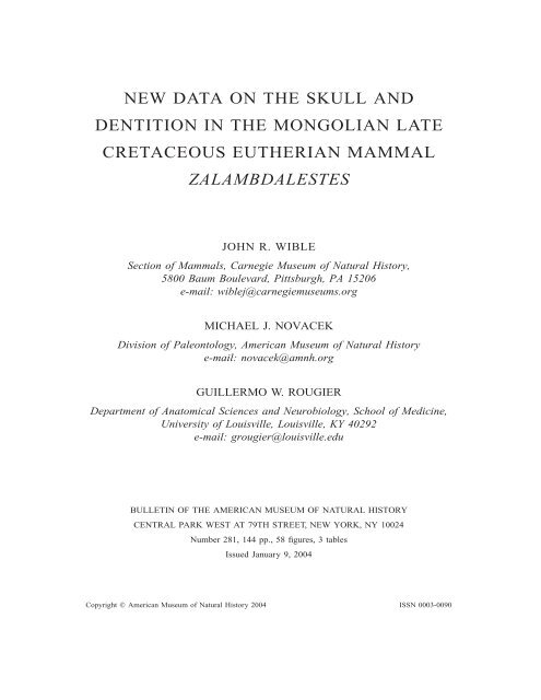
Fig. 7. The rostrum <strong>of</strong> Zalambdalestes lechei PSS-MAE 130 in ventral view. Pattern represents<br />
matrix. Abbreviations: art, artifactual opening; C, upper canine; cr, crest; I1a, upper first incisor alveolus;<br />
I2a, upper second incisor alveolus; I3, upper third incisor; I3a, upper third incisor alveolus; inf,<br />
incisive foramen; mx, maxilla; pmx, premaxilla; rp, rostral process <strong>of</strong> premaxilla.<br />
1971) in identifying the double-rooted, trenchant<br />
tooth in the maxilla as the canine. This<br />
identification is supported by the close positional<br />
relationship <strong>of</strong> this tooth with the<br />
maxillary incisor in PSS-MAE 130. Among<br />
the MAE specimens, the canine is preserved<br />
on both sides <strong>of</strong> PSS-MAE 108 (figs. 11, 23,<br />
25), 130 (figs. 26, 29), and 131, and on the<br />
right side <strong>of</strong> PSS-MAE 129 (fig. 31).<br />
UPPER PREMOLARS: Following Simpson<br />
(1928a), there are three premolars (P2–4)<br />
just rostral to the molars in AMNH 21704,<br />
21708 (fig. 32), and 21709. In contrast, Kielan-Jaworowska<br />
(1969a) reported four premolars<br />
(P1–4) in ZPAL MgM-I/14 and 16<br />
(fig. 4B, D). Her descriptions (ibid.: 186–<br />
188) are repeated here with differences in the<br />
MAE specimens noted.<br />
‘‘P1 small, two-rooted, slightly compressed,<br />
asymmetrical, with a single cusp situated<br />
in the prolongation <strong>of</strong> anterior root. No<br />
cingulum. In lateral view, the tooth (excluding<br />
roots), has the appearance <strong>of</strong> a scalene<br />
triangle, with anterior crest directed down-
2004 WIBLE ET AL.: ZALAMBDALESTES<br />
23<br />
wards, posterior crest longer, directed diagonally.<br />
A very short diastema between P1<br />
and P2’’ (Kielan-Jaworowska, 1969a: 186).<br />
The P1 preserved on the left side <strong>of</strong> PSS-<br />
MAE 108 conforms to this pattern (figs. 11,<br />
23); the crown is missing from the right P1.<br />
The P1 is also preserved on the right side <strong>of</strong><br />
PSS-MAE 129, but is severely worn (fig.<br />
31). As in AMNH 21704, 21708 (fig. 32),<br />
and 21709, the P1 is wholly lacking in PSS-<br />
MAE 130 (fig. 8). This is also true for Barunlestes<br />
(fig. 51B; ZPAL MgM-I/77, Kielan-Jaworowska,<br />
1975a; ZPAL MgM-I/104,<br />
PIN 3142–701, Kielan-Jaworowska and Tr<strong>of</strong>imov,<br />
1980).<br />
P2 ‘‘is somewhat larger than P1, two-rooted,<br />
compressed laterally, with a prominent<br />
main cusp and a minute anterior basal cusp.<br />
A lingual cingulum extends from the anterior<br />
cusp to a point opposite the middle <strong>of</strong> the<br />
main cusp, and is absent from the posterior<br />
half <strong>of</strong> the tooth’’ (Kielan-Jaworowska,<br />
1969a: 186). The P2 is preserved in PSS-<br />
MAE 108 (figs. 11, 23), 129 (fig. 31), and<br />
130 (figs. 8, 9), but it is only in the last that<br />
the morphology <strong>of</strong> this tooth can be checked.<br />
As preserved on both sides, the P2 conforms<br />
to the pattern described by Kielan-Jaworowska<br />
(1969a) with one exception: the lingual<br />
cingulum is not present in the anterior<br />
part <strong>of</strong> the tooth but near the midpoint, just<br />
posterior to the level <strong>of</strong> the main cusp. As<br />
apparent on the left side <strong>of</strong> PSS-MAE 108<br />
(figs. 11, 23), the P2 is somewhat larger (longer<br />
and wider) than the P1, also noted by<br />
Kielan-Jaworowska (1969a). However, the<br />
P1 projects farther ventrally than the P2 (fig.<br />
11); the reconstruction by Kielan-Jaworowska<br />
(1969a: fig. 4B) shows the reverse (fig.<br />
4B).<br />
‘‘P3 is subtriangular in occlusal view, with<br />
a large main cusp (paracone) situated close<br />
to the external border and a spur-like protocone.<br />
On the posterior crest <strong>of</strong> the paracone<br />
V (in lateral view), there is a very small cuspule,<br />
which is identified here as an incipient<br />
metacone. At the posterior end <strong>of</strong> this crest,<br />
in the prolongation <strong>of</strong> the paracone and metacone<br />
cusps, there is a large, prominent metastyle.<br />
The stylar shelf is not actually developed,<br />
however, one can recognize in the posterior<br />
part <strong>of</strong> the tooth, in addition to the metastyle,<br />
two minute styles hardly discernible.<br />
In front <strong>of</strong> these minute styles, the outer part<br />
<strong>of</strong> the tooth is smooth; in the most anterior<br />
part a parastyle is present. The spur-like protocone<br />
is asymmetrical, directed obliquely<br />
antero-medially, much lower than the paracone’’<br />
(Kielan-Jawrowska, 1969a: 186–188).<br />
The P3 preserved on the left side <strong>of</strong> PSS-<br />
MAE 108 (figs. 11, 23) and on the right side<br />
<strong>of</strong> PSS-MAE 129 (fig. 31) are heavily worn<br />
and only visible in lateral view. In PSS-MAE<br />
130 (figs. 8, 9), the right and left P3 are well<br />
preserved, with only slight wear to the crest<br />
running from the paracone to the metastylar<br />
area, and by and large conform to the above<br />
pattern. One difference concerns the crest between<br />
the paracone and metastyle. Although<br />
only one cuspule in this crest (the incipient<br />
metacone) is mentioned in the description by<br />
Kielan-Jaworowska, two cuspules <strong>of</strong> unequal<br />
size are shown in her reconstructions (1969a:<br />
fig. 4A, B). The crest between the paracone<br />
and metastyle in PSS-MAE 130 has a distinct<br />
incipient metacone and two small, more labial<br />
cuspules (fig. 9): the lower one in the<br />
metastylar position (stylar cusp E) and the<br />
other in the intersection <strong>of</strong> the postmetacrista<br />
and postprotocrista. The two minute styles<br />
anterior to stylar cusp E described by Kielan-<br />
Jaworwoska (1969a) are more pronounced in<br />
PSS-MAE 130, in particular the more anterior<br />
one, which is situated immediately posterior<br />
to the ect<strong>of</strong>lexus. The parastyle (stylar<br />
cusp A) is also more pronounced in PSS-<br />
MAE 130, and posterior to it is a very weak<br />
style (stylar cusp B). Finally, there is a low,<br />
but distinct paraconule in PSS-MAE 130,<br />
particularly on the left side, without wings.<br />
Most <strong>of</strong> these differences are probably the<br />
result <strong>of</strong> additional wear on the ZPAL specimens.<br />
‘‘P4 is similar in structure to P3, but has<br />
a protocone developed as in true molars. In<br />
lateral view, it is smaller than P3. On the<br />
posterior crest <strong>of</strong> paracone V, there is a small<br />
cuspule identified as an incipient metacone,<br />
similar to that in P3, a prominent metastyle<br />
being situated in its posterior prolongation.<br />
The stylar shelf is developed although narrower<br />
in the middle than in the molars. In<br />
addition to the prominent parastyle and metastyle,<br />
there are three hardly discernible, minute<br />
styles, two posterior ones situated close<br />
to the metastyle, anterior—close to the par-
24 BULLETIN AMERICAN MUSEUM OF NATURAL HISTORY NO. 281<br />
Fig. 8. Stereophotographs <strong>of</strong> the upper dentition <strong>of</strong> Zalambdalestes lechei PSS-MAE 130 in ventral<br />
(A) and left lateral (B) views.<br />
astyle. The protocone is situated lower than<br />
the paracone, directed transversely, with a<br />
slight anterior deviation. The precingulum<br />
and postcingulum are lacking. The surface<br />
between the protocone and paracone [trigon<br />
basin] is hollowed out by wear, retaining a<br />
very characteristic transverse ridge in the<br />
middle, absent from other teeth’’ (Kielan-Jaworowska,<br />
1969a: 188). As was true <strong>of</strong> the<br />
P3, the P4s in PSS-MAE 108 (figs. 11, 23)<br />
and 129 (fig. 31) are heavily worn and provide<br />
few morphological details. In PSS-MAE<br />
130 (figs. 8, 9, 10A, C), both right and left<br />
P4 are well preserved, with only slight wear<br />
to the crest between the paracone and incipient<br />
metacone, and in most regards follow<br />
the description <strong>of</strong> Kielan-Jaworowska<br />
(1969a). However, as reported above for the<br />
P3, the crest connecting the paracone and<br />
metastyle in PSS-MAE 130 has an incipient<br />
metacone and two cuspules in the metastylar<br />
region (fig. 10A, C): in the E position (i.e.,<br />
the metastyle) and lingual to it. The three<br />
styles between the parastyle and metastyle<br />
that Kielan-Jaworowska (1969a) described as<br />
‘‘hardly discernible’’ are slightly more pronounced<br />
in PSS-MAE 130 (fig. 10A, C). The<br />
anterior one (stylar cusp B) is separated from<br />
the parastyle by a notch; the posterior two<br />
are immediately posterior to the ect<strong>of</strong>lexus.<br />
Of these three, the middle (stylar cusp D?) is<br />
the most distinct, although not as prominent
2004 WIBLE ET AL.: ZALAMBDALESTES<br />
25<br />
Fig. 8.<br />
Continued.<br />
as the parastyle. Finally, on the pre- and postprotocristae<br />
are a low paraconule, with preand<br />
postparaconule cristae, and a worn metaconule<br />
without wings (fig. 10A, C). Presumably<br />
additional wear to these structures produced<br />
the transverse ridge in the trigon observed<br />
by Kielan-Jaworowska (1969a).<br />
UPPER MOLARS: The molars in the CAE<br />
specimens described by Gregory and Simpson<br />
(1926a) and Simpson (1928a) are heavily<br />
worn and provide only general clues to their<br />
morphology. In contrast, the ZPAL specimens<br />
show morphological detail (Kielan-Jaworowska,<br />
1969a) from which Crompton<br />
and Kielan-Jaworowska (1978) were able to<br />
reconstruct the pattern <strong>of</strong> molar occlusion<br />
during mastication. The best preserved, least<br />
worn molar series belong to ZPAL MgM-I/<br />
14, 16, and 43 (Crompton and Kielan-Jaworowska,<br />
1978: figs. 9, 11; Kielan-Jaworowska,<br />
1984a: pl. 14); the M2 from the last<br />
is shown diagrammatically in figure 5. The<br />
descriptions by Kielan-Jaworowska (1969a:<br />
188–189) based on ZPAL MgM-I/14 and 16<br />
are repeated here, with differences noted for<br />
PSS-MAE 130, the only MAE specimen<br />
with well-preserved molars (figs. 8, 9).<br />
‘‘M1 and M2 are similarly developed and<br />
are described here together, M1 being somewhat<br />
wider transversely than M2 . . . The<br />
paracone and metacone are situated strongly<br />
labially, the paracone higher than the meta-
26 BULLETIN AMERICAN MUSEUM OF NATURAL HISTORY NO. 281<br />
Fig. 9.<br />
The left P2–M3 <strong>of</strong> Zalambdalestes lechei PSS-MAE 130 in labial and occlusal views.<br />
cone. The parastyle and metastyle are prominent,<br />
the stylar shelf well developed (particularly<br />
at the ends). On the stylar shelf, in<br />
addition to the parastyle and metastyle, there<br />
are two or three minute accessory styles,<br />
hardly discernible, the mesostyle not being<br />
developed. The protocone is situated lower<br />
than the paracone and metacone. Incipient<br />
conules are very indistinct. Precingulum and<br />
postcingulum are lacking. The surface between<br />
the protocone and paracone-metacone<br />
W is strongly concave, hollowed out by<br />
wear’’ (Kielan-Jaworowska, 1969a: 188–<br />
189). Additional details based on ZPAL<br />
MgM-I/43 (fig. 5) are shown in the diagrams<br />
<strong>of</strong> M2 in Crompton and Kielan-Jaworowska<br />
(1978: fig. 9) and in the stereophotographs<br />
in Kielan-Jaworowska (1984a: pl. 14). Specifically,<br />
a stylocone (stylar cusp B) is figured,<br />
as are winged paraconule and metaconule<br />
approximating the paracone and metacone<br />
(see also Butler, 1990). Clemens and<br />
Lillegraven (1986) described the conules in<br />
Zalambdalestes as weak. In PSS-MAE 130<br />
(figs. 9, 10B, D), a weak stylocone (stylar<br />
cusp B) is present on the M1 but is imperceptible<br />
on the stylar shelf <strong>of</strong> M2. Also, in<br />
the metastylar region <strong>of</strong> the M2, the stylar<br />
shelf is reduced, with no cusps and almost<br />
no ridge. Wear to the M1 and M2 has eroded<br />
the conules. As noted by Kielan-Jaworowska<br />
(1969a), the paracone is higher than the<br />
metacone, but the size disparity is slight. As<br />
preserved, the protocone is somewhat lower<br />
than the paracone and metacone, but this also<br />
may be the result <strong>of</strong> wear. Additional features
2004 WIBLE ET AL.: ZALAMBDALESTES<br />
27<br />
Fig. 10. The left P4 and M1 <strong>of</strong> Zalambdalestes lechei PSS-MAE 130 in labial (A, B) and occlusal<br />
(C, D) views. Abbreviations: D, stylar cusp D; efl, ect<strong>of</strong>lexus; mes, mesostyle (stylar cusp E); met,<br />
metacone; metl, metaconule; par, paracone; parl, paraconule; pas, parastyle (stylar cusp A); pmcc,<br />
postmetacrista cusp; popc, postprotocrista; pro, protocone; prpc, preprotocrista; st, stylocone (stylar<br />
cusp B).
28 BULLETIN AMERICAN MUSEUM OF NATURAL HISTORY NO. 281<br />
coded by Rougier et al. (1998) include paracone<br />
and metacone bases that are separate; a<br />
preprotocrista that does not extend labially<br />
beyond the base <strong>of</strong> the paracone; a postprotocrista<br />
that does extend labially beyond the<br />
base <strong>of</strong> the metacone; a centrocrista between<br />
the paracone and metacone that is V-shaped<br />
(though not to the same extent as in some<br />
metatherians); and an ect<strong>of</strong>lexus that is very<br />
shallow, although deeper on the M1 than M2.<br />
‘‘M3 is a small tooth. The metacone is distinctly<br />
lower than the paracone. The parastyle<br />
is distinct, the stylar shelf is present only<br />
in the anterior half <strong>of</strong> the tooth, while the<br />
metastyle is lacking. The protocone is situated<br />
lower than the paracone-metacone. On<br />
the postprotocrista, near the metacone base,<br />
is a curvature outwards. The surface <strong>of</strong> the<br />
crown is hollowed out by wear, the conules<br />
not discernible’’ (Kielan-Jaworowska,<br />
1969a: 189). The M3 in PSS-MAE 130 has<br />
distinct winged conules, with the metaconule<br />
clearly better developed than the paraconule,<br />
possibly an artifact <strong>of</strong> wear (figs. 8A, 9). In<br />
fact, the ‘‘curvature outwards’’ observed by<br />
Kielan-Jaworowska (1969a) is the postmetaconule<br />
crista. The conules are proportionally<br />
closer to the paracone and metacone than<br />
in the M1 and M2 (see Crompton and Kielan-Jaworowska,<br />
1978: fig. 9), which may be<br />
a consequence <strong>of</strong> the smaller crown size <strong>of</strong><br />
the M3.<br />
LOWER INCISORS AND CANINE: Zalambdalestes<br />
preserves three lower incisors, which<br />
Gregory and Simpson (1926a) and Kielan-<br />
Jaworowska (1969a, 1984a; Kielan-Jaworowska<br />
and Tr<strong>of</strong>imov, 1981) designated as i1,<br />
i2, and i3. If the primitive eutherian condition<br />
is four lower incisors (Rougier et al.,<br />
1998; Ji et al., 2002), it is uncertain which<br />
tooth positions are represented by the three<br />
lower incisors in Zalambdalestes. Behind its<br />
enlarged, procumbent, anterior incisor, Kulbeckia<br />
has three single-rooted alveoli (fig.<br />
51C), which Archibald and Averianov<br />
(2003) identify as i2–4. These authors noted<br />
that the position <strong>of</strong> the i3 and i4 in Kulbeckia<br />
resembles the i2 and i3 in Zalambdalestes<br />
and, therefore, suggested that Zalambdalestes<br />
does not preserve or has lost the i2. Because<br />
these authors expressed some uncertainty<br />
about the existence <strong>of</strong> the i2 alveolus<br />
in Kulbeckia, we choose not to follow their<br />
numeration for Zalambdalestes. We follow<br />
all previous authors (e.g., Gregory and Simpson,<br />
1926a; Kielan-Jaworowska, 1969a,<br />
1984a) in identifying the three incisors in Zalambdalestes<br />
as i1–3, but note the possibility<br />
that our i2 and 3 might be equivalent to the<br />
i3 and 4 <strong>of</strong> Kulbeckia.<br />
One <strong>of</strong> the most distinctive features <strong>of</strong> the<br />
dentition <strong>of</strong> Zalambdalestes is the enlarged,<br />
procumbent, anterior lower incisor, which we<br />
label as i1. It was described first by Gregory<br />
and Simpson (1926a) based chiefly on<br />
AMNH 21704, which preserves the bulk <strong>of</strong><br />
the tooth, minus the tip. However, because <strong>of</strong><br />
poor specimen preservation, the description<br />
by Gregory and Simpson (1926a: 17) was<br />
very limited: ‘‘much enlarged, procumbent,<br />
and occludes between the enlarged upper incisors’’<br />
[the I1 <strong>of</strong> our report]. The three lower<br />
jaws described by Kielan-Jaworowska<br />
(1969a: ZPAL MgM-I/4 and 14) and Kielan-<br />
Jaworowska and Tr<strong>of</strong>imov (1981: PIN 3143–<br />
501) did not provide additional details, as<br />
only the i1 alveolus and/or root were preserved.<br />
Two specimens (ZPAL MgM-I/43<br />
and 77) preserving part <strong>of</strong> the i1 crown have<br />
been figured by Kielan-Jaworowska et al.<br />
(1979: fig. 12–2A) and Kielan-Jaworowska<br />
(1984a: pl. 14, 15, 17), but not described.<br />
Recently, Fostowicz-Frelik and Kielan-Jaworowska<br />
(2002) have published more details<br />
from two specimens <strong>of</strong> Z. lechei, ZPAL<br />
MgM-I/43 and 51. In both, damage to the<br />
inferior border <strong>of</strong> the mandible revealed that<br />
the root <strong>of</strong> the lower incisor was present, and<br />
open below the p4 and anterior part <strong>of</strong> m1.<br />
Two <strong>of</strong> the MAE specimens are the first<br />
Zalambdalestes to preserve essentially the<br />
entire i1. In Z. lechei PSS-MAE 108, in<br />
which the jaws are in articulation, the left i1<br />
is nearly complete (figs. 11, 23). The tip<br />
shows some breakage and most <strong>of</strong> the enamel<br />
is missing (eroded). The tooth is 7.0 mm<br />
in length from alveolus to tip and extends<br />
almost to the rostral tip <strong>of</strong> the premaxilla,<br />
passing just medial to the I1. Details <strong>of</strong> the<br />
crown morphology are obscured by matrix<br />
left in place to buttress the tooth. The right<br />
i1 is broken <strong>of</strong>f at the alveolus, revealing an<br />
oval root. Additionally, bone is missing<br />
along the inferior margin <strong>of</strong> the right mandible,<br />
revealing that the root extends posteriorly<br />
at least below the front edge <strong>of</strong> the p4.
2004 WIBLE ET AL.: ZALAMBDALESTES<br />
29<br />
Fig. 11. Stereophotographs <strong>of</strong> the anterior part <strong>of</strong> the skull and lower jaws <strong>of</strong> Zalambdalestes lechei<br />
PSS-MAE 108 in ventral (A) and lateral (B) views.
30 BULLETIN AMERICAN MUSEUM OF NATURAL HISTORY NO. 281<br />
In cf. Zalambdalestes sp. PSS-MAE 131, in<br />
which the jaws are in articulation but the premaxillae<br />
are displaced, both the right and left<br />
i1 are retained (see Novacek et al., 1997: fig.<br />
3). The teeth are oval in cross section near<br />
the alveolus but subtriangular approaching<br />
the tip, which shows little or no wear. Additionally,<br />
the teeth have enamel restricted,<br />
in cross section, to the buccal half from the<br />
alveolus to the tip. The left i1 preserved in<br />
cf. Zalambdalestes sp. PSS-MAE 145 conforms<br />
to the same pattern, except that the tip<br />
is missing. Incisors with restricted enamel<br />
occur in a variety <strong>of</strong> extinct and extant mammals,<br />
including various multituberculates<br />
(Simmons, 1993), rodents, and lagomorphs<br />
(Koenigswald, 1985, 1988; Thenius, 1989).<br />
The enlongated, procumbent i1 is damaged<br />
in all specimens <strong>of</strong> Barunlestes (fig. 51B;<br />
Kielan-Jaworowska, 1975a; Fostowicz-Frelik<br />
and Kielan-Jaworowska, 2002), and its root<br />
is open and reaches posteriorly under the<br />
posterior part <strong>of</strong> m1 (Fostowicz-Frelik and<br />
Kielan-Jaworowska, 2002). As in PSS-MAE<br />
131, according to McKenna (1994: 57), the<br />
i1 <strong>of</strong> Barunlestes has ‘‘enamel restricted to<br />
an outer U-shaped band.’’ It is unclear what<br />
specimen <strong>of</strong> Barunlestes McKenna used for<br />
his observation, and it may even be that <strong>of</strong><br />
a new genus, related to eurymylids (Fostowicz-Frelik<br />
and Kielan-Jaworowska, 2002). In<br />
Kulbeckia, the open root <strong>of</strong> the enlongated,<br />
procumbent i1 extends below p2, and the<br />
enamel <strong>of</strong> this tooth is thickened on all but<br />
the dorsolingual surface, where the enamel is<br />
either very thin or absent (Archibald et al.,<br />
2001; Archibald and Averianov, 2003).<br />
These authors noted that the incidence <strong>of</strong> an<br />
apical root opening may reflect the young<br />
age <strong>of</strong> the specimen or may indicate that the<br />
tooth was gliriform or ever-growing.<br />
In Zalambdalestes PSS-MAE 129 (fig.<br />
39), the exposed root <strong>of</strong> the i1 was investigated<br />
by Thomas Martin (Freie Universität,<br />
Berlin) in hopes <strong>of</strong> obtaining a sample <strong>of</strong><br />
enamel for analysis with scanning electron<br />
microscopy. He reported that enamel was<br />
lacking from the root, suggesting that the incisor<br />
is not ever-growing or gliriform, a condition<br />
that does occur in rodents, lagomorphs,<br />
vombatid marsupials, and the primate<br />
Daubentonia among others (Koenigswald,<br />
1985). Subsequently, however, we<br />
have modified our view regarding the absence<br />
<strong>of</strong> enamel based on cf. Zalambdalestes<br />
sp. PSS-MAE 145, in which the root <strong>of</strong> the<br />
left i1 is exposed posteriorly to the level <strong>of</strong><br />
the canine and reveals enamel continuous<br />
from the crown to the root. In Kulbeckia,<br />
enamel on the i1 is reported to extend to at<br />
least within 1 mm <strong>of</strong> the apical root opening<br />
(Archibald and Averianov, 2003).<br />
Behind the enlongated i1 in Zalambdalestes<br />
(AMNH 21704), Gregory and Simpson<br />
(1926a) found three small, styliform, singlerooted<br />
teeth that exhibit decreasing procumbency<br />
posteriorly. The identification by<br />
Gregory and Simpson <strong>of</strong> the first two teeth<br />
as the i2–3 and the third as the lower canine<br />
has been accepted by all subsequent authors<br />
(except Archibald and Averianov, 2003; see<br />
above). Confirming the incidence <strong>of</strong> three<br />
styliform teeth behind the i1 are four Zalambdalestes<br />
specimens housed in Warsaw<br />
(fig. 4B; ZPAL MgM-I/4, 13, 43, and 51:<br />
Kielan-Jaworowska, 1969a, 1984a) and one<br />
in Moscow (PIN 3143–501: Kielan-Jaworowska<br />
and Tr<strong>of</strong>imov, 1981). The only Barunlestes<br />
specimen preserving the anterior<br />
lower dentition is said to exhibit the same<br />
pattern (fig. 51B; ZPAL MgM-I/77: Kielan-<br />
Jaworowska, 1975a). Kulbeckia differs in<br />
that it likely had four lower incisors and its<br />
lower canine is either two-rooted or with a<br />
single bifurcated root (fig. 51C; Archibald<br />
and Averianov, 2003).<br />
Two <strong>of</strong> the MAE Zalambdalestes preserve<br />
details <strong>of</strong> the anterior lower dentition. On the<br />
right side <strong>of</strong> PSS-MAE 108, the i2–3 and c<br />
are broken near their alveoli, with the broken<br />
bases showing an increase in size posteriorly.<br />
The i2–3 are subcircular at their bases,<br />
whereas the canine is more ovoid. On the left<br />
side <strong>of</strong> the specimen (figs. 11, 23), the i2 and<br />
c are preserved. The former is procumbent<br />
and fingerlike with a slight increase in diameter<br />
toward the tip; the latter is semiprocumbent<br />
and widest at its ovoid base, tapering<br />
toward the tip. Separating the i2 and c is<br />
a wide gap, which contains the alveolus for<br />
the i3. On the left side <strong>of</strong> PSS-MAE 129, the<br />
broken root <strong>of</strong> the i1 and c and the alveoli<br />
<strong>of</strong> the i2–3 conform to the pattern described<br />
above for PSS-MAE 108.<br />
In PSS-MAE 108, the upper and lower<br />
jaws are articulated in a fashion that reflects
2004 WIBLE ET AL.: ZALAMBDALESTES<br />
31<br />
an occlusive position that was possible during<br />
life (figs. 11, 23). The anterolabial tip <strong>of</strong><br />
the enlarged i1 is in close proximity to the<br />
lingual surface <strong>of</strong> the I1, and these teeth likely<br />
contacted one another (figs. 11, 23). It is<br />
doubtful that any contact between the i1 and<br />
I2 was possible. The i2 and i3 are situated in<br />
the long diastema between the I2 and C and<br />
may not have contacted any upper teeth because<br />
that would have required a considerable<br />
protrusion <strong>of</strong> the lower jaw. The canines<br />
likely contacted one another, with the lower<br />
one situated just anterior to the upper (figs.<br />
11, 23).<br />
LOWER PREMOLARS: In their original description<br />
and diagnosis <strong>of</strong> Zalambdalestes<br />
lechei, Gregory and Simpson (1926a) reported<br />
three lower premolars, p2–4. The p2<br />
description was taken from AMNH 21704:<br />
‘‘p2 smaller than p3 and more procumbent.’’<br />
The description (ibid.: 17) <strong>of</strong> the p3–4 was<br />
based on AMNH 21707 and 21708 (figs. 12,<br />
13): p3 ‘‘trenchant, two-rooted, consisting <strong>of</strong><br />
a single cusp with a small unbasined heel’’;<br />
and p4 ‘‘molariform, similar to m1 but higher<br />
and with a much shorter talonid and relatively<br />
elongated trigonid.’’ Although not described,<br />
the drawings <strong>of</strong> AMNH 21704 and<br />
21708 (ibid.: figs. 15, 17, 18) show a diastema<br />
between the p2 and p3 (see fig. 12). Our<br />
reexamination <strong>of</strong> these specimens in light <strong>of</strong><br />
ZPAL, PIN, and MAE materials reveals that<br />
the tooth originally designated by Gregory<br />
and Simpson (1926a) as the p2 is actually<br />
the p1 and that the p2 is missing. Kielan-<br />
Jaworowska (1984a: pl. 15) illustrated a right<br />
lower jaw <strong>of</strong> Z. lechei (ZPAL MgM-I/43)<br />
that apparently exhibits the same pattern (i.e.,<br />
a diastema between the p1 and p3, and no<br />
alveolus for the p2); the left lower jaw <strong>of</strong> the<br />
specimen has the p2 in place.<br />
Kielan-Jaworowska (1969a) described the<br />
dentition in three Zalambdalestes specimens<br />
(ZPAL MgM-I/4, 14, and 16), each <strong>of</strong> which<br />
had four double-rooted lower premolars (p1–<br />
4) (fig. 4B). However, the discrepancy in the<br />
premolar count with the specimens described<br />
by Gregory and Simpson (1926a) was not<br />
noted. The descriptions <strong>of</strong> the premolars by<br />
Kielan-Jaworowska (ibid.: 189) are repeated<br />
here; the MAE specimens do not reveal any<br />
additional details about crown morphology:<br />
‘‘Between c and p1 there is a short diastema<br />
. . . p1 is trenchant, very slightly procumbent,<br />
with a prominent main cusp and a minute<br />
accessory cusp situated posteriorly. p2<br />
is situated after a very short diastema behind<br />
p1. It is very small, insignificantly procumbent,<br />
with anterior main cusp and a minute<br />
posterior one. p3 is larger than p1 and p2,<br />
consisting <strong>of</strong> a main cusp, with a minute accessory<br />
cusp in front <strong>of</strong> it, and small, unbasined<br />
heel with one low cusp (entoconid).<br />
The entoconid is situated lingually, the labial<br />
part <strong>of</strong> the heel sloping steeply downwards.<br />
p4 is submolariform, highest <strong>of</strong> all the teeth,<br />
with three-cusped trigonid and an unbasined<br />
talonid. The protoconid is highest, somewhat<br />
connate (in posterior view) at the base with<br />
the lower metaconid. The paraconid is much<br />
lower and smaller than the protoconid and<br />
metaconid, situated at the anterior edge <strong>of</strong><br />
the tooth. The talonid is much shorter than<br />
that <strong>of</strong> m1, with a single cusp situated lingually<br />
and the labial part steeply sloping<br />
downwards.’’ In subsequent reports (Kielan-<br />
Jaworowska and Tr<strong>of</strong>imov, 1981; Kielan-Jaworowska,<br />
1984a), four more Zalambdalestes<br />
preserving four lower premolars were illustrated<br />
(ZPAL MgM-I/13, 43, and 51; PIN<br />
3143–501). The only exception is the right<br />
side <strong>of</strong> ZPAL MgM-I/43 in which the p2 is<br />
missing (see above). In addition, Kielan-Jaworowska<br />
and Tr<strong>of</strong>imov (1981) suggested<br />
that the p4 in ZPAL MgM-I/14, a very young<br />
individual, is a deciduous tooth, because it<br />
has an anterior basal cusp, which is lacking<br />
in all other specimens.<br />
Two MAE Zalambdalestes provide information<br />
regarding the lower premolars. PSS-<br />
MAE 108 (figs. 11B, 23) repeats the pattern<br />
<strong>of</strong> four premolars in the Warsaw and Moscow<br />
specimens (with the exception <strong>of</strong> the<br />
right side <strong>of</strong> ZPAL MgM-I/43). In PSS-MAE<br />
129 (fig. 31), the right lower jaw has only<br />
three teeth preserved anterior to the m1; the<br />
left lower jaw is severely damaged. The posterior<br />
two teeth, the p3–4, are separated from<br />
the anterior one by a very short gap. We interpret<br />
the anterior tooth as the p1; it is too<br />
large to be the p2, which in all other Zalambdalestes<br />
is considerably smaller than the<br />
p1. Consequently, PSS-MAE 129 resembles<br />
AMNH 21704 and 21708 and the right side<br />
<strong>of</strong> ZPAL MgM-I/43 in lacking the p2. This<br />
is also true for Barunlestes (fig. 51B; ZPAL
32 BULLETIN AMERICAN MUSEUM OF NATURAL HISTORY NO. 281<br />
MgM-I/77, Kielan-Jaworowska, 1975a;<br />
ZPAL MgM-I/90, 107, and 135, PIN 3142–<br />
701, Kielan-Jaworowska and Tr<strong>of</strong>imov,<br />
1980). Kielan-Jaworowska and Tr<strong>of</strong>imov<br />
(1980: 168–169) mistakenly identified alveoli<br />
for the p2 in ZPAL MgM-I/90 and 135<br />
(Kielan-Jaworowska, personal commun.).<br />
LOWER MOLARS: The three MAE Zalambdalestes<br />
specimens with lower jaws do not<br />
provide details on lower molar crown morphology<br />
because the jaws are in occlusion<br />
(figs. 11B, 30, 31). The most detailed description<br />
<strong>of</strong> the lower molars <strong>of</strong> Zalambdalestes<br />
is that <strong>of</strong> ZPAL MgM-I/14 by Kielan-<br />
Jaworowska (1969a: 189), which is repeated<br />
here (fig. 5): ‘‘m1 and m2 are similarly<br />
shaped. m2 is lower and smaller than m1.<br />
The trigonid in m1 is smaller than in p4, the<br />
talonid larger. The paraconid and metaconid<br />
are connate at their bases, the former being<br />
distinctly smaller and lower than the latter.<br />
The protoconid is somewhat higher than the<br />
metaconid. The talonid is strongly basined,<br />
with poorly defined cusps. One can recognize<br />
the higher entoconid and lower hypoconid<br />
which slightly projects laterally. (A low hypoconulid<br />
was reconstructed on the m2 by<br />
Crompton and Kielan-Jaworowska [1978]<br />
based on ZPAL MgM-I/43.) m3 is the smallest<br />
<strong>of</strong> the molars, narrower and shorter than<br />
m2, with very small trigonid. The paraconid<br />
is vestigial, strongly connate with the metaconid,<br />
the protoconid is lower than the metaconid.<br />
The talonid is comparatively large,<br />
strongly basined, with three cusps. The entoconid<br />
is the highest, the hypoconulid situated<br />
close to it and insignificantly lower, the<br />
hypoconid the lowest.’’ Based on Crompton<br />
and Kielan-Jaworowska (1978: fig. 10), we<br />
add here that the talonid is wider and longer<br />
than the trigonid on m1–3. The paraconid is<br />
lingually positioned, in alignment with the<br />
metaconid. The metacristid is transversely<br />
oriented, and the cristid obliqua is labially<br />
placed, at the base <strong>of</strong> the protoconid. The<br />
hypoconulid is in a posteromedial position,<br />
slightly nearer the entoconid than hypoconid.<br />
DENTAL FORMULA: As already noted, a variety<br />
<strong>of</strong> dental formulae have been reported<br />
for Zalambdalestes lechei. In our reconsideration<br />
<strong>of</strong> this topic, we have found the following<br />
formulae: I2–3/i3, C1/c1, P3–4/p3–<br />
4, M3/m3. Regarding the upper incisors, two<br />
are found in ZPAL MgM-I/16 and the left<br />
sides <strong>of</strong> PSS-MAE 108 (figs. 11, 23) and<br />
131; three are found in PSS-MAE 130 (figs.<br />
7, 8) and the right side <strong>of</strong> PSS-MAE 108.<br />
Regarding the premolars, four are preserved<br />
in both the upper and lower jaws in ZPAL<br />
MgM-I/13, 14, and 16 and PSS-MAE 108<br />
(figs. 11B, 23), and three are preserved (P2–<br />
4 and p1, 3–4) in both upper and lower jaws<br />
in AMNH 21704 and 21708 (figs. 12, 32).<br />
The only specimen with an uneven count in<br />
the upper and lower jaws is PSS-MAE 129,<br />
which has P1–4 and p1, 3–4 (fig. 31). We<br />
have no evidence that any <strong>of</strong> the preserved<br />
teeth in any Zalambdalestes are retained deciduous<br />
teeth, other than perhaps the p4 in<br />
ZPAL MgM-I/14, as suggested by Kielan-Jaworowska<br />
and Tr<strong>of</strong>imov (1981). In fact, in<br />
PSS-MAE 130, the P3–4 show considerably<br />
less wear than the M1–3 (figs. 8, 9), suggesting<br />
that they are replacement teeth that<br />
erupted after the molars. It may be that the<br />
observed variation in upper incisor and upper<br />
and lower premolar number is representative<br />
<strong>of</strong> different taxa among the current sample.<br />
Other than tooth number, there are few dental<br />
and cranial differences that can be used to<br />
distinguish the available sample, especially<br />
when one accounts for differing states <strong>of</strong><br />
preservation and tooth wear. Before taxonomic<br />
revision can be addressed, the ZPAL<br />
and PIN specimens must be reevaluated in<br />
light <strong>of</strong> all the MAE material, which we have<br />
not yet done.<br />
The dental formula reported for Barunlestes<br />
butleri by Kielan-Jaworowska (1975a) is<br />
I?/i3, C1/c1, P3/p3, M3/m3 (fig. 51B). We<br />
identify the lower incisors in Barunlestes as<br />
i1–3, but note the possibility raised by Archibald<br />
and Averianov (2003) for Zalambdalestes<br />
that the distal two incisors in Barunlestes<br />
may be homologous with the i3 and<br />
i4 <strong>of</strong> Kulbeckia. Our dental formula for Zalambdalestes<br />
affects the diagnosis <strong>of</strong> Barunlestes,<br />
which preserves three upper premolars,<br />
identified as P2–4, and three lower premolars,<br />
identified as p1, 3–4. The absence <strong>of</strong><br />
the P1 and p2 was noted amongst the features<br />
distinguishing Barunlestes and Zalambdalestes<br />
by Kielan-Jaworowska (1975a; see<br />
also Kielan-Jaworowska et al., 1979, 2000;<br />
Kielan-Jaworowska and Tr<strong>of</strong>imov, 1980).<br />
However, the utility <strong>of</strong> these as diagnostic
2004 WIBLE ET AL.: ZALAMBDALESTES<br />
33<br />
Fig. 12.<br />
view.<br />
Stereophotographs <strong>of</strong> the lower jaws <strong>of</strong> Zalambdalestes lechei AMNH 21708 in occlusal<br />
features <strong>of</strong> Barunlestes is questioned by our<br />
report above <strong>of</strong> the absence <strong>of</strong> these teeth in<br />
some specimens <strong>of</strong> Zalambdalestes.<br />
The dental formula reported by Archibald<br />
et al. (2001) for Kulbeckia kulbecke (I3, 4,<br />
or 5/i4, C1/c1, P4/p4, M3/m3) (figs. 51C, 52)<br />
differs from that in Zalambdalestes with regard<br />
to the incisor and premolar count. There<br />
are four premolars in both the upper and lower<br />
jaws in all relevant preserved specimens<br />
<strong>of</strong> Kulbeckia, as occurs in some Zalambdalestes.<br />
Archibald and Averianov (2003) identified<br />
these as P1, 2, 4, 5 and p1, 2, 4, 5,<br />
based on the interpretation that position 3 is<br />
lost in early eutherians. As noted in the Materials<br />
and Methods, we chose to number the<br />
premolars as 1 through 4 in this report.<br />
Therefore, premolars 4 and 5 <strong>of</strong> Archibald<br />
and Averianov (2003) correspond to numbers<br />
3 and 4 <strong>of</strong> our report. The lower incisor<br />
count in Kulbeckia is four, whereas that <strong>of</strong><br />
Zalambdalestes is three. We question the<br />
number <strong>of</strong> upper incisors in Kulbeckia. The<br />
one specimen preserving evidence for the<br />
upper incisors, URBAC 99–53, has only<br />
three alveoli (fig. 52), but Archibald et al.<br />
(2001) and Archibald and Averianov (2003)<br />
speculated that as many as five incisors<br />
might have been present. We see no basis for<br />
any speculation beyond the evidence <strong>of</strong> the<br />
three upper incisor alveoli, which resembles<br />
some Zalambdalestes.<br />
LOWER JAW<br />
Given their poor preservation, the CAE<br />
specimens <strong>of</strong> Zalambdalestes lechei (figs.<br />
12–14) provided only a few general features<br />
<strong>of</strong> lower jaw anatomy for the descriptions by<br />
Gregory and Simpson (1926a). In contrast,<br />
the sample in Warsaw and Moscow available<br />
to Kielan-Jaworowska and Tr<strong>of</strong>imov (1981)
34 BULLETIN AMERICAN MUSEUM OF NATURAL HISTORY NO. 281<br />
included several specimens preserving considerable<br />
detail <strong>of</strong> the lower jaw, most notably<br />
the left lower jaw <strong>of</strong> PIN 3143–501.<br />
The angle was broken <strong>of</strong>f in this specimen,<br />
but ‘‘the preserved part indicates that it has<br />
been inflected medially’’ (ibid.: 4), also reported<br />
for Barunlestes by Kielan-Jaworowska<br />
(1975a).<br />
Four MAE specimens <strong>of</strong> Zalambdalestes<br />
preserve lower jaws, PSS-MAE 108, 129,<br />
135, and 145; in each instance, both right and<br />
left jaws are in articulation. The first two<br />
specimens show details <strong>of</strong> the external surface<br />
only, as the internal is obscured from<br />
preparation (figs. 11, 23, 31, 33, 39); little <strong>of</strong><br />
the jaw structure remains in PSS-MAE 135<br />
(figs. 30, 38); and until further preparation is<br />
completed, we do not consider the lower<br />
jaws <strong>of</strong> PSS-MAE 145. We reconstruct the<br />
lower jaw in lateral view (fig. 15), address<br />
the salient features <strong>of</strong> external lower jaw<br />
anatomy here, and, for the sake <strong>of</strong> completeness,<br />
repeat Kielan-Jaworowska and Tr<strong>of</strong>imov’s<br />
(1981) observations on the internal<br />
structure.<br />
The lower jaw in Zalambdalestes is elongate<br />
and anteriorly tapering, with two mental<br />
foramina <strong>of</strong> unequal size (not clearly preserved<br />
in any MAE specimen), the larger one<br />
below the middle <strong>of</strong> p3 and the other below<br />
the anterior root <strong>of</strong> p1, according to Kielan-<br />
Jaworowska and Tr<strong>of</strong>imov (1981). The most<br />
distinctive aspect <strong>of</strong> the anterior mandible is<br />
the large root for i1, which extends back to<br />
a point below at least the last premolar in<br />
PSS-MAE 108 and 129; as noted above, Fostowicz-Frelik<br />
and Kielan-Jaworowska (2002)<br />
reported that the root <strong>of</strong> the lower incisor is<br />
open and extends below the p4 and anterior<br />
part <strong>of</strong> m1. The symphysis extends to the<br />
front <strong>of</strong> p3 in PSS-MAE 129 (fig. 39); Kielan-Jaworowska<br />
and Tr<strong>of</strong>imov (1981) reported<br />
it reaching as far as the p2. The coronoid<br />
process is high and its apex is hooklike, with<br />
a strongly concave posterior edge on the<br />
right side <strong>of</strong> PSS-MAE 129 (fig. 31) and on<br />
the left side <strong>of</strong> PSS-MAE 108 (fig. 23). This<br />
pr<strong>of</strong>ile is not rendered in previously published<br />
figures <strong>of</strong> Zalambdalestes and Barunlestes<br />
(figs. 4B, 51B; Kielan-Jaworowska,<br />
1975a: fig. 2), and firsthand preliminary examination<br />
<strong>of</strong> the material in the Polish–Mongolian<br />
collections suggests the difference<br />
may either reflect variation in the sample or<br />
in some cases damage to this region <strong>of</strong> the<br />
specimen. The angle <strong>of</strong> the coronoid to the<br />
alveolar margin is approximately 60 in PSS-<br />
MAE 108 (fig. 23) and 55 on the right side<br />
<strong>of</strong> PSS-MAE 129 (fig. 31). A lower angle<br />
had been reported previously for Z. lechei:<br />
ca. 45 by Gregory and Simpson (1926a) and<br />
ca. 49 in adults and ca. 42 in juveniles by<br />
Kielan-Jaworowska and Tr<strong>of</strong>imov (1981).<br />
Among the CAE specimens, the angle is<br />
close to 45 in AMNH 21704, but in the holotype,<br />
21708, it is higher, approximately 54<br />
(fig. 13).<br />
As seen in PSS-MAE 108 (fig. 23) and<br />
129 (fig. 31), the ascending ramus <strong>of</strong> the<br />
mandible has a well-developed masseteric<br />
fossa that extends to the lower edge <strong>of</strong> the<br />
jaw. It is strongly demarcated anteriorly by a<br />
very prominent coronoid crest (masseteric<br />
crest <strong>of</strong> Kielan-Jaworowska and Tr<strong>of</strong>imov,<br />
1981), which becomes narrower as it nears<br />
the tip <strong>of</strong> the coronoid process. A distinct,<br />
but low masseteric line (crest) inferior to the<br />
fossa is preserved in PSS-MAE 129 (fig. 31);<br />
the corresponding area is worn in PSS-MAE<br />
108. There is no indication <strong>of</strong> a labial mandibular<br />
foramen at the rear <strong>of</strong> the base <strong>of</strong> the<br />
coronoid crest on the MAE or AMNH specimens,<br />
or on the high-resolution CT scans <strong>of</strong><br />
PSS-MAE 108. Such a foramen has been reported<br />
by Kielan-Jaworowska and Dashzeveg<br />
(1989) for Zalambdalestes lechei and Z.<br />
nymbulakensis, a junior synonym <strong>of</strong> Sorlestes<br />
budan according to Nessov et al. (1994).<br />
For Z. lechei, Kielan-Jaworowska and Dashzeveg<br />
cited a plate <strong>of</strong> photographs <strong>of</strong> PIN<br />
3143–501 in Kielan-Jaworowska and Tr<strong>of</strong>imov<br />
(1981: pl. 2). The lateral view <strong>of</strong> the<br />
lower jaw in their figure 2a shows cracks and<br />
a large artifactual opening in the masseteric<br />
fossa. In addition, there does appear to be a<br />
foramen behind the coronoid crest at the level<br />
<strong>of</strong> the m3 trigonid cusps. As noted by Kielan-Jaworowska<br />
and Dashzeveg (1989), the<br />
opening in PIN 3143–501 is situated higher<br />
and is larger than the labial mandibular foramen<br />
in Early Cretaceous Prokennalestes.<br />
As preserved in PSS-MAE 108 (fig. 23),<br />
a low condyloid crest running anteroventrally<br />
from the condyle toward the coronoid crest<br />
divides the masseteric fossa into subequal<br />
parts: a larger, deeper dorsal part and a small-
2004 WIBLE ET AL.: ZALAMBDALESTES<br />
35<br />
Fig. 13.<br />
view.<br />
Stereophotographs <strong>of</strong> the right lower jaw <strong>of</strong> Zalambdalestes lechei AMNH 21708 in lateral<br />
er, nearly flat ventral part (see also Kielan-<br />
Jaworowska and Tr<strong>of</strong>imov, 1981). The condyle<br />
in PSS-MAE 108 (fig. 23) and 129 (fig.<br />
31) is positioned notably high relative to the<br />
angle. The condyle is markedly broadened<br />
transversely, with an extensive convex surface<br />
dorsally and posteriorly for contact with<br />
the glenoid fossa. Both the medial and lateral<br />
margins <strong>of</strong> the articular surface in PSS-MAE<br />
108 are drawn into triangular, transversely<br />
oriented processes, the one on the medial<br />
side being much more delicate.<br />
The angular process is fully preserved on<br />
the left side <strong>of</strong> PSS-MAE 108 (figs. 23, 33).<br />
It is distinctively hooklike in lateral outline,<br />
similar to that figured for Zalambdalestes by<br />
Kielan-Jaworowska (fig. 4B; 1975a: fig. 2),<br />
although it is even longer. Viewed from behind<br />
or below (fig. 33), the angle shows no<br />
significant medial inflection (contra Kielan-<br />
Jaworowska and Tr<strong>of</strong>imov, 1981). A crack<br />
separates the angle on the right mandible <strong>of</strong><br />
AMNH 21708 from the ascending ramus<br />
(figs. 13, 14). The angle has been repositioned<br />
and glued following the contours, and<br />
it shows no medial inflection. The angular<br />
process in PIN 3143–501, the specimen <strong>of</strong><br />
Zalambdalestes described by Kielan-Jaworowska<br />
and Tr<strong>of</strong>imov (1981: pl. 1), is complete<br />
except for its tip, and the stereophotograph<br />
<strong>of</strong> the occlusal view appears to show<br />
a slight medial deviation to the posteroventrally<br />
directed process compared with PSS-<br />
MAE 108. This is much less than the more<br />
medially deviated posteroventral angular<br />
process in Barunlestes (Kielan-Jaworowska,<br />
1975a: pl. 1, fig. 1e) and Asioryctes (see Kielan-Jaworowska,<br />
1981: pl. 6). We think that<br />
most <strong>of</strong> the descriptions <strong>of</strong> medially inflected<br />
angles in Cretaceous eutherians are the result
36 BULLETIN AMERICAN MUSEUM OF NATURAL HISTORY NO. 281<br />
Fig. 14. Stereophotographs <strong>of</strong> the lower jaws <strong>of</strong> Zalambdalestes lechei AMNH 21708 in left oblique<br />
view to see the medial surface <strong>of</strong> the right ascending ramus.<br />
Fig. 15. Reconstruction <strong>of</strong> the right lower jaw <strong>of</strong> Zalambdalestes lechei in lateral view. Dental<br />
formula is i3, c, p4, m3, as occurs in PSS-MAE 108 and in ZPAL MgM-I/13, 14, and 16; p2 is lacking<br />
in PSS-MAE 129 and in AMNH 21704 and 21708. Mental foramina are reconstructed based on Kielan-<br />
Jaworowska and Tr<strong>of</strong>imov (1981). Abbreviations: an, (mandibular) angle; cc, condyloid crest; coc,<br />
coronoid crest; con, (mandibular) condyle; cor, coronoid process; maf, masseteric fossa; mf, mental<br />
foramina.
2004 WIBLE ET AL.: ZALAMBDALESTES<br />
37<br />
<strong>of</strong> definitional problems. We follow the terminology<br />
proposed by Sánchez-Villagra and<br />
Smith (1997) and agree with these authors<br />
that no Cretaceous eutherian has a medially<br />
inflected angle, a feature fairly widespread<br />
within Metatheria (Rougier et al., 1998).<br />
Lagomorphs and some rodents have an angle<br />
that is thickened on its medial surface or bent<br />
medially, but not to the same extent as in<br />
metatherians. The Cretaceous eutherian that<br />
comes closest to the metatherian form <strong>of</strong> the<br />
angle is Montanalestes from the Albian-Aptian<br />
<strong>of</strong> Montana (see Cifelli, 1999).<br />
Kielan-Jaworowska and Tr<strong>of</strong>imov (1981:<br />
4) described the following features <strong>of</strong> the internal<br />
anatomy <strong>of</strong> the lower jaw <strong>of</strong> PIN<br />
3143–501: ‘‘The symphyseal surface is<br />
markedly rugose and is bounded from above<br />
in the middle part by a ridge. Behind the<br />
symphysis the surface <strong>of</strong> the mandibular ramus<br />
is slightly swollen below p3 and anterior<br />
root <strong>of</strong> m1. On the lower part <strong>of</strong> the ascending<br />
ramus <strong>of</strong> the coronoid process there is a<br />
distinct, oval, rugose swelling, 1.8 mm long<br />
and 1.2 mm wide, possibly a remnant <strong>of</strong> the<br />
coronoid bone, fused with the dentary. Behind<br />
and below this swelling there is a ridge<br />
(medial flange <strong>of</strong> Kermack et al., 1973),<br />
which extends posteriorly and reaches the<br />
condyle. The mandibular foramen is large,<br />
situated ca. 5.2 mm behind the last molar.<br />
Extending posteriorly from the mandibular<br />
foramen there is a very distinct tear-dropshaped<br />
fossa, pointed backwards and upwards.<br />
The fossa is delimited from above by<br />
the medial flange. At the anterior part <strong>of</strong> the<br />
fossa there is a faint groove which extends<br />
posteriorly, parallel to the medial flange and<br />
disappears in the middle length <strong>of</strong> the fossa.’’<br />
Regarding the possible occurrence <strong>of</strong> the<br />
coronoid bone suggested by Kielan-Jaworowska<br />
and Tr<strong>of</strong>imov (1981), the type specimen<br />
AMNH 21708 (fig. 14), although<br />
cracked in this area, as well as the CT scans<br />
<strong>of</strong> PSS-MAE 108 show no such swelling like<br />
that described in PIN 3143–501, and elsewhere<br />
(Rougier et al., 1998) we have scored<br />
this element as absent for Zalambdalestes.<br />
We do not know the meaning <strong>of</strong> the swelling<br />
described by Kielan-Jaworwoska and Tr<strong>of</strong>imov<br />
(1981). It is positioned higher up the<br />
coronoid process than the obvious coronoid<br />
facet present in Vincelestes (Rougier, 1993)<br />
or the rugosity also <strong>of</strong> uncertain meaning<br />
present in Prokennalestes (Kielan-Jaworowska<br />
and Dashzeveg, 1989).<br />
SKULL<br />
Skulls are known for two <strong>of</strong> the CAE<br />
specimens <strong>of</strong> Zalambdalestes lechei (AMNH<br />
21704 and 21708). However, with the exception<br />
<strong>of</strong> the snout and palate in AMNH 21708<br />
(fig. 1), there are few details <strong>of</strong> cranial anatomy<br />
that are recoverable from either specimen.<br />
Not surprisingly, Gregory and Simpson’s<br />
(1926a) observations on the skull <strong>of</strong> Z.<br />
lechei were limited and general. They provided<br />
a restoration <strong>of</strong> the skull in lateral view<br />
(ibid.: fig. 16), based largely on the holotype,<br />
which delimits only the premaxillary bone,<br />
and illustrated few features beyond the infraorbital<br />
foramen.<br />
Van Valen (1964: fig. 2) presented a new<br />
restoration <strong>of</strong> the skull <strong>of</strong> Zalambdalestes in<br />
lateral and palatal views said to be ‘‘drawn<br />
and slightly reconstructed by S. B. McDowell’’<br />
redrawn here as figure 4A and C. Although<br />
based on the CAE specimens, this<br />
restoration provided more sutural details than<br />
those by Gregory and Simpson. Delimited<br />
were the bones on the palate and in the orbitotemporal<br />
region, but there were no new<br />
accompanying descriptions. Our study <strong>of</strong> the<br />
CAE specimens confirms the sutural details<br />
<strong>of</strong> four bones (premaxilla, maxilla, lacrimal,<br />
and squamosal) in the reconstruction by Van<br />
Valen; the remainder is conjectural and does<br />
not agree with the morphology <strong>of</strong> the MAE<br />
specimens.<br />
A detailed description <strong>of</strong> the skull <strong>of</strong> Zalambdalestes<br />
lechei based largely on ZPAL<br />
MgM-I/14 and 16 was published in Kielan-<br />
Jaworowska (1984a). Most <strong>of</strong> the external<br />
surfaces <strong>of</strong> the skull were described, with the<br />
exception <strong>of</strong> the basicranium, which was not<br />
preserved in any specimens in the Warsaw or<br />
Moscow collections. Kielan-Jaworowska included<br />
a fairly detailed reconstruction <strong>of</strong> the<br />
skull in ventral view (ibid.: fig. 1), with the<br />
basicranium based on that known in Barunlestes<br />
(Kielan-Jaworowska and Tr<strong>of</strong>imov,<br />
1980) redrawn here as figure 4D. The sutural<br />
relations depicted in her reconstruction are<br />
for the most part not supported by the morphology<br />
preserved in the MAE specimens. A
38 BULLETIN AMERICAN MUSEUM OF NATURAL HISTORY NO. 281<br />
Fig. 16. Reconstruction <strong>of</strong> the skull <strong>of</strong> Zalambdalestes lechei in (clockwise from upper left) dorsal,<br />
ventral, and right lateral views, illustrating the cranial measurements in table 3. Abbreviations: BBP,<br />
breadth <strong>of</strong> bony palate across M1; BR, breadth <strong>of</strong> rostrum; CH, height <strong>of</strong> ascending mandibular ramus<br />
at condylar process; IB, interorbital breadth; LR, length <strong>of</strong> rostrum; MD, depth <strong>of</strong> mandible at m1;<br />
ML, length <strong>of</strong> mandible; NB, nuchal breadth; ONL, occipitonasal length; PCL, premaxillary-condylar<br />
length; PL, palatal length; POL, preorbital length; PPL, postpalatal length; RH, height <strong>of</strong> ascending<br />
mandibular ramus at coronoid process; RL, length <strong>of</strong> ascending mandibular ramus behind m3; ZB,<br />
greatest zygomatic breadth.
2004 WIBLE ET AL.: ZALAMBDALESTES<br />
39<br />
more featureless reconstruction <strong>of</strong> the skull<br />
<strong>of</strong> Z. lechei in lateral view was published<br />
elsewhere (Kielan-Jaworowska, 1975a: fig.<br />
2A; Kielan-Jaworowska et al., 1979: fig. 12–<br />
1C) redrawn here as figure 4B. Finally, partial<br />
endocranial casts <strong>of</strong> ZPAL MgM-I/14<br />
and 16 were described in Kielan-Jaworowska<br />
(1984b).<br />
Based on the MAE specimens, we reconstruct<br />
the skull <strong>of</strong> Zalambdalestes lechei in<br />
dorsal, lateral (with and without the zygoma),<br />
and ventral view (figs. 42–44). The<br />
skull <strong>of</strong> Zalambdalestes is notable for its<br />
large size compared to the other described<br />
Mongolian Cretaceous eutherians (fig. 51;<br />
Kielan-Jaworowska, 1984a, 1984c; Novacek<br />
et al., 1997). Certain specimens <strong>of</strong> Z. lechei<br />
(e.g., PSS-MAE 108 and 130) range nearly<br />
to 50 mm in length from the anterior apex <strong>of</strong><br />
the premaxilla to the occipital condyles; cf.<br />
Zalambdalestes sp. PSS-MAE 145 has a<br />
skull length <strong>of</strong> more that 60 mm. Barunlestes<br />
is a close second in size, ranging between 35<br />
and 40 mm (Kielan-Jaworowska and Tr<strong>of</strong>imov,<br />
1980). Kulbeckia kulbecke, the most<br />
complete specimen <strong>of</strong> which preserves the<br />
snout and anterior orbit, is probably comparable<br />
in skull size to Barunlestes. The asioryctitheres<br />
Kennalestes, Asioryctes, and<br />
Ukhaatherium range between 26 and 30 mm<br />
(Kielan-Jaworowska, 1981; personal obs.).<br />
Daulestes nessovi from the Coniacian <strong>of</strong> Uzbekistan<br />
is estimated to be just under 20 mm<br />
(McKenna et al., 2000). The zalambdalestid<br />
nasal-facial region in front <strong>of</strong> the cheekteeth<br />
is expanded into a long, narrow snout, more<br />
so in Zalambdalestes than in Barunlestes or<br />
Kulbeckia. The zygomatic arches, unknown<br />
in Barunlestes and Kulbeckia, are slender<br />
and strongly expanded laterally in Zalambdalestes,<br />
and the nuchal and sagittal crests<br />
are distinct in Zalambdalestes and Barunlestes.<br />
The braincase in Zalambdalestes and Barunlestes<br />
is more inflated and the mesocranial<br />
region shorter than in the asioryctitheres<br />
Kennalestes, Asioryctes (Kielan-Jaworowska,<br />
1984a, 1984c), and Ukhaatherium (personal<br />
obs.).<br />
NASAL-FACIAL REGION: The premaxilla in<br />
Zalambdalestes lechei is expanded, contributing<br />
more than one-third the length <strong>of</strong> the<br />
preorbital region in lateral view (fig. 44A).<br />
On the left side <strong>of</strong> PSS-MAE 130, the premaxilla<br />
is very well preserved, better than in<br />
any other specimen collected to date, and<br />
only the rostralmost tip is missing (figs. 7,<br />
8A). It shows that the premaxilla anterior to<br />
the I1 alveolus, which we refer to as the rostral<br />
process, is considerably longer than in<br />
the reconstructions by Gregory and Simpson<br />
(1926a: fig. 16), Van Valen (fig. 4A; 1964:<br />
fig. 2), and Kielan-Jaworowska (fig. 4B;<br />
1975a: fig. 2A). In lateral view (fig. 26), the<br />
rostral process lies in a more dorsal plane<br />
than the alveolar margin <strong>of</strong> the premaxilla<br />
and maxilla. It is also nearly horizontal,<br />
whereas the snout behind it is bent somewhat<br />
downward. The rostral process projects considerably<br />
farther forward than does the nasal<br />
bone (as interpreted from PSS-MAE 108 and<br />
AMNH 21708), producing a much more<br />
oblique external nasal aperture than in previous<br />
reconstructions (fig. 44A). The facial<br />
suture between the premaxilla and maxilla is<br />
oblique (figs. 22, 23, 26, 31). At the alveolar<br />
margin, it is positioned one-third the distance<br />
between the I2 and C, based on PSS-MAE<br />
108 (fig. 23) and AMNH 21708 (fig. 22).<br />
Dorsally, it contacts the nasal above the posterior<br />
canine root in AMNH 21708 (fig. 22)<br />
and PSS-MAE 129 (fig. 31) or anterior to the<br />
canine in PSS-MAE 108 (figs. 11B, 23) and<br />
130 (fig. 26). Some degree <strong>of</strong> premaxillary<br />
expansion is commonly associated with the<br />
development <strong>of</strong> large, long-rooted incisors<br />
and a wide diastema, but the geometric relationships<br />
<strong>of</strong> this bone with adjacent skull<br />
elements varies notably among mammalian<br />
groups. Although the premaxilla is extensively<br />
exposed in Zalambdalestes, it does not<br />
extend posteriorly to contact the frontals, as<br />
in rodents and lagomorphs (figs. 54, 55).<br />
Most <strong>of</strong> the nasal bone, including its sutural<br />
relationships, is well preserved in<br />
AMNH 21708 (fig. 17) and PSS-MAE 108<br />
(fig. 18), 129 (fig. 21), and 130 (fig. 19).<br />
Only one specimen, PSS-MAE 130, preserves<br />
details <strong>of</strong> the nasal’s anterior end (fig.<br />
19). The rostralmost projection <strong>of</strong> the nasal<br />
is just <strong>of</strong>f the midline. It extends slightly farther<br />
forward than does the premaxilla’s contribution<br />
to the lateral wall <strong>of</strong> the external<br />
nasal aperture (figs. 42, 44A), resulting in<br />
what Wible and Rougier (2000) termed the<br />
nasal overhang <strong>of</strong> the nares. The nasal does<br />
not extend nearly as far as does the rostral
40 BULLETIN AMERICAN MUSEUM OF NATURAL HISTORY NO. 281<br />
Fig. 17. Stereophotograph <strong>of</strong> the skull <strong>of</strong> Zalambdalestes lechei AMNH 21708 in dorsal view, with<br />
accompanying line drawing. Gray pattern represents matrix. Abbreviations: fr, frontal; ju, jugal; lac,<br />
lacrimal; mx, maxilla; na, nasal; nc, nuchal crest; nfs, nas<strong>of</strong>rontal suture; pa, parietal; pmx, premaxilla;<br />
sc, sagittal crest; sq, squamosal; ssf, subsquamosal foramen; tlc, translacrimal canal.<br />
process <strong>of</strong> the premaxilla in that aperture’s<br />
floor (fig. 44A). This results in the oblique<br />
external nasal aperture mentioned above.<br />
Near the midpoint in the anterior border <strong>of</strong><br />
the nasal is a short notch, the anterior nasal<br />
notch (fig. 19). Both the nasal overhang <strong>of</strong><br />
the nares and anterior nasal notches are features<br />
found in primitive mammaliaforms<br />
(Lillegraven and Krusat, 1991; Wible and<br />
Rougier, 2000).<br />
In dorsal view, the nasals are narrow and<br />
elongate for most <strong>of</strong> their length, but above<br />
the P1–P2 embrasure, they expand to form a<br />
wide sutural contact with the frontals posteriorly<br />
and with the lacrimals posterolaterally<br />
(figs. 17–21). The nas<strong>of</strong>rontal suture is irregular,<br />
best exemplified on the left side <strong>of</strong> PSS-<br />
MAE 108 (fig. 18). Its lateral half is essentially<br />
horizontal, at the level <strong>of</strong> the posterior<br />
root <strong>of</strong> the P4. Medially, there is an arcuate<br />
process <strong>of</strong> the nasal overlapping the frontal<br />
and extending to the level <strong>of</strong> the M1, and on<br />
the midline, sharp processes <strong>of</strong> the frontals<br />
fit into a narrow V-shaped notch in the nasals
2004 WIBLE ET AL.: ZALAMBDALESTES<br />
41<br />
Fig. 17.<br />
Continued.<br />
in AMNH 21708 (fig. 17) and PSS-MAE<br />
108 (fig. 18). In PSS-MAE 129 (fig. 21) and<br />
130 (fig. 19), the nas<strong>of</strong>rontal contact on the<br />
midline is much flatter. The very broad exposure<br />
<strong>of</strong> the nasal isolates the frontal from<br />
contact with the facial moiety <strong>of</strong> the maxilla.<br />
The reconstruction <strong>of</strong> Zalambdalestes by Van<br />
Valen (1964: fig. 2) incorrectly shows the<br />
frontal contacting the maxilla (fig. 4A). Nasolacrimal<br />
contact also occurs in the prototribosphenidan<br />
Vincelestes and a variety <strong>of</strong><br />
archaic metatherians and eutherians (Rougier<br />
et al., 1998), and it represents a retention <strong>of</strong><br />
the primitive therian condition in Zalambdalestes.<br />
Kielan-Jaworowska and Tr<strong>of</strong>imov<br />
(1980: 170) reported that the upper rim <strong>of</strong><br />
the orbit in Barunlestes was formed by the<br />
lacrimal and nasal, and in their figure 1 (<strong>of</strong><br />
PIN 3142–701), the nasal is shown at the anterodorsal<br />
edge <strong>of</strong> the orbit. Our observations<br />
<strong>of</strong> Barunlestes (ZPAL MgM-I/77 and PIN<br />
3142–701) and specimens <strong>of</strong> Zalambdalestes,<br />
however, reveal that although the nasal<br />
is considerably expanded posteriorly, the<br />
frontal, not the nasal, is proximate to the orbit.<br />
Thus, the prominent anterodorsal rim <strong>of</strong><br />
the orbit is a composite <strong>of</strong> the lacrimal<br />
(where the rim is most pronounced) and the<br />
frontal. A similar arrangement is reported for<br />
Kulbeckia (fig. 51C; Archibald et al., 2001;<br />
Archibald and Averianov, 2003).<br />
The lacrimal in Zalambdalestes is a prominent<br />
element with a facial process that is<br />
broad and arcuate in PSS-MAE 108 (fig. 18)<br />
and 130 (fig. 19), broad and trapezoidal in<br />
PSS-MAE 135 (fig. 20), and broad and triangular<br />
in AMNH 21708 (fig. 17). The intrusion<br />
<strong>of</strong> the lacrimal in the facial region<br />
also shows some variation; it is more intrusive<br />
in the AMNH 21708 than in PSS-MAE<br />
108, 130, and 135. These specimens, except<br />
PSS-MAE 130, preserve a well-developed<br />
foramen in the facial process <strong>of</strong> the lacrimal<br />
(figs. 17, 18, 20), which is discussed below<br />
(see Orbitotemporal Region).<br />
The maxilla in PSS-MAE 108 and 130 and<br />
AMNH 21708 is extensive for most <strong>of</strong> the<br />
length <strong>of</strong> the posterior rostrum, but is limited<br />
dorsally by the development <strong>of</strong> the nasal and<br />
the facial processes <strong>of</strong> the lacrimal and jugal<br />
(figs. 22, 23, 26). The antorbital depression<br />
<strong>of</strong> the maxilla is rather shallow, with an infraorbital<br />
foramen (for passage <strong>of</strong> the infraorbital<br />
nerve and vessels) directly above the<br />
embrasure between P2 and P3. The infraorbital<br />
foramen is large and subcircular, slightly<br />
larger dorsoventrally than mediolaterally.<br />
There is no clearly demarcated antorbital pit<br />
for origin <strong>of</strong> the snout muscles as is strikingly<br />
characteristic <strong>of</strong> leptictids and lipotyphlan<br />
insectivorans (Novacek, 1986a). Kielan-Jaworowska<br />
(1984a) noted that the suture between<br />
the maxilla and jugal was unclear in<br />
the Warsaw specimens <strong>of</strong> Zalambdalestes<br />
(fig. 4B) (and also in Barunlestes, fig. 51B),<br />
whereas the reconstruction by Van Valen<br />
(1964: fig. 2) has the maxilla excluded from<br />
the orbital rim by contact between the jugal<br />
and lacrimal (fig. 4A). The sutures in this<br />
region are distinct on the left side <strong>of</strong> AMNH<br />
21708 (fig. 22) and PSS-MAE 108 (fig. 23)
42 BULLETIN AMERICAN MUSEUM OF NATURAL HISTORY NO. 281<br />
Fig. 18. Stereophotograph <strong>of</strong> the skull <strong>of</strong> Zalambdalestes lechei PSS-MAE 108 in dorsal view, with<br />
accompanying line drawing. Gray pattern represents matrix. Abbreviations: at, atlas; cor, coronoid<br />
process; fr, frontal; lac, lacrimal; mx, maxilla; na, nasal; nc, nuchal crest; pa, parietal; pmx, premaxilla;<br />
pop (br), postorbital process (broken); sc, sagittal crest; sq, squamosal; ssf, subsquamosal foramen; tl,<br />
temporal line; tlc, translacrimal canal.<br />
and 130 (fig. 26), and they confirm Van Valen’s<br />
reconstruction. The jugal is a sizable element;<br />
it contacts the lacrimal and thus forms<br />
the inferior part <strong>of</strong> the orbital rim. It also<br />
forms most <strong>of</strong> the anterior part <strong>of</strong> the zygomatic<br />
process in ventral view (figs. 33, 35).<br />
The posterior edge <strong>of</strong> the short zygomatic<br />
process <strong>of</strong> the maxilla is at the level <strong>of</strong> the<br />
metacone <strong>of</strong> M2 (fig. 35). The suture between<br />
the maxilla and jugal is arcuate in<br />
PSS-MAE 108 (fig. 23), but is more angular<br />
in PSS-MAE 130 (fig. 26), divisible into an<br />
oblique part in the anterior three-fourths and<br />
a more horizontal part in the posterior onefourth.<br />
Only the anteriormost part <strong>of</strong> the<br />
maxillo-jugal suture is preserved in AMNH<br />
21708 (fig. 22).<br />
PALATE: Poorly known from previously<br />
described specimens <strong>of</strong> Zalambdalestes (see<br />
fig. 32), the palatal process <strong>of</strong> the premaxilla<br />
is very well preserved on the left side <strong>of</strong><br />
PSS-MAE 130, with only the rostralmost tip<br />
missing (figs. 7, 8A). The rostral process <strong>of</strong><br />
the premaxilla, the portion anterior to the I1
2004 WIBLE ET AL.: ZALAMBDALESTES<br />
43<br />
Fig. 18.<br />
Continued.<br />
alveolus, is longer and less rounded than in<br />
the reconstruction by Kielan-Jaworowska<br />
(fig. 4D; 1984a: fig. 1). The lateral edge <strong>of</strong><br />
the rostral process immediately anterior to I1<br />
has a sharp, longitudinal crest, delimiting the<br />
palatal and facial components <strong>of</strong> the rostral<br />
process (figs. 7, 8A). A similar crest is found<br />
on the premaxilla in extant armadillos, where<br />
it marks the lower limit <strong>of</strong> the attachment <strong>of</strong><br />
a muscle inserting into the skin <strong>of</strong> the nose<br />
(Wible and Gaudin, in prep.), the nasalis <strong>of</strong><br />
Uekermann (1912). Medial to the crest in Zalambdalestes,<br />
the palatal component <strong>of</strong> the<br />
rostral process presents a concave surface,<br />
deepest anteriorly, that appears continuous<br />
across the midline (figs. 7, 8A). There is a<br />
large opening in this depression in PSS-MAE<br />
130, but it is an artifact. Based on the position<br />
<strong>of</strong> the elongated i1 in PSS-MAE 108<br />
(fig. 11A), this depression was likely the resting<br />
place for the tip <strong>of</strong> that tooth.<br />
The interpremaxillary suture on the palate,<br />
as interpreted from PSS-MAE 130 (figs. 7,<br />
8A) and AMNH 21708 (fig. 32), extends<br />
from the tip <strong>of</strong> the rostrum to the level <strong>of</strong> the<br />
back edge <strong>of</strong> the I1 alveolus. From that point,<br />
based on these specimens plus PSS-MAE<br />
108 (figs. 11, 33), the posterior border <strong>of</strong> the<br />
premaxilla runs obliquely, encountering the<br />
alveolar margin between the I2 and C or I3<br />
(when present). This resembles the premaxillary-maxillary<br />
suture illustrated for AMNH<br />
21708 by Gregory and Simpson (1926a: fig.<br />
14), but differs from the reconstructions by<br />
Van Valen (fig. 4C; 1964: fig. 2) and Kielan-<br />
Jaworowska (fig. 4D; 1984a: fig. 1). The latter<br />
authors extend the interpremaxillary suture<br />
more posteriorly, which results in a more<br />
transverse premaxillary-maxillary suture.<br />
The exact morphology <strong>of</strong> the premaxillarymaxillary<br />
suture at the alveolar margin and<br />
on the face in PSS-MAE 130 is unclear due<br />
to breakage (figs. 7, 8A). As preserved, the<br />
I3 is wholly within the maxilla, but as mentioned<br />
above, a facet on the maxilla may<br />
have contained a process <strong>of</strong> the premaxilla<br />
contributing to the anterior rim <strong>of</strong> the I3 alveolus.<br />
We think this interpretation unlikely,<br />
because PSS-MAE 108 (figs. 11A, 33) and<br />
AMNH 27108 (fig. 32), in which this region<br />
is well preserved, have no such process on<br />
the premaxilla. Within the premaxillary-maxillary<br />
suture at the level just posterior and<br />
medial to the alveolus <strong>of</strong> I2 is a small, elongate<br />
opening on the right side in PSS-MAE<br />
108 (figs. 11A, 33) and on the left in PSS-<br />
MAE 130 (figs. 7, 8A). We interpret these<br />
openings as the incisive foramen, previously<br />
unknown for Zalambdalestes (fig. 4C, D) or<br />
Barunlestes, although they are unusual in<br />
their lateral position and their posteroventromedial<br />
orientation. Incisive foramina generally<br />
are positioned nearer the midline and<br />
face ventrally or anteroventrally in mammals,<br />
even in forms (e.g., rodents, lagomorphs)<br />
with large premaxillae containing<br />
roots for large incisors (Starck, 1995). Incisive<br />
foramina even nearer the alveolar margin<br />
than those in PSS-MAE 108 and 130 occur<br />
in some forms (e.g., the soricid Scutisorex,<br />
CM 86647), but we are not aware <strong>of</strong> any<br />
forms that have incisive foramina with a sim-
44 BULLETIN AMERICAN MUSEUM OF NATURAL HISTORY NO. 281<br />
Fig. 19. Stereophotograph <strong>of</strong> the skull <strong>of</strong> Zalambdalestes lechei PSS-MAE 130 in dorsal view, with<br />
accompanying line drawing. Gray pattern represents matrix. Abbreviations: ch, cerebral hemisphere; fr,<br />
frontal; ju, jugal; lac, lacrimal; mx, maxilla; na, nasal; naf, nasal facet on frontal; ob, olafactory bulb;<br />
pa, parietal; pal, palatine; pmx, premaxilla; sq, squamosal; ssf, subsquamosal foramen; sss, superior<br />
sagittal sinus; ts, transverse sinus; ver, vermis <strong>of</strong> cerebellum.<br />
ilar orientation to those in PSS-MAE 108<br />
and 130.<br />
The palatal processes <strong>of</strong> the maxillae are<br />
nearly complete in PSS-MAE 130, the major<br />
damage being an artifactual hole immediately<br />
anterior to the palatine bone (figs. 8A, 35.<br />
The maxillae form two elongate, shallow,<br />
concave surfaces on either side <strong>of</strong> the midline.<br />
Anteriorly, they taper to a narrow pointed<br />
end at the level <strong>of</strong> the I2 alveolus that is<br />
inserted into a V-shaped notch between the<br />
two premaxillae. As evidenced by PSS-MAE<br />
108 and AMNH 21708 (fig. 32), there is a<br />
low, raised ridge on the anterior part <strong>of</strong> the
2004 WIBLE ET AL.: ZALAMBDALESTES<br />
45<br />
Fig. 19.<br />
Continued.<br />
intermaxillary suture that ends at the level <strong>of</strong><br />
the canine. Only the rostralmost part <strong>of</strong> the<br />
intermaxillary suture in PSS-MAE 130<br />
shows this ridge; behind that the left and<br />
right maxillae have been separated postmortally<br />
to a slight extent (figs. 7, 8A). Based on<br />
ZPAL MgM-I/14 and 16, Kielan-Jaworowska<br />
(1984a) reported a major palatine foramen<br />
(her greater palatine foramen) in the<br />
maxilla opposite the protocone <strong>of</strong> P3 with a<br />
very short groove extending forward from it<br />
(fig. 4D). The major palatine foramen in<br />
PSS-MAE 130 differs in two regards (figs.<br />
8A, 35). First, it is slightly more posterior,<br />
more so on the left than on the right, opposite<br />
the posterior part <strong>of</strong> the P3. Second, it is double,<br />
with a smaller posterolateral opening<br />
separated by a narrow bridge from a larger<br />
anteromedial opening, which has a short,<br />
faint groove extending forward from it.<br />
There is a major palatine foramen on the<br />
right side <strong>of</strong> AMNH 21708 (fig. 32). As preserved,<br />
it is a single large opening and is<br />
even more posterior than in PSS-MAE 130,<br />
opposite the protocone <strong>of</strong> P4 (figs. 4C, 32).<br />
A second smaller opening appears to lie opposite<br />
the protocone <strong>of</strong> M1 (not figured by<br />
Van Valen). The nature <strong>of</strong> this second aperture<br />
is unclear (a second major palatine foramen,<br />
an accessory palatine foramen, or artifact);<br />
PSS-MAE 130 shows no such opening,<br />
but its palate is damaged in this region<br />
(figs. 8A, 35).<br />
The horizontal portion <strong>of</strong> the palatine bone<br />
has been poorly known for Zalambdalestes,<br />
but most <strong>of</strong> it is well preserved in PSS-MAE<br />
130; the exception is the anterior portion,<br />
which is missing as is the posteromedial portion<br />
<strong>of</strong> the maxillae (figs. 8A, 35). As reconstructed<br />
here, the palatal processes <strong>of</strong> the palatines<br />
are proportionally very small (fig. 43),<br />
smaller than the elements reconstructed by<br />
Kielan-Jaworowska (fig. 4D; 1984a: fig. 1)<br />
for Zalambdalestes based on Barunlestes. It<br />
also does not appear to contain any accessory<br />
palatine foramen (lesser palatine foramen <strong>of</strong><br />
Kielan-Jaworowska) near its anterior margin<br />
or a more central location, such as those included<br />
in the reconstructions in Van Valen<br />
(fig. 4C; 1964: fig. 2) and Kielan-Jaworowska<br />
(fig. 4D; 1984a: fig. 1). The lateral suture<br />
with the maxilla can be discerned on the left<br />
side <strong>of</strong> PSS-MAE 130 (figs. 8A, 35). It runs<br />
longitudinally medial to the molars. Only the<br />
posterior portion <strong>of</strong> the suture itself, between<br />
the minor palatine foramen (posterior palatine<br />
foramen <strong>of</strong> Kielan-Jaworowska, 1984a)<br />
and the M2, is preserved. Anterior to this to<br />
the level <strong>of</strong> the M1 protocone, there is a<br />
ridge on the maxilla that indicates where the<br />
palatine abutted, but that bone is missing in<br />
this interval. We were unable to identify the<br />
palatines’ anterior suture with the maxillae,<br />
at least in part because <strong>of</strong> damage that resulted<br />
in a gaping hole. We tentatively reconstruct<br />
that suture near the anteriormost<br />
preserved limit <strong>of</strong> the palatine, at the level<br />
<strong>of</strong> the mesial edge <strong>of</strong> M1 (fig. 43). A raised<br />
ridge runs the length <strong>of</strong> the preserved inter-
46 BULLETIN AMERICAN MUSEUM OF NATURAL HISTORY NO. 281<br />
Fig. 20. Stereophotograph <strong>of</strong> the skull <strong>of</strong> Zalambdalestes lechei PSS-MAE 135 in dorsal view, with<br />
accompanying line drawing. Gray pattern represents matrix; parallel lines are damaged surfaces. Abbreviations:<br />
fr, frontal; lac, lacrimal; lacf, lacrimal foramen; ma, mandible; mx, maxilla; na, nasal; nc,<br />
nuchal crest; pa, parietal; paf, parietal facet on frontal; pop, postorbital process; sc, sagittal crest; sq,<br />
squamosal; tl, temporal line; tlc, translacrimal canal.<br />
palatine suture, which is filled with matrix,<br />
and expands laterally into the strong postpalatine<br />
torus (figs. 35, 43). Kielan-Jaworowska<br />
(1984a) reported the postpalatine torus<br />
as very faint based on ZPAL MgM-I/14<br />
and 16, the condition scored for Zalambdalestes<br />
by Rougier et al. (1998) in their phylogenetic<br />
analysis. In PSS-MAE 130, immediately<br />
lateral to the postpalatine torus and<br />
medial to M3, is a large, oval opening, the<br />
minor palatine foramen, which communicates<br />
directly with the anteroventral portion<br />
<strong>of</strong> the orbit (fig. 35). The bones forming the<br />
foramen are clearly seen on the left side <strong>of</strong><br />
PSS-MAE 130. Anteriorly, medially, and<br />
posteromedially, it is the palatine, laterally<br />
the maxilla, and posteriorly the pterygoid.<br />
This contrasts with the reconstruction <strong>of</strong> Kielan-Jaworowska<br />
(fig. 4D; 1984a: fig. 1),<br />
which places the maxilla on the posterior<br />
border <strong>of</strong> the foramen, and <strong>of</strong> Van Valen (fig.<br />
4C; 1964: fig. 2), which shows only a posterior<br />
palatine notch, the condition preserved<br />
in AMNH 21708 (fig. 32) but probably the<br />
result <strong>of</strong> damage. The minor palatine foramen<br />
in life was slightly smaller than as<br />
shown on the left side <strong>of</strong> PSS-MAE 130, in<br />
which the part <strong>of</strong> the maxilla and palatine<br />
forming the anterior border are damaged<br />
(figs. 8A, 35). Anteromedial to the minor<br />
palatine foramen in PSS-MAE 130, in the<br />
anterolateral surface <strong>of</strong> the base <strong>of</strong> the postpalatine<br />
torus is a small, anteriorly directed,<br />
circular foramen <strong>of</strong> uncertain function. Present<br />
bilaterally, this aperture seems too far<br />
posteriorly positioned to be an accessory pal-
2004 WIBLE ET AL.: ZALAMBDALESTES<br />
47<br />
Fig. 20.<br />
Continued.<br />
atine foramen. Wible (2003) reported similar<br />
foramina piercing the postpalatine torus in<br />
the didelphid Monodelphis.<br />
ORBITOTEMPORAL REGION: Virtually nothing<br />
about the orbit was known from the CAE<br />
specimens <strong>of</strong> Zalambdalestes (see fig. 19),<br />
according to Gregory and Simpson (1926a).<br />
The reconstruction by Van Valen (fig. 4A;<br />
1964: fig. 2) does include sutures in the orbit,<br />
but we cannot identify or confirm these on<br />
the CAE or MAE specimens. The Warsaw<br />
material has provided information only about<br />
the principal orbital foramina (Kielan-Jaworowska,<br />
1984a). Four MAE specimens, PSS-<br />
MAE 108 (figs. 23–25), 129 (fig. 31), 130<br />
(figs. 26–29), and 135 (fig. 30), preserve aspects<br />
<strong>of</strong> the orbital region, and between<br />
them, most features can be recognized, although<br />
not all sutures can be reconstructed<br />
with total certainty. The anterior orbit is best<br />
preserved in PSS-MAE 108 and 135. The<br />
posterior orbit is best represented in PSS-<br />
MAE 130, although the identification <strong>of</strong><br />
some sutures is confounded by numerous<br />
cracks. Finally, aspects <strong>of</strong> the temporal fossa<br />
are preserved in PSS-MAE 108, 130, and<br />
135, as well as in AMNH 21708.<br />
The lacrimal is a large element at the anterodorsal<br />
apex <strong>of</strong> the orbit. In addition to its<br />
facial process described above, the lacrimal<br />
forms a large wedge in the anterior wall <strong>of</strong><br />
the orbit, based on the left side <strong>of</strong> PSS-MAE<br />
108 (fig. 24), 130, and 135. Its contacts within<br />
the orbit are with the maxilla inferiorly<br />
and the frontal medially. The suture with the<br />
maxilla, preserved in PSS-MAE 108 (fig. 24)<br />
and 135, is essentially straight, except at its<br />
lateral terminus where is runs obliquely dorsally.<br />
At the medial edge <strong>of</strong> the maxillo-lacrimal<br />
suture is the maxillary foramen, the<br />
posterior entrance into the infraorbital canal,<br />
which is a transversely oriented, elongate,<br />
oval aperture (fig. 24). No specimen preserves<br />
the entire suture with the frontal, but<br />
enough is preserved between PSS-MAE 108<br />
(fig. 24), 130, and 135 to say that the suture<br />
was largely vertical. Two lacrimal foramina<br />
are preserved in a shallow depression just inside<br />
the orbital rim above the suture between<br />
the lacrimal and maxilla on the left side in<br />
PSS-MAE 108 (fig. 24) and 135. The orbital<br />
rim lateral to the more superior lacrimal foramen<br />
is thickened and bears a weak lacrimal<br />
tubercle (fig. 23; also in AMNH 21708, figs.<br />
4A, 19; see Van Valen, 1964: fig. 2). Gregory<br />
and Simpson (1926a) reported only a single<br />
lacrimal foramen (but see below). Preserved<br />
on both sides in PSS-MAE 108 (fig. 24) and<br />
135, dorsomedial to the lacrimal foramina is<br />
another aperture within the lacrimal somewhat<br />
more recessed from the orbital rim; the<br />
corresponding part <strong>of</strong> the lacrimal is missing<br />
in PSS-MAE 130. This oval foramen runs<br />
through a short canal that opens on the facial<br />
process <strong>of</strong> the lacrimal. The facial opening<br />
<strong>of</strong> this canal is just outside the orbital rim in<br />
PSS-MAE 108 (figs. 18, 23), whereas it is<br />
separated by a wider bridge <strong>of</strong> bone from the<br />
orbital rim in PSS-MAE 135 (figs. 20, 30).<br />
This canal was discovered upon restudy <strong>of</strong><br />
the left side <strong>of</strong> AMNH 21708 (figs. 17, 22),<br />
with a facial aperture resembling that in PSS-<br />
MAE 135. We think the orbital aperture <strong>of</strong><br />
this canal was mistakenly identified as the<br />
lacrimal foramen by Gregory and Simpson<br />
(1926a); the part <strong>of</strong> the lacrimal bone showing<br />
two lacrimal foramina in PSS-MAE 108<br />
and 135 is not preserved in AMNH 21708.
48 BULLETIN AMERICAN MUSEUM OF NATURAL HISTORY NO. 281<br />
Fig. 21. Stereophotograph <strong>of</strong> the skull <strong>of</strong> cf. Zalambdalestes sp. PSS-MAE 129 in dorsal view, with<br />
accompanying line drawing. Part <strong>of</strong> the endocranial surface <strong>of</strong> the petrosal is exposed. Gray pattern<br />
represents matrix; parallel lines are damaged surfaces. Abbreviations: fr, frontal; mx, maxilla; na, nasal;<br />
pe, petrosal; pmx, premaxilla.<br />
The meaning <strong>of</strong> this canal through the lacrimal<br />
is uncertain, as no such structure exists<br />
among other mammals (Gregory, 1920;<br />
Muller, 1934; personal obs.). Some elephant<br />
shrews (e.g., Macroscelides proboscidens<br />
CM 40789) have a small foramen into the<br />
facial process <strong>of</strong> the lacrimal that must serve<br />
a nutritive function, because it has no orbital<br />
aperture. In extant placentals, a variety <strong>of</strong><br />
nerves and vessels cross the orbital rim to<br />
reach the face (Sisson, 1910; Greene, 1963;<br />
Evans, 1993). The only ones to be enclosed<br />
in bone in some forms are the frontal nerves<br />
and vessels, but this is invariably in the frontal<br />
bone (Sisson, 1910; Williams et al.,<br />
1989). Rather than the frontal bone, these<br />
nerves and vessels may have been enclosed<br />
in the lacrimal in Zalambdalestes. Interestingly,<br />
the frontal bone and postorbital process<br />
(see below) are situated fairly close to<br />
the facial aperture <strong>of</strong> this canal in PSS-MAE<br />
108 (fig. 18) and 135 (fig. 20). For descriptive<br />
purposes, we coin the term ‘‘translacrimal<br />
canal’’ for the structure in Zalambdalestes.<br />
After our discovery <strong>of</strong> the translacrimal<br />
canal in Zalambdalestes, Archibald and Averianov<br />
(2003) reported a very similar structure<br />
for Kulbeckia (fig. 51C). The only significant<br />
difference between the two is that the<br />
passageway in Kulbeckia is much nearer the
2004 WIBLE ET AL.: ZALAMBDALESTES<br />
49<br />
Fig. 21.<br />
Continued.<br />
probable suture with the jugal and is therefore<br />
farther removed from the postorbital<br />
process <strong>of</strong> the frontal.<br />
The maxilla in PSS-MAE 108 (fig. 24),<br />
130 (fig. 27), and 135 is limited to the anteroventral<br />
part <strong>of</strong> the orbit and shows appreciable<br />
inflation over the crypt that houses<br />
the cheekteeth. In PSS-MAE 130, the tips <strong>of</strong><br />
the left molar roots are exposed and project<br />
into the orbit dorsal to the maxilla (fig. 27);<br />
the specimen’s right side has not been<br />
cleared <strong>of</strong> matrix in this area. It is likely that<br />
the molar roots were visible during life in<br />
PSS-MAE 130, with the apertures transmitting<br />
alveolar nerves, but the current exposure<br />
is chiefly the result <strong>of</strong> postmortem dorsoventral<br />
crushing. Kielan-Jaworowska (1984a:<br />
113) noted ‘‘numerous foramina alveolaria<br />
posteriora, in which fragments <strong>of</strong> molar roots<br />
are visible’’ in Zalambdalestes. A few such<br />
small foramina are present in the maxilla <strong>of</strong><br />
PSS-MAE 135, but the molar roots are not<br />
exposed. At the anterior apex <strong>of</strong> the orbit, as<br />
stated above, the maxilla forms the floor and<br />
the lacrimal the ro<strong>of</strong> <strong>of</strong> the maxillary foramen,<br />
the large posterior opening into the infraorbital<br />
canal. Based on the left side <strong>of</strong><br />
PSS-MAE 108, the medial aspect <strong>of</strong> this aperture<br />
is formed by a third element, which in<br />
fact appears to form the bulk <strong>of</strong> the anterior<br />
and medial wall <strong>of</strong> the orbit (fig. 24). We<br />
identify this element as the frontal (see below).<br />
Extending posteriorly from the maxillary<br />
foramen is an elongate trough in the orbital<br />
wall, preserved on the left side <strong>of</strong> PSS-<br />
MAE 108 (fig. 24) and on the right side <strong>of</strong><br />
PSS-MAE 135. The swollen maxilla forms<br />
the ventral aspect <strong>of</strong> this trough, but the bulk<br />
<strong>of</strong> this feature is within the element described<br />
above as the frontal. Kielan-Jaworowska<br />
(1984a) identified this element as palatine<br />
based on ZPAL MgM-I/14, but PSS-MAE<br />
108 (fig. 24) and 135 have no preserved sutures<br />
that delimit this from what is clearly<br />
frontal more dorsally in the orbit. At the posterior<br />
terminus <strong>of</strong> this trough is a large aperture<br />
in PSS-MAE 108 (fig. 24), in the same<br />
position as the small opening in ZPAL<br />
MgM-I/14 that Kielan-Jaworowska (1984a)<br />
identified as the sphenopalatine foramen. We<br />
agree, but note that the aperture in PSS-MAE<br />
108 differs in that it is elongated (fig. 24) and<br />
partially divided into a smaller, more anteriorly<br />
directed anteroventral foramen and a<br />
larger, more medially directed posterodorsal<br />
foramen, which we think transmitted the major<br />
palatine nerves and vessels to the palate<br />
and the caudal nasal nerves and vessels to<br />
the nasal cavity, respectively. The dog, for<br />
example, has two separate foramina, which<br />
are termed the caudal palatine and sphenopalatine<br />
foramina (Evans, 1993). Forming<br />
the anteroventral border <strong>of</strong> the foramen<br />
transmitting the major palatine nerves and<br />
vessels in PSS-MAE 108 is the maxilla (fig.<br />
24). The palatine completes the ventral border<br />
<strong>of</strong> that foramen as well as that for the<br />
caudal nasal nerves and vessels. The frontal<br />
appears to be the only other element contributing<br />
to the borders <strong>of</strong> the two foramina (see
50 BULLETIN AMERICAN MUSEUM OF NATURAL HISTORY NO. 281<br />
Fig. 22. Stereophotograph <strong>of</strong> the skull <strong>of</strong> Zalambdalestes lechei AMNH 21708 in lateral view, with<br />
accompanying line drawing. Gray pattern represents matrix. Abbreviations: as, alisphenoid; C, upper<br />
canine (broken); con, (mandibular) condyle; fr, frontal; I2, upper second incisor (broken); i<strong>of</strong>, infraorbital<br />
foramen; ju, jugal; lac, lacrimal; M1, upper first molar; M2, upper second molar; M3, upper third<br />
molar; mx, maxilla; na, nasal; P3, upper third premolar; P4, upper fourth premolar; pa, parietal; pgp,<br />
postglenoid process; pmx, premaxilla; ppr/ptp, paroccipital process/posttympanic process; smf, suprameatal<br />
foramen; sq, squamosal; ssf, subsquamosal foramen; tlc, translacrimal canal.
2004 WIBLE ET AL.: ZALAMBDALESTES<br />
51<br />
below). Posteroventral to the sphenopalatine<br />
foramen in PSS-MAE 108 is a large notch<br />
between the maxilla and palatine. Based on<br />
the left side <strong>of</strong> PSS-MAE 130, we know this<br />
notch is closed by the pterygoid posteriorly<br />
to form the large minor palatine foramen<br />
(figs. 8A, 27, 35).<br />
As interpreted here, the frontal is the largest<br />
bone in the orbitotemporal region (fig.<br />
44A, B), although its contribution is not preserved<br />
intact in any MAE specimen. The left<br />
side <strong>of</strong> PSS-MAE 108 preserves the most,<br />
with the only parts missing being in the vicinity<br />
<strong>of</strong> the lacrimal and orbitosphenoid (fig.<br />
24). Based on PSS-MAE 108, the frontal appears<br />
to be the only element in the medial<br />
wall <strong>of</strong> the anterior orbit. It is delimited by<br />
sutures from the lacrimal anteriorly, the maxilla<br />
ventrally (between the maxillary and<br />
sphenopalatine foramina), and the palatine<br />
ventrally (posterior to the sphenopalatine foramen).<br />
It is possible that what we interpret<br />
to be frontal dorsal to the palatine in PSS-<br />
MAE 108 might contain an additional small<br />
element, although it is not our preferred reconstruction.<br />
The frontal has cracks in it running<br />
dorsally and posteriorly from the sphenopalatine<br />
foramen. The dorsal crack extends<br />
for about two-thirds the height <strong>of</strong> the orbit<br />
and is truly an artifact. However, the posterior<br />
crack, which is irregular but roughly horizontal,<br />
might delimit a narrow quadrangular<br />
element between the frontal and palatine,<br />
posterior to the sphenopalatine foramen. If<br />
this element is not part <strong>of</strong> the frontal, then it<br />
is an unusual anterior process <strong>of</strong> the orbitosphenoid.<br />
Unfortunately, continuity with the<br />
orbitosphenoid cannot be established in PSS-<br />
MAE 108 because <strong>of</strong> damage. PSS-MAE<br />
130, the only other specimen preserving this<br />
region, has numerous cracks on both sides<br />
(figs. 26–29), but it does not have a horizontal<br />
crack resembling that in PSS-MAE 108.<br />
What is preserved on the left side <strong>of</strong> PSS-<br />
MAE 130 is an oblique crack that delimits a<br />
small triangular element dorsal to the palatine<br />
(fig. 27). This element might be an unusual<br />
anterior process <strong>of</strong> the orbitosphenoid,<br />
with a different shape than the larger quadrangular<br />
element in PSS-MAE 108 (fig. 24),<br />
or the elements in both specimens are part <strong>of</strong><br />
the frontal. The latter is our preferred reconstruction<br />
(fig. 44A, B).<br />
The posterior contacts <strong>of</strong> the frontal are<br />
not as well preserved. Behind the palatine,<br />
the frontal abuts in succession the orbitosphenoid,<br />
alisphenoid, and parietal (fig. 44A,<br />
B). The area <strong>of</strong> contact between the frontal<br />
and the anterior edge <strong>of</strong> the orbitosphenoid<br />
is preserved in only one specimen, PSS-<br />
MAE 130 (fig. 29). However, identification<br />
<strong>of</strong> a suture is hindered by numerous cracks<br />
here. We think that the anterior suture between<br />
the frontal and the orbitosphenoid lies<br />
at a level about halfway between the optic<br />
and ethmoidal foramina. Supporting this interpretation<br />
is PSS-MAE 130, in which the<br />
posterodorsal half <strong>of</strong> the frontal is missing<br />
from both sides (figs. 26, 29) and the left<br />
orbitosphenoid preserves a facet along its entire<br />
dorsal aspect less than halfway up the<br />
orbital wall indicating where the frontal<br />
overlaid it (fig. 27). This facet shows that the<br />
dorsal suture between the orbitosphenoid and<br />
frontal was slightly concave inferiorly. On<br />
the specimen’s right side, the anterior end <strong>of</strong><br />
this facet curves ventrally a short distance<br />
posterior to a major vertical crack, about<br />
halfway between the ethmoidal and optic foramina<br />
(fig. 26). The ethmoidal foramen lies<br />
anterior to this crack, apparently entirely<br />
within the frontal; the optic foramen is posterior<br />
to this crack within the orbitosphenoid.<br />
Little information on the contact between the<br />
frontal and alisphenoid is preserved. Only<br />
the posterior part <strong>of</strong> the fronto-alisphenoid<br />
suture shows on the left side <strong>of</strong> PSS-MAE<br />
108, at the juncture <strong>of</strong> these bones with the<br />
parietal. At their suture, the dorsal surface <strong>of</strong><br />
the alisphenoid overlies the frontal. Most <strong>of</strong><br />
the dorsal surface <strong>of</strong> the alisphenoid is preserved<br />
on the left side <strong>of</strong> AMNH 21708, but<br />
the fronto-parietal suture is unclear here (fig.<br />
22). However, the frontal must have contacted<br />
the alisphenoid, because the large crack<br />
along the anterior aspect <strong>of</strong> the alisphenoid<br />
is within the frontal bone dorsally.<br />
Several major orbital foramina are either<br />
within the frontal or between the frontal and<br />
another element (fig. 44B). The contributions<br />
<strong>of</strong> the frontal to the maxillary and sphenopalatine<br />
foramina have been described<br />
above. In PSS-MAE 130, at a level posterior<br />
to the minor palatine foramen, there is a large<br />
opening more than halfway up the orbital<br />
wall, which we identify as the ethmoidal fo-
52 BULLETIN AMERICAN MUSEUM OF NATURAL HISTORY NO. 281<br />
Fig. 23. Stereophotograph <strong>of</strong> the skull <strong>of</strong> Zalambdalestes lechei PSS-MAE 108 in left lateral view,<br />
with accompanying line drawing. Gray pattern represents matrix. Abbreviations: an, (mandibular) angle;<br />
at, atlas; ax, axis; C, upper canine; c, lower canine; ec, ectotympanic; fr, frontal; I1, upper first incisor;<br />
I2, upper second incisor; i1, lower first incisor; i2, lower second incisor; i<strong>of</strong>, infraorbital foramen; ju,<br />
jugal; lact, lacrimal tubercle; mx, maxilla; na, nasal; P1, upper first premolar; P2, upper second pre-
2004 WIBLE ET AL.: ZALAMBDALESTES<br />
53<br />
ramen (figs. 26, 27, 29). Interpreted to be<br />
within the frontal (see above), this oval aperture<br />
is significantly larger on the right side,<br />
as a result <strong>of</strong> damage (breakage on the right<br />
and crushing on the left). An ethmoidal foramen<br />
is similarly situated on the right side<br />
<strong>of</strong> PSS-MAE 135 and is comparable in size<br />
to that on the left side <strong>of</strong> PSS-MAE 130. The<br />
anterior portion <strong>of</strong> the ethmoidal foramen is<br />
preserved on the left side <strong>of</strong> PSS-MAE 108,<br />
along the broken posterior edge <strong>of</strong> the frontal<br />
bone (fig. 24), but the incidence <strong>of</strong> this foramen<br />
in the remaining specimens cannot be<br />
evaluated. This portion <strong>of</strong> the orbit was not<br />
preserved in the specimens described by Kielan-Jaworowska<br />
(1984a). An anterior opening<br />
<strong>of</strong> the orbitotemporal canal (sinus canal<br />
foramen) was identified by Kielan-Jaworowska<br />
(1984b: pl. 30, fig. 1a) in a stereophotograph<br />
<strong>of</strong> ZPAL MgM-I/16 anterodorsal<br />
to what we interpret as the superior orbital<br />
fissure based on PSS-MAE 130. We have<br />
used this location for the foramen in our reconstruction,<br />
at the junction <strong>of</strong> the frontal,<br />
alisphenoid, and orbitosphenoid (fig. 44B).<br />
The only specimen with a potential opening<br />
in this position is the left side <strong>of</strong> PSS-MAE<br />
130, which has an irregular (possibly artifactual?)<br />
opening at the junction <strong>of</strong> the orbitosphenoid<br />
and alisphenoid, posterodorsal to<br />
the optic foramen (fig. 27). Damage to the<br />
right side <strong>of</strong> PSS-MAE 130 has exposed the<br />
orbitotemporal canal, preserved as a small,<br />
anteriorly directed, artifactual opening about<br />
halfway up the temporal fossa, at the level<br />
<strong>of</strong> the foramen rotundum, presumably between<br />
the alisphenoid and parietal, with a<br />
groove extending from it (fig. 29). On the left<br />
side <strong>of</strong> PSS-MAE 108, there is an oval opening<br />
slightly more posteriorly positioned, near<br />
the fronto-parietal-alisphenoid juncture. This<br />
aperture is not the anterior opening <strong>of</strong> the<br />
orbitotemporal canal, because it is located<br />
too far posteriorly and opens directly laterally<br />
rather than anteriorly. Although the edges<br />
<strong>of</strong> this aperture appear to be natural, it is<br />
interpreted as an artifact. Another aperture<br />
associated with the frontal is located posterodorsal<br />
to the sphenopalatine foramen, at the<br />
level <strong>of</strong> the minor palatine foramen on the<br />
left side <strong>of</strong> PSS-MAE 108. This oval opening<br />
is in the horizontal crack described above<br />
that we interpret to be within the frontal (figs.<br />
23, 24). Given that this opening is lacking in<br />
PSS-MAE 130 (figs. 26, 27), we interpret it<br />
as an artifact.<br />
The palatine has a long, narrow, roughly<br />
quadrangular contribution to the orbital mosaic<br />
in Zalambdalestes, forming the medial<br />
wall <strong>of</strong> the minor palatine foramen and extending<br />
between the pterygoid inferiorly, alisphenoid<br />
posteriorly, frontal superiorly, and<br />
maxilla anteriorly in PSS-MAE 108 and 130<br />
(figs. 24, 27). Our estimate is that the palatine’s<br />
contribution in the orbit, as in the palate,<br />
is not very extensive (fig. 44B). Distinct<br />
sutures separate the palatine and frontal at<br />
and posterior to the sphenopalatine foramen<br />
on the left side <strong>of</strong> PSS-MAE 108 (figs. 23,<br />
24) and the palatine and the pterygoid in<br />
PSS-MAE 130 (fig. 27). The contact between<br />
the palatine and the anterior process <strong>of</strong> the<br />
alisphenoid is preserved on the left side <strong>of</strong><br />
PSS-MAE 130, along a major vertical crack<br />
running through the entire side wall <strong>of</strong> the<br />
braincase (fig. 27).<br />
Based on the left side <strong>of</strong> PSS-MAE 130,<br />
the pterygoid forms the posterior border <strong>of</strong><br />
the minor palatine foramen and extends posteriorly<br />
first as a near horizontal lamina that<br />
quickly becomes near vertical to become the<br />
principal component <strong>of</strong> the thin entopterygoid<br />
crest (fig. 35). In addition to contributing<br />
to the entopterygoid crest, the pterygoid<br />
contributes to the medial surface <strong>of</strong> the base<br />
<strong>of</strong> the more posterolaterally situated ectopterygoid<br />
crest, sending a triangular flange<br />
that underlies the alisphenoid (figs. 35, 36,<br />
37A, B). The entopterygoid crest is well preserved<br />
on both sides <strong>of</strong> PSS-MAE 108 (figs.<br />
33, 34A) and on the right side <strong>of</strong> 130 (fig.<br />
35). In lateral view, it is shaped like an ob-<br />
←<br />
molar; p1, lower first premolar; p2, lower second premolar; p3, lower third premolar; pa, parietal; pal,<br />
palatine; pgp, postglenoid process; pmx, premaxilla; ppr/ptp, paroccipital process/posttympanic process;<br />
pt, pterygoid; ptc, posttympanic crest; ptf, posttemporal foramen; smf, suprameatal foramen; sq,<br />
squamosal; tlc, translacrimal canal.
54 BULLETIN AMERICAN MUSEUM OF NATURAL HISTORY NO. 281<br />
Fig. 24. Stereophotograph <strong>of</strong> the left orbit <strong>of</strong> Zalambdalestes lechei PSS-MAE 108 in oblique lateral<br />
view, with accompanying line drawing. Gray pattern represents matrix; parallel lines are damaged surfaces.<br />
Abbreviations: an, (mandibular) angle; ef, ethmoidal foramen; fr, frontal; ju, jugal; lac, lacrimal;<br />
lacf, lacrimal foramen; ma, mandible; mx, maxilla; mxf, maxillary foramen; pal, palatine; spf, sphenopalatine<br />
foramen; tlc, translacrimal canal; tr, trough.<br />
tuse triangle, with the posterior side being<br />
the shortest. The lateral surface <strong>of</strong> the crest<br />
is concave for the attachment <strong>of</strong> the medial<br />
pterygoid muscle (figs. 26, 29). The crest’s<br />
posteroventral tip is the hamulus, which is<br />
pointed in PSS-MAE 108 (figs. 33, 34A) and<br />
more hooklike in 130, although possibly<br />
damaged (figs. 35, 36).
2004 WIBLE ET AL.: ZALAMBDALESTES<br />
55<br />
Fig. 25.<br />
Stereophotograph <strong>of</strong> the skull <strong>of</strong> Zalambdalestes lechei PSS-MAE 108 in right lateral view.<br />
Preserved in only PSS-MAE 130 and<br />
more completely on the left side, the orbitosphenoid<br />
is a small, roughly quadrangular<br />
element that reaches only halfway up the orbital<br />
wall (fig. 44B). Its anterior contact with<br />
the frontal cannot be precisely distinguished,<br />
but must lie between the ethmoidal and optic<br />
foramina, the former being enclosed within<br />
the frontal and the latter within the orbitosphenoid.<br />
Inferiorly, the left side <strong>of</strong> PSS-<br />
MAE 130 preserves a narrow contact, concave<br />
inferiorly, between the orbitosphenoid<br />
and the anterior process <strong>of</strong> the alisphenoid<br />
(fig. 27). Posteriorly, part <strong>of</strong> the suture between<br />
the orbitosphenoid and alisphenoid is<br />
preserved on both sides <strong>of</strong> PSS-MAE 130<br />
immediately dorsal to the superior orbital fissure,<br />
with the latter bone overlying the former.<br />
The orbitosphenoid’s dorsal suture with<br />
the frontal has already been described. Preserved<br />
on both sides in PSS-MAE 130 are<br />
two major foramina (figs. 27, 29), one entirely<br />
within the orbitosphenoid and the other<br />
between that bone and the alisphenoid. The<br />
former, the optic foramen (for the optic nerve<br />
and ophthalmic artery), is elongate, anteriorly<br />
directed, and located in the anteroventral<br />
aspect <strong>of</strong> the orbitosphenoid, dorsomedial to<br />
the midpoint <strong>of</strong> the entopterygoid crest. The<br />
latter, the superior orbital fissure (for the oc-
56 BULLETIN AMERICAN MUSEUM OF NATURAL HISTORY NO. 281
2004 WIBLE ET AL.: ZALAMBDALESTES<br />
57<br />
ulomotor [III], trochlear [IV], abducens [VI],<br />
and ophthalmic [V 1 ] nerves, and the ophthalmic<br />
veins), is a much larger aperture,<br />
more anterolaterally directed, and roughly<br />
heart-shaped with the apex pointing posteriorly.<br />
It is situated in the posteroventral aspect<br />
<strong>of</strong> the orbitosphenoid, with that bone forming<br />
the medial wall and the alisphenoid the<br />
lateral. Neither the optic foramen nor the superior<br />
orbital fissure have been described<br />
previously for Zalambdalestes, although both<br />
apertures are visible in a stereophotograph <strong>of</strong><br />
ZPAL MgM-I/16 published in Kielan-Jaworowska<br />
(1984b: pl. 30).<br />
The alisphenoid is not completely preserved<br />
in any specimen, but the bulk <strong>of</strong> its<br />
contribution to the temporal fossa can be reconstructed<br />
from both sides <strong>of</strong> PSS-MAE<br />
130 (figs. 26, 27, 29) and the left sides <strong>of</strong><br />
PSS-MAE 108 and AMNH 21708 (fig. 22).<br />
Like the orbitosphenoid in front <strong>of</strong> it, the alisphenoid<br />
reaches roughly halfway up the<br />
braincase wall (fig. 44A, B), based on PSS-<br />
MAE 108 and AMNH 21708. However, the<br />
alisphenoid is a much more substantial element<br />
than the orbitosphenoid, more than<br />
twice as long in the anteroposterior dimension.<br />
The alisphenoid’s contacts with the orbitosphenoid<br />
and palatine anteriorly, the<br />
frontal superiorly, and the pterygoid inferiorly<br />
have already been described. Its posterior<br />
contact with the parietal and squamosal<br />
is immediately anterior to the zygomatic process<br />
<strong>of</strong> the squamosal and glenoid fossa,<br />
based on the left sides <strong>of</strong> PSS-MAE 108 and<br />
130 (fig. 26). Immediately posterior to the<br />
superior orbital fissure, which lies between<br />
the alisphenoid and orbitosphenoid, is a<br />
smaller, anteriorly directed foramen entirely<br />
within the alisphenoid (figs. 27, 29). This is<br />
the foramen rotundum (for the maxillary<br />
nerve [V 2 ]), previously unknown for Zalambdalestes.<br />
The superior orbital fissure and<br />
foramen rotundum are recessed between the<br />
lateral braincase wall and the obliquely oriented<br />
ectopterygoid crest, the dorsal surface<br />
<strong>of</strong> which is formed by the alisphenoid and<br />
provides attachment area for the external<br />
pterygoid muscle. Although thin, the ectopterygoid<br />
crest is well preserved bilaterally in<br />
PSS-MAE 108 (figs. 33, 34) and 130 (figs.<br />
35, 37A, B). As with the entopterygoid crest,<br />
the ectopterygoid is shaped like an obtuse<br />
triangle with the posterior side the shortest<br />
(figs. 26, 29). A narrow anterior process extends<br />
forward from the main body <strong>of</strong> the alisphenoid<br />
between the orbitosphenoid and<br />
pterygoid on both sides in PSS-MAE 130<br />
(fig. 27). The better preserved left side shows<br />
a clear contact between the anterior process<br />
and palatine. A similar anterior process <strong>of</strong> the<br />
alisphenoid in contact with the palatine is<br />
found in a various extinct and extant therians<br />
(Muller, 1934; Novacek, 1986a; Hershkovitz,<br />
1992; Wible, 2003).<br />
Along the orbital rim, the left frontal <strong>of</strong><br />
PSS-MAE 135 has a short, triangular postorbital<br />
process directed laterally and slightly<br />
posteriorly, just behind the suture with the<br />
lacrimal (fig. 20). The base <strong>of</strong> this process is<br />
damaged but identifiable on the right side <strong>of</strong><br />
PSS-MAE 108 (fig. 18) and on the left side<br />
<strong>of</strong> AMNH 21708 (fig. 17). The postorbital<br />
process has not been noted previously for<br />
Zalambdalestes (fig. 4A, B), but this is likely<br />
the result <strong>of</strong> specimen damage. On its dorsal<br />
ro<strong>of</strong>, the frontal in PSS-MAE 108, 129, 135,<br />
and AMNH 21708 shows a distinct but low<br />
temporal line that forms the lateral edge <strong>of</strong><br />
the skull ro<strong>of</strong> behind the postorbital process<br />
(figs. 17, 18, 20, 21). At a level just anterior<br />
to the ethmoidal foramen, the temporal line<br />
leaves the lateral edge <strong>of</strong> the skull ro<strong>of</strong> and<br />
runs posteromedially to the midline <strong>of</strong> the<br />
←<br />
Fig. 26. Stereophotograph <strong>of</strong> the skull <strong>of</strong> Zalambdalestes lechei PSS-MAE 130 in left lateral view,<br />
with accompanying line drawing. Gray pattern represents matrix. Abbreviations: as, alisphenoid; C,<br />
upper canine; ecpc, ectopterygoid crest; ef, ethmoidal foramen; fr, frontal; I1a, upper first incisor alveolus;<br />
I2a, upper second incisor alveolus; i<strong>of</strong>, infraorbital foramen; lac, lacrimal; M1, upper first molar;<br />
M2, upper second molar; M3, upper third molar; mx, maxilla; P2, upper second premolar; P3, upper<br />
third premolar; P4, upper fourth premolar; pa, parietal; pgp, postglenoid process; pmx, premaxilla; ppr/<br />
ptc, paroccipital process/posttympanic process; pt, pterygoid; ptc, posttympanic crest; ptf, posttemporal<br />
foramen; smf, suprameatal foramen; sq, squamosal; ssf, subsquamosal foramen.
58 BULLETIN AMERICAN MUSEUM OF NATURAL HISTORY NO. 281<br />
Fig. 27. Stereophotograph <strong>of</strong> the left orbit <strong>of</strong> Zalambdalestes lechei PSS-MAE 130 in oblique lateral<br />
view, with accompanying line drawing. Gray pattern represents matrix. Abbreviations: ap, anterior<br />
process <strong>of</strong> alisphenoid; ef, ethmoidal foramen; fr, frontal; fro, foramen rotundum; ju, jugal; mpf, minor<br />
palatine foramen; mx, maxilla; opf, optic foramen; os, orbitosphenoid; otc?, orbitotemporal canal?; pal,<br />
palatine; pt, pterygoid; s<strong>of</strong>, superior orbital fissure; sq, squamosal; V 3 br, foramina for branches <strong>of</strong><br />
mandibular division <strong>of</strong> trigeminal nerve.<br />
skull. Here the right and left temporal lines<br />
converge to form a sagittal crest just anterior<br />
to the fronto-parietal suture. Based on PSS-<br />
MAE 108 and 135 and AMNH 21708, the<br />
fronto-parietal suture parallels, lying lateral<br />
to, the temporal line, such that the frontals<br />
form a V-shaped posterior process fit between<br />
inverse V-shaped anterior processes on
2004 WIBLE ET AL.: ZALAMBDALESTES<br />
59<br />
Fig. 28. Stereophotograph <strong>of</strong> the left orbit <strong>of</strong> Zalambdalestes lechei PSS-MAE 130 in oblique lateral<br />
view, showing the roots <strong>of</strong> the left molars projecting through the floor <strong>of</strong> the orbit.<br />
the parietals (figs. 17, 18, 20). The parietal<br />
overlaps the frontal to a considerable degree;<br />
the anterior aspect <strong>of</strong> this bone is lost on both<br />
sides in PSS-MAE 108, on the left side in<br />
135, and on the right side <strong>of</strong> AMNH 21708,<br />
revealing a V-shaped facet on the frontal that<br />
extends posteriorly beyond the level <strong>of</strong> the<br />
fronto-parietal suture on the midline (figs.<br />
17, 18, 20). The parietal shows a strong convex<br />
expansion to form the ro<strong>of</strong> <strong>of</strong> the braincase<br />
(figs. 17–20), the squamosal being restricted<br />
to the lateral wall (figs. 42, 44A, B).<br />
A low but distinct sagittal crest runs the entire<br />
length <strong>of</strong> the parietals to the nuchal<br />
(lambdoidal) crest. The sagittal crest is most<br />
pronounced posteriorly in AMNH 21708<br />
(fig. 17), but not in PSS-MAE 108 (fig. 18)<br />
and 135 (fig. 20). The nuchal crest is very<br />
pronounced in AMNH 21708 (fig. 17), but<br />
poorly preserved in the MAE specimens<br />
(figs. 18, 20). There is an indication <strong>of</strong> a faint<br />
rugosity or sculpturing on the dorsal and lateral<br />
surfaces <strong>of</strong> the parietal in the area <strong>of</strong><br />
attachment for the temporalis muscles. A suture<br />
between the parietals and an interparietal<br />
bone in the posterior skull ro<strong>of</strong> is not discernible.<br />
Most <strong>of</strong> the posterior skull ro<strong>of</strong> is<br />
lacking in PSS-MAE 130, although some<br />
bone is retained over the area <strong>of</strong> contact between<br />
the cerebral hemispheres and the cerebellum<br />
(fig. 19). There appears to be two<br />
layers <strong>of</strong> bone here, which in light <strong>of</strong> the<br />
ontogeny <strong>of</strong> this region in some extant placentals<br />
(Zeller, 1983, 1987; Schrenk, 1989)<br />
could represent the interparietal overlain by<br />
the parietals. However, in light <strong>of</strong> the extensive<br />
damage to this portion <strong>of</strong> the skull in<br />
PSS-MAE 130, we refrain from including an<br />
interparietal in our reconstruction (fig. 42).<br />
As observed by Gregory and Simpson<br />
(1926a), a narrow, complete zygoma, strongly<br />
arched laterally, is preserved on the left<br />
side <strong>of</strong> the holotype <strong>of</strong> Zalambdalestes lechei<br />
AMNH 21708 (figs. 17, 22, 32). Sutures delimit<br />
its contributing elements (fig. 4A; Van<br />
Valen, 1964: fig. 2). A similar shaped, complete<br />
zygoma is also preserved bilaterally in<br />
ZPAL MgM-I/16 (fig. 4B; Kielan-Jaworowska,<br />
1969a, 1984a) and on the left side in<br />
PSS-MAE 130 (figs. 19, 26, 35). The jugal<br />
is the principal element <strong>of</strong> the anterior zygoma<br />
(also contributing to the lower third <strong>of</strong>
60 BULLETIN AMERICAN MUSEUM OF NATURAL HISTORY NO. 281
2004 WIBLE ET AL.: ZALAMBDALESTES<br />
61<br />
the orbital rim) and the squamosal <strong>of</strong> the posterior<br />
zygoma. In fact, in lateral view in PSS-<br />
MAE 130, the jugal is the only arch element<br />
visible, except at the rear where it is overlain<br />
by the squamosal (fig. 26). This is also the<br />
overall pattern in AMNH 21708 (fig. 22), although<br />
Van Valen (1964: fig. 2) incorrectly<br />
reconstructed a prominent zygomatic process<br />
<strong>of</strong> the maxilla (fig. 4A). On the face in PSS-<br />
MAE 130, the jugal has a broad connection<br />
via an arcuate suture with the lacrimal anterodorsally<br />
and the short zygomatic process <strong>of</strong><br />
the maxilla anteriorly and ventrally (fig. 26).<br />
Opposite the M3, the jugal narrows dorsoventrally<br />
to a considerable extent and then<br />
tapers to a small prong that occupies a facet<br />
on the ventral surface <strong>of</strong> the squamosal at the<br />
lateral edge <strong>of</strong> (but not within) the glenoid<br />
fossa (figs. 35, 36, 37A, B). Running the<br />
length <strong>of</strong> the lateral surface <strong>of</strong> the jugal is a<br />
low ridge for the attachment <strong>of</strong> the masseter;<br />
it begins opposite M1, at the anteroventral<br />
corner <strong>of</strong> the bone, and arches posterodorsally<br />
such that it ultimately achieves the dorsal<br />
border <strong>of</strong> the jugal near the glenoid (figs.<br />
26, 27). On the medial surface <strong>of</strong> the zygoma,<br />
the jugal anteriorly is strongly furcate,<br />
with the dorsal process longer and broader<br />
than the ventral process. Also visible on the<br />
medial surface is a deep facet occupied by<br />
the anterior end <strong>of</strong> the zygomatic process <strong>of</strong><br />
the squamosal, which has been displaced<br />
somewhat posteriorly. This facet is opposite<br />
the ethmoidal foramen in the orbit. The zygomatic<br />
process <strong>of</strong> the squamosal in AMNH<br />
21708 (fig. 22) is considerably longer than<br />
in PSS-MAE 130 (fig. 26), reaching to the<br />
level <strong>of</strong> the last upper molar.<br />
The squamosal contribution to the posterior<br />
zygoma is notable in showing distinct,<br />
horizontal flaring above the region <strong>of</strong> the<br />
glenoid fossa (fig. 19). Concomitantly, the<br />
glenoid fossa for articulation <strong>of</strong> the lower<br />
jaw on the ventral surface <strong>of</strong> the posterior<br />
root <strong>of</strong> the zygoma is broad and shallow<br />
(figs. 35, 37A, B). In lateral view in PSS-<br />
MAE 108 and 130 and in AMNH 21708, the<br />
ventral aspect <strong>of</strong> the squamosal behind the<br />
glenoid fossa has three well-developed, ventrally<br />
directed processes: from front to back<br />
the postglenoid process (which continues forward<br />
as the entoglenoid process), the posttympanic<br />
crest (see Basicranium and Auditory<br />
Region), and the slightly longer, stouter<br />
posttympanic process (figs. 22, 23, 27). The<br />
posterior aspect <strong>of</strong> the posttympanic process<br />
lies on the occiput, completing the nuchal<br />
crest, and is formed chiefly by the mastoid<br />
portion <strong>of</strong> the petrosal bone in the form <strong>of</strong><br />
the paroccipital process, based on the left<br />
side <strong>of</strong> PSS-MAE 130 (figs. 37A, B, 40). Extending<br />
between the postglenoid and posttympanic<br />
processes is the medially curved<br />
suprameatal bridge, arching over the external<br />
acoustic meatus (figs. 22, 26, 37C). This<br />
thick bridge in part comprises two horizontal<br />
parallel ridges, separated by a depression.<br />
Located within this depression is a very large<br />
suprameatal foramen on the left side <strong>of</strong> PSS-<br />
MAE 108 (fig. 23) and 130 (fig. 26), and<br />
AMNH 21708 (fig. 22), as also occurs in leptictids<br />
(fig. 53), didelphid marsupials, and<br />
certain xenarthrans and primates (see Novacek,<br />
1986a: table 3, character 41). Two suprameatal<br />
foramina are present in Ukhaatherium<br />
(personal obs.) and only one in Kennalestes<br />
(fig. 51E), Asioryctes (fig. 51D), and<br />
Daulestes (fig. 51F), which was called a<br />
subsquamosal foramen by Kielan-Jaworowska<br />
(1981) and McKenna et al. (2000); we<br />
restrict the latter term for foramina dorsal to<br />
the suprameatal bridge. A well-developed,<br />
ovoid subsquamosal foramen lies in the temporal<br />
fossa above the suprameatal foramen<br />
←<br />
Fig. 29. Stereophotograph <strong>of</strong> the skull <strong>of</strong> Zalambdalestes lechei PSS-MAE 130 in right lateral view,<br />
with accompanying line drawing. Gray pattern represents matrix. Abbreviations: as, alisphenoid; C,<br />
upper canine; ecpc, ectopterygoid crest; ef, ethmoidal foramen; egp, entoglenoid process; fo, foramen<br />
ovale; fr, frontal; fro, foramen rotundum; I3, upper third incisor; M1, upper first molar; M2, upper<br />
second molar; M3, upper third molar; mx, maxilla; na, nasal; oc, occipital condyle; opf, optic foramen;<br />
os, orbitosphenoid; otg, orbitotemporal canal; P2, upper second premolar; P3, upper third premolar; P4,<br />
upper fourth premolar; p, paraflocculus; pmxf, premaxilla facet on maxilla; pt, pterygoid; s<strong>of</strong>, superior<br />
orbital fissure; sq, squamosal.
62 BULLETIN AMERICAN MUSEUM OF NATURAL HISTORY NO. 281<br />
Fig. 30. Stereophotograph <strong>of</strong> the skull <strong>of</strong> Zalambdalestes lechei PSS-MAE 135 in left lateral view,<br />
with accompanying line drawing. Gray pattern represents matrix; parallel lines are damaged surfaces.<br />
Abbreviations: con, (mandibular) condyle; fr, frontal; hhu, head <strong>of</strong> humerus; ju, jugal; lac, lacrimal;<br />
mx, maxilla; na, nasal; pa, parietal; pop, postor bital process; sq, squamosal; tl, temporal line; tlc,<br />
translacrimal canal.
2004 WIBLE ET AL.: ZALAMBDALESTES<br />
63<br />
Fig. 31. Stereophotograph <strong>of</strong> the skull <strong>of</strong> cf. Zalambdalestes sp. PSS-MAE 129 in right lateral view,<br />
with accompanying line drawing. Gray pattern represents matrix; parallel lines are damaged surfaces.<br />
Abbreviations: C, upper canine; con, (mandibular) condyle; cor, coronoid process; fr, frontal; I2, upper<br />
second incisor; ma, mandible; maf, masseteric fossa; mx, maxilla; na, nasal; P1, upper first premolar;<br />
P2, upper second premolar; P3, upper third premolar; p2, lower second premolar; p3, lower third<br />
premolar; pe, petrosal; pmx, premaxilla.
64 BULLETIN AMERICAN MUSEUM OF NATURAL HISTORY NO. 281<br />
Fig. 32. Stereophotograph <strong>of</strong> the skull <strong>of</strong> Zalambdalestes lechei AMNH 21708 in ventral view, with<br />
accompanying line drawing. Gray pattern represents matrix; parallel lines are damaged surfaces. Abbreviations:<br />
C, upper canine (broken); con, (mandibular) condyle; egp, entoglenoid process; I2, upper<br />
second incisor (broken); ju, jugal; M1, upper first molar; M2, upper second molar; M3, upper third<br />
molar; mapf, major palatine foramen; mx, maxilla; P2a, upper second premolar alveolus; P3, upper<br />
third premolar; P4, upper fourth premolar; pgf, postglenoid foramen; pgp, postglenoid process; pmx,<br />
premaxilla; ppr/ptp, paroccipital process/posttympanic process; sq, squamosal.<br />
and bridge in PSS-MAE 108 (fig. 18) and on<br />
the left side <strong>of</strong> PSS-MAE 130 (figs. 19, 26)<br />
and AMNH 21708 (figs. 17, 22) and is visible<br />
in the stereophotograph <strong>of</strong> ZPAL MgM-<br />
I/16 (Kielan-Jaworowska, 1984b: pl. 30, fig.<br />
1a). The incidence <strong>of</strong> the subsquamosal foramen<br />
cannot be confirmed in the remaining<br />
MAE or CAE specimens as this part <strong>of</strong> the<br />
temporal fossa is not sufficiently preserved<br />
or exposed. More dorsally, the squamosal<br />
contacts the parietal along a roughly horizontal<br />
suture, based on PSS-MAE 108 and
2004 WIBLE ET AL.: ZALAMBDALESTES<br />
65<br />
Fig. 32.<br />
Continued.<br />
135 and AMNH 21708 (figs. 18, 23, 44A,<br />
B). As in the case <strong>of</strong> the dorsal exposure <strong>of</strong><br />
the parietal, there is some surface rugosity on<br />
the temporal moiety <strong>of</strong> the squamosal.<br />
BASICRANIUM AND AUDITORY REGION: The<br />
basicranium <strong>of</strong> Zalambdalestes lechei is essentially<br />
unknown in specimens from the<br />
CAE or the Polish–Mongolian and Soviet–<br />
Mongolian Expeditions; the exception is that<br />
the basicranial surface <strong>of</strong> the squamosal is<br />
preserved, albeit poorly, in the type AMNH<br />
21708 (fig. 32). In the MAE collection, the<br />
basicranium, from the choanae to the occipital<br />
condyles, has been prepared in PSS-<br />
MAE 130 and preserves exquisite morphological<br />
detail and most sutural relationships<br />
(figs. 35–37). Consequently, this specimen<br />
provides the bulk <strong>of</strong> the descriptions that follow.<br />
In PSS-MAE 108, most <strong>of</strong> the basicranium<br />
has been prepared, except immediately<br />
posterior to the choanae and near the jaw<br />
joints and left ectotympanic bone (figs. 33,<br />
34). In general, it is not as well preserved as<br />
that in PSS-MAE 130 (e.g., the sutures are<br />
not as clear), yet it provides some details not<br />
shown in the latter. For our description, we<br />
divide the basicranium into anterior and posterior<br />
parts. The anterior part or mesocranium<br />
includes the area between the choanae<br />
and the anterior extent <strong>of</strong> the ear region; the<br />
posterior part includes the remainder <strong>of</strong> the<br />
braincase floor.<br />
The most prominent features <strong>of</strong> the mesocranium<br />
are the paired, triangular-shaped<br />
ento- and ectopterygoid crests (figs. 33–35).<br />
For descriptive purposes, we divide the<br />
downward-trending entopterygoid crests into<br />
anterior and posterior halves, which exhibit<br />
different orientations and compositions from<br />
each other. The posterior half <strong>of</strong> the entopterygoid<br />
crest is formed entirely by the pterygoid<br />
bone and is essentially vertical, although<br />
its ventral end converges medially<br />
below the nasopharyngeal trough (fig. 35).<br />
The crest’s anterior half also has a vertical<br />
component, but its ventral end is flared laterally,<br />
at the same level as the back <strong>of</strong> the<br />
palate. Forming the medial surface <strong>of</strong> the<br />
vertical component are the pterygoid bone<br />
dorsally and the perpendicular plate <strong>of</strong> the<br />
palatine bone ventrally. The pterygoid forms<br />
the lateral surface <strong>of</strong> the vertical component<br />
as well as the laterally flared ventral end<br />
(figs. 43, 44B); the reconstruction by Kielan-<br />
Jaworowska (1984a: fig. 1), which is based<br />
on Barunlestes, shows the maxilla forming<br />
the latter (fig. 4D).<br />
The major elements <strong>of</strong> the ro<strong>of</strong> <strong>of</strong> the nasopharyngeal<br />
trough between the entopterygoid<br />
crests in PSS-MAE 130 are the paired<br />
pterygoid bones (fig. 35). These elements<br />
contact each other on the midline throughout<br />
the bulk <strong>of</strong> their length, except anteriorly<br />
where they are separated by an unpaired ossification<br />
(see below). The pterygoids can be<br />
traced into the nasal cavity, where they contribute<br />
to the lateral ro<strong>of</strong>; additional details<br />
<strong>of</strong> the composition <strong>of</strong> the ro<strong>of</strong> anterior to the<br />
choanae are hidden by matrix. The posterior<br />
extent <strong>of</strong> the pterygoids is well indicated by<br />
a roughly transverse suture between the back<br />
<strong>of</strong> the entopterygoid crest and the carotid fo-
66 BULLETIN AMERICAN MUSEUM OF NATURAL HISTORY NO. 281<br />
TABLE 3<br />
Cranial Measurements (mm) <strong>of</strong> Zalambdalestes<br />
ramen (figs. 35, 37A, B). In addition to the<br />
pterygoids, the anterior third <strong>of</strong> the nasopharyngeal<br />
ro<strong>of</strong> has what appears to be a narrow,<br />
flat midline element, which tapers to a<br />
point posteriorly. This element has been distorted<br />
and does not lie flat on the skull base,<br />
but we think it did in life. If this truly is a<br />
midline element and not artifactually produced,<br />
it must be the posterior end <strong>of</strong> the<br />
vomer. Unfortunately, we cannot confirm this<br />
by tracing this element into the nasal cavity<br />
because <strong>of</strong> poor preservation. Lateral to the<br />
possible vomer, the contribution <strong>of</strong> the pterygoids<br />
to the nasopharyngeal ro<strong>of</strong> is essentially<br />
flat. However, posterior to the possible<br />
vomer, there is a low crest at the interpterygoid<br />
suture that becomes more prominent<br />
posteriorly. This crest divides the posterior<br />
part <strong>of</strong> the nasopharyngeal ro<strong>of</strong> into two concave<br />
channels. Causing some distortion in<br />
the nasopharyngeal ro<strong>of</strong> posterior to the possible<br />
vomer is a transverse crack through the<br />
braincase and skull base that amazingly<br />
enough did not damage the underlying, delicate<br />
entopterygoid crests.<br />
The obliquely trending ectopterygoid<br />
crests are more posterolaterally positioned<br />
than the entopterygoids and, based on PSS-<br />
MAE 130, are formed chiefly by the alisphenoid<br />
(figs. 35, 37A, C). The pterygoid forms<br />
most <strong>of</strong> the anterior half <strong>of</strong> the medial surface<br />
<strong>of</strong> the ectopterygoid crest, with only a<br />
thin exposure <strong>of</strong> alisphenoid along the lateral<br />
edge. The ectopterygoid crest was identified<br />
as the pterygoid process <strong>of</strong> the basisphenoid<br />
in Barunlestes by Kielan-Jaworowska and<br />
Tr<strong>of</strong>imov (1980) and in Zalambdalestes by<br />
Kielan-Jaworowska (1984a), but in extant<br />
mammals (e.g., Tupaia, Zeller, 1983) it<br />
forms as part <strong>of</strong> the alisphenoid. The lateral<br />
surfaces <strong>of</strong> both the ento- and ectopterygoid<br />
crests in Z. lechei have well-developed fossae<br />
for the attachment <strong>of</strong> the medial and lateral<br />
pterygoid muscles, respectively (figs. 26,<br />
29).<br />
The skull base between the ectopterygoid<br />
crests is formed by the pterygoids anteriorly<br />
and the basisphenoid posteriorly (figs. 35,<br />
37A, C). The individual basicranial components<br />
<strong>of</strong> the sphenoid—presphenoid, basisphenoid,<br />
and alisphenoid—are not delimited<br />
by sutures in PSS-MAE 108 or 130. These<br />
elements form a unity in the adult skull but<br />
are treated here as separate entities that cor-
2004 WIBLE ET AL.: ZALAMBDALESTES<br />
67<br />
respond with similar structures in living<br />
mammals for which the individual ossification<br />
centers are known. The most conspicuous<br />
feature is a midline crest continuous with<br />
that described above along the interpterygoid<br />
suture. This crest is preserved in PSS-MAE<br />
108 (fig. 33) and 130 (fig. 35), but it shows<br />
some differences. In the former it gradually<br />
decreases in height and in width at its dorsal<br />
base, from front to back, with its posterior<br />
limit lying at a point opposite the carotid foramina<br />
(fig. 33). In the latter, the crest appears<br />
to have its deepest extension in front<br />
<strong>of</strong> the carotid foramina, with an abrupt edge<br />
and a much lower crest continuing posteriorly<br />
between the carotid foramina (fig. 35).<br />
Based on PSS-MAE 130, the crest lies at the<br />
juncture <strong>of</strong> the pterygoids and basisphenoid<br />
(figs. 35, 37A, C). Sutures clearly delimit it<br />
from the pterygoids, but because <strong>of</strong> poor<br />
preservation it is unclear whether it is continuous<br />
with the basisphenoid posteriorly.<br />
We think that this crest is either part <strong>of</strong> the<br />
basisphenoid or parasphenoid (see below).<br />
On either side <strong>of</strong> the midline crest, the pterygoid<br />
shows a longitudinal trough for the nasopharynx<br />
that is continuous anteriorly with<br />
that between the entopterygoid crests and<br />
that ends posteriorly just in front <strong>of</strong> the carotid<br />
foramina. The concavity <strong>of</strong> these<br />
troughs accentuates the height <strong>of</strong> the midline<br />
crest.<br />
The location and form <strong>of</strong> the midline crest<br />
is similar to that described by Kielan-Jaworowska<br />
and Tr<strong>of</strong>imov (1980) for Barunlestes,<br />
except that the latter also has a distinct hooklike<br />
process at its posterior end. These authors<br />
identified the crest in Barunlestes as the<br />
median process <strong>of</strong> the presphenoid. We think<br />
that this term is not reflective <strong>of</strong> the condition<br />
in either Zalambdalestes or Barunlestes,<br />
as in both the crest is beneath parts <strong>of</strong> the<br />
skull base that are in the position <strong>of</strong> the basisphenoid<br />
<strong>of</strong> extant mammals. Additionally,<br />
the basisphenoid may not be the only bone<br />
<strong>of</strong> origin for this crest. A similar midline<br />
ridge in, for example, Morganucodon (Kermack<br />
et al., 1981), the multituberculate<br />
Lambdopsalis (Miao, 1988), and the prototribosphenidan<br />
Vincelestes (Rougier, 1993)<br />
has been identified as the parasphenoid,<br />
which is reported to be fused to the basisphenoid.<br />
The history <strong>of</strong> the parasphenoid<br />
among mammals is not well understood.<br />
Among Recent forms, it is lacking in monotremes<br />
(Kuhn, 1971; Zeller, 1989) and has<br />
only been described in a handful <strong>of</strong> therians,<br />
including pouch young Didelphis virginiana<br />
(Presley and Steel, 1978), prenatal Zaedyus<br />
minutus (Reinbach, 1952, 1955), juvenile<br />
Cynocephalus volans ( Galeopithecus philippinensis,<br />
Parker, 1885), and adult Camelus<br />
bactrianus (Starck, 1967). At least in the<br />
opossum and colugo, the parasphenoid appears<br />
to be only variably present, as the element<br />
is not named in other comprehensive<br />
studies <strong>of</strong> cranial ontogeny in these forms<br />
(e.g., Toeplitz, 1920; Henckel, 1929; Halbsguth,<br />
1973). Until more is known about the<br />
occurrence <strong>of</strong> this enigmatic element among<br />
extant therians, we cannot rule out its presence<br />
in Zalambdalestes.<br />
Behind the troughs for the nasopharynx on<br />
the pterygoids, the basisphenoid has roughly<br />
the shape <strong>of</strong> a square in PSS-MAE 108 (fig.<br />
33) and 130 (figs. 35, 37A, C). At the level<br />
<strong>of</strong> the postglenoid process, the basisphenoid<br />
meets the basioccipital at a horizontal suture,<br />
which is wider in PSS-MAE 130 than in 108.<br />
Lateral to that suture, the basisphenoid contacts<br />
the petrosal at a suture that exhibits differences<br />
between the two specimens. In PSS-<br />
MAE 108, the suture is oblique, but in 130<br />
it is straight (longitudinal) posteriorly, but<br />
then bends laterally at the tip <strong>of</strong> the promontorium’s<br />
anterior pole (fig. 37A, C). Anterior<br />
to the petrosal, the lateral edge <strong>of</strong> the basisphenoid<br />
runs forward (obliquely toward the<br />
midline in PSS-MAE 108 and straight in<br />
130), forming the anteromedial border <strong>of</strong> the<br />
elongate, narrow, elliptical piriform fenestra,<br />
the gap in the skull base between the petrosal<br />
and sphenoid. At the anteriormost extent <strong>of</strong><br />
the piriform fenestra, the basisphenoid arches<br />
laterally into the base <strong>of</strong> the ectopterygoid<br />
crest <strong>of</strong> the alisphenoid, which forms the<br />
obliquely oriented, lateral border <strong>of</strong> the piriform<br />
fenestra. In front <strong>of</strong> the petrosal and<br />
separated from that bone by a suture is a sliver<br />
<strong>of</strong> bone that runs dorsomedially into the<br />
piriform fenestra on both sides in PSS-MAE<br />
130 (fig. 37A, B). Preliminary study <strong>of</strong> highresolution<br />
CT scans <strong>of</strong> PSS-MAE 130 reveals<br />
that this process is continuous with the<br />
posterior clinoid process at the posterolateral<br />
corner <strong>of</strong> the hypophyseal fossa and there-
68 BULLETIN AMERICAN MUSEUM OF NATURAL HISTORY NO. 281<br />
Fig. 33. Stereophotograph <strong>of</strong> the skull <strong>of</strong> Zalambdalestes lechei PSS-MAE 108 in ventral view, with<br />
accompanying line drawing. Gray pattern represents matrix. Abbreviations: an, (mandibular) angle; at,<br />
atlas; C, upper canine; cf, carotid foramen; con, (mandibular) condyle; ec, ectotympanic; ecpc, ectopterygoid<br />
crest; egp, entoglenoid process; enpc, entopterygoid crest; fr, frontal; hf, hypoglossal foramina;<br />
I1, upper first incisor; I2, upper second incisor; i1, lower first incisor; inf, incisive foramen; jf,<br />
jugular foramen; ju, jugal; M1, upper first molar; ma, mandible; mx, maxilla; P1, upper first premolar;<br />
P2, upper second premolar; P3, upper third premolar; P4, upper fourth premolar; pf/fo, piriform fenestra/<br />
foramen ovale; pmx, premaxilla; ppr/ptp, paroccipital process/posttympanic process; pr, promontorium;<br />
tp, tympanic process <strong>of</strong> Kielan-Jaworowska (1981).<br />
fore is part <strong>of</strong> the basisphenoid. There is no<br />
element like this in PSS-MAE 108 (fig. 33),<br />
but that is likely a product <strong>of</strong> inferior preservation.<br />
Kielan-Jaworowska and Tr<strong>of</strong>imov<br />
(1980: fig. 2) illustrated a very similar structure<br />
on the right basicranium <strong>of</strong> Barunlestes<br />
(PIN 3142–701) (see fig. 4D).<br />
Positioned in the basisphenoid on either<br />
side <strong>of</strong> the midline, opposite the anterior extent<br />
<strong>of</strong> the piriform fenestrae, are oval carotid<br />
foramina for the passage <strong>of</strong> the internal carotid<br />
arteries into the Circle <strong>of</strong> Willis at the<br />
base <strong>of</strong> the brain (figs. 33, 35, 37A, B). Leading<br />
into the carotid foramina from behind are
2004 WIBLE ET AL.: ZALAMBDALESTES<br />
69<br />
Fig. 33.<br />
Continued.<br />
shallow but distinct, elongate sulci in the basisphenoid<br />
(fig. 37A). These carotid sulci begin<br />
posteriorly at the basisphenoid-petrosal<br />
suture, anteromedial to the main bulge <strong>of</strong> the<br />
promontorium. In the interval between the<br />
carotid foramina, carotid sulci, and basioccipital,<br />
the basisphenoid is essentially flat in<br />
PSS-MAE 108 (fig. 33). However, in PSS-<br />
MAE 130, it bears three rounded, longitudinal<br />
prominences: one on the midline, representing<br />
the posterior continuation <strong>of</strong> the mesocranial<br />
midline crest, and two on either<br />
side, forming the medial borders <strong>of</strong> the carotid<br />
sulci (figs. 35, 37A).<br />
The alisphenoid includes the ectopterygoid<br />
crest and, dorsolateral to it, the contribution<br />
to the orbitotemporal fossa described<br />
above (figs. 43, 44A, B). Sutures delimiting<br />
the alisphenoid from the more dorsolaterally<br />
situated squamosal anteromedial to the glenoid<br />
fossa are visible on both sides <strong>of</strong> PSS-<br />
MAE 108 and on the left <strong>of</strong> PSS-MAE 130<br />
(fig. 35); the alisphenoid approaches but does<br />
not contribute to the glenoid. Extending posterolaterally<br />
from the base <strong>of</strong> the ectopterygoid<br />
crest in PSS-MAE 130 is a delicate process<br />
on the alisphenoid (fig. 37A, B). On the<br />
medial side <strong>of</strong> this process is the piriform<br />
fenestra and on the lateral side is the foramen<br />
ovale for the mandibular division <strong>of</strong> the trigeminal<br />
nerve (V 3 ). In PSS-MAE 108, this<br />
process is wholly missing (presumably broken),<br />
and the piriform fenestra and foramen<br />
ovale are confluent (fig. 33). The foramen<br />
ovale and piriform fenestra are separated by<br />
the base <strong>of</strong> the ectopterygoid crest in Barunlestes<br />
(Kielan-Jaworowska and Tr<strong>of</strong>imov,<br />
1980: fig. 2). In PSS-MAE 130, the narrow,<br />
obliquely oriented foramen ovale lies on the<br />
lateral wall <strong>of</strong> the braincase (figs. 26, 29),<br />
and the bulk <strong>of</strong> the aperture is enclosed by<br />
the alisphenoid; the squamosal and petrosal<br />
form only the posteriormost border (fig.<br />
37A–C).<br />
Immediately anteroventral to the foramen<br />
ovale, in the posterior base <strong>of</strong> the left ectopterygoid<br />
crest <strong>of</strong> the alisphenoid <strong>of</strong> PSS-<br />
MAE 130, there is a small aperture (fig. 37A,<br />
B), which in their data matrix Rougier et al.<br />
(1998) interpreted as the posterior entrance<br />
to an alisphenoid canal, previously unreported<br />
in Zalambdalestes or Barunlestes. In extant<br />
mammals, such an aperture transmits the<br />
ramus infraorbitalis <strong>of</strong> the stapedial artery or<br />
maxillary artery, usually a substantial vessel,<br />
forward to the orbit (Wible, 1987). Additional<br />
preparation has revealed that this aperture<br />
does indeed lead into a short canal that opens<br />
at a small, anteriorly directed foramen on the<br />
dorsolateral surface <strong>of</strong> ectopterygoid crest.<br />
From this foramen, a short groove runs anteroventrally<br />
into another small foramen directed<br />
medially into the substance <strong>of</strong> the ectopterygoid<br />
crest. To verify this canal, the<br />
specimen’s right ectopterygoid crest was also<br />
cleaned <strong>of</strong> matrix. A small aperture was<br />
found in the posterior base <strong>of</strong> the ectopterygoid<br />
crest, but it was situated more dorsolaterally<br />
than on the left side. Moreover, no<br />
egress for this foramen on the dorsolateral
70 BULLETIN AMERICAN MUSEUM OF NATURAL HISTORY NO. 281<br />
Fig. 34. Stereophotographs <strong>of</strong> the basicranium <strong>of</strong> Zalambdalestes lechei PSS-MAE 108 in ventral<br />
(A) and oblique (B) views. The latter shows the left ectotympanic bone with its fingerlike posterior crus<br />
and its laterally expanded anterior crus.
2004 WIBLE ET AL.: ZALAMBDALESTES<br />
71<br />
surface <strong>of</strong> the right ectopterygoid crest was<br />
found. In PSS-MAE 108, the right ectopterygoid<br />
crest was prepared and showed no apertures<br />
whatsoever, either in the posterodorsal<br />
base or on the dorsolateral surface (fig.<br />
33); the left side was not cleaned <strong>of</strong> matrix<br />
to provide a buttress for the delicate ectotympanic<br />
bone (see below). We now think that<br />
the small, variably present canal through the<br />
left ectopterygoid crest in PSS-MAE 130 is<br />
not an alisphenoid canal; that is, its principal<br />
occupant was not the ramus infraorbitalis or<br />
maxillary artery. Similar, small, variably present<br />
canals or foramina through the ectopterygoid<br />
crest transmitting branches <strong>of</strong> the mandibular<br />
nerve have been described in some<br />
extant forms, including leporids (Frick and<br />
Heckmann, 1955) and various primates<br />
(Hershkovitz, 1977). In fact, in light <strong>of</strong> its<br />
position, the canal in PSS-MAE 130 likely<br />
transmitted nerves and/or vessels to the medial<br />
pterygoid muscle, as do similar structures<br />
in some cercopithecoids (Hershkovitz,<br />
1977). Also, as discussed in the Vascular Reconstruction,<br />
a more plausible route for the<br />
well-developed ramus infraorbitalis or maxillary<br />
artery already exists in Zalambdalestes.<br />
Posterior to the basisphenoid is the basioccipital,<br />
and posterolateral to that are the<br />
paired exoccipitals (fig. 43). The basioccipital<br />
and exoccipital bones are not delimited<br />
by sutures in PSS-MAE 108 (fig. 33) and<br />
130 (figs. 35, 37A), and we describe them<br />
based on their usual positions in extant mammals.<br />
The basioccipital typically stretches<br />
from the basisphenoid suture to the odontoid<br />
notch in the anterior part <strong>of</strong> the foramen<br />
magnum, whereas the exoccipitals form the<br />
bulk <strong>of</strong> the condyles, contribute to the closure<br />
<strong>of</strong> the jugular foramina, and enclose the<br />
hypoglossal nerves (Starck, 1967). In Zalambdalestes,<br />
the anterior part <strong>of</strong> the basioccipital<br />
is roughly square-shaped and featureless;<br />
its sutures with the petrosals follow<br />
a gentle curve in a nearly sagittal plane (figs.<br />
33, 35, 37A, C). This part <strong>of</strong> the basioccipital<br />
appears to be wider in PSS-MAE 130 (fig.<br />
35) than in 108 (fig. 33), but the right petrosal<br />
in the latter has been displaced somewhat<br />
medially. Posteriorly, the basioccipital more<br />
than doubles in width as the suture with the<br />
petrosal runs obliquely, posterolaterally toward<br />
the jugular foramen. Halfway between<br />
the basisphenoid-basioccipital suture and the<br />
U-shaped odontoid notch, on either side <strong>of</strong><br />
the midline in PSS-MAE 130, are large, oval<br />
fossae, slightly obliquely oriented (figs. 35,<br />
37A, B). These fossae, presumably for welldeveloped<br />
rectus capitis muscles, are largely<br />
in the basioccipital, but their anterolateral<br />
part is in the posteromedial aspect <strong>of</strong> the<br />
promontorium <strong>of</strong> the petrosal and their posterolateral<br />
part, by the jugular foramen, is in<br />
the bone we identify as the exoccipital. In<br />
PSS-MAE 108 (fig. 33), these fossae are present<br />
in the basioccipital but are not as well<br />
developed as in PSS-MAE 130.<br />
The most conspicuous feature on the exoccipital<br />
is the large, prominent condyle (fig.<br />
43). Our descriptions <strong>of</strong> the condyle are<br />
based on PSS-MAE 130 (figs. 35, 37A, 40),<br />
because in 108 the condyle is partially obscured<br />
by the articulated cervical vertebrae.<br />
The extensive articular surface on the condyle<br />
can be divided into two ovoid lobes: (1)<br />
a larger posterolateral one, also visible on the<br />
occiput, forming the ventrolateral margin <strong>of</strong><br />
the foramen magnum (fig. 40); and (2) a<br />
smaller anteromedial one, largely bordering<br />
the odontoid notch but not continuous across<br />
the midline (figs. 35, 37A, B). The axes <strong>of</strong><br />
these two lobes are not in the same plane or<br />
direction; the former is more dorsal and is<br />
oriented from anterolateral to posteromedial,<br />
whereas the latter is the reverse. Preserved<br />
on both sides <strong>of</strong> PSS-MAE 108, between the<br />
posterolateral lobe <strong>of</strong> the condyle and the<br />
fossa for the rectus capitis, are two subequal<br />
hypoglossal foramina (fig. 33): the slightly<br />
larger anterior one situated immediately in<br />
front <strong>of</strong> the posterior one. The left side <strong>of</strong><br />
PSS-MAE 130 also preserves the hypoglossal<br />
foramina (fig. 37A, B), but its anterior<br />
opening is actually divided into two subequal<br />
apertures. Anterolateral to the hypoglossal<br />
foramina is the subcircular jugular foramen,<br />
which is subequal to the round window on<br />
the back <strong>of</strong> the promontorium <strong>of</strong> the petrosal.<br />
The exoccipital forms the medial border <strong>of</strong><br />
the jugular foramen. The posterior border <strong>of</strong><br />
the jugular foramen is formed by a narrow,<br />
triangular process, the bulk <strong>of</strong> which is on<br />
the petrosal; the exoccipital contributes the<br />
medial one-third and this small prominence
72 BULLETIN AMERICAN MUSEUM OF NATURAL HISTORY NO. 281<br />
Fig. 35. Stereophotograph <strong>of</strong> the skull <strong>of</strong> Zalambdalestes lechei PSS-MAE 130 in ventral view, with<br />
accompanying line drawing. Gray pattern represents matrix. Abbreviations: art, artifact; bo, basioccipital;<br />
C, upper canine; ecpc, ectopterygoid crest; ef, ethmoidal foramen; enpc, entopterygoid crest; gf,<br />
glenoid fossa; I1a, upper first incisor alveolus; I2a, upper second incisor alveolus; i<strong>of</strong>, infraorbital<br />
foramen; inf, incisive foramen; ju, jugal; M1, upper first molar; M2, upper second molar; M3, upper<br />
third molar; mpf, minor palatine foramina; mapf, major palatime foramina; mx, maxilla; oc, occipital<br />
condyle; P2, upper second premolar; P3, upper third premolar; P4, upper fourth premolar; pf, piriform<br />
fenestra; pms, palatomaxillary suture; pmx, premaxilla; ppt, postpalatine torus; pr, promontorium; pt,<br />
pterygoid; sq, squamosal; vo, vomer.<br />
represents a paracondylar process <strong>of</strong> the exoccipital<br />
(sensu Evans, 1993).<br />
Lateral to the basisphenoid, basioccipital,<br />
and exoccipitals are the paired petrosal<br />
bones, the principal elements <strong>of</strong> the posterior<br />
basicranium (fig. 43). PSS-MAE 130 preserves<br />
exquisite detail <strong>of</strong> the petrosal (figs.<br />
35, 37), whereas this bone is not so well pre-
2004 WIBLE ET AL.: ZALAMBDALESTES<br />
73<br />
Fig. 35.<br />
Continued.<br />
served in PSS-MAE 108 (figs. 33, 34). In<br />
extant mammals, the petrosal can be divided<br />
into two parts: the pars cochlearis, anteroventrally<br />
placed and housing the cochlea, and<br />
the pars canalicularis, posterodorsally placed<br />
and housing the vestibule and semicircular<br />
canals. In Zalambdalestes, the pars cochlearis,<br />
the main feature <strong>of</strong> which is the promontorium,<br />
is a large, oval element, which comprises<br />
the bulk <strong>of</strong> the petrosal visible in ventral<br />
view (fig. 37A, C). The pars canalicularis<br />
lies posterior and lateral to the pars cochlearis<br />
in ventral view and forms the mastoid<br />
exposure on the occiput (figs. 37A, 40).<br />
Centrally located on the posterior half <strong>of</strong><br />
the pars cochlearis is the promontorium <strong>of</strong><br />
the petrosal, a low, obliquely oriented, teardrop-shaped<br />
prominence (fig. 37A, C). The<br />
promontorium represents the actual housing<br />
for the cochlear duct, which Kielan-Jaworowska<br />
(1984b) reported to be one whorl<br />
based on an endocast <strong>of</strong> ZPAL MgM-I/16<br />
(but see below). Flat flanges extend medially,<br />
anteriorly, and anterolaterally from the promontorium,<br />
with that extending anteriorly being<br />
the broadest (fig. 37A). A small, faint<br />
depression on the anterolateral flange in PSS-<br />
MAE 130 likely housed the attachment <strong>of</strong> the<br />
tensor tympani muscle. Two apertures open<br />
into the back <strong>of</strong> the promontorium: the fenestra<br />
cochleae (round window) posteriorly<br />
and the fenestra vestibuli (oval window) posterolaterally.<br />
The fenestra cochleae faces<br />
posterolaterally and is shaped like a half-circle<br />
with its dorsal margin straight. The borders<br />
<strong>of</strong> the anteroventrolaterally facing, elliptical<br />
fenestra vestibuli are well preserved on<br />
the left side <strong>of</strong> PSS-MAE 130 and yield a<br />
stapedial ratio (length/width; see Segall,<br />
1970) <strong>of</strong> 2.3. The contours <strong>of</strong> the rim <strong>of</strong> the<br />
fenestra vestibuli are flat anteriorly, broadly<br />
concave dorsolaterally, with a raised crest<br />
posteriorly, and with a narrow concavity ventromedially.<br />
The last is in the posterior third<br />
<strong>of</strong> the ventromedial border and is continuous<br />
with a well-developed groove on the back <strong>of</strong><br />
the promontorium that extends to just in front<br />
<strong>of</strong> the fenestra cochleae. This groove along<br />
with the concavity in the dorsolateral border<br />
<strong>of</strong> the fenestra vestibuli provided passage for<br />
the stapedial artery. In light <strong>of</strong> the position<br />
<strong>of</strong> the groove, the stapedial artery must have<br />
penetrated the intracrural (obturator) foramen<br />
<strong>of</strong> the stapes, as in extant placentals preserving<br />
the artery (Novacek and Wyss, 1986b;<br />
Wible, 1987; Gaudin et al., 1996). A shallow<br />
stapedial groove is also preserved on both<br />
sides <strong>of</strong> PSS-MAE 108. Neither specimen<br />
has a transpromontorial sulcus for the internal<br />
carotid artery (see below) or a rostral<br />
tympanic process for contact with a bullar<br />
element (figs. 33, 37A). As mentioned above,<br />
the posteromedial aspect <strong>of</strong> the promontorium<br />
in PSS-MAE 130 bears a fossa continuous<br />
with that for the rectus capitis on the<br />
basioccipital and exoccipital (fig. 37A, B).<br />
This fossa is not evident on the promontorium<br />
in PSS-MAE 108 (fig. 33).<br />
Anteromedial to the fenestra vestibuli on<br />
both sides <strong>of</strong> PSS-MAE 130 is a narrow,
74 BULLETIN AMERICAN MUSEUM OF NATURAL HISTORY NO. 281<br />
Fig. 36. Stereophotograph <strong>of</strong> the posterior part <strong>of</strong> the skull <strong>of</strong> Zalambdalestes lechei PSS-MAE 130<br />
in ventral view.<br />
shallow, anterolaterally directed depression<br />
(fig. 37A, B); a similar, but fainter depression<br />
is found on the left side <strong>of</strong> PSS-MAE 108.<br />
On the right side <strong>of</strong> PSS-MAE 130, this depression<br />
begins just in front <strong>of</strong> the medial<br />
extent <strong>of</strong> the groove for the stapedial artery,<br />
whereas on the left the two appear continuous<br />
(fig. 37A, B). Laterally, this depression<br />
ends abruptly posterior to the fossa for the<br />
tensor tympani muscle, at the level <strong>of</strong> the<br />
lateral lip <strong>of</strong> the fenestra vestibuli. If this depression<br />
housed a neurovascular structure,<br />
the most likely candidate is a tympanic<br />
nerve, which follows a similar course in<br />
some extant placentals (MacPhee, 1981;<br />
Evans, 1993). This depression did not contain<br />
a transpromotorial internal carotid artery,<br />
because it is too small compared with<br />
the caliber <strong>of</strong> the carotid foramen, and a<br />
well-developed carotid sulcus is present on<br />
the basisphenoid-petrosal suture. However,<br />
because the anterior terminus <strong>of</strong> this depression<br />
is not near another groove, canal, or foramen,<br />
it may not have housed a neurovascular<br />
structure. Rather than a neural groove,<br />
this depression may have been a facet for the<br />
articulation <strong>of</strong> the posterior crus <strong>of</strong> the ectotympanic<br />
(see below).<br />
The aspects <strong>of</strong> the pars canalicularis visible<br />
in ventral view are best preserved in PSS-<br />
MAE 130 (fig. 37A). Behind the promontorium<br />
are two depressions, which are bordered<br />
posteriorly by a broad, low-lying, continuous<br />
wall with two prominences situated roughly
2004 WIBLE ET AL.: ZALAMBDALESTES<br />
75<br />
behind the two depressions. The medial <strong>of</strong><br />
the two depressions lies immediately behind<br />
the fenestra cochleae. It is the cochlear fossula<br />
(fig. 37B), which housed a diverticulum<br />
<strong>of</strong> the cavum tympani, the air-filled sac occupying<br />
the middle-ear space. The squareshaped<br />
cochlear fossula is fairly smooth and<br />
flat, although it slopes ventrally from front to<br />
back, forming a small pocket above its posterior<br />
wall. Bordering the cochlear fossula<br />
medially is the jugular foramen. The prominence<br />
behind the cochlear fossula is the triangular<br />
process on the petrosal and exoccipital<br />
mentioned above as forming the posterior<br />
wall <strong>of</strong> the jugular foramen. Well preserved<br />
in PSS-MAE 108, the triangular process is<br />
directed anteroventrally, ending at the same<br />
level as the ventralmost extent <strong>of</strong> the promontorium<br />
(figs. 33, 37C). The triangular process<br />
is equivalent in position to what Kielan-<br />
Jaworowska (1981) called the tympanic process<br />
on the petrosal in the asioryctithere Kennalestes<br />
or to what MacPhee (1981) called<br />
the medial section <strong>of</strong> the caudal tympanic<br />
process <strong>of</strong> the petrosal in various extant placentals.<br />
We follow Kielan-Jaworowska in<br />
calling this the tympanic process here, but<br />
note that the condition in Zalambdalestes differs<br />
from that in Kennalestes because <strong>of</strong> the<br />
contribution from the exoccipital. The lateral<br />
border <strong>of</strong> the cochlear fossula is formed by<br />
a thin, low-lying lamina <strong>of</strong> bone that represents<br />
the posterior continuation <strong>of</strong> the broad<br />
crista interfenestralis, which separates the fenestrae<br />
vestibuli and cochleae on the back <strong>of</strong><br />
the pars cochlearis (fig. 37B). Posteriorly,<br />
this thin extension <strong>of</strong> the crista interfenestralis<br />
abuts the lateral aspect <strong>of</strong> the tympanic<br />
process.<br />
The lateral <strong>of</strong> the two depressions, lying<br />
immediately behind the fenestra vestibuli, is<br />
much larger and deeper than the cochlear<br />
fossula, from which it is separated by the thin<br />
crista interfenestralis. The medial two-thirds<br />
<strong>of</strong> this ovoid depression is the fossa for the<br />
stapedius muscle; the lateral one-third is the<br />
sulcus for the facial nerve leading to the stylomastoid<br />
notch (fig. 37B). As with the tympanic<br />
process, the prominence in the posterolateral<br />
wall <strong>of</strong> the stapedius fossa and facial<br />
sulcus is triangular and formed by two elements.<br />
However, it is much larger than the<br />
tympanic process, projecting farther ventrally,<br />
and is formed chiefly by squamosal, with<br />
the petrosal comprising its posterior base,<br />
taken from the left side <strong>of</strong> PSS-MAE 130.<br />
The squamosal’s portion <strong>of</strong> this prominence<br />
is equivalent to what usually (e.g., Novacek,<br />
1986a) is referred to as the posttympanic process<br />
<strong>of</strong> the squamosal, and we follow this<br />
terminology here. The petrosal’s contribution<br />
to the base <strong>of</strong> this process is equivalent in<br />
position to what Rougier et al. (1992) called<br />
the paroccipital process in the prototribosphenidan<br />
Vincelestes or to what MacPhee<br />
(1981) called the lateral section <strong>of</strong> the caudal<br />
tympanic process <strong>of</strong> the petrosal in various<br />
extant placentals. We follow the former terminology<br />
for the petrosal base to reflect the<br />
homology <strong>of</strong> the process in Zalambdalestes<br />
with that in more basal taxa. We use the term<br />
caudal tympanic process <strong>of</strong> the petrosal sensu<br />
Wible (1990) for the entire wall posterior<br />
to the facial sulcus, stapedius fossa, cochlear<br />
fossula, and jugular foramen, medial to the<br />
paroccipital process. Using this terminology,<br />
the tympanic process behind the cochlear<br />
fossula is a prominence on the caudal tympanic<br />
process.<br />
Immediately anteromedial to the paroccipital<br />
process on the left sides <strong>of</strong> PSS-MAE<br />
108 and 130 (fig. 37A, B) is the stylomastoid<br />
notch, by which the facial nerve (VII) exited<br />
the middle-ear space. As evident from the<br />
latter specimen, the stylomastoid notch is in<br />
both the squamosal and petrosal, with the<br />
squamosal underlying the petrosal. Forming<br />
the anteromedial margin <strong>of</strong> the stylomastoid<br />
notch on the left side <strong>of</strong> PSS-MAE 130 is a<br />
posteromedially directed process on the petrosal.<br />
This is the tympanohyal (fig. 37B),<br />
after the stapes the most proximal ossification<br />
in Reichert’s cartilage, the cartilage <strong>of</strong><br />
the second branchial arch. This fingerlike<br />
process is expanded at its tip, which bears a<br />
posteromedially and ventrally directed facet<br />
for the articulation <strong>of</strong> the stylohyal, the next<br />
distal ossification in the hyoid apparatus,<br />
which is not preserved. Extending anteriorly<br />
from the tympanohyal is a low-lying ridge,<br />
the crista parotica (fig. 37B), which marks<br />
the point <strong>of</strong> embryonic articulation <strong>of</strong> Reichert’s<br />
cartilage and the pars canalicularis in<br />
extant mammals (De Beer, 1937; Starck,<br />
1967). The crista parotica in PSS-MAE 130<br />
forms a border between two structures (fig.
76 BULLETIN AMERICAN MUSEUM OF NATURAL HISTORY NO. 281<br />
37B). Medially, between the crista parotica<br />
and the lateral rim <strong>of</strong> the fenestra vestibuli is<br />
the fairly broad and deep facial sulcus, which<br />
transmitted the facial nerve from the facial<br />
foramen in front to the stylomastoid notch<br />
behind. The facial foramen lies opposite the<br />
anterior rim <strong>of</strong> the fenestra vestibuli (fig.<br />
37B). Lateral and anterior to the crista parotica<br />
is a much larger, shallower, obliquely<br />
oriented depression in the tympanic ro<strong>of</strong>,<br />
which is formed by the part <strong>of</strong> the pars canalicularis<br />
called the tegmen tympani (fig.<br />
37B; De Beer, 1937; MacPhee, 1981; Wible,<br />
1992). This ovoid depression is the epitympanic<br />
recess (fig. 37B), which housed the<br />
mallear-incudal articulation. The posterolateral<br />
part <strong>of</strong> the epitympanic recess is floored<br />
by the squamosal and the anteromedial part<br />
is open on the skull base. Just lateral to the<br />
crista parotica is a small, distinct pocket continuous<br />
with the epitympanic recess, the fossa<br />
incudis (fig. 37B), which housed the crus<br />
breve <strong>of</strong> the incus. The fossa incudis is almost<br />
entirely in the petrosal, with the squamosal<br />
contributing only a small part <strong>of</strong> its<br />
lateral wall. Anterolateral to the fossa incudis,<br />
in the anterolateral corner <strong>of</strong> the part <strong>of</strong><br />
the epitympanic recess floored by the squamosal,<br />
is a laterally directed, oval foramen.<br />
This aperture lies between the petrosal and<br />
squamosal and is subequal in size to the fenestra<br />
vestibuli. Kielan-Jaworowska and Tr<strong>of</strong>imov<br />
(1980) identified a similar foramen in<br />
Barunlestes as the apertura externa canalis<br />
facialis, but we have already identified a facial<br />
foramen in a more appropriate position<br />
in PSS-MAE 130, that is, confluent with the<br />
facial sulcus. We think this second, more anterolateral<br />
aperture in PSS-MAE 130 is more<br />
likely to have transmitted the ramus superior<br />
<strong>of</strong> the stapedial artery (fig. 37B, D; see below).<br />
Extending anteroventromedially from the<br />
epitympanic recess is a narrow process that<br />
represents the rostral continuation <strong>of</strong> the tegmen<br />
tympani (fig. 37B). Best preserved on<br />
the left side <strong>of</strong> PSS-MAE 130, the tegmen<br />
tympani tapers to a rostral tip, which contacts<br />
a narrow process <strong>of</strong> the alisphenoid that extends<br />
posterolaterally from the base <strong>of</strong> the<br />
ectopterygoid crest, behind the foramen<br />
ovale for the mandibular nerve. The ventral<br />
surface <strong>of</strong> the tegmen tympani bears a distinct<br />
groove, which leads forward from the<br />
foramen <strong>of</strong> the ramus superior. From the orientation<br />
<strong>of</strong> this groove, it appears that its<br />
principal occupant was directed toward the<br />
piriform fenestra. Similarly situated grooves<br />
are called the groove for the ramus inferior<br />
<strong>of</strong> the stapedial artery (e.g., Vincelestes,<br />
Fig. 37. The left ear region <strong>of</strong> Zalambdalestes lechei PSS-MAE 130 in ventral view (A) with<br />
accompanying line drawings (B, C, and D). Pattern represents matrix. In panel B, parallel lines represent<br />
the broken tympanic process, which is reconstructed with dashed lines; stippled areas are facets; and<br />
fossa incudis, crista parotica, and postglenoid foramen, which are hidden by bone, are indicated by<br />
dashed lines. In panel C, the ectotympanic bone known from PSS-MAE 108 is reconstructed. In panel<br />
D, the major arteries and veins are reconstructed. Abbreviations: ac, anterior crus <strong>of</strong> ectotympanic; adm,<br />
arteria diploëtica magna; as, alisphenoid; bo, basioccipital; bs, basisphenoid; cca, common carotid artery;<br />
cf, carotid foramen; ci, crista interfenestralis; c<strong>of</strong>, cochlear fossula; cp, crista parotica; eam, external<br />
acoustic meatus; ec, ectotympanic; ecf, ectotympanic facet; ecpc, ectopterygoid process; egp,<br />
entoglenoid process; eo, exoccipital; er, epitympanic recess; evpgf, emissary vein <strong>of</strong> postglenoid foramen;<br />
fi, fossa incudis; fo, foramen ovale; frc, fossa for rectus capitis muscle; frs, foramen for ramus<br />
superior; fv, fenestra vestibuli; gf, glenoid fossa; gsa, groove for stapedial artery; hf, hypoglossal foramina;<br />
ica, internal carotid artery; ips, inferior petrosal sinus; jf, jugular foramen; ju, jugal; on, odontoid<br />
notch; pc, posterior crus <strong>of</strong> ectotympanic; pcf, facet for posterior crus; pcp, posterior clinoid<br />
process; pf, piriform fenestra; pgf, postglenoid foramen; pgp, postglenoid process; ppr, paroccipital<br />
process; pr, promontorium; ps?, parasphenoid?; pt, pterygoid; ptc, posttympanic crest; ptp, posttympanic<br />
process; ri, ramus inferior; rio, ramus infraorbitalis; rm, ramus mandibularis; rme, recessus meatus;<br />
rs, ramus superior; sa, stapedial artery; sf, fossa for stapedius muscle; smn, stylomastoid notch;<br />
sq, squamosal; th, tympanohyal; tp, tympanic process <strong>of</strong> Kielan-Jaworowska (1981); tt, tegmen tympani;<br />
ttf, fossa for tensor tympani muscle; V 3 br, branch <strong>of</strong> mandibular division <strong>of</strong> trigeminal nerve; vv,<br />
vertebral vein.<br />
→
2004 WIBLE ET AL.: ZALAMBDALESTES<br />
77
78 BULLETIN AMERICAN MUSEUM OF NATURAL HISTORY NO. 281<br />
Fig. 37.<br />
Continued.<br />
Rougier et al., 1992) or the glaserian fissure<br />
(e.g., Leptictis, Novacek, 1986a). We use the<br />
former term here to reflect the homology <strong>of</strong><br />
the sulcus in Zalambdalestes with more basal<br />
taxa and because we think the primary occupant<br />
<strong>of</strong> this sulcus to be the ramus inferior<br />
(see below). The primary occupant <strong>of</strong> the<br />
glaserian fissure in extant placentals is the<br />
chorda tympani nerve, although the ramus<br />
inferior is also sometimes present (Klaauw,<br />
1931; MacPhee, 1981). As preserved in PSS-<br />
MAE 130, the tip <strong>of</strong> the tegmen tympani<br />
contributes to the posterior border <strong>of</strong> the foramen<br />
ovale (fig. 37B). Dorsal to the anterior<br />
tip <strong>of</strong> the tegmen tympani is an aperture in<br />
the petrosal, the hiatus Fallopii (fig. 37B),
2004 WIBLE ET AL.: ZALAMBDALESTES<br />
79<br />
Fig. 38.<br />
Stereophotograph <strong>of</strong> the skull <strong>of</strong> Zalambdalestes lechei PSS-MAE 135 in ventral view.<br />
which transmitted the greater petrosal nerve,<br />
a branch <strong>of</strong> the facial nerve. The hiatus does<br />
not open endocranially but just below the<br />
level <strong>of</strong> the piriform fenestra.<br />
Anteromedial to the tip <strong>of</strong> the tegmen tympani<br />
is the obliquely oriented, elongate, narrow<br />
piriform fenestra (fig. 37B). The borders<br />
<strong>of</strong> the piriform fenestra are as follows: posteriorly,<br />
the tegmen tympani; posteromedially,<br />
the anterior and anterolateral flange <strong>of</strong><br />
the pars cochlearis; anteromedially and anteriorly,<br />
the basisphenoid; and laterally, the<br />
alisphenoid. Kielan-Jaworowska and Tr<strong>of</strong>imov<br />
(1980) identified the anterior part <strong>of</strong> a<br />
similar gap in the skull base in Barunlestes<br />
as the foramen for the stapedial artery. However,<br />
we think that this is the piriform fenestra,<br />
which in both Zalambdalestes and Barunlestes<br />
transmitted the ramus inferior <strong>of</strong><br />
the stapedial artery into the cranial cavity en<br />
route to the orbit (see below). On the right<br />
side <strong>of</strong> one specimen <strong>of</strong> Barunlestes (PIN<br />
3142–701), Kielan-Jaworowska and Tr<strong>of</strong>imov<br />
(1980) described a small foramen (for<br />
their ramus meningea <strong>of</strong> the stapedial artery)<br />
in the anterior flange <strong>of</strong> the pars cochlearis.<br />
We think this to be an artifact, as no such<br />
aperture is present in Zalambdalestes or any<br />
other eutherian known to us.<br />
The squamosal forms the lateral aspect <strong>of</strong><br />
the basicranium (fig. 37A, C). At the anterolateral<br />
margin <strong>of</strong> the squamosal is the zygomatic<br />
process, the posterior edge <strong>of</strong> which is<br />
at the level <strong>of</strong> the basisphenoid-basioccipital<br />
suture, well anterior to the fenestra vestibuli.<br />
The zygomatic process bears almost the entire<br />
articular surface <strong>of</strong> the glenoid fossa, the<br />
bulk <strong>of</strong> which lies external to the molars<br />
(figs. 32, 33, 35). This was also noted by<br />
Gregory and Simpson (1926a) based on the<br />
holotype <strong>of</strong> Zalambdalestes lechei (AMNH<br />
21708), but Kielan-Jaworowska (1984a) re-
80 BULLETIN AMERICAN MUSEUM OF NATURAL HISTORY NO. 281<br />
Fig. 39. Stereophotograph <strong>of</strong> the skull <strong>of</strong> cf. Zalambdalestes sp. PSS-MAE 129 in ventral view,<br />
with accompanying line drawing. Gray pattern represents matrix; parallel lines are badly worn teeth.<br />
Abbreviations: con, (mandibular) condyle; i1can, canal (broken) for root <strong>of</strong> lower first incisor; I2, upper<br />
second incisor; M1, upper first molar; M2, upper second molar; M3, upper third molar; ma, mandible;<br />
P2, upper second premolar; P3, upper third premolar; P4, upper fourth premolar; pe, petrosal.<br />
stored the glenoid more medially, reaching<br />
nearly to the foramen ovale on the lateral<br />
braincase wall (fig. 4D). The glenoid is a<br />
large, shallow, horizontally arranged, oval<br />
depression, more transverse than anteroposteriorly<br />
elongate (fig. 37A, B). A prominent<br />
but thin postglenoid process is preserved on<br />
both sides <strong>of</strong> PSS-MAE 108 (figs. 23, 33)<br />
and on the left side <strong>of</strong> 130 (figs. 26, 37A,<br />
C). The postglenoid process comes to a point<br />
that projects ventrally to the same level as<br />
the posttympanic process <strong>of</strong> the squamosal<br />
(figs. 22, 26). As noted by Kielan-Jaworowska<br />
(1984a), the postglenoid process borders<br />
only the posteromedial aspect <strong>of</strong> the<br />
glenoid; the posterolateral aspect has no rim<br />
and is slightly angled posterodorsally (fig.<br />
37A). A prominent, thin crest, continuous<br />
but slightly lower than the postglenoid process,<br />
curves anteromedially from the postglenoid,<br />
ending abruptly at the posterolateral<br />
corner <strong>of</strong> the foramen ovale (fig. 37A). As<br />
shown on the left side <strong>of</strong> PSS-MAE 130, this<br />
crest also comes to a point, but it does not<br />
project as far ventrally as that on the postglenoid<br />
process; the apex is roughly halfway<br />
between that on the postglenoid process and<br />
the foramen ovale. Gregory and Simpson<br />
(1926a) applied the term entoglenoid process<br />
for the entire continuous crest stretching
2004 WIBLE ET AL.: ZALAMBDALESTES<br />
81<br />
Fig. 39.<br />
Continued.<br />
from the posteromedial aspect <strong>of</strong> the glenoid<br />
fossa to the foramen ovale, because <strong>of</strong> the<br />
resemblance to the condition described by<br />
Gregory (1910) in extant ‘‘zalambdodont’’<br />
insectivorans. In contrast, Kielan-Jaworowska<br />
(1984a) labeled the entire complex the<br />
postglenoid process. Rather than a single<br />
continuous crest, two separate entities are<br />
present in some forms (e.g., Notharctus, Leptictis),<br />
the posterolateral one by the glenoid<br />
fossa termed the postglenoid process and the<br />
anteromedial one the entoglenoid process<br />
(McDowell, 1958; MacPhee, 1981). We follow<br />
this usage here, employing entoglenoid<br />
process for the lower crest extending anteromedially<br />
from the postglenoid process. The<br />
posteromedial surface <strong>of</strong> the entoglenoid<br />
process in PSS-MAE 130 (fig. 37B) and on<br />
the right side <strong>of</strong> PSS-MAE 108 (fig. 33)<br />
bears an anteromedially directed facet, demarcated<br />
posteriorly by a low vertical crest.<br />
Based on the left side <strong>of</strong> PSS-MAE 108, the<br />
facet articulated with the anterior crus <strong>of</strong> the<br />
ectotympanic bone (see below).<br />
At the posteromedial corner <strong>of</strong> the glenoid<br />
fossa in PSS-MAE 130 (fig. 37B) and the<br />
right side <strong>of</strong> PSS-MAE 108, just in front <strong>of</strong><br />
the entoglenoid and postglenoid processes, is<br />
an obliquely oriented, slitlike postglenoid foramen.<br />
This foramen is also preserved on the<br />
left side <strong>of</strong> the holotype <strong>of</strong> Zalambdalestes<br />
lechei (AMNH 21708), although it was not<br />
previously recognized (fig. 32). The postglenoid<br />
(retroarticular) foramen is the point<br />
<strong>of</strong> exit for the major drainage <strong>of</strong> the transverse<br />
sinus in extant therians (Wible, 1990).<br />
Typically, this aperture lies behind the postglenoid<br />
process (Novacek, 1986a), but there<br />
are exceptions (e.g., Camelus dromedarius,<br />
Smuts and Bezuidenhout, 1987) in which it<br />
lies anterior to the process as in PSS-MAE<br />
130 and AMNH 21708. Kielan-Jaworowska<br />
and Tr<strong>of</strong>imov (1980) tentatively recognized<br />
the postglenoid foramen as posterior to the<br />
postglenoid process in Barunlestes, which is<br />
in contrast to the anterior position we found<br />
in Zalambdalestes. However, we found the<br />
foramen anterior to the postglenoid process<br />
in two specimens <strong>of</strong> Barunlestes, ZPAL<br />
MgM-I/77 and PIN 3142–701.<br />
Behind the glenoid region in PSS-MAE<br />
108 (fig. 33) and 130 (figs. 35, 37A, C) is a<br />
broad, concave, posterodorsolaterally directed<br />
trough in the squamosal for the external<br />
acoustic meatus. The dorsal surface <strong>of</strong> this<br />
part <strong>of</strong> the squamosal forms the floor for the<br />
epitympanic recess, fossa incudis, and foramen<br />
for the ramus superior described above.<br />
Forming the steep anterior wall <strong>of</strong> this U-<br />
shaped trough are the entoglenoid and postglenoid<br />
processes. The equally steep posterior<br />
wall, best seen on the left side <strong>of</strong> PSS-<br />
MAE 130, can be divided into anteromedial<br />
and posterolateral segments separated by the<br />
stylomastoid notch. The posterolateral segment<br />
is formed by the posttympanic process<br />
<strong>of</strong> the squamosal, the stout, ventrally directed,<br />
triangular process, whose base is composed<br />
<strong>of</strong> the petrosal bone (paroccipital process<br />
in fig. 37B). The anteromedial segment<br />
is formed by a sharp, anteromedially curving<br />
crest on the squamosal, which approximates
82 BULLETIN AMERICAN MUSEUM OF NATURAL HISTORY NO. 281<br />
the promontorium <strong>of</strong> the petrosal, anteroventral<br />
to the fenestra vestibuli (fig. 37B).<br />
This crest underlies the base <strong>of</strong> the tympanohyal,<br />
partially obscuring that element in<br />
ventral view. A similar crest was identified<br />
as the tympanohyal in Barunlestes by Kielan-Jaworowska<br />
and Tr<strong>of</strong>imov (1980), but<br />
this must be incorrect if the structure in question<br />
is on the squamosal, as is clearly the<br />
case in Zalambdalestes. We have not encountered<br />
a similar structure in the literature.<br />
We propose the term posttympanic crest (fig.<br />
37C) for this structure in Zalambdalestes<br />
(and presumably also Barunlestes). This term<br />
reflects its position behind the tympanum,<br />
while preserving its identity separate from<br />
the true posttympanic process.<br />
A notable feature preserved on the left<br />
side <strong>of</strong> PSS-MAE 108 is an ectotympanic<br />
(figs. 33, 34B), which to date has not been<br />
reported for any zalambdalestid. In preparing<br />
the basicranium, the medial aspect <strong>of</strong> the<br />
near vertical ectotympanic was encountered<br />
first. It presented the form <strong>of</strong> a simple, incomplete<br />
ring. To preserve the delicate ectotympanic,<br />
preparation <strong>of</strong> the left basicranium<br />
was halted. In the taxon-character matrix<br />
in Rougier et al. (1998: character 142),<br />
we reported that the ectotympanic <strong>of</strong> Zalambdalestes<br />
was ‘‘ring-like’’ based on what<br />
was then known in PSS-MAE 108. After the<br />
publication <strong>of</strong> Rougier et al. (1998), some<br />
additional preparation to the left basicranium<br />
<strong>of</strong> PSS-MAE 108 was done for illustrative<br />
purposes. From this, we discovered that the<br />
ectotympanic was not a simple element, but<br />
that its anterior part was expanded laterally.<br />
Full preparation <strong>of</strong> the delicate ectotympanic<br />
is not possible, but enough has now been uncovered<br />
to allow the following description<br />
and reconstruction (fig. 37C).<br />
The ectotympanic is roughly C-shaped, although<br />
the posterior crus does not bend to<br />
finish the ‘‘C’’ nearly to the same extent as<br />
the anterior one. This element is oriented<br />
vertically, essentially at a 90 angle to the<br />
skull base, below and medial to the trough<br />
for the external acoustic meatus. This position<br />
likely approximates the orientation <strong>of</strong><br />
the ectotympanic in the living animal (see<br />
below). The anterior and posterior crura, the<br />
anterior and posterior halves <strong>of</strong> the ectotympanic,<br />
exhibit strikingly different morphologies:<br />
the latter is very thin and delicate,<br />
whereas the former is expanded laterally to<br />
partially enclose the external acoustic meatus.<br />
The posterior crus <strong>of</strong> the ectotympanic is<br />
round in cross section and <strong>of</strong> uniform small<br />
diameter throughout most <strong>of</strong> its length. It is<br />
separated from the ventromedial aspect <strong>of</strong><br />
the posttympanic crest <strong>of</strong> the squamosal by<br />
a few grains <strong>of</strong> sandstone matrix, but may<br />
have abutted this element during life. Where<br />
the posterior crus bends dorsally and then anteriorly<br />
to form one arm <strong>of</strong> the ‘‘C’’, it has<br />
a slight kink in it that appears unnatural and<br />
may represent some minor damage. The part<br />
<strong>of</strong> the posterior crus that extends forward<br />
from the kink tapers slightly and has an extensive<br />
abutment on the promontorium medial<br />
to the fenestra vestibuli. It appears that<br />
this abutment is natural and would be within<br />
the depression on the promontorium described<br />
above.<br />
The anterior crus <strong>of</strong> the ectotympanic still<br />
shows the ringlike form <strong>of</strong> the posterior one,<br />
although it is not quite as delicate. Moreover,<br />
added to the ring on the anterior crus is a<br />
thin flange <strong>of</strong> bone arching dorsolaterally. In<br />
the part <strong>of</strong> the anterior crus proximal to the<br />
posterior crus, this flange is not very pronounced.<br />
However, as the anterior crus<br />
curves dorsally, the flange is expanded to<br />
form the anterior face and ro<strong>of</strong> <strong>of</strong> the part <strong>of</strong><br />
the external acoustic meatus proximal to the<br />
tympanum. This flange has an extensive contact<br />
with the squamosal bone, on the facet on<br />
the medial aspect <strong>of</strong> the entoglenoid process<br />
described above and the troughlike surface<br />
behind. Because <strong>of</strong> the curve <strong>of</strong> the flange,<br />
it forms a channel for the external meatus<br />
that is widest near the tympanum’s attachment<br />
to the anterior crus and that narrows<br />
laterally passing between the postglenoid<br />
process and posttympanic crest. This wider<br />
part <strong>of</strong> the external meatus proximal to the<br />
tympanum is termed the recessus meatus<br />
acustici externi by Klaauw (1931), shortened<br />
to recessus meatus by McDowell (1958). The<br />
tip <strong>of</strong> the anterior crus in PSS-MAE 108 repeats<br />
the delicate form <strong>of</strong> the posterior crus;<br />
it approximates, but does not appear to be in<br />
direct contact with the sliver <strong>of</strong> alisphenoid<br />
between the piriform fenestra and foramen<br />
ovale behind the ectopterygoid crest.
2004 WIBLE ET AL.: ZALAMBDALESTES<br />
83<br />
The ectotympanic preserved in PSS-MAE<br />
108 presents what appears to be an odd combination<br />
<strong>of</strong> features. It is essentially vertical,<br />
with a simple posterior crus contacting the<br />
promontorium and an anterior crus expanded<br />
laterally to partially enclose the recessus meatus<br />
and contacting the entoglenoid process<br />
<strong>of</strong> the squamosal. The vertical orientation is<br />
unusual compared to the more oblique position<br />
(45 angle) reported by Kielan-Jaworowska<br />
(1981, 1984c) for Asioryctes and<br />
Kennalestes (but see below), but in fact is<br />
the condition found in most extant therians<br />
(Klaauw, 1931). The lack <strong>of</strong> expansion <strong>of</strong> the<br />
posterior crus coupled with the lateral expansion<br />
<strong>of</strong> the anterior is also found in the aardvark<br />
and most lipotyphlans (personal obs.),<br />
and a walled recessus meatus occurs in many<br />
forms with a bony meatus (Klaauw, 1931).<br />
Finally, contact between the anterior crus and<br />
the entoglenoid process is found in some tenrecids<br />
and erinaceids (fig. 58; Klaauw, 1931;<br />
MacPhee, 1981). In the reconstruction <strong>of</strong> the<br />
skull base in Asioryctes and Kennalestes by<br />
Kielan-Jaworowska (1984c: fig. 1), the anterior<br />
crus approximates the entoglenoid process<br />
(fig. 56), but it is unclear whether contact<br />
occurred. Such contact does occur in<br />
Ukhaatherium, based on a facet on the appropriate<br />
face <strong>of</strong> the entoglenoid process in<br />
PSS-MAE 110 (personal obs.).<br />
Kielan-Jaworowska (1981) reported ectotympanics<br />
in two specimens <strong>of</strong> Asioryctes<br />
and one <strong>of</strong> Kennalestes. She (ibid.: 56) reconstructed<br />
this element as ‘‘large, forming<br />
three-quarters <strong>of</strong> a ring, open in posterolateral-dorsal<br />
part, arranged obliquely (about<br />
45) to the horizontal plane.’’ Ukhaatherium<br />
is reported to have a similar fusiform ectotympanic<br />
bone (Novacek et al., 1997; Rougier<br />
et al., 1998). After the discovery <strong>of</strong> the<br />
ectotympanic in Zalambdalestes, we revisited<br />
the morphology <strong>of</strong> that bone in Ukhaatherium<br />
(PSS-MAE 102) and found it to be<br />
remarkably similar in shape, that is, ringlike<br />
with a partial bony external acoustic meatus.<br />
We think that it also occupied a similar position<br />
on the skull base, that is, vertical and<br />
in contact with the entoglenoid process <strong>of</strong> the<br />
squamosal, based on PSS-MAE 110. We also<br />
revisited the descriptions <strong>of</strong> the ectotympanic<br />
in Asioryctes and Kennalestes by Kielan-Jaworowska<br />
(1981). She noted that this bone<br />
in both taxa is widest at its anteromedial<br />
edge, with both crura rounded but with the<br />
posterior narrower than the anterior. This is<br />
the shape that the ectotympanic has in Zalambdalestes<br />
and Ukhaatherium. As reported<br />
by Kielan-Jaworowska (1981), the two specimens<br />
<strong>of</strong> Asioryctes that preserve the ectotympanic<br />
exhibit different degrees <strong>of</strong> inclination<br />
for that element. In ZPAL MgM-I/56,<br />
it is roughly 45 to the horizontal, whereas<br />
in ZPAL MgM-I/98, it is almost vertical.<br />
Kielan-Jaworowska accepted the former as<br />
the natural position and treated the latter condition<br />
as the result <strong>of</strong> distortion. In light <strong>of</strong><br />
the condition reported by us in Zalambdalestes<br />
and Ukhaatherium, we think it is likely<br />
that the almost vertical condition <strong>of</strong> the ectotympanic<br />
in ZPAL MgM-I/98 is the original<br />
position.<br />
An ectotympanic has also been identified<br />
for Daulestes by McKenna et al. (2000).<br />
Found isolated in matrix at the rear <strong>of</strong> the<br />
skull, this element is roughly U-shaped, with<br />
the transverse portion between the crura expanded,<br />
although not to the same extent reported<br />
in Asioryctes by Kielan-Jaworowska<br />
(1981). Whether this element is vertical or<br />
oblique in its orientation in Daulestes is<br />
wholly unknown. It seems doubtful that the<br />
anterior crus contacted the entoglenoid process,<br />
which as preserved is very small in<br />
length and height.<br />
The vertically oriented ectotympanic with<br />
its laterally expanded anterior crus in PSS-<br />
MAE 108 is <strong>of</strong> interest with respect to the<br />
possible condition <strong>of</strong> the auditory bulla. In<br />
no zalambdalestids are bony bullae preserved.<br />
There are no substantial processes <strong>of</strong><br />
the basisphenoid, alisphenoid, petrosal, or<br />
squamosal that might indicate contributions<br />
to a bulla. Likewise, it is now evident that<br />
the ectotympanic is not expanded medially<br />
and does not form a bullar component.<br />
Therefore, an ossified bulla was either absent<br />
in zalambdalestids or was formed by one or<br />
more independent structures, generically referred<br />
to as entotympanics, a condition found<br />
in leptictids (fig. 53), tupaiids, and a variety<br />
<strong>of</strong> other placental taxa (Butler, 1956; Mc-<br />
Dowell, 1958; Novacek, 1977, 1986a;<br />
MacPhee, 1979). The presence <strong>of</strong> a partial<br />
bony external acoustic meatus in Zalambdalestes<br />
does not in and <strong>of</strong> itself require a
84 BULLETIN AMERICAN MUSEUM OF NATURAL HISTORY NO. 281<br />
partial or complete bony auditory bulla. The<br />
aardvark (CM 57994) and Solenodon (CM<br />
18069) have ectotympanics reminiscent <strong>of</strong><br />
that in PSS-MAE 108, but with no substantial<br />
ossified bulla.<br />
Another small piece <strong>of</strong> bone 2 mm in<br />
length was found in the vicinity <strong>of</strong> the left<br />
ectotympanic in PSS-MAE 108. This flat element<br />
was found in contact with the ventralmost<br />
surface <strong>of</strong> the ectotympanic and<br />
from there projected ventrally. This element<br />
tapers to a blunt point at its ventral end and<br />
is widest at the opposite pole, which presents<br />
a slightly oblique face. The surface in contact<br />
with the ectotympanic is flat and the opposite<br />
surface is convex. This element is clearly not<br />
in life position, but its identity is elusive. Of<br />
the auditory ossicles, its morphology is most<br />
reminiscent <strong>of</strong> the manubrium <strong>of</strong> the malleus,<br />
which contacts the tympanum, but it appears<br />
to be too massive for that. Perhaps it is a<br />
broken, more distant segment <strong>of</strong> the hyoid<br />
arch.<br />
OCCIPUT: A partial occiput <strong>of</strong> Zalambdalestes<br />
(ZPAL MgM-I/16) has been described<br />
previously by Kielan-Jaworowska (1984a).<br />
More complete occiputs are preserved in<br />
PSS-MAE 108 and 130. In the former (figs.<br />
23, 25), the articulated cervical vertebrae<br />
conceal the foramen magnum and occipital<br />
condyles, and in the latter (fig. 40), the only<br />
substantial damage is that the bulk <strong>of</strong> the nuchal<br />
crest and most <strong>of</strong> the bone on the right<br />
mastoid exposure have been lost.<br />
Distinct sutures delimit the elements <strong>of</strong> the<br />
occiput in PSS-MAE 108 and 130 (fig. 40):<br />
the paired mastoid portions <strong>of</strong> the petrosal<br />
laterally, the paired exoccipitals ventromedially,<br />
and the supraoccipital dorsomedially.<br />
The mastoid is distinguished from the exoccipital<br />
and supraoccipital by a roughly vertical<br />
suture. Ventrally, this suture lies between<br />
the tympanic process <strong>of</strong> the petrosal<br />
and the paracondylar process <strong>of</strong> the exoccipital<br />
(sensu Evans, 1993). From there, it runs<br />
dorsally between the prominence on the mastoid<br />
formed by the posterior semicircular canal<br />
and the occipital condyle. About onethird<br />
<strong>of</strong> the way up the occiput, the suture<br />
runs somewhat medially to its intersection<br />
with the suture between the exoccipital and<br />
supraoccipital. Dorsal to that, the mastoid suture<br />
runs vertically and then slightly laterally<br />
to the nuchal crest. The suture between the<br />
exoccipital and supraoccipital is essentially<br />
transverse, about halfway up the occiput, intersecting<br />
the foramen magnum near its dorsal<br />
limit.<br />
The supraoccipital has a curved dorsal<br />
border on the nuchal crest and largely<br />
straight lateral and ventral borders, with one<br />
significant exception. In the middle <strong>of</strong> its<br />
ventral border, the supraoccipital has an upside-down,<br />
broad, U-shaped notch, which<br />
forms a cap on the otherwise ovoid foramen<br />
magnum. According to Kielan-Jaworowska<br />
(1984a: 113), the supraoccipital in ZPAL<br />
MgM-I/16 ‘‘is perforated by an unestablished<br />
number <strong>of</strong> small foramina’’, in which she reconstructs<br />
a venous connection with the sigmoid<br />
sinus. We are able to confirm this observation<br />
in PSS-MAE 108, in which there<br />
are numerous small foramina, variable in size<br />
and number, near the nuchal crest and somewhat<br />
ventral to it. Similar foramina occur in<br />
Vincelestes (Rougier et al., 1992) and in the<br />
dog, where they transmit a communication<br />
between the occipital emissary vein and the<br />
transverse sinus (Evans, 1993).<br />
The bulk <strong>of</strong> the foramen magnum is within<br />
the exoccipitals (fig. 40), the exception being<br />
the small contributions from the supraoccipital<br />
dorsally and the basioccipital ventrally.<br />
As seen in PSS-MAE 130, the occipital condyles<br />
enclose roughly the ventral one-third<br />
<strong>of</strong> the foramen magnum. The area <strong>of</strong> the condyle<br />
visible on the occiput is oval and<br />
obliquely oriented, from posteroventromedial<br />
to anterodorsolateral. Lateral to the condyle<br />
is the dorsal condylar fossa, within which on<br />
the left side there is a minute foramen; the<br />
fossa on the right side has no such opening.<br />
Similar foramina in the dog transmit the condyloid<br />
vein, a tributary <strong>of</strong> the sigmoid sinus<br />
(Evans, 1993).<br />
The remaining major element on the occiput<br />
is the mastoid exposure <strong>of</strong> the petrosal<br />
bone (fig. 40). The ventral edge <strong>of</strong> this is<br />
formed by the paroccipital process laterally<br />
and extending medially from that the caudal<br />
tympanic process. Also visible on the occiput<br />
ventrolateral to the paroccipital process is the<br />
tip <strong>of</strong> the posttympanic process <strong>of</strong> the squamosal.<br />
The mastoid is not flat, but is gently<br />
curved, reflecting the contour <strong>of</strong> the enclosed<br />
paraflocculus <strong>of</strong> the cerebellum. Additional-
2004 WIBLE ET AL.: ZALAMBDALESTES<br />
85<br />
Fig. 40. Stereophotograph <strong>of</strong> the skull <strong>of</strong> Zalambdalestes lechei PSS-MAE 130 in posterior view,<br />
with accompanying line drawing. Gray pattern represents matrix. Abbreviations: ctpp, caudal tympanic<br />
process <strong>of</strong> petrosal; eo, exoccipital; fm, foramen magnum; lmf, lower mastoid foramen; lscp, lateral<br />
semicircular canal prominence; oc, occipital condyle; pe, petrosal; pgp, postglenoid process; ppr, paroccipital<br />
process; pscp, posterior semicircular canal prominence; ptf, posttemporal foramen; so, supraoccipital;<br />
umf, upper mastoid foramen.<br />
ly, the mastoid is oriented obliquely with regard<br />
to the rest <strong>of</strong> the occiput and thus is also<br />
partially visible in direct lateral view. Two<br />
prominences over semicircular canals are<br />
found on the mastoid. Lateral to the suture<br />
with the exoccipital and supraoccipital is the<br />
vertical prominence <strong>of</strong> the posterior semicircular<br />
canal, which spans nearly the entire<br />
height <strong>of</strong> the mastoid. Closer to the ventral<br />
margin <strong>of</strong> the mastoid is the horizontal prominence<br />
<strong>of</strong> the lateral semicircular canal,<br />
which spans nearly the entire breadth <strong>of</strong> the<br />
mastoid. These two prominences intersect at<br />
the ventromedial margin <strong>of</strong> the mastoid.<br />
Bone loss on the right side <strong>of</strong> PSS-MAE 130<br />
has exposed two apertures on the ventromedial<br />
and ventrolateral aspects <strong>of</strong> the mastoid,<br />
respectively. The former aperture contains<br />
the unnamed common channel that bifurcates<br />
into the posterior and lateral semicircular canals,<br />
whereas the latter contains the lateral<br />
semicircular canal.<br />
Kielan-Jaworowska (1984a) reported two<br />
foramina, one dorsolateral and the other ventromedial,<br />
in the upper part <strong>of</strong> the left mastoid<br />
<strong>of</strong> ZPAL MgM-I/16. She designated<br />
these the upper and lower mastoid foramina.<br />
Both foramina are preserved on the right side
86 BULLETIN AMERICAN MUSEUM OF NATURAL HISTORY NO. 281<br />
<strong>of</strong> PSS-MAE 108, lying in the same place<br />
figured by Kielan-Jaworowska (1984a: pl.<br />
16, fig. 1b). Only the upper <strong>of</strong> the two mastoid<br />
foramina appears to be preserved on the<br />
right side <strong>of</strong> PSS-MAE 130, but there is a<br />
crack through the area where the lower foramen<br />
is to be expected (fig. 40). However,<br />
they differ from those in ZPAL MgM-I/16 in<br />
that the upper one is medial to the lower. As<br />
pointed out by Kielan-Jaworowska (1984a),<br />
the mastoid foramen is related to the sigmoid<br />
sinus; in the dog, it transmits a communication<br />
between the sigmoid sinus and the occipital<br />
emissary vein (Evans, 1993). In extant<br />
therians, there is usually only one mastoid<br />
foramen per side, but Boyd (1930) reported<br />
multiple mastoid foramina on one or both<br />
sides in 10.3% <strong>of</strong> a sample <strong>of</strong> 1500 human<br />
skulls. What is unusual about the mastoid foramina<br />
in ZPAL MgM-I/16 and PSS-MAE<br />
108 and 130 is their position entirely within<br />
the petrosal, at a considerable distance from<br />
the exoccipital suture (fig. 40). The typical<br />
position for the mastoid foramen is at the suture<br />
<strong>of</strong> the mastoid and exoccipital (and<br />
sometimes supraoccipital). Although the<br />
mastoid foramen is situated entirely within<br />
the petrosal in some forms (e.g., Lemur, Saban,<br />
1963), it still approximates the exoccipital<br />
suture. The position <strong>of</strong> the mastoid foramina<br />
reported for ZPAL MgM-I/16 and for<br />
PSS-MAE 108 and 130 is reminiscent <strong>of</strong> that<br />
reported for the asioryctithere Kennalestes,<br />
but not Asioryctes, by Kielan-Jaworowska<br />
(1981).<br />
In addition to the mastoid foramen, we<br />
found a posttemporal foramen on the occiput,<br />
previously unrecognized for Zalambdalestes.<br />
On the left side <strong>of</strong> PSS-MAE 130 is an elongate<br />
gap in the suture between the mastoid<br />
and squamosal, above the prominence for the<br />
lateral semicircular canal (figs. 26, 40). That<br />
this gap is not an artifact is confirmed by the<br />
presence <strong>of</strong> a notch in the same position on<br />
both sides <strong>of</strong> PSS-MAE 108 (fig. 23), where<br />
the suprameatal bridge meets the nuchal<br />
crest. This notch is in the petrosal and would<br />
have been closed laterally by the squamosal<br />
to complete a foramen. The stereophotograph<br />
<strong>of</strong> the occiput <strong>of</strong> ZPAL MgM-I/16 in Kielan-<br />
Jaworowska (1984a: pl. 16, fig. 1b) shows a<br />
gap in the same place on the specimen’s left<br />
side. In extant mammals, the corresponding<br />
aperture, the posterior opening <strong>of</strong> the posttemporal<br />
canal, transmits the arteria diploëtica<br />
magna and accompanying vein (Wible,<br />
1987; Rougier et al., 1992). In placentals,<br />
this artery connects with the stapedial system<br />
endocranially via the ramus superior.<br />
ENDOCRANIAL CAST: Pending our description<br />
and analysis <strong>of</strong> high-resolution CT<br />
scans, the MAE sample <strong>of</strong> Zalambdalestes<br />
provides little new information on the internal<br />
structure <strong>of</strong> the endocranium. Kielan-Jaworowska<br />
(1984b, 1986) has already published<br />
on well-preserved endocasts <strong>of</strong> Zalambdalestes,<br />
as well as Barunlestes, Asioryctes,<br />
and Kennalestes, which represent the<br />
oldest known eutherian endocasts. They have<br />
been described by Kielan-Jaworowska as lissencephalic,<br />
with very large olfactory bulbs,<br />
widely separated cerebral hemispheres in<br />
their posterior regions, a large dorsal midbrain<br />
exposure, and a comparatively short<br />
and wide cerebellum with well-developed<br />
cerebellar hemispheres. This brain condition<br />
was designated by Kielan-Jaworowska<br />
(1986) as eumesencephalic, in contrast to the<br />
brain structure characteristic <strong>of</strong> multituberculates<br />
and Triconodontidae. She (1984b)<br />
concluded that Mongolian Cretaceous eutherians<br />
were probably more macrosmatic than<br />
most Tertiary and extant mammals, and she<br />
claimed this indicated a preference for nocturnal<br />
behavior in which olfaction and hearing<br />
were important senses. She tentatively<br />
estimated encephalization quotients 0.36 for<br />
Kennalestes gobiensis, 0.56 for Asioryctes<br />
nemegtensis, and 0.70 for Zalambdalestes<br />
lechei.<br />
PSS-MAE 130 (fig. 19) preserves most <strong>of</strong><br />
the dorsal and lateral surfaces <strong>of</strong> the endocast<br />
described for Zalambdalestes by Kielan-Jaworowska<br />
(1984b) based on ZPAL MgM-I/<br />
14 and 16. Preserved in PSS-MAE 130 are<br />
the lateral surfaces <strong>of</strong> the posterior part <strong>of</strong><br />
the olfactory bulbs, most <strong>of</strong> the dorsal and<br />
lateral surfaces <strong>of</strong> the cerebral hemispheres,<br />
the dorsal surface <strong>of</strong> the vermis and cerebellar<br />
hemispheres, and the outer surface <strong>of</strong> the<br />
right paraflocculus. The vessels preserved on<br />
the endocast <strong>of</strong> PSS-MAE 130 are discussed<br />
below in the Vascular Reconstruction. The<br />
endocranial dimensions (in mm) that we can<br />
measure in PSS-MAE 130 are comparable to<br />
those reported by Kielan-Jaworowska
2004 WIBLE ET AL.: ZALAMBDALESTES<br />
87<br />
(1984b). The width <strong>of</strong> the olfactory bulb is<br />
3.6 (3.5 in ZPAL MgM-I/14). The length and<br />
width <strong>of</strong> the cerebral hemispheres are 9.2 and<br />
12.8 (10.3 and 12.5 in ZPAL MgM-I/16),<br />
and the width <strong>of</strong> the cerebellum is 9.8 (9.5<br />
in ZPAL MgM-I/16). PSS-MAE 130 confirms<br />
Kielan-Jaworowska’s (1984b) observations<br />
that the olfactory bulbs are not covered<br />
posteriorly by the cerebrum and that the<br />
cerebral hemispheres diverge strongly posteriorly<br />
(fig. 19). There is a large gap between<br />
the cerebrum and cerebellum covered<br />
by the parietal in PSS-MAE 130, in the position<br />
where Kielan-Jaworowska (1984b) reported<br />
a broad midbrain exposure in ZPAL<br />
MgM-I/16. As noted by Kielan-Jaworowska<br />
(1984b), the central part <strong>of</strong> the cerebellum,<br />
the vermis, is elevated somewhat compared<br />
to the neighboring cerebellar hemispheres,<br />
the surfaces <strong>of</strong> which are poorly preserved<br />
in the ZPAL specimens. In PSS-MAE 130,<br />
the well-preserved right cerebellar hemisphere<br />
is smooth-walled and is not demarcated<br />
from the vermis by a furrow (fig. 19).<br />
The right paraflocculus is 4.2 mm high and<br />
2.6 mm wide, which is considerably larger<br />
than that reconstructed by Kielan-Jaworowska<br />
(1984b: fig. 2). Kielan-Jaworowska<br />
(1984b) was unsure <strong>of</strong> the size <strong>of</strong> the paraflocculus<br />
in ZPAL MgM-I/16, because it was<br />
formed from calcite rather than red sandstone<br />
as was the remainder <strong>of</strong> the brain. The furrow<br />
on the ventrolateral cerebrum tentatively<br />
identified as the rhinal fissure in ZPAL<br />
MgM-I/16 by Kielan-Jaworowska (1984b) is<br />
not visible in PSS-MAE 130.<br />
PSS-MAE 129 preserves the endocranial<br />
surface <strong>of</strong> a partial right petrosal bone (figs.<br />
21, 41). Much <strong>of</strong> the pars cochlearis is intact,<br />
but only the most proximal pars canalicularis<br />
is represented. The element is roughly triangular,<br />
with the base being the broken edge<br />
<strong>of</strong> the proximal pars canalicularis, including<br />
the anterior edge <strong>of</strong> the subarcuate fossa, and<br />
the apex being the anterior pole <strong>of</strong> the pars<br />
cochlearis. The most conspicuous feature,<br />
centrally located on the pars cochlearis, is a<br />
large, subcircular depression housing the<br />
shallow internal acoustic meatus. Within the<br />
meatus are two distinct openings separated<br />
by a low transverse septum, which represent<br />
the foramina acusticum superius and inferius.<br />
The foramen acusticum inferius, the larger<br />
medial opening, is elliptical and transmits<br />
branches <strong>of</strong> the vestibulocochlear nerve into<br />
the inner ear. Preservation does not allow us<br />
to identify whether the foramen acusticum<br />
inferius has a cribriform surface for the transmission<br />
<strong>of</strong> the fascicles <strong>of</strong> the cochlear nerve<br />
(tractus spiralis foraminosus) as in other therians<br />
(Meng and Fox, 1995a, 1995b) including<br />
a petrosal referred to the Early Cretaceous<br />
eutherian Prokennalestes (Wible et al.,<br />
2001). The foramen acusticum superius, the<br />
smaller lateral opening, is subcircular and<br />
transmits the facial nerve to the middle ear<br />
as well as another branch <strong>of</strong> the vestibulocochlear<br />
nerve to the inner ear. The lateral<br />
edge <strong>of</strong> the foramen acusticum superius is<br />
formed by what appears to be a thin prefacial<br />
commissure (fig. 41), but this has not yet<br />
been fully prepared. Beginning at the anterior<br />
pole and running posteriorly along the medial<br />
edge less than half the length <strong>of</strong> the pars<br />
cochlearis is a vascular sulcus, which we interpret<br />
for the inferior petrosal sinus. We<br />
think that this vessel left the skull via the<br />
anterior part <strong>of</strong> the jugular foramen, which is<br />
represented by a notch at the posteromedial<br />
broken edge <strong>of</strong> the par cochlearis (fig. 41).<br />
In the dorsal edge <strong>of</strong> this notch is a small<br />
foramen, which we identify as the cochlear<br />
canaliculus (aqueductus cochleae) for the<br />
perilymphatic duct. On the posterior surface<br />
<strong>of</strong> the broken edge <strong>of</strong> the pars canalicularis<br />
is a smooth-walled depression that represents<br />
the anteriormost part <strong>of</strong> what was apparently<br />
a large subarcuate fossa. Housed within the<br />
subarcuate fossa was the paraflocculus <strong>of</strong> the<br />
cerebellum, which was well developed based<br />
on PSS-MAE 130 (fig. 19) and the endocast<br />
<strong>of</strong> ZPAL MgM-I/16 (Kielan-Jaworowska,<br />
1984b, 1986). Visible on the broken edge <strong>of</strong><br />
the pars canalicularis on either side <strong>of</strong> the<br />
preserved part <strong>of</strong> the subarcuate fossa are<br />
cross sections through endocasts <strong>of</strong> components<br />
<strong>of</strong> the membranous labyrinth <strong>of</strong> the inner<br />
ear. Ventromedial to the subarcuate fossa<br />
is the small circular endocast <strong>of</strong> the endolymphatic<br />
duct, which runs within the aqueductus<br />
vestibuli. Dorsal to this is the much larger<br />
crus commune, which connected the anterior<br />
and posterior semicircular canals with the<br />
vestibule. Dorsolateral to the subarcuate fossa<br />
is the anterior semicircular canal, and ventral<br />
to it is the lateral semicircular canal.
88 BULLETIN AMERICAN MUSEUM OF NATURAL HISTORY NO. 281<br />
Fig. 41. Stereophotograph <strong>of</strong> the right petrosal <strong>of</strong> cf. Zalambdalestes sp. PSS-MAE 129 in dorsal<br />
view, with accompanying line drawing. Gray pattern represents matrix; parallel lines are damaged surfaces.<br />
Abbreviations: fai, foramen acusticum inferius; fas, foramen acusticum superius; jf, jugular foramen;<br />
pfc, prefacial commissure; sips, sulcus for inferior petrosal sinus.<br />
Based on the endocast <strong>of</strong> ZPAL MgM-I/<br />
16, Kielan-Jaworowska (1984b: 162) reported<br />
the cochlea as ‘‘consisting <strong>of</strong> only one<br />
whorl’’ in Zalambdalestes. However, using<br />
West’s (1985) method <strong>of</strong> measuring cochlear<br />
curvature on the illustrations in Kielan-Jaworowska<br />
(1984: fig. 2C, pl. 31, fig. 1b, c),<br />
one and one-quarter turns or 450 appears to<br />
be a better estimate for Zalambdalestes (see<br />
also Wible et al., 2001). West (1985: 1092)<br />
measured the number <strong>of</strong> turns in the spiral<br />
‘‘starting at the inflection point at the round<br />
window, where the cochlear duct leaves the<br />
basal hook region to begin its spiral course,<br />
continuing up the apex <strong>of</strong> the cochlea where<br />
the cochlear duct terminates.’’<br />
VASCULAR RECONSTRUCTION<br />
The major foramina, grooves, and canals<br />
associated with the cranial vascular system<br />
have already been noted in the above descriptions.<br />
We provide a comprehensive restoration<br />
<strong>of</strong> the cranial vessels here to account<br />
for the pathways between the major conduits<br />
into and out <strong>of</strong> the skull. The basis for this
2004 WIBLE ET AL.: ZALAMBDALESTES<br />
89<br />
Fig. 42. Reconstruction <strong>of</strong> the skull <strong>of</strong> Zalambdalestes lechei in dorsal view. Gray areas are holes.<br />
Abbreviations: ano, anterior nasal notch; as, alisphenoid; fr, frontal; ju, jugal; lac, lacrimal; mx, maxilla;<br />
na, nasal; pa, parietal; pmx, premaxilla; pop, postorbital process; sc, sagittal crest; sq, squamosal; ssf,<br />
subsquamosal foramen; tlc, translacrimal canal.<br />
restoration and the terminology employed are<br />
our own published and unpublished studies<br />
<strong>of</strong> the cranial vessels occurring in extant<br />
mammals (Wible, 1984, 1986, 1987, 1990;<br />
Rougier et al., 1992; Wible and Hopson,<br />
1995; and references cited therein). Explicit<br />
models for reconstructing s<strong>of</strong>t tissues in fossils<br />
and for evaluating levels <strong>of</strong> confidence<br />
in those inferences have been proposed by<br />
Bryant and Russell (1992) and Witmer<br />
(1995). In formulating hypotheses <strong>of</strong> s<strong>of</strong>t-tissue<br />
reconstruction here, we follow recent<br />
phylogenetic analyses (e.g., Archibald, 1996;<br />
Nessov et al., 1998; Rougier et al., 1998; Ji<br />
et al., 2002) that identify Zalambdalestes as<br />
an eutherian. Furthermore, our preliminary<br />
hypothesis is that Zalambdalestes falls outside<br />
<strong>of</strong> Placentalia, the crown group for the<br />
living placentals (Novacek et al., 1997; Novacek,<br />
1997; see Discussion). In light <strong>of</strong> this<br />
hypothesis, following the terminology proposed<br />
by Witmer (1995), the extant phylo-
90 BULLETIN AMERICAN MUSEUM OF NATURAL HISTORY NO. 281<br />
Fig. 43. Reconstruction <strong>of</strong> the skull <strong>of</strong> Zalambdalestes lechei in ventral view. Gray areas are holes.<br />
Abbreviations: as, alisphenoid; bo, basioccipital; bs, basisphenoid; ecpc, ectopterygoid crest; enpc,<br />
entopterygoid crest; eo, exoccipital; gf, glenoid fossa; inf, incisive foramen; ju, jugal; mapf, major<br />
palatine foramina; mpf, minor palatine foramen; mx, maxilla; pal, palatine; pe, petrosal; pmx, premaxilla;<br />
ppt, postpalatine torus; pr, promontorium; ps?, parasphenoid; pt, pterygoid; sq, squamosal; vo,<br />
vomer.<br />
genetic bracket (minimally, the first two extant<br />
outgroups) for Zalambdalestes is placentals<br />
and marsupials. Inferences that are based<br />
on s<strong>of</strong>t-tissue structures and osteological correlates<br />
occurring in both extant outgroups are<br />
considered more decisive than those occurring<br />
in only one.<br />
VEINS: In extant therians, the veins <strong>of</strong> the<br />
head can be divided into the intracranial dural<br />
sinus system and the extracranial veins<br />
draining the dural sinuses and superficial<br />
structures. The components <strong>of</strong> the former are<br />
conservative among extant therians, but the<br />
extracranial veins vary considerably (Gelderen,<br />
1924; Wible, 1990; Wible and Hopson,<br />
1995). The major drainage <strong>of</strong> the dural sinus<br />
system is through the postglenoid foramen<br />
and foramen magnum in marsupials (Gelderen,<br />
1924; Archer, 1976) and through these<br />
same apertures plus the internal jugular fo-
2004 WIBLE ET AL.: ZALAMBDALESTES<br />
91<br />
ramen in most placentals (Gelderen, 1924;<br />
Butler, 1967).<br />
The specimen <strong>of</strong> Zalambdalestes providing<br />
the most information about the dural sinuses<br />
is the well-preserved endocast, ZPAL<br />
MgM-I/16, described by Kielan-Jaworowska<br />
(1984b). In addition to preserving the outer<br />
contour <strong>of</strong> the brain, it shows casts for several<br />
venous sinuses. Situated in the depression<br />
posterodorsal to the cerebral hemispheres<br />
were the right and left transverse sinuses.<br />
These met dorsally on the midline at<br />
the large confluence <strong>of</strong> sinuses. At a level<br />
anterodorsal to the paraflocculus <strong>of</strong> the cerebellum,<br />
the transverse sinus divided into<br />
two unequal size channels.<br />
The larger and longer distributary <strong>of</strong> the<br />
transverse sinus ran ventrally between the cerebrum<br />
and paraflocculus. Its point <strong>of</strong> exit<br />
from the braincase cannot be traced, but was<br />
likely the postglenoid vein out the postglenoid<br />
foramen in the squamosal bone (fig. 37B,<br />
D) in light <strong>of</strong> the anatomy <strong>of</strong> living therians<br />
(Wible, 1990). Gelderen (1924) has shown<br />
that the endocranial portion <strong>of</strong> this vein, between<br />
the transverse sinus and postglenoid<br />
foramen, develops differently in marsupials<br />
on the one hand and in placentals on the other.<br />
To reflect this difference, he applied different<br />
terms: sphenoparietal emissary vein in<br />
marsupials and capsuloparietal emissary vein<br />
in placentals. In light <strong>of</strong> the purported phylogenetic<br />
position <strong>of</strong> Zalambdalestes outside<br />
Placentalia and without developmental evidence<br />
for Zalambdalestes, we cannot choose<br />
which <strong>of</strong> these two patterns was present. For<br />
now, we employ the noncommittal term emissary<br />
vein <strong>of</strong> the postglenoid foramen for<br />
this vein in Zalambdalestes.<br />
The smaller and shorter distributary <strong>of</strong> the<br />
transverse sinus was the sigmoid sinus,<br />
which ran posteriorly a short distance dorsal<br />
to the paraflocculus. Its endocast cannot be<br />
traced to an extracranial exit. Consequently,<br />
we have no direct evidence whether the sigmoid<br />
sinus left the braincase via the foramen<br />
magnum as in marsupials or the jugular foramen<br />
as in placentals. However, given the<br />
subequal size <strong>of</strong> the jugular foramen relative<br />
to the fenestra cochleae in Zalambdalestes,<br />
as in extant marsupials (Rougier et al., 1998),<br />
it seems likely that the principal exit was as<br />
the vertebral vein out the foramen magnum<br />
(fig. 37D). The small jugular foramen in Zalambdalestes<br />
transmitted the inferior petrosal<br />
sinus, based on PSS-MAE 129 (see below),<br />
along with cranial nerves IX, X, and XI,<br />
which did not leave much room for the sigmoid<br />
sinus. Endocasts <strong>of</strong> the asioryctitheres<br />
Kennalestes and Asioryctes have been described<br />
by Kielan-Jaworowska (1984b), but<br />
these provide no information regarding the<br />
exit <strong>of</strong> the sigmoid sinus.<br />
The endocast <strong>of</strong> PSS-MAE 130 preserves<br />
the confluence <strong>of</strong> sinuses, the left and right<br />
transverse sinuses, and the most proximal<br />
part <strong>of</strong> the left sigmoid sinus (fig. 19). In<br />
addition, rostral to the confluence <strong>of</strong> sinuses<br />
between the cerebral hemispheres is the posterior<br />
part <strong>of</strong> the superior sagittal sinus. This<br />
can be traced forward only a short distance<br />
to where the skull ro<strong>of</strong> is preserved.<br />
The only other component <strong>of</strong> the dural sinus<br />
system for which there is explicit evidence<br />
is the inferior petrosal sinus. This vessel<br />
occupied the groove on the endocranial<br />
surface <strong>of</strong> the petrosal, near the basioccipital<br />
suture, <strong>of</strong> PSS-MAE 129 (fig. 41) and exited<br />
the braincase through the anterior part <strong>of</strong> the<br />
jugular foramen (fig. 37D). In extant therians,<br />
the inferior petrosal sinus connects the<br />
cavernous sinus around the hypophysis (pituitary<br />
gland) with the internal jugular vein.<br />
In marsupials, there is usually a canal for the<br />
inferior petrosal sinus between the basisphenoid,<br />
basioccipital, and petrosal that ends at<br />
a separate opening anterior to the jugular foramen<br />
(Archer, 1976; Wible, 1990). In placentals,<br />
the course <strong>of</strong> this vein is either extracranial<br />
(i.e., ventral to the basioccipital-petrosal<br />
suture), endocranial, or intramural (Wible,<br />
1983). Rougier et al. (1998) coded the<br />
course <strong>of</strong> the inferior petrosal sinus as intramural<br />
for Zalambdalestes, assuming that the<br />
posterior part <strong>of</strong> the endocranial groove in<br />
the petrosal <strong>of</strong> PSS-MAE 129 was closed to<br />
a canal by the basioccipital.<br />
ARTERIES: In extant therians, three major,<br />
paired arteries supply blood to the adult<br />
head: the internal carotid and vertebral arteries<br />
supply the brain, and the external carotid<br />
artery supplies extracranial s<strong>of</strong>t tissues<br />
(Tandler, 1899, 1901). During development,<br />
an additional major paired artery, the stapedial<br />
artery <strong>of</strong>f the internal carotid, forms in<br />
all therians and sends branches that accom-
92 BULLETIN AMERICAN MUSEUM OF NATURAL HISTORY NO. 281<br />
Fig. 44. Reconstruction <strong>of</strong> the skull <strong>of</strong> Zalambdalestes lechei in right lateral views. Parallel lines<br />
represent cut surfaces. (A) Entire skull; (B) zygoma and rostrum removed; and (C) with major arteries<br />
(black for extracranial and gray for intracranial). Abbreviations: adm, arteria diploëtica magna; ap,<br />
anterior process <strong>of</strong> alisphenoid; as, alisphenoid; ea, ethmoidal artery; ecpc, ectopterygoid crest; ef,<br />
ethmoidal foramen; egp, entoglenoid process; enpc, entopterygoid crest; eo, exoccipital; fa, frontal<br />
artery; fo, foramen ovale; fr, frontal; fro, foramen rotundum; ju, jugal; ioa, infraorbital artery; i<strong>of</strong>,<br />
infraorbital foramen; la, lacrimal artery; lac, lacrimal; mapa, major palatine artery; mpa, minor palatine<br />
artery; mpf, minor palatine foramen; mx, maxilla; na, nasal; oa, ophthalmic artery; oc, occipital condyle;<br />
opf, optic foramen; otc, orbitotemporal canal; pa, parietal; pal, palatine; pe, petrosal; pop, postorbital<br />
process; pmx, premaxilla; ppr/ptp, paroccipital process/posttympanic process; pt, pterygoid; ptc, posttympanic<br />
crest; ptf, posttemporal foramen; ri, ramus inferior; rio, ramus infraorbitalis; rm, ramus mandibularis;<br />
ro, ramus orbitalis; rp, rostral process <strong>of</strong> premaxilla; rs, ramus superior; rso, ramus supraorbitalis;<br />
rt, ramus temporalis; sa, stapedial artery; smf, suprameatal foramen; so, supraoccipital; s<strong>of</strong>,<br />
superior orbital fissure; spa, sphenopalatine artery; spf, sphenopalatine foramen; sq, squamosal; ssf,<br />
subsquamosal foramen; tlc, translacrimal canal; V 3 br, branch <strong>of</strong> mandibular division <strong>of</strong> trigeminal nerve.<br />
pany the divisions <strong>of</strong> the trigeminal nerve<br />
(Tandler, 1902; Wible, 1984, 1987). However,<br />
over the course <strong>of</strong> development in many<br />
placentals and in all marsupials, the stapedial<br />
artery involutes and most or all <strong>of</strong> its end<br />
branches are annexed to the external carotid<br />
artery (Tandler, 1899, 1901; Bugge, 1974,<br />
1978, 1979; Wible, 1984, 1987).<br />
As discussed by Wible (1986), in extant<br />
mammals, the internal carotid artery (with<br />
accompanying vein and sympathetic nerve)<br />
follows one <strong>of</strong> three principal courses beneath<br />
the basicranium en route to its entrance<br />
into the cranial cavity at the carotid foramen:<br />
(1) on the promontorium or transpromontorial,<br />
(2) through the substance <strong>of</strong> the tympanic<br />
wall or perbullar, or (3) medial to the<br />
tympanic wall or extrabullar. The artery’s extracranial<br />
course may or may not be marked<br />
by a groove or canal on the ventral basicranial<br />
surface, which complicates the reconstruction<br />
<strong>of</strong> this vessel in extinct forms. In<br />
addition, there are a few instances in extant<br />
placentals (e.g., Varecia variegata) in which<br />
the internal carotid artery involutes in the<br />
adult and the principal occupant <strong>of</strong> the carotid<br />
groove and foramen is the internal carotid<br />
nerve (Conroy and Wible, 1978). The<br />
bones enclosing the carotid foramen also<br />
vary among extant mammals; this aperture is<br />
either wholly within the basisphenoid or between<br />
the basisphenoid and petrosal (De<br />
Beer, 1937), sometimes even in the medial<br />
part <strong>of</strong> the piriform fenestra (Wible, 1986).<br />
In Zalambdalestes, the carotid foramen<br />
and the groove leading to it are entirely within<br />
the basisphenoid (fig. 37A, B). We deem<br />
it likely that the internal carotid artery, and<br />
not just the nerve, were present, because the<br />
former is the principal occupant <strong>of</strong> similar<br />
structures in the vast majority <strong>of</strong> taxa in the<br />
extant phylogenetic bracket (marsupials and<br />
most placentals). Given the extreme medial<br />
position <strong>of</strong> the carotid groove in Zalambda-
2004 WIBLE ET AL.: ZALAMBDALESTES<br />
93<br />
Fig. 44.<br />
Continued.<br />
lestes (fig. 37D), we think it most likely that<br />
its internal carotid artery had an extrabullar<br />
course, which among extant forms is found<br />
in monotremes, marsupials, perissodactyls,<br />
hyracoids, and some rodents and lagomorphs<br />
(Wible, 1986). It has been suggested (e.g.,<br />
Presley, 1979) that a medial position for the<br />
artery is the primitive condition, preserved in<br />
monotremes, marsupials, and some placentals.<br />
However, in light <strong>of</strong> recent phylogenies<br />
(e.g., Rougier et al., 1996; Hu et al., 1997;<br />
Ji et al., 1999, 2002), a transpromontorial<br />
groove is preserved in two proximate outgroups<br />
to Theria, i.e., multituberculates (Wible<br />
and Rougier, 2000) and the prototribosphenidan<br />
Vincelestes (Rougier et al., 1992),<br />
and also in Daulestes (McKenna et al., 2000)<br />
and a petrosal from the Mongolian Early<br />
Cretaceous referred to the basal eutherian<br />
Prokennalestes (Wible et al., 1997, 2001).<br />
Consequently, the extrabullar course in marsupials<br />
and Zalambdalestes has most likely
94 BULLETIN AMERICAN MUSEUM OF NATURAL HISTORY NO. 281<br />
been convergently acquired. In light <strong>of</strong> the<br />
position <strong>of</strong> the carotid foramen, a medial<br />
pathway for the internal carotid is also indicated<br />
in Barunlestes (fig. 4D; Kielan-Jaworowska<br />
and Tr<strong>of</strong>imov, 1980) and asioryctitheres<br />
(Kielan-Jaworowska, 1981; personal<br />
obs.). A transpromontorial groove is also<br />
lacking from isolated petrosals referred by<br />
Archibald et al. (2001) to the following Late<br />
Cretaceous Asian taxa: Kulbeckia kulbecke,<br />
Eoungulatum kudukensis, Parazhelestes minor,<br />
and Parazhelestes robustus. With only<br />
isolated petrosals, it is not possible to choose<br />
whether the internal carotid was medially positioned,<br />
as in Zalambdalestes, or on the promontorium,<br />
with no sulcus for it, as occurs<br />
in some extant lipotyphlans (MacPhee,<br />
1981).<br />
As discussed by Wible (1987), in extant<br />
placentals, the only structure to occupy a<br />
groove on the promontorium directed at the<br />
fenestra vestibuli is the stapedial artery. This<br />
branch <strong>of</strong> the internal carotid artery invariably<br />
penetrates the intracrural foramen in the<br />
stapes (Novacek and Wyss, 1986b; Wible,<br />
1987). Grooves directed at the fenestra vestibuli<br />
are lacking in extant monotremes and<br />
marsupials, in which the stapedial artery is<br />
absent with one exception; in the platypus,<br />
this vessel is present, but it runs wholly lateral<br />
to the promontorium and fenestra vestibuli<br />
(Tandler, 1899; Wible, 1987). Beyond<br />
the stapes, in its embryonic pattern in placentals<br />
and the platypus, the stapedial artery<br />
has two major branches: the ramus superior,<br />
supplying a ramus supraorbitalis with the<br />
ophthalmic nerve, and the ramus inferior,<br />
supplying a ramus infraorbitalis and mandibularis<br />
with the maxillary and mandibular<br />
nerves, respectively (Tandler, 1902; Wible,<br />
1984, 1987; Wible and Hopson, 1995).<br />
Zalambdalestes had a well-developed stapedial<br />
artery, given the size <strong>of</strong> the groove on<br />
the promontorium directed at the fenestra<br />
vestibuli (fig. 37B, D). In light <strong>of</strong> the extrabullar<br />
course for the internal carotid, the stapedial<br />
artery likely arose outside the middle<br />
ear (fig. 37D), as occurs in the platypus and<br />
some rodents among extant mammals (Wible,<br />
1984, 1987), and then penetrated the<br />
(membranous?) tympanic wall. Within the<br />
middle ear, the stapedial artery ran in its<br />
groove to the fenestra vestibuli and likely<br />
penetrated an intracrural foramen in the stapes.<br />
Beyond the stapes, a well-developed foramen<br />
and groove indicate the presence <strong>of</strong><br />
the ramus superior and ramus inferior, respectively<br />
(fig. 37D). A groove on the promontorium<br />
for the stapedial artery is also<br />
found, for example, in Barunlestes (Kielan-<br />
Jaworowska and Tr<strong>of</strong>imov, 1980), Kulbeckia<br />
(Archibald et al., 2001), Kennalestes (Kielan-Jaworowska,<br />
1981), Ukhaatherium<br />
(Rougier et al., 1998), Prokennalestes (Wible<br />
et al., 1997, 2001), Daulestes (McKenna et<br />
al., 2000), Vincelestes (Rougier et al., 1992),<br />
and some multituberculates (Kielan-Jaworowska<br />
et al., 1986; Wible and Rougier,<br />
2000).<br />
The ramus superior <strong>of</strong> the stapedial artery<br />
is indicated in Zalambdalestes by the laterally<br />
directed foramen between the petrosal<br />
and squamosal (fig. 37B, D), which leads<br />
into the cranial cavity. Among extant mammals,<br />
similarly situated foramina for the ramus<br />
superior occur in the platypus and some<br />
placentals, although in the former the foramen<br />
is between the petrosal and the lamina<br />
obturans (Wible and Hopson, 1995), an independent<br />
ossification that fuses with the petrosal<br />
to form the anterior lamina in the adult<br />
(Presley, 1981; Zeller, 1989). Among extinct<br />
taxa, the foramen for the ramus superior<br />
(pterygoparoccipital foramen) appears to be<br />
entirely within the petrosal in multituberculates<br />
(Kielan-Jaworowska et al., 1986; Wible<br />
and Rougier, 2000), Vincelestes (Rougier et<br />
al., 1992), and Prokennalestes (Wible et al.,<br />
2001), and between the petrosal and squamosal<br />
in triconodontids (Rougier et al.,<br />
1996), a petrosal possibly <strong>of</strong> the symmetrodont<br />
Gobiodon from the Mongolian Early<br />
Cretaceous (Wible et al., 1995), leptictids<br />
(Rougier et al., 1998), and probably also Barunlestes<br />
(see Kielan-Jaworowska and Tr<strong>of</strong>imov,<br />
1980: fig. 2).<br />
Other foramina in Zalambdalestes indicate<br />
the presence <strong>of</strong> three sorts <strong>of</strong> branches that<br />
arose from the ramus superior within and<br />
then exited from the cranial cavity (fig. 44C).<br />
(1) The ramus supraorbitalis (and accompanying<br />
vein) utilized the foramen in the back<br />
<strong>of</strong> the orbit identified by Kielan-Jaworowska<br />
(1984b: pl. 30, fig. 1a) on a stereophotograph<br />
<strong>of</strong> ZPAL MgM-I/16. She labeled this aperture<br />
as the sinus canal foramen, which is
2004 WIBLE ET AL.: ZALAMBDALESTES<br />
95<br />
equivalent to the term we employ, the anterior<br />
opening <strong>of</strong> the orbitotemporal canal. We<br />
have also found the orbitotemporal canal,<br />
presumably between the alisphenoid and parietal,<br />
on the right side <strong>of</strong> PSS-MAE 130 (fig.<br />
29). Posterior to and continuous with this, the<br />
exposed brain endocast has a longitudinal<br />
sulcus that indicates the position <strong>of</strong> the orbitotemporal<br />
vessels. Within the orbit, the ramus<br />
supraorbitalis likely sent arteries with<br />
the end branches <strong>of</strong> the ophthalmic nerve,<br />
that is, a lacrimal artery, an ethmoidal artery<br />
in the ethmoidal foramen, and a frontal artery<br />
perhaps passing through the canal in the lacrimal<br />
onto the face. Among extant mammals,<br />
the anterior opening <strong>of</strong> the orbitotemporal<br />
canal transmits the ramus supraorbitalis (and<br />
accompanying vein) in the echidna (Tandler,<br />
1901; Rougier et al., 1992) and various placentals<br />
(Wible, 1984, 1987), and among extinct<br />
taxa is found, for example, in most multituberculates<br />
(Kielan-Jaworowska et al.,<br />
1986; Wible and Rougier, 2000), Vincelestes<br />
(Rougier et al., 1992), asioryctitheres (Kielan-Jaworowska,<br />
1981; Rougier et al., 1998),<br />
leptictids (Rougier et al., 1998), and probably<br />
Daulestes (McKenna et al., 2000). (2) Temporal<br />
rami to the temporalis muscle utilized<br />
the subsquamosal and suprameatal foramina<br />
on the side wall <strong>of</strong> the braincase above the<br />
external acoustic meatus (figs. 18, 23). Subsquamosal<br />
foramina for temporal rami are<br />
widely distributed among extant (and extinct)<br />
therians (Cope, 1880; Cartmill and MacPhee,<br />
1980; Novacek, 1986a; Wible, 1987), but suprameatal<br />
foramina are more restricted (Novacek,<br />
1986a). Among extant mammals, the<br />
suprameatal foramen is known to transmit an<br />
artery and accompanying vein, but there are<br />
two different sources for the artery. In Didelphis,<br />
it arises from the ramus superior<br />
remnant in the cranial cavity and exits the<br />
skull to supply the temporalis muscle (Wible,<br />
1987), whereas in Tarsius, the posterior auricular<br />
artery sends a meningeal branch into<br />
the cranial cavity (MacPhee and Cartmill,<br />
1986). We think it more likely that the suprameatal<br />
foramen in Zalambdalestes exhibited<br />
the opossum pattern. As in Zalambdalestes,<br />
the suprameatal and subsquamosal foramina<br />
co-occur in Didelphis, with both<br />
transmitting temporal rami (Wible, 1987);<br />
the tarsier has only a suprameatal foramen<br />
(MacPhee and Cartmill, 1986). (3) The arteria<br />
diploëtica magna utilized the posttemporal<br />
foramen on the occiput (fig. 40). Similar<br />
foramina are widely distributed among<br />
extant mammals (and their extinct relatives)<br />
and transmit the arteria diploëtica magna and<br />
accompanying vein (Hyrtl, 1853, 1854;<br />
Tandler, 1899, 1901; Wible, 1987; Rougier et<br />
al., 1992, 1996). This artery either serves as<br />
an anastomosis between the occipital artery<br />
and ramus superior (e.g., Tachyglossus, Dasypus,<br />
Tandler, 1901; Bugge, 1979) or as a<br />
branch <strong>of</strong> the ramus superior to the epaxial<br />
muscles <strong>of</strong> the occiput (e.g., Petrogale, Wible,<br />
1987).<br />
The ramus inferior <strong>of</strong> the stapedial artery<br />
is indicated in Zalambdalestes by the welldeveloped<br />
groove on the tegmen tympani extending<br />
forward from the foramen for the ramus<br />
superior (fig. 37B, D). Among extant<br />
mammals, a similar groove transmits the ramus<br />
inferior in the platypus, where it lies on<br />
the contribution from the lamina obturans to<br />
the lateral trough (Wible and Hopson, 1995),<br />
and in various placentals (e.g., Echinosorex,<br />
Cartmill and MacPhee, 1980). Among extinct<br />
mammals, a similar groove on the petrosal<br />
occurs in multituberculates, where it is<br />
enclosed in a canal (Wible and Rougier,<br />
2000), triconodontids (Rougier et al., 1996),<br />
the petrosal possibly <strong>of</strong> Gobiodon (Wible et<br />
al., 1995), and Vincelestes (Rougier et al.,<br />
1992).<br />
As is the case with the ramus inferior in<br />
the platypus and the vast majority <strong>of</strong> placentals<br />
(Tandler, 1899; Wible, 1987), the bulk <strong>of</strong><br />
the blood in this vessel in Zalambdalestes<br />
probably continued forward to the orbit as<br />
the ramus infraorbitalis with accompanying<br />
veins to ramify with the end branches <strong>of</strong> the<br />
maxillary nerve. En route, near the foramen<br />
ovale, the ramus inferior may also have supplied<br />
a ramus mandibularis with the mandibular<br />
nerve, as in most placentals (Tandler,<br />
1899; Wible, 1987). Among extant mammals,<br />
the ramus infraorbitalis (or maxillary<br />
artery <strong>of</strong> some authors, e.g., Evans, 1993)<br />
follows one <strong>of</strong> three sorts <strong>of</strong> pathways to the<br />
orbit from the ear region (Wible, 1987): (1)<br />
ventral to the skull base or extracranial, (2)<br />
dorsal to the skull base or endocranial, or (3)<br />
through an alisphenoid canal in the skull base<br />
or intramural. Given that an alisphenoid ca-
96 BULLETIN AMERICAN MUSEUM OF NATURAL HISTORY NO. 281<br />
nal is lacking in Zalambdalestes, we are left<br />
with either an extra- or endocranial course.<br />
Of these two, the latter is the more likely,<br />
because the groove for the ramus inferior in<br />
the tegmen tympani in PSS-MAE 130 directed<br />
the ramus infraorbitalis toward the piriform<br />
fenestra, which in turn opened into the<br />
cranial cavity (fig. 37D). An endocranial<br />
course is unusual among extant forms, being<br />
found in dipodoid rodents (Bugge, 1971; Wible,<br />
1984) and some chiropterans (Kallen,<br />
1977; Wible and Davis, 2000), but has also<br />
been reconstructed for Asioryctes (Kielan-Jaworwoska,<br />
1981), Vincelestes (Rougier et al.,<br />
1992), and multituberculates (Wible and<br />
Rougier, 2000). In the extant forms, the endocranial<br />
ramus infraorbitalis enters the orbit<br />
with the maxillary nerve via the confluent<br />
superior orbital fissure and foramen rotundum.<br />
In Zalambdalestes, it likely accompanied<br />
the maxillary nerve through the foramen<br />
rotundum. Within the orbit (fig. 44C),<br />
the ramus infraorbitalis likely sent an infraorbital<br />
artery into the infraorbital canal, a<br />
major palatine and sphenopalatine artery into<br />
the sphenopalatine foramen, and a minor palatine<br />
artery into the minor palatine foramen.<br />
A fourth branch that may have arisen from<br />
the ramus infraorbitalis was a ramus orbitalis.<br />
In extant monotremes and various placentals,<br />
this vessel connects the ramus infraorbitalis<br />
and supraorbitalis (Tandler, 1899,<br />
1901; Bugge, 1974; Wible, 1984, 1987). It<br />
may have arisen within the orbit, the usual<br />
condition, or within the cranial cavity and<br />
then exited via the superior orbital fissure as<br />
in some megachiropterans (Wible, 1984).<br />
Among extant mammals, an ophthalmic<br />
artery derived from the cerebral circulation<br />
and accompanying the optic nerve is found<br />
in the echidna, marsupials, and many placentals<br />
(Tandler, 1899, 1901; Bugge, 1974; Wible,<br />
1984). In light <strong>of</strong> this distribution, an<br />
ophthalmic artery likely occupied the optic<br />
foramen in Zalambdalestes (fig. 44C).<br />
DISCUSSION<br />
PREVIOUS PHYLOGENETIC ANALYSES<br />
To date, Zalambdalestes has been included<br />
as a terminal taxon in seven recent phylogenetic<br />
analyses that have published data<br />
matrices: Hu (1993), Archibald (1996), Nessov<br />
et al. (1998), Rougier et al. (1998), Meng<br />
and Wyss (2001), Archibald et al. (2001),<br />
and Ji et al. (2002). Unfortunately, these<br />
studies, with the exception <strong>of</strong> Archibald et<br />
al. (2001), were specifically designed to test<br />
the phylogenetic relationships <strong>of</strong> Late Cretaceous<br />
taxa other than Zalambdalestes: Anagalidae<br />
in the case <strong>of</strong> Hu (1993), ‘‘Zhelestidae’’<br />
in the case <strong>of</strong> Archibald (1996) and<br />
Nessov et al. (1998), Deltatheridiidae in the<br />
case <strong>of</strong> Rougier et al. (1998), Tribosphenomys<br />
in the case <strong>of</strong> Meng and Wyss (2001),<br />
and Eomaia in the case <strong>of</strong> Ji et al. (2002).<br />
None <strong>of</strong> these studies, including Archibald et<br />
al. (2001), includes enough taxonomic and<br />
morphological breadth to evaluate the phylogenetic<br />
relationships <strong>of</strong> Zalambdalestes.<br />
Nevertheless, we use these studies as the<br />
starting point in our discussion, because our<br />
descriptions <strong>of</strong> the MAE specimens have affected<br />
some <strong>of</strong> the character scoring by these<br />
authors. We amended and reran these matrices<br />
employing the same parameters specified<br />
in the original publications in order to evaluate<br />
the impact <strong>of</strong> the MAE specimens. Although<br />
we do not agree with the choice <strong>of</strong><br />
all characters in these analyses, we did not<br />
eliminate any from our reanalyses. A comprehensive<br />
phylogenetic analysis designed to<br />
test the phylogenetic relationships <strong>of</strong> basal<br />
therians, including Zalambdalestes, and<br />
crown-group Placentalia is a goal <strong>of</strong> our ongoing<br />
research.<br />
HU (1993): Hu (1993) constructed a data<br />
matrix <strong>of</strong> 28 dental characters across 17 extinct<br />
Asian endemic eutherians and a hypothetical<br />
outgroup to evaluate the relationships<br />
<strong>of</strong> the Anagalidae. Six taxa with considerable<br />
missing data were removed from the Hennig<br />
86 analysis, which produced a single tree<br />
with Pseudictops and Astigale as the successive<br />
outgroups to Anagalidae (fig. 45A). Of<br />
the 28 characters, we amend the coding <strong>of</strong><br />
two for Zalambdalestes.<br />
(1) Incisor formula—I3/i3 (0) or I2–3/i3,<br />
I2/i2–3, or I2/i2 (1). Hu (1993: character 1)<br />
scored Zalambdalestes as having three upper<br />
and lower incisors. Our report has shown the<br />
upper incisor count to be variable, with some<br />
Zalambdalestes specimens (e.g., AMNH<br />
21708) having only two premaxillary incisors<br />
and others (e.g., PSS-MAE 130) having<br />
an additional incisor in the maxilla. Conse-
2004 WIBLE ET AL.: ZALAMBDALESTES<br />
97<br />
Fig. 45. Relationships <strong>of</strong> Anagalidae and outgroups. A. The most parsimonious tree produced by<br />
Hennig 86 analysis <strong>of</strong> Hu’s matrix <strong>of</strong> 28 dental characters (Hu, 1993: fig. 7(I)). Anagalidae is the clade<br />
with Eosigale and Huaiyangale at the base. B. The strict consensus <strong>of</strong> two equally most parsimonious<br />
trees produced by PAUP analysis <strong>of</strong> Hu’s matrix with our amendments to two character states for<br />
Zalambdalestes; the same result is achieved whether the multistate characters are treated unordered or<br />
ordered.<br />
quently, we scored Zalambdalestes as I2–3/<br />
i3.<br />
(2) Canines—double-rooted (0), singlerooted<br />
and moderate (1), or single-rooted and<br />
large (2). Hu (1993: character 2) scored Zalambdalestes<br />
as having double-rooted canines.<br />
The upper canine is double rooted, but<br />
the lower has only one root and is reduced<br />
compared to the upper canine. Consequently,<br />
we scored Zalambdalestes as polymorphic:<br />
double-rooted for the upper, and single-rooted<br />
and moderate for the lower.<br />
Amending the scoring <strong>of</strong> Zalambdalestes<br />
for these two characters had an effect on the<br />
number <strong>of</strong> equally most parsimonious trees<br />
and on the topology <strong>of</strong> the strict consensus<br />
tree produced through analysis with PAUP<br />
(Sw<strong>of</strong>ford, 2002). Using his matrix <strong>of</strong> 18<br />
characters across 18 taxa, Hu (1993) recovered<br />
one most parsimonious tree with Hennig<br />
86 that placed Zalambdalestes and Kennalestes<br />
as the successive outgroups to the remaining<br />
endemic Asian taxa (Anagalidae,<br />
Pseudictops, and Astigale) (fig. 45A). Our<br />
amended matrix recovered two equally most<br />
parsimonious trees with PAUP, one <strong>of</strong> which<br />
was identical to Hu’s (1993) tree. The second<br />
tree reversed the positions <strong>of</strong> Zalambdalestes<br />
and Kennalestes. The strict consensus <strong>of</strong><br />
these two trees has a trichotomy with Zalambdalestes,<br />
Kennalestes, and the remaining<br />
endemic Asian taxa (fig. 45B). The same<br />
results were achieved whether the multistate<br />
characters were ordered or not.<br />
In conclusion, the original matrix <strong>of</strong> Hu<br />
(1993) and our amended version <strong>of</strong>fer limited<br />
tests <strong>of</strong> Zalambdalestes affinities, because<br />
they sample only one other Cretaceous<br />
eutherian and include only a narrow sample<br />
<strong>of</strong> dental characters.<br />
ARCHIBALD (1996) AND NESSOV ET AL.<br />
(1998): Archibald (1996) constructed a data<br />
matrix <strong>of</strong> 18 characters <strong>of</strong> the upper postcanine<br />
dentition across 19 Late Cretaceous terminal<br />
taxa to evaluate the relationships <strong>of</strong> the<br />
‘‘Zhelestidae’’ from Uzbekistan. Nessov et<br />
al. (1998) added two more outgroups, Purgatorius<br />
and Tribosphenomys, to the matrix<br />
<strong>of</strong> Archibald (1996). The results <strong>of</strong> both<br />
analyses with PAUP identified ‘‘Zhelestidae’’<br />
as a paraphyletic assemblage <strong>of</strong> basal members<br />
<strong>of</strong> the ungulate lineage, and Archibald<br />
(1996) coined the term Ungulatomorpha for<br />
the clade including ‘‘Zhelestidae’’ and Ungulata<br />
(represented by Protungulatum in his<br />
analysis). Of the 18 characters employed by<br />
these authors, we amend the coding <strong>of</strong> four<br />
for Zalambdalestes or Barunlestes.<br />
(1) Stylar shelf—wide (0) or narrow (1).<br />
Archibald (1996: character e) and Nessov et<br />
al. (1998: character 5) scored Zalambdalestes<br />
as polymorphic for stylar shelf width, which<br />
we think not reflective in light <strong>of</strong> the taxa<br />
considered by these authors. Scored as nar-
98 BULLETIN AMERICAN MUSEUM OF NATURAL HISTORY NO. 281<br />
row in their matrix was the stylar shelf on<br />
the very similar molars <strong>of</strong> Barunlestes. We<br />
follow Clemens and Lillegraven (1986), who<br />
categorized the stylar shelf development in<br />
both taxa as ‘‘narrowed’’ (see figs. 9, 10D)<br />
in comparisons with most <strong>of</strong> the outgroup<br />
taxa considered by Archibald (1996) and<br />
Nessov et al. (1998).<br />
(2) Ect<strong>of</strong>lexus—deep (0), shallow (1), or<br />
none (2). Archibald (1996: character m) and<br />
Nessov et al. (1998: character 13) scored Zalambdalestes<br />
as having a deep ect<strong>of</strong>lexus and<br />
Barunlestes as polymorphic: deep and shallow.<br />
We are uncertain how to evaluate this<br />
character, because a particular tooth position<br />
was not specified and the condition <strong>of</strong> the<br />
ect<strong>of</strong>lexus varies from tooth to tooth (fig. 9).<br />
Nevertheless, the condition in Zalambdalestes<br />
resembles that in Barunlestes, Gypsonictops,<br />
and Cimolestes, all <strong>of</strong> which were said<br />
to be deep and shallow (0 and 1), and we<br />
score Zalambdalestes accordingly.<br />
(3) Metacone or metaconal swelling on P5<br />
(or ultimate upper premolar)—absent (0) or<br />
present (1). Archibald (1996: character i) and<br />
Nessov et al. (1998: character 9) scored Zalambdalestes<br />
as having a metaconal swelling<br />
on the last upper premolar (figs. 9, 10A), the<br />
condition noted by Kielan-Jaworowska<br />
(1969a). In contrast, Barunlestes was scored<br />
as not having a metaconal swelling. This<br />
does not agree with Kielan-Jaworowska and<br />
Tr<strong>of</strong>imov’s (1980: 178) observation ‘‘that an<br />
incipient metacone is here [in Barunlestes]<br />
somewhat larger and more strongly pronounced’’<br />
than in Zalambdalestes. We follow<br />
Kielan-Jaworowska and Tr<strong>of</strong>imov here.<br />
(4) Base <strong>of</strong> paracone and metacone—<br />
merged (0) or separate (1). Archibald (1996:<br />
character p) and Nessov et al. (1998: character<br />
16) scored the base <strong>of</strong> the paracone and<br />
metacone as separate for Zalambdalestes (as<br />
did Rougier et al., 1998: character 30) and<br />
as merged for Barunlestes. Kielan-Jaworowska<br />
and Tr<strong>of</strong>imov (1980: 178) reported<br />
the M1–3 to be heavily worn or damaged in<br />
all specimens <strong>of</strong> Barunlestes, but noted these<br />
teeth ‘‘do not differ in any details from those<br />
<strong>of</strong> Zalambdalestes.’’ In light <strong>of</strong> this and the<br />
stereophotographs <strong>of</strong> PIN 3142–701 in Kielan-Jaworowska<br />
and Tr<strong>of</strong>imov (1980: pl. 2,<br />
fig. 1b), we score the paracone and metacone<br />
as separate in Barunlestes.<br />
Amending the scoring <strong>of</strong> Zalambdalestes<br />
and Barunlestes for these four characters had<br />
an effect on the number <strong>of</strong> equally most parsimonious<br />
trees and on the topology <strong>of</strong> the<br />
strict consensus tree produced through analysis<br />
with PAUP (Sw<strong>of</strong>ford, 2002), following<br />
the methods specified in Archibald (1996)<br />
and Nessov et al. (1998). Using their matrix<br />
<strong>of</strong> 18 characters across 19 taxa, Archibald<br />
(1996) and Nessov et al. (1998) recovered<br />
four equally most parsimonious trees, the<br />
strict consensus <strong>of</strong> which placed Zalambdalestes<br />
and Barunlestes as sister taxa in a<br />
clade with Asioryctes, in turn grouped in a<br />
polytomy with Batodon, Cimolestes, and<br />
Gypsonictops (fig. 46A). The grouping <strong>of</strong> zalambdalestids<br />
and Asioryctes was based on a<br />
single synapomorphy: postparaconular and<br />
premetaconular cristae are weak or absent.<br />
Our amended matrix recovered 44 equally<br />
most parsimonious trees, the strict consensus<br />
<strong>of</strong> which placed Zalambdalestes and Barunlestes<br />
as sister taxa at a polytomy with all the<br />
other nonungulatomorph taxa, with the exception<br />
<strong>of</strong> Paranyctoides and Sailestes,<br />
which were allied with Ungulatomorpha (fig.<br />
46B). Using the matrix <strong>of</strong> 18 characters<br />
across 21 taxa (adding Purgatorius and Tribosphenomys),<br />
Nessov et al. (1998) recovered<br />
414 equally most parsimonious trees,<br />
the strict consensus <strong>of</strong> which placed Zalambdalestes<br />
and Barunlestes as sister taxa at a<br />
polytomy with Asioryctes, Batodon, Cimolestes,<br />
Kennalestes, and a clade composed <strong>of</strong><br />
all the remaining terminal taxa except the<br />
basally positioned hypothetical ancestor and<br />
Otlestes that did not identify a monophyletic<br />
Ungulatomorpha (fig. 46C). Our amended<br />
matrix recovered 150 equally most parsimonious<br />
trees, with the strict consensus<br />
grouping Zalambdalestes and Barunlestes as<br />
sister taxa in a clade with Cimolestes, which<br />
was the outgroup <strong>of</strong> all the remaining terminal<br />
taxa except the basally positioned hypothetical<br />
ancestor and Otlestes (fig. 46D). In<br />
fact, Zalambdalestes and Barunlestes were<br />
actual taxonomic equivalents (Wilkinson,<br />
1995) in our analysis, with the same scoring<br />
for all characters. Supporting the grouping <strong>of</strong><br />
zalambdalestids and Cimolestes was a single<br />
synapomorphy: the linguo-labial width <strong>of</strong><br />
M3 is markedly narrowed.<br />
In conclusion, the original matrices <strong>of</strong>
2004 WIBLE ET AL.: ZALAMBDALESTES<br />
99<br />
Archibald (1996) and Nessov et al. (1998) as<br />
well as our amended versions, reinforced the<br />
remarkable similarities in the postcanine dentitions<br />
<strong>of</strong> Zalambdalestes and Barunlestes,<br />
supporting the close phylogenetic relationships<br />
<strong>of</strong> these taxa. However, only a limited<br />
context for evaluating the broader relationships<br />
<strong>of</strong> zalambdalestids was <strong>of</strong>fered in these<br />
analyses, because <strong>of</strong> the narrow taxonomic<br />
and morphological scope.<br />
ROUGIER ET AL. (1998): Rougier et al.<br />
(1998) constructed a data matrix <strong>of</strong> 156 craniodental<br />
characters across 48 taxa to evaluate<br />
the relationships <strong>of</strong> Deltatheridiidae from the<br />
Late Cretaceous <strong>of</strong> Mongolia and Uzbekistan.<br />
The results <strong>of</strong> their analysis identified<br />
Deltatheridiidae as basal metatherians, at a<br />
trichotomy with Holoclemensia and a clade<br />
<strong>of</strong> all other metatherians. In their description<br />
<strong>of</strong> the ear region <strong>of</strong> Prokennalestes, Wible et<br />
al. (2001) amended the scoring <strong>of</strong> ten basicranial<br />
character states in the Rougier et al.<br />
(1998) matrix, eight <strong>of</strong> which were based on<br />
the examination <strong>of</strong> the new specimens <strong>of</strong> Zalambdalestes<br />
described here. These changes<br />
did not alter the number and topology <strong>of</strong> the<br />
most parsimonious trees recovered by Rougier<br />
et al. (1998). In addition to the changes<br />
made by Wible et al. (2001), we amend 13<br />
more characters states in Zalambdalestes. All<br />
21 <strong>of</strong> the changes to the scoring <strong>of</strong> Zalambdalestes<br />
in the Rougier et al. (1998) matrix<br />
are treated below; nine are resolutions <strong>of</strong> uncertainties<br />
and the remainder are changes in<br />
coding.<br />
(1) Centrocrista—straight (0) or V-shaped<br />
(1). Rougier et al. (1998: character 31)<br />
scored the upper molar centrocrista (the crest<br />
between the paracone and metacone, i.e.,<br />
postparacrista and premetacrista) as V-<br />
shaped for Zalambdalestes. In reviewing this<br />
character, we note that the centrocrista in Zalambdalestes<br />
(figs. 9, 10D) is not as straight<br />
as in other Cretaceous eutherians (e.g., Kennalestes,<br />
Asioryctes), but it is also not as extremely<br />
V-shaped as occurs in some metatherians<br />
(e.g., Pucadelphys, Didelphis). We<br />
now think that it more correctly should be<br />
coded as straight, the condition found in other<br />
Cretaceous eutherians.<br />
(2) Labial mandibular foramen—present<br />
(0) or absent (1). Rougier et al. (1998: character<br />
70) scored the labial mandibular foramen<br />
as absent for Zalambdalestes based on<br />
the MAE and CAE specimens and on the descriptions<br />
in Kielan-Jaworowska and Tr<strong>of</strong>imov<br />
(1981). Rougier et al. were unaware <strong>of</strong><br />
the later observation in Kielan-Jaworowska<br />
and Dashzeveg (1989) <strong>of</strong> what appears to be<br />
a labial mandibular foramen in PIN 3143–<br />
501. In light <strong>of</strong> that description, we score Zalambdalestes<br />
as polymorphic.<br />
(3) Condyle shape—ovoid (0) or cylindrical<br />
(1). Rougier et al. (1998: character 71)<br />
coded the shape <strong>of</strong> the mandibular condyle<br />
as cylindrical for Zalambdalestes. Additional<br />
preparation reveals that it is more ovoid in<br />
PSS-MAE 108 and 129.<br />
(4) Lacrimal foramen number—double (0)<br />
or single (1). The number <strong>of</strong> lacrimal foramina<br />
was reported as unknown for Zalambdalestes<br />
by Rougier et al. (1998: character<br />
89). PSS-MAE 108 and 135 have two lacrimal<br />
foramina, in addition to the translacrimal<br />
canal, the unusual canal that penetrates from<br />
the orbital to the facial process <strong>of</strong> the lacrimal,<br />
which may have transmitted frontal vessels<br />
and nerves (figs. 24, 44C). We think that<br />
the single lacrimal foramen reported for Zalambdalestes<br />
by Gregory and Simpson<br />
(1926a) is really the orbital aperture <strong>of</strong> this<br />
unusual canal. Consequently, we score Zalambdalestes<br />
as having two lacrimal foramina.<br />
(5) Postpalatine torus—absent (0) or present<br />
(1). Kielan-Jaworowska (1984a: 111) reported<br />
the postpalatine torus in Zalambdalestes<br />
to be ‘‘very faint, developed only laterally,<br />
while in the middle it widens anteriorly<br />
and becomes confluent with the<br />
remaining portion <strong>of</strong> the palatine bone.’’<br />
Rougier et al. (1998: character 95) treated the<br />
postpalatine torus as a binary character and,<br />
following Kielan-Jaworowska (1984a), coded<br />
it as absent for Zalambdalestes. This appears<br />
to be the condition in AMNH 21708<br />
(fig. 32) and in stereophotographs <strong>of</strong> ZPAL<br />
MgM-I/14 and 16 (Kielan-Jaworowska,<br />
1969a: pl. 27, figs. 1a, 2). However, at least<br />
in AMNH 21708, the poor preservation may<br />
have affected this structure. In contrast, PSS-<br />
MAE 130 has a sizable postpalatine torus<br />
(fig. 35) that is as well developed as those in<br />
other taxa (e.g., leptictids) coded as strong<br />
by Rougier et al. (1998). In light <strong>of</strong> these<br />
apparent differences, the most reasonable so-
100 BULLETIN AMERICAN MUSEUM OF NATURAL HISTORY NO. 281<br />
Fig. 46. Relationships <strong>of</strong> ‘‘zhelestids’’ and outgroups. A. The strict consensus <strong>of</strong> four equally most<br />
parsimonoius trees produced by PAUP analysis <strong>of</strong> Nessov et al.’s matrix <strong>of</strong> 18 characters <strong>of</strong> the upper<br />
postcanine dentition among Late Cretaceous taxa plus Protungulatum donnae (Nessov et al., 1998: fig.<br />
24B). Ungulatomorpha is the clade including the archaic ungulate P. donnae plus ‘‘zhelestids’’, with<br />
Aspanlestes aptap as the basalmost member. B. The strict consensus <strong>of</strong> 44 equally most parsimonious<br />
trees produced by PAUP analysis <strong>of</strong> the matrix employed in panel A with our amendments to three
2004 WIBLE ET AL.: ZALAMBDALESTES<br />
101<br />
lution is to treat this as polymorphic for Zalambdalestes.<br />
(6) Palatine reaches infraorbital canal—<br />
present (0) or absent (1). Rougier et al.<br />
(1998: character 98) utilized as a character<br />
whether the palatine reaches the maxillary<br />
foramen, the posterior opening <strong>of</strong> the infraorbital<br />
canal. They scored the condition in<br />
Zalambdalestes as unknown. It is clear on<br />
the left side <strong>of</strong> PSS-MAE 108 (fig. 24) that<br />
a third element, in addition to the maxilla and<br />
lacrimal, contributes to the rim <strong>of</strong> the infraorbital<br />
canal. Kielan-Jaworowska (1984a) interpreted<br />
this to be palatine based on ZPAL<br />
MgM-I/14, but we think it more likely is<br />
frontal based on PSS-MAE 108. Consequently,<br />
we score the palatine as not reaching the<br />
infraorbital foramen.<br />
(7) Pterygoids contact on midline—present<br />
(0) or absent (1). Rougier et al. (1998:<br />
character 99) scored Zalambdalestes as having<br />
pterygoids that do not meet on the midline<br />
based on PSS-MAE 130. Additional<br />
preparation <strong>of</strong> the nasopharyngeal trough in<br />
this specimen has revealed an interpterygoid<br />
suture (fig. 35). Consequently, we amend our<br />
earlier score.<br />
(8) Orbitotemporal canal—present (0) or<br />
absent (1). The incidence <strong>of</strong> the orbitotemporal<br />
canal, which transmits the ramus superior<br />
to the orbit, was reported as unknown<br />
in Zalambdalestes by Rougier et al. (1998:<br />
character 103). However, Kielan-Jaworowska<br />
(1984b: pl. 30, fig. 1a) labeled an anterior<br />
opening <strong>of</strong> the orbitotemporal canal (her sinus<br />
canal foramen) on a stereophotograph <strong>of</strong><br />
ZPAL MgM-I/16. Moreover, the canal itself<br />
is preserved on the right side <strong>of</strong> PSS-MAE<br />
130 (fig. 29). Consequently, we follow Wible<br />
et al. (2001) in scoring the orbitotemporal<br />
canal as present.<br />
(9) Alisphenoid canal—absent (0) or present<br />
(1). Rougier et al. (1998: character 107)<br />
scored the alisphenoid canal for the ramus<br />
infraorbitalis as present for Zalambdalestes,<br />
based on the small canal through the base <strong>of</strong><br />
the left ectopterygoid crest in PSS-MAE 130<br />
(fig. 37B). Subsequent preparation has revealed<br />
that the canal is not present on the<br />
right side <strong>of</strong> the same specimen or on either<br />
side <strong>of</strong> PSS-MAE 108. Additionally, it now<br />
seems likely in light <strong>of</strong> the groove on the<br />
tegmen tympani directing the ramus inferior<br />
toward the piriform fenestra that the ramus<br />
infraorbitalis (the occupant <strong>of</strong> the alisphenoid<br />
canal in extant mammals) followed an endocranial<br />
course to the orbit (figs. 37D, 44C).<br />
Consequently, we score the alisphenoid canal<br />
as absent.<br />
(10) Foramen ovale composition—in petrosal<br />
(anterior lamina) (0), between petrosal<br />
and alisphenoid (1), or in alisphenoid or between<br />
alisphenoid and squamosal (2). Rougier<br />
et al. (1998: character 111) included a<br />
character about the composition <strong>of</strong> the foramen<br />
ovale in their analysis. Wible et al.<br />
(2001) have already noted that the second<br />
character state (1) was incorrectly written as<br />
‘‘between petrosal and squamosal’’ and<br />
amended it to the state shown above. The<br />
foramen ovale was scored as positioned between<br />
the petrosal and alisphenoid for Zalambdalestes<br />
by Rougier et al. (1998), based<br />
on PSS-MAE 108, which we now know to<br />
be damaged in light <strong>of</strong> PSS-MAE 130. The<br />
latter specimen clearly shows the foramen<br />
ovale to be between the alisphenoid and<br />
squamosal, with a small contribution from<br />
the tegmen tympani <strong>of</strong> the petrosal (fig.<br />
37B).<br />
(11) Glenoid fossa shape—concave, open<br />
anteriorly (0) or trough-like (1). Of these two<br />
states, the former is representative <strong>of</strong> the condition<br />
in Zalambdalestes (fig. 37A, B).<br />
Rougier et al. (1998: character 115) mistakenly<br />
scored the shape <strong>of</strong> the glenoid fossa as<br />
polymorphic.<br />
(12) Glenoid process <strong>of</strong> jugal—present,<br />
←<br />
character states for Zalambdalestes and one for Barunlestes. C. The strict consensus <strong>of</strong> 414 equally<br />
most parsimonious trees produced by PAUP analysis <strong>of</strong> Nessov et al.’s matrix <strong>of</strong> Late Cretaceous taxa<br />
plus P. donnae, Purgatorius unio, and Tribosphenomys minutus (Nessov et al., 1998: fig. 25A). D. The<br />
strict consensus <strong>of</strong> 150 equally most parsimonious trees produced by PAUP analysis <strong>of</strong> the matrix<br />
employed in panel C with our amendments to three character states for Zalambdalestes and one for<br />
Barunlestes.
102 BULLETIN AMERICAN MUSEUM OF NATURAL HISTORY NO. 281<br />
with articular facet (0), present, without facet<br />
(1), or absent (2). In the reconstruction <strong>of</strong> the<br />
skull <strong>of</strong> Zalambdalestes by Kielan-Jaworowska<br />
(1984a: fig. 1), the jugal extends near<br />
to but does not attain the glenoid fossa and<br />
thus does not have a glenoid process (fig.<br />
4D). Kielan-Jaworowska noted that the suture<br />
between the jugal and squamosal is well<br />
preserved on the right side <strong>of</strong> ZPAL MgM-<br />
I/16 and shown on a stereophotograph <strong>of</strong> that<br />
specimen (her pl. 27, fig. 2). Although we<br />
were unable to see the suture in that figure,<br />
we (Rougier et al., 1998: character 116)<br />
scored Zalambdalestes according to the description<br />
by Kielan-Jaworowska, that is, the<br />
glenoid process is absent. In contrast, the left<br />
side <strong>of</strong> PSS-MAE 130 shows that the jugal<br />
clearly has a glenoid process reaching to the<br />
fossa (fig. 37A, C), but that it does not have<br />
an articular facet. In light <strong>of</strong> our uncertainty<br />
about the condition in ZPAL MgM-I/16, we<br />
choose to score Zalambdalestes as polymorphic<br />
for this character, either with the glenoid<br />
process absent or with one that does not have<br />
a facet.<br />
(13) Tympanic aperture <strong>of</strong> hiatus Fallopii—in<br />
ro<strong>of</strong> through petrosal (0), at anterior<br />
edge <strong>of</strong> petrosal (1), or absent (2). Rougier<br />
et al. (1998: character 123) reported the condition<br />
in Zalambdalestes as unknown. Subsequent<br />
preparation on the left ear region <strong>of</strong><br />
PSS-MAE 130 has revealed a hiatus Fallopii<br />
for the greater petrosal nerve at the anterior<br />
edge <strong>of</strong> the tegmen tympani (fig. 37B; see<br />
also Wible et al., 2001).<br />
(14) Complete wall separating cavum supracochleare<br />
from cavum epiptericum—absent<br />
(0) or present (1). Rougier et al. (1998:<br />
character 128) scored the condition <strong>of</strong> the<br />
wall separating the cavum supracochleare<br />
(for the facial ganglion) from the cavum<br />
epiptericum (for the trigeminal ganglion) as<br />
unknown for Zalambdalestes. However,<br />
based on the endocranial surface <strong>of</strong> the petrosal<br />
preserved in cf. Zalambdalestes sp.<br />
PSS-MAE 129, it is apparent that a complete<br />
wall is present (fig. 41; see also Wible et al.,<br />
2001).<br />
(15) Paroccipital process (sensu Wible and<br />
Hopson, 1993) orientation and shape—vertical<br />
(0), slanted, projecting anteroventrally<br />
as flange toward back <strong>of</strong> promontorium (1),<br />
or indistinct or absent (2). Rougier et al.<br />
(1998: character 131) scored the paroccipital<br />
process as vertical for Zalambdalestes. However,<br />
sutures preserved in PSS-MAE 130 reveal<br />
that the bulk <strong>of</strong> the element in question<br />
is on the squamosal bone (as the posttympanic<br />
process) and that the paroccipital process<br />
is reduced (fig. 37A, B). Consequently,<br />
Wible et al. (2001) scored the paroccipital<br />
process as indistinct for Zalambdalestes.<br />
(16) Stapedius fossa—twice the size <strong>of</strong> fenestra<br />
vestibuli (0) or small and shallow (1).<br />
Rougier et al. (1998: character 139) scored<br />
the condition <strong>of</strong> the stapedius fossa as unknown<br />
in Zalambdalestes. Additional preparation<br />
on PSS-MAE 130 has uncovered a<br />
stapedius fossa that is considerably larger<br />
than the fenestra vestibuli (fig. 37B; see also<br />
Wible et al., 2001).<br />
(17) Medial process <strong>of</strong> squamosal in tympanic<br />
cavity—absent (0) or present (1). Muizon<br />
(1994) has shown that borhyaenid marsupials<br />
have a medial process <strong>of</strong> the squamosal<br />
that borders the epitympanic recess<br />
anterolaterally. Rougier et al. (1998: character<br />
141) included this character in their analysis<br />
and scored the condition in Zalambdalestes<br />
as unknown. Subsequent preparation<br />
on PSS-MAE 108 and 130 reveals that the<br />
medial process <strong>of</strong> the squamosal is absent<br />
(fig. 37A, C).<br />
(18) Ectotympanic—ringlike (0), fusiform<br />
(1), or expanded (2). When the ectotympanic<br />
was first discovered in PSS-MAE 108, only<br />
its medial aspect was prepared to protect this<br />
fragile element. What was exposed was a<br />
ringlike structure, the state scored for Zalambdalestes<br />
by Rougier et al. (1998: character<br />
142). Additional preparation has revealed<br />
an element with a ringlike posterior<br />
crus and an anterior crus expanded laterally<br />
(figs. 34B, 37C), and we amend our intitial<br />
scoring to fusiform.<br />
(19) Foramina for temporal rami—on petrosal<br />
(0), on parietal and/or squama <strong>of</strong> squamosal<br />
(1), or absent (2). Rougier et al. (1998:<br />
character 143) scored the absent state for Zalambdalestes,<br />
but subsquamosal foramina<br />
transmitting temporal rami have been subsequently<br />
identified in the squamosal <strong>of</strong> PSS-<br />
MAE 108 (figs. 18, 23) and AMNH 21708<br />
(figs. 17, 22; see also Wible et al., 2001).<br />
(20) Foramen for ramus superior <strong>of</strong> stapedial<br />
artery—on petrosal (0), on petrosal-
2004 WIBLE ET AL.: ZALAMBDALESTES<br />
103<br />
Fig. 47. Relationships among selected Cretaceous and early Tertiary eutherians. A. The eutherian<br />
portion <strong>of</strong> the strict consensus <strong>of</strong> 144 equally most parsimonious trees produced by PAUP analysis <strong>of</strong><br />
Rougier et al.’s matrix <strong>of</strong> 156 craniodental characters (Rougier et al., 1998: supplementary information;<br />
see also Wible et al., 2001). B. The eutherian portion <strong>of</strong> the strict consensus <strong>of</strong> 288 equally most<br />
parsimonious trees produced by PAUP analysis <strong>of</strong> the Rougier et al. matrix with our amendments to 21<br />
character states for Zalambdalestes.<br />
squamosal suture (1), or absent (2). The condition<br />
<strong>of</strong> the foramen for the ramus superior<br />
was reported as unknown in Zalambdalestes<br />
by Rougier et al. (1998: character 145). Subsequent<br />
preparation on the left side <strong>of</strong> PSS-<br />
MAE 130 shows that the foramen for the ramus<br />
superior lies on the suture between the<br />
petrosal and squamosal (fig. 37B; see also<br />
Wible et al., 2001).<br />
(21) Ascending canal—intramural (0), intracranial<br />
(1), or absent (2). Rougier et al.<br />
(1998: character 152) scored the condition <strong>of</strong><br />
the ascending canal, which transmits the ramus<br />
superior, as unknown in Zalambdalestes.<br />
Given that the foramen for the ramus superior<br />
is now known for PSS-MAE 130 (fig.<br />
37B), an ascending canal must have been<br />
present as well. However, it is uncertain<br />
whether the ascending canal was intramural<br />
or intracranial (see also Wible et al., 2001).<br />
Amending the scoring <strong>of</strong> Zalambdalestes<br />
for these 21 characters has an effect on the<br />
number <strong>of</strong> equally most parsimonious trees<br />
and on the topology <strong>of</strong> the strict consensus<br />
tree produced through analysis with PAUP,<br />
following the methods specified in Rougier<br />
et al. (1998) and the amendments in Wible<br />
et al. (2001). In these two analyses, which<br />
only included seven eutherian taxa altogether,<br />
Zalambdalestes was identified as the sister<br />
taxon <strong>of</strong> leptictids in a clade with zhelestids<br />
in the strict consensus <strong>of</strong> 144 equally most<br />
parsimonious trees (fig. 47A). This clade, in<br />
turn, was allied with a clade composed <strong>of</strong><br />
Asioryctes and Kennalestes. Three synapomorphies<br />
allied Zalambdalestes and leptictids:<br />
stylar cusp A is very small to indistinct,<br />
last lower premolar is molariform, and mandibular<br />
condyle is very high relative to the<br />
toothrow. Our amended analysis doubles the<br />
number <strong>of</strong> equally most parsimonious trees<br />
to 288, the strict consensus <strong>of</strong> which has a<br />
single difference compared with that in<br />
Rougier et al. (1998) and Wible et al. (2001):<br />
the clade <strong>of</strong> Asioryctes and Kennalestes is no<br />
longer supported and these taxa fall at a multichotomy<br />
with the clade composed <strong>of</strong> Zalambdalestes<br />
and leptictids plus zhelestids<br />
(fig. 47B).<br />
In conclusion, this matrix, either the original<br />
or amended, <strong>of</strong>fers a limited test <strong>of</strong> Zalambdalestes<br />
affinities, because it samples<br />
only seven eutherian taxa and includes only<br />
limited characters to test eutherian interrelationships,<br />
as evidenced by the collapse <strong>of</strong> the<br />
tenuous phyletic structure inside Eutheria recovered<br />
in previous studies after our modifications<br />
<strong>of</strong> the scoring here.<br />
MENG AND WYSS (2001): Meng and Wyss<br />
(2001) constructed a matrix <strong>of</strong> 82 osteological<br />
characters across 36 taxa in order to test<br />
the affinities <strong>of</strong> the rodentiaform Tribosphenomys<br />
from the late Paleocene <strong>of</strong> Inner Mongolia,<br />
China. Their results confirmed the basal<br />
position <strong>of</strong> Tribosphenomys relative to<br />
crown-group Rodentia and also supported<br />
the superorder Glires (Rodentia Lagomorpha).<br />
Of the 82 osteological characters, we<br />
amend the scoring <strong>of</strong> 15 here for Zalambdalestes<br />
and Barunlestes.<br />
(1) Incisor enamel—completely surrounds<br />
the tooth (0) or is restricted primarily to the<br />
labial surface (1). Meng and Wyss (2001:<br />
character 1) scored Zalambdalestes as having<br />
enamel that completely surrounds the tooth.<br />
However, we score the derived state, because
104 BULLETIN AMERICAN MUSEUM OF NATURAL HISTORY NO. 281<br />
the specimens that preserve this character, cf.<br />
Zalambdalestes sp. PSS-MAE 131 and 145,<br />
have enamel restricted to the labial surface<br />
on the i1. McKenna (1994) reported restricted<br />
enamel for Barunlestes, but did not identify<br />
a specimen number. It is uncertain, but<br />
the specimen in question may be a new genus<br />
related to eurymylids (Fostowicz-Frelik<br />
and Kielan-Jaworowska, 2002). Consequently,<br />
we agree with Meng and Wyss that the<br />
condition in Barunlestes is unknown.<br />
(2) Upper incisors—more than three pairs<br />
(0) or three or fewer pairs (1). Meng and<br />
Wyss (2001: character 4) scored Barunlestes<br />
as having the derived state, but we are unaware<br />
<strong>of</strong> any specimen in which the incisor<br />
count can be observed or deduced (Kielan-<br />
Jaworowska, 1975a; Kielan-Jaworowska and<br />
Tr<strong>of</strong>imov, 1980). Therefore, we amend the<br />
scoring to unknown for Barunlestes.<br />
(3) First upper incisor—present (0) or absent<br />
(1). Meng and Wyss (2001: character 7)<br />
scored the first upper incisor as present in<br />
Zalambdalestes and Barunlestes. This should<br />
be scored as ‘‘?’’ for both taxa. It is uncertain<br />
from which position Zalambdalestes with<br />
two or three upper incisors has lost incisors<br />
from the primitive eutherian condition <strong>of</strong> five<br />
(Rougier et al., 1998; Ji et al., 2002) and, as<br />
stated above, the upper incisor count is not<br />
known for Barunlestes.<br />
(4) First upper incisor—small (0) or large<br />
(1). For the reasons given with the previous<br />
character, this should be ‘‘?’’ for Zalambdalestes<br />
and Barunlestes, and not small as<br />
scored by Meng and Wyss (2001: character<br />
8).<br />
(5) Second deciduous lower incisor—<br />
small and rooted (0), evergrowing and extended<br />
beneath p3 or m1 (1), or beneath m3<br />
and beyond (2). Meng and Wyss (2001: character<br />
9) scored Zalambdalestes and Barunlestes<br />
as having a small and rooted second<br />
deciduous lower incisor. Following the interpretation<br />
<strong>of</strong> Archibald and Averianov (2003),<br />
the second lower incisor is missing in Zalambdalestes<br />
(and by inference Barunlestes).<br />
It is uncertain whether there is a deciduous<br />
tooth at this position and, therefore, this character<br />
should be scored as ‘‘?’’ for both taxa.<br />
(6) Deciduous I2—small and rooted (0) or<br />
large and evergrowing (1). Meng and Wyss<br />
(2001: character 10) scored the second deciduous<br />
upper incisor as small and rooted for<br />
Zalambdalestes and Barunlestes. As stated<br />
above, the upper incisors are not known for<br />
Barunlestes, and it is uncertain whether the<br />
second deciduous upper incisor is present in<br />
Zalambdalestes.<br />
(7) Anterior surface <strong>of</strong> dI2—smooth (0) or<br />
grooved (1). Meng and Wyss (2001: character<br />
11) scored the anterior surface <strong>of</strong> the<br />
second deciduous upper incisor as smooth<br />
for Zalambdalestes and Barunlestes, but this<br />
should be ‘‘?’’ following our logic for the<br />
previous character.<br />
(8) Third upper incisor—present (0) or absent<br />
(1). Meng and Wyss (2001: character<br />
15) scored the third upper incisor as present<br />
in Zalambdalestes and as unknown in Barunlestes.<br />
Following the same rationale for<br />
the character regarding the presence/absence<br />
<strong>of</strong> the first upper incisor, Zalambdalestes<br />
should be scored ‘‘?’’.<br />
(9) Third upper incisor—lateral to I2 (0),<br />
posterior to dI2 (1), or reduced and immediately<br />
posterior to dI2 (2). Following the<br />
logic with the previous character, this should<br />
be scored be scored ‘‘?’’ for Zalambdalestes<br />
and not ‘‘1’’ as was done by Meng and Wyss<br />
(2001: character 16).<br />
(10) Upper canine—present (0), reduced<br />
(1), or absent (2). In their discussion <strong>of</strong> this<br />
character, Meng and Wyss (2001: character<br />
17) stated that both upper and lower canines<br />
were reduced in Barunlestes, but they scored<br />
this taxon with ‘‘0’’. We agree with their discussion<br />
and amend their scoring <strong>of</strong> Barunlestes<br />
to ‘‘1’’.<br />
(11) First upper premolar—present (0) or<br />
absent (1). Meng and Wyss (2001: character<br />
22) scored the first upper premolar as present<br />
for Zalambdalestes. We have shown above<br />
that this character is polymorphic for this<br />
taxon and amend the scoring accordingly.<br />
(12) Second lower premolar—present (0)<br />
or absent (1). Meng and Wyss (2001: character<br />
23) scored the second lower premolar<br />
as present for Zalambdalestes. However, this<br />
has also been shown by us to be polymorphic<br />
for this taxon, and we amend the scoring accordingly.<br />
(13) Incisive foramina—small and anteriorly<br />
positioned (0), posteriorly positioned<br />
and moderately elongate (1), or posteriorly<br />
positioned and greatly elongate (2). Meng
2004 WIBLE ET AL.: ZALAMBDALESTES<br />
105<br />
and Wyss (2001: character 57) scored the incisive<br />
foramina in Zalambdalestes and Barunlestes<br />
as small and positioned near the upper<br />
incisors. The basis for their scoring is<br />
unclear, given that the position <strong>of</strong> the incisive<br />
foramina has not been previously reported<br />
for these taxa (fig. 4B, D; see Gregory and<br />
Simpson, 1926a; Kielan-Jaworowska and<br />
Tr<strong>of</strong>imov, 1980; Kielan-Jaworowska, 1984a).<br />
In light <strong>of</strong> the position and size <strong>of</strong> the incisive<br />
foramina preserved on the right side in<br />
PSS-MAE 108 (fig. 33) and on the left in<br />
PSS-MAE 130 (fig. 7), we score these openings<br />
as small and anteriorly positioned in Zalambdalestes,<br />
but amend the scoring for Barunlestes<br />
to ‘‘?’’.<br />
(14) Postglenoid process—present (0), rudimentary<br />
(1), or absent (2). Meng and Wyss<br />
(2001: character 62) scored the postglenoid<br />
process as present in Zalambdalestes and unknown<br />
for Barunlestes. However, a postglenoid<br />
process resembling that in Zalambdalestes<br />
is present in two specimens <strong>of</strong> Barunlestes,<br />
ZPAL MgM-I/77 and PIN 3142–701.<br />
We therefore amend the scoring <strong>of</strong> Barunlestes.<br />
(15) Postorbital process—absent (0), present<br />
(1), or complete (2). Meng and Wyss<br />
(2001: character 75) scored the postorbital<br />
process as absent in both Zalambdalestes and<br />
Barunlestes. Our descriptions <strong>of</strong> the postorbital<br />
process here are the first for Zalambdalestes;<br />
PSS-MAE 135 has a well-preserved<br />
postorbital process (fig. 20), while the broken<br />
base <strong>of</strong> this process is identifiable on the<br />
right side <strong>of</strong> PSS-MAE 108 (fig. 18) and on<br />
the left side <strong>of</strong> AMNH 21708 (fig. 17). The<br />
postorbital process in PSS-MAE 135 resembles<br />
that scored as present by Meng and<br />
Wyss in Rhombomylus (Li and Ting, 1985)<br />
and Anagale (McKenna, 1963). We therefore<br />
amend the scoring <strong>of</strong> Zalambdalestes to present.<br />
We also amend the scoring <strong>of</strong> Barunlestes<br />
to ‘‘?’’, as the bones <strong>of</strong> the skull ro<strong>of</strong> ‘‘are<br />
either missing or damaged in all available<br />
specimens’’ (Kielan-Jaworowska and Tr<strong>of</strong>imov,<br />
1980: 170).<br />
Amending the scoring <strong>of</strong> Zalambdalestes<br />
and/or Barunlestes for these 15 characters<br />
has an effect on the number <strong>of</strong> equally most<br />
parsimonious trees and on the topology <strong>of</strong><br />
the strict consensus trees produced through<br />
the four initial analyses with PAUP specified<br />
in Meng and Wyss (2001). In the first, second,<br />
and fourth analyses (all characters unordered;<br />
all characters ordered; and some binary<br />
characters ordered as irreversible-up),<br />
the consensus trees reported by Meng and<br />
Wyss (2001: figs. 13, 14) identified the zalambdalestids<br />
Zalambdalestes and Barunlestes<br />
as the outgroup to the clade including<br />
Leptictis, primates, elephant shrews, anagalids,<br />
Pseudictops, and Glires (fig. 48A). The<br />
consensus tree <strong>of</strong> the third analysis <strong>of</strong> Meng<br />
and Wyss (2001: fig. 14) shifted the position<br />
<strong>of</strong> Tupaia from outside the zalambdalestids<br />
to the outgroup <strong>of</strong> the clade including primates,<br />
elephant shrews, anagalids, Pseudictops,<br />
and Glires (fig. 48B). The consensus<br />
trees produced by the four analyses run with<br />
our amended matrix collapsed the nodes<br />
around the zalambdalestids. In the first three<br />
analyses, the zalambdalestids were at a multichotomy<br />
with Tupaia, Leptictis, and the<br />
clade including primates, elephant shrews,<br />
anagalids, Pseudictops, and Glires (fig. 48C),<br />
and in the fourth analysis, Leptictis fell with<br />
the clade including primates, elephant<br />
shrews, anagalids, Pseudictops, and Glires<br />
(fig. 48D). The consensus trees <strong>of</strong> Meng and<br />
Wyss’s (2001) four analyses were based on<br />
44, 20, 20, and 44 equally parsimonious<br />
trees, respectively, whereas ours were based<br />
on 132, 56, 40, and 132. In the Meng and<br />
Wyss (2001) analyses, the monophyly <strong>of</strong> the<br />
zalambdalestids was supported by three synapomorphies:<br />
enlarged i1, significant upper<br />
diastema, and posterior edge <strong>of</strong> anterior zygomatic<br />
root aligning with M2 or M1. Only<br />
the latter two were so identified in our reanalyses.<br />
In conclusion, the matrix <strong>of</strong> Meng and<br />
Wyss (2001), either the original or with our<br />
amendments, <strong>of</strong>fers a limited test <strong>of</strong> zalambdalestid<br />
affinities, because it does not sample<br />
additional relevant placentals and placental<br />
stem taxa or include more relevant characters<br />
from the dentition, cranium, and postcranium.<br />
However, it does serve to demonstrate<br />
the exclusion <strong>of</strong> Zalambdalestes and Barunlestes<br />
from a very close position to Glires.<br />
ARCHIBALD ET AL. (2001): Archibald et al.<br />
(2001) constructed a matrix <strong>of</strong> 70 osteological<br />
characters across 25 taxa to test the affinities<br />
<strong>of</strong> the zalambdalestids Kulbeckia kulbecke,<br />
Zalambdalestes lechei, and Barunles-
106 BULLETIN AMERICAN MUSEUM OF NATURAL HISTORY NO. 281<br />
Fig. 48. Relationships <strong>of</strong> the rodentiaform Tribosphenomys and outgroups. A. The strict consensus<br />
<strong>of</strong> 44 equally most parsimonious trees produced by PAUP analysis <strong>of</strong> the Meng and Wyss matrix <strong>of</strong> 82<br />
characters <strong>of</strong> the dentition, cranium, and postcranium (Meng and Wyss, 2001: fig. 13) with all characters<br />
ordered or with some binary characters ordered as irreversible-up. B. The strict consensus <strong>of</strong> 20 equally<br />
most parsimonious trees produced by PAUP analysis <strong>of</strong> the matrix employed in panel A with some<br />
multistate characters ordered (Meng and Wyss, 2001: fig. 14). C. The strict consensus <strong>of</strong> 132 equally<br />
most parsimonious trees produced by PAUP analysis <strong>of</strong> the matrix employed in panel A with our<br />
amendments to eleven character states for Zalambdalestes and ten for Barunlestes, and all characters<br />
ordered. D. The strict consensus <strong>of</strong> 132 equally most parsimonious trees produced by PAUP (2000)<br />
analysis <strong>of</strong> the matrix employed in panel C with some binary characters as ordered irreversible-up.<br />
tes butleri to other well-described Late Cretaceous<br />
eutherians and to two archaic<br />
members from both Ungulata and Glires.<br />
Their first analysis, which excluded the representatives<br />
from Ungulata and Glires, supported<br />
a monophyletic Zalambdalestidae and<br />
a monophyletic Zhelestidae (fig. 49A). Their<br />
second analysis, including the members <strong>of</strong><br />
Ungulata and Glires, identified Glires (Mimotona<br />
Tribosphenomys), and Barunlestes,<br />
Zalambdalestes, and Kulbeckia as successive<br />
outgroups to Glires (fig. 49C). Of the 70 osteological<br />
characters, we amend the scoring<br />
<strong>of</strong> 14 here for the zalambdalestids.<br />
(1) Number <strong>of</strong> upper and lower premolars—5/5<br />
(0), 4–5/4–5 (1), 4/4 (2), 3–4/4 (3),<br />
3/4 (4), or 2–3/1–3 (5). Zalambdalestes was<br />
the only taxon scored as ‘‘3’’ by Archibald<br />
et al. (2001: character 1), but we have shown<br />
here that its premolar count is either 3/3, 4/<br />
3, or 4/4. We redefine their state 3 to 4/3 and<br />
score Zalambdalestes as ‘‘2’’, ‘‘3’’, and ‘‘5’’.
2004 WIBLE ET AL.: ZALAMBDALESTES<br />
107<br />
Fig. 48.<br />
Continued.<br />
(2) Relative sizes <strong>of</strong> upper incisors—none<br />
are markedly larger than surrounding incisors<br />
(0) or one or two are markedly larger than<br />
surrounding incisors (1). Archibald et al.<br />
(2001: character 3) scored the derived state<br />
for Barunlestes. However, this should be<br />
‘‘?’’, because the upper incisors are not preserved<br />
in this taxon with the exception <strong>of</strong> the<br />
root <strong>of</strong> the premaxillary tooth nearest the<br />
maxillary suture in ZPAL MgM-I/107 (Kielan-Jaworowska<br />
and Tr<strong>of</strong>imov, 1980).<br />
(3) Distance between paracone or metacone<br />
and protocone relative to total anterior<br />
or posterior width, respectively—between 45<br />
and 55% <strong>of</strong> crown width (0), more than 55%<br />
<strong>of</strong> crown width (1), or less than 45% <strong>of</strong><br />
crown width (2). Archibald et al. (2001:<br />
character 19) scored Zalambdalestes as ‘‘0’’<br />
for this character, presumably based on specimens<br />
reported by Kielan-Jaworowska<br />
(1969a). We do not dispute their measurements,<br />
but we obtain different results from<br />
PSS-MAE 130, in which the measurements<br />
for both M1 and M2 are more than 60%.<br />
Consequently, we score Zalambdalestes as<br />
polymorphic for this character, ‘‘0&1’’.<br />
(4) Relative position <strong>of</strong> paraconule and<br />
metaconule on M1–2—both positioned in<br />
nearly the same labiolingual plane nearer<br />
protocone (0), paraconule positioned distinctly<br />
closer to protocone and metaconule nearer<br />
the midpoint or even nearer metacone (1),<br />
both positioned in nearly same labiolingual<br />
plane at midpoint or closer to paracone and<br />
metacone (2), or positioned in near midpoint<br />
but metaconule very small or absent (3).<br />
Archibald et al. (2001: character 20) scored<br />
Zalambdalestes and Barunlestes as unknown
108 BULLETIN AMERICAN MUSEUM OF NATURAL HISTORY NO. 281<br />
for the position <strong>of</strong> the upper molar conules,<br />
noting that all published specimens are worn,<br />
leaving the conular positions as subject to interpretation.<br />
We disagree with regard to Zalambdalestes.<br />
Crompton and Kielan-Jaworowska<br />
(1978: fig. 9B) illustrated the conules<br />
as in the same labiolingual plane and closer<br />
to the paracone and metacone (fig. 5). The<br />
AMNH cast (10154) <strong>of</strong> one <strong>of</strong> the least worn<br />
upper molar series, ZPAL MgM-I/14, shows<br />
clear evidence <strong>of</strong> the paraconule and metaconule<br />
on M1 in the position figured by<br />
Crompton and Kielan-Jaworowska (1978).<br />
We follow these authors in scoring Zalambdalestes<br />
as ‘‘2’’ for this character.<br />
(5) Each internal crista, notably the postparaconular<br />
crista—not distinct but forms<br />
continuous convex surface with convex or<br />
flat conular basin (0), forms distinct wall<br />
(winglike) delineating a flat or convex conular<br />
basin (1), forms distinct ridge delineating<br />
a distinctly concave conular basin (2), or<br />
very reduced or absent (3). As in the previous<br />
character, Archibald et al. (2001: character<br />
21) scored Zalambdalestes and Barunlestes<br />
as unknown for the crests associated<br />
with the upper molar conules. We disagree<br />
with regard to Zalambdalestes. Crompton<br />
and Kielan-Jaworowska (1978: fig. 9B) illustrated<br />
wings for both the paraconule and<br />
metaconule (fig. 5). We agree with these authors<br />
in light <strong>of</strong> the AMNH cast (10154) <strong>of</strong><br />
ZPAL MgM-I/14, which in our opinion indicates<br />
that cristae were present with the paraconule<br />
and metaconule on M1. However,<br />
because we are uncertain <strong>of</strong> the shape <strong>of</strong> the<br />
basin between the cristae, we score Zalambdalestes<br />
as ‘‘1&2’’.<br />
(6) First lower premolar—smaller than or<br />
subequal to second premolar (0), larger (1),<br />
or one or both p1 or p2 are absent. Archibald<br />
et al. (2001: character 36) scored Zalambdalestes<br />
as ‘‘1’’, which is the case when the<br />
p2 is present. However, because the p2 is absent<br />
in some specimens, Zalambdalestes<br />
should be scored polymorphic ‘‘1&2’’.<br />
(7) Second lower premolar roots—two (0),<br />
one (1), or p2 absent (2). Archibald et al.<br />
(2001: character 38) scored Zalambdalestes<br />
with two roots on the p2, which is the case<br />
when the p2 is present. However, because the<br />
p2 is absent in some specimens, Zalambdalestes<br />
should be scored polymorphic ‘‘0&2’’.<br />
(8) Paraconid on penultimate lower premolar—anterior<br />
basal cusp absent (0) or minute,<br />
low anterior basal cusp (1). Archibald<br />
et al. (2001: character 40) scored the anterior<br />
basal cusp as present in Barunlestes, but Kielan-Jaworowska<br />
and Tr<strong>of</strong>imov (1980) specifically<br />
note that this cusp is lacking in this<br />
form. Consequently, we amend the scoring<br />
<strong>of</strong> Barunlestes to ‘‘0’’.<br />
(9) Heel on ultimate lower premolar—notbasined,<br />
trenchant with a single prominent<br />
cusp (0), small basin or flattened area, sometimes<br />
occupying lingual one-half, with a single<br />
cusp (1), or well-developed, basined talonid<br />
(2). Archibald et al. (2001: character 45)<br />
scored a well-developed, basined talonid on<br />
the ultimate lower premolar in three taxa:<br />
Gypsonictops, Zalambdalestes, and Barunlestes.<br />
However, this is not congruent with<br />
the descriptions <strong>of</strong> the last two taxa in which<br />
the talonid is described as ‘‘unbasined’’ (Kielan-Jaworowska,<br />
1984a). Based on casts<br />
available to us <strong>of</strong> Zalambdalestes (ZPAL<br />
MgM-I/14) and Barunlestes (ZPAL MgM-I/<br />
77 and 135), we don’t accept ‘‘unbasined’’<br />
as the appropriate descriptor and amend the<br />
scoring to ‘‘1’’.<br />
(10) Anteroposterior constriction or pinching<br />
<strong>of</strong> trigonid at midpoint <strong>of</strong> para- and protocristid—no<br />
(0) or yes (1). According to<br />
Archibald et al. (2001: character 49), a<br />
unique feature <strong>of</strong> zalambdalestids among the<br />
taxa they considered is a constriction or<br />
pinching <strong>of</strong> the trigonid at the midpoint <strong>of</strong><br />
the para- and protocristid. We agree that this<br />
is present in Kulbeckia and Zalambdalestes,<br />
but score Barunlestes as unknown. The lower<br />
molar series <strong>of</strong> Barunlestes are too worn to<br />
deny or confirm the existence <strong>of</strong> this trait.<br />
(11) Labial mandibular foramen—present<br />
(0), variably present (1), or absent (2). Archibald<br />
et al. (2001: character 55) scored Zalambdalestes<br />
as lacking the labial mandibular<br />
foramen. However, as in our discussion<br />
above <strong>of</strong> Rougier et al. (1998), Zalambdalestes<br />
should be scored as variably present in<br />
light <strong>of</strong> the absence <strong>of</strong> this foramen in the<br />
MAE and CAE specimens and its reported<br />
presence in PIN 3143–501 (Kielan-Jaworowska<br />
and Dashzeveg, 1989).<br />
(12) Stapedial ratio—rounded, less than<br />
1.8 (0) or elliptical, more than 1.8 (1). Archibald<br />
et al. (2001: character 62) reported on
2004 WIBLE ET AL.: ZALAMBDALESTES<br />
109<br />
five petrosals that they confidently referred<br />
to Kulbeckia kulbecke with stapedial ratios<br />
ranging from 1.52 to 2.54, with an average<br />
<strong>of</strong> 1.88. They used the average to score this<br />
taxon as having an elliptical oval window.<br />
Given that both states were present in their<br />
sample, we amend their scoring to polymorphic.<br />
Additionally, a stapedial ratio has not<br />
been reported for Barunlestes nor do published<br />
figures enable one to be determined<br />
(see Kielan-Jaworowska and Tr<strong>of</strong>imov,<br />
1980). Therefore, we score this taxon as unknown.<br />
The remarkable disparity in stapedial<br />
ratios in the sample <strong>of</strong> five petrosals purported<br />
to be K. kulbecke is disconcerting. Little<br />
is known about the variation <strong>of</strong> this feature<br />
in extant mammals (see Novacek and<br />
Wyss, 1986b; Gaudin et al., 1996). However,<br />
such extreme variation is suggestive <strong>of</strong> either<br />
damage to some specimens or the presence<br />
<strong>of</strong> more than one taxonomic unit in the sample.<br />
(13) Lateral flange—restricted to posterolateral<br />
corner <strong>of</strong> promontorium (0) or greatly<br />
reduced or absent (1). Archibald et al. (2001:<br />
character 68) scored Barunlestes as unknown,<br />
but enough <strong>of</strong> the petrosal is preserved<br />
in PIN 3142–701 to report the lateral<br />
flange as greatly reduced or absent.<br />
(14) ‘‘Tympanic process’’ <strong>of</strong> Kielan-Jaworowska<br />
(1981)—absent (0) or present (1).<br />
Archibald et al. (2001: character 70) scored<br />
Barunlestes as unknown, but the ‘‘tympanic<br />
process’’ is preserved on the left side <strong>of</strong> PIN<br />
3142–701.<br />
Amending the scoring <strong>of</strong> Zalambdalestes,<br />
Barunlestes, and/or Kulbeckia for these 14<br />
characters has an effect on the topology <strong>of</strong><br />
the strict consensus trees produced through<br />
the two analyses with PAUP specified in<br />
Archibald et al. (2001). In their analysis <strong>of</strong><br />
Cretaceous taxa only, Archibald et al. (2001)<br />
found 32 most parsimonious trees, <strong>of</strong> which<br />
the consensus identified a polytomy <strong>of</strong> all the<br />
ingroup taxa except that Zalambdalestidae<br />
and Zhelestidae were monophyletic (fig.<br />
49A). Archibald et al.’s (2001) analysis <strong>of</strong><br />
Cretaceous taxa plus Tribosphenomys spp.,<br />
Mimotona spp., Protungulatum spp., and Oxyprimus<br />
spp. resulted in two most parsimonious<br />
trees, <strong>of</strong> which the consensus had<br />
much more resolution (fig. 49C). However,<br />
the monophyly <strong>of</strong> Zalambdalestidae and<br />
Zhelestidae was not supported; the former<br />
was nested as stem taxa to Tribosphenomys<br />
and Mimotona, and the latter was inside an<br />
ungulatomorph clade with Protungulatum<br />
and Oxyprimus. Analysis <strong>of</strong> the Cretaceous<br />
taxa only with our amended matrix recovered<br />
two trees, the consenus <strong>of</strong> which (fig. 49B)<br />
showed much more resolution than that <strong>of</strong><br />
Archibald et al. (2001). The Zalambdalestidae<br />
were united with Gypsonictops based on<br />
two unequivocal synapomorphies (metacingulum<br />
[postprotocrista] formed <strong>of</strong> postmetaconule<br />
crista continuing on to the metastylar<br />
lobe and the m2 trigonid considerably shortened<br />
with the bases <strong>of</strong> the paraconid and<br />
metaconid showing some fusion) and in turn<br />
with the ungulatomorph clade. The outgroups<br />
to this resembled that recovered in<br />
Archibald et al.’s (2001) second analysis (fig.<br />
49C). Analysis <strong>of</strong> the Cretaceous plus Tertiary<br />
taxa with our amended matrix recovered<br />
four most parsimonious trees, the consensus<br />
<strong>of</strong> which (fig. 49D) differed from that <strong>of</strong><br />
Archibald et al. (2001) in that Zalambdalestes<br />
and Barunlestes formed a clade supported<br />
by two unequivocal synapomorphies (reduced<br />
M3/m3, and pre- and postcingula absent)<br />
and in that the relationships <strong>of</strong> Cimolestes<br />
and Kennalestes were unresolved.<br />
In conclusion, although the matrix <strong>of</strong><br />
Archibald et al. (2001), either the original or<br />
with our amendments, includes more relevant<br />
basal eutherians than those discussed<br />
previously, it still <strong>of</strong>fers a very limited test<br />
<strong>of</strong> zalambdalestid affinities with crowngroup<br />
Placentalia. Archibald et al. (2001)<br />
consider only two options, Glires and Ungulatomorpha.<br />
This is too restrictive a test,<br />
which is made even more restrictive by the<br />
dearth <strong>of</strong> well-sampled basal members <strong>of</strong><br />
these two groups.<br />
JI ETAL. (2002): Ji et al. (2002) constructed<br />
a matrix <strong>of</strong> 269 craniodental and postcranial<br />
characters across 42 taxa to test the affinities<br />
<strong>of</strong> the Early Cretaceous eutherian<br />
Eomaia scansoria. Among the taxa included<br />
were 13 Cretaceous eutherians, Protungulatum,<br />
and the extant hedgehog Erinaceus. The<br />
results <strong>of</strong> their preferred analysis placed<br />
Eomaia at the base <strong>of</strong> Eutheria in a polytomy<br />
with Prokennalestes, Murtoilestes, and a<br />
clade <strong>of</strong> the other eutherians included. Of the<br />
269 characters, we amend only two.
110 BULLETIN AMERICAN MUSEUM OF NATURAL HISTORY NO. 281<br />
Fig. 49. Relationships <strong>of</strong> Zalambdalestidae and outgroups. A. The strict consensus <strong>of</strong> 32 equally<br />
most parsimonious trees produced by PAUP analysis <strong>of</strong> the Archibald et al. matrix <strong>of</strong> 70 characters<br />
from the dentition, anterior skull, mandible, and petrosal among Cretaceous taxa (Archibald et al., 2001:<br />
fig. 3a). Zalambdalestidae (Kulbeckia, Zalambdalestes, and Barunlestes) and Zhelestidae (Paranyctoides,
2004 WIBLE ET AL.: ZALAMBDALESTES<br />
111<br />
(1) Coronoid (or its attachment scar)—present<br />
(0) or absent (1). Kielan-Jaworowska<br />
and Tr<strong>of</strong>imov (1981) speculated that the coronoid<br />
bone was fused to the dentary in Zalambdalestes<br />
based on an oval swelling on<br />
the coronoid crest in PIN 3143–501. Ji et al.<br />
(2002: character 10) followed these authors<br />
in scoring the coronoid as present in Zalambdalestes.<br />
Various Cretaceous eutherians<br />
have a raised, roughened area on the medial<br />
side <strong>of</strong> the mandible near the juncture <strong>of</strong> the<br />
ascending and horizontal rami. Included are<br />
Prokennalestes (Kielan-Jaworowska and<br />
Dashzeveg, 1989), Otlestes (Nessov et al.,<br />
1994, pl. 3, fig. 1c), Asioryctes, and Kennalestes<br />
(Kielan-Jaworowska, 1981). These rugosities<br />
are comparable positionally to a depression<br />
that more obviously served as a facet<br />
for the coronoid bone in more basal taxa,<br />
such as Vincelestes (Rougier, 1993), Henkelotherium<br />
(Krebs, 1991), and Kuehneotherium<br />
(Kermack et al., 1968). Among Cretaceous<br />
eutherians, Montanalestes (Cifelli,<br />
1999) differs in that rather than a raised,<br />
roughened area, it has a roughened depression<br />
more reminiscent <strong>of</strong> a coronoid facet. It<br />
has been suggested (e.g., Kielan-Jaworowska,<br />
1981; Kielan-Jaworowska and Dashzeveg,<br />
1989) that the raised, roughened area in<br />
Prokennalestes, Asioryctes, and Kennalestes<br />
results from the fusion <strong>of</strong> the coronoid bone<br />
to the mandible. We are uncertain <strong>of</strong> this interpretation,<br />
but currently have no other appropriate<br />
alternative to account for this morphology.<br />
Consequently, we do not alter Ji et<br />
al.’s (2002) scoring <strong>of</strong> taxa other than Zalambdalestes.<br />
For Zalambdalestes, we score<br />
the coronoid as polymorphic, because the<br />
raised area described in PIN 3143–501,<br />
which is higher up the coronoid crest than<br />
that occurring in the other Cretaceous eutherians,<br />
is lacking in AMNH 21708 (fig. 14)<br />
and PSS-MAE 108 (based on high-resolution<br />
CT scans).<br />
(2) Labial mandibular foramen inside the<br />
masseteric fossa—absent (0) or present (1).<br />
Following Cifelli (1999), Ji et al. (2002:<br />
character 20) scored the labial mandibular<br />
foramen as absent for Montanalestes from<br />
the Early Cretaceous <strong>of</strong> Montana. We recently<br />
studied the only known specimen <strong>of</strong><br />
Montanalestes (OMNH 60793) and report<br />
that a well-developed labial mandibular foramen<br />
is present. We modify the scoring <strong>of</strong><br />
Montanalestes accordingly.<br />
Altering the scoring <strong>of</strong> Zalambdalestes<br />
and Montanalestes for these two characters<br />
has an effect on the number <strong>of</strong> equally most<br />
parsimonious trees and the topology <strong>of</strong> the<br />
strict consensus tree using the methods specified<br />
in Ji et al. (2002). These authors presented<br />
two analyses, one with all multistate<br />
characters unordered and the other with 38<br />
multistate characters ordered. The former recovered<br />
50 most parsimonious trees, <strong>of</strong><br />
which the consensus placed Zalambdalestes<br />
at a polytomy with the asioryctitheres Kennalestes,<br />
Asioryctes, and Ukhaatherium (fig.<br />
50A). This clade was supported by ten unequivocal<br />
synapomorphies, including two<br />
upper canine roots and a crista interfenestralis<br />
connected to the caudal tympanic process<br />
by a curved ridge. The asioryctithere-<br />
Zalambdalestes clade in turn was at a polytomy<br />
with Montanalestes and a clade <strong>of</strong> Late<br />
Cretaceous eutherians plus Protungulatum<br />
and Erinaceus. Our amended matrix recovered<br />
116 most parsimonious trees with the<br />
consensus collapsing the asioryctithere-Zalambdalestes<br />
clade into a polytomy with<br />
←<br />
Gallolestes, Aspanlestes, Zhelestes, Parazhelestes, Eoungulatum, Avitotherium, and Alostera) are monophyletic.<br />
B. The strict consensus <strong>of</strong> two equally most parsimonious trees produced by PAUP analysis<br />
<strong>of</strong> the matrix employed in panel A with our amendments to eight character states for Zalambdalestes,<br />
seven for Barunlestes, and one for Kulbeckia. C. The strict consensus <strong>of</strong> two equally most parsimonious<br />
trees produced by PAUP analysis <strong>of</strong> the matrix employed in panel A with the addition <strong>of</strong> four Tertiary<br />
taxa, Mimotona, Tribosphenomys, Protungulatum, and Oxyprimus (Archibald et al., 2001: fig. 3b). Zalambdalestidae<br />
and Zhelestidae are paraphyletic and are allied with Glires and Ungulata, respectively.<br />
D. The strict consensus <strong>of</strong> four equally most parsimonious trees produced by PAUP analysis <strong>of</strong> the<br />
matrix employed in panel B with the addition <strong>of</strong> Mimotona, Tribosphenomys, Protungulatum, and Oxyprimus.
112 BULLETIN AMERICAN MUSEUM OF NATURAL HISTORY NO. 281<br />
Fig. 50. Relationships <strong>of</strong> Early Cretaceous Eomaia and outgroups. A. The strict consensus <strong>of</strong> 50<br />
equally most parsimonious trees produced by PAUP analysis <strong>of</strong> the Ji et al. matrix <strong>of</strong> 268 dental and<br />
skeletal characters with all characters unordered (Ji et al., 2002: fig. 6a). B. The strict consensus <strong>of</strong> 116<br />
equally most parsimonious trees produced by PAUP analysis <strong>of</strong> the matrix employed in panel A with<br />
our amendments to one character state for Zalambdalestes and one for Montanalestes. C. The strict<br />
consensus <strong>of</strong> three most equally parsimonious trees produced by PAUP analysis <strong>of</strong> the matrix employed<br />
in panel A with some multistate characters ordered (Ji et al., 2002: supplementary information).<br />
Montanalestes, Daulestes, Cimolestes <br />
Gypsonictops, and Ungulatomorpha Erinaceus<br />
(fig. 50B). The second analysis in Ji<br />
et al. (2002) with some ordered characters<br />
resulted in three most parsimonious trees; the<br />
consensus placed Zalambdalestes as the sister<br />
taxon <strong>of</strong> Kennalestes, with Asioryctes and<br />
Ukhaatherium as successive outgroups (fig.<br />
50C). Three unequivocal synapomorphies<br />
united Zalambdalestes and Kennalestes: the<br />
primary cusp a and posterior cusp c on the<br />
ultimate lower premolar subequal; the paraconid<br />
lingually positioned and appressed to<br />
the metaconid; and a nearly transverse paracristid.<br />
The asioryctithere-Zalambdalestes<br />
clade was nested between Montanalestes at<br />
the base and a clade <strong>of</strong> Murtoilestes, Prokennalestes,<br />
and Eomaia, which in turn was the<br />
outgroup to a clade composed <strong>of</strong> the remaining<br />
Late Cretaceous taxa plus Protungulatum<br />
and Erinaceus. Our amended matrix recovered<br />
the same three trees and consensus.<br />
Although the Ji et al. (2002) matrix had<br />
by far the most characters and sampled more<br />
anatomical regions than any analysis discussed<br />
previously, it still <strong>of</strong>fers a limited test<br />
<strong>of</strong> Zalambdalestes relationships. It contains a<br />
limited sampling <strong>of</strong> other extinct and extant<br />
eutherians, as well as <strong>of</strong> nonbasicranial cranial<br />
characters (which is reflective <strong>of</strong> what is<br />
preserved for Eomaia).<br />
COMPARISONS<br />
In the following, the MAE specimens are<br />
compared, in turn, with other Zalambdalestes,<br />
with Barunlestes butleri, with Kulbeckia<br />
kulbecke, with leptictids and insectivorans,<br />
with lagomorphs, rodents, and elephant<br />
shrews, with asioryctitheres, and with zhelestids<br />
in order to address previous hypotheses<br />
<strong>of</strong> the lower and higher level relationships<br />
<strong>of</strong> Zalambdalestes. We do not intend<br />
these comparisons to be definitive, but merely<br />
a preliminary step in assessing the phylogenetic<br />
relationships <strong>of</strong> Late Cretaceous<br />
Mongolian eutherians. Such a broadscale<br />
analysis is a goal <strong>of</strong> our ongoing research,<br />
but is beyond the scope <strong>of</strong> the current paper.<br />
WITHIN ZALAMBDALESTES: As a result <strong>of</strong> our<br />
descriptions <strong>of</strong> the MAE specimens, nearly<br />
all aspects <strong>of</strong> the cranial anatomy <strong>of</strong> Zalambdalestes<br />
lechei are now known in some<br />
detail, and most <strong>of</strong> the gaps in the descriptions<br />
by previous authors have been completed.<br />
However, our descriptions have also
2004 WIBLE ET AL.: ZALAMBDALESTES<br />
113<br />
highlighted differences from those by previous<br />
authors. In most instances, we think that<br />
the exquisite preservation <strong>of</strong> the MAE specimens<br />
has provided us with a sharper image<br />
<strong>of</strong> the anatomy in question. Examples include<br />
our identification <strong>of</strong> the various orbitotemporal<br />
foramina, the incisive foramen,<br />
the postorbital process, and the posttemporal<br />
canal. Yet, we cannot rule out the possibility<br />
that the currently available sample <strong>of</strong> Z. lechei<br />
presents individual variability, as for example<br />
in the degree <strong>of</strong> development <strong>of</strong> the<br />
facial process <strong>of</strong> the premaxilla, the postpalatine<br />
torus, the midline crest in the nasopharyngeal<br />
trough, the glenoid process <strong>of</strong> the jugal,<br />
and the fossa for the rectus capitis muscle<br />
discussed above, or that more than one<br />
taxon exists. The major discrepancies between<br />
our and previous descriptions (e.g.,<br />
Gregory and Simpson, 1926a; Kielan-Jaworowska,<br />
1969a, 1984a) are summarized below,<br />
with the caveat that we have not fully<br />
studied the ZPAL and PIN collections. Although<br />
we had the opportunity to peruse both<br />
collections, this unfortunately occurred at a<br />
preliminary stage in the preparation <strong>of</strong> the<br />
MAE specimens.<br />
(1) Number <strong>of</strong> upper incisors—The most<br />
recent pronouncements on the dental formula<br />
<strong>of</strong> Zalambdalestes (e.g., Kielan-Jaworowska,<br />
1984a; Thenius, 1989) designate three upper<br />
incisors (fig. 4D), even though evidence for<br />
only two exists in the CAE and ZPAL sample.<br />
PSS-MAE 108, 130, and 131 show without<br />
doubt the presence <strong>of</strong> only two incisors<br />
within the premaxilla (figs. 7, 23). However,<br />
a discrepancy regarding a maxillary incisor<br />
exists: it is present in PSS-MAE 130, the<br />
right side <strong>of</strong> PSS-MAE 108, and perhaps<br />
AMNH 21709, and absent in AMNH 21708<br />
and perhaps ZPAL MgM-I/13. In light <strong>of</strong> the<br />
unexpected nature <strong>of</strong> this tooth, the ZPAL<br />
sample should be reexamined.<br />
(2) Number <strong>of</strong> premolars—The current<br />
sample <strong>of</strong> Zalambdalestes exhibits a discrepancy<br />
in the number <strong>of</strong> upper and lower premolars,<br />
although most specimens have four<br />
in both jaws. As stated above, P1 is lacking<br />
in PSS-MAE 130 (fig. 9) and AMNH 21704,<br />
21708, and 21709, whereas p2 is lacking in<br />
PSS-MAE 129 (fig. 31), AMNH 21704 and<br />
21708 (fig. 12), and the right side <strong>of</strong> ZPAL<br />
MgM-I/43. The incidence <strong>of</strong> this variability<br />
appears valid and not the result <strong>of</strong> damage.<br />
(3) Angle <strong>of</strong> coronoid process—As reported<br />
by Gregory and Simpson (1926a),<br />
Kielan-Jaworowska and Tr<strong>of</strong>imov (1981),<br />
and here, the angle <strong>of</strong> the coronoid process<br />
to the alveolar margin varies from 45 to 60<br />
(cf. figs. 4B, 13, 15). Ontogenetic differences<br />
may account for some <strong>of</strong> this variation, as<br />
suggested by Kielan-Jaworowska and Tr<strong>of</strong>imov<br />
(1981), with the coronoid at a lower angle<br />
in juveniles than adults.<br />
(4) Inflection <strong>of</strong> mandibular angle—Based<br />
on the nearly complete angular process in<br />
PIN 3143–501, Kielan-Jaworowska and Tr<strong>of</strong>imov<br />
(1981) described this process as medially<br />
inflected for Zalambdalestes. The stereophotographs<br />
<strong>of</strong> this specimen in occlusal<br />
view (Kielan-Jaworowska and Tr<strong>of</strong>imov,<br />
1981: pl. 1, fig. 1b) appear to show a slight<br />
medial deviation to the angle, but nothing<br />
near the extent <strong>of</strong> the medial inflection that<br />
occurs in most metatherians (Sánchez-Villagra<br />
and Smith, 1997; Sánchez-Villagra and<br />
Wible, 2002). In contrast, the complete angles<br />
in PSS-MAE 108 are not medially inflected,<br />
but posteriorly directed (fig. 33;<br />
Rougier et al., 1998: character 73). We are<br />
unsure whether a difference in the angle exists<br />
between the PIN, MAE, and AMNH<br />
specimens, but we are certain that the angle<br />
in Zalambdalestes is not medially inflected.<br />
(5) Labial mandibular foramen—Kielan-<br />
Jaworowska and Dashzeveg (1989) reported<br />
the presence <strong>of</strong> a labial mandibular foramen<br />
in PIN 3143–501, and the stereophotographs<br />
in Kielan-Jaworowska and Tr<strong>of</strong>imov (1981:<br />
pl. 2, fig. 2a) appear to show such an opening<br />
behind the coronoid crest within the masseteric<br />
fossa, which is damaged in this specimen.<br />
In contrast, the MAE and AMNH specimens<br />
show no sign <strong>of</strong> this aperture (figs. 13,<br />
23, 31).<br />
(6) Coronoid bone remnant—Kielan-Jaworowska<br />
and Tr<strong>of</strong>imov (1981) described an<br />
oval rugosity on the lower part <strong>of</strong> the ascending<br />
ramus <strong>of</strong> the mandible in PIN 3143–<br />
501 and suggested that it might be a remnant<br />
<strong>of</strong> the coronoid bone, fused to the mandible.<br />
No swelling occurs in AMNH 21708 (fig.<br />
14) and PSS-MAE 108 (based on CT scans).<br />
(7) Development <strong>of</strong> facial process <strong>of</strong> premaxilla—The<br />
CAE and MAE sample <strong>of</strong> Za-
114 BULLETIN AMERICAN MUSEUM OF NATURAL HISTORY NO. 281<br />
lambdalestes exhibits two extremes in the<br />
development <strong>of</strong> the facial process <strong>of</strong> the premaxilla.<br />
Its farthest extent is dorsal to the<br />
posterior canine root in AMNH 21708 (fig.<br />
22) and PSS-MAE 129 (fig. 31) or anterior<br />
to the canine in PSS-MAE 108 (figs. 23, 25)<br />
and 130 (fig. 26). Based on the ZPAL sample,<br />
Kielan-Jaworowska (1984a) reported it<br />
dorsal to the canine (fig. 4B).<br />
(8) Position <strong>of</strong> major palatine foramen—<br />
Based on ZPAL MgM-I/14 and 16, Kielan-<br />
Jaworowska (1984a) reported the major<br />
(greater) palatine foramen to be opposite the<br />
protocone <strong>of</strong> P3 (fig. 4D). In contrast, the<br />
major palatine foramen in PSS-MAE 130 is<br />
slightly more posterior, opposite the posterior<br />
part <strong>of</strong> the P3, and, rather than a single aperture,<br />
it has two unequal openings (fig. 35).<br />
In AMNH 21708, the major palatine foramen<br />
is even more posterior, opposite the protocone<br />
<strong>of</strong> P4 (figs. 4C, 32).<br />
(9) Development <strong>of</strong> postpalatine torus—<br />
As mentioned above, Kielan-Jaworowska<br />
(1984a) reported the postpalatine torus to be<br />
very faint based in ZPAL MgM-I/14 and 16,<br />
the condition also occurring in AMNH<br />
21708 (fig. 32). In contrast, PSS-MAE 130<br />
has a substantial postpalatine torus (fig. 35).<br />
(10) Components <strong>of</strong> minor palatine foramen—In<br />
her reconstruction <strong>of</strong> the skull <strong>of</strong><br />
Zalambdalestes lechei, Kielan-Jaworowska<br />
(1984a: fig. 2) placed the minor palatine<br />
(posterior palatine) foramen within the suture<br />
between the maxilla laterally and palatine<br />
medially and stated (ibid.: 111) that ‘‘the<br />
maxilla has a backward extension along the<br />
side <strong>of</strong> the palatine’’ forming the walls <strong>of</strong> the<br />
nasopharyngeal duct (fig. 4D). In contrast,<br />
the left side <strong>of</strong> PSS-MAE 130 clearly shows<br />
that the pterygoid forms the posterolateral<br />
border <strong>of</strong> the minor palatine foramen and<br />
along with the palatine contributes to the<br />
walls <strong>of</strong> the nasopharyngeal duct (fig. 35).<br />
(11) Inclination <strong>of</strong> occiput—Kielan-Jaworowska<br />
(1984a: 111) reported for Zalambdalestes<br />
that ‘‘the occipital plate is inclined<br />
forwards from the condyles as in Kennalestes<br />
and Asioryctes.’’ Unfortunately, she does<br />
not provide any figures <strong>of</strong> specimens that illustrate<br />
this forward inclination. In contrast,<br />
PSS-MAE 108 and 130 have a roughly vertical<br />
occipital plate (figs. 23, 25, 26, 29), and<br />
AMNH 21708 exhibits a slight posterior inclination<br />
(fig. 22).<br />
WITH BARUNLESTES BUTLERI: Comparisons<br />
between Zalambdalestes and the slightly<br />
younger Barunlestes have already been reported<br />
by Kielan-Jaworowska (1975a,<br />
1984a) and Kielan-Jaworowska and Tr<strong>of</strong>imov<br />
(1980). The MAE specimens <strong>of</strong> Zalambdalestes<br />
allow us to expand upon the<br />
observations <strong>of</strong> these authors, but we caution<br />
that we have not yet had the opportunity to<br />
study in detail the ZPAL and PIN specimens<br />
<strong>of</strong> Barunlestes in light <strong>of</strong> the MAE collection.<br />
As a starting point for our comparisons,<br />
we repeat here the cranial features in the revised<br />
diagnosis in Kielan-Jaworowska<br />
(1984a: 108) for Zalambdalestidae, which included<br />
only Zalambdalestes and Barunlestes:<br />
‘‘Skull constricted in front <strong>of</strong> P1, with very<br />
long, narrow snout. Brain case more inflated<br />
than in other contemporaneous proteutherians,<br />
zygomata slender, strongly expanded laterally,<br />
post-orbital constriction present, occipital<br />
plate inclined forwards from the condyles.<br />
Sagittal and nuchal crests present. Palate<br />
without fenestrae, posterior [minor]<br />
palatine foramina very large, oval. Maxilla<br />
extending backwards along the choanae, the<br />
presphenoid [?parasphenoid] with a prominent<br />
median process, foramen rotundum,<br />
very large pterygoid [ectopterygoid] process<br />
<strong>of</strong> basisphenoid [alisphenoid], fissura for<br />
chorda tympani, postglenoid process extending<br />
only opposite the medial part <strong>of</strong> the glenoid<br />
fossa, foramen arteriae stapediae [piriform<br />
fenestra], sulcus arteriae stapediae, no<br />
sulcus arteriae promontorii.’’ Of these features,<br />
three are no longer applicable to Zalambdalestidae:<br />
occipital plate inclined forwards,<br />
maxilla extending backward along the<br />
choanae, and the fissura for the chorda tympani.<br />
The occiput in Zalambdalestes (fig. 4B)<br />
and Barunlestes (fig. 48B) is reported (Kielan-Jaworowska<br />
and Tr<strong>of</strong>imov, 1980; Kielan-<br />
Jaworowska, 1984a) to be inclined forwards<br />
(about 85 to the plane <strong>of</strong> the teeth in the<br />
latter), but as discussed above it is essentially<br />
vertical in PSS-MAE 108 and 130 and is inclined<br />
slightly backwards in AMNH 21708.<br />
In Barunlestes and Zalambdalestes, Kielan-<br />
Jaworowska and Tr<strong>of</strong>imov (1980) and Kielan-Jaworowska<br />
(1984a) identified a narrow
2004 WIBLE ET AL.: ZALAMBDALESTES<br />
115<br />
process <strong>of</strong> the maxilla ventrolateral to the<br />
choanae, posterior to the minor palatine foramen<br />
(fig. 4D), but as discussed above in<br />
PSS-MAE 130, the corresponding area is<br />
clearly formed by the pterygoid bone. In Barunlestes,<br />
Kielan-Jaworowska and Tr<strong>of</strong>imov<br />
(1980) interpreted a deep groove on the alisphenoid<br />
(their basisphenoid) between the<br />
ectopterygoid crest and entoglenoid process<br />
as for the chorda tympani nerve, a branch <strong>of</strong><br />
the facial nerve. In Zalambdalestes, this interval<br />
includes the foramen ovale, and in<br />
PSS-MAE 130, the specimen best preserving<br />
this region, a deep groove on the alisphenoid<br />
is lacking. There is a deep groove on the tegmen<br />
tympani, but its major occupant was the<br />
ramus inferior <strong>of</strong> the stapedial artery (fig.<br />
37A, B, D).<br />
To the above features, we note the following<br />
additional similarities between Zalambdalestes<br />
and Barunlestes, with observations<br />
on the latter taken from the descriptions and/<br />
or illustrations in Kielan-Jaworowska and<br />
Tr<strong>of</strong>imov (1980): deep groove connecting<br />
sphenopalatine and maxillary foramina;<br />
well-developed entopterygoid crest; squamosal<br />
contributing to foramen ovale; rounded,<br />
midline ridge on basisphenoid; narrow<br />
basisphenoid process within piriform fenestra;<br />
course for internal carotid artery medial<br />
to promontorium; short sulcus for stapedial<br />
artery by fenestra vestibuli; promontorium<br />
with flat anteromedial flange; foramen for ramus<br />
superior between petrosal and squamosal;<br />
tegmen tympani narrow; crista interfenestralis<br />
contacting tympanic process; tympanic<br />
process on petrosal-exoccipital suture;<br />
glenoid fossa on braincase; entoglenoid process<br />
continuous with postglenoid process;<br />
well-developed trough for external acoustic<br />
meatus; posttympanic crest present; cerebral<br />
hemispheres that strongly diverge posteriorly;<br />
and, based on Fostowicz-Frelik and Kielan-Jaworowska<br />
(2002), root for i1 open and<br />
extending at least below the anterior part <strong>of</strong><br />
the m1. Based on our preliminary observations<br />
<strong>of</strong> Barunlestes, we add the following<br />
similarities to this list: position <strong>of</strong> the postglenoid<br />
foramen and presence <strong>of</strong> suprameatal<br />
and subsquamosal foramina. Kielan-Jaworowska<br />
and Tr<strong>of</strong>imov (1980) tentatively recognized<br />
the postglenoid foramen as posterior<br />
to the postglenoid process in Barunlestes,<br />
which is in contrast to the anterior position<br />
we found in Zalambdalestes (fig. 37B). However,<br />
we found the foramen anterior to the<br />
postglenoid process in two specimens <strong>of</strong> Barunlestes,<br />
ZPAL MgM-I/77 and PIN 3142–<br />
701. Moreover, the former specimen has both<br />
suprameatal and subsquamosal foramina,<br />
previously not recognized for Barunlestes.In<br />
light <strong>of</strong> these similarities along with the striking<br />
dental resemblances (see, for example,<br />
the data matrix in Archibald, 1996: table 1),<br />
there is little doubt that Zalambdalestes and<br />
Barunlestes are more closely related to each<br />
other than to any other known taxon.<br />
We also note the following differences between<br />
Zalambdalestes and Barunlestes, again<br />
with observations on the latter based on Kielan-Jaworowska<br />
and Tr<strong>of</strong>imov (1980). An<br />
accessory (lesser) palatine foramen in the<br />
maxillo-palatine suture opposite P4 occurs in<br />
Barunlestes but is lacking in PSS-MAE 130<br />
(fig. 35). The minor (posterior) palatine foramen<br />
is between the palatine, maxilla, and<br />
pterygoid in PSS-MAE 130 (fig. 32), but it<br />
is within the maxilla or between the maxilla<br />
and palatine in Barunlestes (see Kielan-Jaworowska<br />
and Tr<strong>of</strong>imov, 1980: fig. 2). Two<br />
lacrimal foramina occur in PSS-MAE 108<br />
(fig. 24) and 135, but only one is reported in<br />
Barunlestes. A postorbital process is present<br />
in PSS-MAE 108 and 135 (fig. 20) but is<br />
lacking (damaged?) in Barunlestes (fig.<br />
51B). The orbital process <strong>of</strong> the palatine contacts<br />
the lacrimal in Barunlestes, but not in<br />
Zalambdalestes. The carotid foramina are<br />
within the basisphenoid in PSS-MAE 108<br />
(fig. 33) and 130 (fig. 37B, C), but they are<br />
between the basisphenoid and petrosal in Barunlestes<br />
(fig. 4D). On the occiput, the occipital<br />
condyles extend about halfway up the<br />
sides <strong>of</strong> the foramen magnum in Barunlestes,<br />
but only about one-third in PSS-MAE 130<br />
(fig. 40). In the lower jaw, Barunlestes has<br />
only one mental foramen, below the posterior<br />
root <strong>of</strong> p3 (fig. 51B), whereas Zalambdalestes<br />
has two, below the anterior root <strong>of</strong> p1 and<br />
the middle <strong>of</strong> p3 (fig. 51A), according to<br />
Kielan-Jaworowska and Tr<strong>of</strong>imov (1981).<br />
Dental differences noted by Kielan-Jaworowska<br />
(1975a) and Kielan-Jaworowska and<br />
Tr<strong>of</strong>imov (1980) include the number <strong>of</strong> roots<br />
on the upper canine (two in Zalambdalestes<br />
versus one in Barunlestes) and the anterior
116 BULLETIN AMERICAN MUSEUM OF NATURAL HISTORY NO. 281<br />
Fig. 51. Reconstructions <strong>of</strong> the skulls <strong>of</strong> Asian Late Cretaceous eutherians in right lateral views to<br />
the same scale: (A) Zalambdalestes lechei, based on our report; (B) Barunlestes butleri, redrawn from<br />
Kielan-Jaworowska (1975a: fig. 2B); (C) Kulbeckia kulbecke, redrawn from Archibald and Averianov<br />
(2003: fig. 7C); (D) Asioryctes nemegetensis, redrawn from Kielan-Jaworowska (1975a: fig. 1B); (E)<br />
Kennalestes gobiensis, redrawn from Kielan-Jaworowska (1975a: fig. 1A); and (F) Daulestes nessovi,<br />
redrawn from McKenna et al. (2000: fig. 7).<br />
basal cusps on P2 and p3 (present in Zalambdalestes<br />
versus absent in Barunlestes).<br />
WITH KULBECKIA KULBECKE: Nessov’s<br />
(1993) original descriptions <strong>of</strong> Kulbeckia<br />
kulbecke from the late Turonian and Coniacian<br />
<strong>of</strong> Uzbekistan have been supplemented<br />
by two recent reviews that include descriptions<br />
<strong>of</strong> new material (Archibald et al., 2001;<br />
Archibald and Averianov, 2003). In addition<br />
to the dentition, this taxon is now known
2004 WIBLE ET AL.: ZALAMBDALESTES<br />
117<br />
Fig. 51.<br />
Continued.<br />
Fig. 52. The palate <strong>of</strong> Kulbeckia kulbecke, redrawn<br />
from Archibald and Averianov (2003, fig.<br />
7E). Dental formula preserved is I3, C, P4, M3.<br />
from an anterior skull fragment that preserves<br />
most <strong>of</strong> the left snout to the midorbit<br />
(figs. 51C, 52), as well as five isolated petrosals.<br />
Based on this new material, in particular,<br />
Archibald et al. (2001) and Archibald and<br />
Averianov (2003) have assigned Kulbeckia<br />
to Zalambdalestidae, with which we concur.<br />
The phylogenetic analysis <strong>of</strong> Cretaceous eutherians<br />
published in Archibald et al. (2001)<br />
identified Kulbeckia as the sister taxon <strong>of</strong> Zalambdalestes<br />
plus Barunlestes; it was in the<br />
analysis that also included four Tertiary taxa<br />
in which zalambdalestids were paraphyletic<br />
(fig. 49C). Archibald and Averianov (2003)<br />
<strong>of</strong>fer the following summary similarities between<br />
Kulbeckia, Zalambdalestes, and Barunlestes:<br />
‘‘a narrow, somewhat elongated<br />
snout, procumbent, enlarged, and open-rooted<br />
medial lower incisor with enamel restricted<br />
to the more ventrolabial surface [and a<br />
root extending at least below p2], and anteroposteriorly<br />
compressed and centrally<br />
pinched molar trigonids.’’ The last is the anteroposterior<br />
constriction or pinching <strong>of</strong> the<br />
trigonid at the midpoint <strong>of</strong> the para- and protocristid<br />
on m1–3 <strong>of</strong> Archibald et al. (2001).
118 BULLETIN AMERICAN MUSEUM OF NATURAL HISTORY NO. 281<br />
Other similarities taken from Archibald et al.<br />
(2001) include the bases <strong>of</strong> paracone and<br />
metacone separate, a crista interfenestralis<br />
connected to the caudal tympanic process <strong>of</strong><br />
the petrosal by a curved ridge, a ‘‘tympanic<br />
process’’ <strong>of</strong> Kielan-Jaworowska (1981) present<br />
(the lateral half <strong>of</strong> which is on the petrosal),<br />
and the absence <strong>of</strong> a transpromontorial<br />
sulcus. According to Archibald and Averianov<br />
(2003), Kulbeckia resembles Zalambdalestes<br />
in having a translacrimal canal (although<br />
it is placed more ventrally in Kulbeckia),<br />
a lacrimal tubercle, a weak postorbital<br />
process on the frontal, the infraorbital<br />
foramen at the level <strong>of</strong> the penultimate premolar,<br />
the second upper incisor more laterally<br />
compressed than the first, a probable<br />
two-rooted upper canine, and the mental foramina<br />
below p1 and p3 (fig. 51A, C). The<br />
condition <strong>of</strong> the first three features is unknown<br />
in Barunlestes, whereas the last two<br />
differ in that taxon (see above). Finally, Kulbeckia<br />
resembles some specimens <strong>of</strong> Zalambdalestes<br />
in having a small third upper<br />
incisor within the maxilla.<br />
According to Archibald and Averianov<br />
(2003), Kulbeckia differs from Zalambdalestes<br />
and Barunlestes in the ‘‘probable retention<br />
<strong>of</strong> four lower incisors, bifurcated or tworooted<br />
lower canine, relatively smaller or absent<br />
diastemata between anterior teeth, more<br />
lingually placed cristid obliqua, less reduced<br />
M3 and m3’’, lack <strong>of</strong> a metaconal swelling<br />
on P4, lingually placed conules on M1 and<br />
M2 (with distinct, low internal cristae), upper<br />
molar protocones usually with small vertical<br />
crenulation or small cuspule on the anteroand<br />
posterolingual margins <strong>of</strong> well-developed<br />
cingula, and trigonids not as anteroposteriorly<br />
shortened. Additionally, Kulbeckia<br />
differs from Zalambdalestes in having what<br />
appears to be a foramen for the frontal diploic<br />
vein (Thewissen, 1989) in the frontal<br />
bone ventral to the postorbital process, vertically<br />
implanted upper incisors, I1 and I2<br />
subequal, I2 in the premaxillary-maxillary<br />
suture (fig. 51A, C), and a lower stapedial<br />
ratio, 1.52–2.54 with an average <strong>of</strong> 1.88 for<br />
Kulbeckia (but see comments above) versus<br />
2.3 for Zalambdalestes.<br />
WITH LEPTICTIDS AND INSECTIVORANS: As<br />
noted earlier, Gregory and Simpson (1926a)<br />
speculated based largely on dental evidence<br />
that zalambdalestids were stem ‘‘zalambdadonts’’.<br />
After studying the better preserved<br />
dentition <strong>of</strong> AMNH 21709 (Simpson,<br />
1928a), Simpson (1928b) revisited the wider<br />
relationships <strong>of</strong> zalambdalestids and altered<br />
his earlier conclusion. He disassociated Zalambdalestes<br />
from ‘‘zalambdadonts’’ and allied<br />
them with leptictids, noting the remarkable<br />
similarity in molar structure. Close relationships<br />
between zalambdalestids and early<br />
Tertiary leptictids have been supported<br />
subsequently by McDowell (1958), Van Valen<br />
(1967), Kielan-Jaworowska (1969a), and<br />
Clemens (1973). More recent comparisons<br />
have uncovered few derived resemblances in<br />
the dentition or skull <strong>of</strong> zalambdalestids and<br />
leptictids (Novacek, 1986a; Clemens and Lillegraven,<br />
1986).<br />
Among the few cranial resemblances <strong>of</strong><br />
zalambdalestids and leptictids that are unusual<br />
in their incidence among eutherians are a<br />
large, common recess for the foramina transmitting<br />
the major palatine and caudal nasal<br />
nerves and vessels (known in Zalambdalestes)<br />
and a suprameatal foramen (known in<br />
Zalambdalestes and Barunlestes) (fig. 53).<br />
According to Novacek (1986a), the sphenopalatine<br />
recess also occurs in lipotyphlans,<br />
carnivorans, and aardvarks, and the suprameatal<br />
foramen is also in didelphids as well<br />
as some xenarthans and euprimates. The<br />
spenopalatine recess also occurs in asioryctitheres<br />
(Kielan-Jaworowska, 1981), and the<br />
suprameatal foramen may be primitive, because<br />
it is also present in basal metatherians<br />
(Rougier et al., 1998) and in asioryctitheres<br />
(see below). Other cranial similarities (fig.<br />
53) include a long anterior process <strong>of</strong> the alisphenoid<br />
that contacts the palatine (known in<br />
Zalambdalestes), well-develped ecto- and entopterygoid<br />
crests (known in Zalambdalestes<br />
and Barunlestes), and a small palatine contribution<br />
to the orbital wall (known in Zalambdalestes<br />
and Barunlestes). A long anterior<br />
process <strong>of</strong> the alisphenoid in contact<br />
with the palatine also occurs in some extant<br />
placentals (fig. 54; Muller, 1934; McDowell,<br />
1958) and marsupials (Wible, 2003), but it is<br />
lacking in Vincelestes (Rougier, 1993; Hopson<br />
and Rougier, 1993), the basal metatherian<br />
Pucadelphys (Marshall and Muizon,<br />
1995), asioryctitheres (Kielan-Jaworowska,<br />
1981), Daulestes (McKenna et al., 2000),
2004 WIBLE ET AL.: ZALAMBDALESTES<br />
119<br />
Fig. 53. The skull and lower jaw <strong>of</strong> the leptictid Leptictis dakotensis in right lateral view, redrawn<br />
from Novacek (1986a: figs. 1, 10). Parallel lines indicate the cut surfaces <strong>of</strong> the zygoma. Abbreviations:<br />
an, (mandibular) angle; ap, anterior process <strong>of</strong> alisphenoid; as, alisphenoid; asc, alisphenoid canal; con,<br />
(mandibular) condyle; ecpc, ectopterygoid crest; en, entotympanic; enpc, entopterygoid crest; fdv, foramina<br />
for frontal diploic vein; fr, frontal; lact, lacrimal tubercle; ma, mandible; mx, maxilla; os,<br />
orbitosphenoid; pa, parietal; pal, palatine; pmx, premaxilla; smf, suprameatal foramen; sq, squamosal;<br />
ssf, subsquamosal foramen.<br />
and the Eocene palaeoryctid Eoryctes<br />
(Thewissen and Gingerich, 1989). Well-developed<br />
ecto- and entopterygoid crests also<br />
occur in asioryctitheres (fig. 56B; Kielan-Jaworowska,<br />
1981) and some extant placentals<br />
(Novacek, 1986a), but they are lacking in<br />
Vincelestes (Rougier, 1993), Daulestes (Mc-<br />
Kenna et al., 2000), and Eoryctes (Thewissen<br />
and Gingerich, 1989). Regarding the palatine<br />
in the orbit, a small contribution also occurs<br />
in some extant placentals (fig. 57; Muller,<br />
1934; Novacek, 1980, 1985), but it is more<br />
extensive in Vincelestes (Rougier, 1993),<br />
metatherians (Wible, 2003), asioryctitheres<br />
(Kielan-Jaworowska, 1981), Daulestes (Mc-<br />
Kenna et al., 2000), and Eoryctes (Thewissen<br />
and Gingerich, 1989). Leptictids resemble<br />
Barunlestes, and not Zalambdalestes, in that<br />
the palatine contacts the lacrimal (fig. 53).<br />
Leptictids differ from zalambdalestids in numerous<br />
features including the presence <strong>of</strong> a<br />
well-developed entotympanic bulla, absence<br />
<strong>of</strong> a foramen rotundum, broad dorsal contact<br />
between the maxilla and frontal, and small<br />
facial exposure <strong>of</strong> the lacrimal (fig. 53).<br />
Two recent phylogenetic analyses have<br />
linked leptictids and Zalambdalestes. In<br />
Rougier et al.’s (1998) analysis <strong>of</strong> the relationships<br />
<strong>of</strong> Deltatheridiidae, which included<br />
seven eutherian taxa altogether, Zalambdalestes<br />
was identified as the sister taxon <strong>of</strong><br />
Leptictis in a clade with ‘‘zhelestids’’ (fig.
120 BULLETIN AMERICAN MUSEUM OF NATURAL HISTORY NO. 281<br />
47A). Three synapomorphies allied Zalambdalestes<br />
and Leptictis: stylar cusp A is very<br />
small to indistinct, last lower premolar is molariform,<br />
and mandibular condyle is very<br />
high relative to the toothrow (fig. 53). In<br />
Archibald et al.’s (2001) analysis <strong>of</strong> Cretaceous<br />
eutherians plus four Tertiary taxa as<br />
well as in our amended matrix, Gypsonictops,<br />
a Cretaceous leptictidan according to<br />
McKenna and Bell (1997), grouped with the<br />
‘‘zalambdalestids’’ plus Mimotona and Tribosphenomys<br />
(fig. 49C, D). Supporting this<br />
were two unequivocal synapomorphies: the<br />
metacingulum (postprotocrista) formed <strong>of</strong><br />
the postmetaconule crista continuing on to<br />
the metastylar lobe, and the m2 trigonid considerably<br />
shortened with the bases <strong>of</strong> the<br />
paraconid and metaconid showing some fusion.<br />
We have stated above that both the<br />
Rougier et al. (1998) and Archibald et al.<br />
(2001) analyses were very limited tests <strong>of</strong> the<br />
phylogenetic relationships <strong>of</strong> Zalambdalestes,<br />
because <strong>of</strong> the low number <strong>of</strong> relevant<br />
taxa and characters. Interestingly, in the Ji et<br />
al. (2002) analysis, which might be considered<br />
more inclusive in that it has postcranial<br />
as well as cranial characters, Gypsonictops<br />
did not fall with Zalambdalestes, but was allied<br />
with Cimolestes and ‘‘ungulatomorphs’’<br />
and Erinaceus (fig. 50A, C). Also, in Meng<br />
and Wyss (2001), Leptictis did not fall with<br />
Zalambdalestes Barunlestes (fig. 48A, C).<br />
Gregory and Simpson’s (1926a: 14) original<br />
claim that Zalambdalestes was a stem<br />
‘‘zalambdadont’’ has not been repeated by<br />
any subsequent authors. Gregory and Simpson<br />
(1926a) did not clarify what they meant<br />
by ‘‘zalambdadont’’, although it seems likely<br />
they were using it in the sense <strong>of</strong> Gill (1883),<br />
who coined the term for mammals with<br />
broad molars surmounted by W-shaped ridges<br />
that he placed in the infraorder Zalambdadonta<br />
containing Chrrsochloridae, Solenodontidae,<br />
and Tenrecidae. (Recent DNA sequence<br />
analyses do not support a monophyletic<br />
Lipotyphla with golden moles and<br />
tenrecids separated from other lipotyphlans<br />
in Afrotheria along with elephant shrews, sirenians,<br />
hyracoids, and proboscideans<br />
[Springer et al., 1997; Stanhope et al., 1998;<br />
Madsen et al., 2001].) As noted by Asher et<br />
al. (2002: 16), ‘‘Gregory and Simpson<br />
(1926) [1926a] did not provide an etymological<br />
definition <strong>of</strong> the family Zalambdalestidae<br />
or genus Zalambdalestes. However, previous<br />
usage <strong>of</strong> the root <strong>of</strong> this nomen by Gill suggests<br />
that Gregory and Simpson (1926)<br />
[1926a] chose their generic and familial nomina<br />
based on the perceived shape <strong>of</strong> the animal’s<br />
cheek teeth. The type specimen <strong>of</strong> Zalambdalestes<br />
lechei (AMNH 21708) has<br />
heavily worn teeth [figs. 12, 32], showing<br />
transversely elongate upper molars and lowers<br />
with a tall trigonid. Material recovered<br />
since Gregory and Simpson’s description<br />
(e.g., Kielan-Jaworowska, 1968 [1969a])<br />
demonstrates that in contrast to the golden<br />
moles, solenodons, and tenrecs to which Gill<br />
was referring, zalambdalestids possess prominent<br />
metacones on their upper molars, broad<br />
talonids on their lowers, and a primary occlusal<br />
relationship between protocone and<br />
talonid basin (Kielan-Jaworowska, 1968<br />
[1969a]: 187).’’ Asher et al.’s (2002: 15)<br />
conclusion is that zalambdalestids are not anatomically<br />
zalambdadont, which they recognize<br />
‘‘when the primary occlusal relationship<br />
between upper and lower cheek teeth occurs<br />
between the paracone and hyp<strong>of</strong>lexid, primary<br />
shearing occurs between the preparacrista<br />
and protocristid, and the metacone and<br />
talonid basin are reduced or absent.’’<br />
Beyond a superficial resemblance in molar<br />
outline, there is little dental evidence linking<br />
zalambdalestids and ‘‘zalambdadonts’’.<br />
However, as discussed below under asioryctitheres,<br />
there are some features <strong>of</strong> the basicranium<br />
that are shared between zalambdalestids,<br />
asioryctitheres, and some ‘‘zalambdadonts’’.<br />
WITH LAGOMORPHS, RODENTS, AND ELE-<br />
PHANT SHREWS: Van Valen (1964) allied zalambdalestids<br />
with the early Tertiary Asian<br />
anagalids, pseudictopids, and eurymylids,<br />
which he considered to be basal lagomorphs.<br />
His tentative phylogeny (ibid.: fig. 3) depicted<br />
pseudictopids (and anagalids) as the sister<br />
group <strong>of</strong> lagomorphs, but in the text (ibid.:<br />
489) he suggested that Zalambdalestes may<br />
be closer to the rabbit clade, based on the<br />
shared ‘‘enlargement <strong>of</strong> the lower medial incisor<br />
. . . with the concomitant development<br />
<strong>of</strong> diastemata in front <strong>of</strong> P2 and p3.’’ Mc-<br />
Kenna (1975: 34) wrote that ‘‘Zalambdalestes<br />
is cladistically a Cretaceous lagomorph’’,<br />
and he included Zalambdalestidae and Du-
2004 WIBLE ET AL.: ZALAMBDALESTES<br />
121<br />
plicidentata as sister taxa along with Pseudictopidae<br />
in Lagomorpha. In support <strong>of</strong> the<br />
zalambdalestid-duplicidentate relationship,<br />
McKenna <strong>of</strong>fered the enlargement <strong>of</strong> the first<br />
lower incisor and the second upper incisor<br />
(the I1 <strong>of</strong> this report). McKenna (1994: 57)<br />
enumerated additional resemblances between<br />
Barunlestes and the modern groups <strong>of</strong> lagomorphs<br />
(fig. 54): ‘‘an enlarged pair <strong>of</strong> anterior<br />
incisors accompanied by other, smaller<br />
rear incisors, a developing gap between the<br />
incisors and the cheek-teeth, and inner enamel<br />
<strong>of</strong> the upper cheek-teeth that sometimes<br />
enters the tooth sockets.’’ He also noted that<br />
the lower incisor <strong>of</strong> Barunlestes extends far<br />
back in the jaw and has enamel that ‘‘is restricted<br />
to an outer U-shaped band <strong>of</strong> singlelayered<br />
enamel, like that <strong>of</strong> later lagomorphs.’’<br />
As noted above, it is uncertain<br />
what specimen <strong>of</strong> Barunlestes provided Mc-<br />
Kenna with the observation on restricted<br />
enamel, and it may in fact be a new genus<br />
<strong>of</strong> eurymylid (see Fostowicz-Frelik and Kielan-Jaworowska,<br />
2002). One <strong>of</strong> the features<br />
McKenna reported for Barunlestes does not<br />
occur in the MAE and CAE Zalambdalestes,<br />
that is, there is no evidence <strong>of</strong> hypsodonty<br />
on the upper cheekteeth, which are well preserved<br />
in PSS-MAE 130.<br />
Several authors have questioned the homologies<br />
<strong>of</strong> the enlarged lower incisor<br />
shared by Zalambdalestes and lagomorphs<br />
(and rodents). Both Luckett (1985) and<br />
Meng and Wyss (2001) have noted that the<br />
tooth in question in lagomorphs (and rodents)<br />
is the retained deciduous second incisor<br />
(Moss-Salentijn, 1978; Ooè, 1980;<br />
Luckett, 1985), whereas these authors scored<br />
the tooth in Zalambdalestes as the first incisor.<br />
The recent report <strong>of</strong> four lower incisors<br />
in Kulbeckia (fig. 51C; Archibald et al.,<br />
2001; Archibald and Averianov, 2003) supports<br />
the latter interpretation. The primitive<br />
eutherian formula included four lower incisors<br />
(Rougier et al., 1998; Ji et al., 2002), the<br />
condition found in Early Cretaceous Prokennalestes<br />
(Sigogneau-Russell et al., 1992; personal<br />
obs.) and Eomaia (Ji et al., 2002) and<br />
in Late Cretaceous Asioryctes (Kielan-Jaworowska,<br />
1975a) and Ukhaatherium (Novacek<br />
et al., 1997). Zalambdalestes (and Barunlestes)<br />
with a lower incisor count <strong>of</strong> three<br />
(fig. 51A, B) has lost one from the ancestral<br />
formula, but it is uncertain from which position.<br />
Consequently, the enlarged incisor in<br />
Zalambdalestes (and Barunlestes) could be<br />
either the first or second from the ancestral<br />
eutherian formula <strong>of</strong> four. However, Kulbeckia<br />
with four lower incisors, with the enlarged<br />
one the first, supports that the enlarged<br />
tooth in Zalambdalestes (and Barunlestes)<br />
as the i1.<br />
To search for additional craniodental features<br />
allying zalambdalestids with lagomorphs,<br />
we compared our observations on<br />
Zalambdalestes with the character lists and<br />
distributions for gliriform mammals published<br />
in Novacek (1985, 1986a), Li and<br />
Ting (1985), Luckett (1985), Li et al. (1989),<br />
Dashzeveg et al. (1998), and Meng and Wyss<br />
(2001). A number <strong>of</strong> derived features are<br />
shared between zalambdalestids and lagomorphs,<br />
but none are unique to the two<br />
groups. (1) Palatine size on the palate (Novacek,<br />
1985) is subequal to or smaller than<br />
the premaxilla (also in rodents, dermopterans,<br />
and perhaps plesiadapiforms). (2) Orbital<br />
exposure <strong>of</strong> the palatine (Novacek, 1985)<br />
is narrowed at the base <strong>of</strong> the orbital wall<br />
(also in rodents, leptictids, and plesiadapiforms)<br />
(see above; figs. 53–55). (3) Sphenopalatine<br />
foramen (Novacek, 1985) is at the<br />
maxillo-palatine suture (also in rodents, lipotyphlans,<br />
and perhaps anagalids) (figs. 54,<br />
55). (4) Ectopterygoid crest on the alisphenoid<br />
(Novacek, 1986a) is distinct (also in rodents,<br />
macroscelidids, leptictids, archontans,<br />
erinaceids, and asioryctitheres) (figs. 53, 54,<br />
56). (5) Dorsal exposure <strong>of</strong> the alisphenoid<br />
in the orbital wall (Novacek, 1986a) is broad<br />
(also in asioryctitheres, leptictids, and rodents)<br />
(figs. 53, 54). (6) Internal carotid artery<br />
(Novacek, 1985, 1986a) is medially positioned<br />
(also in asioryctitheres, metatherians,<br />
monotremes, and some rodents, xenarthrans,<br />
chiropterans, ungulates, and<br />
carnivorans, Wible, 1986). (7) Hypoglossal<br />
foramina (Novacek, 1986a) are double (or<br />
triple) and closely spaced (also in rodents,<br />
metatherians, and Vincelestes, Rougier et al.,<br />
1996). (8) Mandibular condyle (Novacek,<br />
1985, 1986a) is relatively high compared to<br />
the toothrow (also in rodents, macroscelidids,<br />
lipotyphlans, archontans, and some xenarthrans<br />
and ungulates), although the condition<br />
in lagomorphs differs from Zalambdalestes
122 BULLETIN AMERICAN MUSEUM OF NATURAL HISTORY NO. 281<br />
Fig. 54. The skull and lower jaw <strong>of</strong> the lagomorph Prolagus sardus, a Quaternary ochotonid, in<br />
right lateral view, redrawn from Dawson (1969: figs. 2, 3, 6). Parallel lines indicate the cut surfaces <strong>of</strong><br />
zygoma. Abbreviations: an, (mandibular) angle; as, alisphenoid; con, (mandibular) condyle; cor, coronoid<br />
process; e?, ethmoid?; eam, external acoustic meatus; ec, ectotympanic; ecpc, ectopterygoid crest;<br />
enpc, entopterygoid crest; fr, frontal; i<strong>of</strong>, infraorbital foramen; ma, mandible; mal, masseteric line; mf,<br />
mental foramina; mx, maxilla; mxfe, maxillary fenestra; na, nasal; opf, optic foramen; os, orbitosphenoid;<br />
pa, parietal; pal, palatine; pe, petrosal; pmx, premaxilla; spf, sphenopalatine foramen; sq, squamosal.<br />
in that the condylar process is the highest<br />
process on the mandible (figs. 53–55). In<br />
light <strong>of</strong> their known distributions, features 4,<br />
5, and 7 above may in fact be primitive for<br />
eutherians rather than being derived. Finally,<br />
in lagomorphs as in Zalambdalestes and leptictids,<br />
the alisphenoid contacts the palatine<br />
(also in some rodents) (figs. 53–55). However,<br />
the lagomorph (and rodent) condition is<br />
not via a distinct anterior process that extends<br />
rostral to the optic foramen.<br />
Perusal <strong>of</strong> the above list reveals that all <strong>of</strong><br />
the craniodental features that zalambdalestids<br />
share with lagomorphs also occur in rodents,<br />
although no features are unique to rodents,<br />
lagomorphs, and zalambdalestids. Lagomorphs<br />
and rodents share a remarkable number<br />
<strong>of</strong> additional derived features (see Novacek,<br />
1985, 1986a; Novacek, et al., 1988;<br />
Meng et al., 1994; Shoshani and McKenna,<br />
1998; Dashzeveg et al., 1998; Meng and<br />
Wyss, 2001) not present in zalambdalestids.<br />
Included on the list <strong>of</strong> cranial features uniting<br />
lagomorphs and rodents in the superorder<br />
Glires to the exclusion <strong>of</strong> zalambdalestids are<br />
(figs. 54, 55): incisive foramina large; premaxilla-frontal<br />
contact; palatal exposure <strong>of</strong><br />
the premaxilla and maxilla subequal; alisphenoid<br />
canal present; accessory foramen ovale<br />
present; sphenoidal foramina for the masseteric<br />
and buccinator nerves present; glenoid<br />
fossa elongate with no postglenoid process;<br />
and auditory bulla formed by expanded ectotympanic.<br />
In light <strong>of</strong> the strong morphological<br />
support for Glires, we consider lagomorph<br />
affinities for zalambdalestids as supported<br />
by McKenna (1975, 1994) to be highly<br />
unlikely.
2004 WIBLE ET AL.: ZALAMBDALESTES<br />
123<br />
Fig. 55. The skull and lower jaws <strong>of</strong> the rodent Cocomys lingchaensis, an early Eocene ctenodactyloid,<br />
in right lateral view. The skull is redrawn from Li et al. (1989: fig. 2), and the lower jaw is from<br />
CM-VP 31369. Dashed lines represent broken surfaces. Abbreviations: as, alisphenoid; asc, alisphenoid<br />
canal; con, (mandibular) condyle; foa, foramen ovale accessories, fr, frontal; i<strong>of</strong>, infraorbital foramen;<br />
ip, interparietal; lac, lacrimal; M3, upper third molar; ma, mandible; maf, masseteric fossa; mx, maxilla;<br />
na, nasal; opf, optic foramen; os, orbitosphenoid; pa, parietal; pal, palatine; pe, petrosal; pgf, postglenoid<br />
foramen; pmx, premaxilla; sq, squamosal.<br />
An alternative hypothesis is the placement<br />
<strong>of</strong> zalambdalestids as an outgroup to the extant<br />
orders Lagomorpha and Rodentia, as<br />
supported recently by Archibald et al. (2001)<br />
(fig. 49C). In a volume on the evolutionary<br />
relationships <strong>of</strong> rodents, Li and Ting (1985)<br />
and Luckett (1985) addressed the affinities <strong>of</strong><br />
Glires to zalambdalestids amongst other taxa.<br />
These authors noted numerous shared derived<br />
features from the dentition and skull<br />
allying lagomorphs, rodents, eurymylids, and<br />
mimotonids, but virtually no support for allying<br />
these forms with either zalambdalestids,<br />
anagalids, or pseudictopids. A more recent<br />
study <strong>of</strong> basal Glires by Dashzeveg et<br />
al. (1998) included Barunlestes, but that<br />
form did not share a single derived state from<br />
the dentition and lower jaw with any other<br />
taxon considered. Similarly, Meng and Wyss<br />
(2001), who as discussed above considered<br />
many more relevant taxa in their phylogenetic<br />
analysis than did Archibald et al.<br />
(2001), found no support for a zalambdalestid-Glires<br />
clade (fig. 48A, C). Consequently,<br />
we consider Glires affinities for zalambdalestids<br />
unlikely.<br />
A third hypothesis is inclusion <strong>of</strong> zalambdalestids<br />
in a higher level clade including the<br />
extant orders Lagomorpha, Rodentia, and<br />
Macroscelidea, suggested as a possibility by<br />
Novacek et al. (1988) and adopted by Mc-<br />
Kenna and Bell (1997) as their emended Anagalida<br />
(see also Shoshani and McKenna,<br />
1998). Morphological support for the grouping<br />
<strong>of</strong> these three extant orders is weak, with<br />
only three synapomorphies identified to date<br />
(Novacek et al., 1988). One <strong>of</strong> these is a s<strong>of</strong>ttissue<br />
feature concerning the orientation <strong>of</strong><br />
the embryonic disc toward the mesometrial<br />
pole <strong>of</strong> the uterus at implantation (Novacek
124 BULLETIN AMERICAN MUSEUM OF NATURAL HISTORY NO. 281<br />
and Wyss, 1986a), which is not identifiable<br />
in fossils. The two hard-tissue features concern<br />
separate canals in the alisphenoid for<br />
branches <strong>of</strong> the mandibular nerve, present in<br />
lagomorphs, many rodents, and most macroscelidids<br />
(Novacek and Wyss, 1986a),<br />
and perhaps anagalids (Shoshani and Mc-<br />
Kenna, 1998), and the course <strong>of</strong> the ramus<br />
inferior <strong>of</strong> the stapedial artery (and lesser petrosal<br />
nerve) dorsal to the tympanic ro<strong>of</strong>,<br />
present in lagomorphs, rodents, macroscelidids,<br />
some chiropterans, and possibly leptictids<br />
(Novacek, 1986a; Wible, 1987; Novacek<br />
et al., 1988; Wible and Davis, 2000). Zalambdalestids<br />
do not have separate, well-developed<br />
canals for branches <strong>of</strong> the mandibular<br />
nerve, although there is a small, presumed<br />
nervous canal through the left ectopterygoid<br />
crest <strong>of</strong> PSS-MAE 130 (fig. 37B).<br />
Moreover, the zalambdalestid ramus inferior<br />
runs in a groove on the tegmen tympani, ventral<br />
to the tympanic ro<strong>of</strong> (fig. 37D). Nevertheless,<br />
zalambdalestids do exhibit a variant<br />
regarding the course <strong>of</strong> the rostral continuation<br />
<strong>of</strong> the ramus inferior, the ramus infraorbitalis.<br />
In the extant forms named above, according<br />
to Wible (1987), the stapedial artery<br />
passes through a foramen in the tympanic<br />
ro<strong>of</strong> and bifurcates into the ramus superior<br />
and ramus inferior. The ramus inferior continues<br />
forward within the cranial cavity and<br />
exits via one <strong>of</strong> three apertures: the piriform<br />
fenestra in lagomorphs, chiropterans, and<br />
most rodents (Wible, 1987); an aperture in or<br />
near the foramen ovale in macroscelidids<br />
(Bugge, 1972; MacPhee, 1981); or the confluent<br />
superior orbital fissure/foramen rotundum<br />
in some bats (Kallen, 1977) and dipodoid<br />
rodents (Bugge, 1971). The artery in zalambdalestids<br />
in front <strong>of</strong> the ear region likely<br />
followed a course like that in some bats and<br />
dipodoid rodents, dorsal to the sphenoid and<br />
into the orbit via the foramen rotundum (figs.<br />
37D, 44C). Rather than allying zalambdalestids<br />
with any particular extant anagalidans,<br />
however, a course for the ramus inferior dorsal<br />
to the sphenoid may support a zalambdalestid-asioryctithere<br />
clade or be primitive<br />
for eutherians in that it is also probably the<br />
condition in Vincelestes (Rougier et al.,<br />
1992).<br />
WITH ASIORYCTITHERES: Recently published<br />
cladograms in Archibald (1996) and<br />
Nessov et al. (1998) have allied Zalambdalestes<br />
with Asioryctes (fig. 46A, C), which is<br />
known from the Barun Goyot Formation<br />
(Kielan-Jaworowska, 1975a) and perhaps<br />
Ukhaa Tolgod (Dashzeveg et al., 1995). In<br />
Archibald (1996) and Nessov et al. (1998),<br />
Zalambdalestes and Barunlestes are sister<br />
taxa in a clade with Asioryctes, which is well<br />
removed from Kennalestes, the other Late<br />
Cretaceous Mongolian eutherian considered<br />
by these authors, which occurs in Bayn Dzak<br />
(Kielan-Jaworowska, 1969a) and Ukhaa Tolgod<br />
(Dashzeveg et al., 1995). In Novacek<br />
(1997), Zalambdalestes, Asioryctes, and placentals<br />
form a trichotomy; the position <strong>of</strong><br />
Kennalestes is not considered. Novacek et al.<br />
(1997) named a new taxon Asioryctitheria to<br />
include the asioryctids Asioryctes and<br />
Ukhaatherium plus Kennalestes. Based on<br />
the presence <strong>of</strong> epipubics in Ukhaatherium<br />
and Zalambdalestes along with several other<br />
primitive features, Novacek et al. (1997)<br />
considered both asioryctitheres and zalambdalestids<br />
to be basal eutherian lineages. Mc-<br />
Kenna et al. (2000) tentatively referred Daulestes<br />
nessovi from the Coniacian <strong>of</strong> Uzbekistan<br />
to Asioryctitheria. These authors noted<br />
a general resemblance in the skull and dentition<br />
between Daulestes and asiorycitheres,<br />
in particular Asioryctes and Ukhaatherium,<br />
but identified few derived similarities beyond<br />
the position for the postglenoid foramen anterior<br />
to the postglenoid process, which in<br />
fact is damaged in Daulestes. Finally, in Ji<br />
et al. (2002) Zalambdalestes was united with<br />
the asioryctitheres Asiorcytes, Ukhaatherium,<br />
and Kennalestes (fig. 50A, C); Daulestes<br />
was not allied with these taxa, but with<br />
‘‘ungulatomorphs’’, Erinaceus, Cimolestes,<br />
and Gypsonictops.<br />
In light <strong>of</strong> the above phylogenetic hypotheses,<br />
comparison <strong>of</strong> Zalambdalestes and<br />
asioryctitheres seems warranted. The skull<br />
and dentition <strong>of</strong> Asioryctes and Kennalestes<br />
have been described in detail by Kielan-Jaworowska<br />
(1969a, 1975a, 1981), but a full<br />
description <strong>of</strong> Ukhaatherium is still in preparation<br />
(Novacek et al., in prep.). In their recent<br />
evaluation <strong>of</strong> deltatheridiid relationships,<br />
Rougier et al. (1998) published some<br />
comparisons between zalambdalestids and<br />
asioryctitheres, both in the text and data matrix.<br />
We expand on these comparisons here.
2004 WIBLE ET AL.: ZALAMBDALESTES<br />
125<br />
Fig. 56. The skull <strong>of</strong> Asioryctes nemegtensis (A) and Kennalestes gobiensis (B) in ventral view,<br />
redrawn from Kielan-Jaworwoska (1984c: fig. 1). Abbreviations: ec, ectotympanic; ecpc, ectopterygoid<br />
process; egp, entoglenoid process; enpc, entopterygoid process; gf, glenoid fossa; I4, upper fourth<br />
incisor; I5, upper fifth incisor; pf, piriform fenestra; pr, promontorium; ptc, posttympanic crest; tp,<br />
tympanic process <strong>of</strong> Kielan-Jaworowska (1981).<br />
Zalambdalestes and asioryctitheres exhibit<br />
the following primitive features not found<br />
typically in more advanced eutherians: the<br />
last upper incisor in the maxilla or in the premaxillary-maxillary<br />
suture (also known in<br />
Kulbeckia) (figs. 7, 51C–E, 56); the nasals<br />
expanded broadly posteriorly contacting the<br />
lacrimals (also known in Kulbeckia and Barunlestes)<br />
(figs. 42, 51B–E); lacrimal tubercle<br />
present (also known in Kulbeckia); pterygoids<br />
meet on the midline; glenoid fossa almost<br />
entirely on the zygoma and not the<br />
braincase proper (also known in Barunlestes)<br />
(figs. 4D, 43, 56); and epipubic bones present<br />
(also known in Barunlestes). In addition to<br />
these, Kielan-Jaworowska (1984a: 115) noted<br />
the following primitive characters shared<br />
by zalambdalestids, Kennalestes, and Asioryctes:<br />
‘‘the inclination <strong>of</strong> the occipital plate<br />
upwards and forwards from the condyles;<br />
posterior position <strong>of</strong> f. ovale; large promontorium;<br />
relatively posterior position <strong>of</strong> the<br />
glenoid fossa, opposite the anterior half <strong>of</strong><br />
the promontorium; long jugal, reaching back<br />
[sic] the glenoid fossa; medial position <strong>of</strong> the<br />
internal carotid artery; medial inflection <strong>of</strong><br />
the angular process <strong>of</strong> the dentary.’’ Of these<br />
characters, we have noted above that the occiput<br />
is not inclined forwards and the angular<br />
process is not medially inflected in Zalamb-
126 BULLETIN AMERICAN MUSEUM OF NATURAL HISTORY NO. 281<br />
dalestes. Sánchez-Villagra and Smith (1997)<br />
have claimed that the angle is also not medially<br />
inflected in Kennalestes and Asioryctes<br />
(contra Kielan-Jaworowska, 1975a; Kielan-<br />
Jaworowska et al., 1979), and Rougier et al.<br />
(1998: character 73) scored the angle as posteriorly<br />
directed for these taxa. Additionally,<br />
we are uncertain <strong>of</strong> the polarity <strong>of</strong> one character<br />
on Kielan-Jaworowska’s (1984a) list:<br />
the medial position <strong>of</strong> the internal carotid artery.<br />
In addition to Zalambdalestes and asioryctitheres,<br />
a medial position <strong>of</strong> the internal<br />
carotid is found in metatherians and monotremes<br />
(Wible, 1986), but a lateral<br />
(transpromontorial) position occurs in the<br />
prototribosphenidan Vincelestes (Rougier et<br />
al., 1992), a petrosal from the Mongolian<br />
Early Cretaceous tentatively referred to the<br />
basal eutherian Prokennalestes (Wible et al.,<br />
1997, 2001), and Daulestes (McKenna et al.,<br />
2000). Rather than a primitive feature, a medial<br />
position may be a derived trait potentially<br />
allying zalambdalestids and asioryctitheres.<br />
Zalambdalestes and asioryctitheres also<br />
share some features that are <strong>of</strong> somewhat<br />
limited distribution among eutherians. In<br />
fact, Zalambdalestes exhibits several features<br />
that Novacek et al. (1997) employed in their<br />
diagnosis <strong>of</strong> Asioryctitheria: the position <strong>of</strong><br />
the postglenoid foramen in front <strong>of</strong> rather<br />
than behind the postglenoid buttress (also<br />
known in Barunlestes); the postglenoid process<br />
continuous with the entoglenoid process<br />
(also known in Barunlestes) (figs. 4D, 37A,<br />
C, 56A); a fingerlike tympanic process behind<br />
the fenestra cochleae connected to the<br />
promontorium via a distinct crista interfenestralis<br />
(also known in Kulbeckia and Barunlestes)<br />
(figs. 37C, 56B); and a large piriform<br />
fenestra in the anterior ro<strong>of</strong> <strong>of</strong> the middle ear<br />
(also known in Barunlestes) (figs. 4D, 37A,<br />
B, 56A). In fact, the resemblance in the last<br />
<strong>of</strong> these features is perhaps even more unusual<br />
in that the piriform fenestra apparently<br />
transmitted the ramus inferior <strong>of</strong> the stapedial<br />
artery in both Zalambdalestes and Asioryctes<br />
(Kielan-Jaworowska, 1981). However, we<br />
add that there are differences in the degree<br />
<strong>of</strong> development <strong>of</strong> two <strong>of</strong> these features, the<br />
postglenoid process and the crista interfenestralis.<br />
In Zalambdalestes, the postglenoid<br />
process borders only the posteromedial aspect<br />
<strong>of</strong> the glenoid fossa, whereas it buttresses<br />
the entire posterior aspect in asioryctitheres.<br />
The crista interfenestralis in Zalambdalestes<br />
is a low-lying, delicate ridge, deeply<br />
recessed from the caudal tympanic process <strong>of</strong><br />
the petrosal; in asioryctitheres it is much<br />
stouter and higher, at roughly the same level<br />
as the caudal tympanic process. Zalambdalestes<br />
and Barunlestes also share with asioryctitheres<br />
a deep groove connecting the<br />
sphenopalatine and maxillary foramina, distinct<br />
ectopterygoid and entopterygoid crests<br />
(figs. 4D, 43, 56B), foramen ovale between<br />
the alisphenoid and the squamosal; a suprameatal<br />
foramen, a flat anteromedial flange on<br />
the promontorium, and a posttympanic crest<br />
<strong>of</strong> the squamosal (figs. 4D, 37A, C, 56). Zalambdalestes<br />
and asioryctitheres share a<br />
common recess for the foramina transmitting<br />
the major palatine and caudal nasal nerves<br />
and vessels, mastoid foramina within the petrosal<br />
bone, and a fusiform ectotympanic.<br />
Two mastoid foramina have been described<br />
for Asioryctes by Kielan-Jaworowska (1981:<br />
fig. 2), with the position <strong>of</strong> the upper one<br />
reminiscent <strong>of</strong> those in Zalambdalestes (fig.<br />
40). A single, more laterally placed mastoid<br />
foramen was reported for Kennalestes (Kielan-Jaworowska,<br />
1981: fig. 5). Mastoid foramina<br />
within the petrosal are unusual<br />
among mammals, although they have also<br />
been reported for the basal metatherians Pucadelphys<br />
(Marshall and Muizon, 1995: fig.<br />
18) and Mayulestes (Muizon, 1998: fig. 9) in<br />
positions like those in Zalambdalestes and<br />
Asioryctes. The fusiform ectotympanic <strong>of</strong><br />
Zalambdalestes and asioryctitheres is unusual<br />
in that the expansion is lateral, supporting<br />
the external acoustic meatus, rather than medial<br />
in the bullar floor (figs. 37C, 56). In addition,<br />
in Zalambdalestes and Ukhaatherium,<br />
the anterior crus <strong>of</strong> the ectotympanic contacts<br />
the entoglenoid process. This also appears to<br />
be the case in Kennalestes (see Kielan-Jaworowska,<br />
1981: fig. 6).<br />
Novacek et al. (1997) have already commented<br />
that some <strong>of</strong> the features supporting<br />
Asioryctitheria (and allying it with Zalambdalestes)<br />
are similar to those occurring in<br />
some lipotyphlan insectivorans. Rare among<br />
placentals, entoglenoid processes are widely<br />
distributed among lipotyphlans (McDowell,<br />
1958; MacPhee, 1981; MacPhee et al.,
2004 WIBLE ET AL.: ZALAMBDALESTES<br />
127<br />
Fig. 57. The skull and lower jaw <strong>of</strong> the tenrecid Potamogale velox CM 40781 in right lateral view<br />
(reversed from the original). Dashed line is suture between frontal and parietal, which is drawn based<br />
on CM 16034. Abbreviations: an, (mandibular) angle; as, alisphenoid; asc, alisphenoid canal; bs, basisphenoid;<br />
con, (mandibular) condyle; cor, coronoid process; ec, ectotympanic; enpc, entopterygoid<br />
crest; fr, frontal; i<strong>of</strong>, infraorbital foramen; m3, lower third molar; ma, mandible; mx, maxilla; na, nasal;<br />
os, orbitosphenoid; pa, parietal; pal, palatine; pe, petrosal; pmx, premaxilla; sq, squamosal; ssf, subsquamosal<br />
foramen.<br />
1988), although they are lacking in Eocene<br />
forms such as Diacodon, the dormaalid Macrocranion,<br />
and the amphilemurid Pholidocercus<br />
(MacPhee et al., 1988). According to<br />
MacPhee (1981) and MacPhee et al. (1988),<br />
the entoglenoid process is enlarged in tenrecs,<br />
Solenodon, and some erinaceids (e.g.,<br />
Echinosorex), but is barely evident in soricids<br />
and other erinaceids (e.g., some Erinaceus).<br />
However, the lipotyplan entoglenoid<br />
process is a rather confined, stubby process<br />
(fig. 58) and not a thin, elongate ridge continuous<br />
with a better developed postglenoid<br />
process as in Zalambdalestes, Barunlestes,<br />
and asioryctitheres. Moreover, the postglenoid<br />
foramen lies posterior to the entoglenoid<br />
process in most lipotyphlans and not anterior<br />
as in the Mongolian Late Cretaceous<br />
taxa. The exception to this is in some erinaceids<br />
(e.g., Hemiechinus, MacPhee et al.,<br />
1988: fig. 4A) in which the foramen is anterior<br />
to the process. Many lipotyphlans also<br />
have a fusiform ectotympanic reminiscent <strong>of</strong><br />
the element in Zalambdalestes and asioryctitheres<br />
with the expansion flooring the external<br />
acoustic meatus and not the middle ear<br />
(fig. 58; for examples, see illustrations in<br />
MacPhee et al., 1988; Frost et al., 1991). Interestingly,<br />
the anterior crus <strong>of</strong> the ectotympanic<br />
in some extant lipotyphlans contacts<br />
the entoglenoid process (e.g., Parechinus,<br />
CM 45275, fig. 58; Potamogale, CM 40781;<br />
Solenodon, CM 18069). Another resemblance<br />
is a fingerlike tympanic process connected<br />
with the promontorium via a crista interfenestralis,<br />
which occurs in some extant<br />
erinaceids and tenrecids (MacPhee, 1981). A<br />
low tympanic process is illustrated for the<br />
Eocene erinaceomorphs Diacodon, Macrocranion,<br />
and Pholidocercus by MacPhee et
128 BULLETIN AMERICAN MUSEUM OF NATURAL HISTORY NO. 281<br />
Fig. 58. The skull and lower jaw <strong>of</strong> the erinaceid Parechinus aethiopicus CM 45275 in lateral view.<br />
Abbreviations: an, (mandibular) angle; as, alisphenoid; asc, alisphenoid canal; bs, basisphenoid; con,<br />
(mandibular) condyle; cor, coronoid process; ec, ectotympanic; ecpc, ectopterygoic process; egp, entoglenoid<br />
process; enpc, entopterygoid crest; fr, frontal; ip, interparietal; ju, jugal, ma, mandible; mx,<br />
maxilla; na, nasal; os, orbitosphenoid; pa, parietal; pal, palatine; pe, petrosal; pgf, postglenoid process;<br />
pgp, postglenoid process; pmx, premaxilla; sq, squamosal.<br />
al. (1988: figs. 6, 10A, 11). However, there<br />
is a major distinction in the crests forming<br />
the posterior wall <strong>of</strong> the middle ear: in Zalambdalestes<br />
and asioryctitheres, the caudal<br />
tympanic process forms a continuous wall<br />
behind the stapedius fossa (from the tympanic<br />
process to the stylomastoid notch), but<br />
such a wall is entirely lacking in the lipotyphlans<br />
(MacPhee, 1981; MacPhee et al.,<br />
1988). A large piriform fenestra is found in<br />
tenrecids and soricids (McDowell, 1958;<br />
MacPhee, 1981) and perhaps Diacodon<br />
(MacPhee et al., 1988: fig. 11), but it does<br />
not tramsmit the ramus inferior. Moreover,<br />
large piriform fenestrae occur in Early Tertiary<br />
members <strong>of</strong> various placental lineages<br />
(e.g., rodents, Wahlert, 1974; ungulates,<br />
Thewissen, 1990). Lastly, an ectopterygoid<br />
crest <strong>of</strong> the alisphenoid is present in erinaceids<br />
(fig. 58), but also in various other placentals<br />
(e.g., tree shrews, elephant shrews,<br />
Novacek, 1986a).<br />
In addition to the resemblances noted<br />
above, there is a long list <strong>of</strong> differences between<br />
Zalambdalestes and asioryctitheres.<br />
Kielan-Jaworowska (1984a: 115) noted the<br />
following for zalambdalestids: ‘‘in addition<br />
to the differences in the dentition: greater<br />
size, a more elongated and tubular snout, a<br />
relatively shorter mesocranial region, a relatively<br />
more expanded brain case, the presence<br />
<strong>of</strong> a large posterior [minor] palatine fo-
2004 WIBLE ET AL.: ZALAMBDALESTES<br />
129<br />
ramen, the presence <strong>of</strong> a median process <strong>of</strong><br />
the presphenoid [?parasphenoid] . . . the absence<br />
<strong>of</strong> a basisphenoid wing, the presence<br />
<strong>of</strong> f. rotundum (which in Asioryctes in [sic]<br />
confluent with sphenorbital fissure).’’ To<br />
these, we add the following. The maxilla<br />
contributes to the medial orbital wall in<br />
asioryctitheres, but not in zalambdalestids.<br />
Zalambdalestes has a posterior opening to<br />
the posttemporal canal, lacking in asioryctitheres.<br />
There is only one subsquamosal foramen<br />
in Zalambdalestes, but multiple ones<br />
in asioryctitheres. Finally, there is a single<br />
hypoglossal foramen in asioryctitheres, but<br />
two or three in zalambdalestids. In addition<br />
to these cranial differences are numerous<br />
postcranial differences between Zalambdalestes<br />
and asioryctitheres, with some <strong>of</strong> the<br />
specializations in the former more reminiscent<br />
<strong>of</strong> those in placentals (Kielan-Jaworowska,<br />
1977, 1978; Horovitz et al., 1998;<br />
Horovitz, 2000).<br />
WITH ZHELESTIDS: Novacek et al. (2000)<br />
described a new Late Cretaceous Mongolian<br />
eutherian from the Red Rum (Kholbot) locality,<br />
near Ukhaa Tolgod, based on a left<br />
maxillary fragment containing the ?deciduous<br />
P4, M1, and erupting M2. The bunodont<br />
teeth are reminiscent <strong>of</strong> those <strong>of</strong> zhelestids,<br />
in particular Parazhelestes and Eoungulatum,<br />
from the Coniacian <strong>of</strong> Uzbekistan and contrast<br />
with the sectorial teeth <strong>of</strong> asioryctitheres<br />
and zalambdalestids. The only other mammal<br />
from Red Rum is a zalambdalestid, represented<br />
by two damaged skulls and several<br />
fragmentary jaws and postcrania. The broad<br />
dimensions <strong>of</strong> the heavily worn cheekteeth<br />
on the skulls closely resemble those <strong>of</strong> the<br />
new maxillary fragment, suggesting that it<br />
also is a new taxon <strong>of</strong> zalambdalestid. If this<br />
is the case, then the possibility exists that zalambdalestids<br />
and zhelestids are closely related.<br />
This also implies that zalambdalestids<br />
are closely related to ungulates, if Archibald’s<br />
(1996) and others hypothesis <strong>of</strong> zhelestid-ungulate<br />
affinities is correct.<br />
Novacek et al.’s (2000) hypothesis <strong>of</strong> zalambdalestid-zhelestid<br />
affinities has not been<br />
supported in any <strong>of</strong> the recent phylogenetic<br />
analyses (figs. 46, 47, 49, 50). Nevertheless,<br />
once these new specimens from Red Rum<br />
have been described, then testing the affinities<br />
<strong>of</strong> all the Mongolian Late Cretaceous eutherians<br />
is in order. Arguing against closer<br />
affinities between zalambdalestids and zhelestids<br />
than between zalambdalestids and<br />
asioryctitheres is the structure <strong>of</strong> isolated petrosals<br />
that Archibald et al. (2001) have referred<br />
to various zhelestids, including Parazhelestes<br />
and Eoungulatum. These petrosals<br />
do not show the unique features shared by<br />
zalambdalestids and asioryctitheres, including<br />
the crista interfenestralis connected to the<br />
caudal tympanic process <strong>of</strong> the petrosal and<br />
the fingerlike tympanic process. Moreover,<br />
these petrosals have a short, vertical prootic<br />
canal like that described by Wible et al.<br />
(2001) in Prokennalestes.<br />
CONCLUSIONS<br />
In conjunction with the studies <strong>of</strong> Gregory<br />
and Simpson (1926a), Simpson (1928a,<br />
1928b), Kielan-Jaworowska (1969a, 1981,<br />
1984a, 1984b, 1984c, 1986), and Crompton<br />
and Kielan-Jaworowska (1978), our descriptions<br />
<strong>of</strong> the MAE specimens recently collected<br />
from the Djadokhta Formation and<br />
equivalents have made Zalambdalestes the<br />
most completely known Late Cretaceous therian<br />
from the standpoint <strong>of</strong> cranial morphology,<br />
including the disposition <strong>of</strong> the major<br />
cranial nerves, arteries, and veins. The only<br />
parts <strong>of</strong> the skull not yet described are the<br />
nasal cavity and endocranial floor, and we<br />
hope to remedy this in the future through the<br />
study <strong>of</strong> high-resolution CT scans.<br />
Recent phylogenetic analyses (e.g., Archibald,<br />
1996; Nessov et al., 1998; Rougier et<br />
al., 1998; Ji et al., 2002), including our<br />
amendments to the published matrices based<br />
on the new specimens described herein, have<br />
confirmed the eutherian identity <strong>of</strong> Zalambdalestes<br />
accepted by previous authors. Also<br />
confirmed are the close phylogenetic relationships<br />
<strong>of</strong> Zalambdalestes and Barunlestes<br />
from the slightly younger Mongolian Barun<br />
Goyot Formation (see Archibald, 1996; Nessov<br />
et al., 1998; Archibald et al., 2001; and<br />
our amendments) and <strong>of</strong> these taxa with Kulbeckia<br />
from the late Turonian and Coniacian<br />
<strong>of</strong> Uzbekistan (Archibald et al., 2001; and<br />
our amendments). However, no phylogenetic<br />
analyses published to date include sufficient<br />
taxonomic and morphological breadth to as-
130 BULLETIN AMERICAN MUSEUM OF NATURAL HISTORY NO. 281<br />
sess the broader relationships <strong>of</strong> these three<br />
zalambdalestid taxa.<br />
Our studies have revealed a number <strong>of</strong><br />
primitive features <strong>of</strong> the skull <strong>of</strong> Zalambdalestes,<br />
which viewed in the context <strong>of</strong> the<br />
recently reported epipubic bones in this taxon<br />
(Novacek et al., 1997), support a position<br />
for zalambdalestids within Eutheria outside<br />
<strong>of</strong> extant placentals (Novacek, 1997). Included<br />
among the primitive cranial features <strong>of</strong><br />
Zalambdalestes are: the last upper incisor in<br />
the maxilla; nasals expanded posteriorly to<br />
contact the lacrimals; double lacrimal foramina;<br />
pterygoids meeting on the midline; jugal<br />
extending posteriorly to the glenoid process;<br />
and a relatively posterior position <strong>of</strong> the<br />
glenoid fossa, opposite the anterior half <strong>of</strong><br />
the promontorium <strong>of</strong> the petrosal, almost entirely<br />
on the zygoma and not the braincase<br />
proper. This evidence contraverts the few<br />
craniodental apomorphies that have been cited<br />
as allying Zalambdalestes with certain<br />
placentals, such as lagomorphs (McKenna,<br />
1994) and Glires (Archibald et al., 2001).<br />
Along with these primitive features, Zalambdalestes<br />
exhibits numerous cranial specializations.<br />
As noted by Kielan-Jaworowska<br />
(1975a, 1984a), Kielan-Jaworowska and Tr<strong>of</strong>imov<br />
(1980), Archibald (1996), and Archibald<br />
et al. (2001), the taxon with which Zalambdalestes<br />
shares the most craniodental<br />
apomorphies is the other well-known Mongolian<br />
zalambdalestid Barunlestes. These<br />
taxa share fewer specializations with the older<br />
zalambdalestid Kulbeckia (Archibald et<br />
al., 2001; Archibald and Averianov, 2003).<br />
Beyond Zalambdalestidae, the clade with<br />
which Zalambdalestes shares the most cranial<br />
specializations is the Mongolian Late<br />
Cretaceous Asioryctitheria. Included among<br />
these features are: the postglenoid foramen<br />
in front <strong>of</strong> rather than behind the postglenoid<br />
process; the postglenoid and entoglenoid<br />
processes continuous; a fusiform ectotympanic<br />
expanded dorsolaterally beneath the<br />
external acoustic meatus and contacting the<br />
entoglenoid process; a fingerlike tympanic<br />
process posterior to the fenestra cochleae<br />
connected via a distinct crista interfenestralis<br />
to the promontorium; a large piriform fenestra<br />
in the anterior ro<strong>of</strong> <strong>of</strong> the middle ear,<br />
which transmitted the ramus inferior <strong>of</strong> the<br />
stapedial artery; foramen ovale situated between<br />
the alisphenoid and squamosal; a suprameatal<br />
foramen; a medially positioned internal<br />
carotid artery; and mastoid foramina<br />
entirely within the petrosal bone. In light <strong>of</strong><br />
these unusual similarities, we think that a zalambdalestid-asioryctithere<br />
clade is the most<br />
reasonable working hypothesis for these<br />
forms (see Ji et al., 2002). Endemic Asian<br />
clades from the Late Cretaceous have also<br />
been reported for multituberculates (Rougier<br />
et al., 1997; Kielan-Jaworowska and Hurum,<br />
1997), squamates (Gao and Norell, 2000),<br />
and dinosaurs (Currie, 2000; Sereno, 2000).<br />
Testing <strong>of</strong> the zalambdalestid-asioryctithere<br />
hypothesis awaits broadscale phylogenetic<br />
analysis, a goal <strong>of</strong> our future research. Interestingly,<br />
some <strong>of</strong> the unusual characters <strong>of</strong><br />
the ear region shared by zalambdalestids and<br />
asioryctitheres are reminiscent <strong>of</strong> features<br />
occurring in various lipotyphlans (e.g., tenrecids,<br />
solenodontids, and erinaceids), the<br />
most notable exception being the medially<br />
positioned internal carotid artery. Extensive<br />
testing is needed in order to assess the possibility<br />
<strong>of</strong> a close phylogenetic relationship<br />
between the Mongolian Cretaceous eutherians<br />
and lipotyphlans. If such a relationship<br />
existed, epipubic bones might have been lost<br />
independently and other postcranial specializations<br />
(Kielan-Jaworowska, 1977, 1978;<br />
Horovitz, 2000) convergently acquired in<br />
(some?) lipotyphlans and other placentals.<br />
As reported by us previously (Wible et al.,<br />
1998), the current sample <strong>of</strong> partial and complete<br />
crania referred to Zalambdalestes lechei<br />
exhibits a striking degree <strong>of</strong> individual variation.<br />
Included among the variable features<br />
are: the presence/absence <strong>of</strong> a third incisor in<br />
the maxilla, P1, and p2; the degree <strong>of</strong> development<br />
<strong>of</strong> the postpalatine torus; and the position<br />
and number <strong>of</strong> the major palatine foramina.<br />
Despite this variability, we have chosen<br />
for now to retain all individuals in one<br />
species. However, we add that the entire<br />
sample <strong>of</strong> Z. lechei (CAE, ZPAL, PIN, and<br />
MAE) should be restudied to assess the possibility<br />
that more than one taxon is represented.<br />
ACKNOWLEDGMENTS<br />
We are grateful to Z<strong>of</strong>ia Kielan-Jaworowska<br />
for access to Cretaceous therian mam-
2004 WIBLE ET AL.: ZALAMBDALESTES<br />
131<br />
mals collected by the Polish–Mongolian<br />
Expeditions housed with the Institute <strong>of</strong><br />
Paleobiology in Warsaw, and for her enthusiastic<br />
support and interest as well as many<br />
lively discussions on Mesozoic mammals.<br />
We also thank Thomas Martin for discussions<br />
on incisor enamel in the MAE specimens<br />
<strong>of</strong> Zalambdalestes, and Dave Archibald<br />
and Robert Asher for access to unpublished<br />
manuscripts. We also are grateful to<br />
Yaoming Hu, the <strong>American</strong> <strong>Museum</strong> <strong>of</strong><br />
<strong>Natural</strong> <strong>History</strong>, for unpublished observations,<br />
Rich Cifelli, Oklahoma <strong>Museum</strong> <strong>of</strong><br />
<strong>Natural</strong> <strong>History</strong>, for access to Montanalestes,<br />
and Mary Dawson, Section <strong>of</strong> Vertebrate<br />
Paleontology, Carnegie <strong>Museum</strong> <strong>of</strong><br />
<strong>Natural</strong> <strong>History</strong>, for access to the mandible<br />
<strong>of</strong> Cocomys illustrated in figure 55 and for<br />
discussions on Glires. We thank Timothy<br />
Rowe, Richard Ketcham, and Matthew<br />
Colbert <strong>of</strong> the High Resolution X-ray Computed<br />
Tomography Facility at the Department<br />
<strong>of</strong> Geological Sciences <strong>of</strong> the University<br />
<strong>of</strong> Texas at Austin. The skillful<br />
preparation by Amy Davidson, AMNH, has<br />
made our descriptions <strong>of</strong> these remarkable<br />
specimens possible. Three illustrators have<br />
participated in the completion <strong>of</strong> the figures.<br />
Ed Heck, AMNH (all photographs except<br />
figure 40 and accompanying line<br />
drawings except that with figure 23; and<br />
figure 44); Gina Scanlon, CMNH (figures<br />
4–6, 16, 23, 42, 43, and 45–58, plus final<br />
labeling and layout); and Mark Klingler,<br />
CMNH (figures 7, 9, 10, and 37). We also<br />
thank T.J. Gaudin and I. Horovitz for review<br />
<strong>of</strong> the manuscript and discussion <strong>of</strong><br />
issues raised herein. Funding for this report<br />
was provided by the Jaffe Foundation,<br />
Richard and Lynette Jaffe, the National<br />
Science Foundation (NSF grants BSR 91-<br />
19212, DEB-930070, DEB-9407999, DEB-<br />
9527811, DEB-9625431, DEB-9996051,<br />
DEB-9996172, DEB-0129031, DEB-<br />
0129061, and DEB-0129127), the Philip<br />
M. McKenna Foundation, IREX, the Eppley<br />
Foundation, the National Geographic<br />
Society, and the Frick Laboratory Endowment.<br />
REFERENCES<br />
Archer, M. 1976. The basicranial region <strong>of</strong> marsupicarnivores<br />
(Marsupialia), interrelationships<br />
<strong>of</strong> carnivorous marsupials, and affinities <strong>of</strong> the<br />
insectivorous peramelids. Zoological Journal <strong>of</strong><br />
the Linnean Society 59: 217–322.<br />
Archibald, J.D. 1982. A study <strong>of</strong> Mammalia and<br />
geology across the Cretaceous-Tertiary boundary<br />
in Garfield County, Montana. University <strong>of</strong><br />
California Publication in Geological Sciences<br />
122: 1–286.<br />
Archibald, J.D. 1996. Fossil evidence for a Late<br />
Cretaceous origin <strong>of</strong> ‘‘ho<strong>of</strong>ed’’ mammals. Science<br />
272: 1150–1153.<br />
Archibald, J.D., and A.O. Averianov. 2003. The<br />
Late Cretaceous placental mammal Kulbeckia.<br />
Journal <strong>of</strong> Vertebrate Paleontology 23: 404–<br />
419.<br />
Archibald, J.D., A.O. Averianov, and E.G. Ekdale.<br />
2001. Late Cretaceous relatives <strong>of</strong> rabbits,<br />
rodents, and other extant eutherian mammals.<br />
Nature 414: 62–65.<br />
Asher, R.J., M.C. McKenna, R.J. Emry, A.R. Tabrum,<br />
and D.G. Kron. 2002. Morphology and<br />
relationships <strong>of</strong> Apternodus and other extinct<br />
zalambdadont, placental mammals. Bulletin <strong>of</strong><br />
the <strong>American</strong> <strong>Museum</strong> <strong>of</strong> <strong>Natural</strong> <strong>History</strong> 273:<br />
1–117.<br />
Averianov, A.O. 1997. New Late Cretaceous<br />
mammals <strong>of</strong> southern Kazakhstan. Acta Palaeontologica<br />
Polonica 42: 243–256.<br />
Averianov, A.O. 2000. Mammals from the Mesozoic<br />
<strong>of</strong> Kirgizstan, Uzbekistan, Kazakhstan<br />
and Tadzhikistan. In M.J. Benton, M.A. Shishkin,<br />
D.M. Unwin, and E.N. Kurochkin (editors),<br />
The age <strong>of</strong> dinosaurs in Russia and Mongolia:<br />
627–652. Cambridge: Cambridge University<br />
Press.<br />
Averianov, A., and L. Nessov. 1995. A new Cretaceous<br />
mammal from the Campanian <strong>of</strong> Kazakhstan.<br />
Neues Jahrbuch für Geologie und Paläontologie,<br />
Monatshefte 1995: 65–74.<br />
Bazhanov, V.S. 1972. First Mesozoic Mammalia<br />
(Beleutinus orlovi Bazhanov) from the USSR.<br />
Teriologiya 1: 74–80. [In Russian with English<br />
summary]<br />
Boyd, G.I. 1930. The emissary foramina <strong>of</strong> the<br />
cranium in man and the anthropoids. Journal <strong>of</strong><br />
Anatomy 65: 108–121.<br />
Bryant, H.N., and A. P. Russell. 1992. The role<br />
<strong>of</strong> phylogenetic analysis in the inference <strong>of</strong> unpreserved<br />
attributes <strong>of</strong> extinct taxa. Philosophical<br />
Transactions <strong>of</strong> the Royal Society <strong>of</strong> London<br />
B 337: 405–418.<br />
Bugge, J. 1971. The cephalic arterial system in<br />
mole-rats (Spalacidae), bamboo rats (Rhizomyidae),<br />
jumping mice and jerboas (Dipodoidea)<br />
and dormice (Gliroidea) with special reference<br />
to the systematic classification <strong>of</strong> rodents.<br />
Acta Anatomica 79: 165–180.<br />
Bugge, J. 1972. The cephalic arterial system in<br />
the insectivores and the primates with special<br />
reference to the Macroscelidoidea and Tu-
132 BULLETIN AMERICAN MUSEUM OF NATURAL HISTORY NO. 281<br />
paioidea and the insectivore-primate boundary.<br />
Zeitschrift für Anatomie und Entwicklungsgeschichte<br />
135: 279–300.<br />
Bugge, J. 1974. The cephalic arterial system in<br />
insectivores, primates, rodents and lagomorphs,<br />
with special reference to the systematic classification.<br />
Acta Anatomica 87(suppl. 62): 1–159.<br />
Bugge, J. 1978. The cephalic arterial system in<br />
carnivores, with special reference to the systematic<br />
classification. Acta Anatomica 101: 45–<br />
61.<br />
Bugge, J. 1979. Cephalic arterial pattern in New<br />
World edentates and Old World pangolins with<br />
special reference to their phylogenetic relationships<br />
and taxonomy. Acta Anatomica 105: 37–<br />
46.<br />
Butler, H. 1967. The development <strong>of</strong> the mammalian<br />
dural venous sinuses with especial reference<br />
to the post-glenoid vein. Journal <strong>of</strong><br />
Anatomy 102: 33–56.<br />
Butler, P.M. 1956. The skull <strong>of</strong> Ictops and the<br />
classification <strong>of</strong> the Insectivora. Proceedings,<br />
Zoological Society <strong>of</strong> London 126: 453–481.<br />
Butler, P.M. 1990. Early trends in the evolution <strong>of</strong><br />
tribosphenic molars. Biological Reviews 65:<br />
529–552.<br />
Butler, P.M., and Z. Kielan-Jaworowska. 1973. Is<br />
Deltatheridum a marsupial? Nature 245: 105–<br />
106.<br />
Cartmill, M., and R.D.E. MacPhee. 1980. Tupaiid<br />
affinities: the evidence <strong>of</strong> the carotid arteries<br />
and cranial skeleton. In W.P. Luckett (editor),<br />
Comparative biology and evolutionary relationships<br />
<strong>of</strong> tree shrews: 95–132. New York: Plenum<br />
Press.<br />
Chiu, Z.-X., and C.-K. Li. 1977. Miscellaneous<br />
mammalian fossils from the Paleocene <strong>of</strong> Qianshan<br />
Basin, Anhui. Vertebrata PalAsiatica 15:<br />
94–102. [In Chinese]<br />
Chow, M. M. 1953. Remarks on the two Mesozoic<br />
mammals from the northeastern provinces.<br />
Acta Palaeontologica Sinica 1: 150–156.<br />
Cifelli, R.L. 1993. Early Cretaceous mammal<br />
from North America and the evolution <strong>of</strong> marsupial<br />
dental characters. Proceedings <strong>of</strong> the National<br />
Academy <strong>of</strong> Science <strong>of</strong> the United States<br />
<strong>of</strong> America 90: 9413–9416.<br />
Cifelli, R.L. 1999. Tribosphenic mammal from the<br />
North <strong>American</strong> Early Cretaceous. Nature 401:<br />
363–366.<br />
Clemens, W.A., Jr. 1973. Fossil mammals <strong>of</strong> the<br />
type Lance Formation Wyoming. Part III. Eutheria<br />
and summary. University <strong>of</strong> California<br />
Publication in Geological Sciences 94: 1–102.<br />
Clemens, W.A., and J.A. Lillegraven. 1986. New<br />
Late Cretaceous, North <strong>American</strong> advanced<br />
therian mammals that fit neither the marsupial<br />
nor eutherian molds. In K.M. Flanagan and J.A.<br />
Lillegraven (editors), Vertebrates, phylogeny,<br />
and philosophy. Contribution to Geology The<br />
University <strong>of</strong> Wyoming Special Paper 3: 55–<br />
86.<br />
Clemens, W.A., J.A. Lillegraven, E.H. Lindsay,<br />
and G.G. Simpson. 1979. Where, when, and<br />
what—a survey <strong>of</strong> known Mesozoic mammal<br />
distribution. In J.A. Lillegraven, Z. Kielan-Jaworowska,<br />
and W.A. Clemens (editors), Mesozoic<br />
mammals, the first two-thirds <strong>of</strong> mammalian<br />
history: 7–58. Berkeley: University <strong>of</strong><br />
California Press.<br />
Conroy, G.C., and J.R. Wible. 1978. Middle ear<br />
morphology <strong>of</strong> Lemur variegatus: implications<br />
for primate paleontology. Folia Primatologica<br />
29: 81–85.<br />
Cope, E.D. 1880. On the foramina perforating the<br />
posterior part <strong>of</strong> the squamosal bone <strong>of</strong> the<br />
Mammalia. Proceedings <strong>of</strong> the <strong>American</strong> Philosophical<br />
Society 18: 452–461.<br />
Crompton, A.W., and Z. Kielan-Jaworowska.<br />
1978. Molar structure and occlusion in Cretaceous<br />
therian mammals. In P.M. Butler and<br />
K.A. Joysey (editors), Development, function<br />
and evolution <strong>of</strong> teeth: 249–287. London: Academic<br />
Press.<br />
Currie, P.J. 2000. Theropods from the Cretaceous<br />
<strong>of</strong> Mongolia. In M.J. Benton, M.A. Shishkin,<br />
D.M. Unwin, and E.N. Kurochkin (editors),<br />
The age <strong>of</strong> dinosaurs in Russia and Mongolia:<br />
434–455. Cambridge: Cambridge University<br />
Press.<br />
Dashzeveg, D., J.-L. Hartenberger, T. Martin, and<br />
S. Legendre. 1998. A peculiar minute Glires<br />
(Mammalia) from the early Eocene <strong>of</strong> Mongolia.<br />
In K.C. Beard and M.R. Dawson (editors),<br />
Dawn <strong>of</strong> the age <strong>of</strong> mammals in Asia.<br />
Bulletin <strong>of</strong> Carnegie <strong>Museum</strong> <strong>of</strong> <strong>Natural</strong> <strong>History</strong><br />
34: 194–209.<br />
Dashzeveg, D., M.J. Novacek, M.A. Norell, J.M.<br />
Clark, L.M. Chiappe, A. Davidson, M.C. Mc-<br />
Kenna, L. Dingus, C. Swisher, and A. Perle.<br />
1995. Extraordinary preservation in a new vertebrate<br />
assemblage from the Late Cretaceous <strong>of</strong><br />
Mongolia. Nature 374: 446–449.<br />
Dawson, M.R. 1969. Osteology <strong>of</strong> Prolagus sardus,<br />
a Quarternary ochotonid (Mammalia, Lagomorpha).<br />
Palaeovertebrata, Montpellier 2:<br />
157–190.<br />
De Beer, G.R. 1937. The development <strong>of</strong> the vertebrate<br />
skull. Oxford: Clarendon Press.<br />
Elftman, H.O. 1929. Functional adaptations <strong>of</strong> the<br />
pelvis in marsupials. Bulletin <strong>of</strong> the <strong>American</strong><br />
<strong>Museum</strong> <strong>Natural</strong> <strong>History</strong> 53: 189–232.<br />
Evans, H.E. 1993. Miller’s anatomy <strong>of</strong> the dog.<br />
Philadelphia: W.B. Saunders.<br />
Fleischer, G. 1973. Studien am Skelett des Gehororgans<br />
der Säugetiere, einschliesslich des<br />
Menschen. Säugetierkunde Mitteilungen 21:<br />
131–239.<br />
Fostowicz-Frelik, Ł., and Z. Kielan-Jaworowska.<br />
2002. Lower incisor in zalambdalestid mam-
2004 WIBLE ET AL.: ZALAMBDALESTES<br />
133<br />
mals (Eutheria) and its phylogenetic significance.<br />
Acta Palaeontologica Polonica 47: 177–<br />
180.<br />
Frick, H., and U. Heckmann. 1955. Ein Beitrag<br />
zur Morphogenese des Kaninchenschädels.<br />
Acta Anatomica 24: 268–314.<br />
Frost, D.R., W.C. Wozencraft, and R.S. H<strong>of</strong>fmann.<br />
1991. Phylogenetic relationships <strong>of</strong><br />
hedgehogs and gymnures (Mammalia: Insectivora:<br />
Erinaceidae). Smithsonian Contributions to<br />
Zoology 518: 1–69.<br />
Gao, K., and M.A. Norell. 2000. Taxonomic composition<br />
and systematics <strong>of</strong> Late Cretaceous lizard<br />
assemblages from Ukhaa Tolgod and adjacent<br />
localitites, Mongolian Gobi Desert. Bulletin<br />
<strong>of</strong> the <strong>American</strong> <strong>Museum</strong> <strong>of</strong> <strong>Natural</strong> <strong>History</strong><br />
249: 1–118.<br />
Gaudin, T.J., J.R. Wible, J.A. Hopson, and W.D.<br />
Turnbull. 1996. Reexamination <strong>of</strong> the morphological<br />
evidence for the Cohort Epitheria<br />
(Mammalia, Eutheria). Journal <strong>of</strong> Mammalian<br />
Evolution 3: 31–79.<br />
Gelderen, C. van. 1924. Die Morphologie der Sinus<br />
durae matris. Zweiter Teil. Die vergleichende<br />
Ontogenie der neurokraniellen Venen<br />
der Vögel und Säugetiere. Zeitschrift für Anatomie<br />
und Entwickelungsgeschichte 74: 432–<br />
508.<br />
Gill, T. 1883. On the classification <strong>of</strong> the insectivorous<br />
mammals. Bulletin <strong>of</strong> the Philosophical<br />
Society <strong>of</strong> Washington 5: 118–120.<br />
Gould, G.C. 1997. Systematic revision <strong>of</strong> the Erinaceidae<br />
(Mammalia)—a comprehensive phylogeny<br />
based on the morphology <strong>of</strong> all known<br />
taxa. Ph.D. dissertation, Columbia University,<br />
New York.<br />
Gould, G.C. 2001. The phylogenetic resolving<br />
power <strong>of</strong> discrete dental morphology among<br />
extant hedgehogs and the implications for their<br />
fossil record. <strong>American</strong> <strong>Museum</strong> Novitates<br />
3340: 1–52.<br />
Gradziński, R., Z. Kielan-Jaworowska, and T.<br />
Maryańska. 1977. Upper Cretaceous Djadokhta,<br />
Barun Goyot and Nemegt formations <strong>of</strong><br />
Mongolia, including remarks on previous subdivisions.<br />
Acta Geologica Polonica 27: 281–<br />
317.<br />
Greene, E.C. 1963. The anatomy <strong>of</strong> the rat. Transactions<br />
<strong>of</strong> the <strong>American</strong> Philosophical Society,<br />
new series 27: 1–370.<br />
Gregory, W.K. 1910. The orders <strong>of</strong> mammals.<br />
Bulletin <strong>of</strong> the <strong>American</strong> <strong>Museum</strong> <strong>of</strong> <strong>Natural</strong><br />
<strong>History</strong> 27: 1–524.<br />
Gregory, W.K. 1920. Studies in comparative myology<br />
and osteology, no. IV. A review <strong>of</strong> the<br />
evolution <strong>of</strong> the lacrymal bone <strong>of</strong> vertebrates<br />
with special reference to that <strong>of</strong> mammals. Bulletin<br />
<strong>of</strong> the <strong>American</strong> <strong>Museum</strong> <strong>of</strong> <strong>Natural</strong> <strong>History</strong><br />
42: 95–263.<br />
Gregory, W.K., and G.G. Simpson. 1926a. Cretaceous<br />
mammal skulls from Mongolia. <strong>American</strong><br />
<strong>Museum</strong> Novitates 225: 1–20.<br />
Gregory, W.K., and G.G. Simpson. 1926b. Cretaceous<br />
mammal skulls from Mongolia. Nature<br />
118: 698–699.<br />
Griffiths, M. 1978. The biology <strong>of</strong> monotremes.<br />
New York: Academic Press.<br />
Guan, J. 1998. The fossil animals <strong>of</strong> China. Beijing:<br />
China Ocean Press.<br />
Halbsguth, A. 1973. Das Cranium eines Foeten<br />
des Flattermaki Cynocephalus volans (Galeopithecus<br />
volans) (Mammalia. Dermoptera) von<br />
63 mm SchStlg. Inaugural-Dissertation zur Erlangung<br />
des Doktorgrades der gesamten Medizin<br />
des Fachbereciches Humanmedizin der Johann<br />
Wolfgang Goethe-Universität zu Frankfurt<br />
am Main.<br />
Henckel, K.O. 1929. Die Entwicklung des Schädels<br />
von Galeopithecus temmincki Waterh. und<br />
irhe Bedeutung für die stammesgeschichtliche<br />
und systematische Stellung der Galeopithecidae.<br />
Gegenbaurs Morphologisches Jahrbuch<br />
62: 179–205.<br />
Hershkovitz, P. 1977. Living New World monkeys<br />
(Platyrrhini), with an introduction to Primates,<br />
vol. 1. Chicago: University <strong>of</strong> Chicago Press.<br />
Hershkovitz, P. 1992. The South <strong>American</strong> gracile<br />
mouse opossums, genus Gracilianus Gardner<br />
and Creighton, 1989 (Marmosidae, Marsupialia):<br />
a taxonomic review with notes on general<br />
morphology and relationships. Fieldiana Zoology<br />
70: 1–56.<br />
Hopson, J.A., and G.W. Rougier. 1993. Braincase<br />
structure in the oldest known skull <strong>of</strong> a therian<br />
mammal: implications for mammalian systematics<br />
and cranial evolution. <strong>American</strong> Journal<br />
<strong>of</strong> Science 293-A: 268–299.<br />
Hopson, J.A., G.F. Engelmann, G.W. Rougier, and<br />
J.R. Wible. 1999. Skull <strong>of</strong> a paurodontid mammal<br />
(Holotheria, Dryolestoidea) from the Late<br />
Jurassic <strong>of</strong> Colorado. Journal <strong>of</strong> Vertebrate Paleontology<br />
19(suppl. to 3): 52A–53A.<br />
Horovitz, I. 2000. The tarsus <strong>of</strong> Ukhaatherium<br />
nessovi (Eutheria, Mammalia) from the Late<br />
Cretaceous <strong>of</strong> Mongolia: an appraisal <strong>of</strong> the<br />
evolution <strong>of</strong> the ankle in basal therians. Journal<br />
<strong>of</strong> Vertebrate Paleontology 20: 547–560.<br />
Horovitz, I., G.W. Rougier, and M.J. Novacek.<br />
1998. New postcranial remains <strong>of</strong> Late Cretaceous<br />
eutherian mammals from Mongolia.<br />
Journal <strong>of</strong> Vertebrate Paleontology 18(suppl. to<br />
3): 52A.<br />
Hu, Y. 1993. Two new genera <strong>of</strong> Anagalidae (Anagalida,<br />
Mammalia) from the Paleocene <strong>of</strong><br />
Qianshan, Anhui and the phylogeny <strong>of</strong> anagalids.<br />
Vertebrata PalAsiatica 31: 153–182.<br />
Hu, Y., Y. Wang, Z. Luo, and C. Li. 1997. A new<br />
symmetrodont mammal from China and its implications<br />
for mammalian evolution. Nature<br />
390: 137–142.
134 BULLETIN AMERICAN MUSEUM OF NATURAL HISTORY NO. 281<br />
Hyrtl, J. 1853. Beiträge zur vergleichenden Angiologie.<br />
IV. Das arterielle Gefäss-system der<br />
Monotremen. Denkschriften Akademie der<br />
Wissenschaften, Wien, Mathematisch-Naturwissenschaften<br />
Klasse 5: 1–20.<br />
Hyrtl, J. 1854. Beiträge zur vergleichenden Angiologie.<br />
V. Das arterielle Gefäss-system der<br />
Edentaten. Denkschriften Akademie der Wissenschaften,<br />
Wien, Mathematisch-Naturwissenschaften<br />
Klasse 6: 21–64.<br />
Jerzykiewicz, T., P.J. Currie, D.A. Eberth, P.A.<br />
Johnston, E.H. Koster, and J.-J. Zheng. 1993.<br />
Djadokhta Formation correlative strata in Chinese<br />
Inner Mongolia: an overview <strong>of</strong> the stratigraphy,<br />
sedimentary geology, and paleontology<br />
and comparisons with the type locality in<br />
the pre-Altai Gobi. Canadian Journal <strong>of</strong> Earth<br />
Sciences 30: 2180–2195.<br />
Ji, Q., Z.-X. Luo, and S.-A. Ji. 1999. A Chinese<br />
triconodont mammal and mosaic evolution <strong>of</strong><br />
the mammalian skeleton. Nature 398: 326–330.<br />
Ji, Q., Z.-X. Luo, C.-X. Yuan, J.R. Wible, J.-P.<br />
Zhang, and J.A. Georgi. 2002. The earliest<br />
known eutherian mammal. Nature 416: 816–<br />
822.<br />
Kallen, F.C. 1977. The cardiovascular system <strong>of</strong><br />
bats: structure and function. In W.A. Wimsatt<br />
(editor), Biology <strong>of</strong> bats, vol. III: 289–483.<br />
New York: Academic Press.<br />
Kermack, D.M., K.A. Kermack, and F. Mussett.<br />
1968. The Welsh pantothere Kuehneotherium<br />
praecursoris. Zoological Journal <strong>of</strong> the Linnean<br />
Society 47: 407–423.<br />
Kermack, K.A., F. Mussett, and H.W. Rigney.<br />
1973. The lower jaw <strong>of</strong> Morganucodon. Zoological<br />
Journal <strong>of</strong> the Linnean Society 53: 87–<br />
175.<br />
Kermack, K.A., F. Mussett, and H.W. Rigney.<br />
1981. The skull <strong>of</strong> Morganucodon. Zoological<br />
Journal <strong>of</strong> the Linnean Society 71: 1–158.<br />
Kielan-Jaworowska, Z. 1969a. Preliminary data<br />
on the Upper Cretaceous eutherian mammals<br />
from Bayn Dzak, Gobi Desert. Palaeontologia<br />
Polonica 19: 171–191.<br />
Kielan-Jaworowska, Z. 1969b. Discovery <strong>of</strong> a<br />
multituberculate marsupial bone. Nature 222:<br />
1091–1092.<br />
Kielan-Jaworowska, Z. 1975a. Preliminary description<br />
<strong>of</strong> two new eutherian genera from the<br />
Late Cretaceous <strong>of</strong> Mongolia. Palaeontologia<br />
Polonica 33: 5–16.<br />
Kielan-Jaworowska, Z. 1975b. Possible occurrence<br />
<strong>of</strong> marsupial bones in Cretaceous eutherian<br />
mammals. Nature 255: 698–699.<br />
Kielan-Jaworowska, Z. 1977. Evolution <strong>of</strong> the<br />
therian mammals in the Late Cretaceous <strong>of</strong><br />
Asia. Part II. Postcranial skeleton in Kennalestes<br />
and Asioryctes. Palaeontologia Polonica 37:<br />
65–83.<br />
Kielan-Jaworowska, Z. 1978. Evolution <strong>of</strong> the<br />
therian mammals in the Late Cretaceous <strong>of</strong><br />
Asia. Part III. Postcranial skeleton in Zalambdalestidae.<br />
Palaeontologia Polonica 38: 5–41.<br />
Kielan-Jaworowska, Z. 1979. Pelvic structure and<br />
nature <strong>of</strong> reproduction in Multituberculata. Nature<br />
277: 402–403.<br />
Kielan-Jaworowska, Z. 1981. Evolution <strong>of</strong> the<br />
therian mammals in the Late Cretaceous <strong>of</strong><br />
Asia. Part IV. Skull structure in Kennalestes<br />
and Asioryctes. Palaeontologia Polonica 42:<br />
25–78.<br />
Kielan-Jaworowska, Z. 1982. Marsupial-placental<br />
dichotomy and paleogeography <strong>of</strong> Cretaceous<br />
Theria. In E.M. Gallitelli (editor), Paleontology,<br />
essential <strong>of</strong> historical geology, proceedings <strong>of</strong><br />
first international meeting: 367–383. Modena.<br />
S.T.E.M. Mucchi.<br />
Kielan-Jaworowska, Z. 1984a. Evolution <strong>of</strong> the<br />
therian mammals in the Late Cretaceous <strong>of</strong><br />
Asia. Part V. Skull structure in Zalambdalestidae.<br />
Palaeontologia Polonica 46: 107–117.<br />
Kielan-Jaworowska, Z. 1984b. Evolution <strong>of</strong> the<br />
therian mammals in the Late Cretaceous <strong>of</strong><br />
Asia. Part VI. Endocranial casts <strong>of</strong> eutherian<br />
mammals. Palaeontologia Polonica 46: 157–<br />
171.<br />
Kielan-Jaworowska, Z. 1984c. Evolution <strong>of</strong> the<br />
therian mammals in the Late Cretaceous <strong>of</strong><br />
Asia. Part VII. Synopsis. Palaeontologia Polonica<br />
46: 173–183.<br />
Kielan-Jaworowska, Z. 1986. Brain evolution in<br />
Mesozoic mammals. In K.M. Flanagan and J.A.<br />
Lillegraven (editors), Vertebrates, phylogeny,<br />
and philosophy. Contributions to Geology The<br />
University <strong>of</strong> Wyoming Special Papers 3: 21–<br />
34.<br />
Kielan-Jaworowska, Z., and D. Dashzeveg. 1989.<br />
Eutherian mammals from the Early Cretaceous<br />
<strong>of</strong> Mongolia. Zoologica Scripta 18: 347–355.<br />
Kielan-Jaworowska, Z., and J.H. Hurum. 1997.<br />
Djadochtatheria—a new suborder <strong>of</strong> multituberculate<br />
mammals. Acta Palaeontologica Polonica<br />
42: 201–242.<br />
Kielan-Jaworowska, Z., and L.A. Nessov. 1990.<br />
On the metatherian nature <strong>of</strong> the Deltatheroida,<br />
a sister group <strong>of</strong> the Marsupialia. Lethaia 23:<br />
1–10.<br />
Kielan-Jaworowska, Z., and B.A. Tr<strong>of</strong>imov. 1980.<br />
Cranial morphology <strong>of</strong> the Cretaceous eutherian<br />
mammal Barunlestes. Acta Palaeontologica<br />
Polonica 25: 167–185.<br />
Kielan-Jaworowska, Z., and B.A. Tr<strong>of</strong>imov. 1981.<br />
A new occurrence <strong>of</strong> the Late Cretaceous eutherian<br />
mammal Zalambdalestes. Acta Palaeontologica<br />
Polonica 26: 3–7.<br />
Kielan-Jaworowska, Z., J.G. Eaton, and T.M.<br />
Bown. 1979. Theria <strong>of</strong> metatherian-eutherian<br />
grade. In J.A. Lillegraven, Z. Kielan-Jaworowska,<br />
and W.A. Clemens (editors), Mesozoic<br />
mammals: the first two-thirds <strong>of</strong> mammalian
2004 WIBLE ET AL.: ZALAMBDALESTES<br />
135<br />
history: 182–191. Berkeley: University <strong>of</strong> California<br />
Press.<br />
Kielan-Jaworowska, Z., R. Presley, and C. Poplin.<br />
1986. The cranial vascular system in taeniolabidoid<br />
multituberculate mammals. Philosophical<br />
Transactions <strong>of</strong> the Royal Society <strong>of</strong> London<br />
B 313: 525–602.<br />
Kielan-Jaworowska, Z., M.J. Novacek, B.A. Tr<strong>of</strong>imov,<br />
and D. Dashzeveg. 2000. Mammals<br />
from the Mesozoic <strong>of</strong> Mongolia. In M.J. Benton,<br />
M.A. Shishkin, D.M. Unwin, and E.N. Kurochkin<br />
(editors), The age <strong>of</strong> dinosaurs in Russia<br />
and Mongolia: 573–626. Cambridge: Cambridge<br />
University Press.<br />
Klaauw, C. J. van der. 1931. The auditory bulla<br />
in some fossil mammals. Bulletin <strong>of</strong> the <strong>American</strong><br />
<strong>Museum</strong> <strong>of</strong> <strong>Natural</strong> <strong>History</strong> 62: 1–352.<br />
Koenigswald, W. von. 1985. Evolutionary trends<br />
in the enamel <strong>of</strong> rodent incisors. In W.P. Luckett<br />
and J.-L. Hartenberger (editors), Evolutionary<br />
relationships among rodents: 227–276. New<br />
York: Plenum Press.<br />
Koenigswald, W. von. 1988. Enamel modifications<br />
in enlarged front teeth among mammals<br />
and the various possible reinforcements <strong>of</strong> the<br />
enamel. In D.E. Russell, J.P. Santoro, and D.<br />
Sigogneau-Russell (editors), Teeth revisited:<br />
Proceedings <strong>of</strong> the VIIth International Symposium<br />
on Dental Morphology: Mémoires du Muséum<br />
national d’Histoire Naturelle, Paris, série<br />
C, 53: 147–167.<br />
Krebs, B. 1991. Das Skelett von Henkelotherium<br />
guimarotae gen. et sp. nov. (Eupantotheria,<br />
Mammalia) aus dem Oberen Jura von Portugal.<br />
Berliner geowissenschaftliche Abhandlungen<br />
(A) 133: 121pp.<br />
Kuhn, H.-J. 1971. Die Entwicklung und Morphologie<br />
des Schädels von Tachyglossus aculeatus.<br />
Abhandlungen der Senckenbergischen<br />
Naturforschenden Gesellschaft 528: 1–192.<br />
Li, C.-K., and S.-Y. Ting. 1985. Possible phylogenetic<br />
relationship <strong>of</strong> Asiatic eurymylids and<br />
rodents, with comments on mimotonids. In W.P.<br />
Luckett and J.-L. Hartenberger (editors), Evolutionary<br />
relationships among rodents: a multidisciplinary<br />
analysis: 35–58. New York: Plenum<br />
Press.<br />
Li, C.-K., J.-J. Zheng, and S.-Y. Ting. 1989. The<br />
skull <strong>of</strong> Cocomys lingchaensis, an early Eocene<br />
ctenodactyloid rodent <strong>of</strong> Asia. In C.C. Black<br />
and M.R. Dawson (editors), Papers on fossil<br />
rodents in honor <strong>of</strong> Albert Elmer Wood. <strong>Natural</strong><br />
<strong>History</strong> <strong>Museum</strong> <strong>of</strong> Los Angeles County<br />
Science Series 33: 179–192.<br />
Lillegraven, J.A., and G. Krusat. 1991. Craniomandibular<br />
anatomy <strong>of</strong> Haldanodon exspectatus<br />
(Docodonta; Mammalia) from the Late Jurassic<br />
<strong>of</strong> Portugal and its implications to the<br />
evolution <strong>of</strong> mammalian characters. Contributions<br />
to Geology The University <strong>of</strong> Wyoming<br />
28: 39–138.<br />
Lillegraven, J.A., Z. Kielan-Jaworowska, and<br />
W.A. Clemens (editors). 1979. Mesozoic mammals:<br />
the first two-thirds <strong>of</strong> mammalian history.<br />
Berkeley: University <strong>of</strong> California Press.<br />
Loope, D.B., L. Dingus, C.C. Swisher III, and C.<br />
Minjin. 1998. Life and death in a Late Cretaceous<br />
dune field, Nemegt Basin, Mongolia. Geology<br />
26: 27–30.<br />
Luckett, W.P. 1985. Superordinal and intraordinal<br />
affinities <strong>of</strong> rodents: developmental evidence<br />
from the dentition and placentation. In W.P.<br />
Luckett and J.-L. Hartenberger (editors), Evolutionary<br />
relationships among rodents: a multidisciplinary<br />
analysis: 227–276. New York:<br />
Plenum Press.<br />
Luo, Z.-X., R. L. Cifelli, and Z. Kielan-Jaworowska.<br />
2001. Dual origin <strong>of</strong> tribosphenic<br />
mammals. Nature 409: 53–57.<br />
Luo, Z.-X., Z. Kielan-Jaworowska, and R.L. Cifelli.<br />
2002. In quest for a phylogeny <strong>of</strong> Mesozoic<br />
mammals. Acta Palaeontologica Polonica<br />
47: 1–78.<br />
MacPhee, R.D.E. 1979. Entotympanics, ontogeny,<br />
and primates. Folia Primatologica 31: 23–47.<br />
MacPhee, R.D.E. 1981. Auditory region <strong>of</strong> primates<br />
and eutherian insectivores. Contributions<br />
to Primatology 18: 1–282.<br />
MacPhee, R.D.E., and M. Cartmill. 1986. Basicranial<br />
structures and primate systematics. In<br />
D.R. Swindler and J. Erwin (editors), Comparative<br />
primate biology, vol. 1. Systematics, evolution,<br />
and anatomy: 219–275. New York: Alan<br />
R. Liss.<br />
MacPhee, R.D.E., M.J. Novacek, and G. Storch.<br />
1988. Basicranial morphology <strong>of</strong> Early Tertiary<br />
erinaceomorphs and the origin <strong>of</strong> primates.<br />
<strong>American</strong> <strong>Museum</strong> Novitates 2921: 1–42.<br />
Madsen, O., M. Scully, C.J. Douady, D.J. Kao,<br />
R.W. DeBry, R. Adkins, H.A. Amrine, M.J.<br />
Stanhope, W.W. deJong, and M.S. Springer.<br />
2001. Parallel adpative radiations in two major<br />
clades <strong>of</strong> placental mammals. Nature 409: 610–<br />
614.<br />
Marshall, L.G., and C. de Muizon. 1995. Part II.<br />
The skull. In L.G. Marshall, C. de Muizon, and<br />
D. Sigogneau-Russell (editors), Pucadelphys<br />
andinus (Marsupialia, Mammalia) from the early<br />
Paleocene <strong>of</strong> Bolivia. Mémoires du Muséum<br />
National d’Histoire Naturelle 165: 21–90.<br />
Matthew, W.D., W. Granger, and G.G. Simpson.<br />
1929. Additions to the fauna <strong>of</strong> the Gashato<br />
Formation <strong>of</strong> Mongolia. <strong>American</strong> <strong>Museum</strong><br />
Novitates 376: 1–12.<br />
McDowell, S. B., Jr. 1958. The Greater Antillean<br />
insectivores. Bulletin <strong>of</strong> the <strong>American</strong> <strong>Museum</strong><br />
<strong>of</strong> <strong>Natural</strong> <strong>History</strong> 115: 113–214.<br />
McKenna, M.C. 1963. New evidence against tupaioid<br />
affinities <strong>of</strong> the mammalian family An-
136 BULLETIN AMERICAN MUSEUM OF NATURAL HISTORY NO. 281<br />
agalidae. <strong>American</strong> <strong>Museum</strong> Novitates 2158:<br />
1–16.<br />
McKenna, M.C. 1975. Toward a phylogenetic<br />
classification <strong>of</strong> the Mammalia. In W.P. Luckett<br />
and F.S. Szalay (editors), Phylogeny <strong>of</strong> the Primates:<br />
a multidisciplinary approach: 21–46.<br />
New York: Plenum Press.<br />
McKenna, M.C. 1994. Early relatives <strong>of</strong> flopsy,<br />
mopsy, and cottontail. <strong>Natural</strong> <strong>History</strong> 103(4):<br />
56–58.<br />
McKenna, M.C., and S.K. Bell. 1997. Classification<br />
<strong>of</strong> mammals above the species level. New<br />
York: Columbia University Press.<br />
McKenna, M.C., Z. Kielan-Jaworowska, and J.<br />
Meng. 2000. Earliest eutherian mammal skull,<br />
from the Late Cretaceous (Coniacian) <strong>of</strong> Uzbekistan.<br />
Acta Palaeontologica Polonica 45: 1–<br />
54.<br />
Meng, J., and R.C. Fox. 1995a. Therian petrosals<br />
from the Oldman and Milk River Formations<br />
(Late Cretaceous), Alberta. Journal <strong>of</strong> Vertebrate<br />
Paleontology 15: 122–130.<br />
Meng, J., and R.C. Fox. 1995b. Osseous inner ear<br />
structures and hearing in early marsupials and<br />
placentals. Zoological Journal <strong>of</strong> the Linnean<br />
Society 115: 47–71.<br />
Meng, J., Y.-M. Hu, and C.-K. Li. 2003. The osteology<br />
<strong>of</strong> Rhombomylus (Mammalia, Glires):<br />
implications for phylogeny and evolution <strong>of</strong><br />
Glires. Bulletin <strong>of</strong> the <strong>American</strong> <strong>Museum</strong> <strong>of</strong><br />
<strong>Natural</strong> <strong>History</strong> 275: 1–247.<br />
Meng, J., and A.R. Wyss. 2001. The morphology<br />
<strong>of</strong> Tribosphenomys (Rodentiaformes, Mammalia):<br />
phylogenetic implications for basal Glires.<br />
Journal <strong>of</strong> Mammalian Evolution 8: 1–71.<br />
Meng, J., A.R. Wyss, and M.R. Dawson. 1994.<br />
Primitive fossil rodent from Inner Mongolia<br />
and its implications for mammalian phylogeny.<br />
Nature 370: 134–136.<br />
Miao, M. 1988. Skull morphology <strong>of</strong> Lambdopsalis<br />
bulla (Mammalia, Multituberculata) and<br />
its implications to mammalian evolution. Contributions<br />
to Geology The University <strong>of</strong> Wyoming<br />
Special Paper 4: 1–104.<br />
Moss-Salentijn, L. 1978. Vestigial teeth in the rabbit,<br />
rat and mouse; their relationship to the<br />
problem <strong>of</strong> lacteal dentitions. In P.M. Butler<br />
and K.A. Joysey (editors), Development, function<br />
and evolution <strong>of</strong> teeth: 13–29. London: Academic<br />
Press.<br />
Muizon, C. de. 1994. A new carnivorous marsupial<br />
from the Palaeocene <strong>of</strong> Bolivia and the<br />
problem <strong>of</strong> marsupial monophyly. Nature 370:<br />
208–211.<br />
Muizon, C. de. 1998. Mayulestes ferox, a borhyaenoid<br />
(Metatheria, Mammalia) from the early<br />
Paleocene <strong>of</strong> Bolivia. Phylogenetic and palaeobiologic<br />
implications. Geodiversitas 20:<br />
19–142.<br />
Muller, J. 1934. The orbitotemporal region <strong>of</strong> the<br />
skull <strong>of</strong> the Mammalia. Archives Neerlandaises<br />
de Zoologie 1: 118–259.<br />
Musser, G.G., M.D. Carleton, E.M. Brothers, and<br />
A.L. Gardner. 1998. Systematic studies <strong>of</strong> oryzomyine<br />
rodents (Muridae, Sigmodontinae): diagnoses<br />
and distributions <strong>of</strong> species formerly<br />
assigned to Oryzomys ‘‘capito.’’ Bulletin <strong>of</strong> the<br />
<strong>American</strong> <strong>Museum</strong> <strong>of</strong> <strong>Natural</strong> <strong>History</strong> 236: 1–<br />
376.<br />
Nessov, L.A. 1982. Ancient mammals <strong>of</strong> the SSR.<br />
Yezhyegodnik Vsyesoyuznogo Palyeontologichyeskogo<br />
Obshchyestva 25: 228–242. [In Russian]<br />
Nessov, L.A. 1985a. New mammals from the Cretaceous<br />
<strong>of</strong> Kyzylkum. Vestnik Leningradskogo<br />
Universiteta 17: 8–18. [In Russian with English<br />
summary]<br />
Nessov, L.A. 1985b. Rare osteichthyans, terrestrial<br />
lizards and mammals in the Cretaceous deposits<br />
<strong>of</strong> Middle Asia. Ezhegodnik Vsesoyuznogo<br />
Paleontologicheskogo Obshchestva 28:<br />
199–219. [In Russian]<br />
Nessov, L.A. 1987. Results <strong>of</strong> search and study<br />
on the Cretaceous and early Paleogene mammals<br />
on the territory <strong>of</strong> the USSR. Ezhegodnik<br />
Vsesoyuznogo Paleontologicheskogo Obshchestva<br />
30: 199–218. [In Russian]<br />
Nessov, L.A. 1993. New Mesozoic mammals <strong>of</strong><br />
middle Asia and Kazahkstan and comments<br />
about evolution <strong>of</strong> theri<strong>of</strong>aunas <strong>of</strong> Cretaceous<br />
coastal plains <strong>of</strong> Asia. Trudy Zoologicheskogo<br />
Instituta, Rossiiskaya Akademiya Nauk 249:<br />
105–133. [In Russian with English summary]<br />
Nessov, L.A. 1997. Cretaceous nonmarine vertebrates<br />
<strong>of</strong> northern Eurasia. St. Petersburg: Institute<br />
<strong>of</strong> the Earth’s Crust, St. Petersburg University,<br />
218 pp. [In Russian with English summary]<br />
Nessov, L.A., and Z. Kielan-Jaworowska. 1991.<br />
Evolution <strong>of</strong> the Cretaceous Asian therian<br />
mammals. In Z. Kielan-Jaworowska, N. Heintz,<br />
and H.A. Nakrem (editors), Fifth symposium<br />
on Mesozoic terrestrial ecosystems and biota:<br />
extended abstracts. Contributions from the Paleontological<br />
<strong>Museum</strong> University <strong>of</strong> Oslo 364:<br />
51–52.<br />
Nessov, L.A., and B.A. Tr<strong>of</strong>imov. 1979. The oldest<br />
insectivore <strong>of</strong> the Cretaceous <strong>of</strong> the Uzbek<br />
SSR. Doklady Akademiya Nauk SSSR 247:<br />
952–954. [In Russian; English translation <strong>of</strong><br />
G.V. Shkurkin in 1981 in Doklady Akademiya<br />
Nauk SSSR 247(1–6): 237–239].<br />
Nessov, L.A., D. Sigogneau-Russell, and D.E.<br />
Russell. 1994. A survey <strong>of</strong> Cretaceous tribosphenic<br />
mammals from middle Asia (Uzbekistan,<br />
Kazakhstan and Tajikistan), <strong>of</strong> their geological<br />
setting, age and faunal environment. Palaeovertebrata<br />
Montpellier 23: 51–92.<br />
Nessov, L.A., J.D. Archibald, and Z. Kielan-Jaworowska.<br />
1998. Ungulate-like mammals from
2004 WIBLE ET AL.: ZALAMBDALESTES<br />
137<br />
the Late Cretaceous <strong>of</strong> Uzbekistan and a phylogenetic<br />
analysis <strong>of</strong> Ungulatomorpha. In K.C.<br />
Beard and M.R. Dawson (editors), Dawn <strong>of</strong> the<br />
age <strong>of</strong> mammals in Asia. Bulletin <strong>of</strong> Carnegie<br />
<strong>Museum</strong> <strong>of</strong> <strong>Natural</strong> <strong>History</strong> 34: 40–88.<br />
Nomina Anatomica Veterinaria, 3rd edition. 1994.<br />
Vienna: Adolf Holzhausen’s Successors.<br />
Novacek, M.J. 1977. Aspects <strong>of</strong> the problem <strong>of</strong><br />
variation, origin and evolution <strong>of</strong> the eutherian<br />
auditory bulla. Mammal Review 7: 131–149.<br />
Novacek, M.J. 1980. Cranioskeletal features in tupaiids<br />
and selected Eutheria as phylogenetic evidence.<br />
In W.P. Luckett (editor), Comparative<br />
biology and evolutionary relationships <strong>of</strong> tree<br />
shrews: 35–93. New York: Plenum Press.<br />
Novacek, M.J. 1985. Cranial evidence for rodent<br />
affinities. In W.P. Luckett and J.-L. Hartenberger<br />
(editors), Evolutionary relationships among<br />
rodents: a multidisciplinary analysis: 59–81.<br />
New York: Plenum Press.<br />
Novacek, M.J. 1986a. The skull <strong>of</strong> leptictid insectivorans<br />
and the higher-level classification<br />
<strong>of</strong> eutherian mammals. Bulletin <strong>of</strong> the <strong>American</strong><br />
<strong>Museum</strong> <strong>of</strong> <strong>Natural</strong> <strong>History</strong> 183: 1–112.<br />
Novacek, M.J. 1986b. The primitive eutherian<br />
dental formula. Journal <strong>of</strong> Vertebrate Paleontology<br />
6: 191–196.<br />
Novacek, M.J. 1992. Mammalian phylogeny:<br />
shaking the tree. Nature 356: 121–125.<br />
Novacek, M.J. 1993. Reflections on higher mammalian<br />
phylogenetics. Journal <strong>of</strong> Mammalian<br />
Evolution 1: 3–30.<br />
Novacek, M.J. 1997. Mammalian evolution: an<br />
early record bristling with evidence. Current<br />
Biology 7: R489–R491.<br />
Novacek, M.J., and A.R. Wyss. 1986a. Higherlevel<br />
relationships <strong>of</strong> the Recent eutherian orders:<br />
morphological evidence. Cladistics 2:<br />
257–287.<br />
Novacek, M.J., and A.R. Wyss. 1986b. Origin and<br />
transformation <strong>of</strong> the mammalian stapes. In<br />
K.M. Flanagan and J.A. Lillegraven (editors),<br />
Vertebrates, phylogeny, and philosophy. Contributions<br />
to Geology The University <strong>of</strong> Wyoming<br />
Special Paper 3: 35–53.<br />
Novacek, M.J., A.R. Wyss, and M.C. McKenna.<br />
1988. The major groups <strong>of</strong> eutherian mammals.<br />
In M. J. Benton (editor), The phylogeny and<br />
classification <strong>of</strong> tetrapods, vol. 2, Mammals:<br />
31–71. Oxford: Clarendon Press.<br />
Novacek, M.J., M.A. Norell, L. Dingus, and D.<br />
Dashzeveg. 1996. Dinosaurs, mammals, birds,<br />
and lizards from the Late Cretaceous Ukhaa<br />
Tolgod fauna, Mongolia. Journal <strong>of</strong> Vertebrate<br />
Paleontology 19(suppl. to 3): 56A.<br />
Novacek, M.J., G.W. Rougier, D. Dashzeveg, and<br />
M.C. McKenna. 2000. New eutherian mammal<br />
from the Late Cretaceous <strong>of</strong> Mongolia and its<br />
bearing on the origins <strong>of</strong> the modern placental<br />
radiation. Journal <strong>of</strong> Vertebrate Paleontology<br />
20(supplement to 3): 61A.<br />
Novacek, M.J., G.W. Rougier, J.R. Wible, M.C.<br />
McKenna, D. Dashzeveg, and I. Horovitz.<br />
1997. Epipubic bones in eutherian mammals<br />
from the Late Cretaceous <strong>of</strong> Mongolia. Nature<br />
389: 483–486.<br />
Ooè, T. 1980. Développement embryonnaire des<br />
incisives chez le lapin (Oryctolagus cuniculus<br />
L.). Interprétation de la formule dentaire. Mammalia<br />
44: 259–269.<br />
Parker, W.K. 1885. On the structure and development<br />
<strong>of</strong> the skull in the Mammalia. Part III.<br />
Insectivora. Philosophical Transactions <strong>of</strong> the<br />
Royal Society <strong>of</strong> London 176: 121–275.<br />
Presley, R. 1979. The primitive course <strong>of</strong> the internal<br />
carotid artery in mammals. Acta Anatomica<br />
103: 238–244.<br />
Presley, R. 1981. Alisphenoid equivalents in placentals,<br />
marsupials, monotremes and fossils.<br />
Nature 294: 668–670.<br />
Presley, R., and F.L.D. Steel. 1978. The pterygoid<br />
and ectopterygoid in mammals. Anatomy and<br />
Embryology 154: 95–110.<br />
Reig, O.A., J.A.W. Kirsch, and L.G. Marshall.<br />
1987. Systematic relationships <strong>of</strong> the living and<br />
Neocenozoic <strong>American</strong> ‘‘opossum-like’’ marsupials<br />
(suborder Didelphimorphia), with comments<br />
on the classification <strong>of</strong> these and <strong>of</strong> the<br />
Cretaceous and Paleogene New World and European<br />
metatherians. In M. Archer (editor),<br />
Possums and opossums: studies in evolution,<br />
vol. 1: 1–89. Chipping Norton: Surrey Beatty<br />
& Sons.<br />
Reilly, S.M, and T.D. White. 2003. Hypaxial motor<br />
patterns and the function <strong>of</strong> epipubic bones<br />
in primitive mammals. Science 299: 400–402.<br />
Reinbach, W. 1952. Über einen Rest des Parasphenoids<br />
bei einem rezenten Säugetier. Zeitschrift<br />
für Morphologie und Anthropologie 43:<br />
195–205.<br />
Reinbach, W. 1955. Das Cranium eines Embryos<br />
des Gürteltieres Zaedyus minutus (65 mm Sch.-<br />
St.). Gegenbaurs Morphogisches Jahrbuch 95:<br />
79–141.<br />
Rich, T.H., P. Vickers-Rich, A. Constantine, T.F.<br />
Flannery, L. Kool, and N. van Klaveren. 1997.<br />
A tribosphenic mammal from the Mesozoic <strong>of</strong><br />
Australia. Science 278: 1438–1442.<br />
Rich, T. H., P. Vickers-Rich, A. Constantine, T.F.<br />
Flannery, L. Kool, and N. van Klaveren. 1999.<br />
Early Cretaceous mammals from Flat Rocks,<br />
Victoria, Australia. Records <strong>of</strong> the Queen Victoria<br />
<strong>Museum</strong> 106: 1–34.<br />
Rich, T.H., T.F. Flannery, R. Trusler, L. Kool, N.<br />
van Klaveren, and P. Vickers-Rich. 2001. A<br />
second tribosphenic mammal from the Mesozoic<br />
<strong>of</strong> Australia. Records <strong>of</strong> the Queen Victoria<br />
<strong>Museum</strong> 110: 1–9.<br />
Rich, T.H., T.F. Flannery, R. Trusler, L. Kool, N.
138 BULLETIN AMERICAN MUSEUM OF NATURAL HISTORY NO. 281<br />
van Klaveren, and P. Vickers-Rich. 2002. Evidence<br />
that monotremes and ausktribosphenids<br />
are not sistergroups. Journal <strong>of</strong> Vertebrate Paleontology<br />
22: 466–469.<br />
Rougier, G.W. 1993. Vincelestes neuquenianus<br />
Bonaparte (Mammalia, Theria) un primitivo<br />
mamífero del Cretácico Inferior de la Cuenca<br />
Neuquina. Dissertation, University <strong>of</strong> Buenos<br />
Aires.<br />
Rougier, G.W., J.R. Wible, and J.A. Hopson.<br />
1992. Reconstruction <strong>of</strong> the cranial vessels in<br />
the Early Cretaceous mammal Vincelestes neuquenianus:<br />
implications for the evolution <strong>of</strong> the<br />
mammalian cranial vascular system. Journal <strong>of</strong><br />
Vertebrate Paleontology 12: 188–216.<br />
Rougier, G.W., J.R. Wible, and J.A. Hopson.<br />
1996. Basicranial anatomy <strong>of</strong> Priacodon fruitaensis<br />
(Triconodontidae, Mammalia) from the<br />
Late Jurassic <strong>of</strong> Colorado, and a reappraisal <strong>of</strong><br />
mammaliaform interrelationships. <strong>American</strong><br />
<strong>Museum</strong> Novitates 3183: 1–38.<br />
Rougier, G.W., M.J. Novacek, and D. Dashzeveg.<br />
1997. A new multituberculate from the Late<br />
Cretaceous locality Ukhaa Tolgod, Mongolia.<br />
Considerations on multituberculate interrelationships.<br />
<strong>American</strong> <strong>Museum</strong> Novitates 3191:<br />
1–26.<br />
Rougier, G.W., J.R. Wible, and M.J. Novacek.<br />
1998. Implications <strong>of</strong> Deltatheridium specimens<br />
for early marsupial history. Nature 396:<br />
459–463.<br />
Saban, R. 1963. Contribution à l’étude de l’os<br />
temporal des primates. Descriptions chez<br />
l’homme et les prosimiens. Anatomie comparée<br />
et phylogénie. Mémoires du Muséum National<br />
d’Histoire Naturelle, Paris, séries A 29: 1–378.<br />
Sánchez-Villagra, M.R., and K.K. Smith. 1997.<br />
Diversity and evolution <strong>of</strong> the marsupial mandibular<br />
angular process. Journal <strong>of</strong> Mammalian<br />
Evolution 4: 119–144.<br />
Sánchez-Villagra, M.R., and J.R. Wible. 2002.<br />
Patterns <strong>of</strong> evolutionary transformation in the<br />
petrosal bone and some basicranial features in<br />
marsupial mammals, with special reference to<br />
didelphids. Journal <strong>of</strong> Zoological Systematics<br />
and Evolutionary Research 40: 26–45.<br />
Schrenk, F. 1989. Zur Schädelentwicklung von<br />
Ctenodactylus gundi (Rothman 1776) (Mammalia:<br />
Rodentia). Courier Forschungsinstitut<br />
Senckenberg 108: 1–241.<br />
Segall, W. 1970. Morphological parallelisms <strong>of</strong><br />
the bulla and auditory ossicles in some insectivores<br />
and marsupials. Fieldiana Zoology 51:<br />
169–205.<br />
Sereno, P.C. 2000. The fossil record, systyematics,<br />
and evolution <strong>of</strong> pachycephalosaurs and ceratopsians<br />
from Asia. In M.J. Benton, M.A.<br />
Shishkin, D.M. Unwin, and E.N. Kurochkin<br />
(editors), The age <strong>of</strong> dinosaurs in Russia and<br />
Mongolia: 480–532. Cambridge: Cambridge<br />
University Press.<br />
Shikama, T. 1947. Teilhardosaurus and Endotherium,<br />
new Jurassic Reptilia and Mammalia<br />
from the Husin Coal-field, South Manchuria.<br />
Japan Academy Proceedings 23: 76–84.<br />
Shoshani, J., and M.C. McKenna. 1998. Higher<br />
taxonomic relationships among extant mammals<br />
based on morphology, with selected comparisons<br />
<strong>of</strong> results from molecular data. Molecular<br />
Phylogenetics and Evolution 9: 572–584.<br />
Sigogneau-Russell, D., D. Dashzeveg, and D.E.<br />
Russell. 1992. Further data on Prokennalestes<br />
(Mammalia, Eutheria inc. sed.) from the Early<br />
Cretaceous <strong>of</strong> Mongolia. Zoologica Scripta 21:<br />
205–209.<br />
Simmons, N.B. 1993. Phylogeny <strong>of</strong> Multituberculata.<br />
In F.S. Szalay, M.J. Novacek, and M.C.<br />
McKenna (editors), Mammal phylogeny: Mesozoic<br />
differentiation, multituberculates, monotremes,<br />
early therians, and marsupials: 146–<br />
164. New York: Springer-Verlag.<br />
Simpson, G.G. 1928a. Further notes on Mongolian<br />
Cretaceous mammals. <strong>American</strong> <strong>Museum</strong><br />
Novitates 329: 1–14.<br />
Simpson, G.G. 1928b. Affinities <strong>of</strong> the Mongolian<br />
Cretaceous insectivores. <strong>American</strong> <strong>Museum</strong><br />
Novitates 330: 1–11.<br />
Simpson, G.G. 1945. The principles <strong>of</strong> classification<br />
and a classification <strong>of</strong> mammals. Bulletin<br />
<strong>of</strong> the <strong>American</strong> <strong>Museum</strong> <strong>of</strong> <strong>Natural</strong> <strong>History</strong><br />
85: 1–350.<br />
Sisson, S. 1910. Textbook <strong>of</strong> veterinary anatomy.<br />
Philadelphia: W.B. Saunders.<br />
Slaughter, B.H. 1965. A therian from the Lower<br />
Cretaceous (Albian) <strong>of</strong> Texas. Postilla 93: 1–<br />
18.<br />
Slaughter, B.H. 1971. Mid-Cretaceous (Albian)<br />
therians <strong>of</strong> the Butler Farm local fauna. In D.M.<br />
Kermack and K.A. Kermack (editors), Early<br />
mammals. Zoological Journal <strong>of</strong> the Linnean<br />
Society 50(suppl. 1): 131–143.<br />
Smuts, M.M.S., and A J. Bezuidenhout. 1987.<br />
Anatomy <strong>of</strong> the dromedary. Oxford: Clarendon<br />
Press.<br />
Springer, M.S., G.C. Cleven, O. Madsen, W.W.<br />
deJong, V.G. Waddell, H.M. Armine, and M.J.<br />
Stanhope. 1997. Endemic African mammals<br />
shake the phylogenetic tree. Nature 388: 61–<br />
64.<br />
Stanhope, M.J., V.G. Waddell, O. Madsen, W.W.<br />
deJong, S.B. Hedges, G.C. Cleven, D. Kao, and<br />
M.S. Springer. 1998. Molecular evidence for<br />
multiple origins <strong>of</strong> Insectivora and for a new<br />
order <strong>of</strong> endemic African insectivore mammals.<br />
Proceedings <strong>of</strong> the National Academy <strong>of</strong> Science<br />
<strong>of</strong> the United States <strong>of</strong> America 95: 9967–<br />
9972.<br />
Starck, D. 1967. Le crâne des mammifères. In P.-
2004 WIBLE ET AL.: ZALAMBDALESTES<br />
139<br />
P. Grassé (editor), Traité de Zoologie, Vol. 16,<br />
part 1: 405–549, 1095–1102. Paris: Masson.<br />
Starck, D. 1995. Lehrbuch der speziellen Zoologie.<br />
Band II: Wirbeltiere. Teil 5/1: Säugetiere.<br />
Jena: Gustav Fischer.<br />
Sw<strong>of</strong>ford, D. L. 2002. PAUP*. Phylogenetic analysis<br />
using parsimony (*and other methods).<br />
Version 4. Sunderland, MA: Sinauer Associates.<br />
Szalay, F.S., and M.C. McKenna. 1971. Beginning<br />
<strong>of</strong> the age <strong>of</strong> mammals in Asia: the Late Paleocene<br />
Gashato fauna, Mongolia. Bulletin <strong>of</strong> the<br />
<strong>American</strong> <strong>Museum</strong> <strong>of</strong> <strong>Natural</strong> <strong>History</strong> 144:<br />
269–318.<br />
Tandler, J. 1899. Zur vergleichenden Anatomie<br />
der Kopfarterien bei den Mammalia. Denkschriften<br />
Akademie der Wissenschaften, Wien,<br />
Mathematisch-Naturwissenschaftliche Klasse<br />
67: 677–784.<br />
Tandler, J. 1901. Zur vergleichenden Anatomie<br />
der Kopfarterien bei den Mammalia. Anatomische<br />
Hefte 18: 327–368.<br />
Tandler, J. 1902. Zur Entwicklungsgeschichte der<br />
Kopfarterien bei den Mammalia. Gegenbaurs<br />
Morphologishes Jahrbuch 30: 275–373.<br />
Thenius, E. 1989. Zähne und Gebiß der Säugetiere.<br />
In J. Niethammer, H. Schliemann, and D.<br />
Starck (editors), Handbook <strong>of</strong> zoology, vol.<br />
VIII, Mammalia, part 56. Berlin: Walter de<br />
Gruyter, 513 pp.<br />
Thewissen, J.G.M. 1989. Mammalian frontal diploic<br />
vein and the human foramen caecum. Anatomical<br />
Record 223: 242–244.<br />
Thewissen, J.G.M. 1990. Evolution <strong>of</strong> Paleocene<br />
and Eocene Phenacodontidae (Mammalia, Condylarthra).<br />
University <strong>of</strong> Michigan Papers on<br />
Paleontology 29: 1–107.<br />
Thewissen, J.G.M., and P.D. Gingerich. 1989.<br />
Skull and endocranial cast <strong>of</strong> Eoryctes melanus,<br />
a new palaeoryctid (Mammalia: Insectivora)<br />
from the early Eocene <strong>of</strong> western North America.<br />
Journal <strong>of</strong> Vertebrate Paleontology 9: 459–<br />
470.<br />
Ting, S.-Y., and J.-J. Zheng. 1989. The affinities<br />
<strong>of</strong> Interogale and Anchilestes and the origin <strong>of</strong><br />
Tillodontia. Vertebrata PalAsiatica 27: 77–86.<br />
Toeplitz, C. 1920. Bau und Entwicklung des<br />
Knorpelschädels von Didelphis marsupialis.<br />
Zoologica, Stuttgart 27: 1–84.<br />
Uekermann, A. 1912. Untersuchungen über die<br />
Gesichtsmuskulatur der Xenarthra. Zeitschrift<br />
für Wissenschaftliche Zoologie 102: 377–424.<br />
Van Valen, L. 1964. A possible origin for rabbits.<br />
Evolution 18: 484–491.<br />
Van Valen, L. 1967. New Paleocene insectivores<br />
and insectivore classification. Bulletin <strong>of</strong> the<br />
<strong>American</strong> <strong>Museum</strong> <strong>of</strong> <strong>Natural</strong> <strong>History</strong> 135:<br />
217–284.<br />
Wahlert, J. H. 1974. The cranial foramina <strong>of</strong> protrogomorphous<br />
rodents; an anatomical and phylogenetic<br />
study. Bulletin <strong>of</strong> the <strong>Museum</strong> <strong>of</strong><br />
Comparative Zoology 146: 363–410.<br />
West, C.D. 1985. The relationship <strong>of</strong> the spiral<br />
turns <strong>of</strong> the cochlea and the length <strong>of</strong> the basilar<br />
membrane to the range <strong>of</strong> audible frequencies<br />
in ground dwelling mammals. Journal <strong>of</strong><br />
the Acoustic Society <strong>of</strong> America 77: 1091–<br />
1101.<br />
Wible, J.R. 1983. The internal carotid artery in<br />
early eutherians. Acta Palaeontologica Polonica<br />
28: 281–293.<br />
Wible, J.R. 1984. The ontogeny and phylogeny <strong>of</strong><br />
the mammalian cranial arterial pattern. Ph.D.<br />
dissertation, Duke University, Durham, NC.<br />
Wible, J.R. 1986. Transformations in the extracranial<br />
course <strong>of</strong> the internal carotid artery in<br />
mammalian phylogeny. Journal <strong>of</strong> Vertebrate<br />
Paleontology 6: 313–325.<br />
Wible, J.R. 1987. The eutherian stapedial artery:<br />
character analysis and implications for superordinal<br />
relationships. Zoological Journal <strong>of</strong> the<br />
Linnean Society 91: 107–135.<br />
Wible, J.R. 1990. Late Cretaceous marsupial petrosal<br />
bones from North America and a cladistic<br />
analysis <strong>of</strong> the petrosal in therian mammals.<br />
Journal <strong>of</strong> Vertebrate Paleontology 10: 183–<br />
205.<br />
Wible, J.R. 1992. Further examination <strong>of</strong> the basicranial<br />
anatomy <strong>of</strong> the Megachiroptera: a reply<br />
to A.J. King. Acta Anatomica 143: 309–<br />
316.<br />
Wible, J.R. 2003. On the cranial osteology <strong>of</strong> the<br />
short-tailed opossum Monodelphis brevicaudata<br />
(Marsupialia, Didelphidae). Annals <strong>of</strong> Carnegie<br />
<strong>Museum</strong> 72(3): 1–66.<br />
Wible, J.R., and D.L. Davis. 2000. Ontogeny <strong>of</strong><br />
the chiropteran basicranium, with reference to<br />
the Indian false vampire bat, Megaderma lyra.<br />
In R.A. Adams and S.C. Pedersen (editors.),<br />
Ontogeny, functional ecology and evolution <strong>of</strong><br />
bats: 214–246. New York: Cambridge University<br />
Press.<br />
Wible, J.R., and J.A. Hopson. 1993. Basicranial<br />
evidence for early mammal phylogeny. In F.S.<br />
Szalay, M.J. Novacek, and M.C. McKenna (editors),<br />
Mammal phylogeny: Mesozoic differentiation,<br />
multituberculates, monotremes, early<br />
therians, and marsupials: 45–62. New York:<br />
Springer-Verlag.<br />
Wible, J.R., and J.A. Hopson. 1995. Homologies<br />
<strong>of</strong> the prootic canal in mammals and non-mammalian<br />
cynodonts. Journal <strong>of</strong> Vertebrate Paleontology<br />
15: 331–356.<br />
Wible, J.R., and G.W. Rougier. 2000. Cranial<br />
anatomy <strong>of</strong> Kryptobaatar dashzevegi (Mammalia,<br />
Multituberculata), and its bearing on the<br />
evolution <strong>of</strong> mammalian characters. Bulletin <strong>of</strong><br />
the <strong>American</strong> <strong>Museum</strong> <strong>of</strong> <strong>Natural</strong> <strong>History</strong> 247:<br />
1–124.<br />
Wible, J.R., G.W. Rougier, M.C. McKenna, and
140 BULLETIN AMERICAN MUSEUM OF NATURAL HISTORY NO. 281<br />
M.J. Novacek. 1997. Earliest eutherian ear region:<br />
a petrosal <strong>of</strong> ?Prokennalestes from the<br />
Early Cretaceous <strong>of</strong> Khoobur, Mongolia. Journal<br />
<strong>of</strong> Vertebrate Paleontology 17(suppl. to 3):<br />
84A.<br />
Wible, J.R., M.J. Novacek, and G.W. Rougier.<br />
1998. New data on skull structure in the Mongolian<br />
Late Cretaceous eutherian mammal Zalambdalestes.<br />
Journal <strong>of</strong> Vertebrate Paleontology<br />
18(suppl. to 3): 86A.<br />
Wible, J.R., G.W. Rougier, M.J. Novacek, and<br />
M.C. McKenna. 2001. Earliest eutherian ear region:<br />
a petrosal referred to Prokennalestes from<br />
the Early Cretaceous <strong>of</strong> Mongolia. <strong>American</strong><br />
<strong>Museum</strong> Novitates 3322: 1–44.<br />
Wible, J.R., G.W. Rougier, M.J. Novacek, M.C.<br />
McKenna, and D. Dashzeveg. 1995. A mammalian<br />
petrosal from the Early Cretaceous <strong>of</strong><br />
Mongolia: implications for the evolution <strong>of</strong> the<br />
ear and mammaliamorph interrelationships.<br />
<strong>American</strong> <strong>Museum</strong> Novitates 3149: 1–19.<br />
Wilkinson, M. 1995. Coping with abundant missing<br />
entries in phylogenetic inference using parsimony.<br />
Systematic Biology 44: 501–514.<br />
Williams, P.L., R. Warwick, M. Dyson, and L.H.<br />
Bannister (editors). 1989. Gray’s anatomy, 37th<br />
edition. Edinburgh: Churchill-Livingstone.<br />
Witmer, L.M. 1995. The extant phylogenetic<br />
bracket and the importance <strong>of</strong> reconstructing<br />
s<strong>of</strong>t tissues in fossils. In J. Thomason (editor),<br />
Functional morphology in vertebrate paleontology:<br />
19–33. New York: Cambridge University<br />
Press.<br />
Zeller, U. 1983. Zur Ontogenese und Morphologie<br />
des Craniums von Tupaia belangeri (Tupaiidae,<br />
Scandentia, Mammalia). Inaugural-Dissertation<br />
zur Erlangung des Doktorgrades des Fachbereichs<br />
Medizin der Georg-August-Universität<br />
zu Göttingen.<br />
Zeller, U. 1987. Morphogenesis <strong>of</strong> the mammal<br />
skull with special reference to Tupaia. In H.-J.<br />
Kuhn and U. Zeller (editors), Morphogenesis <strong>of</strong><br />
the mammalian skull. Mammalia Depicta 13:<br />
17–50.<br />
Zeller, U. 1989. Die Entwicklung und Morphologie<br />
des Schädels von Ornithorhynchus anatinus<br />
(Mammalia: Prototheria: Monotremata). Abhandlungen<br />
der Senckenbergischen Naturforschenden<br />
Gesellschaft 545: 1–188.<br />
ADDENDUM<br />
At the final submission <strong>of</strong> our manuscript, we<br />
became aware <strong>of</strong> a major monographic treatment<br />
<strong>of</strong> the basal gliroid mammal Rhombomylus by<br />
Meng et al. (2003; see References). These authors<br />
include a broadscale phylogenetic analysis with a<br />
matrix composed <strong>of</strong> 227 dental, cranial, and postcranial<br />
characters across 50 terminal taxa, including<br />
the Mongolian Late Cretaceous eutherians Zalambdalestes,<br />
Barunlestes, Asioryctes, and Kennalestes.<br />
Contra Archibald et al. (2001), in all analyzes<br />
by Meng et al. (2003), the zalambdalestids<br />
are only distantly related to Glires. In the preferred<br />
tree <strong>of</strong> Meng et al. (2003), with all characters<br />
unordered, the sister group <strong>of</strong> Glires is a<br />
clade consisting <strong>of</strong> Anagale, Anagalopsis, and<br />
Pseudictops. The Mongolian Late Cretaceous eutherians<br />
are at the base <strong>of</strong> the tree, with Kennalestes<br />
and Asioryctes as successive outgroups to a<br />
monophyletic Zalambdalestidae composed <strong>of</strong> Zalambdalestes<br />
and Barunlestes. With the editor’s<br />
permission, we have not attempted to address<br />
Meng et al.’s (2003) analysis here, but will do so<br />
in subsequent publications.
2004 WIBLE ET AL.: ZALAMBDALESTES<br />
141<br />
APPENDIX 1<br />
LIST OF ANATOMICAL TERMS<br />
On the left are the terms used here; on the right are references and/or Nomina Anatomica<br />
Veterinaria (NAV) equivalents.
142 BULLETIN AMERICAN MUSEUM OF NATURAL HISTORY NO. 281<br />
APPENDIX 1<br />
(Continued)
2004 WIBLE ET AL.: ZALAMBDALESTES<br />
143<br />
APPENDIX 1<br />
(Continued)
144 BULLETIN AMERICAN MUSEUM OF NATURAL HISTORY NO. 281<br />
APPENDIX 1<br />
(Continued)


